Embed Size (px)
Citation preview

107
Chapter 8
APPLICATIONS: BIOTECHNOLOGY, MEDICINE, AND HEALTHCARE
Contact persons: H. Craighead, Cornell University; K. Leong, Johns Hopkins University
8.1 VISION
Nanotechnology is beginning to allow scientists, engineers, and physicians to work at thecellular and molecular levels to produce major benefits to life sciences and healthcare. Inthe next century, the emerging field of nanotechnology will lead to new biotechnology-based industries and novel approaches in medicine.
8.2 CURRENT SCIENTIFIC AND TECHNOLOGICAL ADVANCES
Major advances in the last several years in scanning probe and scanning optical analyticalmethods permit viewing the vital chemical processes and microscopic structures inbiological systems with unprecedented resolution. These new analytical probes reveal adetailed picture of the microscopic structure of living cells and a view of chemicalprocesses at the molecular scale. The atomic force microscope, for example, can locateand measure the extraordinarily small forces associated with receptor-ligand binding oncell surfaces. Microscopic electrical probes can detect a living cell’s exchange of ionswith its environment or the propagation of electrical signals in nerves. New high-resolution optical instruments, combined with chemically selective light-emittingfluorescent probes, can follow in detail the chemical processes on the surface of andinside a living cell. This analytical capability allows observation of the biochemicalprocesses and interactions of cells in living systems.
Cells contain exquisite naturally occurring “molecular motors.” One of many examplesof these naturally occurring nanomachines is F1-ATPase, which is part of the large,membrane-embedded complex that synthesizes ATP within mitochondria (Figure 8.1).This structure, only about 10 nm in size, is a robust, fully functional rotating motor that ispowered by natural biochemical processes. In 1998 the Amersham Pharmacia Biotechand Science Prize was awarded to Hiroyuki Noji, a young Japanese scientist whodemonstrated the function of this molecular motor by attaching a long actin filament tothe rotating part of the motor and observing the rotation in an optical microscope. Thedetailed understanding of the structure and function of this motor protein and othermacromolecular assemblies essential for life is an area of growing scientific importance.
During the last few years, scientists have developed the technology for rapidly mappingthe genetic information in DNA and RNA molecules, including detection of mutationsand measurement of expression levels. This technology uses DNA microchip arrays thatadapt some of the lithographic patterning technologies of the integrated circuit industry.

8. Applications: Biotechnology, Medicine, and Healthcare108
Figure 8.1. The molecular motor protein F1-ATPase. Illustrated here is an experiment reported inScience, in which an actin filament is attached to a motor protein to provide load to and allowvisualization of the motor rotation (reprinted with permission from Noji 1998, ©1998American Association for the Advancement of Science).
This is now a commercial technology and is finding its way into biotechnology researchand industrial utilization. Work on new types of chemical arrays should expand thisapproach of parallel biological information processing to analysis of proteins and otherbiomolecules. Miniaturization of allied analytical processes such as electrophoresis willlead to increases in throughput and reduced cost for other important methods of analysissuch as DNA sequencing and fingerprinting. For example, new research (Turner et al.1998) is aimed at replacing the tedious, slow, and expensive process of DNA sequencingin slab gels with miniaturized integrated microfabricated analytical systems (Figure 8.2).
The active area of thedevice (PMMA after development).
14 mm
0.8 mm
5 µm
Loading window
Channel with sparse supports
200 nm
Figure 8.2. Photomosaic of a DNA separation chip. The image is pieced together from twelve opticalmicrographs. The inset shows a small region 0.8 mm long containing dense pillars that act as amolecular sieve to separate DNA molecules according to size. Conventional gelelectrophoresis works essentially the same way, and for this reason these nanofabricatedstructures are called “artificial gels.” This technology, while far from commercialization, hasthe potential to revolutionize DNA separation techniques by providing an inexpensive, durable,and reproducible medium for DNA electrophoresis (courtesy S.W. Turner, Cornell Univ.).

8. Applications: Biotechnology, Medicine, and Healthcare 109
Using biological systems as a model, scientists are attempting to build ever morecomplex systems that are capable of self-assembly. As the sizes of components becomesmaller and manipulation of these components becomes impracticably slow, the need forself-assembling systems is rising. Complex biological systems provide models fromwhich to design components that can come together in only one way to form the desiredthree-dimensional nanoarchitectural system. Similarly, scientists are using strategieslearned from biological systems to design new materials. Spider silk is one of thestrongest materials known. Its molecular structure is being used to design bettercomposite polymer systems of increasing strength and utility.
Nanoparticles considerably smaller than one micron in diameter have been used inrevolutionary ways to deliver drugs and genes into cells. The particles can be combinedwith chemical compounds that are ordinarily insoluble and difficult for cells tointernalize. The derivatized particles can then be introduced into the bloodstream withlittle possibility of clogging the capillaries and other small blood vessels, as in the case ofinsoluble powders. The efficacy and speed of drug action in the human body can therebybe dramatically enhanced. In similar ways, nanoparticles carrying DNA fragments canbe used to incorporate specific genes into target cells (Figure 8.3).
Figure 8.3. Pictured here is the “Gene Gun,” a system that uses nanoparticles to deliver genetic materialto transfect plant and animal cells. In this system, submicron gold particles coated with DNAare accelerated with a supersonic expansion of helium gas. The particles leave the front of thedevice at high velocity and penetrate the cell membrane and nuclear membrane, thusdelivering the genetic material to the nucleus (courtesy Bio-Rad Laboratories).
The ability of DNA to undergo highly controlled and hierarchical assembly makes it idealfor applications in nanobiotechnology. For example, DNA has been used to designlattices that readily assemble themselves into predictable, two-dimensional patterns.These arrays are composed of rigid DNA tiles, about 60 nm2, formed by antiparallelstrands of DNA linked together by a double-crossover motif analogous to the crossoversthat occur in meiosis. The precise pattern and periodicity of the tiles can be modified byaltering DNA sequence, allowing the formation of specific lattices with programmablestructures and features at a nanometer scale. This approach has the potential to lead tothe use of designed DNA crystals as scaffolds for the crystallization of macromolecules,as materials for use as catalysts, as molecular sieves, or as scaffolds for the assembly of

8. Applications: Biotechnology, Medicine, and Healthcare110
molecular electronic components or biochips in DNA-based computers. Similarly,biological-molecule-based scaffolding could take advantage of the unique structuralcharacteristics of RNA molecules, of polypeptide chains, or of the highly specificinteractions that occur between DNA and proteins or between RNA and proteins.
Devices that are currently in use to control the interactions of DNA on surfaces can havebroader applications for controlling nanoassembly. These devices use electric fields tocontrol the movement of particles toward or away from microscopic sites on the devicesurface. Charged biological molecules (DNA, RNA, protein) and analytes, cells, andother nanoscale or microscale charged particles can be precisely organized.
8.3 GOALS FOR THE NEXT 5-10 YEARS: BARRIERS AND SOLUTIONS
The advances noted above and others involving nanofabrication and nanosynthesis areenabling significant new opportunities for scientific research and commercialapplications.
The integration and miniaturization of fluid control, or fluidics, with photonics andelectronics is a trend that will lead to a paradigm change in chemical synthesis andanalysis. Industries that have not previously been considered high-tech will betransformed by nanofabrication technology in the twenty-first century.
Given the inherent nanoscale of receptors, pores, and other functional components ofliving cells, the detailed monitoring and analysis of these components will be madepossible by the development of a new class of nanoscale probes. Nanotechnology willimprove the sensitivity and integration of analytical methods to yield a more coherentevaluation of life processes. The ability to manipulate cells and integrate them withcomplex inorganic devices and probes will permit scientists to perform a new class ofexperiments and ask new questions about basic cell functions. For example, integratedcellular systems grown in culture could replace and thus spare animals used for testingdrugs and hazardous materials.
Nanoscale sensors. Integrated nanoscale sensors could monitor the condition of a livingorganism, the environment, or components of the nutrient supply, sampling a range ofconditions with a high degree of sensitivity. With arrays of ultraminiaturized sensors thatsample a range of chemicals or conditions, the confidence level and specificity ofdetection would be much greater than is now possible with separate macroscopic sensors.As has been seen with electronic integrated circuits, as the level of device integrationincreases and the volume of production grows, the costs of highly complex unitsdecreases. One can project that in the next century highly sophisticated, small, andinexpensive sensors employing nanotechnology will be available and used routinely inmany parts of our lives.
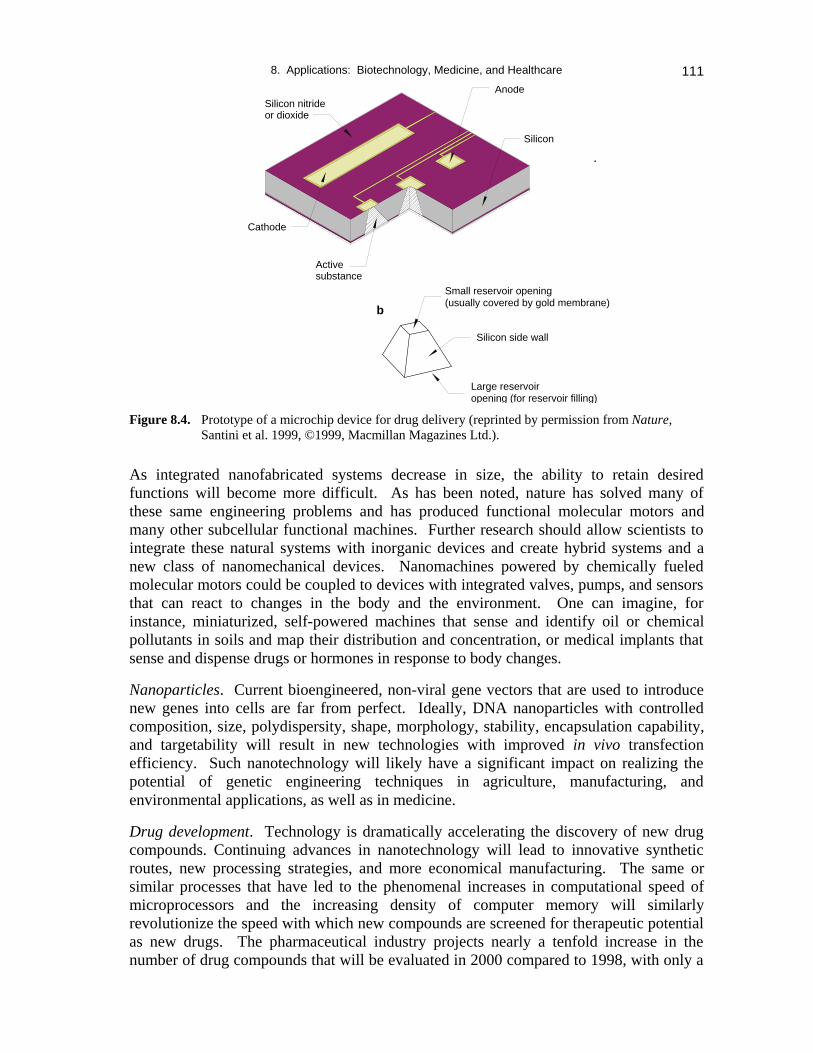
Nanomachines. To date, development of miniaturized devices is based mostly onnonbiological principles. An example of an autonomous miniaturized controlled-releasedimplantable device (a solid-state silicon microchip) for drug delivery applications isillustrated in Figure 8.4. The microchip can release a single or multiple chemicalsubstance(s) on demand. In addition to drug delivery, this technology may also find usein such areas as diagnostics, analytical chemistry, and others.
8. Applications: Biotechnology, Medicine, and Healthcare 111
Figure 8.4. Prototype of a microchip device for drug delivery (reprinted by permission from Nature,Santini et al. 1999, ©1999, Macmillan Magazines Ltd.).
As integrated nanofabricated systems decrease in size, the ability to retain desiredfunctions will become more difficult. As has been noted, nature has solved many ofthese same engineering problems and has produced functional molecular motors andmany other subcellular functional machines. Further research should allow scientists tointegrate these natural systems with inorganic devices and create hybrid systems and anew class of nanomechanical devices. Nanomachines powered by chemically fueledmolecular motors could be coupled to devices with integrated valves, pumps, and sensorsthat can react to changes in the body and the environment. One can imagine, forinstance, miniaturized, self-powered machines that sense and identify oil or chemicalpollutants in soils and map their distribution and concentration, or medical implants thatsense and dispense drugs or hormones in response to body changes.
Nanoparticles. Current bioengineered, non-viral gene vectors that are used to introducenew genes into cells are far from perfect. Ideally, DNA nanoparticles with controlledcomposition, size, polydispersity, shape, morphology, stability, encapsulation capability,and targetability will result in new technologies with improved in vivo transfectionefficiency. Such nanotechnology will likely have a significant impact on realizing thepotential of genetic engineering techniques in agriculture, manufacturing, andenvironmental applications, as well as in medicine.
Drug development. Technology is dramatically accelerating the discovery of new drugcompounds. Continuing advances in nanotechnology will lead to innovative syntheticroutes, new processing strategies, and more economical manufacturing. The same orsimilar processes that have led to the phenomenal increases in computational speed ofmicroprocessors and the increasing density of computer memory will similarlyrevolutionize the speed with which new compounds are screened for therapeutic potentialas new drugs. The pharmaceutical industry projects nearly a tenfold increase in thenumber of drug compounds that will be evaluated in 2000 compared to 1998, with only a
Active substance
Silicon
Silicon side wall
Large reservoiropening (for reservoir filling)
Small reservoir opening(usually covered by gold membrane)
b
Silicon nitrideor dioxide
Anode
Cathode

8. Applications: Biotechnology, Medicine, and Healthcare112
modest miniaturization of technology. If the trend is similar to that of microelectronics,the rate could grow exponentially. Arrays of nanodrops, each a mere nanoliter involume, but holding a small cell culture sample, are being used to place hundreds ofthousands of cell culture assays on a laboratory desktop, revolutionizing the speed withwhich new pharmaceuticals can be screened for activity. The time required for newdrugs to reach patients could thus be reduced, saving human lives.
Drug delivery. Drug and gene delivery will continue to impact significantly on thepractice of medicine. Nanotechnology as applied to drug delivery systems willundoubtedly dramatically improve the therapeutic potential of many water-insoluble andunstable drugs. Microsensors interfaced to a nanoscale drug delivery system coulddispense precise amounts of drugs for optimum functionality and minimum toxicity.However, significant challenges still remain in synthesis and processing of drug-carriernanoparticles at the industrial scale. Nanotechnology may also help reach the hithertoelusive goal of active drug targeting to selected cells within the body. Nanotechnologythat can further reduce the size and reproducibly attach targeting ligands to the drug-loaded nanoparticles may help localize the drug to the desired tissues in the body. Thesenanoparticles may also be valuable tools for molecular and cell biologists to studyfundamental cellular processes such as receptor-mediated endocytosis and intracellulartrafficking.
Interfaces between biological and other materials. In the repair of the human body withprosthetics or artificial replacement parts, mechanical attachment to the body, oralternatively, rejection by the body, occurs at biological interfaces. The nanoscalechemical and topographical details of the implanted materials determine the reaction ofthe body. If we can gain sufficient understanding and control of these biologicalreactions to surface nanostructure, we may be able to control the rejection of artificialimplants. Similarly, it may be possible to surround implanted tissue with ananofabricated barrier that would thwart the rejection mechanisms of the host, allowingwider utilization of donated organs. Ultimately, better materials and understanding oftheir interaction with the body may lead to implants that the body will not only accept,but that will actually become integrated into the body. Nanofabrication and nanosynthesisgive us powerful new tools to address these important medical issues for which a greatdeal of research is still necessary.
Various bio-inspired ideas are discussed in other chapters (e.g., Chapter 4, on synthesis,and Chapter 6, on nanodevices).
8.4 SCIENTIFIC AND TECHNOLOGICAL INFRASTRUCTURE
The infrastructure needs for nanobiology are similar to those for other fields: multiuserfacilities to provide access to specialized technologies, funding mechanisms andorganization structures that encourage and support multidisciplinary teams and areresponsive to rapid technological change, and training of a new generation of scientistsand engineers who are prepared to maximally exploit this new knowledge.
The teaming of physical scientists, engineers, biologists, and health professionals will berequired for research and development efforts. The universities should be supported with

8. Applications: Biotechnology, Medicine, and Healthcare 113
grants for training new undergraduate, graduate, and postdoctoral students in theseinterdisciplinary areas.
8.5 R&D INVESTMENT AND IMPLEMENTATION STRATEGIES
• Fund basic science and technology development needed for future biotechnology,health, and national security (biowarfare, nanobiodevices, and survivability) needs.This must include basic research in the cell and molecular biology of the manynaturally occurring nanomachines within cells.
• Fund efforts to train clinicians in the use of the emerging technologies and theirintegration into medical instruction.
• Promote funding in proposals with rapid turnaround times for exploratory, agileresponse to developing opportunities uncovered by advances in nanotechnology.
• Encourage interdisciplinary cooperation of academic, industrial, and Federallaboratories.
• Support coordinated research by teams that represent the required diversity ofdisciplines, at sufficient magnitude to make rapid progress.
8.6 PRIORITIES AND CONCLUSIONS
• Exploratory research should be encouraged and new ideas promoted aggressively inthe area of nanobiotechnology.
• A systematic investigation should be undertaken of natural structures with intrinsicpatterns at the nanoscale, as well as in use of the identified nanoscale patterns for newmaterials and devices.
• Interaction of biomolecules with inert materials is an area of special interest both formedical application and for understanding the role of environment on the origin andevolution of life on Earth.
• It is important to support universities in interdisciplinary training of undergraduateand graduate students at the intersection of biological, physical, and engineeringsciences.
8.7 EXAMPLES OF CURRENT ACHIEVEMENTS AND PARADIGM SHIFTS
8.7.1 Special Attributes of Biological SystemsContact person: L. Jelinski, Louisiana State University
Biological molecules and systems have a number of attributes that make them highlysuitable for nanotechnology applications. For example, proteins fold into preciselydefined three-dimensional shapes, and nucleic acids assemble according to well-understood rules (Figure 8.5). The ribbon diagram of the oxygen-binding proteinmyoglobin, found in muscle cells, is illustrated in the lower portion of the figure, adiagram constructed from atomic coordinates provided by the Protein Data Bank.Antibodies are highly specific in recognizing and binding their ligands, and biologicalassemblies such as molecular motors can perform transport operations. Because of these

8. Applications: Biotechnology, Medicine, and Healthcare114
and other favorable properties, biomolecules, biophysics, and biology are themes that runthrough all of the topics of this report (Jelinski 1999).
proteins fold intoprecisely-defined 3Dstructures
nucleic acids assemble accordingto well-understood rules
antibodies demonstrate highlyspecific binding and recognition
there are evenmolecular motors
Figure 8.5. Examples of biological systems (courtesy L. Jelinski; lower diagram courtesy L. Pollack,Cornell University).
8.7.2 Nanoscience and Nanotechnology in Tissue EngineeringContact person: D.J. Odde, University of Minnesota
Between the typical size of an animal cell, ~10 µm, and that of a protein molecule,~5 nm, is where nanotechnology advances can effect better understanding and control ofliving cells. Achieving greater control of cell behavior will likely facilitate efforts in theemerging area of tissue engineering. Tissue engineering is directed toward using cellsand their molecules in artificial constructs to compensate for lost or impaired bodyfunctions. Commercial ventures are currently spending ~$500 million/year in research,development, production, and marketing. In 1998 the first two tissue-engineeredproducts came on the market after Food and Drug Administration approval (Lysaght1998). These first two products are both engineered skin equivalents, although manymore tissues are at various stages in development and clinical trials. Undoubtedly, a vastarray of new nanotechnologies could potentially facilitate future tissue engineeringefforts, both in basic and applied research. Four procedures are highlighted here asexamples of applications of nanoscience and nanotechnology to tissue engineering.
First, scanning probe microscopy can be used to elucidate the nanometer-scale structureof protein filaments (Hameroff et al. 1990). These filaments include both intracellularand extracellular structures that are linked together via transmembrane receptors toprovide the mechanical continuity that holds tissues together. Second, optical forces inthe form of laser-tweezers can be used to measure motor protein motions on thenanometer scale (Svoboda et al. 1993). Understanding how molecular motors work willhelp us to better understand the fundamental contractile and propulsive properties oftissues. Third, biomaterials can be fabricated that have nanometer-scale featuresrepresenting the imprinted features of specific proteins (Shi et al. 1999). Such imprintedsurfaces could potentially provide highly stable, biospecific surfaces for the long-termmaintenance of an engineered tissue equivalent. Fourth, nano/micro particles, includingliving animal cells, bacteria, and colloidal gold (100 nm), can be optically guided anddeposited in arbitrarily defined three-dimensional arrays, a process called “laser-guideddirect-writing.” As shown in Figure 8.6, individual spinal cord cells can be confined andguided along a laser beam axis to generate a steady stream of particles. By combining

8. Applications: Biotechnology, Medicine, and Healthcare 115
various cell types and biomaterials, arbitrary three-dimensionally patterned cellconstructs can potentially be assembled to more closely mimic the architecture andstructure of native organs (Odde and Renn 1998; Renn et al. 1999).
Figure 8.6. Laser-guided transport of an individual spinal cord cell inside a hollow optical fiber. The laser light comes from the left and imparts a propulsive force on the cell. The laser beam can be directed onto a surface and cells deposited into arbitrary patterns. Subcellular particles (~100-500 nm) are also guided (cell diameter, 9 µm; time interval between frames, 300 msec).
8.7.3 BiodetectionContact Person: J. Murday, Naval Research Laboratory
Nanotechnology promises revolutionary advances in military capability. For instance,the confluence of biology, chemistry, and physics at the nanometer scale is enablingsignificant advances in military sensors for biological and chemical warfare agents.Civilian disaster response teams and commercial medicine will benefit as well. Wecannot afford to respond to a nerve gas attack, such as the 1995 Aum Shinrikyo incident,by carrying a canary as a sensor (Figure 8.7). Defense research and developmentprograms are pursuing many sensor options; two related technologies are nearing fruitionand will have medical applications as well.

8. Applications: Biotechnology, Medicine, and Healthcare116
Figure 8.7. Canary sensor (courtesy Sankei Shimbun).
One is a colorimetric sensor that can selectively detect biological agent DNA; it is incommercial development with successful tests (Figure 8.8) against anthrax andtuberculosis (Mirkin 1999). Compared to present technology, the sensor is simpler, lessexpensive (by about a factor of 10), and more selective—it can differentiate onenucleotide mismatch in a sequence of 24, where 17 constitutes a statistically uniqueidentification.
Figure 8.8. Anthrax detection: when the anthrax target is present, pairs of nanoparticles assembletogether via the DNA filaments and change the color of the respective suspension(courtesy C. Mirkin, Northwestern University).
A complementary effort is based on atomic force microscopy with a sandwichimmunoassay attaching magnetic beads to a microfabricated cantilever sensitive to smalldisplacements (Figure 8.9; Colton 1999). In the laboratory this technology is already 100to 1,000 times more sensitive than conventional immunoassays.
Both colorimetric and magnetic bead technologies might be implemented in detectorarrays that provide simultaneous identification of multiple pathogens. For instance,GMR memory elements can sense the presence of the magnetic beads (Colton 1999).

8. Applications: Biotechnology, Medicine, and Healthcare 117
Figure 8.9. Atomic force microscope (AFM) immunoassay (courtesy Naval ResearchLaboratory; reprinted with permission from Baselt et al. 1996, ©1996 AmericanVacuum Society).
8.7.4 Semiconductor Nanocrystals as Fluorescent Biological LabelsContact person: P. Alivisatos, University of California, Berkeley
For more than a decade there has been an intensive effort to prepare high-qualitynanometer-size colloidal crystals of many common semiconductors. At the onset, thiseffort had a strong focus on fundamental studies of scaling laws, in this case, quantumconfinement of electrons and holes. Over this decade, tremendous advances occurred inboth the spectroscopy and the fabrication methods. This yielded a new class of veryrobust macromolecules with readily tunable emission energy. To the extent thatapplications of this technology were envisioned at the onset, they were focused in thedomain of optoelectronics. Yet quite unexpectedly, it turns out that these colloidalnanocrystals can be used as fluorescent labels for biological tagging experiments.Biological tagging is one of the most widely employed techniques for diagnostics andvisualization. As shown in Figure 8.10, it appears as though for many applications, thecolloidal nanocrystals are advantageous as labels, when compared to existing organicdyes (Bruchez et al. 1998; Chan and Nie 1998). This has led to rapid commercializationof the new nanotechnology.

8. Applications: Biotechnology, Medicine, and Healthcare118
4Significant advantages over conventional dyes:4Reduced photobleaching4Multi-color labeling, parallel screening4Infrared labels, blood diagnostics4Molecular size nanocrystals are bio-compatible, withmany other possible applications
10 year study of scaling laws and synthesisBand gap vs. size in CdSe nanocrystals
5 nmcrystals
bound toactin fibers
3.5 nmcrystalsbound tocell nucleus
Unexpected applications in biological labelingExample: Two-color stain of mouse fibroblast cell
Figure 8.10. Semiconductor nanocrystals as fluorescent biological labels (reprinted withpermission from Bruchez et al. 1998, ©1998 American Association for theAdvancement of Science).
8.7.5 Nanofabrication of DNA “Chips”Contact persons: M. Sussman, University of Wisconsin; P. Brown, Stanford University
DNA detector arrays that today operate in the micron size range provide the potential todo thousands of experiments simultaneously with very small amounts of material. Figure8.11 is an image of a chip with 6,400 microdots, each containing a small amount of adifferent gene in the yeast genome and capable of determining how active that gene is inyeast. Yeast cells were grown under various conditions; the amount of red or yellowlight represents the level of RNA produced from the DNA in that gene, under thoseconditions. Similar experiments using this or related technologies can now be performedwith tens or hundreds of thousands of human genes. By comparing the pattern of geneexpression of normal tissue with cancerous tissues, scientists can discover which fewgenes are being activated or inhibited during a specific disease. This information iscritical to both the scientific and clinical communities in helping to discover new drugsthat inhibit cancer-causing genes. The important point is that these technologies allowphysiological changes in yeast or humans to be characterized, molecule by molecule, injust a few hours. Five years ago, an experiment like this would have taken dozens ofscientists months to complete.

8. Applications: Biotechnology, Medicine, and Healthcare 119
Figure 8.11. The full yeast genome on a chip (Brown 1999).
This technology therefore represents a paradigm shift in the way biologists do research,providing a means for using the vast amounts of information being revealed by theHuman Genome Project. Some scientists have likened this to 150 years ago, when theperiodic table for the chemical elements was discovered, ushering in a century ofbreakthroughs in chemistry. By analogy, the human and plant genome projects mayorganize all biological information in a way that may usher in a century of basic andapplied research in the manipulation of life. Despite the power of the new technology,coupled with genome sequences, it is still in its nascent forms and is largely limited in itssensitivity, selectivity, and requirement for expert operators. Nanotechnology has thepotential to do the following:
• Further reduce the size of the assays, allowing larger numbers of genes to be studiedin each experiment
• Increase their sensitivity, for example, through better detection methods
• Result in wider application of these systems in hospitals, clinics, or perhaps even asreal-time sensors within the body, for example, by enabling new ways to integratesequential steps in lab procedures into ultraminiaturized lab-on-a-chip devices that areless subject to operator error
8.8 REFERENCES
Baselt, D.R., G.U. Lee, and R.J. Colton. 1996. Biosensor based on force microscope technology. Journalof Vacuum Science and Technology B 14(2):789-793.
Brown, P. 1999. http://cmgm.stanford.edu/pbrown/yeastchip.html.
Bruchez, M. Jr., M. Moronne, P. Gin, S. Weiss, and A.P. Alivisatos. 1998. Semiconductor nanocrystals asfluorescent biological labels. Science 281:2013-2016.

8. Applications: Biotechnology, Medicine, and Healthcare120
Chan, W.C.W., and S.M. Nie. 1998. Quantum dot bioconjugates for ultrasensitive nonisotopic detection.Science 281:2016-2018.
Colton, R. 1999. (Chemistry Division, Naval Research Laboratory -- private communication).
Jelinski, L. 1999. Biologically related aspects of nanoparticles, nanostructured materials, and nanodevices.In Nanostructure science and technology. NTSC Report, ed. R.W. Siegel, E. Hu, and M.C. Roco.Baltimore: World Technology Evaluation Center (WTEC). Web sitehttp://itri.loyola.edu/nano/IWGN.Worldwide.Study/. Also published by Kluwer Academic Publishers.
Hameroff, S., et al. 1990. Scanning tunneling microscopy of cytoskeletal proteins: Microtubules andintermediate filaments. J. Vac. Sci. and Tech. A 8:687-691.
Lysaght, M.J. 1998. An economic survey of the emerging tissue engineering industry. Tissue Eng. 4:231-238.
Mirkin, C. 1999. (Department of Chemistry, Northwestern University – private communication).
Noji, H. 1998. The rotary enzyme of the cell: The rotation of F1-ATPase. Science 282: 1844-1845.
Odde, D.J. and M.J. Renn. 1998. Laser-based direct-write lithography of cells. Ann. Biomed. Eng. 26:S-141.
Renn, M.J., et al. 1999. Laser guidance and trapping of mesoscale particles in hollow-core optical fibers.Phys. Rev. Lett. 82:1574-1577.
Santini, J.T. Jr., M.J. Cima and R. Langer. 1999. A controlled-release chip. Nature 397:335-338.
Shi, H., et al. 1999. Template-imprinted nanostructured surfaces for protein recognition. Nature 398:593-597.
Svoboda, K. et al. 1993. Direct observation of kinesin stepping by optical trapping interferometry. Nature365:721-727.
Turner, S.W., A.M. Perez, A. Lopez and H.G. Craighead. 1998. Monolithic nanofluid sieving structuresfor DNA manipulation. J. Vac. Sci. Technol. B. 16(6):3835-3840 (Nov/Dec 1998).