Embed Size (px)
Citation preview
The
The Veterinary Journal 168 (2004) 41–49
Veterinary Journalwww.elsevier.com/locate/tvjl
Review
Postweaning multisystemic wasting syndrome: a reviewof aetiology, diagnosis and pathology
C. Chae *
Department of Veterinary Pathology, College of Veterinary Medicine and School of Agricultural Biotechnology, Seoul National University,
San 56-1, Shillim-dong, Kwanak-Gu 151-742, Seoul, South Korea
Accepted 19 September 2003
Abstract
This review paper concentrates on the aetiology, diagnosis, and pathological aspects of postweaning multisystemic wasting
syndrome (PMWS). PMWS was first recognized in Canada in 1996 as a new emerging disease which caused wasting in postweaned
pigs. Since then, PMWS has been recognized in pigs in many countries. The syndrome is caused by a DNA virus referred to as
porcine circovirus 2 (PCV2), which is classified in the family Circoviridae. PMWS primarily occurs in pigs between 25 and 120 days
of age with the highest number of cases occurring between 60 and 80 days of age.
The diagnosis of PMWS must meet three criteria: (i) the presence of compatible clinical signs, (ii) the presence of characteristic
microscopic lesions, and (iii) the presence of PCV2 within these lesions. In order to establish the diagnosis, techniques are required
that link virus and tissue lesions, such as immunohistochemistry and in situ hybridization, but not polymerase chain reaction or
virus isolation. The three criteria considered separately are not diagnostic of PMWS. For example, the detection of PCV2 alone does
not indicate PMWS but merely PCV2 infection. A hallmark of microscopic lesions of PMWS is granulomatous inflammation in the
lymph nodes, liver, spleen, tonsil, thymus, and Peyer�s patches. Large, multiple, basophilic or amphophilic grape-like intracyto-
plasmic inclusion bodies are often seen in the cytoplasm of macrophages and multinucleated giant cells.
� 2003 Elsevier Ltd. All rights reserved.
Keywords: Diagnosis; Porcine circovirus; PMWS; Postweaning multisytemic wasting syndrome; Review
1. Introduction
In 1996, a new infectious disease in specific-pathogen-
free (SPF) swine herds in Western Canada was identifiedand reported (Clark, 1997; Harding, 1997). Since then,
postweaning multisystemic wasting syndrome (PMWS)
has been recognized in pigs in Asia, North and South
America, and Europe (LeCann et al., 1997; Segales et al.,
1997; Spillane et al., 1998; Choi and Chae, 1999; Hinrichs
et al., 1999; Onuki et al., 1999; Allan and Ellis, 2000;
Choi et al., 2000; Kiss et al., 2000; Vyt et al., 2000;
Wellenberg et al., 2000; Borel et al., 2001; Trujano et al.,2001; Allan et al., 2002; Celer and Carasova, 2002;
Sarradell et al., 2002; Saoulidis et al., 2002) (Fig. 1).
PMWS is an infectious viral disease of postweaned pigs
* Tel.: +82-2-880-1277; fax: +82-2-871-5821.
E-mail address: [email protected] (C. Chae).
1090-0233/$ - see front matter � 2003 Elsevier Ltd. All rights reserved.
doi:10.1016/j.tvjl.2003.09.018
characterized by progressive weight loss, respiratory
signs and jaundice (Clark, 1997; Harding, 1997; Ellis
et al., 1998; Allan et al., 1999; Allan and Ellis, 2000; Choi
and Chae, 2000). The disease, which occurs in herds thatare usually in otherwise in good health, has a low mor-
bidity but a relatively high mortality rate among pigs in
the 5- to 12-week-old age group (Harding and Clark,
1997; Allan and Ellis, 2000; Kim et al., 2002; Pallares
et al., 2002). PMWS is now endemic in many swine-
producing countries and continues to be a major cause of
wasting disease in swine. This review concentrates on the
aetiology, diagnosis, and pathological aspects of PMWS.
2. Aetiology
The viral families Circoviridae and Nanoviridae are
DNA viral pathogens of plants, birds and swine (Lukert
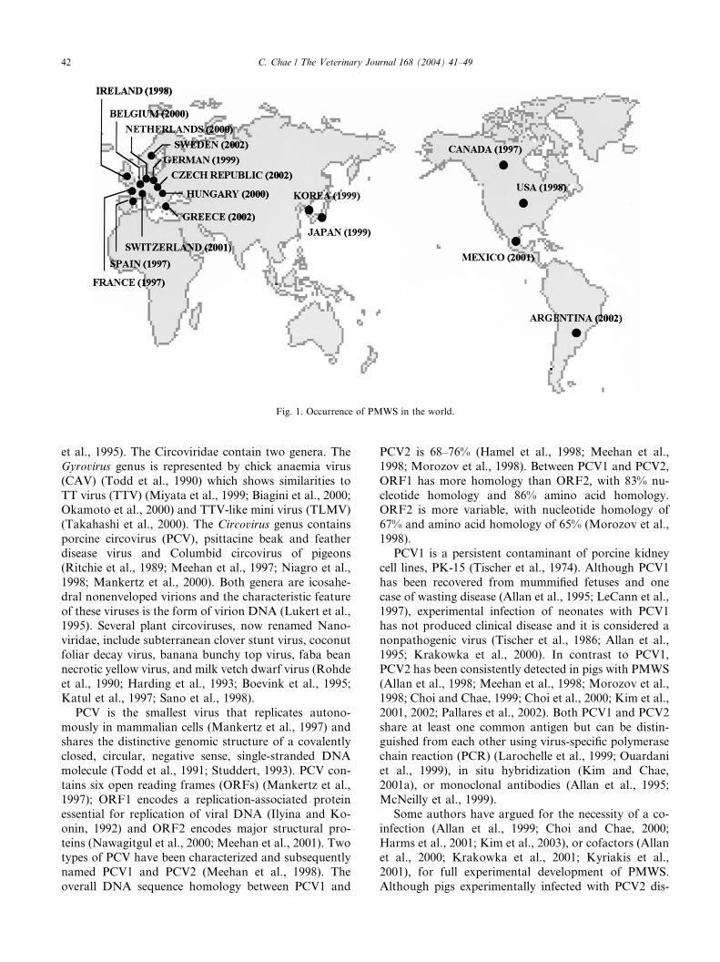
Fig. 1. Occurrence of PMWS in the world.
42 C. Chae / The Veterinary Journal 168 (2004) 41–49
et al., 1995). The Circoviridae contain two genera. The
Gyrovirus genus is represented by chick anaemia virus
(CAV) (Todd et al., 1990) which shows similarities to
TT virus (TTV) (Miyata et al., 1999; Biagini et al., 2000;Okamoto et al., 2000) and TTV-like mini virus (TLMV)
(Takahashi et al., 2000). The Circovirus genus contains
porcine circovirus (PCV), psittacine beak and feather
disease virus and Columbid circovirus of pigeons
(Ritchie et al., 1989; Meehan et al., 1997; Niagro et al.,
1998; Mankertz et al., 2000). Both genera are icosahe-
dral nonenveloped virions and the characteristic feature
of these viruses is the form of virion DNA (Lukert et al.,1995). Several plant circoviruses, now renamed Nano-
viridae, include subterranean clover stunt virus, coconut
foliar decay virus, banana bunchy top virus, faba bean
necrotic yellow virus, and milk vetch dwarf virus (Rohde
et al., 1990; Harding et al., 1993; Boevink et al., 1995;
Katul et al., 1997; Sano et al., 1998).
PCV is the smallest virus that replicates autono-
mously in mammalian cells (Mankertz et al., 1997) andshares the distinctive genomic structure of a covalently
closed, circular, negative sense, single-stranded DNA
molecule (Todd et al., 1991; Studdert, 1993). PCV con-
tains six open reading frames (ORFs) (Mankertz et al.,
1997); ORF1 encodes a replication-associated protein
essential for replication of viral DNA (Ilyina and Ko-
onin, 1992) and ORF2 encodes major structural pro-
teins (Nawagitgul et al., 2000; Meehan et al., 2001). Twotypes of PCV have been characterized and subsequently
named PCV1 and PCV2 (Meehan et al., 1998). The
overall DNA sequence homology between PCV1 and
PCV2 is 68–76% (Hamel et al., 1998; Meehan et al.,
1998; Morozov et al., 1998). Between PCV1 and PCV2,
ORF1 has more homology than ORF2, with 83% nu-
cleotide homology and 86% amino acid homology.ORF2 is more variable, with nucleotide homology of
67% and amino acid homology of 65% (Morozov et al.,
1998).
PCV1 is a persistent contaminant of porcine kidney
cell lines, PK-15 (Tischer et al., 1974). Although PCV1
has been recovered from mummified fetuses and one
case of wasting disease (Allan et al., 1995; LeCann et al.,
1997), experimental infection of neonates with PCV1has not produced clinical disease and it is considered a
nonpathogenic virus (Tischer et al., 1986; Allan et al.,
1995; Krakowka et al., 2000). In contrast to PCV1,
PCV2 has been consistently detected in pigs with PMWS
(Allan et al., 1998; Meehan et al., 1998; Morozov et al.,
1998; Choi and Chae, 1999; Choi et al., 2000; Kim et al.,
2001, 2002; Pallares et al., 2002). Both PCV1 and PCV2
share at least one common antigen but can be distin-guished from each other using virus-specific polymerase
chain reaction (PCR) (Larochelle et al., 1999; Ouardani
et al., 1999), in situ hybridization (Kim and Chae,
2001a), or monoclonal antibodies (Allan et al., 1995;
McNeilly et al., 1999).
Some authors have argued for the necessity of a co-
infection (Allan et al., 1999; Choi and Chae, 2000;
Harms et al., 2001; Kim et al., 2003), or cofactors (Allanet al., 2000; Krakowka et al., 2001; Kyriakis et al.,
2001), for full experimental development of PMWS.
Although pigs experimentally infected with PCV2 dis-
C. Chae / The Veterinary Journal 168 (2004) 41–49 43
played clinical signs and lesions typical of the disease,co-infection with PCV2 and porcine parvovirus (PPV)
also appeared to result in wasting disease (Allan et al.,
1999; Ellis et al., 1999; Choi and Chae, 2000; Kennedy
et al., 2000; Krakowka et al., 2000; Kim et al., 2003).
However, histopathological changes characteristic of
PMWS were induced with PCV2 alone and not with
PPV alone (Allan et al., 1999; Kennedy et al., 2000;
Krakowka et al., 2000; Magar et al., 2000), suggestingthat PCV2 plays the major role. Furthermore, the con-
sistent identification of PCV2 DNA and/or antigen,
closely associated with lesions in a wide range of tissues
from pigs with PMWS, has led to the belief that PCV2 is
the aetiological agent (Meehan et al., 1998; Morozov
et al., 1998; Choi and Chae, 1999; Choi et al., 2000; Kim
et al., 2001; Kim et al., 2002).
3. Definition
Generally, diagnosis of viral diseases in swine is based
on detection of the virus by culture, PCR, immunohis-
tochemistry or in situ hybridization, and/or detection of
virus antibodies by serology. However, diagnosis of
PMWS is different from this general diagnostic ap-proach because PCV2 can be detected in normal healthy
pigs (Allan and Ellis, 2000; Kim et al., 2001; Calsamiglia
et al., 2002). The detection of PCV2 alone does not
necessarily confirm a diagnosis of PMWS. Therefore,
the diagnosis of PMWS must meet three criteria: (i) the
presence of compatible clinical signs, (ii) the presence of
characteristic microscopic lesions, and (iii) the presence
of the PCV2 within these lesions. These three criteriaseparately are not diagnostic of PMWS. Since clinical
signs of PMWS are nonspecific and variable, the pres-
ence of PCV2 DNA or antigen in lymphoid tissues,
demonstrated by in situ hybridization and immunohis-
tochemistry (Choi and Chae, 1999; Choi et al., 2000;
Kim and Chae, 2001b), together with moderate to severe
lymphoid depletion and/or granulomatous lymphade-
nitis, are used as the criteria for the diagnosis. PMWScannot therefore be diagnosed if lymphoid tissues are
not submitted with the specimens provided.
4. Clinical signs
PMWS primarily occurs in pigs between 25 and 120
days of age, with most cases occurring between 60 and80 days of age (Kim et al., 2002). In a recent survey in
Korea, the prevalence was 8.1% (133 cases of PMWS
out of 1634 cases examined), which is very close to the
10.3% (484 cases of PMWS out of 4868 cases examined)
reported in the US (Pallares et al., 2002). Clinical signs
are nonspecific and variable and those listed below in-
clude both field and experimental observations.
In weaned pigs, PMWS is characterized by wastingwith or without respiratory signs, diarrhoea, paleness of
the skin, or icterus (Clark, 1997; Harding, 1997; Ellis
et al., 1998; Allan et al., 1999; Allan and Ellis, 2000;
Choi et al., 2000), and a marked increase in mortality
from single or multiple concurrent bacterial infections
(Madec et al., 2000; Kim et al., 2002) in postweaning
and early finishing pigs. Co-infection with single or
multiple bacteria, and, less frequently, with other vi-ruses, is a severe complicating factor of PMWS and is
clinically important based on observation of increases in
postweaning mortality from 1–2% to 10–25%, when all
other factors apparently remain unchanged.
PMWS is often seen in combination with other viral
and bacterial pathogens such as porcine reproductive
respiratory syndrome virus (PRRSV), swine influenza
virus, porcine parvovirus (PPV), Haemophilus parasuis,Actinobacillus pleuropneumoniae, Streptococcus suis and
Mycoplasma hyopneumoniae (Kim et al., 2002; Pallares
et al., 2002). PCV2 infection alone was found in only
15% of PMWS cases. These co-infections may confuse
or complicate the clinical presentation. Furthermore, the
prevalent co-infecting agents appeared to vary in dif-
ferent countries. For example, in the Republic of Korea,
PRRSV and PPV were the most common agents coex-isting with PCV2 in cases of PMWS (Kim et al., 2002).
However, only one of the 484 cases of PMWS was po-
sitive for PPV in the United States (Pallares et al., 2002).
H. parasuis was the most prevalent bacterial co-infection
in Korea and was found in 32.3% of cases, while in the
US the most prevalent bacterial co-infection was S. suis
in 5.5% of the cases. The fact that many different in-
fectious agents are isolated in cases of PMWS stronglysupports the view that a variety of pathogens may share
a common mechanism in affecting the immune system
(Allan et al., 2000; Krakowka et al., 2001; Kim and
Chae, 2003a) and allow progression of PCV2 infection
to PMWS. Alternatively, PCV2 may initiate lymphoid
depletion, resulting in an increased susceptibility to
other viral and bacterial infections. Immunosuppression
has been confirmed in PMWS-affected swine (Chianiniet al., 2003; Darwich et al., 2003).
5. Histopathology
Histopathologically, PMWS has two characteristic
lesions; granulomatous inflammation (Fig. 2) and the
presence of intracytoplasmic inclusion bodies (Fig. 3).Granulomatous inflammation is seen in the lymph
nodes, liver, spleen, tonsil, thymus, and Peyer�s patches(Table 1) but occurs consistently in superficial inguinal
lymph nodes, and, in our experience, only occasion-
ally in other tissues. This unique lesion is characterized
by infiltrates of epithelioid cells and multinucleated
giant cells (Fig. 2). The epithelioid cells are activated
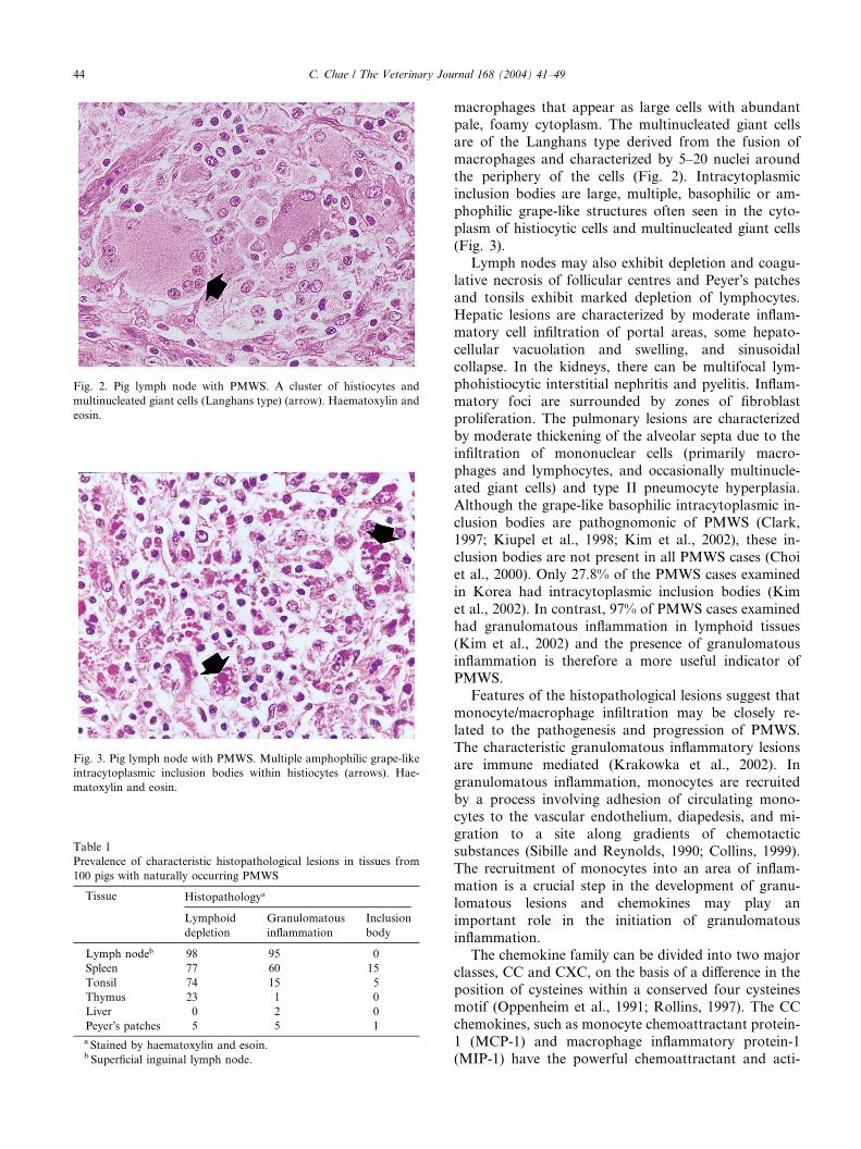
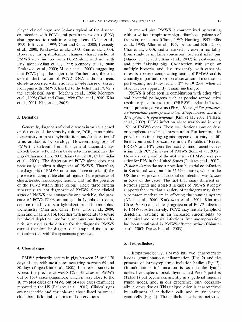
Fig. 3. Pig lymph node with PMWS. Multiple amphophilic grape-like
intracytoplasmic inclusion bodies within histiocytes (arrows). Hae-
matoxylin and eosin.
Table 1
Prevalence of characteristic histopathological lesions in tissues from
100 pigs with naturally occurring PMWS
Tissue Histopathologya
Lymphoid
depletion
Granulomatous
inflammation
Inclusion
body
Lymph nodeb 98 95 0
Spleen 77 60 15
Tonsil 74 15 5
Thymus 23 1 0
Liver 0 2 0
Peyer�s patches 5 5 1
a Stained by haematoxylin and esoin.b Superficial inguinal lymph node.
Fig. 2. Pig lymph node with PMWS. A cluster of histiocytes and
multinucleated giant cells (Langhans type) (arrow). Haematoxylin and
eosin.
44 C. Chae / The Veterinary Journal 168 (2004) 41–49
macrophages that appear as large cells with abundantpale, foamy cytoplasm. The multinucleated giant cells
are of the Langhans type derived from the fusion of
macrophages and characterized by 5–20 nuclei around
the periphery of the cells (Fig. 2). Intracytoplasmic
inclusion bodies are large, multiple, basophilic or am-
phophilic grape-like structures often seen in the cyto-
plasm of histiocytic cells and multinucleated giant cells
(Fig. 3).Lymph nodes may also exhibit depletion and coagu-
lative necrosis of follicular centres and Peyer�s patches
and tonsils exhibit marked depletion of lymphocytes.
Hepatic lesions are characterized by moderate inflam-
matory cell infiltration of portal areas, some hepato-
cellular vacuolation and swelling, and sinusoidal
collapse. In the kidneys, there can be multifocal lym-
phohistiocytic interstitial nephritis and pyelitis. Inflam-matory foci are surrounded by zones of fibroblast
proliferation. The pulmonary lesions are characterized
by moderate thickening of the alveolar septa due to the
infiltration of mononuclear cells (primarily macro-
phages and lymphocytes, and occasionally multinucle-
ated giant cells) and type II pneumocyte hyperplasia.
Although the grape-like basophilic intracytoplasmic in-
clusion bodies are pathognomonic of PMWS (Clark,1997; Kiupel et al., 1998; Kim et al., 2002), these in-
clusion bodies are not present in all PMWS cases (Choi
et al., 2000). Only 27.8% of the PMWS cases examined
in Korea had intracytoplasmic inclusion bodies (Kim
et al., 2002). In contrast, 97% of PMWS cases examined
had granulomatous inflammation in lymphoid tissues
(Kim et al., 2002) and the presence of granulomatous
inflammation is therefore a more useful indicator ofPMWS.
Features of the histopathological lesions suggest that
monocyte/macrophage infiltration may be closely re-
lated to the pathogenesis and progression of PMWS.
The characteristic granulomatous inflammatory lesions
are immune mediated (Krakowka et al., 2002). In
granulomatous inflammation, monocytes are recruited
by a process involving adhesion of circulating mono-cytes to the vascular endothelium, diapedesis, and mi-
gration to a site along gradients of chemotactic
substances (Sibille and Reynolds, 1990; Collins, 1999).
The recruitment of monocytes into an area of inflam-
mation is a crucial step in the development of granu-
lomatous lesions and chemokines may play an
important role in the initiation of granulomatous
inflammation.The chemokine family can be divided into two major
classes, CC and CXC, on the basis of a difference in the
position of cysteines within a conserved four cysteines
motif (Oppenheim et al., 1991; Rollins, 1997). The CC
chemokines, such as monocyte chemoattractant protein-
1 (MCP-1) and macrophage inflammatory protein-1
(MIP-1) have the powerful chemoattractant and acti-
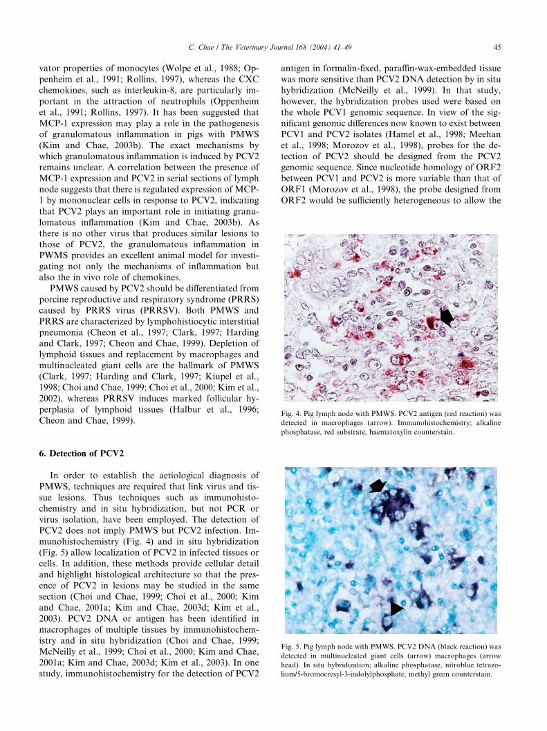
Fig. 4. Pig lymph node with PMWS. PCV2 antigen (red reaction) was
detected in macrophages (arrow). Immunohistochemistry; alkaline
C. Chae / The Veterinary Journal 168 (2004) 41–49 45
vator properties of monocytes (Wolpe et al., 1988; Op-penheim et al., 1991; Rollins, 1997), whereas the CXC
chemokines, such as interleukin-8, are particularly im-
portant in the attraction of neutrophils (Oppenheim
et al., 1991; Rollins, 1997). It has been suggested that
MCP-1 expression may play a role in the pathogenesis
of granulomatous inflammation in pigs with PMWS
(Kim and Chae, 2003b). The exact mechanisms by
which granulomatous inflammation is induced by PCV2remains unclear. A correlation between the presence of
MCP-1 expression and PCV2 in serial sections of lymph
node suggests that there is regulated expression of MCP-
1 by mononuclear cells in response to PCV2, indicating
that PCV2 plays an important role in initiating granu-
lomatous inflammation (Kim and Chae, 2003b). As
there is no other virus that produces similar lesions to
those of PCV2, the granulomatous inflammation inPWMS provides an excellent animal model for investi-
gating not only the mechanisms of inflammation but
also the in vivo role of chemokines.
PMWS caused by PCV2 should be differentiated from
porcine reproductive and respiratory syndrome (PRRS)
caused by PRRS virus (PRRSV). Both PMWS and
PRRS are characterized by lymphohistiocytic interstitial
pneumonia (Cheon et al., 1997; Clark, 1997; Hardingand Clark, 1997; Cheon and Chae, 1999). Depletion of
lymphoid tissues and replacement by macrophages and
multinucleated giant cells are the hallmark of PMWS
(Clark, 1997; Harding and Clark, 1997; Kiupel et al.,
1998; Choi and Chae, 1999; Choi et al., 2000; Kim et al.,
2002), whereas PRRSV induces marked follicular hy-
perplasia of lymphoid tissues (Halbur et al., 1996;
Cheon and Chae, 1999).
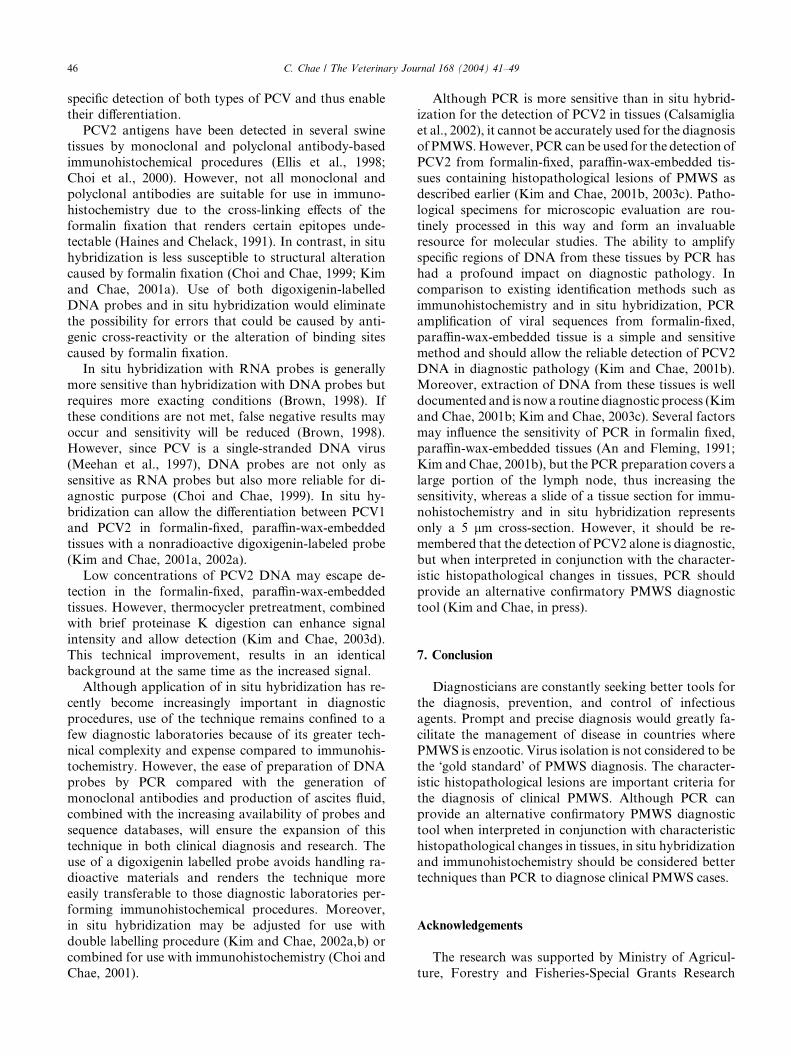
phosphatase, red substrate, haematoxylin counterstain.Fig. 5. Pig lymph node with PMWS. PCV2 DNA (black reaction) was
detected in multinucleated giant cells (arrow) macrophages (arrow
head). In situ hybridization; alkaline phosphatase, nitroblue tetrazo-
lium/5-bromocresyl-3-indolylphosphate, methyl green counterstain.
6. Detection of PCV2
In order to establish the aetiological diagnosis of
PMWS, techniques are required that link virus and tis-
sue lesions. Thus techniques such as immunohisto-
chemistry and in situ hybridization, but not PCR orvirus isolation, have been employed. The detection of
PCV2 does not imply PMWS but PCV2 infection. Im-
munohistochemistry (Fig. 4) and in situ hybridization
(Fig. 5) allow localization of PCV2 in infected tissues or
cells. In addition, these methods provide cellular detail
and highlight histological architecture so that the pres-
ence of PCV2 in lesions may be studied in the same
section (Choi and Chae, 1999; Choi et al., 2000; Kimand Chae, 2001a; Kim and Chae, 2003d; Kim et al.,
2003). PCV2 DNA or antigen has been identified in
macrophages of multiple tissues by immunohistochem-
istry and in situ hybridization (Choi and Chae, 1999;
McNeilly et al., 1999; Choi et al., 2000; Kim and Chae,
2001a; Kim and Chae, 2003d; Kim et al., 2003). In one
study, immunohistochemistry for the detection of PCV2
antigen in formalin-fixed, paraffin-wax-embedded tissuewas more sensitive than PCV2 DNA detection by in situ
hybridization (McNeilly et al., 1999). In that study,
however, the hybridization probes used were based on
the whole PCV1 genomic sequence. In view of the sig-
nificant genomic differences now known to exist between
PCV1 and PCV2 isolates (Hamel et al., 1998; Meehan
et al., 1998; Morozov et al., 1998), probes for the de-
tection of PCV2 should be designed from the PCV2genomic sequence. Since nucleotide homology of ORF2
between PCV1 and PCV2 is more variable than that of
ORF1 (Morozov et al., 1998), the probe designed from
ORF2 would be sufficiently heterogeneous to allow the
46 C. Chae / The Veterinary Journal 168 (2004) 41–49
specific detection of both types of PCV and thus enabletheir differentiation.
PCV2 antigens have been detected in several swine
tissues by monoclonal and polyclonal antibody-based
immunohistochemical procedures (Ellis et al., 1998;
Choi et al., 2000). However, not all monoclonal and
polyclonal antibodies are suitable for use in immuno-
histochemistry due to the cross-linking effects of the
formalin fixation that renders certain epitopes unde-tectable (Haines and Chelack, 1991). In contrast, in situ
hybridization is less susceptible to structural alteration
caused by formalin fixation (Choi and Chae, 1999; Kim
and Chae, 2001a). Use of both digoxigenin-labelled
DNA probes and in situ hybridization would eliminate
the possibility for errors that could be caused by anti-
genic cross-reactivity or the alteration of binding sites
caused by formalin fixation.In situ hybridization with RNA probes is generally
more sensitive than hybridization with DNA probes but
requires more exacting conditions (Brown, 1998). If
these conditions are not met, false negative results may
occur and sensitivity will be reduced (Brown, 1998).
However, since PCV is a single-stranded DNA virus
(Meehan et al., 1997), DNA probes are not only as
sensitive as RNA probes but also more reliable for di-agnostic purpose (Choi and Chae, 1999). In situ hy-
bridization can allow the differentiation between PCV1
and PCV2 in formalin-fixed, paraffin-wax-embedded
tissues with a nonradioactive digoxigenin-labeled probe
(Kim and Chae, 2001a, 2002a).
Low concentrations of PCV2 DNA may escape de-
tection in the formalin-fixed, paraffin-wax-embedded
tissues. However, thermocycler pretreatment, combinedwith brief proteinase K digestion can enhance signal
intensity and allow detection (Kim and Chae, 2003d).
This technical improvement, results in an identical
background at the same time as the increased signal.
Although application of in situ hybridization has re-
cently become increasingly important in diagnostic
procedures, use of the technique remains confined to a
few diagnostic laboratories because of its greater tech-nical complexity and expense compared to immunohis-
tochemistry. However, the ease of preparation of DNA
probes by PCR compared with the generation of
monoclonal antibodies and production of ascites fluid,
combined with the increasing availability of probes and
sequence databases, will ensure the expansion of this
technique in both clinical diagnosis and research. The
use of a digoxigenin labelled probe avoids handling ra-dioactive materials and renders the technique more
easily transferable to those diagnostic laboratories per-
forming immunohistochemical procedures. Moreover,
in situ hybridization may be adjusted for use with
double labelling procedure (Kim and Chae, 2002a,b) or
combined for use with immunohistochemistry (Choi and
Chae, 2001).
Although PCR is more sensitive than in situ hybrid-ization for the detection of PCV2 in tissues (Calsamiglia
et al., 2002), it cannot be accurately used for the diagnosis
of PMWS.However, PCRcan be used for the detection of
PCV2 from formalin-fixed, paraffin-wax-embedded tis-
sues containing histopathological lesions of PMWS as
described earlier (Kim and Chae, 2001b, 2003c). Patho-
logical specimens for microscopic evaluation are rou-
tinely processed in this way and form an invaluableresource for molecular studies. The ability to amplify
specific regions of DNA from these tissues by PCR has
had a profound impact on diagnostic pathology. In
comparison to existing identification methods such as
immunohistochemistry and in situ hybridization, PCR
amplification of viral sequences from formalin-fixed,
paraffin-wax-embedded tissue is a simple and sensitive
method and should allow the reliable detection of PCV2DNA in diagnostic pathology (Kim and Chae, 2001b).
Moreover, extraction of DNA from these tissues is well
documented and is now a routine diagnostic process (Kim
and Chae, 2001b; Kim and Chae, 2003c). Several factors
may influence the sensitivity of PCR in formalin fixed,
paraffin-wax-embedded tissues (An and Fleming, 1991;
Kim and Chae, 2001b), but the PCR preparation covers a
large portion of the lymph node, thus increasing thesensitivity, whereas a slide of a tissue section for immu-
nohistochemistry and in situ hybridization represents
only a 5 lm cross-section. However, it should be re-
membered that the detection of PCV2 alone is diagnostic,
but when interpreted in conjunction with the character-
istic histopathological changes in tissues, PCR should
provide an alternative confirmatory PMWS diagnostic
tool (Kim and Chae, in press).
7. Conclusion
Diagnosticians are constantly seeking better tools for
the diagnosis, prevention, and control of infectious
agents. Prompt and precise diagnosis would greatly fa-
cilitate the management of disease in countries wherePMWS is enzootic. Virus isolation is not considered to be
the �gold standard� of PMWS diagnosis. The character-
istic histopathological lesions are important criteria for
the diagnosis of clinical PMWS. Although PCR can
provide an alternative confirmatory PMWS diagnostic
tool when interpreted in conjunction with characteristic
histopathological changes in tissues, in situ hybridization
and immunohistochemistry should be considered bettertechniques than PCR to diagnose clinical PMWS cases.
Acknowledgements
The research was supported by Ministry of Agricul-
ture, Forestry and Fisheries-Special Grants Research
C. Chae / The Veterinary Journal 168 (2004) 41–49 47
Program (MAFF-SGRP), and Brain Korea 21 Project,Republic of Korea.
References
Allan, G.M., Ellis, J.A., 2000. Porcine circoviruses: a review. Journal
of Veterinary Diagnostic Investigation 12, 3–14.
Allan, G.M., McNeilly, F., Cassidy, J.P., Reilly, G.A.C., Adair, B.,
Ellis, J.A., McNulty, M.S., 1995. Pathogenesis of porcine circovi-
rus, experimental infections of colostrum deprived piglets and
examination of pig fetal material. Veterinary Microbiology 44,
49–64.
Allan, G.M., McNeilly, F., Kennedy, S., Daft, B., Clarke, E.G., Ellis,
J.A., Haines, D.M., Meehan, B.M., Adair, B.M., 1998. Isolation of
porcine circovirus-like viruses from pigs with a wasting disease in
the USA and Europe. Journal of Veterinary Diagnostic Investiga-
tion 10, 3–10.
Allan, G.M., Kennedy, S., McNeilly, F., Foster, J.C., Ellis, J.A.,
Krakowka, S.J., Meehan, B.M., Adair, B.M., 1999. Experimental
reproduction of severe wasting disease by co-infection of pigs with
porcine circovirus and porcine parvovirus. Journal of Comparative
Pathology 121, 1–11.
Allan, G.M., McNeilly, F., Kennedy, S., Meehan, B., Ellis, J.,
Krakowka, S., 2000. Immunostimulation, PCV-2 and PMWS.
Veterinary Record 147, 170–171.
Allan, G.M., McNeilly, F., Meehan, B., Kennedy, S., Johnston, D.,
Ellis, J., Krakowka, S., Fossum, C., Wattrang, E., Wallgren, P.,
2002. Reproduction of PMWS with a 1993 Swedish isolate of PCV-
2. Veterinary Record 150, 255–256.
An, S.F., Fleming, K.A., 1991. Removal of inhibitor(s) of the
polymerase chain reaction from formalin fixed, paraffin wax
embedded tissues. Journal of Clinical Pathology 44, 924–927.
Biagini, P., Attoui, H., Gallian, P., Touinssi, M., Cantaloube, J.F., de
Micco, P., de Lamballerie, X., 2000. Complete sequences of two
highly divergent European isolates of TT virus. Biochemical and
Biophysical Research Communications 271, 837–841.
Boevink, P., Chu, P.W.G., Keese, P., 1995. Sequence of subterranean
clover stunt virus DNA: affinities with the geminiviruses. Virology
207, 354–361.
Borel, N., B€u rgi, E., Kuipel, M., Stevenson, G.W., Mittal, S.K.,
Pospischil, A., Sydler, T, 2001. Three cases of postweaning
multisystemic wasting syndrome (PMWS) due to porcine circovirus
type 2 (PCV2) in Switzerland. Schweizer Archiv fur Tierheilkunde
143, 249–255.
Brown, C., 1998. In situ hybridization with riboprobes: an overview
for veterinary pathologists. Veterinary Pathology 35, 159–167.
Calsamiglia, M., Segales, J., Quintana, J., Rosell, C., Domingo, M.,
2002. Detection of porcine circovirus types 1 and 2 in serum and
tissue samples of pigs with and without postweaning multisystemic
wasting syndrome. Journal of Clinical Microbiology 40,
1848–1850.
Celer Jr., V., Carasova, P., 2002. First evidence of porcine circovirus
type 2 (PCV-2) infection of pigs in the Czech Republic by semi-
nested PCR. Journal of Veterinary Medicine B, Infectious Disease
and Veterinary Public Health 49, 155–159.
Cheon, D.-S., Chae, C., 1999. Distribution of a Korean strain of
porcine reproductive and respiratory syndrome virus in experi-
mentally infected pigs, as demonstrated immunohistochemically
and by in-situ hybridization. Journal of Comparative Pathology
120, 79–88.
Cheon, D.-S., Chae, C., Lee, Y.-S., 1997. Detection of nucleic acids of
porcine reproductive and respiratory syndrome virus in the lungs of
naturally infected piglets as determined by in-situ hybridization.
Journal of Comparative Pathology 117, 157–163.
Chianini, F., Majo, N., Segales, J., Dominguez, J., Domingo, M.,
2003. Immunohistochemical characterisation of PCV2 associate
lesions in lymphoid and non-lymphoid tissues of pigs with
natural postweaning multisystemic wasting syndrome (PMWS).
Veterinary Immunology and Immunopathology 94, 63–75.
Choi, C., Chae, C., 1999. In-situ hybridization for the detection of
porcine circovirus in pigs with postweaning multisystemic
wasting syndrome. Journal of Comparative Pathology 121,
265–270.
Choi, C., Chae, C., 2000. Distribution of porcine parvovirus in porcine
circovirus 2-infected pigs with postweaning multisystemic wasting
syndrome as shown by in-situ hybridization. Journal of Compar-
ative Pathology 123, 302–305.
Choi, C., Chae, C., 2001. Colocalization of porcine reproductive and
respiratory syndrome virus and porcine circovirus 2 in porcine
dermatitis and nepnropathy syndrome by double-labeling techniqe.
Veterinary Pathology 38, 436–441.
Choi, C., Chae, C., Clark, E.G., 2000. Porcine postweaning multisys-
temic wasting syndrome in Korean pig: detection of porcine
circovirus 2 infection by immunohistochemistry and polymerase
chain reaction. Journal of Veterinary Diagnostic Investigation 12,
151–153.
Clark, E.G., 1997. Post-weaning wasting syndrome. Proceedings of the
American Association of Swine Practitioners 28, 499–501.
Collins, T., 1999. Acute and chronic inflammation. In: Cotran, R.S.,
Kumar, V., Collins, T. (Eds.), Robbins Pathologic Basis of Disease.
Saunders, Philadelphia, pp. 50–88.
Darwich, L., Pie, S., Rovira, A., Segales, J., Domingo, M., Oswald,
I.P., Mateu, E., 2003. Cytokine mRNA expression profiles in
lymphoid tissues of pigs naturally affected by postweaning multi-
systemic wasting syndrome. Journal of General Virology 84,
2117–2125.
Ellis, J., Hassard, L., Clark, E., Harding, J., Allan, G., Willson, P.,
Strokappe, J., Martin, K., McNeilly, F, Meehan, B., Todd, D.,
Haines, D., 1998. Isolation of circovirus from lesions of pigs with
postweaning multisystemic wasting syndrome. Canadian Veteri-
nary Journal 39, 44–51.
Ellis, J., Krakowka, S., Lairmore, M., Haines, D., Bratanich, A.,
Clark, E., Allan, G., Konoby, C., Hassard, L., Meehan, B., Martin,
K., Harding, J., Kennedy, S., McNeilly, F., 1999. Reproduction of
lesions of postweaning multisystemic wasting syndrome in gnoto-
biotic piglets. Journal of Veterinary Diagnostic Investigation 11,
3–14.
Haines, D.M., Chelack, B.J., 1991. Technical considerations for
developing enzyme immunohistochemical staining procedures
on formalin-fixed paraffin-embedded tissues for diagnostic
pathology. Journal of Veterinary Diagnostic Investigation 3,
101–112.
Halbur, P.G., Paul, P.S., Frey, M.L., Landgraf, J., Eernisse, K., Meng,
X.-J., Andrews, J.J., Lum, M.A., Rathje, J.A., 1996. Comparison
of the antigen distribution of two US porcine reproductive and
respiratory syndrome virus isolates with that of the Lelystad virus.
Veterinary Pathology 33, 159–170.
Hamel, A.L., Lin, L.L., Nayar, G.P.S., 1998. Nucleotide sequence of
porcine circovirus associated with postweaning multisystemic
wasting syndrome in pigs. Journal of Virology 72, 5262–5267.
Harding, J., 1997. Post-weaning multisystemic wasting syndrome
(PMWS): Preliminary epidemiology and clinical presentation.
Proceedings of the American Association of Swine Practitioners
28, 503.
Harding, J.C., Clark, E.G., 1997. Recognizing and diagnosing
postweaning multisystemic wasting syndrome (PMWS). Swine
Health and Production 5, 201–203.
Harding, R.M., Burns, T.M., Hafner, G., Dietzgen, R.G., Dale, J.L.,
1993. Nucleotide sequence of one component of the banana bunchy
top virus genome contains a putative replicase gene. Journal of
General Virology 74, 323–328.
48 C. Chae / The Veterinary Journal 168 (2004) 41–49
Harms, P.A., Sorden, S.D., Halbur, P.G., Bolin, S.R., Lager, K.M.,
Morozov, I., Paul, P.S., 2001. Experimental reproduction of severe
disease in CD/CD pigs concurrently infected with type 2 porcine
circovirus and porcine reproductive and respiratory syndrome
virus. Veterinary Pathology 38, 528–539.
Hinrichs, U., Ohlinger, V.F., Pesch, S., Wang, L., Tegeler, R.,
Delbeck, F.E.J., Wendt, M., 1999. First report of porcine circo-
virus type 2 infection in Germany. Tier€arztliche Umschau 54,
255–258.
Ilyina, T.V., Koonin, E.V., 1992. Conserved sequence motifs in the
initiator proteins for rolling circle DNA replication encoded by
diverse replicons from eubacteria, eukaryotes and archaebacteria.
Nucleic Acids Research 20, 3279–3285.
Katul, L., Maiss, E., Morozov, S.Y., Vetten, H.J., 1997. Analysis of six
DNA components of the faba bean necrotic yellows virus genome
and their structural affinity to related plant virus genomes.
Virology 233, 247–259.
Kennedy, S., Moffett, D., McNeilly, F., Meehan, B., Ellis, J.,
Krakowka, S., Allan, G.M., 2000. Reproduction of lesions of
postweaning multisystemic wasting syndrome by infection of
conventional pigs with porcine circovirus type 2 alone or in
combination with porcine parvovirus. Journal of Comparative
Pathology 122, 9–24.
Kim, J., Chae, C., 2001a. Differentiation of porcine circovirus 1 and 2
in formalin-fixed, paraffin-embedded tissues from pigs with post-
weaning multisystemic wasting syndrome by in-situ hybridisation.
Research in Veterinary Science 70, 265–269.
Kim, J., Chae, C., 2001b. Optimized protocols for the detection of
porcine circovirus 2 DNA from formalin-fixed paraffin-embedded
tissues using nested polymerase chain reaction and comparison of
nested PCR with in situ hybridization. Journal of Virological
Methods 92, 105–111.
Kim, J., Chae, C., 2002a. Double in situ hybridization for simulta-
neous detection and differentiation of porcine circovirus 1 and 2 in
pigs with postweaning multisystemic wasting syndrome. The
Veterinary Journal 164, 247–253.
Kim, J., Chae, C., 2002b. Simultaneous detection of porcine circovirus
2 and porcine parvovirus in naturally and experimentally coin-
fected pigs by double in situ hybridization. Journal of Veterinary
Diagnostic Investigation 14, 236–240.
Kim, J., Chae, C., 2003a. A comparison of the lymphocyte subpop-
ulations of pigs experimentally infected with porcine circovirus 2
and/or parvovirus. The Veterinary Journal 165, 325–329.
Kim, J., Chae, C., 2003b. Expression of monocyte chemoattractant
protein-1 but not interleukin-8 in granulomatous lesions in
lymph nodes from pigs with naturally occurring postweaning
multisystemic wasting syndrome. Veterinary Pathology 40, 181–
186.
Kim, J., Chae, C., 2003c. Multiplex nested PCR compared with in situ
hybridization for the differentitation of porcine circoviruses and
porcine parvovirus from pigs with postweaning multisystemic
wasting syndrome. Canadian Journal of Veterinary Research 67,
133–137.
Kim, J., Chae, C., 2003d. Optimal enhancement of in-situ hybridiza-
tion for the detection of porcine circovirus 2 in formalin-fixed,
paraffin-wax-embedded tissues using a combined pretreatement of
thermocycler and proteinase K. Research in Veterinary Science 74,
235–240.
Kim, J., Chae, C., 2004. A comparison of virus isolation, polymerase
chain reaction, immunohistochemistry and in situ hybridization for
the detection of porcine circovirus 2 and porcine parvovirus in
experimentally and naturally coinfected pigs. Journal of Veterinary
Diagnostic Investigation, in press.
Kim, J., Choi, C., Han, D.U., Chae, C., 2001. Simultaneous detection
of porcine circovirus 2 and porcine parvovirus in pigs with
postweaning multisystemic wasting syndrome by multiplex PCR.
Veterinary Record 149, 304–305.
Kim, J., Chung, H.-K., Jung, T., Cho, W.-S., Choi, C., Chae, C., 2002.
Postweaning multisystemic wasting syndrome of pigs in Korea:
prevalence, microscopic lesions and coexisting microorganisms.
Journal of Veterinary Medical Science 64, 57–62.
Kim, J., Choi, C., Chae, C., 2003. Pathogenesis of postweaning
multisystemic wasting syndrome reproduced by co-infection with
Korean isolates of porcine circovirus 2 and porcine parvovirus.
Journal of Comparative Pathology 128, 52–59.
Kiss, I., Kecskemeti, S., Tuboly, T., Bajmocy, E., Tanyi, J., 2000. New
pig disease in Hungary: Postweaning multisystemic wasting
syndrome caused by circovirus. Acta Veterinaria Hungarica 48,
469–475.
Kiupel, M., Stevenson, G.W., Mittal, S.K., Clark, E.G., Haines, D.M.,
1998. Circovirus-like viral associated disease in weaned pigs in
Indiana. Veterinary Pathology 35, 303–307.
Krakowka, S., Ellis, J.A., Meehan, B., Kennedy, S., McNeilly, F.,
Allan, G., 2000. Viral wasting syndrome of swine: experimental
reproduction of postweaning multisystemic wasting syndrome in
gnotobiotic swine by coinfection with porcine circovirus 2 and
porcine parvovirus. Veterinary Pathology 37, 254–263.
Krakowka, S., Ellis, J.A., McNeilly, F., Ringler, S., Rings, D.M.,
Allan, G., 2001. Activation of the immune system is the pivotal
event in the production of wasting syndrome disease in pigs
infected with porcine circovirus-2 (PCV-2). Veterinary Pathology
38, 31–42.
Krakowka, S., Ellis, J.A., McNeilly, F., Gilpin, D., Meehan, B.,
McCullough, K., Allan, G., 2002. Immunologic features of porcine
circovirus type 2 infection. Viral Immunology 15, 567–582.
Kyriakis, S.C., Saoulidis, K., Lekkas, S., Miliotis, Ch.C., Papoutsis,
P.A., Kennedy, S., 2001. The effects of immuno-modulation on
the clinical and pathological expression of postweaning multi-
systemic wasting syndrome. Journal of Comparative Pathology
126, 38–46.
Larochelle, R., Antaya, M., Morin, M., Magar, R., 1999. Typing of
porcine circovirus in clinical specimens by multiplex PCR. Journal
of Virological Methods 80, 69–75.
LeCann, P., Albina, E., Madec, F., Cariolet, R., Jestin, A., 1997. Piglet
wasting disease. Veterinary Record 141, 660.
Lukert, P.D., de Boer, G.F., Dale, J.L., Keese, P., McNulty, M.S.,
Randles, J.W., Tisher, I., 1995. The Circoviridae. In: Murphy,
F.A., Fauquet, C.M., Bishop, D.H.L., Ghabrial, S.A., Jarvis,
A.W., Martelli, G.P., Mayo, M.A., Summers, M.D. (Eds.), Virus
Taxonomy. Classification and Nomenclature of Viruses. Sixth
Report of the International Committee on Taxonomy of Viruses.
Springer, Wien, New York, pp. 166–168.
Madec, F., Eveno, E., Morvan, P., Hamon, L., Blanchard, P.,
Cariolet, R., Amenna, N., Morvan, H., Truong, C., Mahe, D.,
Albina, E., Jestin, A., 2000. Post-weaning multisystemic wasting
syndrome (PMWS) in pigs in France: clinical observation from
follow-up studies on affected farms. Livestock Production Science
63, 223–233.
Magar, R., Larochelle, R., Thibault, S., Lamontagne, L., 2000.
Experimental transmission of porcine circovirus type 2 (PCV 2) in
weaned pigs: a sequential study. Journal of Comparative Pathology
123, 258–269.
Mankertz, A., Persson, F., Mankertz, J., Blaess, G., Buhk, H.-J., 1997.
Mapping and characterization of the origin of DNA replication of
porcine circovirus. Journal of Virology 71, 2562–2566.
Mankertz, A., Hattermann, K., Ehlers, B., Soike, D., 2000. Cloning,
and sequencing of columbid circovirus (CoCV), a new circovirus
from pigeons. Archives of Virology 145, 2469–2479.
McNeilly, F., Kennedy, S., Moffett, D., Meehan, B.M., Foster, J.C.,
Clarke, E.G., Ellis, J.A., Haines, D.M., Adair, B.M., Allan, G.M.,
1999. A comparison of in situ hybridization and immunohisto-
chemistry for the detection of a new porcine circovirus in formalin-
fixed tissues from pigs with post-weaning multisystemic wasting
syndrome (PMWS). Journal of Virological Methods 80, 123–128.
C. Chae / The Veterinary Journal 168 (2004) 41–49 49
Meehan, B.M., Creelan, J.L., McNulty, M.S., Todd, D., 1997.
Sequence of porcine circovirus DNA: affinities with plant circovi-
ruses. Journal of General Virology 78, 221–227.
Meehan, B.M., McNeilly, F., Todd, D., Kennedy, S., Jewhurst, V.A.,
Ellis, J.A., Hassard, L.E., Clark, E.G., Haines, D.M., Allan, G.M.,
1998. Characterization of novel circovirus DNAs associated with
wasting syndromes in pigs. Journal of General Virology 79, 2171–
2179.
Meehan, B.M., McNeilly, F., McNair, I., Walker, I., Ellis, J.A.,
Krakowka, S., Allan, G.M., 2001. Isolation and characterization of
porcine circovirus 2 from cases of sow abortion and porcine
dermatitis and nephropathy syndrome. Archives of Virology 146,
835–842.
Miyata, H., Tsunoda, H., Kazi, A., Yamada, A., Khan, M.A.,
Murakami, J., Kamahora, T., Shiraki, K., Hino, S., 1999.
Identification of a novel GC-rich 113-nucleotide region to complete
the circular, single-stranded DNA genome of TT virus, the first
human circovirus. Journal of Virology 73, 3582–3586.
Morozov, I., Sirinarumitr, T., Sorden, S.D., Halbur, P.G., Morgan,
M.K., Yoon, K.-J., Paul, P.S., 1998. Detection of a novel strain of
porcine circovirus in pigs with postweaning multisystemic wasting
syndrome. Journal of Clinical Microbiology 36, 2535–2541.
Nawagitgul, P., Morozov, I., Bolin, S.R., Harms, P.A., Sorden, S.D.,
Paul, P.S., 2000. Open reading frame 2 of porcine circovirus type 2
encodes a major capsid protein. Journal of General Virology 81,
2281–2287.
Niagro, F.D., Forsthoefel, A.N., Lawther, R.P., Kamalanathan, L.,
Ritchie, B.W., Latimer, K.S., Lukert, P.D., 1998. Beak and feather
disease virus and porcine circovirus genomes: intermediates
between the geminiviruses and plant circoviruses. Archives of
Virology 143, 1723–1744.
Okamoto, H., Ukita, M., Nishizawa, T., Kishimoto, J., Hoshi, Y.,
Mizuo, H., Tanaka, T., Miyakawa, Y., Mayumi, M., 2000.
Circular double-stranded forms of TT virus DNA in the liver.
Journal of Virology 74, 5161–5167.
Onuki, A., Abe, K., Togashi, K., Kawashima, K., Taneichi, A.,
Tsunemitsu, H., 1999. Detection of porcine circovirus from lesions
of a pig with wasting disease in Japan. Journal of Veterinary
Medical Science 61, 1119–1123.
Oppenheim, J.J., Zacharie, C.O.C., Mukaida, N., Matsushima, K.,
1991. Properties of the novel proinflammatory supergene ‘‘Inter-
crine’’ cytokine family. Annual Review of Immunology 9, 617–648.
Ouardani, M., Wilson, L., Jette, R., Montpetit, C., Dea, S., 1999.
Multiplex PCR for detection and typing of porcine circoviruses.
Journal of Clinical Microbiology 37, 3917–3924.
Pallares, F.J., Halbur, P.G., Opriessnig, T., Sorden, S.D., Villar, D.,
Janke, B.H., Yaeger, M.J., Larson, D.J., Schwartz, K.J., Yoon,
K.J., Hoffman, L.J., 2002. Porcine circovirus type 2 (PCV-2)
coinfections in US field cases of postweaning multisystemic wasting
syndrome (PMWS). Journal of Veterinary Diagnostic Investigation
14, 515–519.
Ritchie, B.W., Niagro, F.D., Luckert, P.D., Steffens, W.L., Latimer,
K.S., 1989. Characterization of a new virus from cockatoos with
psittacine beak and feather disease. Virology 171, 83–88.
Rohde, W., Randles, J.W., Langridge, P., Hanold, D., 1990. Nucle-
otide sequence of a circular single-stranded DNA associated with
coconut foliar decay virus. Virology 176, 648–651.
Rollins, B.J., 1997. Chemokines. Blood 90, 909–928.
Sano, Y., Wada, M., Hashimoto, Y., Matsumoto, T., Kojima, M.,
1998. Sequences of ten circular ssDNA components associated with
the milk vetch dwarf virus genome. Journal of General Virology 79,
3111–3118.
Saoulidis, K., Kyriakis, S.C., Kennedy, S., Lekkas, S., Miliotis, C.C.,
Allan, G., Balkamos, G.C., Papoutsis, P.A., 2002. First report of
post-weaning multisystemic wasting syndrome and porcine derma-
titis and nephropathy syndrome in pigs in Greece. Veterinary
Medicine B, Infectious Disease and Veterinary Public Health 49,
202–205.
Sarradell, J., Perez, A.M., Andrada, M., Rodriguez, F., Fernandez, A.,
Segales, J., 2002. PMWS in Argentina. Veterinary Record 150,
323.
Segales, J., Sitjar, M., Domingo, M., Dee, S., Del Pozo, M., Noval, R.,
Sacristan, C., De las Heras, A., Ferro, A., Latimer, K.S., 1997.
First report of post-weaning multisystemic wasting syndrome in
pigs in Spain. Veterinary Record 141, 600–601.
Sibille, Y., Reynolds, H.Y., 1990. Macrophages and polymorphonu-
clear neutrophils in lung defense and injury. The American Review
of Respiratory Disease 141, 471–501.
Spillane, P., Kennedy, S., Meehan, B., Allan, G., 1998. Porcine
circovirus infection in the Republic of Ireland. Veterinary Record
143, 511–512.
Studdert, M.J., 1993. Circoviridae: new viruses of pigs, parrots and
chickens. Australian Veterinary Journal 70, 121–122.
Takahashi, K., Iwasa, Y., Hijakata, M., Mishiro, S., 2000. Identifi-
cation of a new human DNA virus (TTV-like mini virus, TLMV)
intermediately related to TT virus, and chicken anemia virus.
Archives of Virology 145, 979–993.
Tischer, I., Rasch, R., Tochtermann, G., 1974. Characterization of
papovavirus- and picornavirus-like particles in permanent pig
kidney cell lines. Zentralblatt fur Bakteriologie, Parasitenkund,
Infecktionskrankheiten und Hygiene – Erste Abteilung Originale –
Reihe A: Medizinische Mikrobiologie und Parasitologie 226,
153–167.
Tischer, I., Mields, W., Wolff, D., Vagt, M., Greim, W., 1986. Studies
on epidemiology and pathogenicity of porcine circovirus. Archives
of Virology 91, 271–276.
Todd, D., Creelan, J.L., Mackie, D.P., Rixon, F., McNulty, M.S.,
1990. Purification and biochemical characterization of chicken
anaemia agent. Journal of General Virology 71, 819–823.
Todd, D., Niagro, F.D., Ritchies, B.W., Curran, W., Allan, G.M.,
Lukert, P.D., Latimer, K.S., Steffens, W.L., McNulty, M.S., 1991.
Comparison of three animals viruses with circular single-stranded
DNA genomes. Archives Virology 117, 129–135.
Trujano, M., Iglesias, G., Segales, J., Palacios, J.M., 2001. PCV-2 from
emaciated pigs in Mexico. Veterinary Record 148, 792.
Vyt, P., Labar, G., Bos, M., Nauwynck, H., Roels, S., Miry, C.,
Pensaert, M., Ducatelle, R., 2000. The ‘‘post-weaning multisys-
temic wasting syndrome’’ in Belgium. Flemish Veterinary Journal
69, 435–440.
Wellenberg, G.J., Pesch, S., Berndsen, F.W., Steverink, P.J., Hunn-
eman, W., Van der Vorst, T.J., Peperkamp, N.H., Ohlinger, V.F.,
Schippers, R., Van Oirschot, J.T., de Jong, M.F., 2000. Isolation
and characterization of porcine circovirus type 2 from pigs showing
signs of post-weaning multisystemic wasting syndrome in The
Netherlands. Veterinary Quarterly 22, 167–172.
Wolpe, S.D., Davatelis, G., Sherry, B., Beutler, B., Hesse, D.G.,
Nguyen, H.T., Moldawer, L.L., Nathan, C.F., Lowry, S.F.,
Cerami, A, 1988. Macrophages secrete a novel heparin-binding
protein with inflammatory and neutrophil chemokinetic properties.
Journal of Experimental Medicine 167, 570–581.