Embed Size (px)
Citation preview
!
!
"#$%!$%!&!'(%)*'+$,)!-&.)/+!+/0$/12!(.!)#/!.(33(1$,4!&+)$53/6!
!
7&+3!8&9/:&,,!&,9!;<+$3!=(+):&,,!
!"#$%&'()*+",'-$./,"0*1($2*3)'%$-'#,"(/'4*3$25."6*!,(7#,7("0*'%&*
8$9"(17.*:/$'#,/;/,/"0*
!"##$%&#'$%!()*>!<==>?!+,?!@AB*@CD>!
EFG6!DH>ADICJD@KLAIAHKIK@IC@ILH!
!
"#/!(..$5$&3!0/+%$(,!5&,!M/!+/)+$/0/9!.+(:!
#))'6JJ111>M/,)#&:>(+4J5(5J!
!
"#$%!'(%)*'+$,)!#&%!M//,!9/'(%$)/9!&)!
#))'6JJ$,.(%5$/,5/>/'.3>5#!
!
Secondary Metabolites from Cyanobacteria: Complex Structures
and Powerful Bioactivities
Karl Gademann* and Cyril Portmann
Swiss Federal Institute of Technology (EPFL)
Chemical Synthesis Laboratory
EPFL-SB-ISIC-LSYNC
CH-1015 Lausanne, Switzerland.
Email: [email protected]
2
Abstract
This review presents natural products from cyanobacteria. Several classes of
secondary metabolites are highlighted. Toxic metabolites from these prokaryotic
photosynthetic organisms include compounds such as microcystin, anatoxin and
saxitoxin, which display hepatotoxicity and neurotoxicity. Their potential as drugs in
cancer therapy is discussed based on the cryptophycin class of potent cytotoxic
agents. The next part of this review highlights iron chelators from cyanobacteria,
including schizokinen, synechobactin and anachelin. The biogenesis of anachelin is
investigated as its mechanism of iron acquisition. Several indole alkaloids are then
reviewed, from simple carbolines such as bauerines and nostocarboline to complex
polycyclic structures such as hapalindole, welwitindolinone and ambiguine. The
latter compounds present fascinating structure combined with powerful bioactivities
and interesting biogenetic pathways. In the last part, protease inhibitors from
cyanobacteria are discussed (cyanopeptolins, micropeptin and oscillapeptin) and
their structure/activity relationships and selectivity for trypsin / chymotrypsin are
presented. All these examples highlight the large structural variety of cyanobacterial
metabolites combined with powerful biological activities. Cyanobacteria can thus be
considered a prime source both for novel bioactive compounds and for leads for
drugs.
Keywords
Natural products – Isolation – Structure determination – Biosynthesis – Medicinal
chemistry – Total synthesis
3
1 Introduction
Cyanobacteria are among the most successful and oldest life forms still
present on earth. These prokaryotic photoautotrophs populate habitats as diverse as
soil, marine and freshwater environments, rocks (endolithic), plants or animals (as
endosymbionts) to ice. This vast variety of different ecological niches, in some of
which tough competition for nutrients rules, requires successful adaptation to vastly
different conditions. In order to adapt to these surroundings, cyanobacteria evolved
to produce a wide variety of different secondary metabolites.[1-6] In particular
cyanobacteria from marine and freshwater sources produce many metabolites which
possess powerful biological activities.[1,3] This review will present different classes of
secondary metabolites generated by cyanobacteria, will highlight their complex
structures and powerful bioactivities and will discuss their relevance to human health
and society.
4
2 Toxic metabolites from cyanobacteria
The class of cyanobacterial secondary metabolites that recently received wide
attention from different scientific communities as well as the general public, are
cyanobacterial toxins or cyanotoxins.[5-7] One of the best studied classes of
cyanobacterial toxins are the microcystins, modified peptides produced by a large
variety of different cyanobacteria such as Anabaena, Microcystis, Hapalasiphon,
Nostoc and Oscillatoria.[8-13] The general structure of microcystins is described by
cyclo(-D-Alal1-Xaa2-D-MeAsp3-Yaa4-Adda5-D-Glu6-Mdha7-), whereas the amino acids
Xaa2 and Yaa4 are highly variable and determine the suffix in the nomenclature of
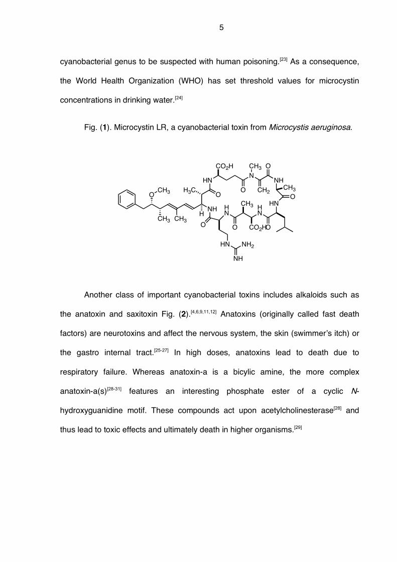
microcystins.[14] For example, the microcystin shown in Fig. (1) is called Microcystin
LR (Xaa2 = Ala, Yaa4 = Arg).[10] So far, more than 50 different variants of
microcystins have been found, all featuring the !-amino acid (2S,3S,8S,9S)-3-
amino-9-methoxy-2,6,8-trimethyl-10-phenyldecy-4(E),6(E)-dienoic acid abbreviated
as Adda.[9,10] This amino acid was shown to be essential for the biological activity of
microcystin via the covalent modification of proteins.[10,15,16] Microcystins are highly
active against protein phosphatase 1 and 2A.[17,18] These proteins are essential for
many signal transduction pathways of eukaryotic cells such as programmed cell
death (apoptosis).[19,20] This enzyme inhibitory activity is also responsible for the
toxicity of microcystins to grazers and thus point to their ecological role as
deterrence chemicals in aquatic ecosystems.[21,22] Recently, microcystins caused
great concerns to human health organizations. Due to the known hepatotoxicity of
microcystins, elevated levels of liver cancer were related to the presence of
microcystin in drinking water.[7] Actually, Microcystis is the most prevalent
5
cyanobacterial genus to be suspected with human poisoning.[23] As a consequence,
the World Health Organization (WHO) has set threshold values for microcystin
concentrations in drinking water.[24]
Fig. (1). Microcystin LR, a cyanobacterial toxin from Microcystis aeruginosa.
Another class of important cyanobacterial toxins includes alkaloids such as
the anatoxin and saxitoxin Fig. (2).[4,6,9,11,12] Anatoxins (originally called fast death
factors) are neurotoxins and affect the nervous system, the skin (swimmer!s itch) or
the gastro internal tract.[25-27] In high doses, anatoxins lead to death due to
respiratory failure. Whereas anatoxin-a is a bicylic amine, the more complex
anatoxin-a(s)[28-31] features an interesting phosphate ester of a cyclic N-
hydroxyguanidine motif. These compounds act upon acetylcholinesterase[28] and
thus lead to toxic effects and ultimately death in higher organisms.[29]
NH
O
CH3 CH3
CH3O
HNN
O
CH3
CH2
NH
O
HN
O
HN
HN
OO
CO2H
CH3
O
CH3
CO2H
HN NH2
NH
H3C
H
6
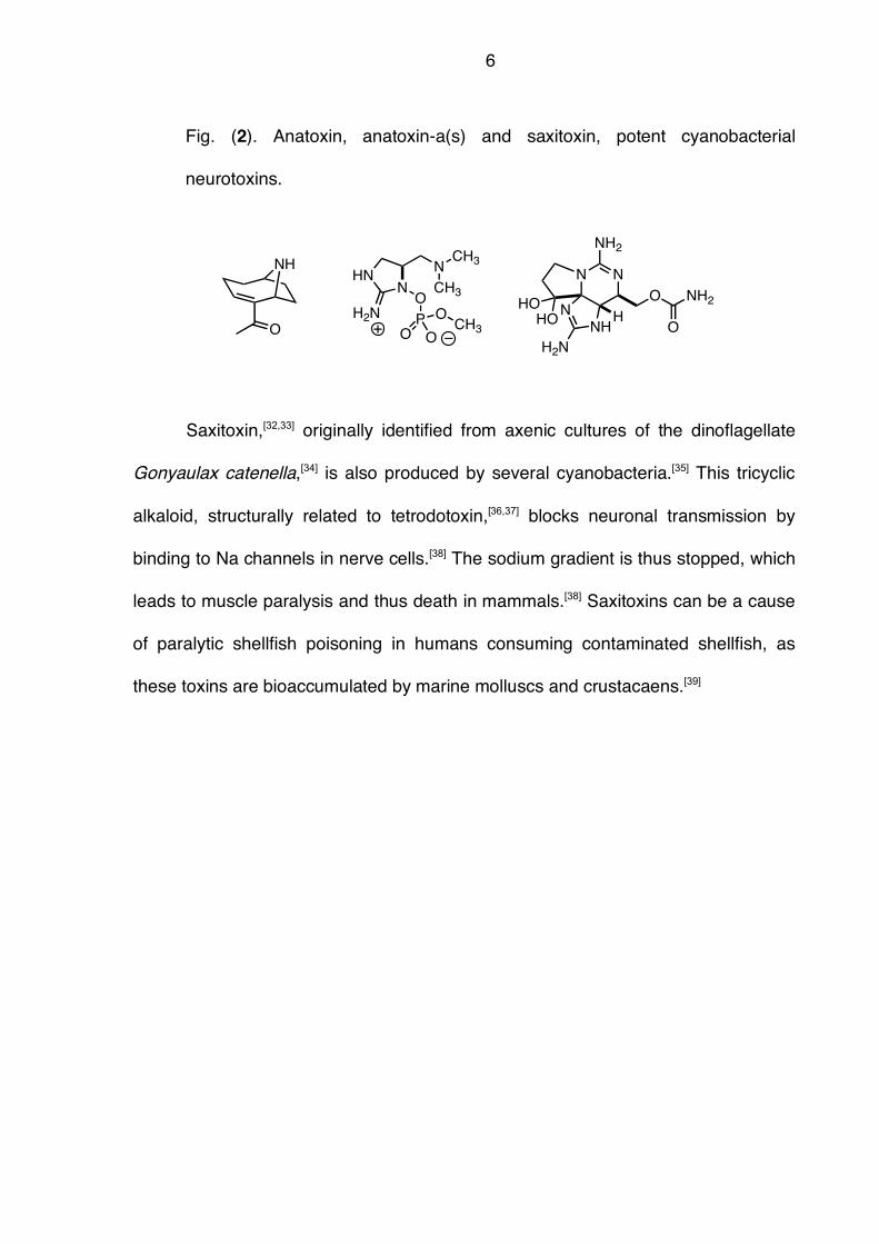
Fig. (2). Anatoxin, anatoxin-a(s) and saxitoxin, potent cyanobacterial
neurotoxins.
Saxitoxin,[32,33] originally identified from axenic cultures of the dinoflagellate
Gonyaulax catenella,[34] is also produced by several cyanobacteria.[35] This tricyclic
alkaloid, structurally related to tetrodotoxin,[36,37] blocks neuronal transmission by
binding to Na channels in nerve cells.[38] The sodium gradient is thus stopped, which
leads to muscle paralysis and thus death in mammals.[38] Saxitoxins can be a cause
of paralytic shellfish poisoning in humans consuming contaminated shellfish, as
these toxins are bioaccumulated by marine molluscs and crustacaens.[39]
NH
O
NHN
H2N
O
P
O O
OCH3
N
CH3
CH3
N N
HO
HON
NH
H2N
O
O
NH2
NH2
H
7
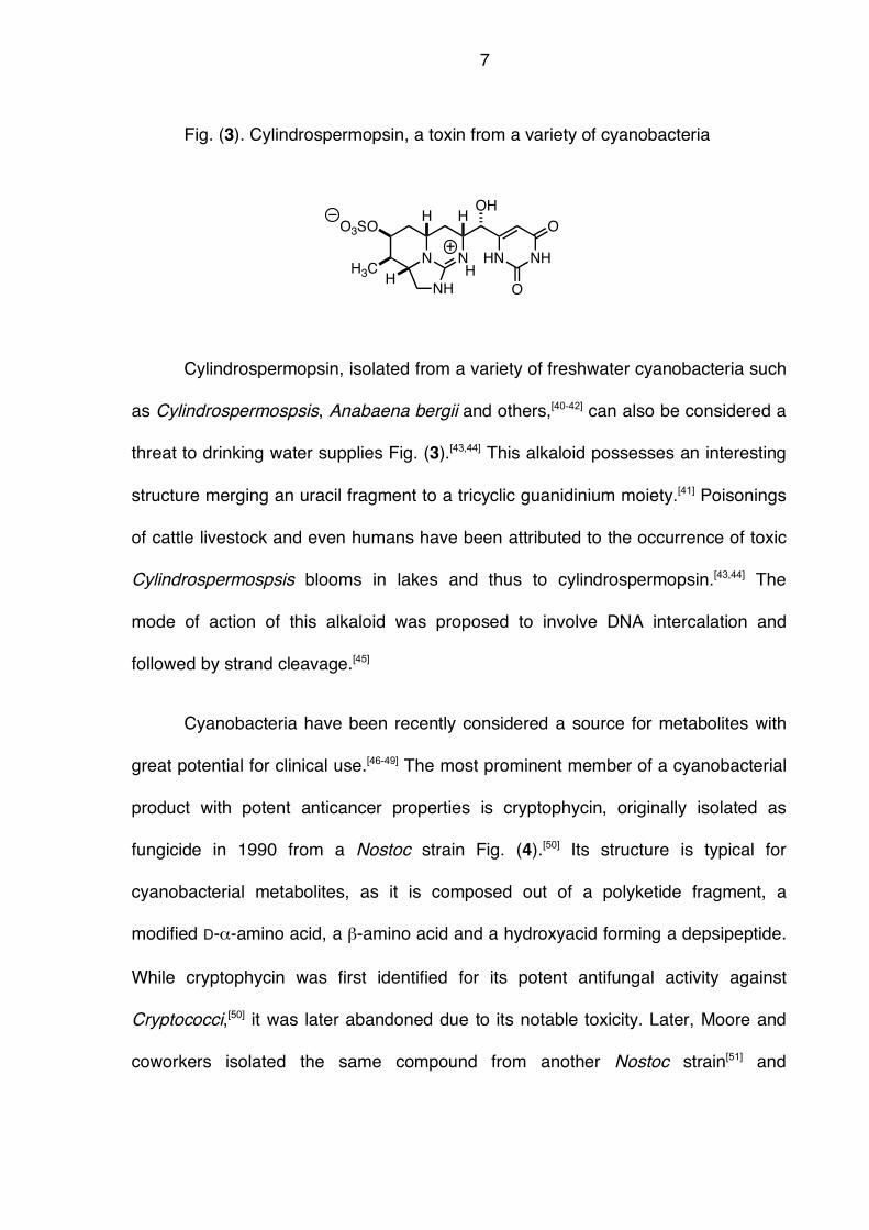
Fig. (3). Cylindrospermopsin, a toxin from a variety of cyanobacteria
Cylindrospermopsin, isolated from a variety of freshwater cyanobacteria such
as Cylindrospermospsis, Anabaena bergii and others,[40-42] can also be considered a
threat to drinking water supplies Fig. (3).[43,44] This alkaloid possesses an interesting
structure merging an uracil fragment to a tricyclic guanidinium moiety.[41] Poisonings
of cattle livestock and even humans have been attributed to the occurrence of toxic
Cylindrospermospsis blooms in lakes and thus to cylindrospermopsin.[43,44] The
mode of action of this alkaloid was proposed to involve DNA intercalation and
followed by strand cleavage.[45]
Cyanobacteria have been recently considered a source for metabolites with
great potential for clinical use.[46-49] The most prominent member of a cyanobacterial
product with potent anticancer properties is cryptophycin, originally isolated as
fungicide in 1990 from a Nostoc strain Fig. (4).[50] Its structure is typical for
cyanobacterial metabolites, as it is composed out of a polyketide fragment, a
modified D-!-amino acid, a "-amino acid and a hydroxyacid forming a depsipeptide.
While cryptophycin was first identified for its potent antifungal activity against
Cryptococci,[50] it was later abandoned due to its notable toxicity. Later, Moore and
coworkers isolated the same compound from another Nostoc strain[51] and
N N
NH
HN NH
O
O
OH
H
HHO3SO
H3C H
8
determined powerful cytotoxicity against tumor cell lines with IC50 value in the order
of 10 pM.[52,53] In addition, cryptophycin was not a substrate for the P-glycoprotein
efflux pump, as activity was found against both drug-sensitive and drug–resistant
tumor cell lines.[53] This cyanobacterial peptide polyketide hybrid induces apoptosis
by blocking the cell cycle at the G2/M phase apparently via inhibition of tubulin
polymerization.[54] The mode of action of cryptophycin combined with its extreme
potency (up to 1000 fold greater than taxol), led to its clinical evaluation as well as
structure/activity relationships (SAR).[55]
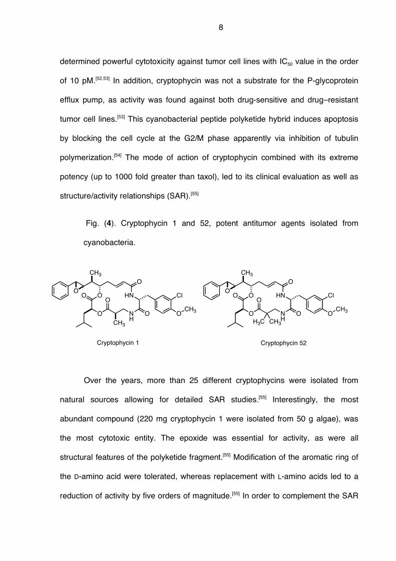
Fig. (4). Cryptophycin 1 and 52, potent antitumor agents isolated from
cyanobacteria.
Over the years, more than 25 different cryptophycins were isolated from
natural sources allowing for detailed SAR studies.[55] Interestingly, the most
abundant compound (220 mg cryptophycin 1 were isolated from 50 g algae), was
the most cytotoxic entity. The epoxide was essential for activity, as were all
structural features of the polyketide fragment.[55] Modification of the aromatic ring of
the D-amino acid were tolerated, whereas replacement with L-amino acids led to a
reduction of activity by five orders of magnitude.[55] In order to complement the SAR
O
O
O
HN
NH
O
O
CH3
O
OCH3
O
Cryptophycin 1 Cryptophycin 52
CH3
Cl O
O
O
HN
NH
O
O
CH3
O
OCH3
O Cl
H3C CH3
9
of the natural isolates, researchers both in academia and in industry (Eli Lilly)
embarked on the synthesis of hundreds of analogs, allowing for a detailed
understanding of the structural requirements for the biological activity of
cryptophycins.[55] Cryptophycin 52 was found to be more stable towards hydrolysis
and was thus selected as the main candidate for clinical development by Eli Lilly
(LY355703).[56,57] While this compound passed phase I clinical studies,[56,57] there is
little data in the literature concerning phase II studies.[58]
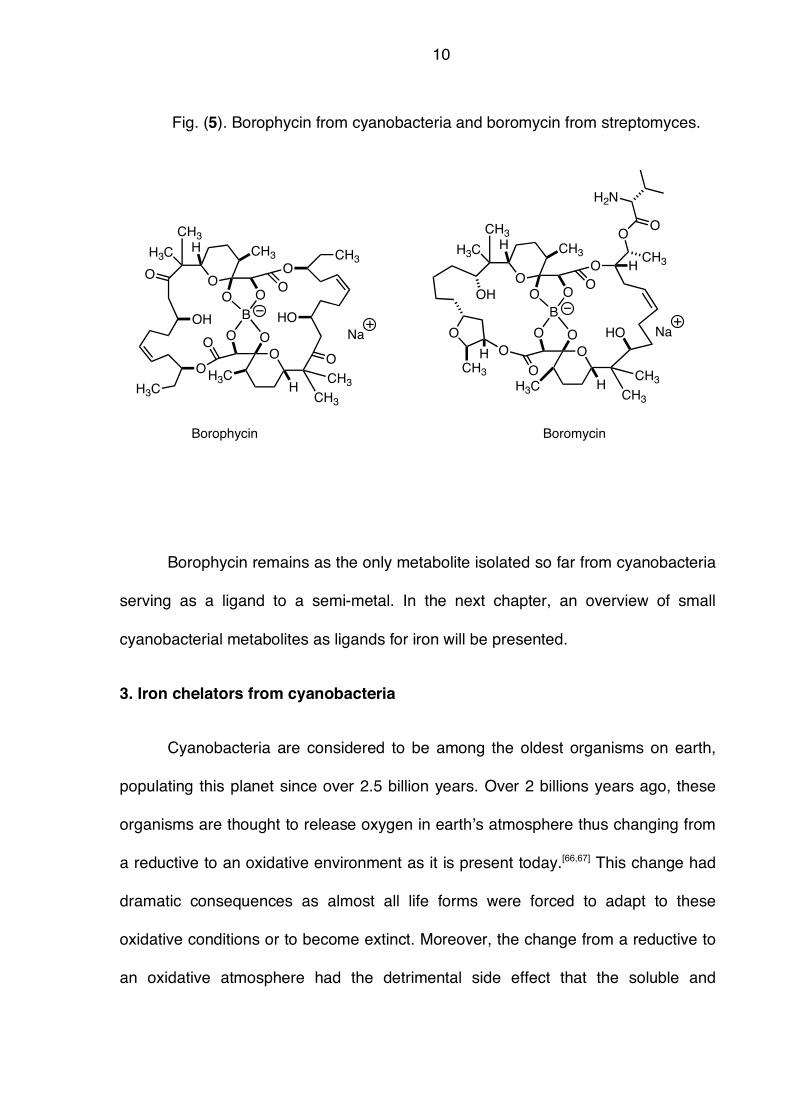
Borophycin is a complex boron containing polyketide, isolated in 1994 by
Moore and coworkers from Nostoc linckia[59] and later from Nostoc spongiaeforme
Fig. (5).[60] This metabolite displayed promising antitumor activity against standard
cancer cell lines (MIC 0.066 mg/mL, LoVo and 3.3 mg/mL KB).[59] The structure was
assigned by spectroscopic methods and both configuration and constitution
unambiguously established by single crystal X-Ray diffraction. The structure can be
regarded as a dimeric macrodiolide, with a borate linking the two strands. Its
structure strongly resembles boromycin from Streptomyces antibioticus, isolated in
1969 by Prelog, Zähner, Keller-Schierlein and coworkers[61] and structure
determination by Dunitz, Prelog and coworkers,[62] aplasmomycin from Streptomyces
griseus[63,64] and tartrolon from Sorangium cellulosum.[65]
10
Fig. (5). Borophycin from cyanobacteria and boromycin from streptomyces.
Borophycin remains as the only metabolite isolated so far from cyanobacteria
serving as a ligand to a semi-metal. In the next chapter, an overview of small
cyanobacterial metabolites as ligands for iron will be presented.
3. Iron chelators from cyanobacteria
Cyanobacteria are considered to be among the oldest organisms on earth,
populating this planet since over 2.5 billion years. Over 2 billions years ago, these
organisms are thought to release oxygen in earth!s atmosphere thus changing from
a reductive to an oxidative environment as it is present today.[66,67] This change had
dramatic consequences as almost all life forms were forced to adapt to these
oxidative conditions or to become extinct. Moreover, the change from a reductive to
an oxidative atmosphere had the detrimental side effect that the soluble and
O
H3C
OO O
O
H3C
CH3
CH3
O
HO
OCH3
OO O
O
CH3
H3C
O
OH
H
CH3H
B
Na
O
O
O O
O
H3C CH3
CH3
O
OO O
O
CH3
H3C
H
CH3H
B
NaO
CH3
OH
HO
O
CH3
O
H2N
Borophycin Boromycin
H
H
11
prevalent Fe(II) salts were oxidized to the corresponding Fe(III) oxide hydrates,
which are almost insoluble.[68] Due to this extremely low bioavailability of iron, the
acquisition, transport and storage of Fe became a central challenge for every
organism.[68] Microorganisms thus evolved sophisticated strategies for Fe
sequestering involving small organic ligands, so called siderophores.[69,70] In fact,
many siderophores from bacteria have been isolated which can be clustered in three
different structural types.[69,70]
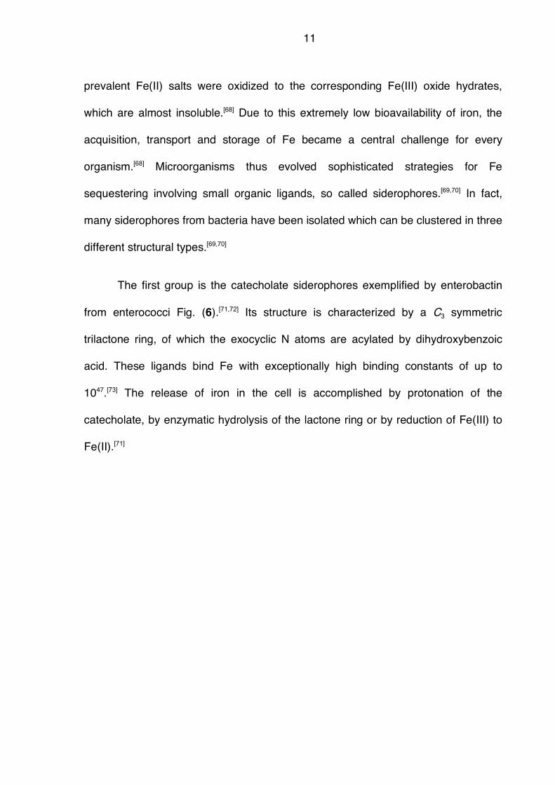
The first group is the catecholate siderophores exemplified by enterobactin
from enterococci Fig. (6).[71,72] Its structure is characterized by a C3 symmetric
trilactone ring, of which the exocyclic N atoms are acylated by dihydroxybenzoic
acid. These ligands bind Fe with exceptionally high binding constants of up to
1047.[73] The release of iron in the cell is accomplished by protonation of the
catecholate, by enzymatic hydrolysis of the lactone ring or by reduction of Fe(III) to
Fe(II).[71]
12
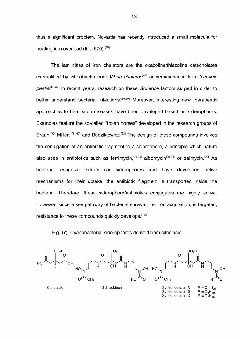
Fig. (6). The three main structural classes of bacterial siderophores.
Another class of siderophores is represented by hydroxamate siderophores
exemplified by desferrioxamine (DFO), isolated by Prelog and collaborators fifty
years ago.[74-76] Key elements are hydroxamic acid units which can chelate to Fe(III)
in a bidentate way. The binding constants of DFO have been determined to be
between 1020 and 1030, depending on the structure and on the pH.[77,78] DFO is a drug
currently used to treat iron overload.[79] Iron is so precious to humans that men have
no means of secreting excess iron out of the body.[80] Excess iron leads to oxidative
stress followed by tissue damage and, ultimately, death.[81] Moreover,
hemochromatosis is the most prevalent genetic disorder in Europe with 1 out of 20
carrying the mismatched gene, [82] and this disease is directly linked to high iron
levels. Iron overload can only be cured by either phlebotomy or treatment with
DFO.[83] The drawback of DFO is that it needs to be administered for 6 days for at
least 12 hours intravenously due to poor pharmacokinetics. Patient compliance is
HN
OO
ONHHN
O
O
O
OO
OH
HO
O
OH
OH
Enterobactin Desferrioxamine B Vibriobacin
HN
OH
OH
O
N
O
N
O
OHHO
HN
O
O
NHO
OH
Me
Me
HO
OH
NH
N
O
O
HO
NCH3
O
HO
N
N
NH3
H
O
O
HO
13
thus a significant problem. Novartis has recently introduced a small molecule for
treating iron overload (ICL-670).[79]
The last class of iron chelators are the oxazoline/thiazoline catecholates
exemplified by vibriobactin from Vibrio cholerae[84] or yersiniabactin from Yersinia
pestis.[85-87] In recent years, research on these virulence factors surged in order to
better understand bacterial infections.[88,89] Moreover, interesting new therapeutic
approaches to treat such diseases have been developed based on siderophores.
Examples feature the so-called “trojan horses” developed in the research groups of
Braun,[90] Miller, [91,92] and Budzikiewicz.[93] The design of these compounds involves
the conjugation of an antibiotic fragment to a siderophore, a principle which nature
also uses in antibiotics such as ferrimycin,[94,95] albomycin[96-98] or salmycin.[99] As
bacteria recognize extracellular siderophores and have developed active
mechanisms for their uptake, the antibiotic fragment is transported inside the
bacteria. Therefore, these siderophore/antibiotics conjugates are highly active.
However, since a key pathway of bacterial survival, i.e. iron acquisition, is targeted,
resistance to these compounds quickly develops.[100]
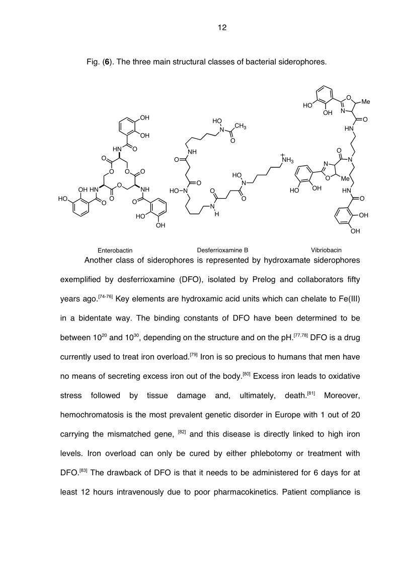
Fig. (7). Cyanobacterial siderophores derived from citric acid.
OH
CO2H
NH
O
NH
O
NHO
CH3O
NOH
OH3C
OH
CO2H
HO
O
OH
O
Citric acid Schizokinen
OH
CO2H
NH
O
NH
O
NHO
CH3O
NOH
OR
Synechobactin ASynechobactin BSynechobactin C
R = C11H23R = C9H19R = C7H15
14
Whereas bacterial siderophores have been actively studied for decades,
there is much less known about cyanobacterial iron chelators. This is surprising, as
cyanobacteria probably caused the transformation of a reductive atmosphere to an
oxidative one and thus rendered the uptake of Fe(III) a central challenge to every
organism.[66,67] Another problem is that cyanobacteria frequently populate marine and
freshwater habitats and are among the most important primary producers. In such
environments, complex siderophores, which are costly to make by the organism
involving many enzymes and pathways), are lost by dilution when secreted.
Therefore, it was assumed for a long time that only small and simple organic
molecules such as simple hydroxamic acids or citrate would be used. Their benefit
would include ease of preparation with the drawback of poorer iron chelation.
The first report on a cyanobacterial siderophore was schizokinen from the
freshwater Anabaena PCC 7120 Fig. (7).[101-103] This compound, originally isolated
from the terrestrial bacterium Bacillus megaterium,[104,105] is a citrate derivative of
which two carboxylate groups are amidated with an acetyl hydroxamic amine. In
2005, the synechobactins were isolated from a coastal marine cyanobacterium,
Synechococcus PCC 7002 and demonstrated to act as siderophores.[106] These
compounds can be considered as derivatives of schizokinen, with one of the
hydroxamic acids replaced by a long fatty acid. The authors suggest that this
amphiphilic structure helps to anchor the siderophore into the membrane of
cyanobacterium and thus prevents its loss by dilution in marine environments. This
notion is supported by several amphiphilic siderophores from marine bacteria such
as marinobactin E, aquachelin D and amphibactin D, which all carry a long
15
hydrophobic chain.[107] The citrate core of both schizokinen and synechobactin are
also found in petrobactin.[108]
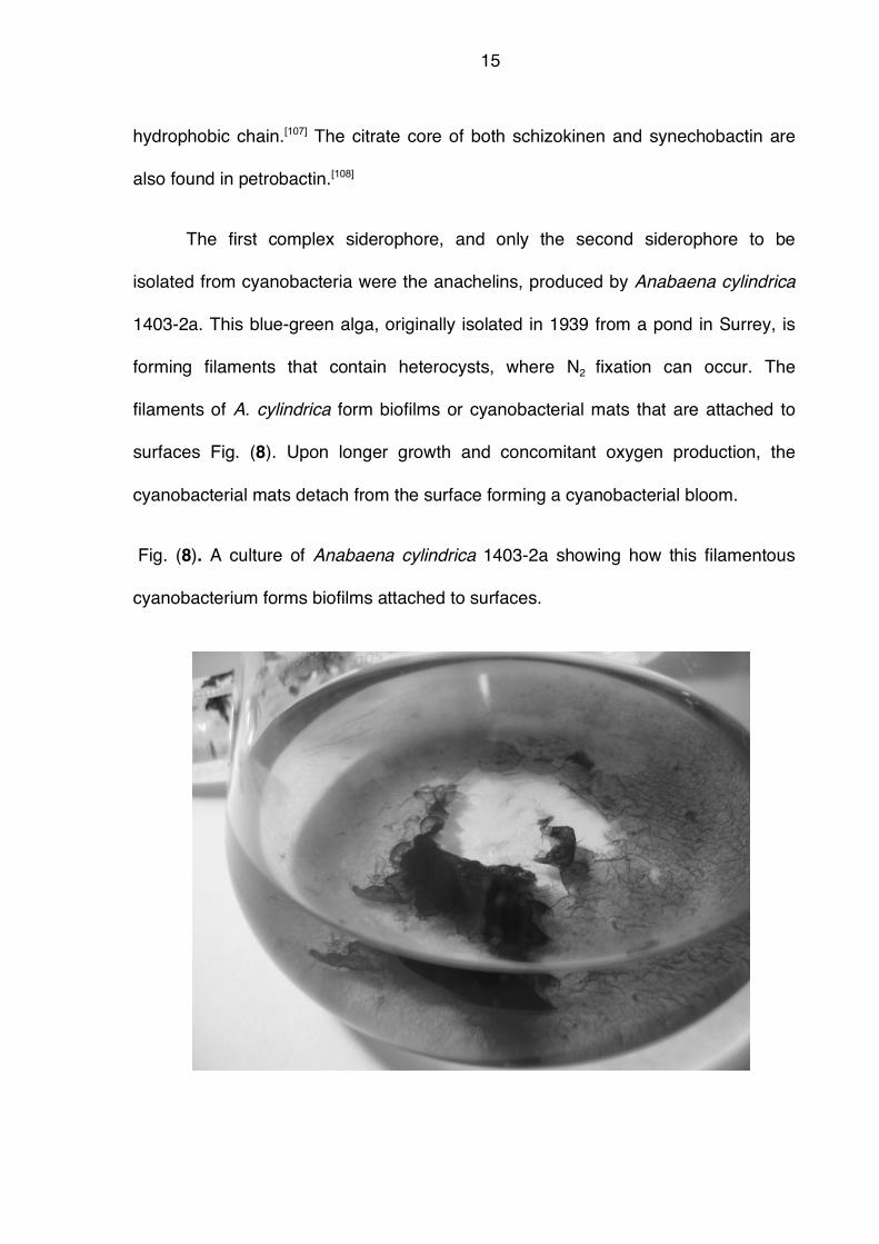
The first complex siderophore, and only the second siderophore to be
isolated from cyanobacteria were the anachelins, produced by Anabaena cylindrica
1403-2a. This blue-green alga, originally isolated in 1939 from a pond in Surrey, is
forming filaments that contain heterocysts, where N2 fixation can occur. The
filaments of A. cylindrica form biofilms or cyanobacterial mats that are attached to
surfaces Fig. (8). Upon longer growth and concomitant oxygen production, the
cyanobacterial mats detach from the surface forming a cyanobacterial bloom.
Fig. (8). A culture of Anabaena cylindrica 1403-2a showing how this filamentous
cyanobacterium forms biofilms attached to surfaces.
16
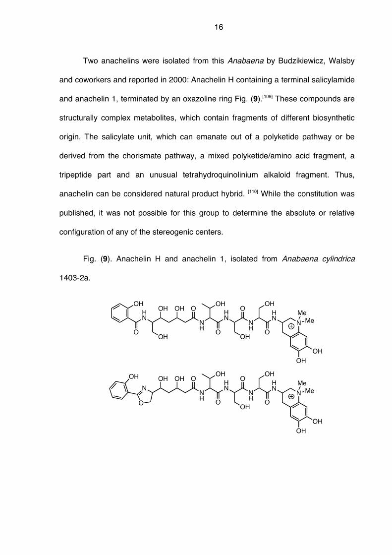
Two anachelins were isolated from this Anabaena by Budzikiewicz, Walsby
and coworkers and reported in 2000: Anachelin H containing a terminal salicylamide
and anachelin 1, terminated by an oxazoline ring Fig. (9).[109] These compounds are
structurally complex metabolites, which contain fragments of different biosynthetic
origin. The salicylate unit, which can emanate out of a polyketide pathway or be
derived from the chorismate pathway, a mixed polyketide/amino acid fragment, a
tripeptide part and an unusual tetrahydroquinolinium alkaloid fragment. Thus,
anachelin can be considered natural product hybrid. [110] While the constitution was
published, it was not possible for this group to determine the absolute or relative
configuration of any of the stereogenic centers.
Fig. (9). Anachelin H and anachelin 1, isolated from Anabaena cylindrica
1403-2a.
NH
HN
O
OH
NH
O
OH
HN
N
OH
Me
Me
OH
OH
O
OOH OH
OH
HN
O
OH
NH
HN
O
OH
NH
O
OH
HN
N
OH
Me
Me
OH
OH
O
OOH OH
N
O
OH
17
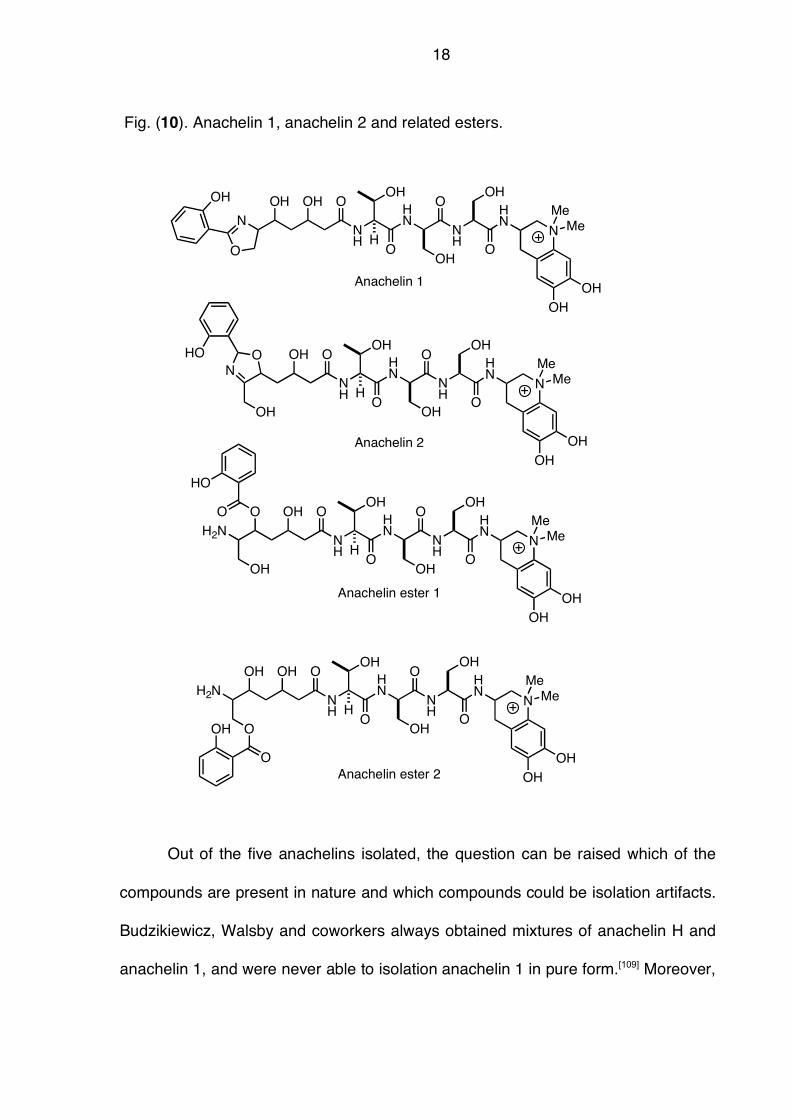
Subsequently, Murakami and coworkers reported the isolation and structure
elucidation of anachelin 1 and additional isomers Fig. (10).[111] In addition to the
terminal oxazoline, the authors also obtained anachelin 2 featuring the oxazoline
ring from C(5)-O to the C(6)-N. Moreover, two related esters were obtained. The
authors were also successful in determining the absolute configuration of the
tripeptide fragment thus establishing the sequence as L-Thr-D-Ser-L-Ser. The
configuration of the polyketide fragment as well as the alkaloid part remained
unassigned.
18
Fig. (10). Anachelin 1, anachelin 2 and related esters.
Out of the five anachelins isolated, the question can be raised which of the
compounds are present in nature and which compounds could be isolation artifacts.
Budzikiewicz, Walsby and coworkers always obtained mixtures of anachelin H and
anachelin 1, and were never able to isolation anachelin 1 in pure form.[109] Moreover,
NH
HN
O
OH
NH
O
OH
HN
N
OH
Me
Me
OH
OH
O
OOH
OH
NH
HN
O
OH
NH
O
OH
HN
N
OH
Me
Me
OH
OH
O
OOH OH
N
O
OH
O
N
HO
NH
HN
O
OH
NH
O
OH
HN
N
OH
Me
Me
OH
OH
O
OOH
OH
NH
HN
O
OH
NH
O
OH
HN
N
OH
Me
Me
OH
OH
O
O
O
H2N
O
H2N
O
HO
OH
H
H
H
H
OH
O
OH
Anachelin 1
Anachelin 2
Anachelin ester 1
Anachelin ester 2
19
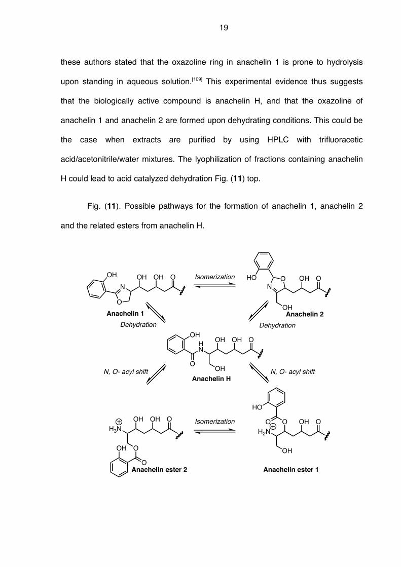
these authors stated that the oxazoline ring in anachelin 1 is prone to hydrolysis
upon standing in aqueous solution.[109] This experimental evidence thus suggests
that the biologically active compound is anachelin H, and that the oxazoline of
anachelin 1 and anachelin 2 are formed upon dehydrating conditions. This could be
the case when extracts are purified by using HPLC with trifluoracetic
acid/acetonitrile/water mixtures. The lyophilization of fractions containing anachelin
H could lead to acid catalyzed dehydration Fig. (11) top.
Fig. (11). Possible pathways for the formation of anachelin 1, anachelin 2
and the related esters from anachelin H.
OOH
OH
OOH OH
N
O
OHO
N
HO
OOH
OH
O
O
H3NO
H2N
O
HO
OH OH
O
OH
Isomerization
Isomerization
OOH OH
OH
HN
O
OH
N, O- acyl shift
Dehydration
Anachelin 1 Anachelin 2
Anachelin H
Anachelin ester 2 Anachelin ester 1
N, O- acyl shift
Dehydration
20
The formation of the esters could be easily explained by an N, O-acyl shift of
the salicylamide under slightly acidic conditions. This shift would be favored due to
the protonation of the amino group in acidic medium. This would provide an
explanation why Murakami and coworkers did not obtain anachelin H, but instead
the isomeric esters.[111] N, O-acyl shifts are frequently observed in peptides and a
well-known example is the formation of iso-cyclosporin derived from cyclosporin via
an N, O-acyl shift of the MeBmt amino acid.[112-116] This migration readily occurs
under acidic conditions and is fully reversible upon the addition of base.[116] Last,
anachelin is the siderophore of an aquatic freshwater siderophore, which should be
resistant to facile hydrolysis. This requirement adds further support to the role of
anachelin H, as anachelin 1 was found to be prone to hydrolysis. All these reasons
provide support for the hypothesis that anachelin H constitutes the actual
siderophore, and that anachelin 1 and 2, as well as the related esters would be
considered derivatives.
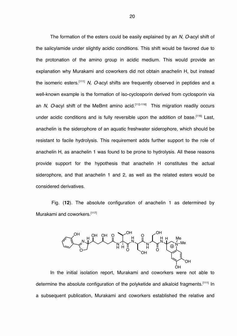
Fig. (12). The absolute configuration of anachelin 1 as determined by
Murakami and coworkers.[117]
In the initial isolation report, Murakami and coworkers were not able to
determine the absolute configuration of the polyketide and alkaloid fragments.[111] In
a subsequent publication, Murakami and coworkers established the relative and
NH
HN
O
OH
NH
O
OH
HN
N
OH
Me
Me
OH
OH
O
OOH OH
N
O
OH
H
H H
21
absolute configuration of anachelin via degradation and spectroscopic analysis and
published their analysis in 2004 Fig. (12).[117] We have independently established the
relative and absolution configuration via a stereodivergent synthesis. [118]
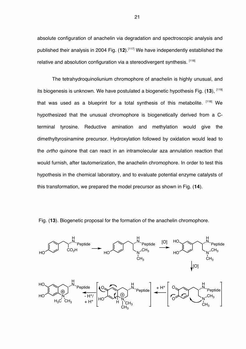
The tetrahydroquinoliunium chromophore of anachelin is highly unusual, and
its biogenesis is unknown. We have postulated a biogenetic hypothesis Fig. (13), [119]
that was used as a blueprint for a total synthesis of this metabolite. [118] We
hypothesized that the unusual chromophore is biogenetically derived from a C-
terminal tyrosine. Reductive amination and methylation would give the
dimethyltyrosinamine precursor. Hydroxylation followed by oxidation would lead to
the ortho quinone that can react in an intramolecular aza annulation reaction that
would furnish, after tautomerization, the anachelin chromophore. In order to test this
hypothesis in the chemical laboratory, and to evaluate potential enzyme catalysts of
this transformation, we prepared the model precursor as shown in Fig. (14).
Fig. (13). Biogenetic proposal for the formation of the anachelin chromophore.
HOCO2H
HN
HO
HN
HO
HNHO
N N
CH3
CH3
O
HNO
N
CH3
CH3HO
HNHO
N
H3C CH3
[O]
Peptide Peptide Peptide
PeptidePeptide
CH3
CH3
[O]
HO
HNO
Peptide
N
CH3
CH3H
+ H+
- H+/
+ H+
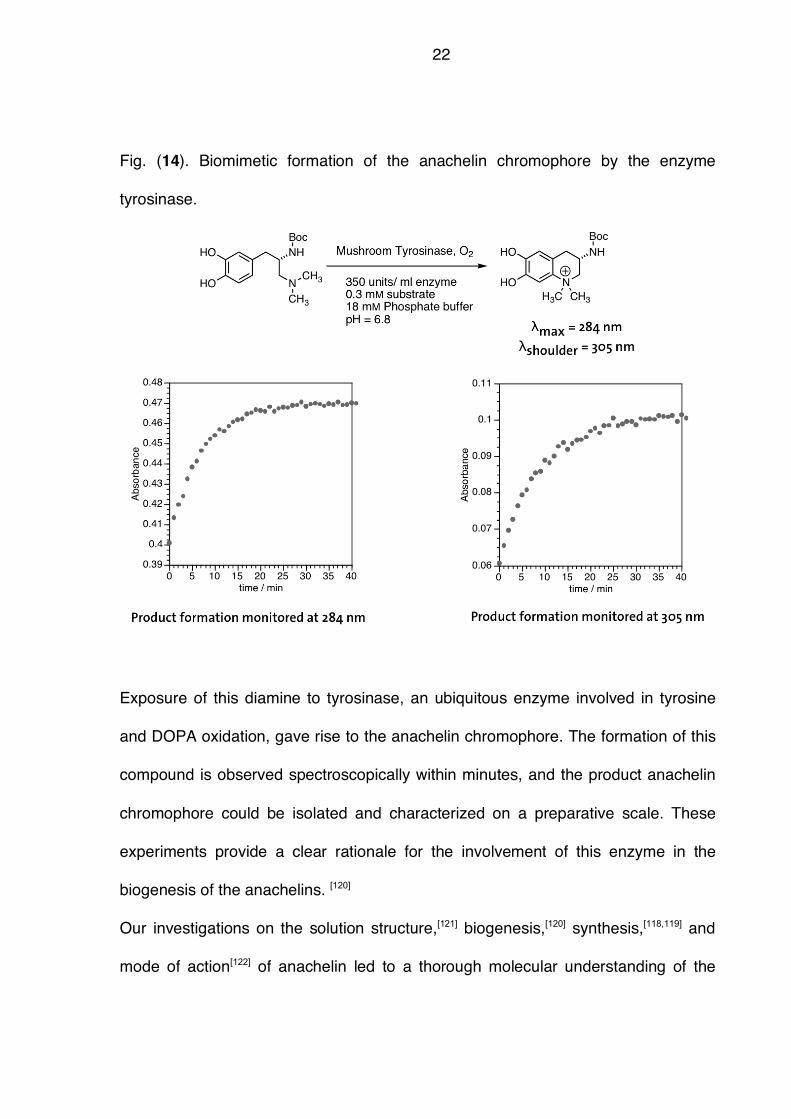
22
Fig. (14). Biomimetic formation of the anachelin chromophore by the enzyme
tyrosinase.
Exposure of this diamine to tyrosinase, an ubiquitous enzyme involved in tyrosine
and DOPA oxidation, gave rise to the anachelin chromophore. The formation of this
compound is observed spectroscopically within minutes, and the product anachelin
chromophore could be isolated and characterized on a preparative scale. These
experiments provide a clear rationale for the involvement of this enzyme in the
biogenesis of the anachelins. [120]
Our investigations on the solution structure,[121] biogenesis,[120] synthesis,[118,119] and
mode of action[122] of anachelin led to a thorough molecular understanding of the
23
different facets of this compound. This knowledge also allowed for the application in
a totally different field, i.e. surface modification in materials science. The
predominant physical form of iron in marine and freshwater environments are iron
oxide hydrates, which are solid minerals. In addition, iron ions are present in stones,
rocks and minerals. Therefore, in order to apprehend iron ions, siderophores must
recognize, bind and sequester iron from solid minerals. The first step of this process
in binding of the siderophore to mineral surfaces. We therefore asked the question
whether it is possible that anachelin binds to metal oxide surfaces. In order to test
this hypothesis, we prepared the hybrid[110] of the anachelin chromophore and
polyethylene glycol (PEG), which is frequently used to render surfaces resistant to
the attachment of proteins. The resulting compound was clearly shown to efficiently
bind to titanium oxide surfaces.[123] Thus, it has been shown that the anachelin
chromophore recognizes metal oxide surfaces as found in minerals. This postulated
first step in iron acquisition is thought to have large implications on the molecular
understanding of iron acquisition of marine and freshwater organisms.
4 Indole alkaloids
Indole alkaloids are among the most frequently encountered classes of
secondary metabolites in higher plants, and to a lesser extent, in microorganisms
and animals.[124] Prominent examples include the ergot, iboga, pyrroloindole (i. e.
physiostigmin), carboline (i. e. harmane, reserpine, yohimbin), aspidosperma and
the strychnos alkaloids (i. e. strychin, brucin and curare).[124] In most of these cases,
potent biological activities are associated with this structural framework, and the
24
isolation and structure elucidation of these compounds was of fundamental
importance for the understanding of their mode of action for the development of
related substances with beneficial effects.
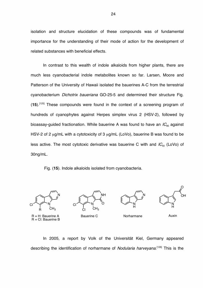
In contrast to this wealth of indole alkaloids from higher plants, there are
much less cyanobacterial indole metabolites known so far. Larsen, Moore and
Patterson of the University of Hawaii isolated the bauerines A-C from the terrestrial
cyanobacterium Dichotrix baueriana GO-25-5 and determined their structure Fig.
(15).[125] These compounds were found in the context of a screening program of
hundreds of cyanophytes against Herpes simplex virus 2 (HSV-2), followed by
bioassay-guided fractionation. While bauerine A was found to have an IC90 against
HSV-2 of 2 µg/mL with a cytotoxicity of 3 µg/mL (LoVo), bauerine B was found to be
less active. The most cytotoxic derivative was bauerine C with and IC50 (LoVo) of
30ng/mL.
Fig. (15). Indole alkaloids isolated from cyanobacteria.
In 2005, a report by Volk of the Universität Kiel, Germany appeared
describing the identification of norharmane of Nodularia harveyana.[126] This is the
N
N
CH3
Cl
R
N
NH
CH3
Cl
Cl
O
R = H: Bauerine A
R = Cl: Bauerine B
Bauerine C
NH
N
Norharmane
NH
OH
O
Auxin
25
first identification of this compound from cyanobacteria. Norharmane is known in
higher plants, bacteria and even in humans. Volk reported anticyanobacterial
activity[127] of this compound against both filamentous and unicellular cyanobacteria,
whereas no activity against green algae could be determined. [128,129] This is in
agreement with earlier reports on the isolation of harmane from a marine
Pseudomonas species by Murakami and coworkers, who also showed
anticyanobacterial activity with green algae being unaffected.[130] These authors also
suggested the use of harmane for the control of toxic algal blooms.[130] Given the
known co-mutagenicity of harmane,[131,132] this suggested approach for algal bloom
control could be questioned. The import plant hormone (phytohormane) auxin or
indolo acetic acid was also recently identified in cyanobacteria of the genus
Nostoc.[133] The production and release of such phytohormones by cyanobacteria
living in symbiotic or parasitic environments could possess significant ecological
consequences.
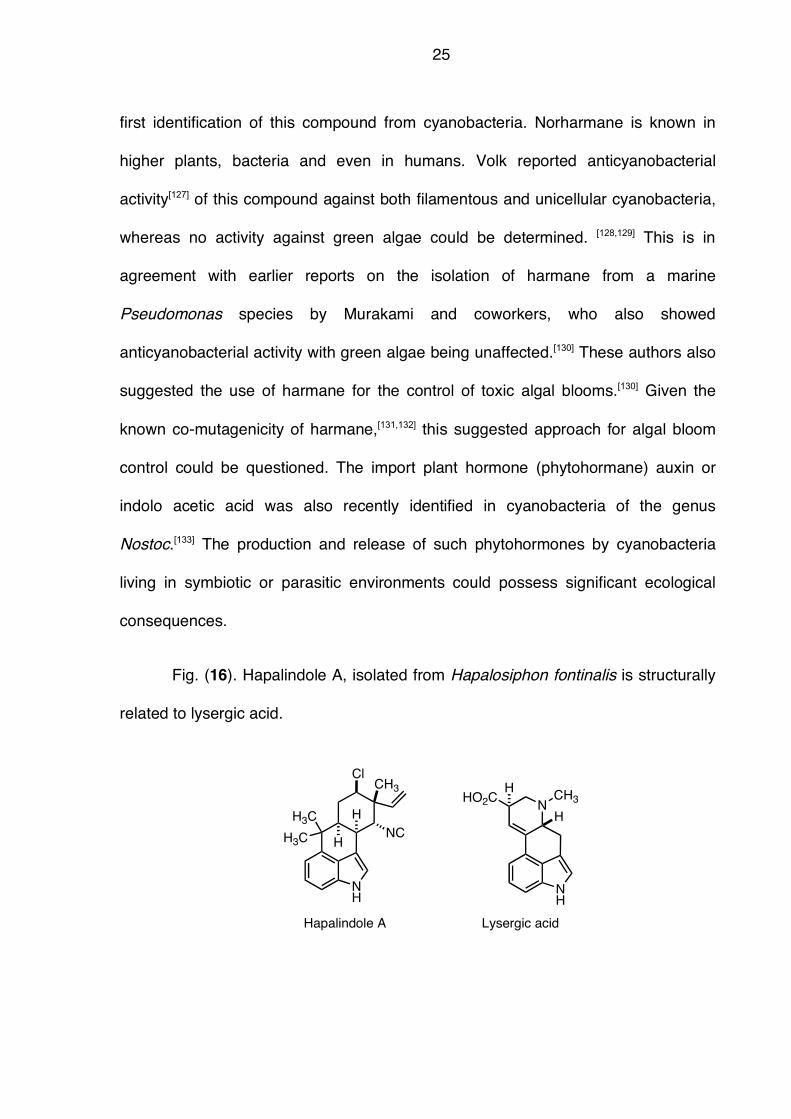
Fig. (16). Hapalindole A, isolated from Hapalosiphon fontinalis is structurally
related to lysergic acid.
NH
NC
CH3
Cl
H
H
H3C
H3C
Hapalindole A
NH
NCH3HO2C
H
H
Lysergic acid
26
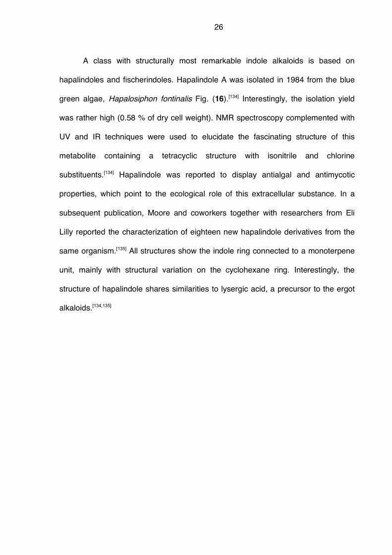
A class with structurally most remarkable indole alkaloids is based on
hapalindoles and fischerindoles. Hapalindole A was isolated in 1984 from the blue
green algae, Hapalosiphon fontinalis Fig. (16).[134] Interestingly, the isolation yield
was rather high (0.58 % of dry cell weight). NMR spectroscopy complemented with
UV and IR techniques were used to elucidate the fascinating structure of this
metabolite containing a tetracyclic structure with isonitrile and chlorine
substituents.[134] Hapalindole was reported to display antialgal and antimycotic
properties, which point to the ecological role of this extracellular substance. In a
subsequent publication, Moore and coworkers together with researchers from Eli
Lilly reported the characterization of eighteen new hapalindole derivatives from the
same organism.[135] All structures show the indole ring connected to a monoterpene
unit, mainly with structural variation on the cyclohexane ring. Interestingly, the
structure of hapalindole shares similarities to lysergic acid, a precursor to the ergot
alkaloids.[134,135]
27
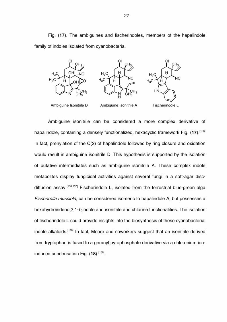
Fig. (17). The ambiguines and fischerindoles, members of the hapalindole
family of indoles isolated from cyanobacteria.
N
NC
CH3
Cl
OH
H
H3C
H3C
CH3
CH3
OOH
Ambiguine Isonitrile D
NC
CH3
Cl
H
H
Fischerindole L
H3C
H3C
HNNH
NC
CH3
Cl
H
H3C
H3C
CH3
CH3
Ambiguine Isonitrile A
H
Ambiguine isonitrile can be considered a more complex derivative of
hapalindole, containing a densely functionalized, hexacyclic framework Fig. (17).[136]
In fact, prenylation of the C(2) of hapalindole followed by ring closure and oxidation
would result in ambiguine isonitrile D. This hypothesis is supported by the isolation
of putative intermediates such as ambiguine isonitrile A. These complex indole
metabolites display fungicidal activities against several fungi in a soft-agar disc-
diffusion assay.[136,137] Fischerindole L, isolated from the terrestrial blue-green alga
Fischerella musciola, can be considered isomeric to hapalindole A, but possesses a
hexahydroindeno[2,1-b]indole and isonitrile and chlorine functionalities. The isolation
of fischerindole L could provide insights into the biosynthesis of these cyanobacterial
indole alkaloids.[138] In fact, Moore and coworkers suggest that an isonitrile derived
from tryptophan is fused to a geranyl pyrophosphate derivative via a chloronium ion-
induced condensation Fig. (18).[138]
28
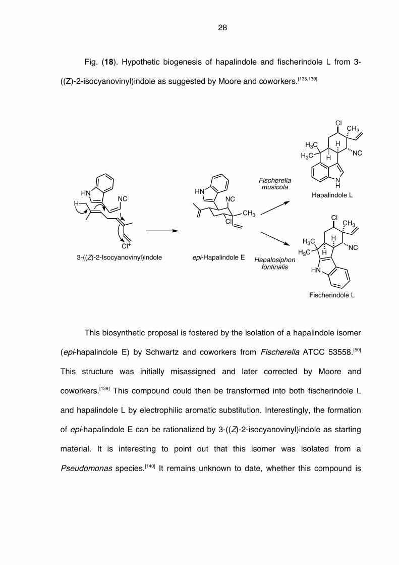
Fig. (18). Hypothetic biogenesis of hapalindole and fischerindole L from 3-
((Z)-2-isocyanovinyl)indole as suggested by Moore and coworkers.[138,139]
This biosynthetic proposal is fostered by the isolation of a hapalindole isomer
(epi-hapalindole E) by Schwartz and coworkers from Fischerella ATCC 53558.[50]
This structure was initially misassigned and later corrected by Moore and
coworkers.[139] This compound could then be transformed into both fischerindole L
and hapalindole L by electrophilic aromatic substitution. Interestingly, the formation
of epi-hapalindole E can be rationalized by 3-((Z)-2-isocyanovinyl)indole as starting
material. It is interesting to point out that this isomer was isolated from a
Pseudomonas species.[140] It remains unknown to date, whether this compound is
HNNC
3-((Z)-2-Isocyanovinyl)indole
HNC
Cl+
CH3
Cl
epi-Hapalindole E
NH
NC
CH3
Cl
H
H
H3C
H3C
Hapalindole L
NC
CH3
Cl
H
H
Fischerindole L
H3C
H3C
HN
Fischerellamusicola
Hapalosiphonfontinalis
HN
29
the biogenetic precursor to the hapalindole and fischerindole families of natural
products.
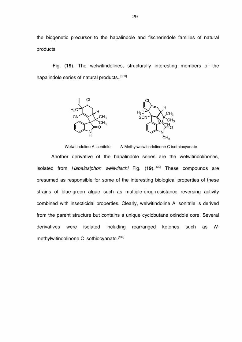
Fig. (19). The welwitindolines, structurally interesting members of the
hapalindole series of natural products..[139]
Another derivative of the hapalindole series are the welwitindolinones,
isolated from Hapalosiphon weilwitschi Fig. (19).[139] These compounds are
presumed as responsible for some of the interesting biological properties of these
strains of blue-green algae such as multiple-drug-resistance reversing activity
combined with insecticidal properties. Clearly, welwitindoline A isonitrile is derived
from the parent structure but contains a unique cyclobutane oxindole core. Several
derivatives were isolated including rearranged ketones such as N-
methylwitindolinone C isothiocyanate.[139]
NH
CH3
CH3
HH3C
CN
O
Welwitindoline A isonitrile
NO
O
CH3
CH3
SCN
HH3C
Cl Cl
N-Methylwelwitindolinone C isothiocyanate
CH3
H
30
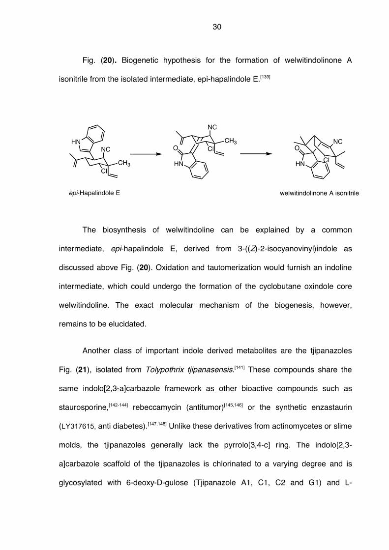
Fig. (20). Biogenetic hypothesis for the formation of welwitindolinone A
isonitrile from the isolated intermediate, epi-hapalindole E.[139]
The biosynthesis of welwitindoline can be explained by a common
intermediate, epi-hapalindole E, derived from 3-((Z)-2-isocyanovinyl)indole as
discussed above Fig. (20). Oxidation and tautomerization would furnish an indoline
intermediate, which could undergo the formation of the cyclobutane oxindole core
welwitindoline. The exact molecular mechanism of the biogenesis, however,
remains to be elucidated.
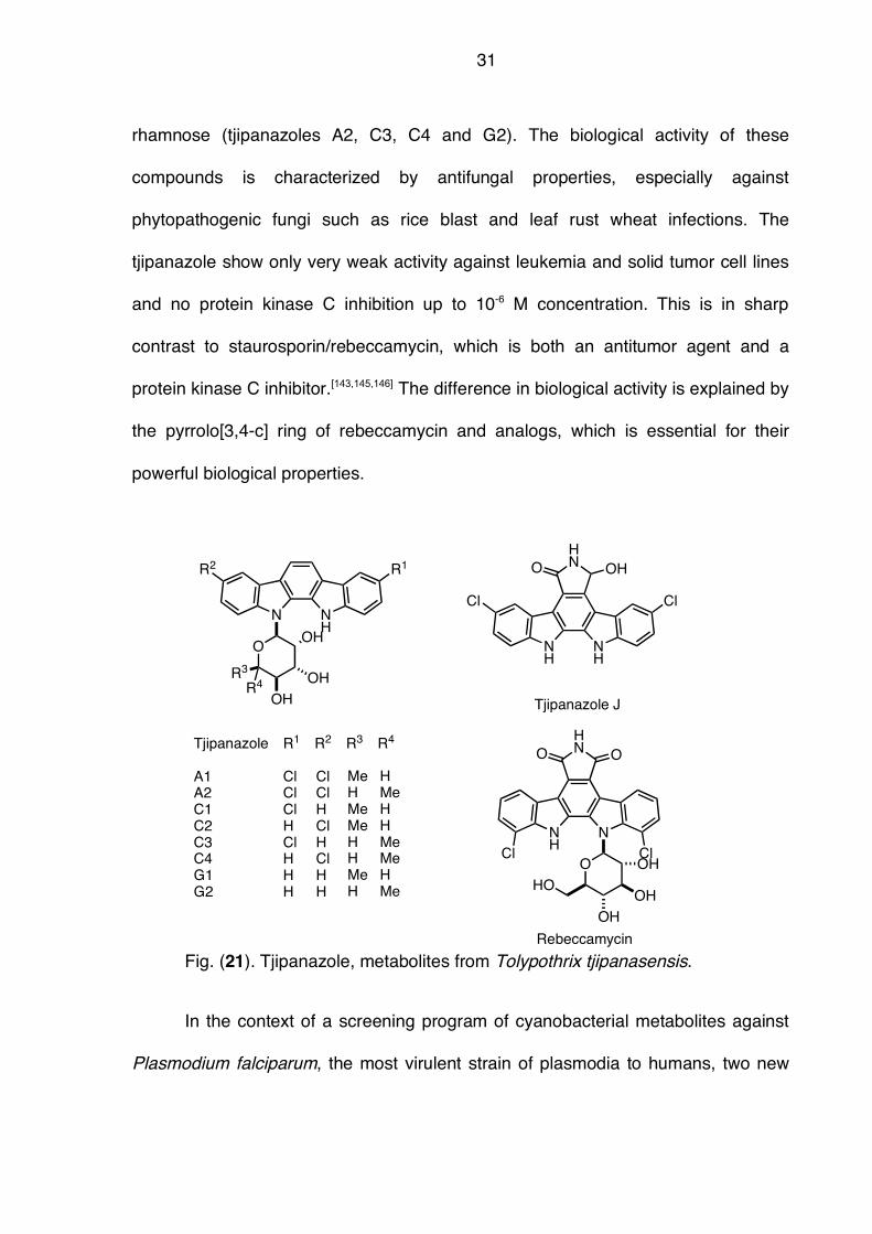
Another class of important indole derived metabolites are the tjipanazoles
Fig. (21), isolated from Tolypothrix tjipanasensis.[141] These compounds share the
same indolo[2,3-a]carbazole framework as other bioactive compounds such as
staurosporine,[142-144] rebeccamycin (antitumor)[145,146] or the synthetic enzastaurin
(LY317615, anti diabetes).[147,148] Unlike these derivatives from actinomycetes or slime
molds, the tjipanazoles generally lack the pyrrolo[3,4-c] ring. The indolo[2,3-
a]carbazole scaffold of the tjipanazoles is chlorinated to a varying degree and is
glycosylated with 6-deoxy-D-gulose (Tjipanazole A1, C1, C2 and G1) and L-
NC
CH3
Cl
epi-Hapalindole E
HN
NC
CH3
Cl
HN
O
HN
ONC
Cl
welwitindolinone A isonitrile
31
rhamnose (tjipanazoles A2, C3, C4 and G2). The biological activity of these
compounds is characterized by antifungal properties, especially against
phytopathogenic fungi such as rice blast and leaf rust wheat infections. The
tjipanazole show only very weak activity against leukemia and solid tumor cell lines
and no protein kinase C inhibition up to 10-6 M concentration. This is in sharp
contrast to staurosporin/rebeccamycin, which is both an antitumor agent and a
protein kinase C inhibitor.[143,145,146] The difference in biological activity is explained by
the pyrrolo[3,4-c] ring of rebeccamycin and analogs, which is essential for their
powerful biological properties.
Fig. (21). Tjipanazole, metabolites from Tolypothrix tjipanasensis.
In the context of a screening program of cyanobacterial metabolites against
Plasmodium falciparum, the most virulent strain of plasmodia to humans, two new
N NH
R2 R1
O
R3
OH
OH
OHR4
Tjipanazole
A1A2C1C2C3C4G1G2
R1 R2 R3 R4
ClClClHClHHH
ClClHClHClHH
MeHMeMeHHMeH
HMeHHMeMeHMe
NH
NH
Cl Cl
HNO OH
Tjipanazole J
NH
N
HNO O
Cl ClO
HO
OH
OH
OH
Rebeccamycin
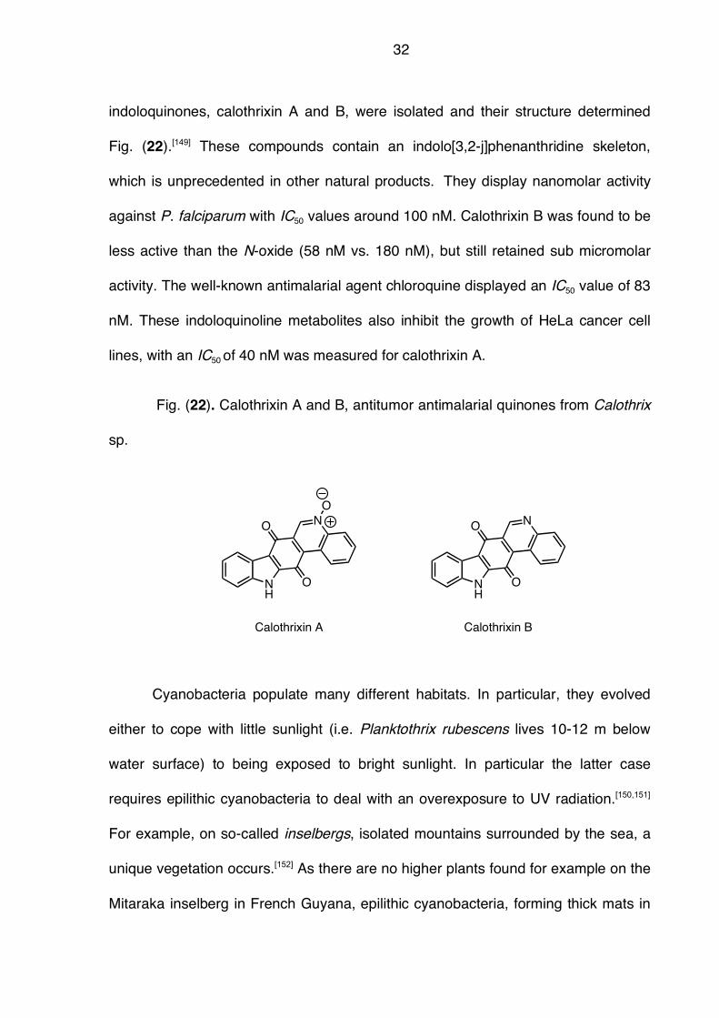
32
indoloquinones, calothrixin A and B, were isolated and their structure determined
Fig. (22).[149] These compounds contain an indolo[3,2-j]phenanthridine skeleton,
which is unprecedented in other natural products. They display nanomolar activity
against P. falciparum with IC50 values around 100 nM. Calothrixin B was found to be
less active than the N-oxide (58 nM vs. 180 nM), but still retained sub micromolar
activity. The well-known antimalarial agent chloroquine displayed an IC50 value of 83
nM. These indoloquinoline metabolites also inhibit the growth of HeLa cancer cell
lines, with an IC50 of 40 nM was measured for calothrixin A.
Fig. (22). Calothrixin A and B, antitumor antimalarial quinones from Calothrix
sp.
Cyanobacteria populate many different habitats. In particular, they evolved
either to cope with little sunlight (i.e. Planktothrix rubescens lives 10-12 m below
water surface) to being exposed to bright sunlight. In particular the latter case
requires epilithic cyanobacteria to deal with an overexposure to UV radiation.[150,151]
For example, on so-called inselbergs, isolated mountains surrounded by the sea, a
unique vegetation occurs.[152] As there are no higher plants found for example on the
Mitaraka inselberg in French Guyana, epilithic cyanobacteria, forming thick mats in
NH
NO
O
O
NH
NO
O
Calothrixin A Calothrixin B
33
polysaccharide matrices, are supposed to be the only living organisms under
intense solar radiation.[152] In order to meet this challenge, such cyanobacteria
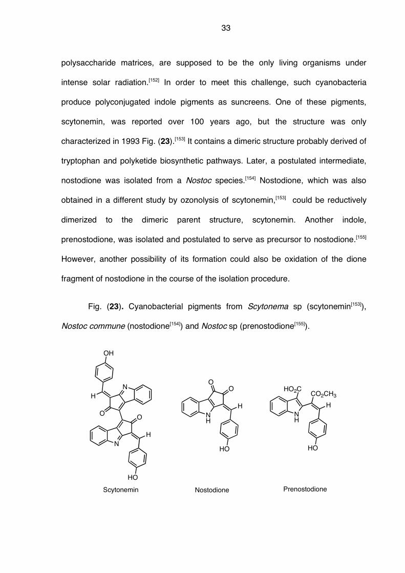
produce polyconjugated indole pigments as suncreens. One of these pigments,
scytonemin, was reported over 100 years ago, but the structure was only
characterized in 1993 Fig. (23).[153] It contains a dimeric structure probably derived of
tryptophan and polyketide biosynthetic pathways. Later, a postulated intermediate,
nostodione was isolated from a Nostoc species.[154] Nostodione, which was also
obtained in a different study by ozonolysis of scytonemin,[153] could be reductively
dimerized to the dimeric parent structure, scytonemin. Another indole,
prenostodione, was isolated and postulated to serve as precursor to nostodione.[155]
However, another possibility of its formation could also be oxidation of the dione
fragment of nostodione in the course of the isolation procedure.
Fig. (23). Cyanobacterial pigments from Scytonema sp (scytonemin[153]),
Nostoc commune (nostodione[154]) and Nostoc sp (prenostodione[155]).
N
O
H
N
O
H
Scytonemin
NH
O
H
O
Nostodione
OH
HO
HO
NH
H
Prenostodione
HO
HO2C CO2CH3
34
NH
N CH3
Cl
Nostocarboline
NH
Cl N
O
N
N
F
O
OH
N
Cl
Nostocarboline/ciprofloxacin hybrid
I
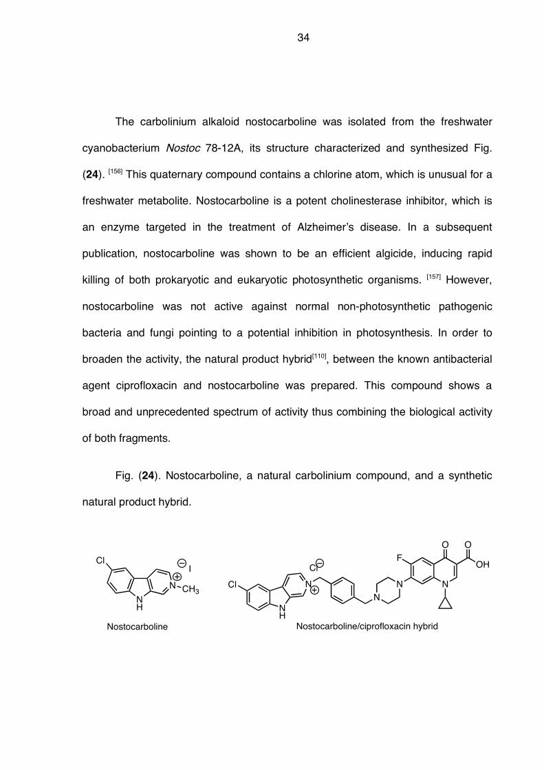
The carbolinium alkaloid nostocarboline was isolated from the freshwater
cyanobacterium Nostoc 78-12A, its structure characterized and synthesized Fig.
(24). [156] This quaternary compound contains a chlorine atom, which is unusual for a
freshwater metabolite. Nostocarboline is a potent cholinesterase inhibitor, which is
an enzyme targeted in the treatment of Alzheimer!s disease. In a subsequent
publication, nostocarboline was shown to be an efficient algicide, inducing rapid
killing of both prokaryotic and eukaryotic photosynthetic organisms. [157] However,
nostocarboline was not active against normal non-photosynthetic pathogenic
bacteria and fungi pointing to a potential inhibition in photosynthesis. In order to
broaden the activity, the natural product hybrid[110], between the known antibacterial
agent ciprofloxacin and nostocarboline was prepared. This compound shows a
broad and unprecedented spectrum of activity thus combining the biological activity
of both fragments.
Fig. (24). Nostocarboline, a natural carbolinium compound, and a synthetic
natural product hybrid.
35
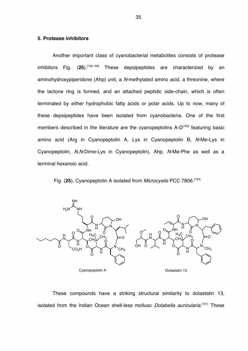
5. Protease inhibitors
Another important class of cyanobacterial metabolites consists of protease
inhibitors Fig. (25).[158,159] These depsipeptides are characterized by an
aminohydroxypiperidone (Ahp) unit, a N-methylated amino acid, a threonine, where
the lactone ring is formed, and an attached peptidic side-chain, which is often
terminated by either hydrophobic fatty acids or polar acids. Up to now, many of
these depsipeptides have been isolated from cyanobacteria. One of the first
members described in the literature are the cyanopeptolins A-D[160] featuring basic
amino acid (Arg in Cyanopeptolin A, Lys in Cyanopeptolin B, N-Me-Lys in
Cyanopeptolin, N,N-Dime-Lys in Cyanopeptolin), Ahp, N-Me-Phe as well as a
terminal hexanoic acid.
Fig. (25). Cyanopeptolin A isolated from Microcystis PCC 7806.[160]
These compounds have a striking structural similarity to dolastatin 13,
isolated from the Indian Ocean shell-less mollusc Dolabella auricularia.[161] These
O NH
NH
O
N
CH3
O
O
NH
H3CCH3
O
N
O
CH3
O
OH
NH
OHN
O
NH
NH
H2N
CO2H
Cyanopeptolin A
O NH
NH
O
N
CH3
O
O
NH
H3CCH3
O
N
O
CH3
O
OH
NH
OHN
O
Dolastatin 13
O
OH

36
seemingly defenseless animals are only attacked by certain carnivorous predators,
which is attributed to a very powerful chemical defense system of the sea slugs.
These secondary metabolites are now thought to originate from the dietary sources
of Dolabella such as cyanobacteria. Further support to this hypothesis is given by
the structural similarity of dolastatin 13 to cyanopeptolins.[162]
37
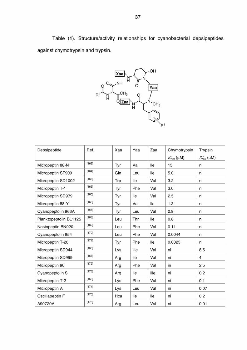
Table (1). Structure/activity relationships for cyanobacterial depsipeptides
against chymotrypsin and trypsin.
Depsipeptide Ref. Xaa Yaa Zaa Chymotrypsin
IC50 (µM)
Trypsin
IC50 (µM)
Micropeptin 88-N [163] Tyr Val Ile 15 ni
Micropeptin SF909 [164] Gln Leu Ile 5.0 ni
Micropeptin SD1002 [165] Trp Ile Val 3.2 ni
Micropeptin T-1 [166] Tyr Phe Val 3.0 ni
Micropeptin SD979 [165] Tyr Ile Val 2.5 ni
Micropeptin 88-Y [163] Tyr Val Ile 1.3 ni
Cyanopeptolin 963A [167] Tyr Leu Val 0.9 ni
Planktopeptolin BL1125 [168] Leu Thr Ile 0.8 ni
Nostopeptin BN920 [169] Leu Phe Val 0.11 ni
Cyanopeptolin 954 [170] Leu Phe Val 0.0044 ni
Micropeptin T-20 [171] Tyr Phe Ile 0.0025 ni
Micropeptin SD944 [165] Lys Ille Val ni 8.5
Micropeptin SD999 [165] Arg Ile Val ni 4
Micropeptin 90 [172] Arg Phe Val ni 2.5
Cyanopeptolin S [173] Arg Ile Ille ni 0.2
Micropeptin T-2 [166] Lys Phe Val ni 0.1
Micropeptin A [174] Lys Leu Val ni 0.07
Oscillapeptin F [175] Hca Ile Ile ni 0.2
A90720A [176] Arg Leu Val ni 0.01
O NH
NH
N
CH3
ONH
O
N
O
CH3
OH
NH
O
R2
Xaa
Yaa
R1
Zaa
38
As over twenty of these depsipeptides were isolated from cyanobacterial
sources, structure/activity relationships on their inhibitory action can be performed.
In table (1), a selection of these compounds is given, with the variable amino acids,
Xaa, Yaa and Zaa highlighted. Several requirements for activity become evident:
a. The key residue for protease inhibition consists of the
aminohydroxypiperidone residue. This amino acid is conserved for all
these depsipeptides.
b. All active compounds share a 19-membered lactone ring, with the amino
acid Zaa acylates the !-OH group of Thr
c. The selectivity for chymotrypsin is dictated by a hydrophobic amino acid
adjacent to Ahp, mostly consisting of Tyr or Leu, with Gln and Trp only
found once. The selectivity for trypsin is achieved by basic amino acids
such as Arg or Lys. An exception is apparently oscillapeptin F with the
highly unusual Hca amino acid.
d. The range of activities is rather broad, spanning three orders of
magnitude against chymotrypsin and four orders of magnitude for
trypsin. The most potent derivatives are micropeptin T-20 (IC 50 = 2.5
nM against chymotrypsin) and A90720A (IC50 = 10 nM against trypsin).
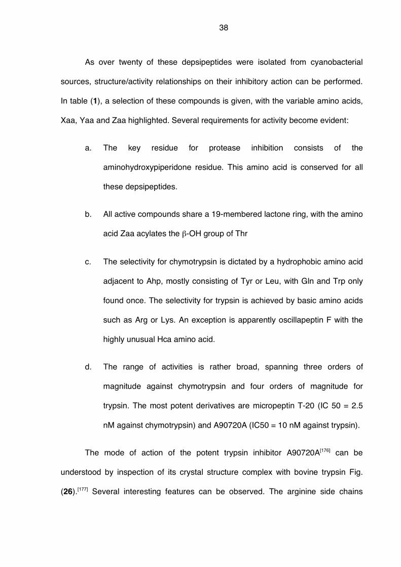
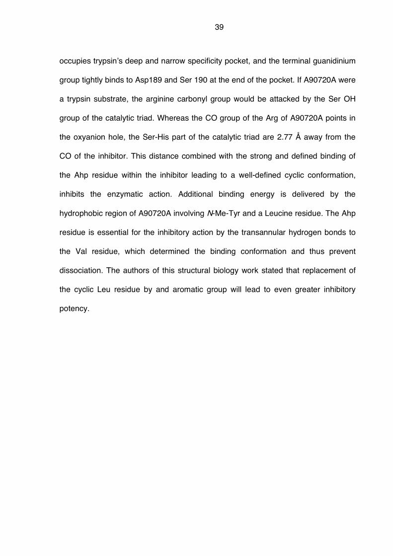
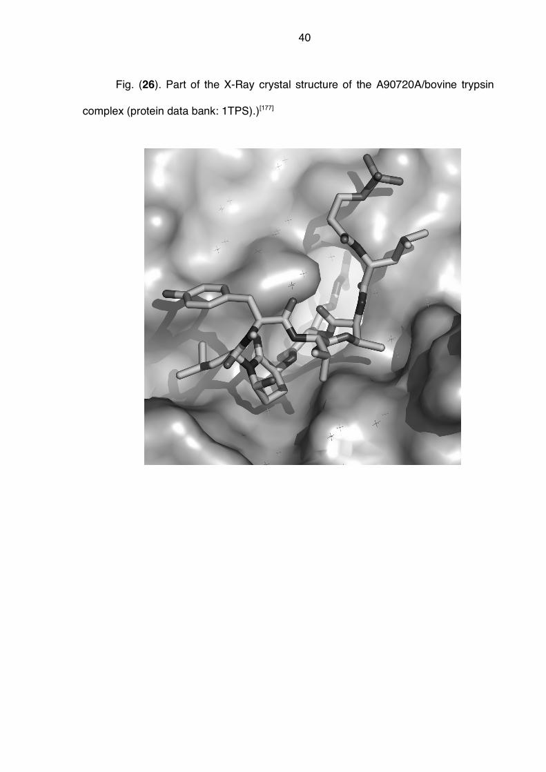
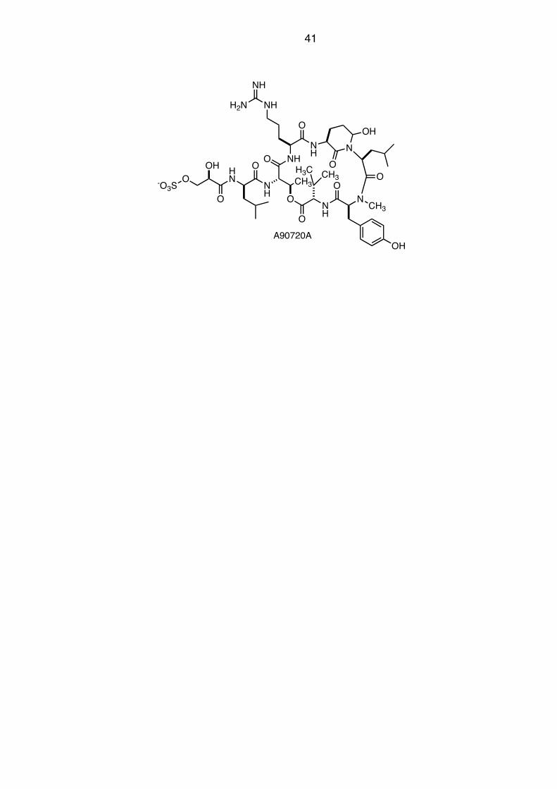
The mode of action of the potent trypsin inhibitor A90720A[176] can be
understood by inspection of its crystal structure complex with bovine trypsin Fig.
(26).[177] Several interesting features can be observed. The arginine side chains
39
occupies trypsin!s deep and narrow specificity pocket, and the terminal guanidinium
group tightly binds to Asp189 and Ser 190 at the end of the pocket. If A90720A were
a trypsin substrate, the arginine carbonyl group would be attacked by the Ser OH
group of the catalytic triad. Whereas the CO group of the Arg of A90720A points in
the oxyanion hole, the Ser-His part of the catalytic triad are 2.77 Å away from the
CO of the inhibitor. This distance combined with the strong and defined binding of
the Ahp residue within the inhibitor leading to a well-defined cyclic conformation,
inhibits the enzymatic action. Additional binding energy is delivered by the
hydrophobic region of A90720A involving N-Me-Tyr and a Leucine residue. The Ahp
residue is essential for the inhibitory action by the transannular hydrogen bonds to
the Val residue, which determined the binding conformation and thus prevent
dissociation. The authors of this structural biology work stated that replacement of
the cyclic Leu residue by and aromatic group will lead to even greater inhibitory
potency.
40
Fig. (26). Part of the X-Ray crystal structure of the A90720A/bovine trypsin
complex (protein data bank: 1TPS).)[177]
41
O NH
NH
O
N
CH3
O
O
NH
H3C CH3
O
N
O
CH3
O
OH
NH
OHN
O
O
NH
NH
H2N
-O3S
OH
OH
A90720A

42
6. Conclusion
This review gave an overview of the large variety of different cyanobacterial
metabolites featuring polyketide, alkaloid, peptides and terpene fragments and any
combination thereof. Whereas some metabolites are of general concern due to their
toxicity such as microcystins, saxitoxins or anatoxins, other display significant
pharmaceutical potential as “drugs from the sea” like the cryptophycins. Some of
these compounds might be produced for deterrence purposes or to render
cyanobacteria unattractive as a food source for grazer (by strong protease
inhibitors). Other compounds that act as iron chelators such as anachelin or as
pigments (scytonemin) have other functions of vital interest to cyanobacteria
populating habitats with extreme conditions such as bare rocks or the open ocean.
All these metabolites demonstrate how small molecules can secure evolutionary
advantage of the producing organisms (in this case cyanobateria) to survive in
vastly different habitats. Moreover, these fascinating structures combined with
powerful biological activities are inspiration to the chemist for the understanding of
natural phenomena on a molecular level and for the development of molecular
solution to the problems our society faces today.
43
7. References
[1] Singh, S.; Kate, B. N.; Banerjee, U. C. Critical Reviews in Biotechnology
2005, 25, 73-95.
[2] Harada, K. Chem. Pharm. Bull. 2004, 52, 889-899.
[3] Burja, A. M.; Banaigs, B.; Abou-Mansour, E.; Burgess, J. G.; Wright, P. C.
Tetrahedron 2001, 57, 9347-9377.
[4] Namikoshi, M.; Rinehart, K. L. J. Ind. Microbiol. Biotechnol. 1996, 17, 373-
384.
[5] Carmichael, W. W. Sci. Am. 1994, 270, 78-86.
[6] Carmichael, W. W. J. Appl. Bacteriol. 1992, 72, 445-459.
[7] Ouellette, A. J. A.; Wilhelm, S. W. Front. Ecol. Environ. 2003, 1, 359-366.
[8] Dawson, R. M. Toxicon 1998, 36, 953-962.
[9] Lakshmana Rao, P. V.; Gupta, N.; Bhaskar, A. S. B.; Jayaraj, R. J. Environ.
Biol. 2002, 23, 215-224.
[10] Gulledge, B. M.; Aggen, J. B.; Huang, H.-B.; Nairn, A. C.; Chamberlin, A. R.
Curr. Med. Chem. 2002, 9, 1991-2003.
[11] Hitzfeld, B. C.; Hˆger, S. J.; Dietrich, D. R. Environ. Health Perspect. 2000,
108, 113-122.
[12] Harada, K.-I. J. Health Sci. 1999, 45, 150-165.
[13] Falconer, I. R. Environ. Toxicol. 1999, 14, 5-12.
[14] Harada, K.-I.; Ogawa, K.; Matsuura, K.; Murata, H.; Suzuki, M.; Watanabe, M.
F.; Itezono, Y.; Nakayama, N. Chem. Res. Toxicol. 1990, 3, 473-481.
44
[15] Rudolph-Bohnera, S.; Mierke, D. F.; Moroder, L. FEBS Lett. 1994, 349, 319-
323.
[16] An, J.-S.; Carmichael, W. W. Toxicon 1994, 32, 1495-1507.
[17] Matsushima, R.; Yoshizawa, S.; Watanabe, M. F.; Harada, K.;
Furusawa, M.; Carmichael, W. W.; Fujiki, H. Biochem. Biophys. Res.
Commun. 1990, 171, 867-874.
[18] Yoshizawa, S.; Matsushima, R.; Watanabe, M. F.; Harada, K.; Ichihara, A.;
Carmichael, W. W.; Fujiki, H. J. Cancer Res. Clin Oncol. 1990, 116, 609-614.
[19] Cohen, P. Ann. Rev. Biochem. 1989, 58, 453-508.
[20] Hunter, T. Cell 1995, 80, 225-236.
[21] Agrawal, M. K.; Bagchi, D.; Bagchi, S. N.; Zitt, A.; Von Elert, E.; Weckesser,
J. Environ. Toxicol. 2005, 20, 314-322.
[22] Agrawal, M. K.; Bagchi, D.; Bagchi, S. N. Hydrobiologia 2001, 464, 37-44.
[23] Codd, G. A.; Morrison, L. F.; Metcalf, J. S. Toxicol. Appl. Pharmacol. 2005,
203, 264-272.
[24] World Health Organization. http://www.who.int/water_sanitation_health/dwq
/chemicals/microcystin/en/ (accessed May, 16th, 2007).
[25] Mansell, H. L., Tetrahedron 1996, 52, 6025-6061.
[26] Koskinen, A. M. P.; Rapoport, H., J. Med. Chem. 1985, 28, 1301-1309.
[27] Devlin, J. P.; Edwards, O. E.; Gorham, P. R.; Hunter, N. R.; Pike, R. K.;
Stavric, B., Can. J. Chem. 1977, 55, 1367-1371.
[28] Mahmood, N. A.; Carmichael, W. W., Toxicon 1987, 25, 1221-1227.
45
[29] Mahmood, N. A.; Carmichael, W. W.; Pfahler, D., Am. J. Vet. Res. 1988, 49,
500-503.
[30] Cook, W. O.; Beasley, V. R.; Dahlem, A. M.; Dellinger, J. A.; Harlin, K. S.;
Carmichael, W. W., Toxicon 1988, 26, 750-753.
[31] Moore, B. S.; Ohtani, I.; De Koning, C. B.; Moore, R. E.; Carmichael, W. W.,
Tetrahedron Lett. 1992, 33, 6595-6598.
[32] Bordner, J.; Thiessen, W. E.; Bates, H. A.; Rapoport, H., J. Am. Chem. Soc.
1975, 97, 6008-6012.
[33] Wong, J. L.; Oesterlin, R.; Rapoport, H., J. Am. Chem. Soc. 1971, 93, 7344-
7345.
[34] Schantz, E. J.; Lynch, J. M.; Vayvada, G.; Matsumoto, K.; Rapoport, H.,
Biochemistry 1966, 5, 1191-1195.
[35] Jackim, E.; Gentile, J., Science 1968, 162, 915-916.
[36] Tsuda, K.; Ikuma, S.; Kawamura, M.; Tachikawa, R.; Sakai, K., Chem.
Pharm. Bull. 1964, 12, 1357-1374.
[37] Goto, T.; Kishi, Y.; Takahashi, S.; Hirata, Y., Tetrahedron 1965, 21, 2059-
2088.
[38] Kao, C. Y.; Nishiyama, A., J. Physiol. 1965, 180, 50-66.
[39] Oshima, Y.; Buckley, L. J.; Alam, M.; Shimizu, Y., Comp. Biochem. Physiol.
1977, 57, 31-34.
[40] Norris, R. L.; Shaw, G. R.; Smith, M. J.; Chiswell, R. K.; Seawright, A. A.;
Moore, M. R.; Eaglesham, G. K.; Pierens, G., Environ. Toxicol. 1999, 14, 163-
165.

46
[41] Banker, R.; Carmeli, S.; Hadas, O.; Teltsch, B.; Porat, R.; Sukenik, A., J.
Phycol. 1997, 33, 613-616.
[42] Harada, K.-I.; Ohtani, I.; Iwamoto, K.; Suzuki, M.; Watanabe, M. F.;
Watanabe, M.; Terao, K., Toxicon 1994, 32, 73-84.
[43] Hawkins, P. R.; Runnegar, M. T.; Jackson, A. R.; Falconer, I. R., Appl.
Environ. Microbiol. 1985, 1292.
[44] Byth, S., Med. J. Austr. 1980, 2, 40-42.
[45] Metcalf, J. S.; Barakate, A.; Codd, G. A., FEMS Microbiol. Lett. 2004, 235,
125-129.
[46] Newman, D. J.; Cragg, G. M., J. Nat. Prod. 2004, 67, 1216-1238.
[47] Newman, D. J.; Cragg, G. M., Curr. Med. Chem. 2004, 11, 1693-1713.
[48] Newman, D. J.; Cragg, G. M.; Snader, K. M., J. Nat. Prod. 2003, 66, 1022-
1037.
[49] Cragg, G. M.; Newman, D. J.; Snader, K. M., J. Nat. Prod. 1997, 60, 52-60.
[50] Schwartz, R. E.; Hirsch, C. F.; Sesin, D. F.; Flor, J. E.; Chartrain, M.;
Fromtling, R. E.; Harris, G. H.; Salvatore, M. J.; Liesch, J. M.; Yudin, K., J.
Ind. Microbiol. 1990, 5, 113-123.
[51] Barrow, R. A.; Hemscheidt, T.; Liang, J.; Paik, S.; Moore, R. E.; Tius, M. A., J.
Am. Chem. Soc. 1995, 117, 2479-2490.
[52] Corbett, T. H.; Valeriote, F. A.; Demchik, L.; Lowichik, N.; Polin, L.;
Panchapor, C.; Pugh, S.; White, K.; Kushner, J.; Rake, J.; Wentland, M.;
Golakoti, T.; Hetzel, C.; Ogino, J.; Patterson, G.; Moore, R., Invest. New
Drugs 1997, 15, 207-218.
47
[53] Smith, C. D.; Zhang, X.; Mooberry, S. L.; Patterson, G. M.; Moore, R. E.,
Cancer Res. 1994, 54, 3779-3784.
[54] Panda, D.; Ananthnarayan, V.; Larson, G.; Shih, C.; Jordan, M. A.; Wilson, L.,
Biochemistry 2000, 39, 14121-14127.
[55] Eggen, M. J.; Georg, G. I., Med. Res. Rev. 2002, 22, 85-101.
[56] Stevenson, J. P.; Sun, W.; Gallagher, M.; Johnson, R.; Vaughn, D.;
Schuchter, L.; Algazy, K.; Hahn, S.; Enas, N.; Ellis, D.; Thornton, D.;
O'Dwyer, P. J., Clin. Cancer Res. 2002, 8, 2524-2529.
[57] Sessa, C.; Weigang-Kohler, K.; Pagani, O.; Greim, G.; Mora, O.; De Pas, T.;
Burgess, M.; Weimer, I.; Johnson, R., Eur. J. Cancer 2002, 38, 2388-2396.
[58] Edelman, M. J.; Gandara, D. R.; Hausner, P.; Israel, V.; Thornton, D.;
DeSanto, J.; Doyle, L. A., Lung Cancer 2003, 39, 197-199.
[59] Hemscheidt, T.; Puglisi, M. P.; Larsen, L. K.; Patterson, G. M. L.; Moore, R.
E.; Rios, J. L.; Clardy, J., J. Org. Chem/ 1994, 59, 3467-3471.
[60] Banker, R.; Carmeli, S., J. Nat. Prod. 1998, 61, 1248-1251.
[61] Hutter, R.; Keller-Schierlein, W.; Knüsel, F.; Prelog, V.; Rodgers, G. C., Jr.;
Suter, P.; Vogel, G.; Voser, W.; Zähner, H., Helv. Chim. Acta 1967, 50, 1533-
1539.
[62] Dunitz, J. D.; Hawley, D. M.; Miklos, D.; White, D. N.; Berlin, Y.; Marusic, R.;
Prelog, V., Helv. Chim. Acta 1971, 54, 1709-1713.
[63] Okami, Y.; Okazaki, T.; Kitahara, T.; Umezawa, H., J. Antibiot. (Tokyo) 1976,
29, 1019-1025.
48
[64] Nakamura, H.; Iitaka, Y.; Kitahara, T.; Okazaki, T.; Okami, Y., J. Antibiot.
(Tokyo) 1977, 30, 714-719.
[65] Schummer, D.; Irschik, H.; Reichenbach, H.; Höfle, G., Liebigs Ann. 1994, 3,
283-289.
[66] Summons, R. E.; Hope, J. M.; Logan, G. A.; Jahnke, L. L., Nature 1999, 400,
554-557.
[67] Blankenship, R. E.; Hartman, H., TIBS 1998, 23, 94-97.
[68] Crichton, R., 'Inorganic Biochemistry of Iron Metabolism: from Molecular
Mechanism to Clinical Consequences', 2nd. Ed. 2001.
[69] Drechsel, H.; Jung, G., J. Pept. Sci. 1998, 4, 147-181.
[70] Raymond, K. N.; Müller, G.; Matzanke, B. F., Top. Curr. Chem. 1984, 123,
49-102.
[71] Stintzi, A.; Barnes, C.; Xu, J.; Raymond, K. N., Proc. Natl. Acad. Sci. U S A
2000, 97, 10691-10696.
[72] Pollack, J. R.; Neilands, J. B., Biochem. Biophys. Res. Commun 1970, 38,
989-992.
[73] Avdeef, A.; Sofen, S. R.; Bregante, T. L.; Raymond, K. N., J. Am. Chem. Soc.
1978, 100, 5362-5370.
[74] Bickel, H.; Hall, G. E.; Keller-Schierlein, W.; Prelog, V.; Vischer, E.; Wettstein,
A., Helv. Chim. Acta 1960, 43, 2129-2138.
[75] Bickel, H.; Keberle, H.; Vischer, E., Helv. Chim. Acta 1963, 46, 1385-1389.
[76] Keller-Schierlein, W.; Merterns, P.; Prelog, V.; Walser, A., Helv. Chim. Acta
1965, 48, 710-723.
49
[77] Schwarzenbach, G.; Schwarzenbach, K., Helv. Chim. Acta 1963, 46, 1390-
1400.
[78] Anderegg, G.; Leplatte.F; Schwarzenbach, G., Helv. Chim. Acta 1963, 46,
1409-1422.
[79] Nick, H.; Acklin, P.; Lattmann, R.; Buehlmayer, P.; Hauffe, S.; Schupp, J.;
Alberti, D., Curr. Med. Chem 2003, 10, 1065-1076.
[80] Finch, C. A.; Huebers, H. A., Clin. Physiol. Biochem. 1986, 4, 5-10.
[81] Haematology: Basic Principles and Practice. Churchill and Livingstone: New
York, 1995.
[82] Kuhn, L. C., TIBS 1999, 24, 164-166.
[83] Liu, Z. D.; Hider, R. C., Med. Res. Rev. 2002, 22, 26-64.
[84] Griffiths, G. L.; Sigel, S. P.; Payne, S. M.; Neilands, J. B., J. Biol. Chem.
1984, 259, 383-385.
[85] Heesemann, J.; Hantke, K.; Vocke, T.; Saken, E.; Rakin, A.; Stojiljkovic, I.;
Berner, R., Mol. Microbio.l 1993, 8, 397-408.
[86] Haag, H.; Hantke, K.; Drechsel, H.; Stojiljkovic, I.; Jung, G.; Zähner, H., J.
Gen. Microbiol. 1993, 139, 2159-2165.
[87] Chambers, C. E.; McIntyre, D. D.; Mouck, M.; Sokol, P. A., Biometals 1996, 9,
157-167.
[88] Clardy, J.; Walsh, C., Nature 2004, 432, 829-837.
[89] Gardner, C. R.; Walsh, C. T.; Almarsson, O., Nat. Rev. Drug Discov. 2004, 3,
926-934.
[90] Braun, V.; Braun, M., Curr. Opin. Microbiol. 2002, 5, 194-201.
50
[91] Roosenberg, J. M., 2nd; Lin, Y. M.; Lu, Y.; Miller, M. J., Curr. Med. Chem.
2000, 7, 159-197.
[92] Murray, A. P.; Miller, M. J., J. Org. Chem. 2003, 68, 191-194.
[93] Budzikiewicz, H., Curr. Top. Med. Chem. 2001, 1, 73-82.
[94] Bickel, H.; Mertens, P.; Prelog, V.; Seibl, J.; Walser, A., Antimicrobial Agents
Chemother. 1965, 5, 951-957.
[95] Sackmann, W.; Reusser, P.; Neipp, L.; Kradolfer, F.; Gross, F., Antibiot.
Chemother. 1962, 12, 34-45.
[96] Maehr, H.; Pitcher, R. G., J. Antibiot. (Tokyo) 1971, 24, 830-834.
[97] Turkova, J.; Mikes, O.; Sorum, F., Experientia 1963, 19, 633-634.
[98] Stapley, E. O.; Ormond, R. E., Science 1957, 125, 587-589.
[99] Vértesy, L.; Aretz, W.; Fehlhaber, H.-W.; Kogler, H., Helv. Chim. Acta 1995,
78, 46-60.
[100] Braun, V., Drug Resistance Updates 1999, 2, 363-369.
[101] Goldman, S. J.; Lammers, P. J.; Berman, M. S.; Sanders-Loehr, J., J.
Bacteriol. 1983, 156, 1144-1150.
[102] Lammers, P. J.; Sanders-Loehr, J., J. Bacteriol. 1982, 151, 288-294.
[103] Simpson, F. B.; Neilands, J. B., J. Phycol. 1976, 12, 44-48.
[104] Lankford, C. E.; Walker, J. R.; Reeves, J. B.; Nabbut, N. H.; Byers, B. R.;
Jones, R. J., J. Bacteriol. 1966, 91, 1070-1079.
[105] Arceneaux, J. L.; Lankford, C. E., Biochem. Biophys. Res. Commun. 1966,
24, 370-375.
[106] Ito, Y.; Butler, A., Limnol. Oceanogr. 2005, 50, 1918-1923.
51
[107] Martinez, J. S.; Carter-Franklin, J. N.; Mann, E. L.; Martin, J. D.; Haygood, M.
G.; Butler, A., Proc. Natl. Acad. Sci U S A 2003, 100, 3754-3759.
[108] Barbeau, K.; Zhang, G.; Live, D. H.; Butler, A., J. Am. Chem. Soc. 2002, 124,
378-379.
[109] Beiderbeck, H.; Taraz, K.; Budzikiewicz, H.; Walsby, A. E., Z. Naturforsch. C
2000, 55, 681-687.
[110] Gademann, K. Chimia 2006, 60, 841-845.
[111] Itou, Y.; Okada, S.; Murakami, M. Tetrahedron 2001, 57, 9093-9099.
[112] Wenger, R. M. Helv. Chim. Acta 1983, 66, 2308-2321.
[113] Wenger, R. M. Helv. Chim. Acta 1984, 67, 502-525.
[114] Wenger, R. M. Helv. Chim. Acta 1983, 66, 2672-2702.
[115] Wenger, R. M. Fortschr. Chem. Org. Naturst. 1986, 50, 123-36.
[116] Wenger, R. M. Angew. Chem. Int. Ed. 1985, 24, 77-85.
[117] Ito, Y.; Ishida, K.; Okada, S.; Murakami, M. Tetrahedron 2004, 60, 9075-
9080.
[118] Gademann, K.; Bethuel, Y. Org. Lett. 2004, 6, 4707-4710.
[119] Gademann, K.; Bethuel, Y. Angew. Chem. Int. Ed. 2004, 43, 3327-3329.
[120] Gademann, K. ChemBioChem 2005, 6, 913-919.
[121] Gademann, K.; Budzikiewicz, H. Chimia 2004, 58, 212-214.
[122] Bethuel, Y.; Gademann, K. J. Org. Chem. 2005, 70, 6258-6264.
[123] Zürcher, S.; Wäckerlin, D.; Bethuel, Y.; Malisova, B.; Textor, M.; Tosatti, S.;
Gademann, K. J. Am. Chem. Soc. 2006, 128, 1064-1065.
52
[124] Bhat, S. V.; Nagasampagni, B. A.; Sivakumar, M. Chemistry of Natural
Products; Springer Verlag: Berlin, 2005.
[125] Larsen, L. K.; Moore, R. E.; Patterson, G. M. L. J. Nat. Prod. 1994, 57, 419-
421.
[126] Volk, R. B. J. Appl. Phycol. 2005, 17, 339-347.
[127] Volk, R. B. J. Appl. Phycol. 2006, 18, 145-151.
[128] Volk, R. B.; Furkert, F. H. Microbiol. Res. 2006, 161, 180-186.
[129] Volk, R. B.; Mundt, S. J. Appl. Phycol. 2007, 19, 55-62.
[130] Kodani, S.; Imoto, A.; Mitsutani, A.; Murakami, M. J. Appl. Phycol. 2002, 14,
109-114.
[131] Sugimura, T. Environ. Health Perspect. 1998, 106, A522-3.
[132] Nagalakshmi, C.; Subbaraman, A. S.; Pednekar, M. D.; Choughuley, A. S.
Indian. J. Exp. Biol. 1984, 22, 84-7.
[133] Sergeeva, E.; Liaimer, A.; Bergman, B. Planta 2002, 215, 229-238.
[134] Moore, R. E.; Cheuk, C.; Patterson, G. M. L. J. Am. Chem. Soc. 1984, 106.
[135] Moore, R. E.; Cheuk, C.; Xu-Qiang, G. Y.; Patterson, G. M. L.; Bonjouklian,
R.; Smitka, T. A.; Mynderse, J. S.; Foster, R. S.; Jones, N. D.;
Swartzendruber, J. K.; Deeter, J. B. J. Org. Chem. 1987, 52, 1036-1043.
[136] Smitka, T. A.; Bonjouklian, R.; Doolin, L.; Jones, N. D.; Deeter, J. B.; Yoshida,
W. Y.; Prinsep, M. R.; Moore, R. E.; Patterson, G. M. L. J. Org. Chem. 1992,
57, 857-861.
[137] Huber, U.; Moore, R. E.; Patterson, G. M. L. J. Nat. Prod. 1998, 61, 1304-
1306.
53
[138] Park, A.; Moore, R. E.; Patterson, G. M. L. Tetrahedron Lett. 1992, 33, 3257-
3260.
[139] Stratmann, K.; Moore, R. E.; Bonjouklian, R.; Deeter, J. B.; Patterson, G. M.
L.; Shaffer, S.; Smith, C. D.; Smitka, T. A. J. Am. Chem. Soc. 1994, 116,
9935-9942.
[140] Evans, J. R.; Napier, E. J.; Yates, P. J Antibiot (Tokyo) 1976, 29, 850-2.
[141] Bonjouklian, R.; Smitka, T. A.; Doolin, L. E.; Molloy, R. M.; Debono, M.;
Shaffer, S. A.; Moore, R. E.; Stewart, J. B.; Patterson, G. M. L. Tetrahedron
1991, 47, 7739-7750.
[142] Furusaki, A.; Hashiba, N.; Matsumoto, T.; Hirano, A.; Iwai, Y.; Omura, S.
Chem Commun. 1978, 800-801.
[143] Tamaoki, T.; Nomoto, H.; Takahashi, I.; Kato, Y.; Morimoto, M.; Tomita, F.
Biochem. Biophys. Res. Commun. 1986, 135, 397-402.
[144] Oka, S.; Kodama, M.; Takeda, H.; Tomizuka, N.; Suzuki, H. Agric. Biol. l
Chem. 1986, 50, 2723-2727.
[145] Nettleton, D. E.; Doyle, T. W.; Krishnan, B.; Matsumoto, G. K.; Clardy, J.
Tetrahedron Lett. 1985, 26, 4011-4014.
[146] Bush, J. A.; Long, B. H.; Catino, J. J.; Bradner, W. T. J. Antibiotics 1987, 40,
668-678.
[147] Fine, H. A.; Kim, L.; Royce, C.; Draper, D.; Haggarty, I.; Ellinzano, H.; Albert,
P.; Kinney, P.; Musib, L.; Thornton, D. J. Clinical Oncology 2005, 23, 115S-
115S.
54
[148] Graff, J. R.; McNulty, A. M.; Hanna, K. R.; Konicek, B. W.; Lynch, R. L.;
Bailey, S. N.; Banks, C.; Capen, A.; Goode, R.; Lewis, J. E.; Sams, L.; Huss,
K. L.; Campbell, R. M.; Iversen, P. W.; Neubauer, B. L.; Brown, T. J.; Musib,
L.; Geeganage, S.; Thornton, D. Cancer Res. 2005, 65, 7462-7469.
[149] Rickards, R. W.; Rothschild, J. M.; Willis, A. C.; de Chazal, N. M.; Kirk, J.;
Kirk, K.; Saliba, K. J.; Smith, G. D. Tetrahedron 1999, 55, 13513-13520.
[150] Garciapichel, F.; Sherry, N. D.; Castenholz, R. W. Photochem. Photobiol.
1992, 56, 17-23.
[151] Garciapichel, F.; Castenholz, R. W. J. Phycol. 1991, 27, 395-409.
[152] Bultel-Ponce, V.; Felix-Theodose, F.; Sarthou, C.; Ponge, J. F.; Bodo, B. J.
Nat. Prod. 2004, 67, 678-681.
[153] Proteau, P. J.; Gerwick, W. H.; Garciapichel, F.; Castenholz, R. Experientia
1993, 49, 825-829.
[154] Kobayashi, A.; Kajiyama, S. I.; Inawaka, K.; Kanzaki, H.; Kawazu, K. Z.
Naturforsch. C 1994, 49, 464-470.
[155] Ploutno, A.; Carmeli, S. J. Nat. Prod. 2001, 64, 544-545.
[156] Becher, P. G.; Beuchat, J.; Gademann, K.; Jüttner, F. J. Nat. Prod. 2005, 68,
1793-1795.
[157] Blom, J. F.; Brütsch, T.; Barbaras, D.; Bethuel, Y.; Locher, H. H.;
Hubschwerlen, C.; Gademann, K. Org. Lett. 2006, 8, 737-740.
[158] Radau, G. Pharmazie 2000, 55, 555-60.
[159] Radau, G.; Schermuly, S.; Fritsche, A. Arch. Pharm. (Weinheim) 2003, 336,
300-9.
55
[160] Martin, C.; Oberer, L.; Ino, T.; Konig, W. A.; Busch, M.; Weckesser, J. J
Antibiot. (Tokyo) 1993, 46, 1550-6.
[161] Pettit, G. R.; Kamano, Y.; Herald, C. L.; Dufresne, C.; Cerny, R. L.; Herald, D.
L.; Schmidt, J. M.; Kizu, H. J. Am. Chem. Soc. 1989, 111, 5015-5017.
[162] Luesch, H.; Harrigan, G. G.; Goetz, G.; Horgen, F. D. Curr. Med. Chem.
2002, 9, 1791-1806.
[163] Yamaki, H.; Sitachitta, N.; Sano, T.; Kaya, K. J. Nat. Prod. 2005, 68, 14-18.
[164] Banker, R.; Carmeli, S. Tetrahedron 1999, 55, 10835-10844.
[165] Reshef, V.; Carmeli, S. Tetrahedron 2001, 57, 2885-2894.
[166] Kodani, S.; Suzuki, S.; Ishida, K.; Murakami, M. FEMS Microbiol. Lett. 1999,
178, 343-348.
[167] Bister, B.; Keller, S.; Baumann, H. I.; Nicholson, G.; Weist, S.; Jung, G.;
Süssmuth, R. D.; Jüttner, F. J. Nat. Prod. 2004, 67, 1755-1757.
[168] Grach-Pogrebinsky, O.; Carmeli, S.; Sedmak, B. Tetrahedron 2003, 59,
8329-8336.
[169] Ploutno, A.; Carmeli, S. Tetrahedron 2002, 58, 9949-9957.
[170] Von Elert, E.; Oberer, L.; Merkel, P.; Huhn, T.; Blom, J. F. J. Nat. Prod. 2005,
68, 1324-1327.
[171] Okano, T.; Sano, T.; Kaya, K. Tetrahedron Lett. 1999, 40, 2379-2382.
[172] Ishida, K.; Murakami, M.; Matsuda, H.; Yamaguchi, K. Tetrahedron Lett.
1995, 36, 3535-3538.
[173] Jakobi, C.; Oberer, L.; Quiquerez, C.; Konig, W. A.; Weckesser, J. FEMS
Microbiol. Lett. 1995, 129, 129-134.
56
[174] Okino, T.; Murakami, M.; Haraguchi, R.; Munekata, H.; Matsuda, H.;
Yamaguchi, K. Tetrahedron Lett. 1993, 34, 8131-8134.
[175] Itou, Y.; Ishida, K.; Shin, H. J.; Murakami, M. Tetrahedron 1999, 55, 6871-
6882.
[176] Bonjouklian, R.; Smitka, T. A.; Hunt, A. H.; Occolowitz, J. L.; Perun Jr., T. J.;
Doolin, L.; Stevenson, S.; Knauss, L.; Wijayaratne, R.; Szewczyk, S.;
Patterson, G. M. L. Tetrahedron 1996, 52, 395-404.
[177] Lee, A. Y.; Smitka, T. A.; Bonjouklian, R.; Clardy, J. Chem. Biol. 1994, 1, 113-
117.