Embed Size (px)
Citation preview

RESEARCH ARTICLE Open Access
Porcine pulmonary valve decellularizationwith NaOH-based vs detergent process:preliminary in vitro and in vivo assessmentsMathieu van Steenberghe1,2* , Thomas Schubert3,4, Sébastien Gerelli5, Caroline Bouzin6, Yves Guiot7,Daela Xhema1, Xavier Bollen8, Karim Abdelhamid9 and Pierre Gianello1
Abstract
Background: Glutaraldehyde fixed xenogeneic heart valve prosthesis are hindered by calcification and lack ofgrowth potential. The aim of tissue decellularization is to remove tissue antigenicity, avoiding the use of glutaraldehydeand improve valve integration with low inflammation and host cell recolonization. In this preliminary study, weinvestigated the efficacy of a NaOH-based process for decellularization and biocompatibility improvement of porcinepulmonary heart valves in comparison to a detergent-based process (SDS-SDC0, 5%).
Methods: Native cryopreserved porcine pulmonary heart valves were treated with detergent and NaOH-based processes.Decellularization was assessed by Hematoxylin and eosin/DAPI/alpha-gal/SLA-I staining and DNA quantification of nativeand processed leaflets, walls and muscles.Elongation stress test investigated mechanical integrity of leaflets and walls (n = 3 tests/valve component) of valves in thenative and treated groups (n = 4/group).Biochemical integrity (collagen/elastin/glycosaminoglycans content) of leaflet-wall and muscle of the valves (n = 4/group)was assessed and compared between groups with trichrome staining (Sirius Red/Miller/Alcian blue).Secondly, a preliminary in vivo study assessed biocompatibility (CD3 and CD68 immunostaining) and remodeling(Hematoxylin and eosin/CD31 and ASMA immunofluorescent staining) of NaOH processed valves implanted inorthotopic position in young Landrace pigs, at 1 (n = 1) and 3 months (n = 2).
Results: Decellularization was better achieved with the NaOH-based process (92% vs 69% DNA reduction in thewall). Both treatments did not significantly alter mechanical properties. The detergent-based process induced asignificant loss of glycosaminoglycans (p < 0,05).In vivo, explanted valves exhibited normal morphology without any sign of graft dilatation, degeneration orrejection. Low inflammation was noticed at one and three months follow-up (1,8 +/− 3,03 and 0,9836 +/− 1,3605CD3 cells/0,12 mm2 in the leaflets). In one animal, at three months we documented minimal calcification in thearea of sinus leaflet and in one, microthrombi formation on the leaflet surface at 1 month. The endoluminal sideof the valves showed partial reendothelialization.(Continued on next page)
* Correspondence: [email protected]ôle de Chirurgie Expérimentale et Transplantation (CHEX), Institut deRecherche Expérimentale et Clinique (IREC), Secteur des Sciences de la Sante,Université Catholique de Louvain, Avenue Hippocrate 55/B1.55.04, B-1200Brussels, Belgium2Service de chirurgie cardiaque et vasculaire, Clinique Cecil, avenue LouisRuchonnet 53, 1003 Lausanne, SwitzerlandFull list of author information is available at the end of the article
© The Author(s). 2018 Open Access This article is distributed under the terms of the Creative Commons Attribution 4.0International License (http://creativecommons.org/licenses/by/4.0/), which permits unrestricted use, distribution, andreproduction in any medium, provided you give appropriate credit to the original author(s) and the source, provide a link tothe Creative Commons license, and indicate if changes were made. The Creative Commons Public Domain Dedication waiver(http://creativecommons.org/publicdomain/zero/1.0/) applies to the data made available in this article, unless otherwise stated.
van Steenberghe et al. Journal of Cardiothoracic Surgery (2018) 13:34 https://doi.org/10.1186/s13019-018-0720-y
(Continued from previous page)
Conclusions: NaOH-based process offers better porcine pulmonary valve decellularization than the detergentprocess. In vivo, the NaOH processed valves showed low inflammatory response at 3 months and partialrecellularization. Regarding additional property of securing, this treatment should be considered for the newgeneration of heart valves prosthesis.
Keywords: Cardiovascular engineering, Heart valve, Xenograft, Decellularization, Biocompatibility, Remodeling
BackgroundCurrently, heart valve prostheses are hindered by severallimitations. Mechanical valves have excellent long-termdurability but require lifelong anticoagulation therapydue to thromboembolic risks. Bioprostheses do not re-quire anticoagulation but show reduced durability andare more prone to degeneration, particularly in youngerpatients. This phenomenon has been related to a morereactive immunity, higher calcium and phosphate me-tabolism and physical activity that play a role in pros-thesis calcifications [1]. Bioprosthetic valves are usuallytreated with glutaraldehyde to prevent immune rejectionof the xenogeneic scaffold. But it has been early shownthat glutaraldehyde modifies mechanical properties ofthe native tissue, is cytotoxic, does not remove tissuephospholipids, induces a release of cell debris, and doesnot completely suppress the immune reaction againstthe graft. These events lead to chronic inflammation andcalcifications [1–5].Finally, cryopreserved homografts with ideal
hemodynamic performance but limited availability alsoshow limited durability because of residual tissue im-munogenicity [6–9]. Mechanical and bioprostheticvalves share another disadvantage: they cannot grow andremodel, therefore resulting in subsequent revision sur-geries in pediatric patients [10].Decellularization of biological valves is an alternative.
This concept involves removing all cellular componentsthat are supposedly immunogenic and may lead to calcifi-cations while minimizing any adverse effect on the com-position, biological activity, and mechanical integrity ofthe matrix. The resulting extracellular matrix (ECM) canbe recellularized by the host and functionally and structur-ally integrated into the body with growth potential [11].As of today, new products are already commercially
available but short term results with some of those im-plants, essentially those of xenogeneic source, did notdemonstrate convincing results in the pediatric popula-tion [12, 13], while others, essentially decellularizedhomografts from allogeneic source seemed to yieldbetter midterm term results [14, 15].Despite these progresses, no gold standard decellulari-
zation process exists. Various decellularization protocolsare proposed in the literature and most popular arethose using detergents [16, 17].
We previously demonstrated enhanced biocompatibil-ity and vascular remodeling of allogeneic and xenogeneicpericardium with a treatment based on NaOH decellu-larization in comparison to the glutaraldehyde fixationand detergent process [18, 19].This NaOH-based process has the particularity of be-
ing inactivator for conventional (virus/bacteria) andnon-conventional (prion) pathogens and therefore im-proves the security of those grafts [20–22].We investigated this treatment as a decellularization
process to improve biocompatibility and remodeling ofxenogeneic pulmonary heart valves for tissue engineer-ing applications.In vitro experiments assessed decellularization, antigen
removal, mechanical and biochemical integrity of por-cine pulmonary heart valves treated with the NaOH-based process (decellularized porcine pulmonary heartvalves with NaOH based-process: DPV) as well as por-cine pulmonary heart valves treated with a conventionaldetergent process (decellularized porcine pulmonaryheart valves with detergent-based process: DDPV) andnative porcine pulmonary heart valves (NPV). Moreover,an in vivo preliminary study was conducted to assessbiocompatibility (inflammation and calcifications) andremodeling (endothelialization and recellularization) at 1and 3 months follow-up of DPV valve in a growing allo-geneic/porcine model.
MethodsSources of matricesFor in vitro study, porcine Landrace hearts were procuredfrom a local slaughterhouse (Eurovlees, Zele, Belgium).For in vivo study, porcine hearts were procured at our la-
boratory, from Landrace pigs weighing 20 kg, used for ab-dominal surgery course/demonstration. Animals wereeuthanized, and hearts harvested respecting the standardsof animal care. The pulmonary valves were then harvested,rinsed with sterile ringer solution and frozen at − 80 °C.
Matrix preparationBefore processing, valves were thawed and washed insterile ringer solution.Two treatment protocols were conducted. The
detergent-based process was based on a conventionaldetergent protocol previously published. Briefly, porcine
van Steenberghe et al. Journal of Cardiothoracic Surgery (2018) 13:34 Page 2 of 12
pulmonary valves were incubated for 48 h in an aqueoussolution containing 0.5% sodium deoxycholate (SDC)and 0.5% sodium dodecyl sulfate (SDS) under continu-ous agitation followed by a 72 h rinsing step in a con-tinuous flow of demineralized water [23, 24].As previously described, the NaOH-based process con-
sists of a succession of baths of acetone, ethanol, NaOHand Hydrogen Peroxyde. This chemical treatment ensuresdefatting, prions/viruses and bacterial inactivation [18].Finally, the valves (Fig. 1a) were frozen at − 80 °C
without solution and individually packed.
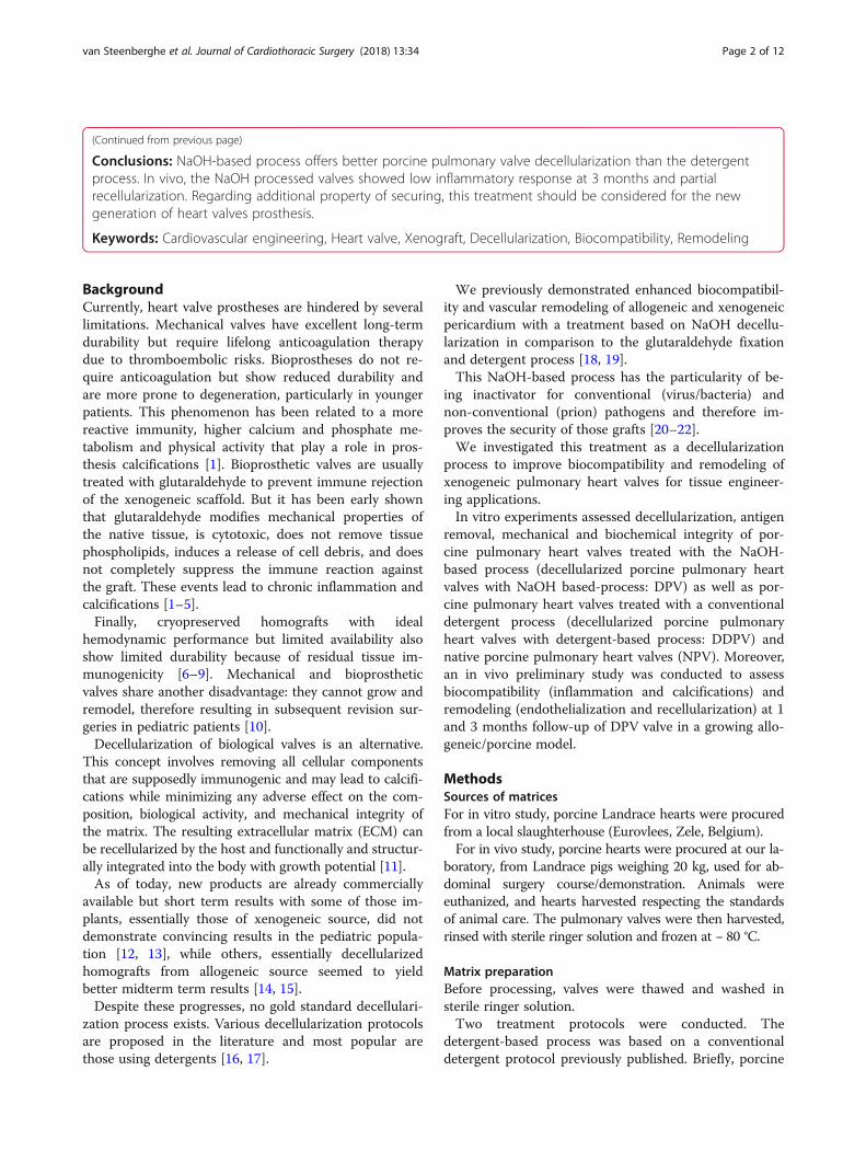
In vitro characterizationValves were individually characterized for leaflet, pul-monary trunk (wall) and muscle structure.For histological assessment, the valves were cut longi-
tudinally to explore wall, leaflet and muscle on the sameslide (Fig. 1b). The slices were immediately fixed over-night in 4% formaldehyde and embedded in paraffin.Serial sections (thickness of 5 μm) were mounted onglass and dried for 12 h at 37 °C.
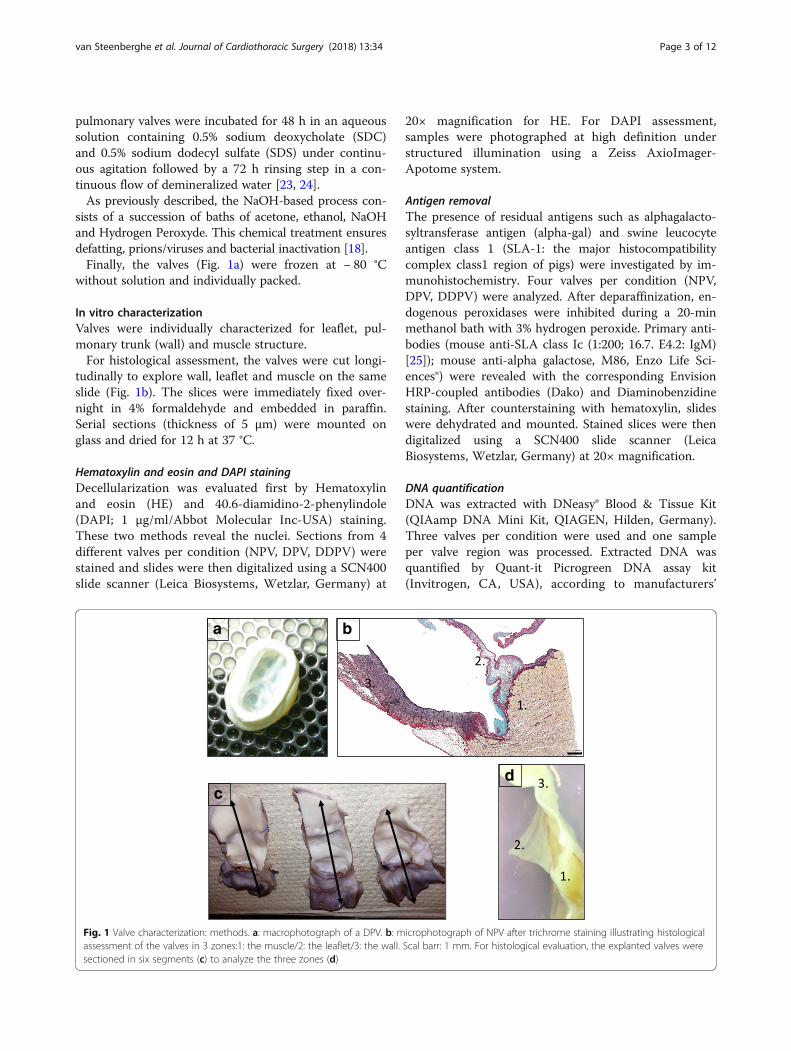
Hematoxylin and eosin and DAPI stainingDecellularization was evaluated first by Hematoxylinand eosin (HE) and 40.6-diamidino-2-phenylindole(DAPI; 1 μg/ml/Abbot Molecular Inc-USA) staining.These two methods reveal the nuclei. Sections from 4different valves per condition (NPV, DPV, DDPV) werestained and slides were then digitalized using a SCN400slide scanner (Leica Biosystems, Wetzlar, Germany) at
20× magnification for HE. For DAPI assessment,samples were photographed at high definition understructured illumination using a Zeiss AxioImager-Apotome system.
Antigen removalThe presence of residual antigens such as alphagalacto-syltransferase antigen (alpha-gal) and swine leucocyteantigen class 1 (SLA-1: the major histocompatibilitycomplex class1 region of pigs) were investigated by im-munohistochemistry. Four valves per condition (NPV,DPV, DDPV) were analyzed. After deparaffinization, en-dogenous peroxidases were inhibited during a 20-minmethanol bath with 3% hydrogen peroxide. Primary anti-bodies (mouse anti-SLA class Ic (1:200; 16.7. E4.2: IgM)[25]); mouse anti-alpha galactose, M86, Enzo Life Sci-ences®) were revealed with the corresponding EnvisionHRP-coupled antibodies (Dako) and Diaminobenzidinestaining. After counterstaining with hematoxylin, slideswere dehydrated and mounted. Stained slices were thendigitalized using a SCN400 slide scanner (LeicaBiosystems, Wetzlar, Germany) at 20× magnification.
DNA quantificationDNA was extracted with DNeasy® Blood & Tissue Kit(QIAamp DNA Mini Kit, QIAGEN, Hilden, Germany).Three valves per condition were used and one sampleper valve region was processed. Extracted DNA wasquantified by Quant-it Picrogreen DNA assay kit(Invitrogen, CA, USA), according to manufacturers’
c
a b
d
Fig. 1 Valve characterization: methods. a: macrophotograph of a DPV. b: microphotograph of NPV after trichrome staining illustrating histologicalassessment of the valves in 3 zones:1: the muscle/2: the leaflet/3: the wall. Scal barr: 1 mm. For histological evaluation, the explanted valves weresectioned in six segments (c) to analyze the three zones (d)
van Steenberghe et al. Journal of Cardiothoracic Surgery (2018) 13:34 Page 3 of 12

protocol. Fluorescence was read at 480 nm and520 nm. Final DNA concentration was expressed inng/mg dry weight.
Mechanical propertiesUniaxial mechanical resistance tests were performed on aminimum of four valves per condition on the three leafletsand three samples of pulmonary wall per valve. Samplesof 15 mm on 15 mm for wall and the whole leaflets wereplaced between two plastic structure with a central hole of8,5 mm diameter where a probe comes in contact and ap-plies pressure on the tissue. Mechanical testing was per-formed, using an Instron traction system with Instronbluehill software (Model 5600, Instron, Canton, MA) witha load-to-failure test set at an elongation rate of 3 mm.min− 1. The load to elongation behavior of the matricesand failure modes were recorded. The structuralproperties of the matrices were represented by stiffness(Nm.m− 1) and ultimate load (N). Stiffness (k) wascalculated as k =ΔF/ΔL where, F is the force applied onthe body and L is the displacement produced by the forcealong the same degree of freedom. These parameters werecompared between native and treated tissues. Tests werenot conducted on muscle.
Biochemical integrityLongitudinal slices from 4 different valves per condition(NPV, DPV, DDPV) were analyzed. Five micrometer sec-tions were stained using a combined Miller, alcian blueand sirius red trichrome, as described by SarathchandraP. [26]. The Miller stains elastin in dark blue, the alcianblue colors the glycosaminoglycans (GAGs) in Cyan andthe sirius red stains collagen in red.Quantification was performed individually on leaflet,
pulmonary wall and pulmonary trunk using Tissue IAsoftware (Leica Biosystems, Dublin, Ireland). Pixelscorresponding to the Miller, Alcian blue and Sirius redstaining were selected separately to create three colorprofiles. Total tissue area was defined by setting an in-tensity threshold (grey value). Results were expressed asa percentage of stained area and calculated as (stainedarea/tissue area) × 100.
In vivo studySurgical procedureAnimals were housed according to the guidelines of theFrench Ministry of Agriculture and Animal Care. Allprocedures were approved by the local Ethics Commit-tee for Animal Care of the Ecole de Chirurgie - Univer-sité de Lorraine, Nancy (D57–547-5).Three female Landrace pigs weighting 40 kg were kept
unfed for 24 h before the operation. A premedication ofketamine (1000 mg) was administered by IM. The ani-mals were then intubated and kept under general
anesthesia throughout the operation. A physiologicalfollow-up (oximetry, pulse, and heart rate) was con-ducted throughout the entire operating procedure. Afterlongitudinal sternotomy, the heart was exposed.Systemic heparinization was achieved with an activatedcoagulation time of 400 s. Pediatric cardiopulmonary by-pass was then placed and turned on. Then, the nativepulmonary artery root was harvested and replaced withDPV with two 5.0 prolene running sutures. After im-plantation, the pigs were weaned off bypass. The cannu-las were removed and the sternum was closed. The pigsreceived low molecular weight heparin prophylaxis(40 mg/day) for 5 days. The animals did not receive anti-biotics. One pig was euthanized at day 30 and 2 pigswere euthanized at day 90. The valves were then re-moved and cut in order to obtain three parts relating toposterior, right and left leaflets and the correspondingsinus, pulmonary wall and muscular base. Finally, theseportions were divided in two parts to obtain six seg-ments (Fig. 1c/d). Tissues were fixed overnight in 4%formaldehyde and embedded in paraffin.
Histological evaluationColoration and staining Hematoxylin and eosin, Mas-son’s trichrome and von Kossa stainings assessed remod-eling/cell infiltration, structure and calcificationsrespectively.Immunohistochemistry for CD3 and CD68 were per-
formed using a Ventana Benchmark XT machine(Roche®, USA) to assess inflammatory reaction. The CD68 is particularly useful as a marker for the various cellsof the macrophage lineage, including monocytes, histio-cytes, giant cells, Kupffer cells. CD3 is highly specific ofall stages of T-cell development [18]. Slides were digita-lized at 20× magnification with a SCN400 slide scanner(Leica, Wetzlar, Germany) and visualized on the DigitalImage Hub (Leica Biosystems, Dublin, Ireland).For immunofluorescent co-staining, 5 μm sections
were subjected to endogenous peroxidases inhibitionfor 20 min and then to specific antigen binding sites for1 h (PBS with 5% BSA and 0.05% Triton). Rabbit anti-CD31 (polyclonal, Abcam, # ab28364, 1/100 dilutionfor rat, 1/50 dilution for pig) and Mouse anti-ASMA(clone 1A4, Abcam #ab7817, 1/100 dilution, for pig),primary antibodies were incubated overnight at 4 °C inPBS containing 1% BSA and 0.05% Triton X-100. Thiswas followed by an incubation with AlexaFluor 568anti-rabbit and AlexaFluor647 anti-mouse (Invitrogen)secondary antibodies, incubated at a 1/1000 dilution for1 h at room temperature. Nuclei were stained withDAPI and labeled sections.Stained sections were digitized using a Pannoramic
P250 FlashIII slide scanner (3DHistech) at 20× magnifi-cation and visualized using CaseViewer.
van Steenberghe et al. Journal of Cardiothoracic Surgery (2018) 13:34 Page 4 of 12

Histomorphometry A minimum of five regions of inter-est [ROI] in the three parts (wall/leaflet/muscle) of thesix segments of the explanted DPV were analyzed at ×20 magnification with a grid representing a surface of 0.12 mm square. CD3 and CD68 immunohistochemicalstaining’s were assessed by point counting as previouslydescribed [18].
Statistical analysisOne-sample Kolmogorov–Smirnov tests and QQ-plotswere used to ensure the normal distribution of values.Results were expressed as means ± SD or in ratios. Thestatistical significance of differences between experimen-tal groups was tested by Student-T or one-way analysisof variance with a Bonferroni’s post hoc test. The statis-tical tests were carried out with PASW 18. Differenceswere considered to be significant at p < 0.05.
ResultsIn vitro characterizationDecellularization and antigen removalStaining for HE, DAPI and alpha-gal and SLA-I waspositive for controls in the three parts of the valve.Decellularization and antigen removal were more
complete for DPV than for DDPV as well for the muscle,wall or leaflet of treated valves. Positive staining foralpha-gal and SLA-I were still detected for DDPV in thethree parts. Cells were also evidenced with hematoxylinand eosin for DDPV muscle and wall (Fig. 2/Table 1).
Mechanical integrityNo differences between NPV, DPV and DDPV regardingelasticity and maximal load of leaflets or pulmonary wallwere detected (Fig. 3a).
DNABetter DNA reduction was achieved with NaOH-basedprocess in comparison with detergent process. The DPVleaflets showed 95% DNA reduction to NPV whileDDPV showed 92% DNA reduction. In the wall ofDDPV, DNA content was reduced to 69% and in theDPV, the content was reduced to 92%. In the muscle,DNA reduction was quite similar for both treatments:96% for DDPV vs 97% for DPV (Fig. 3b/Table 2).
Biochemical integrityMuscle Collagen and elastin staining were maintainedin DPV and DDPV in comparison to NPV. However, sig-nificant reduction of GAGs staining in DDPV muscle incomparison to NPV occurred (p = 0.021) while there wasno difference between DPV and NPV (Fig. 4).
Leaflet Histological examination after trichrome stain-ing revealed evidence of GAGs staining reduction after
both treatments. Software analysis showed GAGs stain-ing was significantly reduced for both DPV and DDPVto NPV with p < 0.05 while elastin staining was signifi-cantly reduced for DDPV to NPV (p = 0.021).Collagen staining was not reduced after both treat-
ments (Fig. 4).
Wall No significant reduction of staining was noticedfor DPV and DDPV in comparison to NPV (Fig. 4).
In vivo studyNo deaths occurred. The three pigs showed regular growth(to reach 60 kg at 1 month and 120 kg at 3 months).
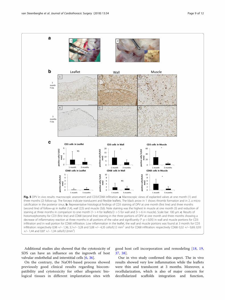
Macroscopic evaluationThe explanted valves exhibited no signs of graft dilata-tion, degeneration or rejection. The luminal surface ofthe arterial wall was similar to the adjacent host artery.At 1 month, the DPV exhibited two translucent, flex-
ible and mobile leaflets while one small thrombus wasdetected in the third one (Fig. 5a1).At 3 months, all leaflets of the two DPVs were translu-
cent, flexible and mobile. We noticed one calcificationin one posterior leaflet sinus (Fig. 5a2).
Histological evaluationInflammation CD3 infiltration was essentially located inthe muscle at 1 month. The CD3 count was significantlythe lowest in the leaflets (p < 0,005). At 3 months, theCD3 infiltration showed the same repartition than at1 month with the highest count in the muscle with p <0,05. At 3 months in comparison to 1 month, infiltrationwas significantly reduced in the wall and in the musclewith p < 0,005 while leaflet infiltration was still low (0,98+/− 1,36 cells/0,12 mm2) (Fig. 5b/c).At 1 month CD68 infiltration was low in all parts of
the valves with the highest count in the muscle part withp < 0,05. The lowest count was in the leaflets (p < 0,05).At 3 months, the CD68 infiltration was still the lowestin the leaflet with p > 0,05 but not statistically differentthan in the wall and in the muscle. The CD68 infiltra-tion significantly decreased at 3 months in the muscle(p = 0,000) (Fig. 5c).
Calcifications Von Kossa staining was positive for onesinus at 3 months.
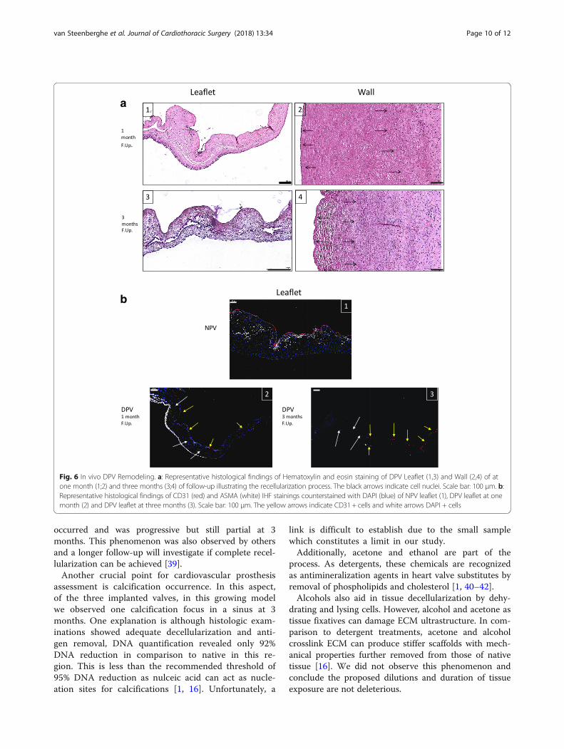
Remodeling HE, AMSA and CD31 staining showedprogressive DPV recellularization occurred at one and 3months (Fig. 6). The interstitial recellularization in-creased with time and cell colonization, was deeper inthe pulmonary trunk at 3 months than at 1 month butwas still partial (Fig. 6a). In a similar way,
van Steenberghe et al. Journal of Cardiothoracic Surgery (2018) 13:34 Page 5 of 12
endothelialisation (endothelial cell monolayer) of DPVwas still partial at 1 month and 3 months (Fig. 6b).
DiscussionThe aim of the present study was firstly to assess the ef-ficacy of a NaOH-based process to decellularize and
maintain biochemical and mechanical properties ofxenogeneic valve in comparison to a standard detergenttreatment. This treatment, a combination of 0.5%SDC/0.5%SDS is largely recommended for xenogeneic tissuesdecellularization as for human valves decellularizationwith good midterm clinical results in pediatric and adultpopulations [14, 23, 27–29]. Secondly a preliminary
Fig. 2 Histological valves characterization: Hematoxylin and eosin/DAPI/Alpha-gal and SLA-1 staining. Representative histology microphotographof muscle (first table)/leaflet (second table) and wall (third table) of NPV (first line), DPV (second line) and DDPV (third line) after Hematoxylin andeosin (HE, first column), DAPI (second column) Alpha-gal (third column) and SLA-1 (fourth column) staining. (Black scale bar: 100 μm and whitescale bar: 200 μm). The black arrows show positive staining. The staining for NPV (both muscle/leaflet and wall) was positive in all conditions (HE/DAPI/Alpha-gal and SLA-1) and was clear
van Steenberghe et al. Journal of Cardiothoracic Surgery (2018) 13:34 Page 6 of 12
study investigated in a growing model biocompatibilityand remodeling/cell recolonization of NaOH-basedprocessed valve.The main decellularization agent of our treatment is
NaOH. The duration of exposure of the tissue to thisagent and the combination with other chemicals offer asupplementary property to inactivate conventional(bacteria and virus) and non-conventional (prion) patho-gen agents that a conventional detergent process doesnot offer, improving grafts security [22]. To our
knowledge, this method for valve decellularization wasnot yet reported. The NaOH, while known as a decellu-larization agent, is not commonly used for biological tis-sue decellularization because it is at risk to denature thetissue. Indeed, decellularization treatments have to re-spect a perfect balance between decellularization andmaintenance of physical properties for optimal in vivofunctionality [30, 31]. Our in vitro and in vivo resultsshowed the treatment did not denature the valves andcan also ensure this balance for very thin structures suchas leaflets.Moreover, our treatment achieved better results in
terms of decellularization and antigen removal than thedetergent-based process. The presence of alpha-gal epi-tope remaining should lead to rapid deterioration of thelast in a clinical scenario as it was observed forSynergraft valves [12, 32].The NaOH-based process led to lower biochemical
modifications of the valves in comparison to detergenttreatment while mechanical properties of detergent-based processed valves were maintained.It was shown that SDS might destabilize the triple hel-
ical domain of collagen and lead to tissue deterioration[33–35]. We noted the use of SDS lead to extracellularmatrix swelling due to destruction of extracellular gly-cosaminoglycans as in our study while we did not ob-serve direct consequences on mechanical tests notaltered in comparison to native valves.
Table 1 Assessment of HE/DAPI/Alpha-gal and SLA-1 stainingsfor muscle/leaflet/wall of control (NPV, n = 4), DPV(n = 4) andDDPV(n = 4)
H.E. DAPI Alpha-gal SLA-1
Muscle NPV +++ +++ +++ +++
DPV – – – +
DDPV + – + ++
Leaflet NPV +++ +++ +++ +++
DPV – – – –
DDPV – – + –
Wall NPV +++ +++ +++ +++
DPV – – – –
DDPV + + + ++
Semi-quantitative numerical scale: –: no staining; +: staining traces; ++: moderatestaining and +++: intense staining
a
b
Fig. 3 Valves characteristics: Mechanical properties and DNA content. a: Stiffness (K: N/mm) and maximal load before rupture (F max: Newton) ofleaflet and wall of NPV, DDPV and DPV. No significant differences were observed between groups. b: DNA content (ng/mg) in muscle, leaflet andwall of NPV, DDPV and DPV
van Steenberghe et al. Journal of Cardiothoracic Surgery (2018) 13:34 Page 7 of 12
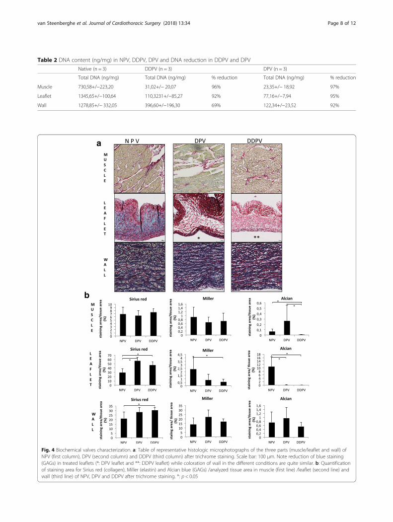
Table 2 DNA content (ng/mg) in NPV, DDPV, DPV and DNA reduction in DDPV and DPV
Native (n = 3) DDPV (n = 3) DPV (n = 3)
Total DNA (ng/mg) Total DNA (ng/mg) % reduction Total DNA (ng/mg) % reduction
Muscle 730,58+/−223,20 31,02+/− 20,07 96% 23,35+/− 18,92 97%
Leaflet 1345,65+/−100,64 110,3231+/−85,27 92% 77,16+/−7,94 95%
Wall 1278,85+/− 332,05 396,60+/−196,30 69% 122,34+/−23,52 92%
a
b
Fig. 4 Biochemical valves characterization. a: Table of representative histologic microphotographs of the three parts (muscle/leaflet and wall) ofNPV (first column), DPV (second column) and DDPV (third column) after trichrome staining. Scale bar: 100 μm. Note reduction of blue staining(GAGs) in treated leaflets (*: DPV leaflet and **: DDPV leaflet) while coloration of wall in the different conditions are quite similar. b: Quantificationof staining area for Sirius red (collagen), Miller (elastin) and Alcian blue (GAGs) /analyzed tissue area in muscle (first line) /leaflet (second line) andwall (third line) of NPV, DPV and DDPV after trichrome staining. *: p < 0.05
van Steenberghe et al. Journal of Cardiothoracic Surgery (2018) 13:34 Page 8 of 12
Additional studies also showed that the cytotoxicity ofSDS can have an influence on the ingrowth of hostvalvular endothelial and interstitial cells [6, 36].On the contrary, the NaOH-based process showed
previously good clinical results regarding biocom-patibility and cytotoxicity for other allogeneic bio-logical tissues in different implantation sites with
good host cell incorporation and remodeling [18, 19,37, 38].Our in vivo study confirmed this aspect. The in vivo
results showed very low inflammation while the leafletswere thin and translucent at 3 months. Moreover, arecellularization, which is also of major concern fordecellularized scaffolds integration and function,
a
b
c
Fig. 5 DPV in vivo results: macroscopic assessment and CD3/CD68 infiltration. a: Macroscopic views of explanted valves at one month (1) andthree months (2) follow-up. The forceps indicate translucent and flexible leaflets. The black arrow in 1 shows thrombi formation and in 2, a microcalcification in the posterior sinus. b: Representative histological findings of CD3 staining of DPV at one month (first line) and three months(second line) of follow-up in leaflet (1;4), wall (2;5) and muscle (3;6). Note staining was the highest in muscle at one month (3) and reduction ofstaining at three months in comparison to one month (1- > 4 for leaflets/2- > 5 for wall and 3- > 6 in muscle). Scale bar: 100 μm. c: Results ofhistomorphometry for CD3 (first line) and CD68 (second line) staining in the three portions of DPV at one month and three months showing adecrease of inflammatory reaction at three months in all portions of the valve and significantly (*: p < 0,05) in wall and muscle portions for CD3infiltration and in wall portion for CD68 infiltration. Low inflammation in the leaflet, the wall and muscle portions was found at 3 month: for CD3infiltration: respectively 0,98 +/− 1,36; 3,1+/− 3,28 and 5,08 +/− 4,35 cells/0,12 mm2. and for CD68 infiltration: respectively CD68: 0,32 +/− 0,69; 0,93+/− 1,44 and 0,87 +/− 1,34 cells/0,12mm2)
van Steenberghe et al. Journal of Cardiothoracic Surgery (2018) 13:34 Page 9 of 12
occurred and was progressive but still partial at 3months. This phenomenon was also observed by othersand a longer follow-up will investigate if complete recel-lularization can be achieved [39].Another crucial point for cardiovascular prosthesis
assessment is calcification occurrence. In this aspect,of the three implanted valves, in this growing modelwe observed one calcification focus in a sinus at 3months. One explanation is although histologic exam-inations showed adequate decellularization and anti-gen removal, DNA quantification revealed only 92%DNA reduction in comparison to native in this re-gion. This is less than the recommended threshold of95% DNA reduction as nulceic acid can act as nucle-ation sites for calcifications [1, 16]. Unfortunately, a
link is difficult to establish due to the small samplewhich constitutes a limit in our study.Additionally, acetone and ethanol are part of the
process. As detergents, these chemicals are recognizedas antimineralization agents in heart valve substitutes byremoval of phospholipids and cholesterol [1, 40–42].Alcohols also aid in tissue decellularization by dehy-
drating and lysing cells. However, alcohol and acetone astissue fixatives can damage ECM ultrastructure. In com-parison to detergent treatments, acetone and alcoholcrosslink ECM can produce stiffer scaffolds with mech-anical properties further removed from those of nativetissue [16]. We did not observe this phenomenon andconclude the proposed dilutions and duration of tissueexposure are not deleterious.
a
b
Fig. 6 In vivo DPV Remodeling. a: Representative histological findings of Hematoxylin and eosin staining of DPV Leaflet (1,3) and Wall (2,4) of atone month (1;2) and three months (3;4) of follow-up illustrating the recellularization process. The black arrows indicate cell nuclei. Scale bar: 100 μm. b:Representative histological findings of CD31 (red) and ASMA (white) IHF stainings counterstained with DAPI (blue) of NPV leaflet (1), DPV leaflet at onemonth (2) and DPV leaflet at three months (3). Scale bar: 100 μm. The yellow arrows indicate CD31 + cells and white arrows DAPI + cells
van Steenberghe et al. Journal of Cardiothoracic Surgery (2018) 13:34 Page 10 of 12
Last, the preservation method that we used is simple, costeffective, and the valve can be easily stored and banked for along time as musculo-skeletal tissues in a tissue bank [18].The results of this preliminary study are encouraging
to consider this NaOH-based process for xenogeneicvalve decellularization. In the clinical setup, xenogeneicsource is advantageous regarding availability. But xeno-geneic tissue transplantation to humans imposes highcaution in view of controversial results with xenogeneicdecellularized gafts [12]. We investigated only alpaha galepitope removal. Others xeno non alpha-gal antigensexist and especially a recently highlighted, highly im-munogenic xenoantigen, N-glycolylneuraminic acid anti-gen. This should be also investigated. The newgenetically modified pigs for these major xenoantigens(alphagalactosyltransferase KO N-glycolylneuraminicacid KO pigs) offer new possibilities in this direction[43]. On the other hand, it would be impossible and in-appropriate to check the disappearance of all xeno anti-gens [44] and, as suggested by G Gerosa, a step back tothe preclinical evaluation in human-like models (non-human primates) is mandatory to assess effectivebiocompatibility [17].Moreover, larger samples with a longer follow-up and
echocardiographic data are prerequisite before consider-ing clinical translation.
ConclusionsThe NaOH-based process does not alter biomechanicalvalve properties and can be used for xenogeneic heartvalve decellularization. It ensures better decellularizationand antigen removal than detregent-based process. In apreliminary in vivo study, the NaOH-based processedvalve showed recellularization, low inflammation, andabsence of structural deterioration. Regarding additionalproperty of graft securing, this treatment should be con-sidered for the new generation valves.
Abbreviationsalpha-gal: Alphagalactosyltransferase antigen; ASMA: Alpha smooth musclecells actin; BSA: Bovine serum albumin; DAPI: 40.6-diamidino-2-phenylindole;DDPV: Decellularized porcine pulmonary heart valves with detergent-basedprocess; DNA: Desoxyribonucleic acid; DPV: Decellularized porcine pulmonaryheart valves with NaOH-based process; HE: Hematoxylin and eosin; MHC-1: Major histocompatibility complex class 1; NPV: Native porcine pulmonaryheart valves; PBS: Phosphate buffered saline; SD: Standard deviation;SDC: Sodium deoxycholate; SDS: Sodium dodecyl sulfate; SLA-1: Swineleucocyte antigen: MHC class 1 region of pigs
AcknowledgementsWe thank Ecole de chirurgie de Nancy for technical assistance, Pascale Segers andEric Legrand (CHEX) for writing and administrative assistance.We thank Prof. P. Astarci (cardiac surgery department, Cliniques universitairesSaint-Luc) for having made possible the realization of the in vitro tests.
FundingThis research did not receive any specific grant from funding agencies in thepublic, commercial or not-for-profit sectors.The content of the work is solely the responsibility of the authors.
Availability of data and materialsDatas are available from corresponding author on a reasonable request.
Authors’ contributionsMvanS, PG, and TS contributed for study conception and design. Data acquisitionwas carried out by MvanS, DX, SG, CB, YG, XB and KA. Analysis and datainterpretation were carried out by MvanS, PG, TS, DX, and CB. The authorsMvanS drafted the manuscript, PG, DX, CB, critically revised the article.All authors read and approved the final manuscript.
Ethics approval and consent to participateAll procedures were approved by the local Ethics Committee for Animal Careof the Ecole de Chirurgie -Université de Lorraine, Nancy (D57–547-5).
Competing interestsThe authors declare that they have no competing interests.
Publisher’s NoteSpringer Nature remains neutral with regard to jurisdictional claims in publishedmaps and institutional affiliations.
Author details1Pôle de Chirurgie Expérimentale et Transplantation (CHEX), Institut deRecherche Expérimentale et Clinique (IREC), Secteur des Sciences de la Sante,Université Catholique de Louvain, Avenue Hippocrate 55/B1.55.04, B-1200Brussels, Belgium. 2Service de chirurgie cardiaque et vasculaire, Clinique Cecil,avenue Louis Ruchonnet 53, 1003 Lausanne, Switzerland. 3Serviced’orthopédie et de traumatologie de l’appareil locomoteur, Cliniquesuniversitaires Saint-Luc, Avenue Hippocrate 10, B-1200 Brussels, Belgium.4Unité de thérapie tissulaire et cellulaire de l’appareil locomoteur, Cliniquesuniversitaires Saint Luc, Avenue Hippocrate 10, B-1200 Brussels, Belgium.5Service de chirurgie cardiaque, Centre hospitalier Annecy-Genevois, siteAnnecy, 1 Avenue de l’Hopital, F-74370 Pringy, France. 6Institut de RechercheExpérimentale et Clinique (IREC), IREC Imaging Platform (2IP), Universitécatholique de Louvain, Avenue Hippocrate 55/B1.55.20, B-1200 Brussels,Belgium. 7Service d’anatomie pathologique, Cliniques universitaires Saint Luc,Avenue Hippocrate 10, B-1200 Brussels, Belgium. 8Institute of Mechanics,Materials and Civil Engineering, Mechatronic, Electrical Energy, and DynamicSystems (MEED), Secteur des Sciences et Technologies, Université Catholiquede Louvain, Place du Levant 2/L5.04.02, B-1348 Louvain-la-Neuve, Belgium.9Service d’oncologie, Centre hospitalier universitaire vaudois, Rue du Bugnon46, CH-1011 Lausanne, Vaud, Switzerland.
Received: 11 December 2017 Accepted: 5 April 2018
References1. Schoen FJ, Levy RJ. Calcification of tissue heart valve substitutes: progress
toward understanding and prevention. Ann Thorac Surg. 2005;79:1072–80.2. Umashankar PR, Mohanan PV, Kumari TV. Glutaraldehyde treatment elicits
toxic response compared to decellularization in bovine pericardium. ToxicolInt. 2012;19:51–8.
3. Manji RA, Zhu LF, Nijjar NK, Rayner DC, Korbutt GS, Churchill TA, Rajotte RV,Koshal A, Ross DB. Glutaraldehyde-fixed bioprosthetic heart valve conduitscalcify and fail from xenograft rejection. Circulation. 2006;114:318–27.
4. Choi SY, Jeong HJ, Lim HG, Park SS, Kim SH, Kim YJ. Elimination of alpha-galxenoreactive epitope: alpha-galactosidase treatment of porcine heart valves.J Heart Valve Dis. 2012;21:387–97.
5. Hu XJ, Dong NG, Shi JW, Deng C, Li HD, Lu CF. Evaluation of a novel tetra-functional branched poly(ethylene glycol) crosslinker for manufacture ofcrosslinked, decellularized, porcine aortic valve leaflets. J Biomed Mater ResB Appl Biomater. 2014;102:322–36.
6. Dohmen PM. Clinical results of implanted tissue engineered heart valves.HSR Proc Intensive Care Cardiovasc Anesth. 2012;4:225–31.
7. Shaddy RE, Hawkins JA. Immunology and failure of valved allografts inchildren. Ann Thorac Surg. 2002;74:1271–5.
8. Carpentier A, Lemaigre G, Robert L, Carpentier S, Dubost C. Biologicalfactors affecting long-term results of valvular heterografts. J ThoracCardiovasc Surg. 1969;58:467–83.
9. Rajani B, Mee RB, Ratliff NB. Evidence for rejection of homograft cardiacvalves in infants. J Thorac Cardiovasc Surg. 1998;115:111–7.
van Steenberghe et al. Journal of Cardiothoracic Surgery (2018) 13:34 Page 11 of 12

10. Ruel M, Chan V, Bedard P, Kulik A, Ressler L, Lam BK, Rubens FD, GoldsteinW, Hendry PJ, Masters RG, Mesana TG. Very long-term survival implicationsof heart valve replacement with tissue versus mechanical prostheses inadults <60 years of age. Circulation. 2007;116:I294–300.
11. Keane TJ, Swinehart IT, Badylak SF. Methods of tissue decellularization used forpreparation of biologic scaffolds and in vivo relevance. Methods. 2015;84:25–34.
12. Simon P, Kasimir MT, Seebacher G, Weigel G, Ullrich R, Salzer-Muhar U,Rieder E, Wolner E. Early failure of the tissue engineered porcine heart valveSYNERGRAFT in pediatric patients. Eur J Cardiothorac Surg. 2003;23:1002–6.
13. Ruffer A, Purbojo A, Cicha I, Glockler M, Potapov S, Dittrich S, Cesnjevar RA.Early failure of xenogenous de-cellularised pulmonary valve conduits–aword of caution! Eur J Cardiothorac Surg. 2010;38:78–85.
14. Sarikouch S, Horke A, Tudorache I, Beerbaum P, Westhoff-Bleck M, BoethigD, Repin O, Maniuc L, Ciubotaru A, Haverich A, Cebotari S. Decellularizedfresh homografts for pulmonary valve replacement: a decade of clinicalexperience. Eur J Cardiothorac Surg. 2016;50:281–90.
15. Brown JW, Elkins RC, Clarke DR, Tweddell JS, Huddleston CB, Doty JR,Fehrenbacher JW, Takkenberg JJ. Performance of the CryoValve SG humandecellularized pulmonary valve in 342 patients relative to the conventionalCryoValve at a mean follow-up of four years. J Thorac Cardiovasc Surg.2010;139:339–48.
16. Crapo PM, Gilbert TW, Badylak SF. An overview of tissue and whole organdecellularization processes. Biomaterials. 2011;32:3233–43.
17. Iop L, Gerosa G. Guided tissue regeneration in heart valve replacement: frompreclinical research to first-in-human trials. Biomed Res Int. 2015;2015:432901.
18. van Steenberghe M, Schubert T, Guiot Y, Bouzin C, Bollen X, Gianello P.Enhanced vascular biocompatibility of decellularized xeno−/allogeneicmatrices in a rodent model. Cell Tissue Bank. 2017;18:249–62.
19. van Steenberghe M, Schubert T, Xhema D, Bouzin C, Guiot Y, Duisit J,Abdelhamid K, Gianello P. Enhanced vascular regeneration with chemically/physically treated bovine/human pericardium in rodent. J Surg Res. 2018;222:167–79.
20. Cornu O, Schubert T, Libouton X, Manil O, Godts B, Van Tomme J, Banse X, DelloyeC. Particle size influence in an impaction bone grafting model. Comparison offresh-frozen and freeze-dried allografts. J Biomech. 2009;42:2238–42.
21. Fawzi-Grancher S, Goebbels RM, Bigare E, Cornu O, Gianello P, Delloye C,Dufrane D. Human tissue allograft processing: impact on in vitro and in vivobiocompatibility. J Mater Sci Mater Med. 2009;20:1709–20.
22. WHO. Decontamination methods for transmissible spongiformencephalopathies. Report of a WHO consultation, Geneva, Switzerland, 23-26 march 1999. In: WHO infection control guidelines for transmissiblespongiform encephalopathies. WHO/CDS/CSR/APH/2000.3; 2009. p. 29–32.
23. Hulsmann J, Grun K, El Amouri S, Barth M, Hornung K, Holzfuss C,Lichtenberg A, Akhyari P. Transplantation material bovine pericardium:biomechanical and immunogenic characteristics after decellularization vs.glutaraldehyde-fixing. Xenotransplantation. 2012;19:286–97.
24. Cebotari S, Tudorache I, Ciubotaru A, Boethig D, Sarikouch S, Goerler A,Lichtenberg A, Cheptanaru E, Barnaciuc S, Cazacu A, Maliga O, Repin O,Maniuc L, Breymann T, Haverich A. Use of fresh decellularized allografts forpulmonary valve replacement may reduce the reoperation rate in childrenand young adults: early report. Circulation. 2011;124:S115–23.
25. Duisit J, Orlando G, Debluts D, Maistriaux L, Xhema D, de Bisthoven YJ, Galli C,Peloso A, Behets C, Lengele B, Gianello P. Decellularization of the porcine eargenerates a biocompatible, nonimmunogenic extracellular matrix platform forface subunit bioengineering. Ann Surg. 2017; Epub ahead of print
26. Sarathchandra P, Smolenski RT, Yuen AH, Chester AH, Goldstein S, HeacoxAE, Yacoub MH, Taylor PM. Impact of gamma-irradiation on extracellularmatrix of porcine pulmonary valves. J Surg Res. 2012;176:376–85.
27. Theodoridis K, Muller J, Ramm R, Findeisen K, Andree B, Korossis S, HaverichA, Hilfiker A. Effects of combined cryopreservation and decellularization onthe biomechanical, structural and biochemical properties of porcinepulmonary heart valves. Acta Biomater. 2016;43:71–7.
28. Booth C, Korossis SA, Wilcox HE, Watterson KG, Kearney JN, Fisher J, InghamE. Tissue engineering of cardiac valve prostheses I: development andhistological characterization of an acellular porcine scaffold. J Heart ValveDis. 2002;11:457–62.
29. Pu L, Wu J, Pan X, Hou Z, Zhang J, Chen W, Na Z, Meng M, Ni H, Wang L, LiY, Jiang L. Determining the optimal protocol for preparing an acellularscaffold of tissue engineered small-diameter blood vessels. J Biomed MaterRes B Appl Biomater. 2017; Epub ahead of print
30. Badylak SF, Freytes DO, Gilbert TW. Extracellular matrix as a biologicalscaffold material: structure and function. Acta Biomater. 2009;5:1–13.
31. Wong ML, Wong JL, Vapniarsky N, Griffiths LG. In vivo xenogeneic scaffoldfate is determined by residual antigenicity and extracellular matrixpreservation. Biomaterials. 2016;92:1–12.
32. Kasimir MT, Rieder E, Seebacher G, Wolner E, Weigel G, Simon P. Presenceand elimination of the xenoantigen gal (alpha1, 3) gal in tissue-engineeredheart valves. Tissue Eng. 2005;11:1274–80.
33. Rieder E, Kasimir MT, Silberhumer G, Seebacher G, Wolner E, Simon P, WeigelG. Decellularization protocols of porcine heart valves differ importantly inefficiency of cell removal and susceptibility of the matrix to recellularizationwith human vascular cells. J Thorac Cardiovasc Surg. 2004;127:399–405.
34. Kasimir MT, Rieder E, Seebacher G, Silberhumer G, Wolner E, Weigel G,Simon P. Comparison of different decellularization procedures of porcineheart valves. Int J Artif Organs. 2003;26:421–7.
35. Bodnar E, Olsen EG, Florio R, Dobrin J. Damage of porcine aortic valve tissuecaused by the surfactant sodiumdodecylsulphate. Thorac Cardiovasc Surg.1986;34:82–5.
36. Caamano S, Shiori A, Strauss SH, Orton EC. Does sodium dodecyl sulfatewash out of detergent-treated bovine pericardium at cytotoxicconcentrations? J Heart Valve Dis. 2009;18:101–5.
37. Dufrane D, Marchal C, Cornu O, Raftopoulos C, Delloye C. Clinicalapplication of a physically and chemically processed human substitute fordura mater. J Neurosurg. 2003;98:1198–202.
38. Dufrane D, Mourad M, van Steenberghe M, Goebbels RM, Gianello P.Regeneration of abdominal wall musculofascial defects by a humanacellular collagen matrix. Biomaterials. 2008;29:2237–48.
39. Navarro FB, Costa FD, Mulinari LA, Pimentel GK, Roderjan JG, Vieira ED,Noronha L, Miyague NI. Evaluation of the biological behavior ofdecellularized pulmonary homografts: an experimental sheep model. RevBras Cir Cardiovasc. 2010;25:377–87.
40. Jorge-Herrero E, Fernandez P, de la Torre N, Escudero C, Garcia-Paez JM,Bujan J, Castillo-Olivares JL. Inhibition of the calcification of porcine valvetissue by selective lipid removal. Biomaterials. 1994;15:815–20.
41. Schmidt CE, Baier JM. Acellular vascular tissues: natural biomaterials fortissue repair and tissue engineering. Biomaterials. 2000;21:2215–31.
42. Mendoza-Novelo B, Cauich-Rodriguez JV. Decellularization, stabilization andfunctionalization of collagenous tissues used as cardiovascular biomaterials. In:Pignatello R, editor. Biomaterials - physics and chemistry. InTech; 2011. p. 159–82.
43. Lee W, Long C, Ramsoondar J, Ayares D, Cooper DK, Manji RA, Hara H.Human antibody recognition of xenogeneic antigens (NeuGc and gal) onporcine heart valves: could genetically modified pig heart valves reducestructural valve deterioration? Xenotransplantation. 2016;23:370–80.
44. Griffiths LG, Choe LH, Reardon KF, Dow SW, Christopher OE.Immunoproteomic identification of bovine pericardium xenoantigens.Biomaterials. 2008;29:3514–20.
van Steenberghe et al. Journal of Cardiothoracic Surgery (2018) 13:34 Page 12 of 12