Embed Size (px)
Citation preview

Polymorphic Ventricular Tachyarrhythmias in the Absence of Organic Heart Disease: Classification, Differential Diagnosis, and Implications for Therapy
Sami Viskin and Bernard Belhassen
Different polymorphic ventricular tachyarrhythmias may cause syncope or cardiac arrest in patients with no heart disease: (1) Catecholamine-sensitive poly- morphic ventricular tachycardia (VT) presents dur- ing childhood: the hallmark is the reproducible provo- cation of atrial and polymorphic ventricular arrhythmias during exercise, despite a normal QT. 13-Blockers are the treatment of choice. (2) In the long QT syndromes (LQTS), malfunction of ion channels leads to prolonged ventdcular repolariza- tion, early afterdepolarizations, and triggered ven- tricular arrhythmias. Therapeutic options include: 13-blockers, genotype-specific therapy, cardiac sym- pathetic denervation, and implantation of pacemak- ers or defibrillators. (3) The "short-coupled variant of torsade de pointes" is a malignant disease that shares several characteristics with idiopathic ven- tricular fibrillation. Although verapamil is frequently recommended, mortality rates remain high. (4) Idio- pathic ventricular fibrillation (VF) with normal electro- cardiogram (ECG) strikes young adults of both genders. In contrast to other polymorphic tachyar- rhythmias, idiopathic VF is not generally related to stress. Also, familial involvement is rare. Therapeu- tic options include implantation of defibrillators and therapy with class 1A drugs. (5) The "Brugada syndrome" and the "syndrome of nocturnal sudden death" strike males almost exclusively. Right bundle branch block (RBBB) with ST elevation in the right precordial leads--the "Brugada sign"--is seen in the ECG of both patient populations. Implantation of defibrillators is recommended. Copyright © 1998 by W.B. Saunders Company
S ustained ventricular arrhythmias generally oc- cur in the setting of organic heart disease.
When such arrhythmias arise in patients with no
structural cardiac abnormalities, they are usually monomorphic3, ~ Although cardiac arrest may rarely occur, the idiopathic monomorphic ven- tricular tachycardias have, in general, a good prognosis with or without therapy.l,2
Polymorphic ventricular tachycardia (VT) and ventricular fibrillation (VF) may also strike osten- sibly healthy patients. Recent advances in our understanding of these more malignant polymor- phic tachyarrhythmias prompt the present review.
Definitions
Polymorphic VT is a rapid VT with changing morphology of the QRS complexes. Torsade de pointes is a polymorphic VT that occurs in the setting of a long QT syndrome (LQTS). The definition of normal heart in the studies reviewed, is in conformity with the requirements consid- ered "mandatory for ruling out organic heart disease. ''3 Accordingly, a "normal heart" is defined when all the following are normal: (1) clinical history (including the absence of arrhythmogenic drugs), (2) blood chemistry (except for high cardiac enzymes or brief hypokalemia ascribed to resuscitation), (3) electrocardiography and exer- cise testing (except for arrhythmias and specific abnormalities of the QRS, ST, and QT segments in special groups, see below), (4) echocardiography,
From the Department of Cardiology, Tel Aviv Sourasky- Medical Center, and Sackler-School of Medicine, Tel Aviv University, Israel.
Address correspondence to Sami Viskin, MD, Depart- ment of Cardiology, Tel Aviv Medical Center, Weizman 6, Tel Aviv 64239, Israel.
Copyright © 1998 by W.B. Saunders Company 0033-0620/98/4101-000258.00/0
Progress in Cardiovascular Diseases, Vol. 41, No. 1 (July/August), 1998: pp 17-34 17

18 VISKIN AND BELHASSEN
and (5) cardiac catheterization with coronary angiography. The limitations of this definition of "apparently normal heart" should be noted (see "differential diagnosis" below).
Classification of Polymorphic Ventricular Tachyarrhythmias
A common characteristic of these arrhythmias is the clinical presentation with syncope and the high incidence of sudden death. Young patients with no evident heart disease are often misdiag- nosed as "epileptics" when these rapid polymor- phic tachyarrhythmias provoke seizures.
Inconsistent terminology has led to confusion. We propose the following classification based on clinical and electrocardiographic characteristics (Table 1). 4-11
Polymorphic VT
Catecholamine-Dependent Polymorphic VT
The hallmark of this disease 8,tz is the reproduc- ible occurrence of polymorphic VT during stress, without QT prolongation (Fig 1). Catecholamin- ergic polymorphic VT mainly affects children. In the largest series, 8 the age at the onset of symp- toms (syncope or cardiac arrest during stress) was 8 + 4 years. Infants, 13 as well as adults, t4,15 have been reported as affected. Familial involvement is common, 8 and autosomal dominant inheritance has been suggested. 14 The resting electrocardio- gram (ECG; including the QT interval) is normal, but sinus bradycardia is common. 8a3 During exercise, a reproducible sequence of events oc- curs (Fig 1): as the sinus rate increases, atrial
TABLE 1. Polymorphic Ventricular Arrhythmias in the Absence of Organic Heart Disease. Classification Based on Clinical and Electrocardiographic Characteristics*
Patient Characteristics Arrhythmia
Precipitation Patient Group (First Series Reported)
a) Age b) Gender Baseline Stress- c) Familial Hx ECG related Mode of Onset Therapy
I. Polymorphic VT Congenital LQTS (Jervell-
Lange Nielsen 4 and Romano-WardS, 6)
Catecholaminergic poly- morphic VT (Leenhardt 8)
Short-coupled variant of torsade de pointes (Leenhardt 7)
II. Ventricular Fibrillation Idiopathic VF with
normal ECG (Belhassen 9)
Idiopathic VF with RBBB and STT (Brugada 1°)
Sleep-death syndrome (ApontC 1)
a) 21 -+ 15yearsl LongQT b) Female > male:l: Abnormal TU morphology c) Common a) 8 +_ 4 years Normal b) M/F = 1.3/1 Sinus bradycardia c) Common a) 35 _+ 10 years Normal b) M/F = 1/1 c) Common
Often§ Pause-dependent[I 15-blockers Long CI Pacemaker
ICD, other~ Always Typical (see text) B-blockers
Rarely Not pause-dependent ICD Short CI
a) 36 _+ 16 years Normal No b) M/F = 1.4/1 c) No a) 46 +_ 7 years RBBB + ST1" Rarely b) M/F > 10/1 c) Common a) 35 _+ 10 years RBBB + ST1" Sleep b) M/F = 10/1 c) Rare
Not pause-dependent ICD Short CI Class 1A
Not pause-dependent ICD Short CI
Not pause-dependent ICD Short CI
NOTE. Therapy is recommended therapy based on limited data. Age is age at the onset of symptoms (mean +_ standard deviation). Gender, M/F = male to female ratio. Familial Hx is familial history of syncope or sudden death.
Abbreviations: CI, coupling interval of the extrasystole precipitating the tachyarrhythmia; RBBB + STT, RBBB with persistent ST segment elevation in the right precordial leads.
*Some overlapping exists between several entities (see text). lAge at onset of symptoms in the international Long QT Registry (reports of infants abound in the literature). :l:For explanations of the female predominance in this disease with autosomal inheritance, see text. §The incidence of stress-related arrhythmias varies according to the genotype. ILTorsade de pointes that is not pause-dependent occurs in infants and patients with severe forms of LQTS. ¶For genotype-specific therapy, see Table 2. The "short-coupled variant of torsade de pointes ''7,12 and idiopathic
ventricular fibrillation 9 may represent the same disease (see text).

POLYMORPHIC VENTRICULAR TACHYARRHYTHMIAS 19
Fig 1. Idiopathic catecholaminergic polymorphic ventricular tachycardia provoked by exercise. This 14-year-old girl had multiple events of stress-related syncope and developed ventricular fibrillation during isoproterenol infusion when tilt-table evaluation was performed somewhere else. Baseline ECG (left margin) is normal. Resting heart rate = 48 beats/min, QT = 0.38 s, QTc = 0.36 s. With increasing levels of exercise, the following events result: (A) atrial fibrillation begins, (B) multiple premature ventricular complexes arise, and (C,D) runs of polymorphic ventricular tachycardia appear. The patient has been asymptomatic for 4 years on 400 mg/d of metoprolol.
arrhythmias and ventricular extrasystoles appear. If the effort continues, salvos of monomorphic or bidirectional VT eventually lead to bursts of polymorphic VT. In contrast to this reproducible provocation with exercise, catecholaminergic poly- morphic VT is not inducible with programmed ventricular stimulation (premature ventricular stimulation) .8
[3-Blockers are the treatment of choice. The maximal dosages that are well tolerated should be prescribed and Holter recordings and exercise tests should be repeated periodically to assure that the degree of sinus tachycardia that precedes the onset of arrhythmias is never reached. Still, 2 of 20 patients (10%) reported in the largest series 8 (as well as 1 of 4 patients in a smaller series) 15 died suddenly in spite of [3-blocker therapy. Thus, additional modes of therapy should be consid- ered. However, limited data suggest that class 1 drugs and amiodarone are ineffective. 8 Finally, the benefits of an implantable cardioverter defibrilla- tor (ICD) should be weighted against the poten- tial proarrhythmic effects of such devices in these patients; stress caused by appropriate or inappro-
priate shocks could prove to be disastrous for patients with catecholamine-sensitive arrhyth- mias.
The Long QT Syndromes
In the LQTS, malfunction of ion channels leads to prolonged ventricular repolarization, early afterde- polarizations (EAD), and triggered ventricular arrhythmias.16-z° The malfunction of ion channels may be the result of gene mutations (congenital LQTS, Table 2) or may be caused by drugs or metabolic abnormalities 16-19,2>28 (acquired LQTS, Table 3). The abnormal repolarization (depicted in the ECG as a prolonged QT interval with odd morphology) begets malfunction of adjunct ion channels, further promoting inward currents dur- ing the late phases of repolarization. The resulting surplus of intracellular positive ions causes EADs (represented in the ECG as characteristically tall U waves), which may be of enough amplitude to depolarize the cell membrane and trigger ventricu- lar arrhythmias. Some regions of the heart, specifi- cally the midmyocardium, are more prone to

20 VISKIN AND BELHASSEN
TABLE 2. Congenital Long QT Syndromes*
{Chromosome} (Gene) Ion-Channel Basis of Prolonged Genotype-Specific
Genotype Affected* Repolarization Therapy
LQT1 [11] (KVLQT1) Reduced potassium outward currents ? Potassium channel Iks
LQT2 {7}: (HERG) Reduced potassium outward currents Potassium channel Ikr
LQT3 {3]: (SCN5A) Sustained sodium inward current Sodium channel SCN5A (intermittent
reopening) LQT4 {4} ? ? LQT5 {21} (KCNE1)26 Reduced potassium outward currents
IsK, a potassium channel subunit that regulates KVLQT1 and HERG
Spironolactone Potassium supplements Beta-blockers79, 80 Sodium channel blockers
(mexiletine)81,82
Abbrev ia t ions : Ikr, rapid component of the delayed rectifier potassium current; Iks, slow component of the delayed rectifier potassium current.
*Detailed reviews of the molecular biology of the congenital LQTS ~648,2° have been published.
display prolongation of repolarizadon and EADs, 29 The resultant heterogeneity of repolarization facili- tates the onset of the characteristic arrhythmia, torsade de pointes. 29,3° Torsade is characterized by QRS complexes of changing amplitude and contour. However, these changes in contour may not be appreciable when only short bursts are recorded or when only single lead recordings are available. 19 The baseline LQT interval and the mode of onset of the arrhythmia (see below) aid in establishing the diagnosis of torsade de pointes.
TABLE 3. Acquired Long QT Syndromes*
Basis of Prolonged Etiology* Repolarization
Antiarrhythmic drugs (class 1Aand class Ill)
Antibiotics (erythromycin, amantadine, pentami- dine, ketoconazole, and others)
Histamine~ receptor antagonists (astemizole, terfenadine)
Cholinergic agonists (cisapride, organophos- pates)
Psychiatric: antidepressants (tricyclic, tetracyclic), phenothiazines, haloperidol
Metabolic abnormalities (hypokalemia, hypomag- nesemia)
Bradyarrhythmias
Blockade of Ikr and other potassium outward cur- rents18,21-23
Inhibition of Ikr potassium outward currents 24
Inhibition of Ikr potassium outward currents 25
inhibition of Ikr potassium outward currents 27
inhibition of ]kr potassium outward currents 23 and other mechanisms TM
Several mechanisms TM
*Reviews of the acquired L Q T S 19'28 have been pub- lished.
Classifications of the LQTS refer to the congeni- tal LQTS as "adrenergic dependent," whereas the acquired LQTS is equated with "pause dependent" torsade de pointes. 18,19,29 The rationale behind this grouping relates to (1) the association be- tween "adrenergic" (stressful) states and arrhyth- mia precipitation in the congenital LQTS 3t and (2) the mode of onset of torsade de pointes, which is invariably preceded by a relatively long cycle, ie, a pause, in the acquired LQTS. 32,33 However, the terms "adrenergic" and "pause depen- dent" are not mutually exclusive. 17,18,34 In fact, a majority of spontaneous torsade episodes, docu- mented in adults with congenital LQTS, are actually pause-dependent (Fig 2).34-36
The age at diagnosis in the International Regis- try was 21 + 15 years, 37 but reports of symptom- atic LQTS in newborns and children abound in the literature. 38-42 In the Registry, 37 the majority of syncopal episodes occurred in association with emotional stress or vigorous physical activity. Arrhythmias that occur during swimming or after arousal by an alarm clock or the telephone ringing are more rare, but still characteristic. More recent data suggest that in some genotypes (LQT3), the majority of arrhythmias are not stress related, 43 and in some families, arrhythmias occur only during sleep, a7,44 In general, symptoms seem to be more common in females. 37 Explanations for this gender variability, which is unexpected for an autosomal genetic disorder, include45: (1) the presence of modifier genes, causing more frequent symptoms in females, or alternatively,

POLYMORPHIC VENTRICULAR TACHYARRHYTHMIAS 21
l I
V 1
I I "
a V F
Fig 2. Pause-dependent torsade de pointes in a 31-year-old woman with congenital long QT syndrome previously reported. 34 The arrhythmia occurred in the absence of drugs. R-R intervals are shown in ms. Top panel: Progressive increment in R-R interval caused by sinus arrhythmia. The eighth sinus complex follows a relatively long R-R interval (885 ms) and shows TU wave changes (arrowhead) from which the first premature ventricular complex originates (*). This extrasystole begins a series of "short-long" sequences perpetuated in the form of ventricular bigeminy (second panel), eventually escalating and leading to torsade de pointes.
premature mortality in males; and (2) overdiagno- sis of LQTS in females (or underdiagnosis in males) because the clinical diagnosis is based on the QT interval, which is longer in females. 45
The diagnosis of a congenital LQTS may be confirmed with molecular biology:. However, only a few specialized centers screen for all identified mutations. 46 Thus, the diagnosis remains largely a clinical one. 4r In a patient with a rate-corrected QT > 0.46 seconds and documented torsade de pointes, the diagnosis is straightforward. In that case, exclusion of secondary forms (Table 3) leads to a clinical diagnosis of congenital LQTS. Of note, before a diagnosis of "acquired" is given to a patient with drug-induced torsade, the possibility that the drug merely unmasked a congenital
LQTS es'49 should be excluded by close follow-up. More challenging, however, is the patient with syncope who has no arrhythmia documentation, a negative family history, and a QT in the upper normal range. In such a case, the following may aid in establishing a diagnosis of LQTS: (1) there is day-to-day variability in the QT interval, and repeated ECGs may eventually reveal a frankly abnormal traceS°; (2) attention should be focused not only on the length, but also on the morphol- ogy of the TU segment: the presence of "biphasic T waves" or "T waves humps" in the left precor- dial or limb leads, is a relatively specific sign of the LQTS (particularly in patients older than 15 years of age with no hypertension or heart dis- ease)51,52; (3) because the LQTS generally has

2 2 VISKIN AND BELHASSEN
autosomal dominant inheritance, review of ECGs of family members may reveal abnormal traces; and (4) although the odds for detecting torsade de pointes during a Holter recording are small, ambulatory electrocardiographic recordings may unmask TU wave alternans or characteristic post- extrasystolic TU changes.
The diagnostic yield of exercise-testing for patients with borderline QT is less clear. This is because studies emphasizing the difference in the response of QT interval to exercise or valsalva (between patients with LQTS and healthy con- trois) 5>55 included patients who had frankly pro- longed QT interval even at rest. Also, the QT response to exercise is not the same in all geno- types. 56 Finally, exercise stress testing is of low yield for arrhythmia provocation. Similarly, tors- ade is rarely provoked with premature ventricular stimulation. 5r Therefore, the reason for perform- ing electrophysiologic studies (EPS) in a patient with suspected LQTS is primarily to exclude other causes of arrhythmias and to record EADs with the use of special catheters. 5s-6° Some au- thors inject adrenaline or isoproterenol for ar- rhythmia provocation, 6° but the diagnostic accu- racy of this intervention is less clear.
Long-term randomized studies of the therapeu- tic modalities for the LQTS have not been per- formed and are not being conducted. It is impor- tant to remember that all the recommendations for therapy in the congenital LQTS are based essentially on observational, uncontrolled stud- ies.
f3-Blockers. More than 20 years ago, Schwartz observed that patients treated with [~-blockers (or cardiac sympathetic denervation) had lower mor- tality (6%) than those treated otherwise or pa- tients left untreated (>60% mortality).31 [3-blocker therapy is associated with a 0.41 relative risk for syncope or cardiac arrest (95% confidence inter- val = 0.21 to 0.81) 61 and remains the mainstay of therapy of the congenital LQTS. However, even a 6% long-term risk for arrhythmia recurrence (with [~-blockers) is unacceptable for this other- wise healthy and young patient population. There- fore, additional therapy is often recommended for those considered to be at high risk.
Left cardiac sympathetic denervation (LCSD). The rationale for recommending surgical sympa- thetic denervation of the heart (performed as left stellectomy, left cervicothoracic sympathectomy,
high thoracic left sympathectomy, or posterior left thoracic sympathectomy) is based on the "sympa- thetic imbalance" hypothesis. 31 This hypothesis states that cardiac efferent sympathetic activity-- higher in the left side--causes or aggravates the LQTS. Demonstration that the LQTS is caused by mutations in genes that encode cardiac ion chan- nels, has called this theory into question. Never- theless, long-term, albeit uncontrolled studies, suggest that LCSD reduces the risk for arrhyth- mias in patients refractory to [~-blocker therapy. 31,62-64 It is plausible that rather than correcting a left versus right "sympathetic imbal- ance," this procedure prevents arrhythmias by maximizing the degree of F-blockade or by block- ing cx-adrenergic pathways as well. 64 Thus, LCSD might be particularly beneficial for patients unwill- ing or unable to take adequate dosages of [~-block- ers. It should be emphasized, however, that pa- tients refractory to [~-blockers have a 6-year sudden death rate of 8% even after sympathetic denervation. 64
Cardiac pacing. Cardiac pacing was initially recommended for patients with LQT and concomi- tant bradyarrhythmias. 42,65 Subsequently, Eldar and others showed that pacing is also beneficial for patients without bradycardia. 63,66-69 The in- verse relation between the heart rate and the duration of repolarization dictates that pacing at faster rates results in a shorter QT interval. In the International LQT Registry, 6 patients who had recurrent symptoms despite pacing had a pro- grammed lower rate limit of -<74 beats/min. 69 Accordingly, Moss et al recommend pacing at 70 to 80 beats/min. 69 Based on the cycle length preceding spontaneous arrhythmias, we recom- mend a lower rate limit of 80 beats/min for adults. 34 This pacing rate will result in a high percentage of paced beats. Therefore, single cham- ber ventricular pacing would not be satisfactory, and the main options are single chamber atrial pacing or dual chamber pacing. Atrial pacing, which involves implantation of less hardware, may be adequate for most patients. However, a few patients (particularly small children with very prolonged QT interval) may develop func- tional atrioventricular block, as a result of the prolonged ventricular repolarization, immedi- ately before the onset of torsade. 39,41,7° In such cases, single chamber atrial pacing will be value- less at the time when it is most needed. Therefore,

POLYMORPHIC VENTRICULAR TACHYARRHYTHMIAS 2 3
dual chamber pacemakers are often used. Of note, when the use of pause-prevention pacing algo- rithms is contemplated (see below), dual cham- ber pacing rather than atrial pacing is mandatory. In such a case, programming a long AV delay (longer than the patient's native PR interval during [3-blocker therapy) will inhibit ventricular pacing most of the time. This will result in considerable savings in battery expenditure. For example, calculations made for a typical dual chamber pacemaker with a 2.7-V battery, pro- grammed to pace at 80 beats/min with a 3.5-V output and 0.5-ms pulse-width and assuming a 500-D resistance in the atrium and the ventricle, suggest the following: (1) constant pacing in both cardiac chambers will' result in battery depletion by the end of 5.9 years; and (2) in contrast, programming a sufficiently long AV delay that will allow constant pacing in the atrium, but essentially sensing-only in the ventricle, will result in a 7.5-year longevity (Albert Maarse, MSc, GuidantYCardiac Pacemakers Inc, personal com- munication, February 1998). This 27% increment in pacemaker longevity is an important consider- ation for young patients who will need several device replacements over the years.
We believe that the main role of pacing in the LQTS is to prevent the sudden pauses that facili- tate the onset of torsade de pointes. 34 In particu- lar, prevention of postextrasystolic pauses is im- perative to interrupt the escalating "short-long sequence" that culminates in torsade. 34 More rapid pacing will shorten postextrasystolic pauses, potentially reducing the risk of torsade. However, pacing at fast rates for many years may lead to a tachycardia-induced cardiomyopathy, n Certainly, the pacing rates that prevent drug-induced pause- dependent torsade (100 to 140 beats/min) 72 are too fast to be practical for long-term manage- ment. Dilated cardiomyopathy has already been ascribed to 12 years of pacing at 110 beats/min in a child with LQTS. r3 A potential alternative method for preventing pause-induced torsade involves the use of specific pause-prevention pacing algorithms (Fig 3), but experience with this approach is very limited, r4,r5
Implantable defibrillators. Reliance on ICDs to treat the LQTS is becoming more frequent. 76 Defibrillators are the treatment of choice when symptoms recur in spite of combined therapy. Also, many would recommend ICD implantation
for all cardiac arrest survivors. Finally, some will argue in favor of ICDs for all patients with symptomatic LQTS. 76 In fact, analysis of implan- tation patterns shows that in 17% of cases, the devices are being used as "first line" of therapy. 76
A preliminary report on the risk of recurrent arrhythmias in 5 of 35 (14%) high-risk patients treated with ~-blockers and cardiac pacing, 77 is likely to increase the number of defibrillator implantations. It is important to remember, how- ever, that unique characteristics of the LQTS could lead to more frequent device-related compli- cations in this patient population. The young age of these patients will inevitably translate into more hardware-related complications in the long term (see below). Also, torsade de pointes tends to recur upon termination, and the stress and sudden pauses produced by ICD shocks may aggravate this tendency for arrhythmia recur- rence. Indeed, a patient with LQTS who received 62 appropriate shocks within a short period has been described. 7s Even during ~-blocker therapy, "multit~le shocks" occurred in 6°/0 of patients with LQT. 76 Programming fast (--> 100 beats/rain) "post- shock pacing rates" may help to prevent this complication.
It is premature to extrapolate to the LQTS the excellent results achieved with ICDs in patients with organic heart disease. Nevertheless, in a small series of LQT patients with ICDs, sudden death has not occurred. 76 The availabili W of defibrillators with dual chamber pacing capabili- ties and pause-preventing pacing algorithms 75 is a very important addition to our therapeutic op- tions.
Genotype-specific therapy. Recent studies r9-82 advocate genotype-specific therapy (Table 2). It should be emphasized that these encouraging data are based on a small number of patients with short-term follow-up.
Short-Coupled Variant of Torsade de Pointes
Terms like "short-coupled variant of torsade de pointes ''7,12 or "inducible torsade with normal QT "83 have been used to describe rapid nonsus- tained tachycardia--of "twisting of the points" morphology--in patients with strictly normal QT. Genotyping of families with LQTS has shown overlapping in the duration of the QT interval between patients affected with a long QT gene

24 VISKIN AND BELHASSEN
A SR = 76
B LRL 70 AV 170 RSD off
C LRL 70 AV 150 RSD 18%
D LRL 60 AV 170 RSD 18%
E LRL 80 AV 200 RSD 15%
1 sec
Fig 3. Use of a pause-preventing pacing algorithm (rate smoothing) to prevent postextrasystolic pauses in a 14-year-old girl with pause-dependent torsade de pointes. 74 (A) Sinus rate 76 beats/rain; a premature complex is followed by a postextrasystolic pause. The ensuing sinus complex has marked post-pause U wave changes (arrow). Ventricular bigeminy (long-short sequence) supervenes. (B) Dual-chamber pacing (C.RI. Vigor 1230, Guidant/ Cardiac Pacemakers Inc, St Paul, MN). There is atrial capture with atrioventricular conduction. Rate smoothing is off. Spontaneous extrasystoles reset the pacemaker and cause short-long sequences (perpetuated as ventricuiar bigeminy). Note the marked postextrasystolic TU changes after each long interval (small arrows). (C) Rate- smoothing down is programmed to 18%. This dictates that successive R-R intervals cannot increase by more than 18%. After 3 sinus complexes there is a spontaneous extrasystole. Because of rate smoothing, pacing occurs at a rate faster than the programmed lower rate: the first paced cycle is 18% longer than the coupling interval of the extrasystole. The next three complexes show atrial pacing with normal atrioventricular conduction. Each paced cycle is 18% longer than the previous one. Note the absence of pauses and the absence of TU changes. (D) An extrasystole occurs during dual chamber pacing at 60 beats/min. As a result of rate smoothing, two relatively fast paced beats ensue and the rate decreases by 18% each cycle. (E) Pacing at 80 beats/min and rate-smoothing down of 15%. An extrasystole is followed by more rapid pacing (each paced cycle increases by 15%). SR, sinus rate; LRL, programmed lower rate limit (beats/rain); AV, AV delay (ms); RSD, rate-smoothing-down (expressed as "percentage of the previous R-R interval"). (Reprinted with permission. TM)
and unaffected relatives, s4 Thus, it could be argued that patients with these variant forms of torsade are affected by a LQTS. However, several observations suggest that this is not the case: the extrasystoles initiating torsade in the LQTS have long coupling intervals (586-4-89 ms) 34 and appear to originate from the U wave or the terminal aspect of a bizarre QTU complex. How- ever, the extrasystoles initiating the polymorphic VT in the torsade variants have a short coupling interval (245 +-_ 28 ms) and rise from the peak of the T wave. 7 Also, the "long-short" sequence that
often precedes the onset of torsade in the LQTS 17,18,34 is absent in the torsade variants/
Overlapping exists between the short-coupled variant of torsade 7,~2,83 and idiopathic VF 2,9,85 (see below): (1) both conditions affect young adults of both genders (age, 36 + 16 years)7,85; (2) the morphology and the very short coupling interval of the extrasystoles initiating nonsustained poly- morphic VT in the torsade variants 7a2,s3 are strikingly similar to those that precipitate sus- tained arrhythmias in idiopathic VFS6; and (3) patients with short-coupled torsade have a high

POLYMORPHIC VENTRICULAR TACHYARRHYTHMIAS 2~
incidence of sudden death (presumably caused by VF),7 whereas episodes of nonsustained polymor- phic VT are observed in idiopathic VE 85,86 How- ever, a positive family history is relatively com- mon in the short-coupled torsade, 7,83 and absent in idiopathic VF with normal ECG. 2,85
The short coupled variant of torsade is a malignant disease. In one series, 7 of 14 patients had recurrent arrhythmias within 5 years. 7 Al- though verapamil is often recommended, 7 the recurrence rate during verapamil therapy is high: in the series by Leenhardt, 27% of patients (3 of 11) treated with verapamil died suddenly or had appropriate ICD-shocks, despite verapamil therapy. 7 In the same series, 2 patients received /3-blockers and both died. 7 In view of the similari- ties between the short-coupled variant of torsade and idiopathic VF, it is of interest to see if the beneficial effects of class 1A drugs on idiopathic VF (see below) can be reproduced in the torsade variants.
VF
Idiopathic VF With Normal Electrocardiogram
In 1987, Belhassen et al reported the first series of idiopathic VE 9 Increasing awareness of this condi- tion has led to widespread recognition. More than 160 patients have been enrolled in the Unex- plained Cardiac Arrest Registry in Europe 3 and a similar registry has been started in the United States. Idiopathic VF represents about 1% of cases of out-of-hospital VE but up to 14% of VF events in patients younger than 40 years of age. 85,87 Both genders are affected. 2,s5,88,89 Our youngest and oldest patients are 14 and 69 years old, respec- tively, but the majority were 25 to 55 years old at the time of diagnosis. Similar demographics have been reported by others. 87,88,9°93
Sudden death, with VF documented during resuscitation, is commonly the first manifesta- tion. 2,85 In contrast to other polymorphic arrhyth- mias, idiopathic VF is generally not precipitated by stress, s5,93 One third of cases have a history of syncope, which is caused by bursts of polymor- phic VT or VF that terminate spontaneously. 85,93 In fact, many of our patients survived because cardiac arrest occurred in the emergency room, shortly after presentation for evaluation of syn-
cope. Nevertheless, we and others have reported patients with syncopal seizures spanning over several years. 92,9<95 Noteworthy are the "arrhyth- mic storms," with numerous episodes of VF clustered in the first 24 hours of hospitalization, seen in 25% of cases. 85,92
Idiopathic VF has a distinctive mode of on- set 85,s6 (Fig 4). In all but one of our cases, 86 and in almost all published reports, 89,9°,93,96-IOI a single extrasystole, with a very short coupling interval, initiated a rapid polymorphic VT that immedi- ately deteriorated to VE 86 Pause-dependent poly- morphic arrhythmias have exceptionally been observed. 15,96 Also, w e 2'9 and other g r o u p s 92,100
have reported high inducibility rates during EPS, with induction of VF in up to 78% of patients. Lower (33% to 50%) inducibility rates have been reported by others. 8r,88,9°,93 These differences are probably related to the patient populations stud- ied: some 9° included patients with catecholamine- sensitive polymorphic or idiopathic monomor- phic VT (arrhythmias that are generally not inducible with programmed stimulation), beside patients with truly idiopathic VE As discussed elsewhere, s6 differences in the protocols used are also responsible for the different inducibility rates.
The etiology of the extrasystoles with ultra- short coupling interval (Fig 4) and the mecha- nisms by which they trigger VF remain specula- tive. Studies in animals I°2 and humans 1°3 have shown a "vulnerable period" in the cardiac cycle during which a single stimulus (of appropriate strength) can reproducibly induce VE The most vulnerable phase coincides with the upslope of the T wave, but the peak and the early phases of the T-wave downslope are also within this vulner- able phase. Some investigators have recorded fractionated endocardial potentials in patients with idiopathic VE 104 Nevertheless, extrapolation of the data on the "vulnerable period" suggests that spontaneous extrasystoles with very short coupling intervals may prompt VF even in the absence of cardiac pathology.
Two therapeutic options exist for idiopathic VF: (1) ICDs (the option favored by most investi- gators), and (2) EPS-guided therapy with class 1A drugs (the option favored by our group). EPS- guided therapy is defined as a drug regimen that renders a patient, who had inducible VF in the baseline study, no longer inducible. Accordingly,
26 VISKIN AND BELHASSEN
. . . . V
a V F ~ a - ^ ^ ^ ^ ' ^ "~ ~ V 6
Fig 4. Idiopathic ventricular fibrillation. A 50-year-old woman referred for neurological evaluation after 10 episodes of syncope during the preceding week. (A) Baseline ECG is normal, except for the presence of multiple premature ventricular complexes with different coupling intervals. Some of these extrasystoles have a very short coupling interval and seem to arise from the peak of the T wave (arrowhead), whereas others ensue in the descending limb of the T wave (arrow). (B) Soon after hospitalization, a presyncopal episode caused by polymorphic ventricular tachycardia is documented. The tachycardia terminates spontaneously, the ensuing sinus rhythm demonstrates noise caused by presyncopal patient movements. (C) Within minutes, an episode of ventricular fibrillation requiring defibrillation occurred. All the arrhythmias started with a premature ventricular complex with very short coupling interval. No further arrhythmias occurred once therapy with quinidine was initiated.
patients with no inducible arrhythmias before the initiation of drugs are not candidates for this approach.
The rationale for selecting ICDs as first line of therapy is evident: because of the absence of organic heart disease, the risk of nonsudden cardiac death is nil. Thus, the mortality risk is entirely dependent on the risk of arrhythmia recurrence, which is high. 85 Patients with idio- pathic VF and implanted defibrillators have a 23% to 57% incidence of "appropriate shocks" (representing potentially lethal arrhythmias), but no overall mortality during 1 to 3.6 years of follow-up. 8a,9°,91 However, ICDs are not problem- free. Because of their young age, patients with idiopathic VF will require repeated device replace- ments over the years. The risk for some complica- tions, like infection, increases after device replace- ments. Therefore, the 2% infection rate reported in prospective ICD trials with only 3 years of fo!low-upi°5 will lead to underestimation of the hazards faced by patients with idiopathic VE Similarly, lead malfunction (potentially leading to oversensing and inappropriate shocks) 1°6 may be discovered only after long-term use; 5 years after
implantation, such complications may be de- tected in 7% of systems.l°7
Reports on the effectiveness of class 1A drugs in idiopathic VF have appeared in the literature for almost 70 years. 94 An example is the patient reported by Moe in 19491°i: following multiple syncopal attacks and VF, this 38-year-old man was treated with quinidine for 43 years until he died of noncardiac illness at the age of 81. l°s Our experience with class IA drugs for idiopathic VF dates back 18 years. By the time implantable defibrillators became readily available, our favor- able experience led us to continue recommending EPS-guided therapy with class 1A drugs. This experience can now be summarized as follows: of 33 patients with idiopathic VE 26 (79%) had inducible VF in the baseline study. All these patients had repeated evaluation after therapy with class 1A drugs and 25 (96%) of them were no longer inducible. Twenty-three of these pa- tients received long-term therapy with class 1A drugs and all these patients are alive and have remained free of sustained arrhythmias, including 15 patients with more than 5 years of follow-up. These results contrast with the inefficacy of other

POLYMORPHIC VENTRICULAR TACHYARRHYTHMIAS 2 7
drugs: recurrence may occur with class 1C drugs, 87,9°,93,1°9 [~-blockers, s7,91,93 verapamil, 93 and amiodarone.S8,91,109
In spite of our favorable experience with class 1A drugs, 2,9,85 most physicians have been reluc- tant to rely on drug therapy, Only 9% of the European Unexplained Cardiac Arrest Registry patients received class 1 drugs} l° This evident lack of confidence in class 1A drugs probably reflects the documented inefficacy of EPS-guided therapy in cardiac arrest patients with organic heart disease, n l ,m Also, two reports of drug failure in idiopathic VF 88,9° are frequently quoted. Thus, it is important to analyze these reports: (1) Wever 9° reported recurrent VF in 1 of 4 patients treated with quinidine; however, this fatality occurred in a patient who never underwent repeated EPS to test for quinidine efficacy; and (2) Meissner 88 reported that 46% of patients with idiopathic VF and ICDs received shocks, despite drug therapy in the majority. However, only one of these patients received class 1A drugs, and only temporarily. 8s
ICDs are the therapy of choice for idiopathic VF: (1) when arrhythmias are not inducible in the baseline studies, (2) when arrhythmias remain inducible despite class 1A drugs, and (3) when patients are not compliant with drug therapy. Patients who have inducible VF on presentation should be informed of the options of defibrillator implantation (an option with negligible mortality risk but small long-term morbidity) or EPS- guided therapy with class 1A drugs (an option with probable small risk for arrhythmic death according to limited data). It should be empha- sized that because of the limited number of patients with long-term follow-up in our series, only limited estimations of the arrhythmic risk can be offered. In other words, because only 19 patients have been treated for at least 3 years, and only 15 patients have been treated for at least 5 years, the estimated 95% confidence limits for the 3-year and 5-year arrhythmic risk are 0% to 13% and 0% to 20%, respectively.
Idiopathic VF With Right Bundle Branch Block and ST-Segment Elevation. The Brugada Syndrome
Since the description by Brugada in 1992, l° nearly 100 patients with idiopathic VF who have a
peculiar pattern in their resting ECG have been reported. 9z,113-11z The electrocardiographic pat- tern (referred to as the "Brugada sign") (Fig 5) has been described as "right bundle branch block (RBBB) with ST elevation in Vi-V3."l°,nS,l~<ns, n9 Gussak 12° prefers the term '~J-waves" to depict the same phenomenon. Experimental studies suggest that the basis for these J waves is a prominent notch-rela ted to the spike-and-dome morphol- o g y - i n the action potential of epicardial cells} 2~ This notch is secondary to transient outward currents, which are more prominent in the right ventricular epicardial areas. Accordingly, the prominent J waves of the Brugada syndrome could reflect variance in the duration of the spike-and-dome components of the action poten- tial in different cardiac regions, possibly identify- ing patients prone to reentrant arrhythmias} 21 The accentuation of J waves, observed in the Brugada syndrome after infusion of adrenergic- blockers or class 1A drugs 115,1~y (two interven- tions that augment the action potential dome), is consistent with this explanation321
Data on the prevalence of the Brugada sign in the healthy population suggest that the Brugada sign is a specific marker of arrhythmic risk. In their original description of the Brugada sign} ° none of the patients in a control group (consisting of 38 patients with RBBB but no arrhythmias) had the peculiar ST elevation, m More recently, "an obvious Brugada sign" was found in 12% of consecutive patients with idiopathic VE but in none of 600 healthy adults (P < 0.005, estimated 95% confidence limit for the incidence of the Brugada sign in the healthy adult population -<0.5% IS. Viskin, unpublished data]). Moreover, Brugada reported that malignant arrhythmias eventually occur in 27% of initially asymptomatic patients who have the Brugada sign. n4 In con- trast, in a Japanese study, all 34 asymptomatic patients with a Brugada sign remained free of cardiac events. 116 It should be noted, however, that the follow-up in this study was only 1.2 + 0.2 years.
It is not clear if the Brugada syndrome and idiopathic VF with normal electrocardiogram rep- resent different disorders or the same disease. The average age (at the time of VF) of patients with and without the Brugada sign is similar (46 + 7 years), n6 Also, both patient groups have a high inducibility rate with programmed ventricular
28
A
V3 / M
B
VISKIN AND BELHASSEN
> b 4
18 Dec 1997
C 25 mm/sec
" T - I i : 1 0 m m mV ~; ': 5 0 , 0 5- ,
i : 1
i i i ! i 12-Oct-1996 ?
i
i i J i
I
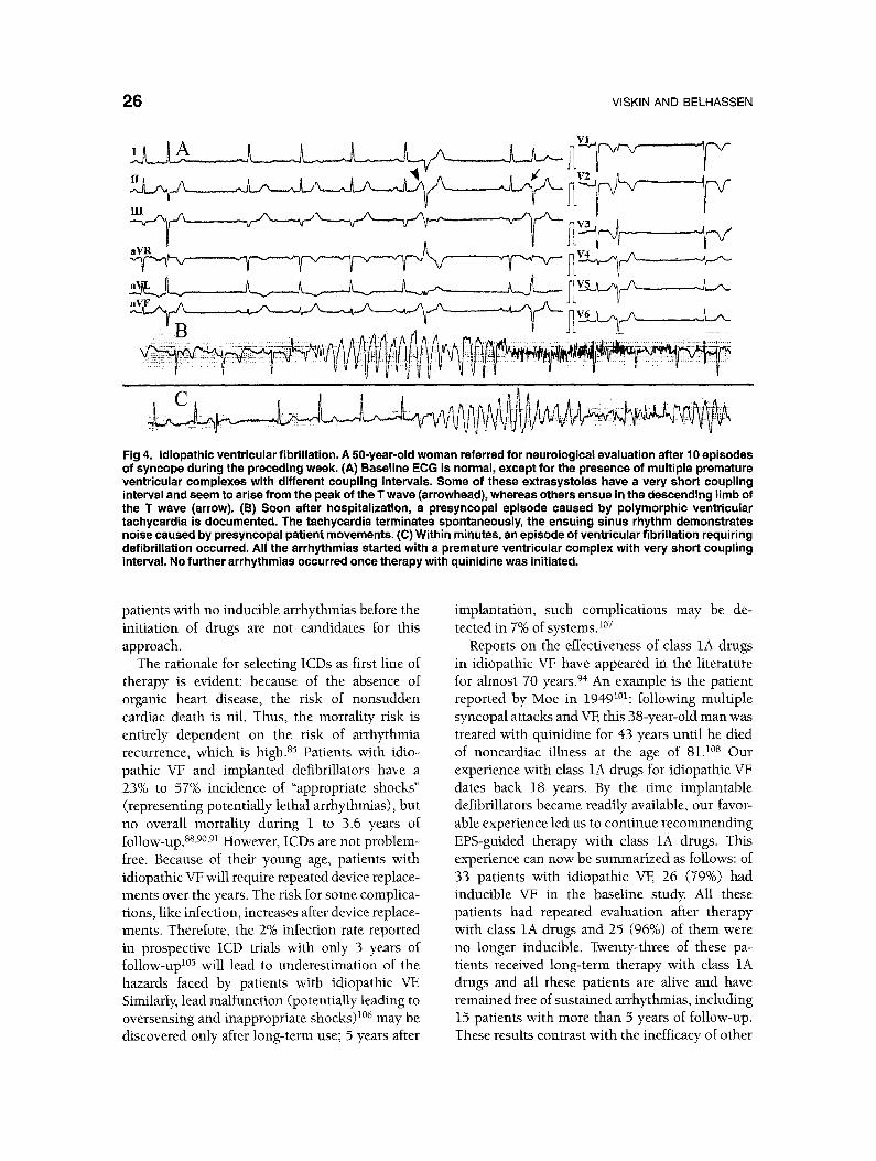
Fig 5. A 22-year-old male reported after an episode of aborted sudden death. 11a (A) RBBB pattern and ST segment elevation in the right precordial leads (typical of the Brugada syndrome) are obvious. (B) Over the years, the degree of ST segment elevation varied, but the typical pattern persisted. (C) Stored endocardial electrogram retrieved from his implanted defibrillator (C.P.I., Ventak P2, 1625, Guidant/Cardiac Pacemakers Inc) during a spontaneous episode of ventricular fibrillation, which occurred 2 years after implantation in the absence of drug therapy.
stimulation: 81% of patients in the series by Brugada 122 and 78% of our patients with normal ECG have inducible VE Moreover, unmasking of a Brugada sign in some patients with otherwise idiopathic VF may occur during long-term fol- low-up or following intravenous administration of drugs. 115,1.r,122 On the other hand, the Brugada syndrome affects primarily males: 89% of the patients with Brugada syndrome gathered by Brugada n4 and all the 21 patients reported by Atarashi n6 and our group are males. For compari- son, the male to female ratio among our patients with idiopathic VF and normal ECG is 18 to 13 (P < 0.001). The male predominance of the Bru- gada syndrome is of interest in view of recent data 123 linking the Brugada sign with the "syn- drome of nocturnal sudden death in Southeast Asians," another form of idiopathic VF, which almost exclusively affects males (see below). Finally, familial involvement is common in the Brugada syndrome n7,nS,n2 and absent in idio- pathic VF with normal ECG. 85 Indeed, genetic
studies recently performed in 6 families with the Brugada syndrome showed different mutations involving sodium channels in 3 families. TM
The main differential diagnosis of the Brugada syndrome is arrhythmogenic right ventricular dysplasia (ARVD), In this disease, fibrous and fatty tissue replace the myocardium, initially in the right ventricle, but eventually in both ven- tricles. 125 In a patient with inverted T waves in the right precordial leads and diffuse ventricular involvement, the diagnosis of dysplasia is evident. However, cardiac arrest may be the first manifesta- tion of focal ARVD325 Ruling-out focal dysplasia may be particularly challenging when a Brugada sign is present. 126 The RBBB pattern of the Bru- gada sign could imply a right ventricular disorder. Also, the catecholamine-sensitive monomorphic VT, anecdotally observed in the Brugada syn- drome n3,nr is of left bundle configuration also suggesting right ventricular origin Finally the Brugada sign has been reported in relatives of patients with documented ARVD n9 However

POLYMORPHIC VENTRICULAR TACHYARRHYTHMIAS 29
patients with the Brugada syndrome do not de- velop structural abnormalities during long-term follow-up. 1°,122 In addition, the arrhythmias ob- served in the two groups differ: although polymor- phic tachycardia has rarely been documented in ARVD, 127 the majority of arrhythmias (either spontaneous or induced) in cases of dysplasia are monomorphic.128,129 This contrasts with the poly- morphic morphology of the arrhythmias in idio- pathic VF with 1°,12z and without 86 the Brugada sign (Fig 5).
Patients with aborted sudden death and a Brugada sign are at high risk for arrhythmia recurrence. ~22 At the present time, there is no data to support the use of antiarrhythmic drugs in the Brugada syndrome, 117 and ICD implantation is the treatment of choice.
The Syndrome of Nocturnal Sudden Death in South East Asian Males
Nocturnal death in previously healthy young men (Pokkuri disease) is a well-recognized entity in Japan. From 1959 to 1961, almost 300 cases of "sudden death of unknown etiology" were re- ported in Tokyo. 13° The male to female ratio was 14 to 1. Moreover, 84% of the unexplained deaths among males occurred during sleep, whereas only 8% of the female fatalities happened at night. 13° Similar "sleep-death syndromes" have been de- scribed in Laos (non-laitai or "sleep death"), the Philippines (bangungut or "arise and moan"), 11 and Thailand.131
Following reports of unexplained death among Laotian refugees in the United States, the Center for Disease Control began active surveillance for unexpected deaths among Asian immigrants in 1981. By 1983, 79 cases with negative postmor- tem examination had been identified. 132 The same pattern was recognized: all but one of the deaths occurred in men, and all but one happened during sleep.
Epidemiological studies have attempted to ex- plain the "sleep-death syndrome" on a nutritional basis, namely, an acquired LQTS related to thia- mine deficiency. 133 However, this is unlikely for several reasons: (1) the QT interval of patients resuscitated from sleep-death syndrome is nor- mal; and (2) females, who have longer QT inter- vals and are afflicted by acquired forms of the LQTS more commonly than males, 134 would be
expected to suffer from this predominantly male disease.
Similarities do exist between the sleep-death syndrome and other forms of idiopathic VE These similarities include the age at the onset of symp- toms (25 to 45 years)130,132 the mode of onset of spontaneous arrhythmias, 123,135 and a high induc- ibility rate of VF during electrophysiologic stud- ies.~23,a36,137 Moreover, according to a recent study, 82% of Thai males with sleep-death syndrome have a resting ECG indistinguishable from the Brugada sign. Interestingly, anecdotal reports sug- gest that class 1A drugs may prevent induction of VF in the laborator)z. B6,B7
Patients with sleep-death syndrome have a high mortality risk, 123,135 but data on therapy is s c a r c e . 123,136,137 Recurrent VE despite therapy with [B-blockers or amiodarone, has been documented, and there is no data on the long-term value of class 1A drugs. ICDs are viewed as the only effective therapy. 123
Polymorphic Ventricular Arrhythmias Without Apparent Heart Disease:
Differential Diagnosis
Onset of stress-related symptoms (syncope or cardiac arrest) during infancy or childhood favors the diagnosis of catecholaminergic polymorphic VT or a congenital LQTS. Female gender strongly points against the Brugada syndrome or a sleep- death syndrome. Obviously, a QT interval of long duration or odd morphology is characteristic of a LQTS. Similarly, an RBBB with ST elevation is the sinequa-non of the Brugada syndrome and is apparently common in the sleep-death syndrome. Finally, documentation of the mode of onset of spontaneous arrhythmias is of importance: a long-short cycle preceding the onset of polymor- phic VT favors a LQTS even in cases with borderline QT. However, arrhythmias precipi- tated by a single extrasystole with ultra-short coupling interval are seen in the short-coupled variant of torsade and the idiopathic VF syn- dromes (idiopathic VF with normal ECG, the Brugada syndrome, and the sleep death syn- drome). Finally, the highly reproducible mode of onset of catecholaminergic polymorphic arrhyth- mias is essentially diagnostic.
Several forms of organic heart disease, which are difficult to exclude without specific testing,

3 0 VISKIN AND BELHASSEN
ought to be considered in the differential diagno- sis following an episode of"cardiac arrest without apparent heart disease." These include: (1) coro- nary-artery spasm in patients with normal coro- nary angiography15,138; (2) a focal ARVD15,118,119,139;
and (3) conditions that facilitate atrioventricular conduction of supraventricular arrhythmias to the point in which very rapid ventricular rates trigger VF. 140 The last category includes patients with enhanced AV nodal conduction 14° or an atrioventricular accessory pathway that is not evident during sinus rhythm because of a left anterolateral location.141 Finally, idiopathic mono- morphic VT, although usually well tolerated, 1,2 may rarely lead to cardiac arrest, especially during strenuous effort. 1,2,142
References
1. Brooks R, Burgess J: Idiopathic ventricular tachycar- dia. A review. Medicine 67:271-294, 1988
2. BelhassenB, ViskinS: Idiopathicventriculartachycar- dia and fibrillation. J Cardiovasc Electrophysiol 4:356- 368, 1993
3. Consensus Statement of the Joint Committees of the Unexplained Cardiac Arrest Registry of Europe and of the Idiopathic Ventricular Fibrillation Registry in the United States: Survivors of out-of-hospital cardiac arrest with apparently normal heart. Need for defini- tion and standardized clinical evaluation. Circulation 95:265-272, 1996
4. Jervell A, Lange-Nielsen F: Congenital deaf-mutism, functional heart disease with prolongation of the Q-T interval and sudden death. Am Heart J 54:59-68, 1957
5. Romano C, Gemme G, Pongiglione R: Aritmie cardi- ache rare dell'eta pediatrica. Clin Pediatr 45:656- 683, 1963
6. Ward O: A new familial cardiac syndrome in children. J Irish Med Assoc 54:103-106, 1964
7. Leenhardt A, Glaser E, Burguera M, et al: Short- coupled variant of torsade de pointes. A new electro- cardiographic entity in the spectrum of idiopathic ventricular tachyarrhythmias. Circulation 89:206- 215, 1994
8. LeenhardtA, Lucet V, Denjoy I, et al: Catecholaminer- gic polymorphic ventricular tachycardia in children. A 7-year follow-up of 21 patients. Circulation 91:1512- 1519, 1995
9. Beihassen B, Shapira I, Shoshani D, et al: Idiopathic ventricular fibrillation: Inducibility and beneficial ef- fects of class I antiarrhythmic agents. Circulation 75:809-816, 1987
10. Brugada P, Brugada J: Right bundle branch block, persistent ST segment elevation and sudden cardiac death: A distinct clinical and electrocardiographic syndrome. A multicenter report. J Am Coil Cardiol 20:1391-1396, 1992
11. Aponte G: The enigma of "Bangungut." Ann Intern Med 52:1258-1263, 1960
12. Coumel P, Lucet V: Les syndromes de torsades de pointes. Varietes et limites. Ann Cardiol Angeiol 35:205-214, 1986
13. Shaw T: Recurrent ventricular fibrillation associated with normal QT intervals. QJM 200:451-462, 1981
14. Myrianthefs M, Cariolou M, EIdar M, et al: Exercise- induced ventricular arrhythmias and sudden cardiac death in a family. Chest 111:1130-1133, 1997
15. Eisenberg S, Scheinman M, Dullet N, et al: Sudden cardiac death and polymorphous ventricular tachycar- dia in patients with normal QT intervals and normal systolic cardiac function. Am J Cardiol 75:687-692, 1995
16. Roden D: Recent advances in the long QT syn- drome. Card Electrophysiol Rev 1:262-263, 1997
17. Roden D, Lazzara R, Rosen M, et al: Multiple mechanisms in the long QT syndrome. Current knowledge, gaps, and future directions. Circulation 94:1996-2012, 1996
18. Tan HL, Hou C J, Lauer MR, et al: Electrophysiologic mechanisms of the long QT interval syndromes and torsade de pointes. Ann Intern Med 122:701-714, 1995
19. Jackman WM, Friday K J, Anderson JL, et al: The long QT syndromes: A critical review, new clinical observations and a unifying hypothesis. Prog Cardio- vasc Dis 31:115-172, 1988
20. Priori SG, Napolitano C, Paganini V, et al: Molecular biology of the long QT syndrome: Impact on manage- ment. Pacing Clin Electrophysiol 20:11-2052-11-2057, 1997
21. Carmeliet E: Electrophysiologic and voltage clamp analysis of the effects of sotalol on isolated cardiac muscle and Purkinje fibers. J Pharmacol Exp Ther 232:817-825, 1985
22. Sanguinetti M, Jurkiewicz N: Two components of cardiac delayed rectifier K + current: Differential sen- sitivity to block by class III antiarrhythmic agents. J Gen Physio196:195-215, 1990
23. Yang T, Roden D: Extracellular potassium modula- tion of drug block of IKr: Implications for torsades de pointes and reverse use-dependence. Circulation 93:407-411,1996
24. Antzelevitch C, Sun Z, Zhang Z, et al: Cellular and ionic mechanisms underlying erythromycin-induced long QT intervals and torsade de pointes. J Am Coil Cardio128:1836-1848, 1996
25. Vorperian V, Zhou Z, Mohammad S, et al: Torsade de pointes with an antihistamine metabolite: Potassium channel blockade with desmethylastemizole. J Am Coil Cardio128:1556-1561, 1996
26. Duggal P, Vesely B, Wattanasirichaigoon D, et al: Mutation of the gene for IsK associated with both Jervell and Lange Nielsen and Romano-Ward forms of long QT syndrome. Circulation 97:142-146, 1998
27. Drolet 13, Khalifa M, Daleau P, et al: Block of the rapid component of the delayed rectifier potassium current by the prokinetic agent cisapride underlies drug-

POLYMORPHIC VENTRICULAR TACHYARRHYTHMIAS 31
related lengthening of the QT interval. Circulation 97:204-210, 1998
28. Martyn R, Somberg J, Kerin N: Proarrhythmia of nonantiarrhythmic drugs. Am Heart J 126:201-205, 1993
29. Antzelevitch C, Sicouri S: Clinical relevance of car- diac arrhythmias generated by afterdepolarizations. Role of M cells in the generation of U waves, triggered activity and torsade de pointes. J Am Coil Cardio123:259-277, 1994
30. EI-Sherif N, Caref E, Yin H: The electrophysiological mechanism of ventricular tachyarrhythmias in the long QT syndrome. Tddimentional mapping of activa- tion and recovery patterns. Circ Res 79:474-492, 1996
31. Schwartz P J, Periti M, Malliani A: The long Q-T syndrome. Am Heart J 89:378-390, 1975
32. Kay GN, Plumb V J, Arciniegas JG, et al: Torsade de pointes: The long-short initiating sequence and other clinical features: Observations in 32 patients. J Am Coil Cardiol 2:806-817, 1983
33. Locati EH, Maison-Blanche P, De]ode P, et al: Spon- taneous sequences of onset of torsade de pointes in patients with acquired prolonged repolarization: Quantitative analysis of Holter recordings. J Am Coil Cardio125:1564-1575, 1995
34. Viskin S, Alia S, Barron H, et al: Mode of onset of torsade de pointes in congenital long QT syndrome. J Am Coil Cardio128:1262-1268, 1996
35. Viskin S, Fish R, Zeltser D, et al: Pause-dependent vs. non pause-dependent initiation of torsade de pointes in the congenital long qt syndrome. Pacing Clin Electrophysio121:1i-851, 1998 (abstr)
36. Gronefeld G, Holtgen R, Hohnloser S: Implantable cardioverter defibrillator therapy in a patient with the idiopathic long QT syndrome. Pacing Clin Electro- physio119:1260-1263, 1996
37. MossAJ, Schwartz P J, Crampton RS, et al: The long QT syndrome. Prospective longitudinal study of 328 families. Circulation 84:1136-1144, 1991
38. Garson A, Dick M, Fournier A, et al: The long QT syndrome in children. An international study of 287 patients. Circulation 87:1866-1872, 1993
39. Van-Hare GF, Franz MR, Roge C, et al: Persistent functional atrioventricular block in two patients with prolonged QT intervals: Elucidation of the mecha- nism of block. Pacing Clin Electrophysiol 13:608- 618, 1990
40. Weintraub R, Gow R, Wilkinson J: The congenital long QT syndromes in childhood. J Am Coil Cardiol 16:674-680, 1990
41. Scott W, Dick M: Two: One atrioventricular block in infants with congenital long QT syndrome. Am J Cardio160:1409-1410, 1987
42. DiSegni E, David D, Katzenstein M, et al: Permanent overdrive pacing for the suppression of recurrent ventricuiar tachycardia in a newborn with long QT syndrome. J Electrocardiol 13:189-192, 1980
43. Hajj R, Zareba W, Rosero S, et al: Adrenergic triggers and non-adrenergic factors associated with
cardiac events in long QT syndrome patients. Pacing Clin Electrophysio120:11-1072, 1997 (abstr)
44. -lobe T, Langen C, Bink-Boelkens M, et al: Late potentials in a bradycardia-dependent long QT syn- drome associated with sudden death during sleep. J Am Coil Cardiol 19:541-549, 1992
45. Vincent GM: Heterogeneity in the inherited long QT syndrome. J Cardiovasc Electrophysiol 6:137-146, 1995
46. Priori SG: Is long QT syndrome entering the era of molecular diagnosis? Heart 77:5-6, 1997
47. Schwartz P J, MossAJ, Vincent GM, et al: Diagnostic criteria for the long QT syndrome. An update. Circula- tion 88:782-784, 1993
48. Broadhurst P, Nathan A: Cardiac arrest in a young woman with the long QT syndrome and concomitant astemizole ingestion. Br Heart J 70:469-470, 1993
49. Hsieh M, Chen S, CHang C, et al: Drug-induced torsade de pointes in one patient with congenital long QT syndrome. Int J Cardio154:85-88, 1996
50. Garcia-Rubira J, Aramburu O, Romero D, et al: Syncope and long QT syndrome with an initially normal QT interval. Int J Cardio140:286-288, 1993
51. Lehmann M, Suzuki F, Fromm B, et ai: T wave "humps" as a potential electrocardiographic marker of the long QT syndrome. J Am Coil Cardiol 24:746- 754, 1994
52. Malfatto G, Beria G, Sala S, et al: Quantitive analysis of T wave abnormalities and their prognostic implica- tions in the idiopathic long QT syndrome. J Am Coil Cardio123:296-301,1994
53. Krahn A, Klein G, Yee R: Hysteresis of the RT interval with exercise. A new marker for the long QT syn- drome? Circulation 96:1551-1556, 1997
54. Mitsutake A, Takeshita A, Kuroiwa A, et al: Useful- ness of the valsalva maneuver in management of the long QT syndrome, Circulation 63:1029-1035, 1981
55. Vincent GM, Jaiswal D, Timothy K: Effects of exer- cise on heart rate, QT, QTc and QT/QS2 in the Romano-Ward inherited long QT syndrome. Am J Cardio168:498-503, 1991
56. Moss A J, Zareba W, Benhorin J, et al: ECG T-wave patterns in genetically distinct forms of the hereditary long QT syndrome [see comments]. Circulation 92: 2929-2934, 1995
57. Bhandari AK, Sapiro W, Morady F, et al: Electrophysi- ologic testing in patients with the long QT syndrome. Circulation 71:63-71, 1985
58. Zhou JT, Zheng LR, Liu WY, et al: Early afterdepolar- ization in the familial long QTU syndrome. J Cardio- vasc Electrophysiol 3:431-436, 1993
59. Shimizu W, Ohe T, Kurita T, et al: Early afterdepolar- izations induced by isoproterenol in patients with congenital long QT syndrome. Circulation 84:1915- 1923, 1991
60. Kawade M, Ohe T, Kamiya T: Provocative testing and drug response in a patient with the long QT syn- drome. Br Heart J 74:67-70, 1995
61. Moss A J, Schwartz P J, Crampton RS, et ai: The long QT syndrome: A prospective international study. Circulation 71:17-21, 1985

32 VISKIN AND BELHASSEN
62. Epstein A, Rosner M, Hageman G, et al: Posterior left thoracic cardiac sympathectomy by surgical division of the sympathetic chain: An alternative approach to the treatment of the long QT syndrome. Pacing Clin Electrophysiol 19:1095-1104, 1996
63. Bhandari AK, Scheinman MM, Morady F, et al: Efficacy of left cardiac sympathectomy in the treat- ment of patients with the long QT syndrome. Circula- tion 70:1018-1023, 1984
64. Schwartz P J, Locati EH, Moss A, et al: Left cardiac sympathetic denervation in the therapy of congenital long QT syndrome. A worldwide report. Circulation 84:503-511, 1991
65. Crawford MH, Karliner JS, O'Rourke RA, et al: Prolonged Q-T interval syndrome. Successful treat- ment with combined ventricular pacing and proprano- Iol. Chest 68:369-371, 1975
66. Wilmer CI, Stein B, Morris DC: Atrioventricular pace- maker placement in Romano-Ward syndrome and recurrent torsades de poJntes. Am J Cardiol 59:171- 172, 1987
67. Eldar M, Griffin JC, Abbott JA, et al: Permanent cardiac pacing in patients with the long QT syn- drome. J Am Coil Cardio110:600-607, 1987
68. EIdar M, Griffin JC, Van Hare GF, et al: Combined use of beta-adrenergic blocking agents and long- term cardiac pacing for patients with the long QT syndrome. J Am Coil Cardio120:830-837, 1992
69. Moss A J, Liu JE, Gottlieb S, et al: Efficacy of permanent pacing in the management of high-risk patients with long QT syndrome. Circulation 84:1524- 1529, 1991
70. Rosenbaum MB, Acunzo RS: Pseudo 2:1 atrioven- tricular block and T wave alternans in the long QT syndromes. J Am Coil Cardiol 18:1363-1366, 1991 (editorial)
71. Shinbane J, Wood M, Jensen N, et al: Tachycardia- induced cardiomyopathy: A review of animal models and clinical studies. J Am Coil Cardiol 29:709-715, 1997
72. Sclarovsky S, Strasberg B, Lewin R, et al: Polymor- phous ventricular tachycardia: Clinical features and treatment. Am J Cardio144:339-345, 1979
73. Klein H, Levi A, Kaplinsky E, et al: Congenital long QT syndrome: Deleterious effect of long-term high- rate ventricular pacing and definitive treatment by cardiac transplantation. Am Heart J 132:1079-1081, 1996
74. Viskin S, Fish R, Roth A, et al: Prevention of torsade de pointes in the congenital long QT syndrome: Use of a pause-prevention pacing algorithm. Heart 79:417- 419, 1998
75. Viskin S, Fish R, Brosh D, et al: Prevention of pause-dependent torsade de pointes and malignant ventricular arrhythmias with a pause-preventing pac- ing algorithm. Pacing Clin Electrophysiol 21:11-826, 1998 (abstr)
76. Gran W, Silka M, Oliver R, et al: Use of implantable cardioverter-defibrillators in the congenital long QT syndrome, Am J Cardio178:703-706, 1996
77. Dorostkar P, Eldar M, Belhassen B, et al: Long-term follow-up of patients with the idiopathic long QT syndrome treated with combined beta-blockers and chronic pacing. Circulation 96:1-1172, 1997 (abstr, suppl I)
78. Saxon L, Shannon K, Wetzel G, et al: Familial long QT syndrome: Electrical storm and implantable car- dioverter device therapy. Am Heart J 131:1037-1039, 1996
79. Compton S, Lux R, Ramsey M, et al: Genetically defined therapy of inherited long QT syndrome. Correction of abnormal repolarization by potassium. Circulation 94:1018-1022, 1996
80. Priori SG, Napolitano C, Cantu F, et al: Differential response to Na+ channel blockade, beta-adrenergic stimulation and rapid pacing in a cellular model mimicking the SCN5A and HERG defects present in the long QT syndrome. Experimental basis for gene- specific therapy. Circ Res 78:1009-1015, 1996
81. Rosero S, Zareba W, Robinson J, et al: Gene specific therapy for long QT syndrome: QT shortening with lidocaine and tocainide in patients with mutation of the sodium channel gene. Ann Noninvasive Electro- cardiol 3:274-278, 1997
82. Schwartz P J, Priori SG, Locati EH, et al: Long QT syndrome patients with mutations of the SCNSA and HERG genes have differential responses to Na+ channel blockade and to increases in heart rate. Implications for gene-specific therapy [see com- ments]. Circulation 92:3381-3386, 1995
83. Strasberg B, Welch W, Palileo E, et ai: Familial JnducJble torsade de poJntes with normal QT interval. Eur Heart J 4:383-390, 1983
84. Vincent GM, Timothy KW, Leppert M, et al: The spectrum of symptoms and QT intervals in carriers of the gene for the long QT syndrome. N Engl J Med 327:846-852, 1992
85. Viskin S, Belhassen B: Idiopathic ventricular fibrilla- tion. Am Heart J 120:661-671,1990
86. Viskin S, Lesh M, Eldar M, et al: Mode of onset of malignant ventricular arrhythmias in idiopathic ven- tricular fibrillation. J Cardiovasc Electrophysiol 8:1115- 1120, 1997
87. Tung R, Shen W, Hammill S, et al: Idiopathic ventricu- lar fibrillation in out-of-hospital cardiac arrest survi- vors. Pacing Clin Electrophysiol 17:1405-1412, 1994
88. Meissner M, Lehmann M, Steinman R, et al: Ventricu- lar fibrillation in patients without significant structural heart disease: A multicenter experience with implant- able cardioverter-defibrillator therapy. J Am Coil Car- dio121:1406-1412, 1993
89. Wever FD: Unfavorable outcome in patients with primary electrical disease who survive ventricular fibrillation, Circulation 89:2456, 1994 (letter)
90. Wever EFD, Hauer RNW, Oomen A, et al: Unfavor- able outcome in patients with primary electrical disease who survived an episode of ventricular fibrillation. Circulation 88:1021-1029, 1993
91, Fan W, Peter C: The Cedars Investigators. Survival and incidence of appropriate shocks in implantable cardioverter defibrillator recipients who have no de-

POLYMORPHIC VENTRICULAR TACHYARRHYTHMIAS 33
tectable structural heart disease. Am J Cardio174:687- 690, 1994
92. Aizawa Y, Naitoh N, Washizuka T, et al: Electrophysi- ological findings in idiopathic recurrent ventricular fibrillation: Special reference to mode of induction, drug testing and long-term outcomes. Pacing Clin Electrophysio119:929-939, 1996
93. Wellens H J J, Lemery R, Smeets JL, et al: Sudden arrhythmic death without overt heart disease. Circu- lation 85:1-92-1-97, 1992 (suppl I)
94. Dock W: Transitory ventricular fibrillation as a cause of syncope and its prevention by quinidine sulfate. Am Heart J 4:709-714, 1929
95. Bechgaard P: Paroxysmal ventricular fibrillation with recovery. Acta Med Scand 132:9-19, 1948
96. Masrani K, Cowley C, Bekheit S, et al: Recurrent syncope over a decade due to idiopathic ventricular fibrillation. Chest 106:1601-1603, 1994
97. Bjerregaard P, Gussak I, Kotar SL: Recurrent syn- cope in a patient with prominent J wave. Am Heart J 127:1426-1429, 1994
98. Dubner S J, Gimeno GM, Elencwajg B, et al: Ventricu- lar fibrillation with spontaneous reversion on ambula- tory ECG in the absence of heart disease. Am Heart J 105:691-693, 1983
99. Ledwich JR, Fay JE: Idiopathic recurrent ventricular fibrillation. Am J Cardio124:255-257, 1969
100. Lemery R, Brugada P, Della Bella P, et al: Ventricular fibrillation in six adults without overt heart disease. J Am Coil Cardio113:911-916, 1989
11)1. Moe T: Morgagni-Adams-Stokes attacks caused by transient recurrent ventricular fibrillation in a patient without apparent heart disease. Am Heart J 37:811- 818, 1949
102. Chen P, Feld G, Mower M, et al: Effects of pacing rate and timing of defibrillation shock on the relation between the defibrillation threshold and the upper limit of vulnerability in open chest dogs. J Am Coil Cardiol 18:1555-1563, 1991
103. Chen P, Feld G, Kriett J, et al: Relation between the upper limit of vulnerability and defibrillation threshold in humans. Circulation 88:186-192, 1993
104. Saumarez R, Heald S, Slade A, et al: Primary ventricular fibrillation is associated with increased paced right ventricular electrogram fractionation. Cir- culation 92:2565-2571, 1995
105. The Antiarrhythmic Versus implantable Defibrillators (AVI D) Investigators: A comparison of antiarrhythmic- drug therapy with implantable defibrillators in pa- tients resuscitated from near-fatal ventricular arrhyth- mias. N Engl J Med 337:1576-1583, 1997
106. Daoud E, Kirsh M, Boiling S, et al: Incidence, presen- tation, diagnosis, and management of malfunctioning implantable cardioverter-defibrillator rate-sensing leads. Am Heart J 128:892-895, 1994
107. Goyal R, Harvey M, Horwood L, et al: Incidence of lead system malfunction detected during implantable defibrillator generator replacement. Pacing Clin Elec- trophysio119:1143-1146, 1996
108. Konty F, Dale J: Self-terminating idiopathic ventricu- lar fibrillation presenting as syncope: A 40-year fol- low-up report. J Intern Med 227:211-213, 1990
109. Frustaci A, Beilocci F, Olsen E: Results of biventricu- lar endomyocardial biopsy in survivors of cardiac arrest with apparently normal hearts. Am J Cardiol 74:890-895, 1994
110. Priori S, Paganini V: Idiopathic ventricular fibrillation: Epidemiology, pathophysiology, primary prevention, immediate evaluation and management, long-term evaluation and management, experimental and theo- retical developments. Card Electrophysiol Rev 1:244- 247, 1997
111. Wever EFD, Hauer RNW, van-Capelie F, et al: Randomized study of implantable defibrillator as first-choice therapy versus conventional strategy in postinfarct sudden death survivors. Circulation 91: 2195-2203, 1995
112. Bocker D, Haverkamp W, Block M, et al: Comparison of d,l-sotalol and implantable defibrillators for treat- ment of sustained ventricular tachycardia or fibrilla- tion in patients with coronary disease. Circulation 94:151-157, 1996
113. Viskin S, Belhassen B: Clinical problem solving: When you only live twice. N Engl J Med 332:1221- 1225, 1995
114. Brugada J, Brugada P: Right bundle branch block and ST segment elevation in leads V1 through V3. A marker for sudden death in patients without demon- strable structural heart disease. Circulation 97:457- 460, 1998
115. Miyazaki T, Mitamura H, Miyoshi S, et al: Autonomic and antiarrhythmic drug modulation of ST segment elevation in patients with Brugada syndrome. J Am Coil Cardio127:1061 -1070, 1996
116. Atarashi H, Ogawa S, Haruni K, et al: Characteristics of patients with right bundle branch block and ST- segment elevation in right precordial leads. Am J Cardio178:581-583, 1996
117. Kasanuki H, Ohnishi S, Ohtuka M, et al: Idiopathic ventricular fibrillation induced with vagal activity in patients without obvious heart disease. Circulation 95:2277-2285, 1997
118. Martini B, Nava A, Thiene G, et al: Ventdcular fibrillation without apparent heart disease: Descrip- tion of six cases. Am Heart J 118:1203-1209, 1989
119. Corrado D, NavaA, Buja G, et ai: Familial cardiomy- opathy underlies syndrome of right bundle branch block, ST segment elevation and sudden death. J Am Coil Cardio127:443-448, 1996
120. Gussak I, Bjerregaard P, Egan TE, et al: ECG phenomenon called the J wave. History, pathophysi- elegy and clinical significance. J Electrocardio128:49- 58, 1995
121. Yan G, Antzelevitch C: Cellular basis for the electro- cardiographic J wave. Circulation 93:372-379, 1996
122. Brugada J, Brugada P: What to do in patients with no structural heart disease and sudden arrhythmic death? Am J Cardio178:69-75, 1996 (suppl 5A)
123. Nademanee K, Veerakul G, Nimmannit S, et al: Arrhythmogenic marker for the sudden unexpected

34 VISKIN AND BELHASSEN
death syndrome in Thai men. Circulation 96:2595- 2600, 1997
124. Chen Q, Kirsch G, Zhang D, et al: Genetic basis and molecular mechanism for idopathic ventricular fibrilla- tion. Nature 392:293, 1998
125. Corrado D, Basso C, Thiene G, et al: Spectrum of clinicopathologic manifestations of arrhythmogenic right ventricular cardiomyopathy/dysplasia: A multi- center study. J Am Coil Cardio130:1512-1520, 1997
128. Scheinman M: Is the Brugada syndrome a distinct clinical entity? J Cardiovasc Electrophysiol 8:332- 336, 1997
127. Fontaine G, Aouate P, Fontaliran F: Repolarization and genesis of cardiac arrhythmias. Role of body surface mapping. Circulation 95:2600-2602, 1997
128. Lecierq JF, Potenza S, Maisonblanche P, et al: Determinants of spontaneous occurrence of sus- tained monomorphic ventricular tachycardia in right ventricular dysplasia. J Am Coil Cardiol 28:720-724, 1996
129. Link M, Wang P, Haugh C, et al: Arrhythmogenic right ventricular dysplasia: clinical results with implantable cardioverter defibrillators. J Intervent Card Electro- physiol 1:41-48, 1997
130. Kobayshi T, Ito Y, Kahijara T, et al: Acute cardiac death of unknown etiology. A preliminary report. Jpn Heart J 3:442-454, 1962
131. Tungsanga K, Sriboonlue P: Sudden unexplained death syndrome in North-Eastern Thailand. Int J Epidemio122:81-87, 1993
132. Baron R, Thacker S, Gorelkin L, et al: Sudden death among Southeast Asian refugees. An unexplained nocturnal phenomenon. JAMA 250:2947-2951,1983
133. Munger R, Prineas R, Crow R, et al: Prolonged QT interval and risk of sudden death in South-East Asian men. Lancet 338:280-281, 1991
134. Makkar R, Fromm B, Steinman R, et al: Female gender as a risk factor for torsade de pointes associ- ated with cardiovascular drugs. JAMA 270:2590- 2597, 1993
135, Hayashi M, Murata M, Satoh M, et al: Sudden nocturnal death in young males from ventricular flutter. Jpn Heart J 26:585-591,1983
136, Otto C, Tauxe R, Cobb L, et al: Ventricular fibrillation causes sudden death in Southeast Asian immi- grants. Ann Intern Med 100:45-47, 1984
137, Gilbert J, Gold R, Haffajee C, et al: Sudden cardiac death in a Southeast Asian immigrant: Clinical, elec- trophysiologic and biopsy characteristics. Pacing Clin Electrophysiol 9:912-914, 1986
138. Myerburg R, Kessler K, Mallon S, et al: Life- threatening ventricular arrhythmias in patients with silent myocardial ischemia due to coronary-artery spasm. N Engl J Med 326:1451-1455, 1992
139. Morgera T, Sinagra G, Viel E, et al: The syndrome of right bundle branch block, persistent ST segment elevation and sudden cardiac death. Which is the histological substrate? Eur Heart J 18:1190-1191, 1997 (letter)
140. Wang Y, Griffin J, Lesh M, et al: Patients with supraventricular tachycardia presenting with aborted sudden death: Incidence, mechanism and long-term follow-up. J Am Coil Cardiol 18:1720-1721,1991
141. Robinson K, Rowland E, Krikler D: Latent pre- excitation: Exposure of anterograde accessory path- way conduction during atrial fibrillation. Br Heart J 59:53-55, 1988
142. Wesley R, Taylor R, Nadamanee K: Catecholamine- sensitive right ventricular tachycardia in the absence of structural heart disease: A mechanism of exercise- induced cardiac arrest. Cardiology 79:237-243, 1991


![Fulminant isolated cardiac sarcoidosis with pericardial effusion … · 2017. 4. 18. · if associated with new ventricular tachyarrhythmias or ... case report[2 ,14 15]. Conduction](https://img.dokumen.tips/doc/110x75/6119971c0f2ccf10175eeb3e/fulminant-isolated-cardiac-sarcoidosis-with-pericardial-effusion-2017-4-18.jpg)