Embed Size (px)
Citation preview
THE FILTRABLE MICROORGANISMS OF THEPLEUROPNEUMONIA GROUP
ALBERT B. SABIN
From The Children's Hospital Research Foundation and the Department ofPediatrics, University of Cincinnati College of Medicine, Cincinnati, Ohio
CONTENTS
General characteristics of the group; methods of study..................... 4Bovine pleuropneumonia, and the etiological agent........................ 13Agalactia of sheep and goats, and the etiological agent................... 16Pleuropneumonia-like microorganisms of dogs; their relation to caninedistemper........................................................... 18
Pleuropneumonia-like microorganisms of rats........................... 20Pleuropneumonia-like micro6rganisms of mice............................ 23Pleuropneumonia-like micro6rganisms of guinea-pigs..................... 31Pleuropneumonia like micro6rganisms of man............................ 32Pleuropneumonia-like microorganisms of sewage and decomposing matter.. 34
L1 and Streptobacillus moniliformis. Symbosis or variation?.......... 35Filtrability and size of minimal reproductive units......................... 39Toxigenicity and immunological reactions.................................. 41Pathology.......................................................... 45Effects of immune serums, vaccines, and chemotherapeutic agents. ..................48Relation to viruses, and other ifitrable and nonifitrable microbes........... 52Classification and nomenclature.......................6................ 5
In 1898, when our knowledge of filtrable viruses was but a fewyears old, the then ultramicroscopic etiological agent of bovinepleuropneumonia was cultivated in a cell-free medium by Nocard,et al. (63). The morphologic studies of Bordet (3) and of Borrel,et al. (4) in 1910, and of others later on established the remarkablepolymorphic character of the micro6rganism; and the filtrationexperiments through graded collodion membranes carried out byElford (32) in 1929 established that the cultures contained "par-ticles," 125 to 150 mui in size, which were capable of reproducingnot only themselves but also the larger and more complex struc-tures. For 25 years after its cultivation in serum broth it wasunique among infectious agents, but was practically alwaysgrouped with the filtrable viruses. In 1923, Bridr6 and Dona-
1
on July 14, 2019 by guesthttp://m
mbr.asm
.org/D
ownloaded from
ALBERT B. SABIN
tien (5) demonstrated not only that the filtrable etiological agentof agalactia of sheep and goats could be cultivated in vitro in thesame manner as the micro6rganism, or virus as it was still called,of bovine pleuropneumonia but also that, excepting the pathogenicand immunologic properties, there was the closest morphologicaland biological resemblance between these two agents (6). Al-though in the ensuing period many communications appeareddealing with the morphology and mode of reproduction of thebovine pleuropneumonia and agalactia micro6rganisms (43, 53,65, 66, 103), another 11 years passed before a new member ofthis group was discovered. In 1934, Shoetensack (86) reportedthe cultivation in cell-free media of morphologically and bio-logically similar micro6rganisms from dogs suffering of distemper,and although insufficient work was done on the relation of thesemicro6rganisms to the virus of canine distemper, it was neverthe-less established that members of the pleuropneumonia groupoccurred in dogs.
In 1935, Klieneberger (44) reported the remarkable observationthat a pleuropneumonia-like microorganism could be demon-strated in all available strains of Streptobacillus moniliformis, agram-negative pleomorphic bacillus which is a normal inhabitantof the nasopharynx of rats and the cause of at least one type ofrat-bite fever in man. Although she has been able to isolate thismicro6rganism (L1) in pure culture and maintain it in continuoussubculture without reversion to Streptobacillus moniliformis, otherinvestigators (21, 24) have challenged her hypothesis that suchsymbiosis existed and suggested that the L1 micro6rganism is avariant of the bacillus. These studies, however, have led to theisolation of other pleuropneumonia-like, pathogenic micro6rgan-isms from rats, unassociated with bacteria and distinct fromL1 (47, 48).The next important contribution in this field came in 1936,
when Laidlaw and Elford (52) reported on a new group of filtrablemicroorganisms which they isolated from raw sewage. Theseresembled the other members of the pleuropneumonia group inthat minute particles, 125 to 175 mu in size, reproduced the same
2
on July 14, 2019 by guesthttp://m
mbr.asm
.org/D
ownloaded from
THE PLEUROPNEUMONIA GROUP
type of polymorphic structures, and gave rise to similar micro-scopic colonies on solid media, but differed in not requiring proteinfor their growth; also they possessed no pathogenic properties.Seiffert (84) confirmed these observations when he reported theisolation of similar micro6rganisms from filtrates of soil, compost,decomposing leaves, and manure.
Late in 1938, Sabin (75) and Findlay, et al. (35) simultaneouslydescribed the isolation of a new pleuropneumonia-like micro6r-ganism from mice which developed a peculiar nervous disease inthe course of routine passage of toxoplasma or lymphocyticchoriomeningitis virus. The studies on this group received a neworientation when it was demonstrated (74, 75, 76) that thisfiltrable microorganism of the mouse, although capable of multi-plying in a cell-free medium in vitro, was an intracellular parasitein vivo, with a special affinity for the mesenchymal cells of thepleura, peritoneum, and joints, and that during the course ofits multiplication a typical neurotropic exotoxin was producedwhich gave rise to choreiform nervous signs. Furthermore, earlyin 1939 Sabin (76) isolated from normal mice another such micro-organism, immunologically distinct from the first, and with suchlimited cellular affinities that it could multiply only in themesenchymal cells of the joints in which it produced a prolifera-tive, progressive, and chronic ankylosing arthritis. Subsequentstudies (77, 80) revealed that normal mice are carriers of thesepathogenic micro6rganisms, especially in their conjunctiva andnasal mucosa, and at least five immunologically distinct typeshave already been described (80). Although attempts to demon-strate members of the pleuropneumonia group in pathologicalmaterial from patients with rheumatic fever and rheumatoidarthritis have met with no success, studies are being continued onhuman beings (80); and Dienes (28) has already brought forthmore than suggestive evidence of their existence in the femalegenital tract.At the present time our knowledge has progressed far enough to
indicate that there exists in nature a distinct group of filtrable,saprophytic and parasitic micro6rganisms, of which the etiological
3
on July 14, 2019 by guesthttp://m
mbr.asm
.org/D
ownloaded from

ALBERT B. SABIN
agent of bovine pleuropneumonia is the prototype, and whichpossess properties that clearly distinguish them from the ordinarybacteria, the filtrable viruses, and the rickettsiae.
GENERAL CHARACTERISTICS OF THE GROUP AND METHODS OF STUDY
Criteria for Identification. The criteria which admit a micro-organism into the pleuropneumonia group are: (1) growth in cell-free culture media with the development of polymorphic struc-tures including, "rings," globules, filaments, and minute, filtrableelementary bodies, usually 125 to 250 m,u in size, which are theminimal reproductive units; and (2) the development on suitablesolid media of characteristic minute colonies which may be assmall as 10 to 20ui and as a rule not larger than 600,u. Thesecharacteristics are shared by the saprophytic as well as the para-sitic members of the group, but the latter are further distinguishedby their inability to grow in cultures that do not contain a highconcentration of serum protein.
Cultivation from Infected Tissues and Exudates. When smears,made with animal tissues or exudates in which a pathogenic mem-ber of the group has multiplied, are stained with the ordinaryaniline dyes or by Gram's method one can find no formed elementssuggesting the presence of a micro6rganism. Furthermore, whensuch tissues or exudates are cultured on ordinary solid or fluidmedia or on media containing less than 5 to 10 per cent of bloodor serum, there is usually no growth. It is for this reason thata number of filtrable infectious agents were believed to beviruses until cultivation on suitable media revealed that they weremembers of the pleuropneumonia group. However, even whensuitable media are employed the primary growth, because it canbe so unlike that which occurs with the familiar bacteria, mayfail to be recognized by the uninitiated. The media usually con-sidered suitable consist of heart-muscle infusion peptone broth oragar (2 per cent) having a pH of 7.6 to 8.0, to which is added 10,20, 30, or even 40 per cent of various animal serums (horse, bovine,rabbit) or human ascitic fluid. Boiled blood has been incorpo-rated in the basic medium by some (24, 45, 86) and glucose byothers (75) because primary isolation of certain strains is thus
4
on July 14, 2019 by guesthttp://m
mbr.asm
.org/D
ownloaded from
THE PLEUROPNEUMONIA GROUP
facilitated. The use of dried meat extracts instead of fresh meatinfusion was enough on one occasion in my experience to makethe difference between growth and no growth.When a suitable fluid medium is inoculated with infected tissue
or exudate which in itself gives rise to appreciable turbidity, thebest procedure to follow is subculture of 0.1 to 0.2 ml. into freshmedium on the 4th day and again on the 7th day. If the initialculture is not clouded by the inoculum the first sign of growthmay be the development of very slight diffuse turbidity, whichcan be appreciated only by comparison with an uninoculatedtube of medium, or by the appearance of a slight granular sedi-ment. An uninoculated tube of the culture medium is, therefore,always incubated along with the inoculated ones. In primarycultures the first turbidity or other evidence of growth may notappear for 3 to 14 days. Subculture should be carried out assoon as growth is suspected, or on the 4th and 7th days if themedium remains clear even if a Giemsa-stained film reveals noformed elements of any kind. I have referred to the latter typeof subculture as "blind passage" (75, 76), because good growthdeveloped on a number of occasions in such subcultures evenwhen the primary culture itself remained negative over a periodof weeks. After several serial passages have been carried outgrowth may become apparent as early as 24 or 48 hours. Thatthe transmissible turbidity which appears in fluid cultures isdue to the growth of a member of the pleuropneumonia groupcan be proved in the following ways: (a) gram-stained smearsreveal no ordinary bacteria and indeed may show no distinct formsof any kind; (in smears of centrifuged sediments suspended insaline, the micro6rganisms, though faintly stained are gram-negative); (b) Giemsa-stained smears reveal characteristic poly-morphic structures; (c) demonstration of large numbers of similarpolymorphic structures in the dark-field; and most important ofall, (d) development of characteristic microscopic colonies whensome of the fluid culture is seeded on solid medium containing thesame kind of protein. In doubtful cases, the dark-field examina-tion may be most misleading since structures which morpho-logically are remarkably similar to these micro6rganisms may be
5
on July 14, 2019 by guesthttp://m
mbr.asm
.org/D
ownloaded from

ALBERT B. SABIN
found in uninoculated tubes especially those incubated at 370 fora long time. These "pseudo" structures, however, fail to show upin Giemsa-stained films and no colonies appear on solid media.The use of 30 per cent serum or ascitic fluid agar for primary
isolation of these microorganisms from infected tissues or exudateoften gives decisive results more quickly, and when the materialto be cultured is contaminated with ordinary bacteria, it is themethod of choice. The tissue is minced to expose a greatersurface which is brought into contact with the soft agar in manydifferent places; if the material to be cultured is fluid, 0.1 ml.of it is poured on and allowed to spread over the agar. Theoptimum colonial development occurs when evaporation of themedium is prevented, which is accomplished by inserting a pieceof filter paper in the cover and sealing the Petri dish with parafilm(a procedure suggested to me by Dr. Homer Swift). Macroscopicexamination of such an agar plate may reveal nothing but thedried inoculum to the uninitiated and -oftentimes to the experi-enced as well. With the aid of a hand lens, however, and some-times with the naked eye it is possible to discern the minutecolonies which may require as little as 2 days' or as much as 7days' incubation to become apparent. These colonies are bestexamined under the microscope, with the substage condenserremoved, using the 10 X ocular and 16 mm. objective with obliqueillumination obtained from a blue light by the concave mirror.Where the growth is not confluent, the isolated colonies appeardistinctly outlined and slightly elevated, with a nipple-like darkercenter or surface vacuolar meshwork, and 10 to 600u in sizedepending on the species and conditions of growth. When suchcolonies are present or suspected, the piece of agar on whichthey occur is cut out and streaked on another agar plate which isincubated as before. Numerous minute colonies usually appearin a few days along the streaks if micro6rganisms of the pleuro-pneumonia group are present. The "pseudo colonies" occurringon certain kinds of serum agar described by Brown, Swift, andWatson (8) are sufficiently different from those of the pleuro-pneumonia group not to cause confusion when their possible occur-rence is appreciated. Once colonial growth is established on
6
on July 14, 2019 by guesthttp://m
mbr.asm
.org/D
ownloaded from
THE PLEUJROPNEUMONIA GROUP
solid medium, and a Gram stain of a film of such colonies revealsno ordinary bacteria, it is advisable to establish growth in fluidmedia by dropping a piece of agar with many such colonies intoa tube of fluid medium containing the same kind of serum orascitic fluid, whichever may have been present in the solidmedium. It may take a few days before growth appears andwhen it does it is usually in the form of granules or flakes closeto the piece of agar, and only rarely as a diffuse turbidity. Aftera number of rapid subcultures (sometimes as many as 6 to 10 arerequired) a culture often changes from granular suspension todiffuse turbidity. When all colonies do not appear the same, itis advisable to subculture single colonies on solid media. In thismanner Sabin and Johnson (80) were able to demonstrate threedistinct immunological types in a culture from the nasal mucosaof a single mouse.When one has thus obtained characteristic growth on fluid and
solid media, it is desirable to demonstrate filtrability preferablythrough suitable gradocol membranes before finally classifyinga micro6rganism as a member of the pleuropneumonia group.Filtration through Berkefeld filters, impervious to Serratiamarcescens, is significant only when it is shown by plating or dilu-tion that a relatively large number of reproductive units, althoughnot necessarily a large proportion of the total, have passedthrough.
General Remarks about Conditions of Growth. The saprophyticmembers of the pleuropneumonia group differ from the parasiticones in that they do not require protein for growth and can mul-tiply at 22°. Although Nocard, et al. (63) first cultivated themicroorganism of bovine pleuropneumonia in a medium contain-ing 4 to 5 per cent of serum, practically all the other members ofthis group require 10 per cent or more of serum for primary iso-lation. After adaptation to growth in vitro, multiplication alsooccurs with smaller concentrations of serum protein. Experi-ments with the micro6rganism of agalactia (6) revealed that whenthe concentration of serum is increased to 80 or 90 per cent growthis retarded, and in pure serum it is apparently completely in-hibited; no growth whatever occurred in serum diluted with physi-
7
on July 14, 2019 by guesthttp://m
mbr.asm
.org/D
ownloaded from

ALBERT B. SABIN
ologic salt solution. In the case of bovine pleuropneumoniagrowth was reported to be entirely arrested when 50 per cent ofthe medium consisted of horse serum (94). Although the func-tion of the protein is still unknown I have observed in workingwith the micro6rganisms isolated from mice that some strains andtypes can become so thoroughly adapted to the protein of onespecies that they fail to grow or will grow very poorly when trans-ferred to a culture medium containing the serum of anotherspecies.Growth occurs both aerobically and anaerobically, but, with
the exception of the microorganisms isolated from dogs, it isless abundant under anaerobic conditions. Whenever the in-fluence of pH has been studied it was found that pH 7.8 to 8.0is optimum for growth. The addition of various sugars improvesgrowth in some instances and not in others. In the case of agalac-tia, the addition of glucose, levulose, galactose, raffinose, arabi-nose, xylose, sucrose and maltose in concentrations of 1 to 2 percent is reported (6) as exerting a retarding effect while lactose andmannitol, for example, have a stimulating effect. Fermentationoften occurs with the production of acid, and when a pH of 7.0or less is reached growth usually ceases. With the mouse micro-organisms I found that in the presence of 0.5 per cent glucose,subculture is no longer possible on the 3rd day of fully adaptedcultures, while without the added sugar positive subcultures canbe obtained even at the end of a month at 37°. This amount ofglucose, however, did not have the same effect on at least one ratstrain (L4) and not quite as rapid an effect on the microorganismof bovine pleuropneumonia.
Reduction of hemoglobin has been observed in cultures of thepleuropneumonia microorganisms, and in tests carried out withDoctor Joel Warren both hemolysis and reduction of hemoglobinwere observed on solid media with the pleuropneumonia, La, andTypes A, B, and C mouse microorganisms but not with L1 and L4.I have also observed that with some strains the yellow pigmentof the added serum disappears with the first signs of growth.Certain metabolic studies on the microorganisms of the grouphave been reported.(41, 71, 72, 98).
8
on July 14, 2019 by guesthttp://m
mbr.asm
.org/D
ownloaded from

THE PI&VROPNEUMONIA GROUP
Preservation of Cultures. Bridr6 and Donatien (6) reportedthat the micro6rganism of agalactia under aerobic conditions at370 lost its reproductive capacity at the end of a month (presenceor absence of added sugar not indicated). Under anaerobicconditions, however, or when the fluid culture was covered withvaseline after aerobic cultivation, it was still possible to obtainpositive subcultures after a sojourn of 22 months at 370; culturesthat were similarly sealed and stored at 00, 60 to 120, and 25°did not survive as long, failing to yield growth even after 5months. In my own work when it was desirable to carry cultureswith sugar added to the medium, they would be stored in therefrigerator and subcultures made at 10- to 14-day intervals,although with some strains it was still possible to obtain growthafter 5 to 6 weeks. Cultures on solid media kept in plates ortubes sealed with parafilm may be subcultured at monthly in-tervals. In order to preserve various strains or types for futurestudies and before their pathogenic or other properties havebeen changed by too many subcultures, Swift's method forpreserving bacteria has been applied (92). Twenty-five ml.or more of full-grown culture is spun at 4000-5000 r.p.m. for onehour in an angle centrifuge. Approximately 10 per cent of thesupernatant liquid is left behind and used for resuspending thesediment. This concentrated suspension is distributed in 0.1ml. amounts in small cotton-plugged tubes, which are thenrapidly frozen with solid C02, and put into a chilled desiccatorcontaining a large dish of P205. The desiccator is placed in aninsulated container over a tin box containing solid CO2 so thatit remains at a temperature of approximately -10° to -20°during the entire period of evacuation and drying which usuallydoes not require more than 24 hours. Suitably dried specimensappear as a white or slightly yellowish bit of foam. The tubesare sealed with picein and stored at room temperature to preventthe seal from cracking. To reconstitute, a small amount ofmedium is added to the dried material and transferred to a tubeor flask of fresh medium. Cultures dried in this manner haveyielded positive subcultures after more than a year of storage.Drying of cultures from the frozen state on the Mudd-Flosdorf
9
on July 14, 2019 by guesthttp://m
mbr.asm
.org/D
ownloaded from
ALBERT B. SABIN
apparatus has for some reason been unsuccessful on a number ofoccasions; on the other hand, I have had no difficulty in preserv-ing the same microorganisms in infected tissue dried with thisapparatus.Morphology and Mode of Reproduction. The existing descrip-
tions of the morphology and mode of reproduction (3, 4, 25, 43,53, 65, 66, 94, 96, 103) have varied a good deal depending on (a)whether the investigator followed the growth on solid or liquidmedia, (b) the type or age of culture used, and (c) the method ofexamination, i.e. stained films, agar fixation, or dark-field. It isremarkable how different is the impression gained of the mor-phology of one of these micro6rganisms from an examination ofpreparations of colonies on solid media, and of stained films ordark-field preparations of growth on liquid media. Quite asidefrom the recognized fact that the growth units may be so plasticas to undergo considerable distortion in films and smears, theusual preparation from a colony on solid medium presents suchlarge discs, globules, and even amorphous masses with chromaticbodies, that the general reaction of utter confusion is not limitedto the uninitiated. Near the edge of such a preparation one canfind structures which are similar to those seen in liquid media.While not denying that studies of growth on solid media are es-sential for the ultimate concept of the true mode or modes ofreproduction, I personally find it much less confusing and defi-nitely more decisive for comparative purposes to study the growthon fluid media either stained or with the dark field. By thesemethods one can find morphological differences even amongdifferent types found in the same species of animal. For ex-ample, the Type A microorganism of the mouse exhibits theelementary-body-like granules, small bacilliform or spirillarforms during the early phases of growth, the rings, triangles,quadrangles, etc. with the denser bodies distributed irregularlythrough these structures; but at no time have streaming filamentsattached to these structures been found such as are regularly seenin the Type B microorganism and in certain other members of thegroup.While various investigators have described many distinct types
10
on July 14, 2019 by guesthttp://m
mbr.asm
.org/D
ownloaded from
THE PLEUJROPNEUMONIA GROUP
of reproduction, the following synthesis by Ledingham (53) forthe micro6rganisms of pleuropneumonia bovis and agalactiacorresponds most to my own observations on many other membersof this group with certain allowances for individual differences:
"Commencing with the filterable viable element we note its spore-like capacity to pullulate to filamentous and ramifying elements. Theprotoplasmic substance of these filaments, whether at their extremitiesor in their course, retains the power to elaborate the more deeply stainedchromatic and consolidated nodes from which further moniliformgrowth proceeds. The term 'moniliform' I use only for convenienceto express the beaded character of the growing filament. Unlike thoseof a moniliform streptobacillus the "beads" exhibit the greatest varietyin size and shape, particularly during the early stages in their develop-ment. In early colonial growths, as I have described and figured, thesechromatic condensations may assume considerable dimensions, bizarreshapes and a quite characteristic differential reaction of their outer andinner parts to the Giemsa stain, and their further pullulation by a uni-polar or multipolar pseudopodial budding process furnishes the greatmass of polymorphic units, rings, spheres, filaments, etc. present incultures at the period of maximal growth."
Growth on Chorioallantoic Membrane and in Tissue Culture.Tang, et al. (95) inoculated 9- to 11-day-old eggs with a cultureof pleuropneumonia bovis. "Oedema and sometimes whitespots" were found throughout the chorioallantoic membrane.Although the embryos were usually dead 3 or 4 days after inocu-lation, positive cultures were obtained only from the chorioallan-toic membrane and from the surface of the embryo but not fromthe internal organs, yolk or amniotic fluid While they saw allthe usual forms of the micro6rganism on dark-field examination,they found no cytoplasmic or intranuclear inclusions in Giemsa-stained scrapings from affected areas of the chorioallantoic mem-brane. Sullivan and Dienes (91) working with micro6rganLsmsisolated from mice (some of which I identified as Type A) re-ported that they were unsuccessful in obtaining growth on thechorioallantoic membranes of chick embryos in the usual manner,but by chilling the embryo to death at 40 prior to incubation theywere able to obtain growth and serial passage. Swift (personal
11
on July 14, 2019 by guesthttp://m
mbr.asm
.org/D
ownloaded from

ALBERT B. SABIN
communication) observed that the Type A micro6rganism ofmice is the only one that grows not at all or very feebly on livingchorioallantoic membranes of chick embryos, while the Type Bmicro6rganism, certain rat strains, and pleuropneumonia growwell; in all instances, however, growth was better with embryosthat were killed by chilling. I was able to obtain growth andserial passage of the Type B micro6rganism in a medium con-sisting of 0.1 ml. of minced mouse embryo tissue and 4.5 ml.Tyrode's solution with no added serum. The multiplication inthis medium appeared to be predominantly intracellular. Sull-ivan and Dienes (91) using mouse micro6rganisms demonstratedgrowth on a minced-chick-embryo-tyrode solution medium aswell as on embryo-tyrode-agar (Zinsser, et al. 107).
Thermal Death Point. While some members of the pleuropneu-monia group are killed by relatively low temperatures, the thermaldeath points of others are in the same range as those of mostviruses and bacteria. Thus the Type A micro6rganism of miceis killed by a temperature of 450 maintained for 15 minutes (74,75), while other types isolated from mice (31) are not affected by450 or 500 in 15 minutes but are killed at these temperatures in30 minutes, or at 550 in 15 minutes. Laidlaw and Elford (52)reported that the majority of "sewage microorganisms" werekilled at 450 in 15 minutes and that none withstood 550 for 5minutes. The micro6rganism of agalactia on the other hand isreported (6) to resist 500 for 1 hours, and 530 for 7- minutesbut not for 10 minutes. The microorganisms of the dog arekilled at 480 in 30 minutes, 500 in 10 to 20 minutes, and 550in 5 minutes (87). The L1 micro6rganism associated withStreptobacillus moniliformis resists 530 for 15 minutes (45). Thefiltrable agent of spontaneous polyarthritis of rats (subsequentlyidentified (1, 49) as belonging to the pleuropneumonia group)is killed by 400 in 4 hours, 420 in 60 to 75 minutes, 440 in 30minutes, 450 in 10 minutes, 460 in 6 minutes, 480 in 4 minutes, and500 in 2 minutes (Collier, (15)). Collier's data are given herein detail because they are extensive enough to indicate the curveof denaturation of the viable material. In contrast to thismarked lability, one may note that the filtrable pyogenic agent
12
on July 14, 2019 by guesthttp://m
mbr.asm
.org/D
ownloaded from
THE PLEUROPNEUMONIA GROUP
from rat sarcomas (identical with the L4 microorganism of therat pleuropneumonia group) was reported (100, 101) to be onlyattenuated at 560 in 30 to 60 minutes and completely inactivatedafter one hour at 600. Another strain of the L4 micro6rganism(at first called L7) isolated from the joints of rats with spon-taneous polyarthritis was reported to be killed by heating at 500for 30 minutes (37). Whether or not this means that differentstrains of the same serological type can have distinct thermaldeath points, is, of course, problematical.
Virulence of Cultures. The pathogenic properties of culturesof the various micro6rganisms will be discussed later on, but itcan be stated here that cultures of some of the members of thisgroup lose their virulence after but a few passages in vitro whileothers remain pathogenic for more than a hundred passages.Furthermore, the medium on which a given strain is grown mayalso modify its pathogenicity (30). While. Findlay, et al. (37)stated that "it seems that all pleuropneumonia-like organismsrequire an adjuvant such as agar or cells to start infection, ifinjected into animals", that has not been found necessary withthe micro6rganisms of bovine pleuropneumonia, agalactia, andthose I isolated from mice.
Bovine Pleuropneumonia and Properties of the Etiological AgentBovine pleuropneumonia is a highly contagious disease of cattle
characterized by extensive consolidation, pleurisy, and subpleuraleffusion affecting usually one and sometimes both lungs. Inyoung calves there is occasionally also joint involvement. Thedisease, one of great economic importance, has been recog-nized in Europe for over 200 years, and at present is distributedthroughout the world with the exception of North America,Western Europe, and India (97). It appeared in the U.S.A.in 1843 but was finally eradicated. The infectious agent wasearly shown to be present in great concentration in the serousexudate or lymph of the lung by subcutaneous inoculation intoother bovines which develop after an incubation period of 8 to15 days an extensive edema spreading from the site of inoculation.Cattle, so inoculated, have fever and often die but never develop
13
on July 14, 2019 by guesthttp://m
mbr.asm
.org/D
ownloaded from
ALBERT B. SABIN
the lung lesions so characteristic of the natural disease or anyother lesion, excepting that directly associated with the site ofinoculation; recovered animals, however, are immune not onlyto reinoculation but also to the natural disease. Although it isgenerally assumed that in nature the disease is transmitted bydroplet infection through the respiratory tract, it is practicallyimpossible to reproduce it experimentally by this route, even afterintratracheal injection of highly virulent lymph, or by any otherroute. In a renewed attempt to produce pleuropneumonia byintratracheal inoculation, Daubney (20) succeeded only once in22 trials with highly virulent lymph. In view of the highlycontagious character of the disease one wonders if the infectiousagent may not have to be carried into the lung tissues by someparasite, a possibility which does not seem to have been inves-tigated thus far, and which might well repay investigation par-ticularly in view of Shope's recent observations on the virus ofswine influenza (89).The infectious agent, which was early shown to pass through
filters retaining the known bacteria (29), could not be grown byordinary procedures or demonstrated in stained preparations ofthe pathological exudates and tissues, until Nocard and Roux incollaboration with their students (63) adopted the collodion sactechnique previously used by Metchnikoff, et al. (57) in studieson cholera. The collodion sacs, containing ordinary broth anda "trace" of virulent lymph, were sealed and implanted into theperitoneal cavity of rabbits and guinea-pigs. After 15 to 20 days
1 An observation by K. F. Meyer (58a) is of great interest with respect to thepredilection of so many members of the pleuro-pneumonia group for the joints.He reported: "Lung-sickness virus collected from a sick animal and used forprophylactic inoculations [tail] in cattle, produced in animals of different ages apolyarthritis serofibrinosa. This phenomenon was not due to individual disposi-tion, as is occasionally observed in calves, but it was specific for the strain ofvirus used by us. The subcutaneous inoculations of synovial liquid of animalsaffected by this particular strain produced, besides the typical local reaction, apoly-arthritis in all animals [bovines] experimented on. The synovial liquidrepresented a pure virus, and the micro-organism of pleuro-pneumonia could becultivated from it. The specific action of the strain became lost in subcultures orby passage through an animal, and no secondary joint affections could be producedsubsequently by inoculation of cultures."
14
on July 14, 2019 by guesthttp://m
mbr.asm
.org/D
ownloaded from
THE PLETTROPNEUMONIA GROUP
growth was present in the sacs implanted in the rabbits but notin the guinea-pigs. Serial passage was accomplished and thecultures were proved to be virulent on subcutaneous injectionin cattle which upon recovery were immune to reinoculation withvirulent lymph. They soon discovered, however, that thislaborious technique was unnecessary, and that they could obtaingrowth in vitro by using an especially rich peptone medium towhich was added a small amount of bovine or rabbit serum.They were also first to make use of a step which all new inves-tigators, who find themselves studying a member of the pleuro-pneumonia group, sooner or later discover for themselves, thatthe ordinary criteria used for judging growth of bacteria may notsuffice; they said: "La culture du microbe de la p6ripneumonieest abondante; pourtant, elle ne provoque qu'un tres l6ger louche,une opalescence A peine sensible du liquide; on est oblige, pour seconvaincre de la realite de la culture, d'examiner comparativement,a cOt4 d'elle, tin tube de meme bouillon non ensemence." [My italics]Subsequent work by Dujardin-Beaumetz (29) established itsgrowth on solid media, and that of Bordet (3) followed by Borrel,et al. (4) revealed for the first time the complex morphology ofthe infectious agent on artificial media. It was Borrel and hiscoworkers (4) who first proposed the name of "Asterococcusmycoides" for this microorganism because it recalls "les principauxcaracteres de ce microbe int6ressant, gaine muqueuse, filamentspseudo-myc6liens, polarit6s multiples"; and it is of interest toquote their prophecy, which took 13 years to come true, namely:"II est difficile en l'etat actuel de le comparer A d'autres typespuisqu'il est le seul connu de son espece, mais on peut d6jApr6voir qu'il ne restera pas isol6 dans ce groupe . . . "A strain of pleuropneumonia bovis which has grown in vitro
for some time imparts a distinct turbidity to the fluid mediumwhich upon shaking exhibits silk-like whorls, that are not seenwith any other member of the group described thus far. Thetendency to form long chains of rings may be responsible for thisproperty. Tang, et al. (94) working with strains isolated in Chinaindicated that no growth was observed in plain broth, blood broth,litmus milk, blood agar, Loeffler's serum and Bordet-Gengou's
15
on July 14, 2019 by guesthttp://m
mbr.asm
.org/D
ownloaded from

ALBERT B. SABIN
medium. They found that "hormone" broth with 2 per centpeptone and 10 per cent horse serum was the best. Certaincarbohydrates are fermented with the production of acid but notgas. "Glucose, fructose, mannose, maltose and dextrin werestrongly fermented, sucrose and trehalose only slightly attacked,while raffinose, inulin, galactose, salicin, xylose, mannitol,arabinose, amygdalin, lactose, dulcitol, iso-dulcitol, sorbitol,inositol, erythritol and adonitol were not acted upon." Theyalso stated that the microorganism was bile-soluble; it reducedhemoglobin in fluid cultures when freshly isolated and for a cer-tain number of passages thereafter, but that old strains lost thisproperty. Five minutes' contact with anaesthetic ether wasenough to kill it.As regards the host range of the infectious agent of pleuro-
pneumonia bovis, Willems (99) established in 1850 that materialwhich infected cattle was innocuous for the goat, sheep, dog,swine, rabbit, guinea-pig, poultry and man. DXujardin-Beau-metz (30) reported that while cultures in bovine-serum brothhad the same limited host range as the original lymph, the samemicroorganisms grown in cultures containing sheep or horseserum, were highly infectious for sheep and goats. In theseanimals the cultures produced not only the marked swelling atthe site of subcutaneous infection but on occasion also fever,polyarthritis, and death. With cultures, presumably grown inhorse-serum broth, Tang, et al. (94) were also able to infect goatsand one water buffalo, but not "white mice, hamsters, albinorats, guinea-pigs, rabbits and cats after subcutaneous, intra-peritoneal, intracerebral, intravenous and in some cases intra-testicular inoculation." [See also Walker (97)]
Agalactia of Sheep and Goats and Properties of the Etiologic AgentAgalactia receives its name from the manifestation which first
drew attention to the disease, but careful clinical and experimentalstudies have established that it is a systemic disease, affectingmales and females alike, with particular involvement of the joints,the eyes, and in lactating sheep and goats the mammary glands(9). Agalactia has been predominantly a European disease,
16
on July 14, 2019 by guesthttp://m
mbr.asm
.org/D
ownloaded from
THE PLEUROPNEUMONIA GROUP
being especially prevalent in the mountainous regions of Italy,France, and Switzerland; in recent years it has occurred in Al-geria. The course of the disease may be acute or chronic. Whiledeath may ensue before the appearance of the usual lesions, thatdoes not occur often and the animals exhibit arthritis, keratitisand occasionally a vesiculo-pustular skin eruption; the lactatingfemales develop mastitis and the scrotum may be inflamed in themales. The infected mammary glands stop secreting milk,develop many indurated nodules, and atrophy. The joints usu-ally affected are those of the carpus and tarsus and less often thefemoro-tibial, humero-radial, coxo-femoral and metacarpals.In most animals the arthritis clears up, while in some the processoccasionally goes on to abscess-formation with involvement ofthe articulating surfaces and ultimate ankylosis. In the chronicform of the disease there may be remissions and exacerbationsof the arthritis without ultimate deformity. There is usuallygeneralized wasting of the musculature. The pregnant animalsabort giving birth to dead fetuses or monsters.The etiological agent has been shown to be filtrable (9, 10).
It is present in the secretions of the infected mammary glands,eyes, and joints, and in the early stages of the disease in the blood.Intravenous inoculation or even feeding of infectious materialcan produce the typical disease with localization in the mammaryglands, joints and eyes, indicating that the agent has a specialaffinity for these tissues. In 1923, Bridr6 and Donatien (5) firstcultivated the infectious agent (from the fluid of an affected joint)in broth containing 20 to 30 per cent of horse serum. The cul-tures so obtained and others grown subsequently from infectedmilk or lymph nodes reproduced the disease in sheep and goats.The cultures were shown to be infective by the cutaneous, sub-cutaneous, intra-articular, and intravenous routes. Bridr6 andDonatien recognized the close morphological, cultural, and bi-ological resemblance of their micro6rganism to that of pleuro-pneumonia bovis, but were able to show that the two weredifferent in their pathogenic properties and that there was noserological relationship or cross immunity between them (6).The microorganism of agalactia grows in cows' and goats'
17
on July 14, 2019 by guesthttp://m
mbr.asm
.org/D
ownloaded from
ALBERT B. BABIN
milk without producing any obvious change in the medium, whileat least one strain of bovine pleuropneumonia microorganismtested by Bridr6 and Donatien (6) failed to grow in milk. Somesugars like glucose, levulose, galactose, raffinose, arabinose, xylose,sucrose, and maltose retard growth while others like lactose,mannitol and erythritol favor it. Good growth occurs in thepresence of staphylococci and certain other bacteria and whenthese organisms are encountered in the first cultures of infectiousmaterial, one need only remove them by filtration through asuitable Berkefeld "V" or Chamberland L1 candle to obtain apure culture of the agalactia agent.
Pleuropneumonia-like Microorganisms of Dogs and Their Relationto Canine Distemper
In 1934, Shoetensack (86), working in Japan, reported the iso-lation of pleuropneumonia-like microorganisms from materialobtained from dogs with distemper. His own illustrations andthe subsequent work done on his strains by Klieneberger (47)leave no doubt that the filtrable micro6rganisms he called As-terococcus canis are indeed, as he himself indicated, members ofthe pleuropneumonia group. Their role in the etiology of caninedistemper is, however, another matter. At first he cultivatedthese mnicroorganisms from the purulent nasal secretions, andinoculated one-half of an agar slant of a 4-day, 7th generationculture subcutaneously into 4 puppies, 2 weasels, 4 guinea-pigs,and 4 rabbits with negative results. Then from a dog, dying ofa spontaneous "distemper-like" disease the same type of micro-organism was grown from the purulent secretions of the eye andnose, from the lung, pericardial fluid, and liver. A filtratefrom the lung of this animal inoculated subcutaneously intoa new dog led to death in 18 days after a severe attack of therespiratory type of distemper. From this dog the same micro-organism was again cultured from the lung, pleural exudate, andliver. Inoculation of the 8th generation of this culture presum-ably produced distemper in 3 puppies. The lungs, spleen, liver,and pericardial fluid of 9 healthy dogs were cultured in a similarmanner but in no instance were these microbrganisms encoun-tered.
18
on July 14, 2019 by guesthttp://m
mbr.asm
.org/D
ownloaded from
THE PLEUROPNEUMONIA GROUP
In 1936, Shoetensack (88) reported that in 14 of 15 dogs whichdied of typical respiratory distemper, A. canis, Type I, could becultured from one or more organs-always from the lungs, andoccasionally from the kidneys, brain, and "glands." A. canis,Type II (biologically and serologically different from Type I)was found in only 3 of these dogs. He believes that Type IIis "seemingly unable to act as the first invader in the case ofcontagious respiratory type of canine distemper, and can beconceived to be acting only a part of the role in the disease,appearing together with the Type I organism in some cases ofcanine distemper of the contagious respiratory type. Sometimesit is also found in spontaneous pneumonia in dogs." (87) Con-cerning prophylaxis with a "dead organism vaccine" preparedfrom pure cultures of A. canis, I and II, Shoetensack states:"The dogs which received two injections of 2 c.c. each of thevaccine have shown results which go to demonstrate the vaccineas a means of prophylaxis against infection from canine dis-temper." (88) The control dogs all died of severe distemper, andpositive cultures of the Type I and II micro6rganisms were ob-tained from them. The 5 vaccinated dogs lived longer, died ofother conditions, and no Asterococci could be cultured from them.A critical examination of the data leaves one in doubt as to the
relationship between the virus of canine distemper and the micro-organisms of the pleuropneumonia group which were so con-stantly present in the exudates and tissues of the sick dogs. Theexisting data do not even permit a statement on the pathogenicityof the dog micro6rganisms, and further study of the entire ques-tion, including the possibility of a double etiology of caninedistemper, certainly seems indicated. In this respect somerecent observations of Pinkerton (70) on mink distemper are ofinterest. Finely minced lung, spleen, kidney, and bladder tissuesfrom eight minks moribund or dying of distemper were spreadover horse-serum-agar slants (Zinsser, Fitzpatrick, Wei medium)and incubated at 37°. While the majority of the cultures re-mained sterile and no microorganisms or definite inclusions werefound in films, several cultures representing lung, kidney, andbladder tissue from one mink and bladder tissue from another,developed irregular focal areas of clouding, 2 to 5 mm. in diam-
19
on July 14, 2019 by guesthttp://m
mbr.asm
.org/D
ownloaded from

ALBERT B. SABIN
eter, in the substance of the medium underlying certain of thetissue fragments. Film preparations revealed a heavy growthof a minute, spiral microorganism which Pinkerton believed tohave certain morphological characteristics in common with thatof bovine pleuropneumonia. This micro6rganism was carriedfor several transfers on the Z.F.W. medium without tissue, andsubinoculations on blood agar remained sterile. Intracellularclusters of the organism were observed in the first culture on theZ.F.W. medium, in which the cells were those of mink tissue, aswell as in plasma-tissue cultures and in Maitland medium usingguinea-pig lung and spleen. It was then found that the micro-organism would also grow in blood broth in which it produced afine clouding. After three transfers in this medium, largeamounts of culture were inoculated into five ferrets known to befree of present or past infection with distemper, three subcu-taneously and two intranasally. All of them remained well for6 weeks and subsequent inoculation with moderate doses of theoriginal mink virus produced typical fatal distemper infectionin all five of the ferrets. I examined some films of this micro-organism which Doctor Pinkerton was- kind enough to send me,and my impression is that it cannot yet be included in the pleuro-pneumonia group chiefly because of the absence of the charac-teristic polymorphic forms and because not enough is known ofits capacity to grow, or its typ~e of growth, on solid media.
Pleuropneumonia-like Microtrganisms of Rats"L1." Since many rats normally carry Streptobacillus monili-
formis in their nasopharynx (90), its symbiont or variant desig-nated L1 by Klieneberger, may be regarded as the first pleuro-pneumonia-like microorganism demonstrated in this species (44,45). The peculiarities of this micro6rganism will be discussedseparately later on, but it may be mentioned here that on at leastone occasion L1 has been isolated directly from the lung of a tamerat independently of Streptobacillus moniliformis (47).
"L3." In 1937, Klieneberger and Steabben (46) examined thelungs of 19 rats exhibiting chronic "bronchopneumonia" or"bronchiectasis" and from 17 they isolated pleuropneumonia-
20
on July 14, 2019 by guesthttp://m
mbr.asm
.org/D
ownloaded from

THE PLEUROPNEUMONIA GROUP
-like micro6rganisms often in pure culture, which they at firstcalled L1. Subsequent immunologic and other studies (47) re-vealed however, that, with at least one exception, they were deal-ing with a distinct micro6rganism which was given the name L3.Lungs of normal appearance from 10 rats were also cultured atthat time but all with negative results. In 1940, Klienebergerand Steabben (50) reported additional studies on the lungs ofrats of different ages comprising a total of 251 laboratory rats and17 wild ones. Lung lesions were present in 108 laboratory ratsbut pleuropneumonia-like micro6rganisms were recovered fromthe lungs of 138; there were altogether 46 out of 139 rats, 8 monthsof age or younger, with lungs of normal appearance which yieldedthese micro6rganisms on culture. The authors refer to all thesemicroorganisms as L3, although there is no indication that morethan 4 strains were typed serologically. There is still the pos-sibility, therefore, that the pleuropneumonia-like micro6rganisms.in the lungs of rats may belong to a variety of serological andbiological types. Among the 17 wild rats they found only onewith lung lesions and that one yielded an L3 microorganism con-firmed by serological typing; the lungs of the remaining 16 gavenegative cultures.Although it has not proved possible to produce any pulmonary
lesions or any other pathogenic effects in rats with cultures of Lsmicroorganisms, Klieneberger and Steabben (50) are inclined toregard them as having some connection with the causation of thenatural disease. Subcutaneous or intraperitoneal inoculation ofL3 cultures, particularly when mixed with agar, into mice hasproduced abscesses but there is as yet no evidence that themicro6rganism was multiplying in the mouse and not in theimplanted agar; no symptoms, however, resulted from intra-cerebral inoculation in mice (50). Doctor Joel Warren informsme that intravenous inoculation of L3 cultures in mice producedno apparent disease.
"L4." Early in 1938, Woglom and Warren (100, 101) reportedthat under certain conditions suspensions of sarcoma 39 pro-duced abscesses upon subcutaneous inoculation in rats. Theseabscesses were transmissible by Berkefeld filtrates containing
21
on July 14, 2019 by guesthttp://m
mbr.asm
.org/D
ownloaded from
ALBERT B. SABIN
no demonstrable bacteria. Upon intravenous injection in ratsthe filtrable agent produced widespread suppuration involvingespecially the extremities, the testis, and the soft tissues about,the head and larger joints. The mouse was even more sus-ceptible to this agent than the rat, but guinea-pigs and seeminglyalso rabbits were refractory; it could be cultivated in serial pas-sage on the chorioallantoic membrane of the chick embryo.Cytoplasmic inclusions were reported in the epithelial cells ofthe skin overlying the abscess.Somewhat later in 1938, Klieneberger (47) reported the isolation
of a new pleuropneumonia-like micro6rganism from the "swollensubmaxillary gland" of a rat. She called it L4 because it wasculturally and serologically different from LI and L3. Uponculturing some of the infective material of Woglom and Warrenshe discovered that the filtrable pyogenic agent was identicalwith the L4 micro6rganism (48). The L4 cultures could producean abscess upon subcutaneous inoculation but had no effectwhatever upon intravenous inoculation. However, when someof the material from an abscess produced by a culture was in-jected intravenously, the effects of the original filtrable agentwere reproduced. Woglom and Warren (102) confirmed thesefindings. In my hands, cultures of an L4 strain given me byDoctor Warren produced "polyarthritis" and suppuration in theinterscapular region upon intravenous inoculation in mice; noneof the mice died and the "polyarthritis" disappeared spontane-ously.
Still another condition, in which the L4 micro6rganism ulti-mately was shown to be of etiological relationship, came to lightin 1938. Collier, (11, 12, 13, 16, 17, 18) working in Java, foundthat the spontaneous "polyarthritis" (swollen extremities) ob-served in a wild rat (Rattus norvegicus) could be transmitted towhite rats in serial passage. The infectious agent, which couldnot be identified with any cultivable bacteria, was shown to bewidespread in the body and capable of producing a polyarthritisregardless of the route of inoculation. Some of the rats died of asystemic infection, and in the survivors the arthritis cleared upspontaneously. Findlay, et al. (37) reported finding a similar
2lt
on July 14, 2019 by guesthttp://m
mbr.asm
.org/D
ownloaded from
THE PLEUROPNEUMONIA. GROUP
spontaneous "polyarthritis" among pure-bred laboratory ratsin London. They were able to transmit this condition to otherrats with bacteria-free, gradocol-membrane filtrates and showedthat the etiological agent is identical with a pleuropneumonia-like micro6rganism which they at first called "L7" but whichwas subsequently found to be serologically identical with L4 (49).This "L7" strain was pathogenic for rats and mice but not for pigs,rhesus and cercopithecus monkeys, rabbits, guinea-pigs, volesand hedgehogs. Later, Beeuwkes (1), working in Java in thesame laboratory with Collier, reported the isolation of two strains,one from the infectious agent of spontaneous polyarthritis of therat and the other from a similar agent obtained from rats afterinoculation with material from a patient with acute rheumaticfever. These two strains appeared to be identical biologicallybut since their serological type was not determined, one cannot,of course, regard them as representatives of L4.Summary. Three distinct biological and serological types
have thus far been found in rats: L1, L3, and L4. L1 occurs usuallyin association with Streptobacillus moniliformis and is not patho-genic without it. L3 has been found in normal lungs and in lungswith chronic bronchiectatic lesions, but it has not yet been foundto be pathogenic in rats and the extent of its pathogenicity formice has yet to be elucidated. L4 has been shown to be the causeof spontaneous polyarthritis of rats and apparently is also latentsomewhere in the body, since it has also showed up in a "swollengland," in transplantable tumors, etc. L4 is pathogenic for ratsand mice. Little is known of the natural habitat of these micro-organisms in rats.
Pleuropreumonia-like Microorganisms of MiceThese mirco6rganisms were first encountered in mice in 1938
during the course of experiments with Toxoplasma (74, 75) andthe virus of lymphocytic choriomeningitis (35); and the first strainwas isolated simultaneously in America and England from thebrains of mice infected with those agents. During the next twoyears a number of other biologically and immunologically distinctspecies were found in mice and it was shown that their natural
23D
on July 14, 2019 by guesthttp://m
mbr.asm
.org/D
ownloaded from

4ALBERT B. SABIN
habitat was in the conjunctiva, the mucosa of the nose andoccasionally trachea, and oddly enough in the brain. Manydifferent stocks of mice were shown to be carriers of these micro-organisms (77, 80). Preliminary experiments have revealed thatnewborn mice do not harbor them, and acquire them only if theirmothers or the other mice with which they are in contact arecarriers. They make their first appearance in the nose; and theeyes have thus far not been found to be infected until after theyhave opened. Mice probably remain carriers during their entirelifetime. Thus far, there have not been encountered any spon-taneous diseases in which any of these microorganisms are theetiological agents. And yet all of the known strains which appearto be harmless in their natural habitat (unless they are the causeof mouse catarrh) have proved to be highly pathogenic underexperimental conditions in which they are brought into contactwith the tissues for which they seem to have a special affinity.Work done with at least two types of this group has indicatedthat while in vitro, they can multiply in cell-free media, in vivothey grow only in association with or within the cells for whichthey have a special affinity. These microorganisms of mice varysufficiently from one another as regards morphology, colony type,pathogenicity or tissue affinities, toxin production and antigenicmake-up to permit their classification into a number of distinctspecies.
Type A (Sabin) and L5 (Findlay, Klieneberger, MacCallum, andMackenzie). While the Type A microorganism was first isolatedfrom the brains of mice used in experiments with Toxoplasma,it has since been found on at least one occasion in the brain of anormal mouse, almost regularly in the eyes (conjunctiva) ofcarriers, in the nasal mucosa and in the lungs of mice which hadreceived nasal instillations of various materials (77, 91). Regard-less of what other type may be carried in the nasal mucosa, themicro6rganisms isolated from the eyes have, thus far, always beenType A. Upon intracerebral injection of brain or other tissuescontaining the Type A infectious agent, or of serum-broth cul-tures, there developed in most mice after an incubation period of1 to 10 days, but usually on the 2nd or 3rd day, a very charac-
24
on July 14, 2019 by guesthttp://m
mbr.asm
.org/D
ownloaded from

THE PLEUROPNEUMONIA GROUP
teristic turning or rolling on the long axis of the body with orwithout other nervous signs. Some of the mice died, some con-tinued with choreiform signs or hydrocephalus for months andyears, while the majority recovered in a few days. Two kinds oflesions were observed in the brain after intracerebral injection:the first, consisting of destruction of the periventricular tissuefollowed by extensive infiltration with polymorphonuclear leu-cocytes, was present in all mice while the second consisting ofalmost complete necrosis and lysis of the posterior pole of thecerebellum was found only in mice which exhibited the turningor rolling signs. Intracutaneous, subcutaneous, intramuscular,intratesticular, or intravenous injection as well as nasal instillationunder ether anaesthesia, or administration by stomach tube oflarge amounts of infected mouse brain was without any obviouseffect. After intra-abdominal or intrathoracic injection, on theother hand, 20 to 40 per cent of the mice developed convulsionsand the characteristic rolling within 17 to 48 hours and diedshortly thereafter, revealing the same cerebellar change but notthe periventricular destructive and inflammatory lesion foundafter intracerebral inoculation. Extensive studies disclosed thatafter intra-abdominal and intrathoracic inoculation the infectiousagent multiplied in the mesothelial cells of the peritoneum orpleura but was not present in the brain, suggesting that thelesions in the brain were caused by a toxin. When this infectiousagent was grown in glucose-serum broth, the cultures were notonly as pathogenic as the infected tissues, but the presence of atrue exotoxin, capable of reproducing the same nervous signs andcerebellar lesion after intravenous injection, was demonstratedin them (75).Primary cultivation of the Type A micro6rganism in fluid media
is facilitated by the addition of 0.5 per cent glucose. About 48hours after growth first becomes grossly apparent in such amedium, the culture is no longer pathogenic or viable. This isassociated with the development of acid, the pH dropping fromabout 7.8 to about 6.0. In the absence of added sugar this doesnot occur and subculture is possible for at least a month. Mor-phologically the elements making up a fluid culture consist
25
on July 14, 2019 by guesthttp://m
mbr.asm
.org/D
ownloaded from

ALBERT B. SABIN
chiefly-of elementary bodies, minute rings or ovals with condensedbodies at one or both poles, and occasionally triangular or quad-rangular structures in which the elementary bodies are linked bythinner bonds. On solid media good growth is obtained onlywhen the moisture is kept in by sealing the Petri dish. Thefully developed colonies vary in size from 20 to 100 A with onlyan occasional well-isolated one reaching 200 to 300 A, and presentcentral areas which are circumscribed, elevated, and darker thanthe rest. Giemsa-stained impression films of such colonies pres-ent in addition to those found in fluid cultures much larger andmore polymorphic structures. When fluid cultures are injectedintravenously into older mice which are not as susceptible to thetoxin as younger ones, or when the microorganisms are injectedafter being separated from the toxin by centrifugation, approxi-mately 20 per cent of the animals develop a polyarthritis whichusually clears up in the course of a few weeks. No cartilagedestruction or ankylosis has been observed with this type.During the course of the arthritis an occasional mouse developschoreiform signs. The pathogenicity of this strain has remainedeven after more than 100 subcultures in vitro. Rabbits, guinea-pigs, and rhesus monkeys have not been found susceptible.The microorganism isolated by Findlay, et al. (35) from mice
with "rolling disease" and called L6 was reported by them to beimmunologically related to or identical with the Type A micro-organism on the basis of cross-immunity tests in mice. However,there are some significant differences in pathogenicity between thetwo. Neither their original infectious agent in the mouse brains,which had a lower titre, nor the L5 cultures were pathogenic afterintraperitoneal or intrathoracic injection. Not only were nervoussigns absent but there also appeared to be no local multiplicationsince it could not be recovered from the liver or spleen 3 days afterinoculation. Furthermore the L5 culture produced no apparentsigns of disease after intracerebral injection unless it was mixedwith agar or virus-infected mouse brain. No cerebellar lesionwags reported and there is as yet no evidence that L5 produces theneurotoxin. It would, therefore, be inadvisable to call the L5 andType A microorganisms identical until more work has been done,
26
on July 14, 2019 by guesthttp://m
mbr.asm
.org/D
ownloaded from

THE PLEUROPNEUMONIA GROUP
although Doctor Homer Swift informs me that the two are sero-logically identical.
In 1939, Sullivan and Dienes (91) produced pneumonia inmice by serial passage of mouse-lung suspensions by means ofnasal instillation under ether anaesthesia. From the lungs ofsuch mice they isolated several strains of pleuropneumonia-likemicro6rganisms. They stated that "when injected intravenously,intraperitoneally or subcutaneously in the two strains of miceat our disposal, our cultures failed to produce any clinical-pathological phenomena." Sullivan and Dienes were kindenough to send me two of their strains which I grew in 10 percent rabbit-serum broth or 30 per cent ascitic-fluid broth with0.5 per cent glucose added in both instances. After severalpassages growth appeared in 24 hours, and intravenous injectionof 0.5 ml. amounts of 24 or 48 hour cultures into 3- to 4-week-oldmice produced within 1 to 2 hours the typical convulsions androlling characteristic of the neurotoxin effect, with the majorityof animals dying in 3 to 4 hours. This toxic effect was com-pletely neutralized by the Type A antiserum, and results ofagglutination tests showed conclusively that the two strainsof Sullivan and Dienes were identical with the Type A micro6r-ganism. While these strains were indeed isolated from lungs withpneumonia, these authors presented no evidence that they werethe cause of the pneumonia or that the cultures could eitherproduce pneumonia or immunize against it. The relationshipbetween the experimentally produced pneumonia in mice and theType A micro6rganisms is, therefore, still to be investigated.
The Type B Microorganism. This micro6rganism was firstcultured from the brain of a normal mouse (75, 76) and has sub-sequently been found in the nasal mucosa (77, 80). Mor-phologically it differs from the Type A microorganism in thatit has more complex and polymorphic structures in fluid media.Especially noticeable on dark-field examination are the long un-dulating and vibrating thin filaments which are attached to thering structures and which are usually first seen during the secondday of grossly apparent growth. The Type B colony differsfrom that of Type A in not having the central, raised, dark,
27
on July 14, 2019 by guesthttp://m
mbr.asm
.org/D
ownloaded from

ALBERT B. SABIN
nipple-like structure but rather what seems to be a meshwork ofvacuoles. Acid is produced in the presence of glucose and thefluid culture loses both its pathogenicity and viability 2 to 3 daysafter growth becomes grossly apparent. It is further differen-tiated from Type A by not producing a toxin, by its great af-finity for the joints and inability to multiply in most other tissues,and by being immunologically distinct. After intracerebralinjection of a culture at the height of growth, the mice remainwell and there is not even multiplication of the micro6rganism.Several rapid brain-to-brain passages lead to the complete dis-appearance of the micro6rganism rather than to adaptation andincreased virulence. Intracutaneous, subcutaneous, intramus-cular or intrathoracic injection or nasal instillation with or with-out ether anaesthesia induced neither arthritis nor any otherlocal or systemic disease. Arthritis was produced in practically100 per cent of mice, however, when 0.5 ml. of a 24- or 48-hourculture was injected intravenously and somewhat less often when1 ml. was injected intra-abdominally. The micro6rganismsdisappear from the peritoneal cavity 24 hours after intra-ab-dominal injection and there is no multiplication in the visceraafter intravenous injection. Swelling of the joints may appear asearly as 4 to 5 days, and then the arthritis is migratory, newjoints becoming involved as others recede. Fusiform swellingsof isolated digits occur frequently. The process is progressiveand chronic leading often to ankylosis, especially of the knees,"wrists" and "ankles", after 2 to 5 months. Although it has notyet proved possible to obtain positive cultures from the jointsbefore or during the first day or two of clinically apparent ar-thritis, there is no difficulty in securing growth in fluid or solidmedia somewhat later and positive cultures have been gotten aslate as 10 weeks after intravenous inoculation. The affectedanimals appear otherwise healthy. Pathological changes arelimited to the joints and consist chiefly of proliferation in thesynovial membrane, the capsule, the perichondrium, and necrosisof the articulating cartilage. Cultures have retained theirpathogenicity even after 50 to 60 passages in vitro but there is anindication that it is diminished after prolonged cultivation in
28
on July 14, 2019 by guesthttp://m
mbr.asm
.org/D
ownloaded from

THE PLEUROPNEUMONIA GROUP
vitro. Inoculation of large amounts of virulent culture into rab-bits and guinea-pigs has been without effect.
Types C, D, E and Other Pleuropneumonia-like MicroorganismsReported in Mice. Several strains of the Type C micro6rganismhave been isolated from the lungs of mice which had previouslyreceived various materials by nasal instillation under etheranaesthesia (77). Type C produces a progressive polyarthritisin mice following intravenous injection, but differs from TypeB in its antigenic make-up and probably also in its capacity tomultiply in other tissues in addition to those of the joints. TypeC produces no neurotoxin and is also immunologically distinct.Four strains of Type D were encountered in the nose, lung,
and brain of mice (80). It is immunologically distinct from theother types, has no neurotoxin, and produces progressive ar-thritis in mice after intravenous injection. The same is true of asingle strain isolated from the nasal mucosa of a mouse and calledType E because it differed in its antigenic make-up from all theothers. Not enough work has yet been done to permit a state-ment on the tissue affinities of these types.
Findlay, et al. (36) inoculated intracerebrally into mice theblood of splenectomized mice containing Eperythrozoon coccoides.From the brains of the inoculated mice they isolated on 6 occa-sions a micro6rganism which they called L6. It is not identicalwith E. coccoides and is serologically distinct from L1, L3, L4, andL5. Its colony has a coarse, globular surface and in liquid mediumit forms little clumps which are composed of fairly large globules(49). It is stated (38) that L6 produces a fatal encephalitis uponintracerebral injection in mice and "arthritic changes" at the siteof inoculation in the pad of the foot in 30 to 50 per cent of animals.Klieneberger (49) also reports the isolation by Dr. H. Jahn, fromthe swollen joint of a mouse, previously inoculated with Strepto-bacillus moniliformis, of a pleuropneumonia-like micro6rganismwhich is not L1, and is called "M55." It is reported to cause ar-thritis but has not been typed; and its relation to the other mem-bers of this group is unknown.Edward (31) isolated a number of strains of pleuropneumonia-
like micro6rganisms from the pneumonic lesions of mice which
29
on July 14, 2019 by guesthttp://m
mbr.asm
.org/D
ownloaded from

ALBERT B. SABIN
had been given nasal instillations under ether anaesthesia of mouselung suspensions. These strains did not form distinct colonieson solid medium, but since it is not stated whether or not theevaporation of moisture was prevented, the lack of colonialdevelopment might have been due solely to the conditions ofcultivation. Fluid cultures, however, presented morphologicalforms similar to those of other members of the group ("granules,ring forms of various sizes, forms showing filamentous budding,and thin bacillary bodies with several rounded swellings alongtheir length") including reproductive units which passed gradocolmembranes with an A.P.D. of 0.45 ;L. "All the strains weretested for their ability to ferment glucose, fructose, galactose,xylose, arabinose, rhamnose, maltose, sucrose, lactose, raffinose,dextrin, inulin, salicin, mannitol, dulcitol and glycerol, but pro-duction of acid from any of these could not be definitely estab-lished. A temperature of 55°C for 15 minutes killed the organ-isms. They resisted 450 and 50°C for 15 minutes but not for 30minutes." With respect to sugar fermentation as well as heatresistance, Edward's strains would appear to differ from the TypeA and Type B micro6rganisms, since the latter definitely produceacid from glucose and do not resist 450 and 500 C for 15 minutes.They are also different in their pathogenic properties in thatintra-abdominal or intravenous injection of cultures of strains1, 2, and 4 (all of one serological type) produced neither arthritisnor any nervous signs suggestive of the presence of a neurotoxin.This is in agreement with the fact that strain 4 was not agglu-tinated by either the Type A or Type B antiserum, but wasagglutinated to 1:160 by the Type C antiserum (Personal com-munication from Doctor Edward). Since the Type C serum hada titre of at least 1: 1000 for the homologous micro6rganism, onecannot regard those strains (i.e. 1, 2, 4, 5, and 6, all of one sero-logical type) as being identical with Type C. Strains 3 and 7were serologically distinct from the others and Doctor Edwardinformed me that Type A antiserum agglutinated strain 3 to1: 320 which only suggests antigenic relationship since this serumagglutinated the homologous microorganism to a dilution of1: 6000. Strain 3 was agglutinated to 1: 40 by the Type C serum
30
on July 14, 2019 by guesthttp://m
mbr.asm
.org/D
ownloaded from

THE PLEUROPNEUMONIA GROUP
and not at all by the Type B serum. Although the relationshipof these strains to Types D and E is unknown, their failure toproduce arthritis would constitute at least one point of difference.Although Edward expressed the belief that the microorganismshe isolated from the lung lesions were actually the cause of thepneumonia, it is to be noted that only the early subculturescould produce pneumonia and that no tests were made to ruleout (a) the presence of a virus, or (b) to show cross-immunitybetween the micro6rganisms and the pneumonia-producing agent.Horsfall and Hahn (42) were able to show that while pleuro-pneumonia-like micro6rganisms were present in the lungs of micewith a spontaneous virus pneumonia, they were not identicalwith the etiological agent of the pneumonia.From this review it is apparent that mice are carriers of a large
variety of immunologically and biologically distinct micro6r-ganisms of the pleuropneumonia group and also that all the strainswhich have been tested are different from those that are known inthe rat. It is of interest to note, therefore, that in at least twoinstances mice which had been quartered with rats in the sameroom or building were found to be carriers of rat micro6rganisms.In 1939, Dr. Joel Warren isolated an L4 micro6rganism fromthe brains of mice used for passage of herpes virus by the intra-cerebral route, and Findlay, et al. (37) of an L3 micro6rganismfrom the brain of an apparently healthy mouse.
Pleuropneumonia-like Microorganisms of Guinea-pigsThe existence of an independent member of the pleuropneu-
monia group has not yet been demonstrated in guinea-pigs, butthat such may be present in association with some of the bacteriais suggested by the work of Klieneberger. In 1935, she reportedthe isolation on two occasions of a gram-positive streptococcusor streptobacillus from the nasopharynx of healthy guinea-pigs.Associated with the bacterial colonies were "minute" colonieswhich she called L2, but these were never isolated and theiressential nature remains obscure (44). In 1940 she describedthe isolation of a culture resembling Streptobacillus moniliformisfrom large cervical abscesses of guinea-pigs. Since S. monili-
31
on July 14, 2019 by guesthttp://m
mbr.asm
.org/D
ownloaded from

ALBERT B. SABIN
formis of rats is not pathogenic for guinea-pigs, it could not bedismissed as merely a cross-infection. From the guinea-pigstrain, she obtained a micro6rganism, resembling L1, but thesecultures were lost with the outbreak of war before any serologicalwork was done with them (49).
Pleuropneumonia-like Microorganisms of ManIn 1937, Gerlach (40) was probably the first to suggest the
presence of filtrable microorganisms of the pleuropneumoniagroup in man. His illustrations and protocols, however, presentconvincing evidence to the contrary and suggest that he misin-terpreted precipitation in the media. In the same year Dienesand Edsall (22) reported the isolation of a pure culture of apleuropneumonia-like micro6rganism from a suppurating Bar-tholin's gland of a laboratory worker. At that time, however,they were inclined to attribute the infection to the patient'scontact with rats. While the strain was lost before its biologicaland serological identity could be established (there was neverany reason for calling it L3), the more recent work of Dienes (28)suggests that contact with rats might have had nothing to dowith the infection. Following the demonstration of the specialpathogenic properties of the Types A and B mouse microor-ganisms, Swift and Brown (93) reported the isolation of pleuro-pneumonia-like microorganisms from patients with rheumaticfever, but they subsequently pointed out that their direct "cul-tures" were misinterpreted while the ones obtained after passagethrough mice were biologically and immunologically identicalwith those normally carried by these animals (77). Many at-tempts by a number of investigators to cultivate such microor-ganisms from exudates and tissues of patients with rheumaticfever or rheumatoid arthritis have all been unsuccessful (38, 77,81, 91).
In view of the fact that mice were shown to carry these micro-organisms in their eyes (conjunctiva) and nose, swab culturesfrom these regions obtained from 100 human beings were studiedby Sabin and Johnson (81), but without success. They observed,
32
on July 14, 2019 by guesthttp://m
mbr.asm
.org/D
ownloaded from

THE PLEUROPNEUMONIA GROUP
however, that when human tonsils were minced and smeared over30 per cent ascitic-fluid-agar plates, minute colonies (20 to 40 ,uin size) resembling those of the pleuropneumonia group sometimesgrew out in between the larger bacterial species. These colonies,which they called "X" colonies, occurred in 3 out of 60 cases inwhich children's tonsils were studied, and on 2 occasions inanother group of 103 children. The "X" colonies, however,could not be passaged either per se, or in the presence of theassociated bacteria or rich filtered extracts of tonsillar tissue, andno studies which would permit a decision concerning their rela-tionship to the pleuropneumonia group were, therefore, possible.In this connection, it is of interest to quote the following sentencefrom a paper by Seiffert (85) dealing with the saprophytic filtrablemicroorganisms in soil, manure, etc.: "Es ist noch zu erwahnendass ich ahnliche Mikroorganismen einmal aus Speichel unddreimal aus Sputum von Bronchiektatikern gewinnen, aber nichtin Passage weiterzuchten konnte." Whether or not there existin the respiratory tract of human beings microorganisms of thepleuropneumonia group with special requirements for in vitro cul-tivation is a question that is raised by these observations andmay well repay further investigation.A recent brief report by Dienes (28) brings the first evidence
that is more than suggestive that such micro6rganisms exist inhuman beings. Cultures from the genital tract of female patientswho had gonorrheal pelvic infections were studied on an ascitic-fluid-agar medium, and pleuropneumonia-like colonies wereencountered in about one-third of the cases. After 48 hoursthese colonies were often only 10 to 20 ,u in diameter, but theydeveloped to considerably larger size on passage. Four strainswere thus isolated in pure culture. Since I disagree with DoctorDienes on some of his morphological interpretations, I was franklyskeptical, until he was good enough to send me not only prep-arations of these colonies but also a Giemsa-stained film of thegrowth in a fluid culture. I found these to be morphologicallyabsolutely typical of the pleuropneumonia group and indis-tinguishable from the forms observed in the cultures of the mouse
33
on July 14, 2019 by guesthttp://m
mbr.asm
.org/D
ownloaded from

ALBERT B. SABIN
micro6rganisms. Doctor Dienes informs me that his strainsgrow and survive in broth cultures but the growth is very scanty.Further studies in this new direction are of the greatest interest.
Pleuropneumonia-like Microorganisms as Saprophytes in Sewageand Other Decomposing Matter
In 1936, Laidlaw and Elford (52) reported a new group offiltrable micro6rganisms, obtained from raw sewage originatingin four London districts, but not detected in London tap water,nor in fecal material from man, pig, rabbit, or rat. Sewage wasfiltered through gradocol membranes with an A.P.D. of 1.0 ,0.8 ,u, or 0.6 ,u and seeded into Fildes's broth or agar. Growthoccurred at 220 and at 370 but was optimum at 300. In brothcultures they found granules, varying in size from small cocciabout 0.5 ,I in size to small dots which were imperfectly resolved,rings, single or paired, with an occasional one showing a thicken-ing or granule at the periphery or a short filament attached tothe margin. The most turbid cultures contained as many as 3to 10 billion reproductive units per ml. Typical pleuropneu-monia-like colonies, i.e. minute, round, granular colonies withwell-marked dark centers and lighter peripheral zones, developedon the solid media. The various strains fell into three serologicaltypes which they called A, B, and C. Pathogenicity was testedby feeding cultures to rats, by intravenous inoculation in rabbits,inhalation, subcutaneous and intra-abdominal inoculation inmice, all with negative results. Cultures from the peritonealcavity of inoculated mice were sterile within 24 hours.
Seiffert (84, 85) confirmed these results by showing that similarfiltrable microorganisms were present in Germany in soil, com-post, decomposing leaves, and manure. At least two of the Ger-man strains are serologically identical with and the others areall related to those isolated in England (49). In addition, Seiffert(84) pointed out that: "Diese mehrfach wiederholten Versuchediirften dafiir sprechen, dass es in der freien Natur neben filtra-blen, an der Grenze der Sichtbarkeit stehenden Mikroorganismenwahrscheinlich auch solche gibt, die nur durch irgendwelcheLebensreaktionen nachweisbar sind." From compost which had
34
on July 14, 2019 by guesthttp://m
mbr.asm
.org/D
ownloaded from
THE PLEUROPNEUMONIA GROUP
been filtered through membranes of smaller porosity he obtained atransmissible, enzyme-like effect, i.e. decomposition of starch,which could be carried on for 5 to 8 passages. There was nodecomposition of starch in corresponding dilution experiments,nor after the addition of chloroform. There was no definiteturbidity in the cultures and no visible growth on solid media.Liquefaction of gelatin could also be obtained for 6 passages.
L1 AND STREPTOBACILLUS MONILIFORMIS. QUESTION OF SYMBIOSISOR VARIATION AND ITS SIGNIFICANCE FOR THE MICRO-
ORGANISMS OF THE PLEUROPNEUMONIA GROUP
Streptobacillus moniliformis (54), a gram-negative pleomorphicbacillus, is a normal inhabitant of the nasopharynx of rats (90).While its pathogenicity for rats is doubtful or nonexistent, itproduces a highly fatal generalized pyogenic infection in mice(55, 56), which because of the localization of the process, that isessentially one of multiple abscess formation, in the heart muscle,joints, and elsewhere has called forth erroneous comparisons withrheumatic fever (55). In human beings, however, S. moniliformisdoes give rise to diseases (rat-bite fever and Haverhill fever)clinically reminiscent of certain manifestations of rheumaticfever, i.e. recurrent attacks of fever, erythema, and polyarthritis(21, 34, 54, 68).From the work reported by Klieneberger and others it became
apparent that all strains of S. moniliformis were associated withor gave rise to another micro6rganism, called L1, which pos-sessed many properties in common with the micro6rganisms ofpleuropneumonia and agalactia. Separation of the two micro-organisms by the usual bacteriological methods failed and it wasnot until Klieneberger (45) found that the two were not equallyresistant to ageiing or heat that such separation was possible.When a 4- to 8-day-old broth culture kept at 370, or a fresh cul-ture heated at about 530 for 5 to 15 minutes was seeded on 10per cent horse-serum-agar plates, there developed after a fewdays' incubation 5 to 300 of the usual, large (3-5 mm.), mixedS. moniliformis colonies and in between them a number of minutecolonies which could be detected with a magnifier or a micro-
35
on July 14, 2019 by guesthttp://m
mbr.asm
.org/D
ownloaded from
ALBERT B. SABIN
scope. When fully developed after about 4 days' incubation thesmaller colonies were about one-fifth the size of the larger mixedvariety and were characterized by a dark, granular centre and aclearer ring zone. Subcultures of the small L1 colonies on thesame medium failed, however, and it was not until Klienebergerused a "special medium" [heart-muscle-infusion-peptone broth(pH 7.6-8.0) with boiled blood (5 per cent defibrinated blood),20 per cent of horse serum, 20 per cent of a sterile filtrate of aS. moniliformis serum-broth culture, and 2 per cent agax] thatisolation and continued maintenance of L1 in "pure" culture waspossible. After many subcultures old strains of L1 were able togrow on 30 per cent horse-serum agar without the added filtrate.When it is grown in fluid medium the broth itself remains clearand large colony clumps are found sticking to the wall or as asediment at the bottom of the tube. Klieneberger has stressedthe necessity of repeated purifications of L1 cultures, since somestrains were found often to revert to S. moniliformis in the earlypassages. She has maintained several strains of L1 cultures forover 100 to 300 passages without reversion to S. moniliformiseither in her own hands or in anybody else's (personal com-munication from Doctors L. Dienes and J. Warren). Becauseof these facts and because a pleuropneumonia-like micoorganism,morphologically, culturally, and immunologically identicalwith L1, was isolated on at least one occasion from the lungsof a rat without S. moniliformis, Klieneberger has maintained thatthe Streptobacillus moniliformis represents an intimate symbiosisbetween the streptobacillus and L1. This concept was challengedby Dienes (23, 24) who suggested that the L1 micro6rganism wasbut a variant of the streptobacillus chiefly on the grounds that thestrains of L1 which he isolated and which apparently grew in pureculture on solid medium reverted to S. moniliformis in broth cul-tures, and partly because, in his opinion, the morphology of theL1 microorganism as well as of other pleuropneumonia-like micro-organisms does not differ materially from that of bacteria.Klieneberger's (49) recent comment on this controversy is veryenlightening:
36
on July 14, 2019 by guesthttp://m
mbr.asm
.org/D
ownloaded from
THE PLEUROPNEUMONIA GROUP
"To make the position quite clear it should be pointed out thatDienes and the writer have both been able to separate the so-called L1form from the parent culture of S. moniliformis and have maintainedit in pure condition. They both agree that the L1 consists of granules,filamentous forms and pleomorphic bodies, while the S. moniliformiscultures contain bacillary chains in addition to these elements. Thetwo chief points of disagreement are the following. Dienes finds hisL1 strains reverting into the parent culture under certain conditions,while the writer's strains [i.e. the highly purified ones-A. B. S.] haveso far not reverted under similar conditions. Dienes believes furtherthat the globular forms are swollen bacilli and that the filamentous formsin the L1 correspond to the bacilli, while the writer believes that theglobular forms develop independently and that they resemble as wellas the filamentous and granular forms elements seen in the cultures ofpleuropneumonia and agalactia....
"The reasons why the writer is still in favor of the symbiosis hypothe-sis are the following. The bacillary elements of S. moniliformis showby their shape, arrangement, multiplication mode and their bacterial"rigidity" that they are true bacilli while the L1 growth contains noordinary bacillary forms, but elements resembling those of the organismof pleuropneumonia bovis. Like pleuropneumonia the L1 cultures con-tain a large amount of small granules of different shape some of whichare not much larger than vaccinia bodies; the globules which form anintegral part of both are not to be compared with swollen bacterial formsas Dienes suggests.... Besides possessing a similar morphology, L1 andthe organisms of the pleuropneumonia group show also the same colonytype.... There is further the regular filterability of L1 and the pleuro-pneumonia-like organisms through some of the coarser filters such asthe Berkefeld V candle. It seems a reasonable conclusion from thesedata that the L1 is itself a pleuropneumonia-like organism and conse-quently unlikely to prove a variant growth form of the streptobacillus.There is the further argument that if the L1 is a variant form of S.moniliformis, the now numerous strains of the L series occurring inde-pendently should be derived from streptobacillary mother strains, butevidence of the existence of such is not forthcoming."
One of the cardinal characteristics of the members of thepleuropneumonia group is the possession of reproductive unitswhich are in the range of 125 to 250 m,u in size. Filtration
37
on July 14, 2019 by guesthttp://m
mbr.asm
.org/D
ownloaded from

ALBERT B. SABIN
through Berkfeld candles, which often permit ordinary bacteriato pass and whose pores are large and irregular, cannot be reliedupon for this essential information. It is regrettable, therefore,that no filtration experiments with the L1 cultures throughgradocol membranes of known A.P.D. have as yet been carriedout or reported. If L1 cultures could be proved to contain suchminute reproductive units, and if these minute units, isolatedfrom early passage cultures by gradocol membrane filtration,were shown to be capable of reproducing S. moniliformis, thecase for the variant hypothesis would be definitely stronger.Since the bacillary elements in a culture of S. moniliformis in-cubated for 2 days or longer undergo rapid disintegration, itwould also be significant to determine whether or not minuteelements of the same order of magnitude which could reproducethe original streptobacilli could be obtained by gradocol filtration.Since S. moniliformis is a "mixed" culture it is not surprisingthat a serum prepared against it agglutinates the L1 micro6r-ganisms as well as or better than a homologous serum, and viceversa. The preliminary results with agglutinin-absorption re-ported by Dawson and Hobby (21) do not warrant the conclusionthat S. moniliformis and L1 have an identical antigenic make-up.It is perhaps of interest in this respect, that Klieneberger (49)found that immune serums against the L4, L5, L6, and Asterococcuscanis I micro6rganisms agglutinated L1 suspensions in titres vary-ing from 1:10 to 1:80 while the immune serums against othertypes as well as various bacterial immune serums and normalserums had no effect on these suspensions. There is a suggestion,therefore, that L1 is not in a class by itself but rather that it maypossess a common antigen with some of the other members ofthe pleuropneumonia group.To what extent, if any, L1 contributes to the pathogenicity of
S. moniliformis is obscure since, in pure culture, it is completelynonpathogenic. It would, therefore, help to elucidate further therelationship between these microorganisms if it were knownwhether or not immunization with L1 produced resistance inmice against infection with S. moniliformis.Knowledge of the true relationship between L1 and S. monili-
38
on July 14, 2019 by guesthttp://m
mbr.asm
.org/D
ownloaded from

THE PLEUROPNEUMONIA GROUP
formis would greatly add to our understanding of the filtrablemicroorganisms of the pleuropneumonia group. For if L1 isproved beyond doubt (proper filtration experinents still need tobe carried out) to belong to this group, its origin as a variantfrom S. moniliformis, if proved, would be an indication of how theother members of this group might have originated at one time.On the other hand, if it is proved to be a symbiont,- the failure,thus far, to find any strain of the streptobacillus, which is freefrom L1 or which can be experimentally separated from it, suggestsan association by far more intimate than that represented byordinary symbiosis. The symbiosis of L1 with Clostridium tetaniand C. tetanomorphum which Klieneberger (47) was able to estab-lish experimentally and to maintain in over 100 passages, differedfrom that of S. moniliformis in that both micro6rganisms couldbe recovered in a pure state. Klieneberger has also encountered*a gram-positive streptococcus or streptobacillus in the nasophar-ynx of guinea-pigs, -a microorganism grown from an extractedhuman tooth (44), and gram-positive cocci from the skin of apig (49) inoculated with swine-pox which gave every appearanceof existing in "symbiosis" with microorganisms similar to L1,but she was unable to separate the Li-like elements in pureculture. With regard to the peculiar large forms which have onoccasion been observed in Bacterium fundiliforme (syn. Fuso-bacterium nucleatum), Hemophilus influenzae and a Flavobacter-ium (26, 27, 49), Klieneberger (49) remarks that "the mereobservation of swollen bodies or globular forms is not sufficientevidence to prove the existence of a filterable, pleuropneumonia-like symbiont or growth phase in these cultures."
FILTRABILITY AND SIZE OF MINIMAL REPRODUCTIVE UNITS
The development of reproductive units or elementary bodies,of the same order of magnitude as that of the larger viruses, intheir life cycle constitutes one of the cardinal characteristics ofthe members of the pleuropneumonia group. Filtration throughthe ordinary bacteriological filters cannot be relied upon toestablish this important property, and only data obtained byfiltering suitably prepared material through gradocol membranes
39
on July 14, 2019 by guesthttp://m
mbr.asm
.org/D
ownloaded from
ALBERT B. SABIN
can be used in estimating the size of the minimal reproductiveunit. Laidlaw and Elford (52), in writing of the sewage micro-organisms, stated: "In its general filtration behavior the organismresembles bovine pleuropneumonia and agalactia and contrastswith viruses and bacteriophages, which, individually, have beenfound to be relatively uniform in particle size as evidenced by thefact that no appreciable drop in filtrate concentration, as com-pared with the original, is detected until the' porosity of themembrane used is about twice the true end-point value." Thismarked drop in titre following filtration through membranes ofrelatively large A.P.D. was also found by Edward (31) in testson one of the strains he isolated from mouse lungs. The smallestunits or elementary bodies actually form only a very smallproportion of the total number of reproductive units that arepresent in a culture.The larger polymorphic structures in the cultures may be a
source of error in filtration experiments and should be removed bycentrifugation and preliminary filtration through membraneswith average pore diameters of 0.7 to 0.9 ju. As an example maybe mentioned the experience with the Type A microorganism ofmice. With the infectious agent in the form of mouse-brainsuspension in broth which had been highly centrifuged and welldiluted, it passed a 980 miu membrane but was retained bymembranes with an A.P.D. of 848 m, or smaller. When the980 m,u membrane filtrate was used for further filtration, itpassed a 720 m,u but was retained by 628 m,t. When a culture ofthe Type A microorganism in serum broth which exhibits only adiffuse slight opalescence was filtered without prelimnnarycentrifugation it passed a 584 m,u membrane but was retained bythe membranes with an A.P.D. of 500 miu or smaller; after pre-liminary, high-speed, horizontal centrifugation of the culture itpassed the 500 m,u membrane but was retained by that with anA.P.D. of 396 m,u. On this basis one might estimate the size ofthe minimal reproductive unit of the Type A micro6rganism at200-300 m,u, but further tests need to be carried out with culturesthat have been better prepared. Thus the same filtration end-point was obtained with a centrifuged culture of the Type B
An
on July 14, 2019 by guesthttp://m
mbr.asm
.org/D
ownloaded from

THE PLEUROPNEUMONIA GROUP
micro6rganism, but when the 584 m,u membrane filtrate was usedfor further filtration it was found that even the 322 m/A membranefiltrate yielded positive cultures. The filtration end-pointsreported by Findlay, et al. (35) for L5 both in tissue suspension andin culture are practically identical with those for Type A givenabove, except that it would appear that they probably did notcentrifuge their culture before filtration. Regarding the filtra-bility of L7 (the etiological agent of polyarthritis in the rat andserologically identical with L4) Findlay, et al. (37) reported thatit passed membranes with an A.P.D. of 560 and 440 m,u but notthat of 300 m,. Since they did not indicate how the culture wastreated preliminary to filtration, the final estimate of size mustawait a more detailed report.
TABLE 1Data on the size of the minimal reproductive units
A.P.D. OF A.P.D. OF TIrrMATEDMICRO6RGANISM MEMBRANE MEMBRANE SIZE oF
FROM PZRMITTING WITCOMPLETE RPODUTIPASSAGE REITENTIONBZODCV
mp =9m mAPleuropneumonia bovis................... 250-300 250 125-175Agalactia.............................. ? 300 150-225Sewage.............................. 260 250 125-175Mouse lung (Edward).................... 450 330 165-247
The pleuropneumonia, agalactia, sewage, and one of Edward'smouse-lung microorganisms are the only ones on which properlyperformed filtration experiments have been reported, althoughadditional tests would be desirable on the mouse organism becausethe gap between A.P.D. 330 mrA and 450 m,u is somewhat toolarge. Table 1 presents some of the essential data.
TOXIGENICITY AND IMMUNOLOGICAL REACTIONS
Toxin ProductionThe first suggestion that a microorganism of the pleuro-
pneumonia group may produce an exotoxin came when Nocard,et al. (63) observed that the presence of collodion sacs, containing
41
on July 14, 2019 by guesthttp://m
mbr.asm
.org/D
ownloaded from
ALBERT B. SABIN
growing cultures of pleuropneumonia bovis, in the abdominalcavity of rabbits often led to extreme cachexia and death. Sincethere were no obvious lesions at necropsy and the microorganismscould be cultured neither from the blood nor from the viscera,and since the implantation of many collodion sacs containing onlyuninoculated medium was not harmful to the rabbits, they con-cluded that they were dealing in all probability with the diffusionfrom the collodion sacs of a toxin elaborated by the growingmicroorganisms. They stated, in fact: "VoilA donc un nouvelexemple d'un animal tres sensible aux toxines d'un microbecontre lequel il est pourtant tout a fait r6fractaire." In keepingwith this is the observation that cows may die after subcutaneousinjection of infected lymph or culture, although the only lesionis at the site of inoculation. It is surprising, therefore, that nowork appears to have been done or reported on attempts toestablish whether or not the microorganism of pleuropneumoniabovis actually gives rise to a true exotoxin.There is, however, one member of the pleuropneumonia group,
i.e. the Type A microorganism of mice, which unquestionablyproduces a true exotoxin during its growth both in vivo and invitro (74, 75). It is called a true exotoxin first because it is presentin sterile filtrates during the early period of growth (i.e. evenbefore there is gross evidence of growth), second because it isthermolabile being destroyed at 500 in 30 minutes but not at 450in the same period (the micro6rganism itself is killed at 450 in15 minutes), and third because it is antigenic being specificallyneutralized by antitoxin. The existence of this toxin wassuspected even before its isolation from cultures, because theextensive brain lesions, which were found in mice dying17 to 48 hotirs after intra-abdominal or intrathoracic injection ofthe infectious agent, were not associated with the presence of anytransmissible agent. The production of this toxin in vivo is ofspecial interest because all the indications are that under thosecircumstances the Type A microorganism multiplied only in themesothelial cells of the peritoneum and pleura. There is thus anexample of a toxin elaborated in the cells of one organ producingits effect by acting on those of another (the brain). The evidence
42
on July 14, 2019 by guesthttp://m
mbr.asm
.org/D
ownloaded from
THE PLEUROPNEUMONIA GROUP
that the toxin produced in vivo is identical with that in vitrois based on the fact that they both give rise to the same clinicalsigns, produce the same neurolytic change in the posterior poleof the cerebellum, and are immunologically identical.The amount of toxin in a culture is relatively small, an intra-
venous injection of 0.1 to 0.5 ml. being required to demonstrateit. It disappears from the cultures after about 2 days of growth.Three-weeks-old mice are most susceptible and animals olderthan 2 months usually do not react. After intravenous injectionof the whole culture or a Seitz filtrate the first nervous signs,i.e., convulsions or rolling, may appear as early as 5 to 10 minutes,usually within 1 to 2 hours, and in older mice may be delayed for6 to 9 hours. Most mice die within a few hours or less afterinoculation; these show no lesions in the brain or anywhere else.The mice that survive with persistent nervous signs, show thetypical neurolytic lesion in the cerebellum as early as 18 hoursafter inoculation. The toxin continues to be produced in cultureseven after more than 100 subcultures in vitro. In preliminarytests on rats carried out together with Doctor Joel Warren it wasfound that intravenous injection of 1 ml. of an active "A" cultureproduced nervous signs and death within a few hours in four 36-day-old animals but was harmless in adult rats.
Similar tests in mice with cultures of all the other pleuro-pneumonia-like micro6rganisms of mice, of L3 and L4 of rats, andof an old passage strain from pleuropneumonia bovis yielded noevidence of the presence of a toxin.
Immunological Reactions, Groups, and TypesImmunization of rabbits with cultures of the microorganisms
of the pleuropneumonia group leads to the development of agglu-tinins, complement-fixing and neutralizing antibodies. Thedevelopment of antibodies during the course of actual infectionin susceptible hosts seems less definite, although some animalswith pleuropneumonia or agalactia have agglutinins. Alsono neutralizing antibodies were found in the serum of recoveredanimals that were resistant to reinfection with the neurolytic,Type A micro6rganism of mice (74), the filtrable pyogenic agent
43
on July 14, 2019 by guesthttp://m
mbr.asm
.org/D
ownloaded from
ALBERT B. SABIN
(L4) of rats (101), and the micro6rganism of rat polyarthritis(13, 18). Neutralizing or protective antibodies, however, areeasily demonstrable in the sera of immunized rabbits (79) andin hyperimmune sera prepared in sheep, goats, and horses (6,30, 64).The members of the pleuropneumonia group have been dif-
ferentiated into a large number of distinct serological types withthe aid of the agglutination reaction. All the strains frompleuropneumonia bovis appear to belong to a single serologicaltype (47, 94) which is not related antigenically to any othermember of the group. The same is true of all the strains fromagalactia. Those found in the dog belong to two distinct sero-logical types which while not related with one another mayaccording to Klieneberger (49) have a common antigen with someof the micro6rganisms of rats and mice. Three distinct sero-logical types have thus far been identified among the micro-organisms isolated from rats, and five distinct types among thoseisolated from mice. The existing data indicate that many of themurine strains have one or more antigens in common, some beingmore closely related than others. The micro6rganisms originallyisolated from sewage were found to belong to three serologicaltypes: A, B, and C, with A and B possessing an antigenic relation-ship. Klieneberger (49) compared all the available saprophyticmicro6rganisms from England (Laidlaw and Elford) and Germany(Seiffert) with one another and with the parasitic ones and found(a) that they all fell into the three original types with Type Cconsisting of a single strain unrelated to any other member of thegroup, and (b) that the saprophytic and parasitic microorganismshad no antigenic relationship, excepting that the L4 serumagglutinated many of the saprophytic strains to a titre of 1:10 or1: 20, although none of the saprophytic sera agglutinated L4.
It is interesting to note (a) that each microorganism that hasdistinguishing characteristics as regards biological and pathogenicproperties is also serologically distinct, and (b) that each animalspecies seems to carry its own serological types, with the possibleexception of some of the rat strains which on rare occasions maybe transmitted to mice under natural conditions.
44
on July 14, 2019 by guesthttp://m
mbr.asm
.org/D
ownloaded from
THE PLEUROPNEUMONIA GROUP
PATHOLOGY
There is probably no pathological picture that is characteristicof effects of the pathogenic members of the pleuropneumoniagroup. Some of them seem to have distinct tissue affinities whileothers have not. Thus the microorganism of agalactia, regardlessof the route of inoculation, can localize in the joints, the eyes, andthe mammary glands of the lactating animals; the Type Bmicroorganism of mice, after intravenous or intraperitonealinjection, will localize and multiply only in the joints; the Type Amicroorganism of mice will multiply in the cells of the peritonealor pleural coverings of the organs but not in their parenchymalcells. On the other hand, there are others that will multiplyat the port of entry without spreading very far. Thus, wheninfected lymph or a virulent culture from pleuropneumonia bovisis injected subcutaneously it produces a severe local reaction butnot pleuropneumonia; the same is also true, to a certain extent,of the L4 microorganism of the rat which will produce abscessesat the site of injection and will multiply wherever it is distributedafter intravenous injection.The question of whether or not cytoplasmic or intranuclear inclu-
sions occur is one which requires further study. Meyer (58), Gaigerand Davis (39) found no such inclusions in bovine pleuropneumo-nia, and Tang, et al. (95) could find none in the cells of the cho-rioallantoic membrane of the chick embryo inoculated with thepleuropneumonia microorganism. Woglom and Warren (101)reported the presence of acidophilic, cytoplasmic inclusions(readily seen in their photographs) in the epithelium overlyingthe abscesses of 12 to 24 hours' duration which were produced bythe filtrable pyogenic agent subsequently shown to be identicalwith L4. These inclusions have not yet been reproduced withcultures, but further study of their relationship to the micro-6rganism is definitely indicated. I found the Type A micro-6rganism of mice scattered throughout the cytoplasm of themesothelial cells of the peritoneum and pleura when Giemsa-stained films of fresh tissue were examined; in sections of fixedtissues, however, I found more compact inclusions in the cyto-
45
on July 14, 2019 by guesthttp://m
mbr.asm
.org/D
ownloaded from

ALBERT B. SABIN
plasm of the pleural cells, but was unable to see them inthe-peritoneal cells over the liver and spleen. Sections of thejoints of mice with experimental arthritis, produced by eithertype Type A or Type B, showed neither inclusions nor anyother intracellular or extracellular structures which could be;identified as the microorganisms.The primary attack of the microorganism of bovine pleuro-
pneumonia appears to be on the connective tissue with thedevelopment of marked edema and large amounts of serousexudate. In the natural disease it is the interlobular and sub-pleural connective tissue network that is affected with subsequentvascular thrombosis and pulmonary necrosis. There may be onlya fibrinous pleurisy over the consolidated lung and no effusionin the pleural sac but with large collections of fluid underneaththe pleura and along the perilobular connective tissue. Atother times, however, the pleura may be covered with a layer offibrin 1 to 2 cm. thick and there may be anywhere from 1 to 20litres of reddish-yellow or yellowish clear exudate in the pleuralcavity (63, 97). After subcutaneous injection of infected lymphor virulent culture the lesion is one of extensive edema, theconnective tissue is gelatinous and masses of clear, amber fluidare found along its extensions. Histologically, K. F. Meyer (58)reported that thick rings of leucocytes and fibrin deposits aroundthe interstitial blood vessels were rather characteristic. Accord-ing to Ziegler (104) the necrosis, interstitial perivascular andmarginal organization processes, and parabronchial changes arediagnostically significant.
In agalactia, the process in the mammary gland is that of aninterstitial mastitis characterized by a disappearance of glandulartissue with invasion of young fibrous tissue. Suppuration isextremely rare. The eye lesion is that of an interstitial paren-chymatous keratitis with infiltration of small cells and vascularproliferation in the thickened cornea. In the joints the processis usually limited to congestion, cellular infiltration and thickeningof the periarticular connective tissue without involvement of thesynovia or cartilage; occasionally, however, there is proliferation,thickening, focal necrosis and cellular infiltration of the synoviaand erosion of the articulating cartilage and more rarely still
46
on July 14, 2019 by guesthttp://m
mbr.asm
.org/D
ownloaded from

THE PLEUROPNEUMONIA GROUP
there is formation of fungous polyps consisting of young con-nective tissue. As a rule, these lesions completely regress andankylosis is very rare. An endarteritic process can be seen in thejoint tissues (9).The pathological changes produced by the Type A micro-
organism of mice vary with the site into which it is inoculated.Its multiplication in the peritoneum and pleura gives rise to nolocal lesions, but the toxin that is elaborated produces not only adistintegration of the posterior pole of the cerebellum but alsointense degenerative changes without inflammatory reaction inthe neopallial cortex, basal ganglia, and nervous part of theretina (74). When the joints are affected after intravenousinjection the process is limited to the periarticular connectivetissue and synovia (proliferation and subacute inflammation)without involving the articular cartilage. There is always com-plete recovery and no ankylosis. With the Type B micro-organism, on the other hand, the pathological changes are limitedto the joints and involve the capsule, synovia, articulatingcartilage and subchondral bone marrow. The essentially pro-liferative character of the process is apparent as early as thesecond day after swelling of the joint is discernible and becomesmore marked subsequently, affecting the capsule, synovia andperichondrium to such an extent that obliteration of the freejoint-space may result. Actual destruction of cartilage has notbeen seen until about 4 weeks after intravenous inoculation, andat 7 weeks one may find the normal articulating cartilage replacedby undifferentiated cells which may be immature chondroblastsor osteoblasts. In a completely ankylosed joint, at about 5months, one finds that considerable ossification has occurredin the distorted articulating surfaces which are joined by densefibrous tissue (76, 78).The pathological effects produced by the L4 micro6rganism of
the rat, either as the filtrable pyogenic agent or as the etiologicalagent of the spontaneous polyarthritis, appear to be quite differentfrom those described above, and are influenced by the fact thatthe micro6organism has no special tissue affinities. Franksuppuration with the formation of an encapsulated abscess occursat the site of subcutaneous injection in the rat, while in the mouse
47
on July 14, 2019 by guesthttp://m
mbr.asm
.org/D
ownloaded from

ALBERT B. SABIN
massive edema develops involving the whole side of the animal,microscopic examination of which reveals extensive hemorrhagicnecrosis with infiltration of polymorphonuclear leucocytes. Afterintravenous injection "the suppurative lesions in the feet involvedthe soft parts only, the bone itself being spared though the marrowwas thickly strewn with polymorphonuclear leucocytes" (101).In the spontaneous polyarthritis of rats the process appears tobe similar and sometimes "while the joint is still swollen, theskin may ulcerate, and the limb below the joint may be gnawedoff by the rat." (37)
EFFECTS OF IMMUNE SERUMS, VACCINES, AND CHEMOTHERAPEUTICAGENTS
Wherever the effect of immune serums has been studied againstnatural or experimental infections produced by members of thepleuropneumonia group, the result has generally been that adistinct protective effect could be obtained when the serum wasadministered before infection but that even early after infectionthere was either no therapeutic effect or only a doubtful one.Nocard, et al. (64) showed this to be the case in pleuropneumoniabovis with hyperimmune bovine serum, and Dujardin-Beaumetz(30) reported a similar result with hyperimmune horse serum.Carr6 (9) demonstrated the protective effect of hyperimmuneserums prepared with agalactia "virus" and Bridr6 and Donatien(6) obtained similar results with cultures of the microorganismalthough they found it more difficult to protect against intra-articular than against subcutaneous inoculation; curative testson a few animals during the first few days of the disease werewithout effect or inconclusive. Sabin and Morgan (79) showedthat potent immune rabbit serum which could completely protectmice against the polyarthritis produced by Type B and thenervous and arthritic manifestations of Type A, had little or noeffect when administered after the first clinical signs of diseasehad appeared.Bovine pleuropneumonia is among the first diseases whose
control was early attempted by protective vaccination. Theearliest successful results were obtained by subcutaneous injection
48
on July 14, 2019 by guesthttp://m
mbr.asm
.org/D
ownloaded from
THE PLEUROPNEUMONIA GROUP
of infectious lymph (serous exudate) into the tail (97, 99). Aftercultivation of the micro6rganism the same results were obtainedwith living cultures, but in both instances the reactions were notonly severe but sometimes fatal. Recently, Purchase (73)studying control of the disease in Rhodesia found that while the11th to the 45th generations of in vitro cultures were harmless tocattle, they gave rise to distinct immunity lasting for 2 years,and yielded fairly satisfactory results in the field; he also notedthat vaccination was of no value when signs of disease hadappeared. Kurotchkin (51), however, reported that in theheavily infected regions of China a field test with 520 animalsrevealed that "whereas no arrest of epizootics occurred whenattenuated culture was used, rapid eradication of the disease wasobtained after immunisation with virulent culture." In agalactia,Bridr6 and Donatien (6) found that: (a) living cultures could notbe used because they produced the disease regardless of the routeof inoculation, and noninfective doses did not give rise to im-munity; (b) even after 100 passages in vitro the cultures couldnot be used because they had not completely lost their virulence;(c) various combinations of serum and cultures yielded no im-munity if disease was prevented; and (d) no immunity wasproduced by cultures killed at 560 for one hour or by formalin.It is of interest, therefore, that Sabin and Morgan (79) found thatvaccination with concentrated suspensions of the mouse Type Bmicroorganism killed at 500 (30 minutes) could protect themajority of mice against the experimental polyarthritis althoughit was practically ineffective when it was begun after the firstclinical signs of disease had appeared.
Effect of Chemotherapeutic AgentsIn 1928, Bridr6, et al. (7) reported that the subcutaneous
injection of the sodium salt of stovarsol (acetylamino-hydroxy-phenylarsonic acid) exerted both a preventive and curativeaction on agalactia contagiosa, as it occurs in nature and as it isproduced experimentally in sheep and goats. It is interestingthat the reason these investigators chose stovarsol for thischemotherapeutic test is that the micro6rganism of agalactia is
49
on July 14, 2019 by guesthttp://m
mbr.asm
.org/D
ownloaded from
ALBERT B. SABIN
stained by the same dyes as the protozoa but not by the usual(aniline) bacterial stains. In 1938, Pigoury (69) indicated thateven a single dose (0.75 gm.) of stovarsol had a distinct therapeuticeffect on the course of the mastitis and arthritis in 27 animals,while among 25 others treated with urotropine and in 5 untreatedcontrols the disease pursued its normal course. He also foundthat a single dose of stovarsol did not protect a well animal formore than a week. Witt is quoted (38) as having observed abeneficial effect of arsenicals in bovine pleuropneumonia.Two organic gold compounds, aurothioglucose (solganal B)
and aurodetoxin, were found capable of preventing the develop-ment of an arthritis in rats which was subsequently shown to becaused by a micro6rganism of the pleuropneumonia group (14,19). When Findlay, et al. (37) described the pleuropneumonia-like microorganism which causes the spontaneous polyarthritisof rats in London, they also confirmed the preventive effect ofaurothioglucose on the development of the experimental disease.In further chemotherapeutic experiments Findlay, et al. (38)studied the capacity of various chemical compounds to prevent(a) the "encephalitis" which results from the intracerebral in-jection of the L4, L5, and L6 micro6rganisms in mice, and (b)the "arthritis" which develops chiefly in the inoculated extremityfollowing the injection of L4, L5, L6, L7 (which serologically isidentical with L4), and a guinea-pig strain of Streptobacillusmoniliformis into the foot-pads of mice and rats. The variouscompounds were administered subcutaneously immediately afterthe infectious agents and daily thereafter for 3 days. Theauthors concluded that (a) as regards prevention of the encephaliticsyndrome and death, sulfanilamide, sulfapyridine, and sodiumsalicylate were without effect, neoarsphenamine, acetarsol (stovar-sol), and to a less extent tryparsamide were slightly active againstLb and L6 but not L4, while aurothioglucose protected the majorityof mice against all three; (b) as regards prevention of the arthritis,sodium salicylate, lithium antimonyl tartrate were without effectand neoarsphenamine had a slight effect when tested against L7,while the organic gold compounds (aurothioglucose, sodiumaurothiomalate, lopion, parmanil, and neosolganal) were highly
50
on July 14, 2019 by guesthttp://m
mbr.asm
.org/D
ownloaded from

THE PLEUROPNEUMONIA GROUP
active against all the micro6rgan.ismstested; (c) "organic goldpreparations are bactericidal to pleuropneumonia-like organismsboth in vitro and in vivo."The "arthritis" produced by these micro6rganisms developed in
2 days and treatment started at that time or later did not appearto result in a disappearance of the lesion in the subsequent 10 days.The authors believed, however, that there "appeared to be someslowing up of the infective process, but since the swelling andarthritis are self-limiting and tend to disappear eventually inthe absence of any treatment, it is not easy to judge how farsolganal B affects the process once it is fully.developed."The curative effect of certain gold compounds was established
in studies carried out by Sabin and Warren (82) on the experi-mental, proliferative chronic arthritis of mice produced byType B. They found not only that a curative effect could still beobtained when treatment was delayed as long as 3 weeks, butalso that a therapeutically active gold compound had no apparenteffect in vitro on the micro6rganism causing the arthritis. Growth,even of minimal inocula, was not inhibited or prevented by con-centrations of sodium aurothiomalate varying from 1 to 200mgm. per cent in the fluid media (a single dose of 2 mgm. cancure mice), and microorganisms grown for several generations inthe presence of gold did not lose the capacity to produce arthritis.While the discrepancy between these results on the effect of goldcompounds in vitro and those of Findlay, Mackenzie, and Mac-Callum may be due to the fact that different micro6rganisms wereused, it is my opinion, rather, that the conclusion of the latterworkers was based on insufficient data (which, as they indicated,they were prevented from enlarging by circumstances beyond theircontrol). Neoarsphenamine, silver arsphenamine, bismuth sub-salicylate, sodium salicylate and colloidal gold were without effecton the chronic arthritis in mice. Sabin and Warren have usedthis experimental disease in mice in a search for chemotherapeuticagents with high curative properties and low toxicity, and haverecently reported the preparation of a new compound, calciumaurothiomalate, which is practically nontoxic in mice and yetproduces a curative effect after a single dose of 1 mgm. (83).
51
on July 14, 2019 by guesthttp://m
mbr.asm
.org/D
ownloaded from
ALBERT B. SABIN
It is of interest to not'that in at least two diseases (agalactiacontagiosa and experimental chronic arthritis in mice) caused bymembers of the pleuropneumonia group, chemotherapeuticagents possess a curative effect which is not exerted by the spe-cific immune serums.
POSITION OF PLEUROPNEUMONIA GROUP IN RELATION TO VIRUSES,
RICKETTSIAE, COCCOBACILLIFORM BODIES, AND OTHER
FILTRABLE AND NONFILTRABLE BACTERIA
For comparative purposes one may perhaps define the filtrablemicroorganisms of the pleuropneumonia group as polymorphicforms, which can (a) multiply in cell-free media and produceminute colonies (usually not exceeding 0.6 mm.) on suitable solidmedia, (b) reproduce in several different ways, and (c) produce,as part of their life cycle, elementary bodies (usually 125-250 m,uin size) capable of reproducing the larger, more polymorphic andcomplex forms. One may add that while they are best visualizedtinctorially by the staining methods used for protozoa, and whilein pathological material or in cultures containing a great deal ofserum they are not revealed by gram-staining or the usual anilinedyes, they can, nevertheless, be so stained (especially in smearsof centrifuged cultures) and are then gram-negative. Whatthey have in common, then, with the viruses is chiefly the smallsize of one phase of their life cycle. The viruses, however, arecharacterized not only by their small size but also by the factthat they do not possess in their make-up the constituents whichwould permit them to exist as autonomous units of life, that is,they must depend upon some more highly organized living cellto supply the materials or functions by which they can multiply(or increase in amount) and thus perpetuate themselves. It hasoccasionally been assumed that the very small size of even thelarger viruses precluded their possession of enough of thoseenzyme systems which make possible the existence of independentliving units. Relatively, therefore, it is of interest that in themicroorganisms of the pleuropneumonia group we have examplesof minute particles, which, while not exceeding in size many of thetypical larger viruses, nevertheless, possess the properties of
52
on July 14, 2019 by guesthttp://m
mbr.asm
.org/D
ownloaded from

THE PLEUROPNEUMONIA GROUP
independent living units. And yet there appears to be, in ad-dition to size, still another link to the viruses. For with certainmembers of the pleuropneumonia group, this property of inde-pendent existence may be only a matter of adaptation. I amreferring to the Type A and Type B microorganisms of micewhich in the animal body do not seem to be able to multiplyuntil they locate in the right type of cell where they may be-seento proliferate in the cytoplasm. Upon being transferred to cell-free media, the assumption of independent life by these minuteparticles seems to depend upon the appearance of a specialmechanism which may be so specific that after adapting them-selves to the serum protein of one animal, they cannot survivewhen the protein of another animal species is substituted. (Per-sonal observations on some strains of the mouse pleuropneumoniagroup). Thus one finds in this group the minutest microbialunits endowed with the living functions of independent growthand reproduction, a property which, in the present state of ourknowledge, clearly differentiates them from the viruses.
Zinsser (105) defined the rickettsiae, as "minute bacillary ordiplococcus-like organisms which are pleomorphic to the extentthat range of size and shape may vary from that of minute,just-visible coccobacilli to longer filaments which sometimesapproach bacillary size. Although not unstainable by ordinarymethods of bacterial staining, as formerly supposed, they arebest brought out by special methods-most useful among whichare Giemsa and the stain of Castaneda." He further said that"the Rickettsiae, in spite of many claims to the contrary, havenever been cultivated without the presence of living cells."More recently Zinsser and Schoenbach (106) indicated that theobligate intracellular parasitism of the pathogenic rickettsiaediffered from that of the viruses in that multiplication of theformer was favored by metabolically inactive cells. Thisobligate intracellular parasitism as well as the absence of themarked polymorphism thus readily differentiate the rickettsiaefrom the pleuropneumonia group.
In 1935, Nelson (59) reported certain minute coccobacilliformbodies which were associated with infectious fowl coryza, and in
53
on July 14, 2019 by guesthttp://m
mbr.asm
.org/D
ownloaded from
ALBERT B. SABIN
later communications he described morphologically and culturallysimilar micro6rganisms as the cause of infectious catarrh ofmice (60) and albino rats (62). The coccobacilliform bodiesas they appear in exudates and in cultures, consisting of mincedchick embryo suspended in Tyrode's solution, are gram-negative.While originally, they do not grow on ordinary media with orwithout blood or serum, Nelson found that they are not obligateintracellular parasites, because they can grow in the Tyrode'ssolution extract of embryonic tissue, free of cells (61). Further-more this diffusible growth factor withstands heating at 1000for 60 minutes. Nelson also found that after many passages onestrain of fowl coryza bodies could grow 4n the fluid blood at thebase of an agar slant. No colonies were seen on the agar butunder similar conditiots microorganisms of the pleuropneumoniagroup are also most unlikely to form colonies. In describing therat catarrh coccobacilliform bodies, Nelson (62) pointed out thatthey exhibited greater pleomorphism than those of fowl coryza;in cultures "at 48 hours it was customary to find small uniformlystained spherical cells, slightly larger ring forms with a centralunstained area, short uniformly stained rods, short forms with abipolar appearance, and small curved or spindle-shaped rods."The mouse catarrh bodies present in exudate could be filteredthrough a gradocol membrane with an A.P.D. of 640 m,u (60).Nelson (62) further states that "it is recognized that there is amorphological similarity between the coccobacilliform bodies,particularly those of mouse and rat catarrh, and the organismsof the pleuropneumonia group." While the apparent ease withwhich the bodies are revealed by the Gram's method and theabsence of colonies on solid media may be regarded as distin-guishing characteristics, it would seem that not enough work hasas yet been done by the methods employed in studying themicro6rganisms of the pleuropneumonia group to permit eitherinclusion or exclusion of the coccobacilliform bodies.As regards certain well-known filtrable bacteria such as the
anaerobic B. pneumosintes (Dialister pneumosintes), the membersof the genus Veillonella including Staphylococcus parvulus (Veil-lonella parvula), Staphylococcus or Micrococcus minimus, etc., and
54
on July 14, 2019 by guesthttp://m
mbr.asm
.org/D
ownloaded from
THE PLEUROPNEUMONIA GROUP
others, almost the only property they possess in common withthe micro6rganisms of the pleuropneumonia group is theirrelatively small size, lacking altogether the capacity to give riseto the considerably larger and more complex polymorphic forms.Among the other numerous species of filtrable, gram-negative,anaerobic bacteria found in the human nasopharynx, Olitsky (67)has described some as being pleomorphic, tenuous, vibrio-likeand forming minute colonies on solid media. Not enough isknown about these microorganisms to permit their arbitraryexclusion from the pleuropneumonia group, and it may indeed beworth while to restudy them by the more recently developedmethods. Laidlaw and Elford (52) writing of the pleuropneu-monia-like sewage micro6rganisms, stated: "We do not considerthat this group is related in any way to the 'filterable forms'of the pathogenic bacteria which have been described by many,e.g., Hadley, Delves, and Klimek (1931), or Kendall (1931 and1932). The existence of these filterable or 'virus' forms is stillcontested, but in any case they arise under ill-defined conditionsor as the result of special treatment (Hadley et al.), or on a partic-ular kind of medium (Kendall). The 'virus' forms are said tomultiply for a time, as such, but may, under appropriate con-ditions, revert to the larger form, which in turn, multipliesas such."When one inquires in what way, if any, the micro6rganisms of
the pleuropneumonia group differ from all the other bacteria,i.e., the class of Schizomycetes one is confronted chiefly withquestions concerning the extreme polymorphism and mode ofreproduction. On these questions there is not much uniformityof opinion at the present time. As the name itself suggestsmultiplication among the Schizomycetes is characteristically bycell fission, with endospores "formed by some species of theEubacteriales, [and] conidia by some of the filamentous forms."(2) Turner (96) discussing the micro6rganism of pleuropneu-monia bovis in 1935 stated: "What distinguishes it most clearlyfrom other members of the class Schizomycetes is 1) its extremepleomorphism, 2) the characteristic polygenethodism, or pro-vision of alternative modes of reproduction, and 3) its peculiar
55
on July 14, 2019 by guesthttp://m
mbr.asm
.org/D
ownloaded from

ALBERT B. SABIN
protean or amoeboid tendency to change its shape relativelyquickly, which is seen nowhere else among the Schizomycetes."When Turner wrote this there were only two species in the pleuro-pneumonia group, and despite his appreciation of the fundamentaldifference between them and the Schizomycetes, he suggested thatthey be classified as a separate order under the Schizomycete8.It seems to me, however, that if one accepts Turner's viewpoint asapplying to the entire group, as I am inclined to do, one cannotclassify these micro6rganisms under the Schizomycetes withoutmodifying the definition of that class.
CLASSIFICATION AND NOMENCLATURE
It is not without misgiving that I attempt to classify and namethe ever-increasing number of microorganisms in this group.That the job has to be done cannot be denied but it is doubtfulthat it can be done without having almost everyone else disagreewith the result. The very title of this review is in a sense mis-leading for it pretends to describe a pleuropneumonia group ofmicroorganisms when, with one exception, they have nothing todo with pleuropneumonia. One thing is certain, and that isthat we cannot go on calling all the known, as well as yet to bediscovered, microorganisms pleuropneumonia-like without twist-ing our tongues and confusing our minds.
In the preceding section on the position of these microorganismsin the microbial world, I set forth the reasons for my belief thatthey belong in a class distinct from the Schizomycetes. It issuggested, therefore, that a new class be established and that itbe called Paramycetes. Because I could not think of anythingas short that would at the same time be descriptive of the cardinalcharacteristics of the group, and because para (7rapa) meaningbeside or alongside suggests the position of this class as beingclose to the others, this name was selected to include all themicroorganisms which because of morphology or mode of re-production cannot be included with either the Schizomycetes orEumycetes. One order to be called Paramycetales is proposed andit is to be characterized by the properties which were previously
56
on July 14, 2019 by guesthttp://m
mbr.asm
.org/D
ownloaded from

THE PLEUROPNEUMONIA GROUP 5
given as applying to all the micro6rganisms of the pleuropneu-monia group. Since the parasitic species are quite distinct fromthe saprophytic ones, it is suggested that two families be estab-lished: family I: Parasitaceae to include all the pathogenic andnonpathogenic micro6rganisms found in animals, all requiring asuitable amount of protein for growth; family II: Saprophytaceaeto include the nonpathogenic micro6rganisms, requiring noprotein for growth, found in sewage, soil, etc. Since the parasiticmembers are most clearly distinguished from one another,biologically and immunologically, according to the species ofanimal they inhabit and since a number of distinct species ofthese micro6rganisms have been found in the same animal, it issuggested that the names of the animals be incorporated in thenames of the genera. Thus:
Family I. ParasitaceaeGenus I. Bovimyces (micro6rganisms of cattle)
Species. Bovimyces pleuropneumoniae, the etiological agent ofpleuropneumonia contagiosa bovis
Genus II. Capromyces (micro6rganisms of goats and sheep)Species. Capromyces agalactiae, the etiological agent of
agalactia contagiosa of goats and sheepGenus III. Canomyces (micro6rganis of the dog)
Species. Canomyces pulmonisI Distinct micro6rganismsCanomyces pulmonis II found in the respiratory
tractGenus IV. Murimyces (microorganisms of rats, occasionally of
mice)Species. Murimyces streptobaciUi-moniliformis, usually in
association with Streptobacillus moniliformis butoccasionally independently (L1)
Murimyces pulmonis, usuaOly in association with cer-tain lung lesions in the rat (L3)
Murimyces arthritidis, etiological agent of sponta-neous arthritis in the rat (L4)
Genus V. Musculomyces (microorganisms found only in mice)Species. Musculomyces neurolyticus, produces an exotoxin
with a neurolytic action especially on cerebellum(Type A)
57
on July 14, 2019 by guesthttp://m
mbr.asm
.org/D
ownloaded from

ALBERT B. SABIN
Species. Musculomyces arthrotropicus, has an almost exclusiveaffiity for joints giving rise to a chronic arthritis(Type B)
Musculomyces histotropicus, pathogenic, with broadertissue affinities (Types C, D, E)
When the existence of distinct species of similar micro-organisms is established in man, guinea-pig, etc., the genera maybe named Hominomyces, Cavimyces, etc. The objection mayjustifiably be raised that it is uncommon to have genera ofmicro6rganisms limited to one species of mammal, but thus farthat has been remarkably true of this microbial group. In thesaprophytic group only two distinct species are recognizable thusfar on the basis of both cultural and immunological differences.It is suggested that the genus be called Sapromyces, and that thespecies be names in honor of Sir Patrick Laidlaw who togetherwith Elford first described these micro6rganisms.The proposed classification and nomenclature of the known
microorganisms of the pleuropneumonia group may thus besummarized as follows:Class. Paramycetes
Order I. ParamycetalesFamily I. Parasitaceae
Genus I. BovimycesSpecies 1. Bovimyces pleuropneumoniae
Genus II. CapromycesSpecies 1. Capromyces agalactiae
Genus III. CanomycesSpecies 1. Canomyces pulmonis ISpecies 2. Canomyces pulmonis II
Genus IV. MurimycesSpecies 1. Murimyces streptobacilli-moniliformisSpecies 2. Murimyces pulmonisSpecies 3. Murimyces arthritidis
Genus V. MusculomycesSpecies 1. Musculomyces neurolyticusSpecies 2. Musculomyces arthrotropicusSpecies 3. Musculomyces histotropicus (mixed species
of many types)*
58
on July 14, 2019 by guesthttp://m
mbr.asm
.org/D
ownloaded from
THE PLEUROPNEUMONIA GROUP
Family II. SaprophytaceaeGenus I. Sapromyces
Species 1. Sapromyces laidlawi ABSpecies 2. Sapromyces laidlawi C
In suggesting this classification I am not unmindful of Leding-ham's (53) recommendation in 1933 that the micro6rganisms ofpleuropneumonia and agalactia be grouped together merely as anew genus in the existing family of Actimonycetaceae, and ofTurner's (96) proposal in 1935 of a new order which he namedBorrelomycetales in honor of Borrel, who according to Turnerfirst demonstrated the characteristic morphology. Since Bordet(3) was the first to describe the complex morphology of thepleuropneumonia microorganism as Borrel et al. (4) point outin their first communication, and since there already exists agenus called Borrelia in the family Spirochaetaceae, the nameBorrelomycetales is an inadvisable choice. If in the course of timethe weight of evidence is in favor of these microorganisms be-longing to the class of Schizomycetes, the remainder of this pro-posed classification and nomenclature might perhaps still beuseful in distinguishing the vaxious members of the so-calledpleuropneumonia group.
REFERENCES(1) BEEUWKEs, H. 1940 Over pleurapneumonieachtige microorganismen,
geisoleerd bij de spontane rattenpolyarthritis en bij een analoog.ziektebeeld, ontstaan na enting van menschelijk materiaal bij der rat.Geneesk und Tijdschr. Nederland.-Indie, 80, 1823-1834.
(2) BERGEY, D. H. and others. 1939 Bergey's Manual of DeterminativeBacteriology. 5th ed., The Williams & Wilkins Co., Baltimore.
(3) BORDET, J. 1910 La morphologie du microbe de la peripneumonie desbovid6s. Ann. inst. Pasteur, 24, 161-167.
(4) BORREL, DUJARDIN-BEAUMETZ, Jeantet et Jouan. 1910 Le microbe de lap6ripneumonie. Ann. inst. Pasteur, 24, 168-179.
(5) BRIDRA, J., ET DONATIEN, A. 1923 Le microbe de l'agalaxie contagieuseet sa culture in vitro. Compt. rend., 177, 841-843.
(6) BRIDRPi, J., ET DONATIEN, A. 1925 Le microbe de l'agalaxie contagieusedu mouton et de la chevre. Ann. inst. Pasteur, 39, 925-951.
(7) BRIDRA, J., DONATIEN, A., ET HILBERT, D. 1928 Le stovarsol, sp6cifiquede l'agalaxie contagieuse du mouton et de la ch6vre. Compt. Rend.,187, 262-263.
59
on July 14, 2019 by guesthttp://m
mbr.asm
.org/D
ownloaded from

60 ALBERT B. SABIN
(8) BROWN, T. M., SWIFT, H. F., AND WATSON, R. F. 1940 Pseudo-coloniessimulating those of pleuropneumonia-like microorganisms. J. Bact.,40, 857-868.
(9) CARRk, H. 1912 L'agalaxie contagieuse de la brebis et de la ch6vre. Ann.inst. Pasteur, 26, 937-972.
(10) CELLI, A., ET DE BLASI, D. 1906 Etiologia dell' agalassia contagiosa dellepecore e capre. Ann. igiene sper., N. S., XVI, 257-299.
(11) COLLIER, W. A. 1938 Onderzoekingen over ratten-polyarthritis. I.Ziekteverloop. Geneeskund. Tijdschr. Nederland.-Indie, 78, 2845-2852.
(12) COLLIER, W. A., EN ESSEVELD, H. II. Het aantoonen van het agens in derat. Ibid., 78, 2961-2968.
(13) COLLIER, W. A. 1939 III. Immuniteit na infectie en na hyperimmuni-satie. Ibid., 79, 782-795.
(14) COLLIER, W. A. 1939 IV. Chemotherapie met goudverbindingen. Ibid.,79, 796-800.
(15) COLLIER, W. A. 1939 V. Houd-baarheid en temperatuurgevoeligheid vanhet agens der ratten-polyarthritis. Ibid., 79, 933-940.
(16) COLLIER, W. A. 1939 VI. De verdeeling van het agens der rattenpoly-arthritis in het organisme. Ibid., 79, 1490-1496.
(17) COLLIER, W. A. 1939 Infectious polyarthritis of rats. J. Path. Bact.,48, 579-589.
(18) COLLIER, W. A. 1939 Untersuchungen uber Ratten-Polyarthritis. Ver-suche zum Nachweis neutralisierender Antistoffe. Schweiz. Z. allg.Path. Bakt., 2, 65-70.
(19) COLLIER, W. A. 1939 Zur Goldtherapie der experimentellen Polyarthritisder Ratte. Z. Immunitats., 95, 132-138.
(20) DAUBNEY, R. 1935 Contagious bovine pleuro-pneumonia. Note on ex-perimental reproduction and infection by contact. J. Comp. Path.Therap., 48, 83-96.
(21) DAWSON, M. H., AND HOBBY, G. L. 1939(a) Pleuropneumonia-likeorganisms as a variant phase of Streptobacillu8 moniliformis. ThirdInternational Congress for Microbiology, Abstracts Commun. p. 21.(b) Rat-bite fever. Trans. Assoc. Am. Physicians, 54, 329-332.
(22) DIENES, L., AND ED5ALL, G. 1937 Observations on the L-organism ofKlieneberger. Proc. Soc. Exptl. Biol. Med., 36, 740-744.
(23) DIENES, L. 1938 Further observations on the L organism of Klieneberger.Proc. Soc. Exptl. Biol. Med., 39, 365-367.
(24) DIENES, L. 1939 L organisms of Klieneberger and Streptobacillus monili-formis. J. Infectious Diseases, 66, 24-42.
(25) DIENES, L., AND SULLIVAN, E. R. 1939 Morphology and nature of pleuro-pneumonia-like micro6rganisms. Proc. Soc. Exptl. Biol. Med., 41,424-426.
(26) DIENES, L. 1939 L type variant forms in cultures of various bacteria.Proc. Soc. Exptl. Biol. Med., 42, 636-640.
(27) DIENES, L. 1940 Origin of L type colonies in bacterial cultures. Proc.Soc. Exptl. Biol. Med., 43, 703-704.
(28) DIENES, L. 1940 Cultivation of pleuropneumonia-like organisms fromfemale genital organs. Proc. Soc. Exptl. Biol. Med., 44, 468-469.
on July 14, 2019 by guesthttp://m
mbr.asm
.org/D
ownloaded from

THE PLEUROPNEUMONIA GROUP 61
(29) Dujardin-Beaumetz, E. 1900 Le microbe de la p6ripneumonie et sa cul-ture. Th6se de Paris. 0. Doin.
(30) DUJARDIN-BEAUMETZ, E. 1906 Transmission de la peripneumonie desbovid6s aux esp6ces ovine et caprine. Ann. inst. Pasteur, 20, 449-468.
(31) EDWARD, D. G.FF. 1940 The occurrence in normal mice of pleuro-pneu-monia-like organisms capable of producing pneumonia. J. Path. Bact.,50, 409-418.
(32) Elford, W. J. 1929 Ultra-filtration methods and their application inbacteriological and pathological studies. Brit. J. Exptl. Path., 10,126-144.
(33) ELFORD, W. J. 1938 The sizes of viruses and bacteriophages, and methodsfor their determination. In Handbuch der Virusforschung, edited byR. Doerr and C. Hallauer, Vienna, 1, 126-181.
(34) FARRELL, E., LORDI, G. H., AND VOGEL, J. 1939 Haverhill fever: reportof a case with review of the literature. Arch. Internal Med., 64, 1-14.
(35) FINDLAY, G. M., KLIENEBERGER, E., MACCALLUM, F. O., AND MACKENZIE,R. D. 1938 Rolling disease. New syndrome in mice associated witha pleuropneumonia-like organism. Lancet, 235, 1511-1513.
(36) FINDLAY, G. M., KLIENEBERGER, E., MACCALLUM, F. O., AND MACKENZIE,R. D. 1939-40 Eperythrozoon in the blood of mice and its possiblerelationship to pleuropneumonia-like organisms. Trans. Roy. Soc.Trop. Med. Hyg., 33, 6-7.
(37) FINDLAY, G. M., MACKENZIE, R. D., MACCALLUM, F. O., AND KLIENE-BERGER, E. 1939 The aetiology of polyarthritis in the rat. Lancet,237, 7-10.
(38) FINDLAY, G. M., MACKENZIE, R. D., AND MACCALLUM, F. 0. 1940 Chemo-therapeutic experiments on pleuropneumonia-like organisms in rodents.Brit. J. Exptl. Path., 21, 13-22.
(39) GAIGER, S. H., AND DAVIES, G. 0. 1932 Veterinary pathology and bact-teriology. London. p. 394 [Quoted from Tang, et al. (94)].
(40) GERLACH, F. 1937 Ergebnisse microbiologischer Untersuchungen beib6sartigen Geschwtilsten. Wien. klin. Wochschr., 50, (II), 1603-1610.
(41) HOLMES, B. E., AND PIRIE, A. 1932 Growth and metabolism of the bovinepleuro-pneumonia virus. Brit. J. Exptl. Path., 13, 364-370.
(42) HORSFALL, F. L., AND HAHN, R. G. 1940 A latent virus in normal micecapable of producing pneumonia in its natural host. J. Exptl. Med.,71, 391-408.
(43) KLIENEBERGER, E. 1934 The colonial development of the organisms ofpleuropneumonia and agalactia on serum agar and variations of themorphology under different conditions of growth. J. Path. Bact.,39, 409-420.
(44) KLIENEBERGER, E. 1935 The natural occurrence of pleuropneumonia-like organisms in apparent symbiosis with Streptobacillus moniliformi8and other bacteria. J. Path. Bact., 40, 93-105.
(45) KLIENEBERGER, E. 1936 Further studies on Streptobacillus moniliformisand its symbiont. J. Path. Bact., 42, 587-598.
(46) KLIENEBERGER, E., AND STEABBEN, D. B. 1937 On a pleuropneumonia-like organism in lung lesions of rats, with notes on the clinical andpathological features of the underlying condition. J. Hyg., 37, 143-152.
on July 14, 2019 by guesthttp://m
mbr.asm
.org/D
ownloaded from

62 ALBERT B. SABIN
(47) KLIENEBERGER, E. 1938 Pleuropneumonia-like organisms of diverseprovenance: some results of an enquiry into methods of differentiation.J. Hyg., 38, 458-476.
(48) KLIENEBERGER, E. 1939 Studies on pleuropneumonia-like organisms:the L4 organism as the cause of Woglom's "pyogenic virus." J. Hyg.,39, 260-265.
(49) KLIENEBERGER, E. 1940 The pleuropneumonia-like organisms: furthercomparative studies and a descriptive account of recently discoveredtypes. J. Hyg., 40, 204-222.
(50) KLIENEBERGER, E., AND STEABBEN, D. B. 1940 On the association of thepleuropneumonia-like organism Ls with bronchiectatic lesions in rats.J. Hyg., 40, 223-227.
(51) KUROTOCHKIN, T. J. 1939 Protective value of virulent and attenuatedcultures of Asterococcus mycoides in bovine pleuropneumonia. ThirdIntern. Congr. Microbiology, Abstracts Commun., p. 22-23.
(52) LAIDLAW, P. P., AND ELFORD, W. J. 1936 A new group of filterable or-ganisms. Proc. Roy. Soc. (London) B, 120, 292-303.
(53) LEDINGHAM, J. C. G. 1933 The growth phases of pleuropneumonia andagalactia on liquid and solid media. J. Path. Bact., 37, 393-410.
(54) LEVADITI, C., NICOLAU, S., ET POINCLOUX, P. 1925 Sur le role 6tiologiquede Streptobacillus moniliformis (nov. spec.) dans l'Frytheme poly-morphe aigu septicemique. Compt. rend., 180, 1188-1190.
(55) LEVADITI, C., NELBIE, R. F., ET SCHOEN, R. 1932 Le rhumatisme infec-tieux spontan6 de la souris provoqu6 par le Streptobacillus moniliformis.Ann. inst. Pasteur, 48, 308-343.
(56) MACKIE, T. J., VAN ROOYEN, C. E., AND GILROY, E. 1933 An epizooticdisease occurring in a breeding stock of mice: bacteriological andexperimental observations. - Brit. J. Exptl. Path., 14, 132-136.
(57) METCHNIKOFF, E., Roux, E., ET TAURELLI-SALIMBENI. 1896 Toxine etantitoxine chol6rique. Ann. inst. Pasteur, 10, 257-282.
(58) MEYER, K. F. 1909 Notes on the pathological anatomy of pleuro-pneu-monia. Commemorative pamphlet Vet. Bact. Lab., Onderstepoort,Trans. Dept. Agr., 135-164.
(58a) MEYER, K. F. 1910 Some Experimental and Epidemiological Observa-tions on a Particular Strain of Pleuro-Pneumonia. Trans. Dept.Agr., Rep. Govt. Vet. Bacteriologist for 1908-1909, Pretoria, pp.159-163.
(59) NELSON, J. B. 1935 Cocco-bacilliform bodies associated with an infec-tious fowl coryza. Science, 82, 43-44.
(60) NELSON, J. B. 1937 Infectious catarrh of mice. I. A natural outbreakof the disease. II. The detection and isolation of coccobacilliformbodies. III. The etiological significance of the coccobacilliformbodies. J. Exptl. Med., 65, 833-860.
(61) NELSON, J. B. 1939 Growth of the fowl coryza bodies in tissue cultureand in blood agar. J. Exptl. Med., 69, 199-209.
(62) NELSON, J. B. 1940 Infectious catarrh of the albino rat. I. Experi-mental transmission in relation to the r6le of Actinobacillus muris.II. The causal relation of coccobacilliform bodies. J. Exptl. Med.,72, 645-662.
on July 14, 2019 by guesthttp://m
mbr.asm
.org/D
ownloaded from

THE PLEUROPNEUMONIA' GROUP 63
(63) NOCARD, Roux, BORREL, SALIMBENI, ET DUJARDIN-BEAUMETZ. 1898 Lemicrobe de la p6ripneumonie. Ann. inst. Pasteur, 12, 240-262.
(64) NOCARD, RoUX, ET DUJARDIN-BEAUMETZ. 1899 etudes sur la peripneu-monie (2e note). Bull. soc. centr. med. veterin., p. 442 [Quoted fromDujardin-Beaumetz (30)].
(65) NOWAK, J. 1929 Morphologie, nature et cycle 6volutif du microbe de laperipneumonie des bovid6s. Ann. inst. Pasteur, 43, 1330-1352.
(66) NOWAK, J., ET LOMINSKI, I. 1934 Morphologie et 6volution du microbede l'agalaxie contagieuse des ovins et des caprins dans les milieuxnutritifs. Ann. inst. Pasteur, 53, 438-451.
(67) OLITSKY, P. K. 1930 Studies on the bacteriology of epidemic influenza.Medicine, 9, 387-411.
(68) PARKER, F., JR., AND HUDSON, N. P. 1926 The etiology of Haverhillfever (Erythema Arthriticum Epidemicum). Am. J. Path., 2, 357-379.
(69) PIGOURY, L. 1938 L'agalaxie contagieuse de la ch6vre au Liban. Bull.soc. path. exot., 31, 194-199.
(70) PINKERTON, H. 1940 Immunological and histological studies on minkdistemper. J. Am. Vet. Med. Assoc., 96, 347-355; also personal com-munication.
(71) PIRIE, A. 1937 The metabolism of filter-passing organism "C" from sew-age. Brit. J. Exptl. Path., 18, 96-102.
(72) PIRIE, A. 1938 The effect of catalase on the respiration of a filterableorganism from sewage. Brit. J. Exptl. Path., 19, 9-17.
(73) PURCHASE, H. S. 1939 Vaccination against contagious bovine pleuro-pneumonia with "culture virus." Vet. Record, 51, 31 and 67; quotedfrom abstract in Vet. Bull. 1939, 9, 705.
(74) SABIN, A. B. 1938 Isolation of a filtrable, transmissible agent with"neurolytic" properties from toxoplama-infected tissues. Science,88, 189-191.
(75) SABIN, A. B. 1938 Identification of the filtrable, transmissible neurolyticagent isolated from toxoplasma-infected tissue as a new pleuro-pneumonia-like microbe. Science, 88, 575-576.
(76) SABIN, A. B. 1939 Experimental proliferative arthritis in mice producedby filtrable, pleuropneumonia-like micro6rganisms. Science, 89,228-229.
(77) SABIN, A. B. 1939 Mice as carriers of pathogenic pleuropneumonia-likemicroorganisms. Science, 90, 18-19.
(78) SABIN, A. B. 1940 Joint pathology at different stages of experimental,proliferative, progressive arthritis in mice. J. Bact., 39, 343.
(79) SABIN, A. B., AND MORGAN, I. M. 1940 Effects of immune serum and vac-cines on experimental arthritis produced by the filtrable micro-organ-isms of the mouse pleuropneumonia group. Arch. Path. 29, 735-736.
(80) SABIN, A. B., AND JOHNSON, B. 1940 Pathogenic pleuropneumonia-likemicro6rganisms in tissues of normal mice and isolation of new im-munological types. Proc. Soc. Exptl. Biol. Med., 44, 569-571.
(81) SABIN, A. B., AND JOHNSON, B. 1940 Search for microorganisms of thepleuropneumonia group in rheumatic and non-rheumatic children.Proc. Soc. Exptl. Biol. Med., 44, 565-569.
on July 14, 2019 by guesthttp://m
mbr.asm
.org/D
ownloaded from

64 ALBERT B. SABIN
(82) SABIN, A. B., AND WARREN, J. 1940 The curative effect of certain goldcompounds on experimental, proliferative, chronic arthritis in mice.J. Bact., 40, 823-856.
(83) SABIN, A. B., AND WARREN, J. 1940 The therapeutic effectiveness of apractically nontoxic new compound (calcium aurothiomalate) in experi-mental, proliferative, chronic arthriti8 of mice. Science, 92, 535-536.
(84) SEIFFERT, G. 1937 Ueber das Vorkommen filtrabler Mikroorganismen inder Natur und ihre Zuchtbarkeit. Zentr. Bakt. Parasitenk., (I Abt.,Orig.) 139, 337-342.
(85) SEIFFERT, G. 1937 Filtrable Mikroorganismen in der freien Natur.Ibid., 140, 168-172.
(86) SHOETENSACK, M. 1934 Pure cultivation of the filtrable virus isolatedfrom canine distemper. Kitasato Arch. Exptl. Med., 11, 277-290.
(87) SHOETENSACK, H. M. 1936 Pure cultivation of filtrable virus isolatedfrom canine distemper (part II). Morphological and cultural featuresof Asterococcus Canis, Type I, N. Sp. and Asterococcus Canis, TypeII, N. Sp. Kitasato Arch. Exptl. Med., 13, 175-184.
(88) SHOETENSACK, H. M. 1936 Studies concerning the relation betweencanine-distemper and Asterococcus Canis, Type I and II (Part I).Kitasato Arch. Exptl. Med., 13, 269-280.
(89) SHOPE, R. E. 1939 An intermediate host for the swine influenza virus.Science, 89, 441-442.
(90) STRANGEWAYS, W. I. 1933 Rats as carriers of Streptobacillus moniliformis.J. Path. Bact., 37, 45-51.
(91) SULLIVAN, E. R., AND DIENEs, L. 1939 Pneumonia in white mice producedby a pleuro-pneumonia-like micro-organism. Proc. Soc. Exptl. Biol.Med., 41, 620-622.
(92) SWIFT, H. F. 1937 A simple method for preserving bacterial cultures byfreezing and drying. J. Bact., 33, 411-421.
(93) SWIFT, H. F., AND BROWN, T. McP. 1939 Pathogenic pleuropneumonia-like microorganisms from acute rheumatic exudates and tissues. Sci-ence, 89, 271-272.
(94) TANG, F. F., WEI, H., MCWHIRTER, D. L., AND EDGAR, J. 1935 An inves-tigation of the causal agent of bovine pleuropneumonia. J. Path.Bact., 40, 391-406.
(95) TANG, F. F., WEI, H., AND EDGAR, J. 1936 Further investigations on thecausal agent of bovine pleuropneumonia. J. Path. Bact., 42, 45-51.
(96) TURNER, A. W. 1935 A study of the morphology and life cycles of theorganism of pleuropneumonia contagiosa boum (Borrelomyces peripneu-moniae Nov. Gen.) by observation in the living state under dark-ground illumination. J. Path. Bact., 41, 1-32.
(97) WALKER, J. 1930 Pleuro-pneumonia contagiosa bovum. A System ofBacteriology in Relation to Medicine. (Medical Research Council, 7,322-335. London.
(98) WARREN, J. 1941 Observations on the biological characteristics oforganisms of the pleuropneumonia group. J. Bact., in press.
(99) WILLEMs, L. 1900 Cinquante ann6es d'inoculation pr6ventive de la p6ri-pneumonie contagieuse des bovid6s (1850-1900). Bruxelles, 1900, p. 21[Quoted from Dujardin-Beaumetz (30)].
on July 14, 2019 by guesthttp://m
mbr.asm
.org/D
ownloaded from

THE PLEUROPNEUMONIA GROUP 65
(100) WOGLOM, W. H., AND WARREN, J. 1938 A pyogenic virus in the rat.Science, 87, 370-371.
(101) WOGLOM, W. H., AND WARREN, J. 1938 A pyogenic filtrable agent in thealbino rat. J. Exptl. Med., 68, 513-528.
(102) WOGLOM, W. H., AND WARREN, J. 1939 The nature of a pyogenic filterableagent in the white rat. J. Hyg., 39, 266-267.
(103) WROBLEWSKI, W. 1931 Morphologie et cycle 6volutif des microbes de lap6ripneumonie des bovid6s et de l'agalaxie contagieuse des chevres etdes moutons. Ann. inst. Pasteur, 47, 94-115.
(104) ZIEGLER, M. 1921 Histologische Untersuchungen uber die Lungenseuchedes Rindes. Z. Infektionskrankh., parasit. Krankh. Hyg. Haustiere,22, 37-75, 128-150, 189-237.
(105) ZINSSER, H., AND BAYNE-JONES, S. 1934 Textbook of Bacteriology. pp.745-746, D. Appleton-Century Company, Inc. New York.
(106) ZINSSER, H., AND SCHOENBACH, E. B. 1937 Studies on the physiologicalconditions prevailing in tissue cultures. J. Exptl. Med., 66, 207-227.
(107) ZINSSER, H., FITZPATRICK, F., AND WEI, H. 1939 A study of Rickettsiaegrown on agar tissue cultures. J. Exptl. Med., 69, 179-190.
on July 14, 2019 by guesthttp://m
mbr.asm
.org/D
ownloaded from

66 ALBERT B. BABIN
PLATE 1*
Figures 1, 2, 9, 10, 11, 12, 13 are reproduced from Ledingham (53) and figure 3from Klieneberger (48). The others are my own and with the exception of figures4 and 16, have not been published previously.
FIG. 1. "Fully developed colonies of pleuropneumonia (strain 'Shanghai') andothers which have not yet acquired the clear peripheral rings. X 70" (J. Path.Bact.)
FIG. 2. "Colonies of pleuropneumonia. Strain 'PP'. X 70" (J. Path.Bact.).
FIG. 3. L4 colonies after 10 days' incubation. X 80 (J. Hyg.).FIG. 4. "X" colonies of Sabin and Johnson (81) grown from human tonsils.
X 150 (From Proc. Soc. Exptl. Biol. Med.).FIG. 5. Type A microorganism of mice. Colonies (3 days' incubation) photo-
graphed with transmitted light. X 100.FIG. 6. Same, photographed with oblique lighting. X 100.FIG. 7. Type B microbrganism of mice. Colonies (3 days' incubation) photo-
graphed with transmitted light. X 100.FIG. 8. Same, photographed with oblique lighting. X 100.FIG. 9. 4-day culture of pleuropneumonia in serum broth, showing condensed
rings and spherical elements. Giemsa. X 1500 (J. Path. Bact.).FIG. 10. 5-day culture of pleuropneumonia in serum broth, showing pre-
dominance of "elementary bodies." Giemsa. X 1200 (J. Path. Bact.).FIG. 11. Impression of 2-day old pleuropneumonia colony on serum agar
showing "moniliform elements" and "yeast-like" bodies. Giemsa. X 2000(J. Path. Bact.).
FIG. 12. Same. "Active peripheral growth with pseudopodial budding of thelarge yeast-like bodies. Giemsa. X 2000" (J. Path. Bact.).
FIG. 13. Impression of 6-day-old pleuropneumonia colony on serum agar."Composite drawing of 'nucleated' bodies with chromatic elements surroundedby blue-stained sheath. Giemsa. X 1500." (I have seen similar structures incultures of the Type B microorganism of mice in the early passages after isolationfrom the animal body; they were present in fluid as well as solid media.) (J.Path. Bact.).
FIG. 14. Mesothelial cell of peritoneum of a mouse inoculated with braintissue infected with a Type A microorganism, that had been maintained byanimal passage and had not been cultured in vitro. Compare the typical forms ofthe microorganism seen in the cytoplasm of the cell with those shown in Fig. 15.Giemsa. X 1600.
FIG. 15. 2-day-old culture in serum-glucose broth of the Type A microorganismof mice. Note relatively simple morphology consisting of elementary bodies andrings exhibiting one or more dense bodies. Giemsa. X 1000.
FIG. 16. 2-day-old culture in serum-glucose broth of the Type B microorganismof mice. Note the more complex morphology especially the filaments "growingout" of the rings. Giemsa. X 1000 (J. Bact., Ref. 82).
* Please note how remarkably different are the forms seen in preparationsfrom fluid cultures as compared with those found in impressions of colonies onsolid media.
on July 14, 2019 by guesthttp://m
mbr.asm
.org/D
ownloaded from
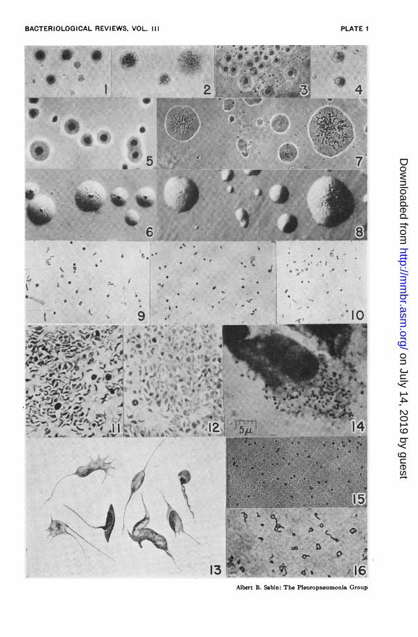
BACTERIOLOGICAL REVIEWS, VOL. III i
* * * - :0;~ 2 ;< j <q-0;tzP$ ;lwl
w~~~~~~~~~~~~~~~~~~kp
03V*S ' n 3 r j IljR;6-~~0-
.* , 4;19 S::.i$ +.' * S-.
jo 0
4 . S
9 |._3* K _- W Aio*;F,$* u@ .*8lp4 **$<*a4
~~~'-~'-wt,-'F Q
Al>~~(Ngt t Js st = *X - * W ^
_10"k, t *9 2i_;JW i.;.r ...$,;.
V~~~
16fa-
i16Albert B. Sabin: The Pleuropneumonia Group
)
PLATE 1
4.
.1
A-
I
..0
I (7':4:-7 14
on July 14, 2019 by guesthttp://m
mbr.asm
.org/D
ownloaded from