Embed Size (px)
Citation preview
J. Algal Biomass Utln. 2015, 6 (3): 47- 59 Planktonic Cyanoprokaryotes and Bacillariophytes of Indian Ramsar Site.
ISSN: 2229 – 6905 47
47
Planktonic Cyanoprokaryota and Bacillariophyta of East Kolkata Wetlands Ecosystem,
a Ramsar Site of India with reference to diversity and taxonomic study.
Anindita Singha Roy and Ruma Pal*
Phycology laboratory, Department of Botany, University of Calcutta, 35, Ballygunge Circular Road, Kolkata - 700019, West Bengal, India. *Corresponding author:[email protected]
Abstract:
The present paper deals with 53 phytoplankton taxa comprising of 34 species belonging to 15 different genera of Cyanoprokaryota and 19 species
grouped under 12 genera of Bacillariophyta, recorded from East Kolkata Wetlands, a Ramsar site. Investigation was carried out for a period of 2 years (January 2012 to January 2014). Emphasis was given on taxonomic identification of the recorded taxa including the scanning electron
microscopic (SEM) investigation. Dominant taxa recorded were Chroococcus dispersus, Synechocystis aquatilis, Merismopedia punctata, M.
glauca, Pseudoanabaena galeata, P. catenata, Anabaenopsis raciborskii, Navicula halophila, Nitzschia palea, Cyclotella striata whose further detailed morphology was studied with the help of SEM.
Key words: Bacillariophyta, Cyanoprokaryota,, East Kolkata Wetlands, Phytoplankton, SEM, Taxonomy
Introduction:
Wetlands are one of the most productive ecosystems on
Earth (Ghermandi et. al. 2008). As per Ramsar Convention,
26 of the total wetlands are designated as Ramsar sites,
among the total of 94 wetlands (natural and manmade)
recorded from India (Ramsar Convention, 2012), one of
which is East Kolkata Wetlands (EKW) of West Bengal.
Wetlands in India may vary from high altitude Himalayan
lakes, followed by wetlands in flood plains of river system,
coastal wetlands, area under paddy cultivation and so on.
On the basis of hydrological, ecological and geological
characteristic, wetlands can be classified as marine (coastal
wetlands), estuarine (including deltas, mangrove swamps),
lacustrine (lakes), riverine (rivers and streams) and
palustrine types (marshes, swamps and bogs) (Cowardin et
al. 1979). Following this classification, EKW our study site
can be grouped under lacustrine wetlands.
Wetlands are known to sustain a varied diversity of
both flora and fauna. Flora includes various forms that
range from microscopic planktonic algae to macrophytes.
In aquatic ecosystem, phytoplanktons play a major role in
maintaining the floral diversity as they are the basis of
aquatic food chain. Wetlands harbor diverse groups of
phytoplanktons, the simplest prokaryotic form being
Cyanoprokaryotes (formerly known as Blue Green Algae)
and many eukaryotic forms. One such eukaryotic form is
Bacillariophyta. Other groups are like Chlorophytes,
Chrysophytes, Euglenophytes, Desmids, etc. In our
previous study we have described the taxonomy and
diversity of planktonic chlorophytes (under
communication). This paper deals with diversity study of
Cyanoprokaryotes and members of Bacillariophyta.
The first Indian phycologist who took an initiative in
studying Cyanonoprokaryotes in India was Kirtikar (1886).
From then onwards began the extensive study of
Cyanoprokaryotes from different fresh, brakish and marine
water bodies, moist soil, and different paddy fields.
(Ramakrishnan and Kannan, 1992; Rao, 1998;
Venkataraman, 1975).Extensive study of Cyanoprokaryotes
and planktonic diatoms has been conducted in different
wetlands of India. Deep et al. (2013) enlisted 55 species of
Cyanoprokayotes, determining the diversity in wetlands of
Sambalpur, Orissa. Sasamal et al. (2005) recorded
planktonic diatom bloom formation in the coastal wetlands
of Orissa. Wetlands of Assam, which is also designated as
Ramsar Site of Bhramaputra river basin had been
thoroughly surveyed by Sharma (2015). He recorded 55
taxa of phytoplanktons, with Bacillariophyceae as
dominant and Cyanoprokaryotes as subdominant groups.
Reports from coastal wetlands in southern and western part
of India, also accounts for taxonomic identification
(Venkataraman, 1939; Subrahmanyan, 1946).
Venkataraman (1939) reported 98 forms of diatoms from
Madras coast. Subrahmanyan (1946) also gave a systematic
account of 171 forms of planktonic diatom from South
Indian coast. 17 different Cyanoprokaryotes were reported
by Sivakumar et al. (2012) from the Coastal wetlands of
Tamil Nadu.
Many reports of EKW include management
programmes to conserve this wetland which spans over the
entire eastern outskirts of a metropolitan city, Kolkata for
the human benefit; however, very few works dealt with the
taxonomy or diversity study of phytoplanktons (Pradhan et
al. 2008; Ray Chaudhuri et al. 2007, 2008; Kundu et al.
2008). For diversity and taxonomic study on phytoplankton
J. Algal Biomass Utln. 2015, 6 (3): 47- 59 Planktonic Cyanoprokaryotes and Bacillariophytes of Indian Ramsar Site.
ISSN: 2229 – 6905 48
48
population no report is available from the study area. The
present communication thus deals with the diversity and
taxonomic enumeration of planktonic members of
Cyanoprokaryota and Bacillariophycean members of East
Kolkata Wetlands.
Material and methods:
Study area
The East Kolkata Wetland (EKW) is a complex of natural
and human made wetland, situated at the eastern outskirts
of the metropolitan city Kolkata, India. 45.93 % of EKW
includes manmade water area (Kundu et al. 2008).This
pond (Captain Bhery) serves the dual purpose of recycling
sewage water of Kolkata metropolitan city and for fish
cultivation extensively. This wetland is declared as a
“Wetland of International Importance” by Government of
India and the Ramsar Convention had declared this wetland
as “Ramsar Site no. 1203” in 19th August 2002 (Ramsar
Secretariate, 2013). The geographic coordinates of EKW
was found to be 88°24.641′ east latitude and 22°33.115‟
north latitude as determined by GARMIN GPS map 76
CSx device. The study area covers an area of almost 450
m² having a depth of 3–4 ft. The sewage water includes
municipal waste, agricultural runoff and industrial effluents
of urban and semi urban areas. The sampling spots were
selected along the transects of the pond and samples were
collected from depth of 1.5-2 cm below surface water of
the wetlands. No epipelic or benthic flora was recorded.
Sampling and taxonomic identification
Phytoplankton sampling was carried out at a regular
interval of 15 days for 2 years from January 2012 to
January 2014. Different transects along the sampling spot
were chosen for the purpose of sampling. Phytoplankton
net of mesh size 20 µ was used for phytoplankton
sampling. Approximately 50L of water was passed through
the plankton net and ultimately concentrated to 300ml
containing huge plankton population. It was then preserved
in 4% formalin and brought back to the laboratory for
taxonomic identification with the help of standard keys
using monograph and relevant available literature viz.
Prescott (1982), Desikachary (1959), Komarek and
Anagnostidis (1999, 2005, 2013), Husted (1930). Samples
were viewed under low, high and oil immersion objective
of Carl- Zeiss Axiostar microscope and microphotographs
of these samples were taken using a digital camera (Canon
T2-T2 1,6x SLR 426115).
Scanning Electron Microscopic study
Phytoplankton samples were washed with saline phosphate
buffer (PBS) for 2-3 times and then centrifuged at 8000
rpm. A drop of washed material was taken on a glass cover
slip (Blue Star) and dried at 20°C. The samples were
repeatedly washed with different ethanol grade and dried at
room temperature. After complete dehydration the cover
slips were placed on carbon tape and put in Quorum (Q 150
TES) gold coater to coat the samples with gold. Images
have been taken at different magnification with the use of
Carl Zeiss EVO 18 (EDS 8100) microscope with Zeiss Inca
Penta FETX 3 (Oxford instruments) attachment.
Results:
A total of 53 taxa of Cyanoprokaryota and Bacillariophyta
along with their seasonal abundance have been recorded
(Table 1). Systematic enumeration of Cyanoprokaryotes
and Bacillariophycean members had been based on
Komarek and Anagnostidis classification (1999, 2005 and
2013) and Husted (1930) respectively. Out of 53 planktonic
taxa, 10 species were found to be dominant and can be
reffered to as major taxa. These are: Chroococcus
dispersus, Synechocystis aquatilis, Merismopedia punctata,
M. glauca, Pseudoanabaena galeata, P. catenata,
Anabaenopsis raciborskii, Nitzschia palea, Navicula
halophila, Cyclotella striata. SEM observation was carried
out for these 10 species for detailed study as distinct cell
wall morphology and constrictions on the wall are not
properly distinct in light microscopy. Taxonomic
enumerations of the identified taxa are as follows:
PHYLUM- CYANOPROKARYOTA-I
ORDER- CHROOCOCCALES
FAMILY- MERISMOPEDIACEAE
1. Merismopedia minima Beck (Fig. 1a)
(Geitler, 1932, p.129 c; Desikachary, 1959, pl. 29, fig. 6;
Komarek and Anagnostidis 1999, p. 175, fig. 222)
Cells pale blue-green, 4 to many in small colonies, 0.5-
0.6µm broad, free swimming, groups of four cells 2-3 ×
3µm.
2. Merismopedia punctata Meyen (Fig. 1b)
(Geitler, 1932, p. 129 c; Desikachary, 1959, pl. 23, fig. 5
and pl. 29, fig. 6; Komarek and Anagnostidis, 1999, p. 172,
fig. 222)
Colonies small, 4-64 cells, about 60 µm broad; cells not
closely packed, spherical or ovoid, 2.5-3.5µm broad, pale
blue-green.
SEM observation: Distinct cell wall morphology visible
(fig. 4e).
3. Merismopedia glauca (Ehrenberg) Naegeli (Fig. 1c)
(Desikachary, 1959, pl. 29, fig. 5; Komarek and
Anagnostidis, 1999, p. 172, fig. 222)
Colonies mostly small with 16-64 cells, 45-50 µm
diameter; cells spherical or oval, closely arranged, 3-6 µm
broad, pale blue-green.
SEM observation: Cell wall clearly visible. Smooth wall,
with distinct constriction between the adjacent cells (fig.
4d)
4. Merismopedia Trolleri Bachmann (Fig. 1d)
(Precott, 1982, pl. no. 101, fig. 5)
Colonies containing 8-16 cells, each with a distinct sheath,
cell contents with pseudovacuoles, appearing brownish or
J. Algal Biomass Utln. 2015, 6 (3): 47- 59 Planktonic Cyanoprokaryotes and Bacillariophytes of Indian Ramsar Site.
ISSN: 2229 – 6905 49
49
purplish because of light refraction; cells 2-3.5 µm
diameter.
5. Synechocystis aquatilis Sauvageau (Fig. 1o)
(Desikachary, 1959, pl. 25, fig. 9; Komarek and
Anagnostidis, 1999, p. 172, fig. 222)
Cells spherical, single or in twos, 4-6 µm broad, pale blue-
green.
SEM observation: Cell wall smooth. Constriction between
the cells distinctly visible (fig. 4a)
6. Coelosphaerium pallidum Lemmermann (Fig. 2e)
(Prescott, 1982, pl. 106, fig. 3; Komarek and Anagnostidis,
1999, p. 172, fig. 222)
An ovate colony of small, ovate cells crowded; cell
contents pale blue-green, without pseudovacuoles; cells 1-
2.5 µm in diameter, 2-3.5µm long.
FAMILY- SYNECHOCOCCACEAE
7. Synechococcus elongatus Naegeli (Fig. 1n)
(Geitler, 1932, p.273, fig. 133 a-c; Desikachary, 1959, pl.
25, figs. 7, 8; Komarek and Anagnostidis, 1999, p. 123, fig.
137)
Cells cylindrical, 1.4-2 µm broad, 1½-3 times as long as
broad, single or 2-4 cells together, contents homogeneous
and light blue-green.
8. Rhabdoderma irregulare (Naumann) Geitler (Fig. 1t)
(Prescott, 1982, pl. 103, figs. 9, 10)
Colony consisting of sigmoid, cylindrical cells irregularly
arranged within a copious, gelatinous envelope; cells 1-2
µm in diameter, 4.5-6 µm long.
9. Rhabdoderma lineare Schmidle and Lauterborn in
Schmidle (Fig. 2a)
(Prescott, 1982, pl. 103, figs. 11, 12)
Colony consisting of cylindrical, nearly straight cells,
colonial envelope transparent and wide; cells 1-2 µm in
diameter, 8-10 µm long; cell contents blue-green,
homogeneous.
10. Rhabdogloea rhaphidioides (R. Et. E Chodat)
Komarek (Fig. 2c)
Basionym- Dactylococcopsis rhaphidioides
(Geitler, 1932, p.281, fig.137; Desikachary, 1959, p. 158,
pl. 29, fig. 3; Prescott, 1982, pl. no. 105, figs. 13-15 all as
Dactylococcopsis rhaphidioides; Komarek and
Anagnostidis 1999, p. 105, fig. 108)
Cells elongate- fusiform, seldom straight, usually arcuate or
sigmoid, narrowed but not sharply pointed at the poles,
arranged in colonies of 2-8 within a hyaline, gelatinous
envelope; cells 1-3 µm in diameter, 5-20 µm long.
11. Rhabdogloea Smithii (Chodat and Chodat) Komarek
(Fig. 2d)
Basionym - Dactylococcopsis Smithii
(Prescott, 1982, pl. 105, figs. 3, 4 as Dactylococcopsis
Smithii; Komarek and Anagnostidis 1999, p. 105, fig.109)
Colony ovate or nearly broadly fusiform, containing 2-8
fusiform cells which are nearly straight or slightly arcuate;
on end of the cell pointed and the other bluntly rounded;
cells 3-4 µm in diameter, 10-15 µm long.
12. Rhabdogloea fascicularis (Lemmermann) Keshri and
Shikdar comb. nov. (Fig. 2g)
Basionym - Dactylococcopsis fascicularis
(Geitler, 1932, p. 283, fig. 138b; Desikachary 1959, p.158,
pl. 29, fig. 3; Prescott, 1982, pl. 105, figs. 10-12 all as
Dactylococcopsis fascicularis)
Colonies composed of 2-8 elongate, arcuate or spirally
sigmoid cells tapering to fine points at the poles, compactly
twisted, cells 1-1.5 µm in diameter, 10-15 µm long.
FAMILY- MICROCYSTACEAE
13. Microcystis aeruginosa Kuetzing (Fig. 2j)
(Prescott, 1982, pl. 101, fig. 5)
A free-floating or sedentary colony of numerous spherical
cells closely and irregularly arranged within copious
mucilage, forming ovate, globose, or irregularly shaped
masses; cell contents pale or bright blue-green.
FAMILY- CHROOCOCCACEAE
14. Chroococcus limneticus Lemmermann (Fig. 1j)
(Geitler, 1932, p. 234, fig. 113a; Desikachary, 1959, pl. 26,
fig. 2; Komarek and Anagnostidis 1999, p.209, fig. 382)
Colonies blue green, cells spherical or subspherical after
division, 2-20, free floating in a gelatinous layer, without
sheath cells 6-6.5 µm diameter, with sheath 8-14µ
diameter, sheath colourless; cells are blue-green.
15. Chroococcus dispersus (V. Keissler) Lemmermann
(Fig. 1k)
(Geitler, 1932, p. 233, fig. 113d; Desikachary, 1959, p.106;
Prescott, 1982, pl. 100, fig. 7, Komarek and Anagnostidis,
1999, p. 286, fig. 373)
Free-floating mucilaginous colony with 4-8 cells, either
solitary and then widely separated from each other or in
groups isolated from each other, blue-green, without sheath
3-4 µm broad, with sheath 5-6 µm broad.
SEM observation: Cell wall smooth with distinct
constrition between the two cells (fig. 4b)
16. Chroococcus dispersus var. minor G.M.Smith (Fig.
1l)
(Prescott, 1982, pl. 100, figs. 1-3)
A variety of Chroococcus dispersus having smaller cells,
1.5-2 µm in diameter, cells without sheath.
17. Chroococcus turgidus (Kuetzing) Naegeli (Fig. 1m)
(Desikachary, 1959, pl. 26, fig. 6)
J. Algal Biomass Utln. 2015, 6 (3): 47- 59 Planktonic Cyanoprokaryotes and Bacillariophytes of Indian Ramsar Site.
ISSN: 2229 – 6905 50
50
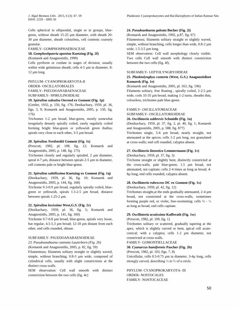
Cells spherical to ellipsoidal, single or in groups, blue-
green, without sheath 15-25 µm diameter, with sheath 20-
30 µm diameter, sheath colourless, cell contents coarsely
granular.
FAMILY- GOMPHOSPHAERIACEAE
18. Gomphoshpaeria aponina Kuetzing (Fig. 2f)
(Komarek and Anagnostidis, 1999)
Cells pyriform or cordate in stages of division, usually
within wide gelatinous sheath; cells 4-5 µm in diameter, 8-
12 µm long.
PHYLUM- CYANOPROKARYOTA-II
ORDER- OSCILLATORIALES
FAMILY- PSEUDOANABAENACEAE
SUBFAMILY- SPIRULINOIDEAE
19. Spirulina subsalsa Oersted ex Gomont (Fig. 1p)
(Geitler, 1932, p. 150, fig. 176; Desikachary, 1959, pl. 36,
figs. 3, 9; Komarek and Anagnostidis, 2005, p. 150, fig.
176)
Trichomes 1-2 µm broad, blue-green, mostly somewhat
irregularly densely spirally coiled, rarely regularly coiled
forming bright blue-green or yellowish green thallus;
spirals very close to each other, 3-5 µm broad.
20. Spirulina Nordstedtii Gomont (Fig. 1s)
(Prescott, 1982, pl. 108, fig. 12; Komarek and
Anagnostidis, 2005, p. 148, fig. 175)
Trichomes closely and regularly spiraled, 2 µm diameter,
spiral 4-7 µm, distance between spirals 2-5 µm in diameter,
cell contents pale or bright blue-green.
21. Spirulina subtilissima Kuetzing ex Gomont (Fig. 1q)
(Desikachary, 1959, pl. 36, fig. 10; Komarek and
Anagnostidis, 2005, p. 144, fig. 168)
Trichome 0.3-0.9 µm broad, regularly spirally coiled, blue-
green or yellowish, spirals 1.5-2.5 µm broad, distance
between spirals 1.25-2 µm.
22. Spirulina laxissima West,G.S. (Fig. 1r)
(Desikachary, 1959, pl. 36, fig. 5; Komarek and
Anagnostidis, 2005, p. 141, fig. 160)
Trichome 0.7-0.8 µm broad, blue-green, spirals very loose,
but regular, 4.5-5.3 µm broad; 12-18 µm distant from each
other, end cells rounded, obtuse.
SUBFAMILY- PSUEDOANABAENOIDEAE
23. Pseudoanabaena catenata Lauterborn (Fig. 2h)
(Komarek and Anagnostidis, 2005, p. 82, fig. 59)
Filamentous; filaments solitary straight or slightly waved,
simple, without branching, 0.8-3 µm wide, composed of
cylindrical cells, usually with slight constrictions at the
distinct cross-walls.
SEM observation: Cell wall smooth with distinct
constriction between the two cells (fig. 4c)
24. Pseudoanbaena galeata Bocher (Fig. 2i)
(Komarek and Anagnostidis, 1992, p.87, fig. 67)
Filamentous; filaments solitary straight or slightly waved,
simple, without branching, cells longer than wide, 0.8-2 µm
wide, 1.5-3.5 µm long.
SEM observation: Cell wall morphology clearly visible.
Two cells Cell wall smooth with distinct constriction
between the two cells (fig. 4f).
SUBFAMILY- LEPTOLYNGBYOIDEAE
25. Planktolyngbya contorta (West, G.S.) Anagnostidiset
Komarek (Fig. 1e)
(Komarek and Anagnostidis, 2005, pl. 163, fig. 196)
Filaments solitary, free floating , spirally coiled, 2-2.5 µm
wide, coils 33-35 µm broad, making 1-2 turns, sheaths thin,
colourless, trichomes pale blue-green.
FAMILY- OSCILLATORIACEAE
SUBFAMILY- OSCILLATORIOIDEAE
26. Oscillatoria subbrevis Schmidle (Fig. 1u)
(Desikachary, 1959, pl. 37, fig. 2, pl. 40, fig. 1; Komarek
and Anagnostidis, 2005, p. 588, fig. 877)
Trichomes single, 5-6 µm broad, nearly straight, not
attenuated at the apices; cells 1-2 µm long, not granulated
at cross-walls; end cell rounded, calyptra absent.
27. Oscillatoria limnetica Lemmermann (Fig. 1v)
(Desikachary, 1959, pl. 37, fig. 3)
Trichome straight or slightly bent, distinctly constricted at
the cross-walls, pale blue-green; 1.5 µm broad, not
attenuated, not capitate; cells 2-4 times as long as broad, 4-
8µ long; end cells rounded, calyptra absent.
28. Oscillatoria rubescens DC ex Gomont (Fig. 1x)
(Desikachary, 1959, pl. 42, fig. 12)
Trichomes straight,at the ends gradually attenuated, 2-4 µm
broad, not constristed at the cross-walls, sometimes
forming purple red, or violet, free-swimming; cells ½ - ⅓
as long as broad, end cells capitate.
29. Oscillatoria acutissima Kufferath (Fig. 1w)
(Prescott, 1982, pl. 109, fig. 1)
Trichomes solitary or scattered, gradually tapering at the
apex, which is slightly curved or bent, apical cell acute-
conical; with a calyptra; cells 1-2 µm diameter, not
constricted at cross-walls.
FAMILY- GOMONTIELLACEAE
30. Cyanarcus hamiformis Pascher (Fig. 2b)
(Prescott, 1982, pl. 103, figs. 7, 8)
Unicellular, cells 0.5-0.75 µm in diameter, 3-4µ long, cells
strongly curved, describing ½ to ⅔ of a circle.
PHYLUM- CYANOPROKARYOTA- III
ORDER- NOSTOCALES
FAMILY- NOSTOCACEAE
J. Algal Biomass Utln. 2015, 6 (3): 47- 59 Planktonic Cyanoprokaryotes and Bacillariophytes of Indian Ramsar Site.
ISSN: 2229 – 6905 51
51
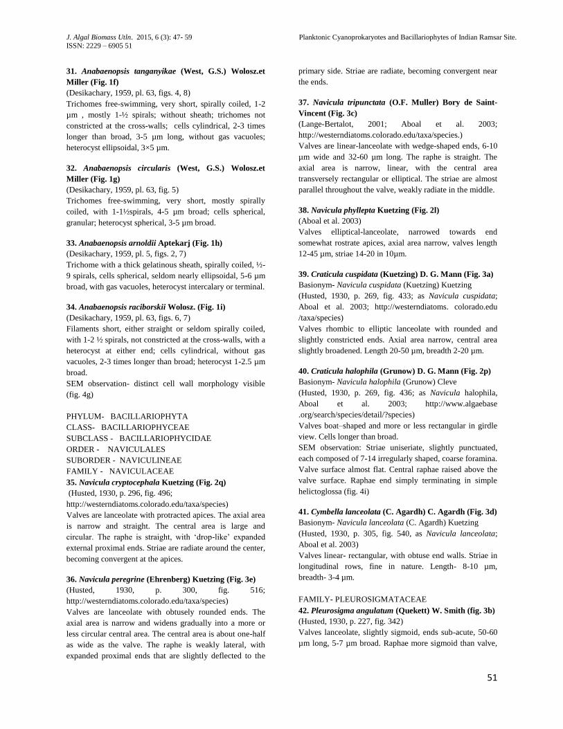
31. Anabaenopsis tanganyikae (West, G.S.) Wolosz.et
Miller (Fig. 1f)
(Desikachary, 1959, pl. 63, figs. 4, 8)
Trichomes free-swimming, very short, spirally coiled, 1-2
µm , mostly 1-½ spirals; without sheath; trichomes not
constricted at the cross-walls; cells cylindrical, 2-3 times
longer than broad, 3-5 µm long, without gas vacuoles;
heterocyst ellipsoidal, 3×5 µm.
32. Anabaenopsis circularis (West, G.S.) Wolosz.et
Miller (Fig. 1g)
(Desikachary, 1959, pl. 63, fig. 5)
Trichomes free-swimming, very short, mostly spirally
coiled, with 1-1½spirals, 4-5 µm broad; cells spherical,
granular; heterocyst spherical, 3-5 µm broad.
33. Anabaenopsis arnoldii Aptekarj (Fig. 1h)
(Desikachary, 1959, pl. 5, figs. 2, 7)
Trichome with a thick gelatinous sheath, spirally coiled, ½-
9 spirals, cells spherical, seldom nearly ellipsoidal, 5-6 µm
broad, with gas vacuoles, heterocyst intercalary or terminal.
34. Anabaenopsis raciborskii Wolosz. (Fig. 1i)
(Desikachary, 1959, pl. 63, figs. 6, 7)
Filaments short, either straight or seldom spirally coiled,
with 1-2 ½ spirals, not constricted at the cross-walls, with a
heterocyst at either end; cells cylindrical, without gas
vacuoles, 2-3 times longer than broad; heterocyst 1-2.5 µm
broad.
SEM observation- distinct cell wall morphology visible
(fig. 4g)
PHYLUM- BACILLARIOPHYTA
CLASS- BACILLARIOPHYCEAE
SUBCLASS - BACILLARIOPHYCIDAE
ORDER - NAVICULALES
SUBORDER - NAVICULINEAE
FAMILY - NAVICULACEAE
35. Navicula cryptocephala Kuetzing (Fig. 2q)
(Husted, 1930, p. 296, fig. 496;
http://westerndiatoms.colorado.edu/taxa/species)
Valves are lanceolate with protracted apices. The axial area
is narrow and straight. The central area is large and
circular. The raphe is straight, with „drop-like‟ expanded
external proximal ends. Striae are radiate around the center,
becoming convergent at the apices.
36. Navicula peregrine (Ehrenberg) Kuetzing (Fig. 3e)
(Husted, 1930, p. 300, fig. 516;
http://westerndiatoms.colorado.edu/taxa/species)
Valves are lanceolate with obtusely rounded ends. The
axial area is narrow and widens gradually into a more or
less circular central area. The central area is about one-half
as wide as the valve. The raphe is weakly lateral, with
expanded proximal ends that are slightly deflected to the
primary side. Striae are radiate, becoming convergent near
the ends.
37. Navicula tripunctata (O.F. Muller) Bory de Saint-
Vincent (Fig. 3c)
(Lange-Bertalot, 2001; Aboal et al. 2003;
http://westerndiatoms.colorado.edu/taxa/species.)
Valves are linear-lanceolate with wedge-shaped ends, 6-10
µm wide and 32-60 µm long. The raphe is straight. The
axial area is narrow, linear, with the central area
transversely rectangular or elliptical. The striae are almost
parallel throughout the valve, weakly radiate in the middle.
38. Navicula phyllepta Kuetzing (Fig. 2l)
(Aboal et al. 2003)
Valves elliptical-lanceolate, narrowed towards end
somewhat rostrate apices, axial area narrow, valves length
12-45 µm, striae 14-20 in 10µm.
39. Craticula cuspidata (Kuetzing) D. G. Mann (Fig. 3a)
Basionym- Navicula cuspidata (Kuetzing) Kuetzing
(Husted, 1930, p. 269, fig. 433; as Navicula cuspidata;
Aboal et al. 2003; http://westerndiatoms. colorado.edu
/taxa/species)
Valves rhombic to elliptic lanceolate with rounded and
slightly constricted ends. Axial area narrow, central area
slightly broadened. Length 20-50 µm, breadth 2-20 µm.
40. Craticula halophila (Grunow) D. G. Mann (Fig. 2p)
Basionym- Navicula halophila (Grunow) Cleve
(Husted, 1930, p. 269, fig. 436; as Navicula halophila,
Aboal et al. 2003; http://www.algaebase
.org/search/species/detail/?species)
Valves boat–shaped and more or less rectangular in girdle
view. Cells longer than broad.
SEM observation: Striae uniseriate, slightly punctuated,
each composed of 7-14 irregularly shaped, coarse foramina.
Valve surface almost flat. Central raphae raised above the
valve surface. Raphae end simply terminating in simple
helictoglossa (fig. 4i)
41. Cymbella lanceolata (C. Agardh) C. Agardh (Fig. 3d)
Basionym- Navicula lanceolata (C. Agardh) Kuetzing
(Husted, 1930, p. 305, fig. 540, as Navicula lanceolata;
Aboal et al. 2003)
Valves linear- rectangular, with obtuse end walls. Striae in
longitudinal rows, fine in nature. Length- 8-10 µm,
breadth- 3-4 µm.
FAMILY- PLEUROSIGMATACEAE
42. Pleurosigma angulatum (Quekett) W. Smith (fig. 3b)
(Husted, 1930, p. 227, fig. 342)
Valves lanceolate, slightly sigmoid, ends sub-acute, 50-60
µm long, 5-7 µm broad. Raphae more sigmoid than valve,
J. Algal Biomass Utln. 2015, 6 (3): 47- 59 Planktonic Cyanoprokaryotes and Bacillariophytes of Indian Ramsar Site.
ISSN: 2229 – 6905 52
52
excentric near the ends. Transverse and oblique striae,
equidistant.
ORDER - THALASSIOPHYSALES
FAMILY - CATENULACEAE
43. Amphora coffeaiformis (C. Agardh) Kuetzing (Fig.
3f)
(Husted, 1930, p. 344, fig. 634)
Frustules in girdle view elliptic lanceolate, truncate. Valves
arcuate on the dorsal margin and straight or slightly
concave on the ventral margin. Ends of the valves slightly
protracted and capitates. Striae delicate. Length 8-10 µm,
breadth- 3-4 µm.
ORDER- MASTOGLOIALES
FAMILY- ACHNANTHACEAE
44. Achnanthes sp. Bory de Saint-Vincent (Fig. 3g)
(Husted, 1930, p. 200, fig. 273-286; Scott and Thomas,
2005)
The frustules are heterovalvar. The raphae valve usually
posses a central area of thickened silica, called fascia or
staurus. The striae are uni-, bi- or triseriate and composed
of areolae covered by complex sieve plates.
ORDER- BACILLARIALES
FAMILY- BACILLARIACEAE
45. Nitzschia acicularis (Kuetzing) W. Smith (Fig. 3l)
(Husted, 1930, p.423, fig. 821; Aboal et al. 2003;
http://westerndiatoms.colorado.edu/taxa/species)
The central part of the valve has nearly parallel sides and a
sharp tapering. The extended distal portions of the valve are
tapered towards fine apices. The striae are not visible with
light microscopy.
46. Nitzschia frustulum (Kuetzing) Grunow (Fig. 2q)
(Husted, 1930, p. 414, fig. 795; Aboal et al. 2003)
Frustules isopolar, bilaterally symmetrical. Cells lie in
valve or girdle view and isolated valves always in valve
view. Valves bilaterally symmetrical, linear to lanceolate,
with sub-rostrate poles. Striae clearly visible under light
microscope. Length 5-50µm, width 3-5µm.
47. Nitzschia palea (Kuetzing) W. Smith (Fig. 3i)
(Opute, 1974; Husted, 1930, p. 414, fig. 801)
Valves linear to lanceolate with short wedge-shaped
tapering ends. Length 40-50µm, breadth 3-5µm.
SEM observation: Valve margins symmetric, convex on
both sides. Central part of the cell showing large central
interspace, eccentric raphae divided by central nodule-
fibulae regularly spaced (fig. 4h)
48. Nitzschia fruticosa (Fig. 3h)
(Husted, 1930 ; Aboal et al, 2003)
More than one valves giving a rosette shaped appearance.
Valves linear to lanceolate with short wedge-shaped
tapering ends. Length 20-30 µm, breadth 2-5 µm.
49. Pseudonitzschia sp. Peragallo (Fig. 2o)
(Husted, 1930)
Cells are narrow and fusiform or boat-shaped, united in
stepped chains with overlapping valve ends. Chains are
motile. Cells are yellow brown in colour. The valve face is
covered with slits and pores. The raphe is off-centric and
not raised above the valve.
CLASS - MEDIOPHYCEAE
SUBCLASS - THALASSIOSIROPHYCIDAE
ORDER - LEPTOCYLINDRALES
FAMILY- LEPTOCYLINDRACEAE
50. Leptocylindrus danicus Cleve (Fig. 3m)
(Scott and Thomas, 2005;
http://westerndiatoms.colorado.edu/taxa/species)
The weakly silicified cells are cylindrical and occur in
chains. Cell length (pervalvar axis) is normally 3-5 times
the cell width (diameter). Adjacent cells are closely
abutted. The valve of one cell is slightly convex, while the
adjacent valve is slightly concave.
51. Aulacoseira granulata (Ehrenberg) Simonsen (Fig.
2k)
(Hauk, 2003, p. 20, pl. 25, figs. 1-10, pl. 26, figs. 1-4)
Basionym- Gaillonella granulata Ehrenberg 1843
Frustules are cylindrical, join face-to-face and form
filamentous colonies. Valves are 4-17 µm in diameter, with
a mantle height of 4-20 µm. The ratio of the mantle height
to valve diameter is usually greater than 0.8 but less than 5.
The mantle has straight sides and the valve face is flat. The
mantle areolae are square. Linking spines are located at the
end of each pervalvar costa. Linking spines are short,
triangular or bifurcated.
ORDER- THALASSIOSIRALES
FAMILY- THALASSIOSIRACEAE
52. Thalassiosira weissflogii (Grunow) Fryxell and Hasle
(Fig. 3j,k)
(http://westerndiatoms.colorado.edu/taxa/species)
Basionym- Micropodiscus weissflogii Grunow in Van
Heurck 1880
Valves are round, flat, with short mantles. The frustules are
relatively lightly silicified. Areolae are fine and details of
their structure are not visible with the light microscope A
single, prominent rimoportula is present on the margin of
the valve.
FAMILY- STEPHANODISCACEAE
53. Cyclotella striata (Kuetzing) Grunow (Fig. 2m)
(Husted 1930, p. 99, fig. 71; Heft 10, p. 100, fig. 67)
J. Algal Biomass Utln. 2015, 6 (3): 47- 59 Planktonic Cyanoprokaryotes and Bacillariophytes of Indian Ramsar Site.
ISSN: 2229 – 6905 53
53
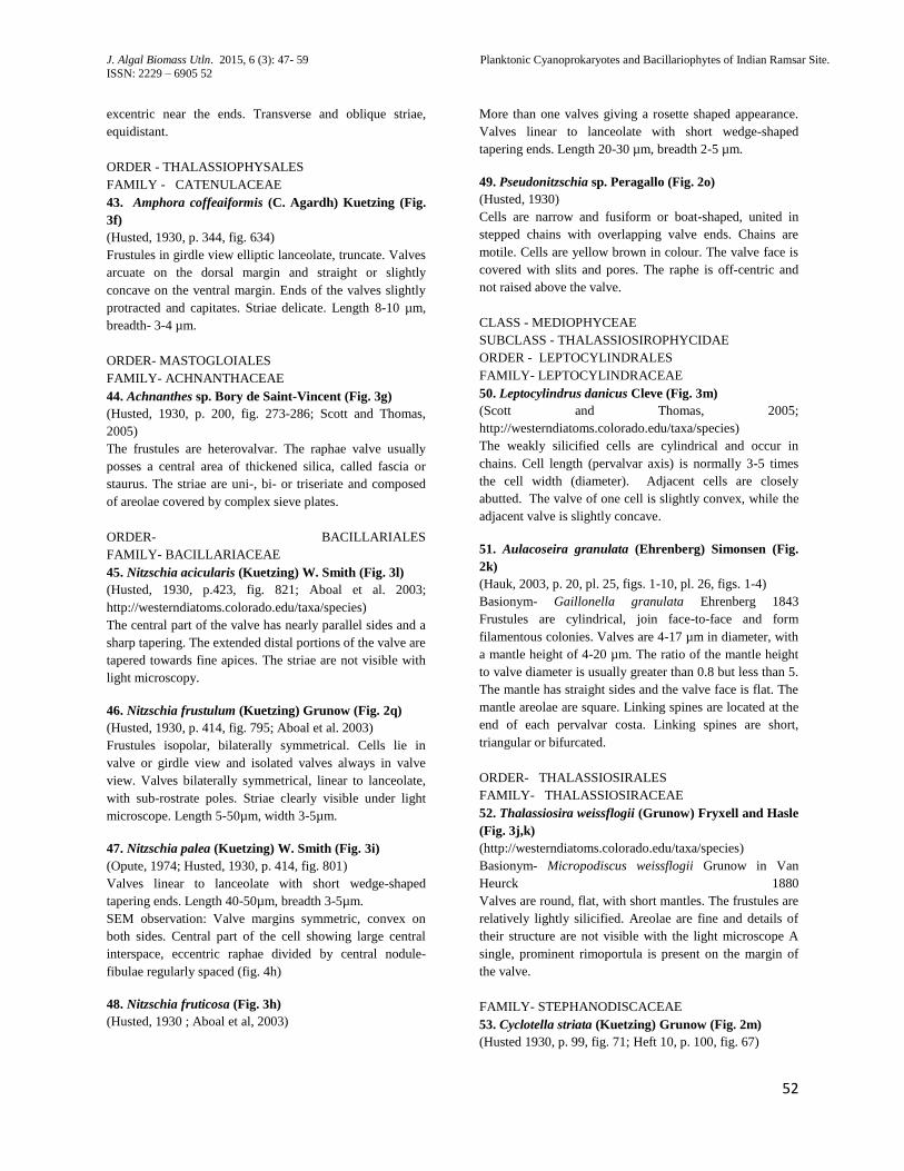
Cells disc- shaped, 6-18 µm in diameter. Valves with more
or less broad, evenly striated border, striae 10-12 in 10 µm.
central portion with pflexesand coarsely punctuate.
SEM observation- The marginal zone consists of ridges
separated by furrows. The shape of ridges is rectangular.
Areolae perforate the cell wall in the marginal zone.
Grabuels cover the entire outer valve surface and
sometimes form dendritic silica structure. Spines present at
the end of furrows on the valve face. They are conical (fig.
4j, k)
Discussion:
East Kolkata Wetlands of West Bengal, India, contributes
dual roles in sewage treatment which largely depends on
phytoplankton population, the main contributor in primary
production in one hand and also helps in cleaning the
pollutants (Pradhan et. al., 2008). One of the important
aspect of this particular wetland is that, unlike others viz.
Delta Marsh, bogs, fens etc. it showed no benthic algal
growth but only phytoplanktons, “the free wanderers”.
Therefore floristic study of phytoplankton population is
important in ecological point of view also. This wetlands is
also economically important for fish production, where
phytoplanktons are directly consumed by fishes. Similar
type of studies on phytoplankton diversity of coastal West
Bengal revealed that the seasonal variation and salinity
level control the phytoplankton abundance (Choudhury and
Pal, 2010, 2011, 2012).
Total 53 taxa comprising of both Cyanoprokaryota and
Bacillariophyta have been reported in this communication
and other 61 taxa from Chlorophyta have already been
communicated (data unpublished). Therefore total 114
phytoplankton taxa have been reported from 2 years‟
regular samplings. From the investigation it became clear
that the phytoplankton community of this wetland is
dominated by Chlorophyte population throughout the year
and the Cyanoprokaryota was found to be the second
dominant group showing maximum abundance in winter
and least abundance during summer. However, members of
Chroococcus sp., Merismopedia sp., Synechococcus sp. and
Spirulina sp. flourished throughout the year with maximum
abundance in winter mainly. Whereas Bacillariophycean
members were recorded throughout the year but their
population size is much smaller than that of
Cyanoprokaryota and Chlorophyta.
Acknowledgement:
We would like to thank University Grant Commission
(UGC), New Delhi, India for their financial assistance. We
would also like to thank Department of Botany and Center
for Research in Nanoscience and Nanotechnology (CRNN)
of University of Calcutta for instrumental facilities.
Reference:
Aboal, M., Alvarez Cobelas, M., Cambra, J., Ector, L.,
2003. Floristic list of non-marine diatoms
(Bacillariophyceae) of Iberian Peninsula, Balearic Islands
and Canary Islands. Updated taxonomy and bibliography.
Diatom Monographs 4: 1-639.
Cowardin, L.M., Carter, V., Golet, F.C., LaRoe, E.T.,
1979. Classification of Wetlands and Deepwater Habitats
of the United States.U.S. Department of the Interior, Fish
and Wildlife Service, Washington, DC.
Choudhury, A.K., Pal. R., 2010. Phytoplankton and
nutrient dynamics of shallow coastal stations at Bay of
Bengal, Eastern Indian Coast, Aquatic Ecology (Kluwer
Acad. Publ.), 44: 55-71.
Choudhury, A.K., Pal, R., 2010. Variations in seasonal
phytoplankton assemblages as a response to Environmental
Changes in the surface waters of a hypo saline coastal
station along the Bhagirathi Hooghly
estuary. Environmental Monitoring and Assessment 179:
531-553.
Choudhury, A.K., Pal, R., 2012. Understanding the
seasonal dynamics of primary productivity in relation to
phytoplankton populations from the Bhagirathi – Hooghly
estuary, eastern Indian coast. Journal of Algal Biomass
Utilization. 3(4): 80–88.
Deep, P.R., Bhattacharya, S., Nayek, B., 2013.
Cyanobacteria in wetlands of industrialized Sambalpur
District of India. Aquatic Biosystems 9: 14.
Desikachary, T.V., 1959. Cyanophyta. In: ICAR
Monographs on Algae, New Delhi.
Geitler, L., 1932. Cyanophyceae. In Rabenhorst‟s
Krytogamenflora 14: 1-1196, Akad. Verlagsges., Leipzig.
Ghermandi, A., Van den Bergh, J.C.J.M., Brander, L.M.,
Nunes, P.A.L.D., 2008. The Economic Value of Wetland
Conservation and Creation: A Meta-Analysis. [Working
Paper 79]. Fondazione Eni Enrico Mattei, Milan, Italy.
Hauk, V., 2003. Atlas of Freshwater Diatoms with brief
key and description. Part-I. Czech Phycology Supplement.
1: 26.
http://westerndiatoms.colorado.edu/taxa/species
Hustedt, F., 1930. Heft 10: Bacillariophyta (Diatomeae)
Zweite Auflage. In: Die Süsswasser-Flora Mitteleuropas.
(Pascher, A. Eds), pp. [i]-vii, [1]-466. Jena: Verlag von
Gustav Fischer.
Kirtikar, 1886. A new species of algae Conferva Thermalis
Birdwood. Journal of the Bombay Natural History Society
1:135-138.
J. Algal Biomass Utln. 2015, 6 (3): 47- 59 Planktonic Cyanoprokaryotes and Bacillariophytes of Indian Ramsar Site.
ISSN: 2229 – 6905 54
54
Komárek, J., 2013. Cyanoprokaryota. III. Heterocytous
genera. – In: Büdel B., Gärtner G., Krienitz L. & Schagerl
M. (eds), Süswasserflora von Mitteleuropa/Freshwater flora
of Central Europe, p. 1130, Springer Spektrum Berlin,
Heidelberg.
Komárek, J., Anagnostidis, K., 1999. Cyanoprokaryota. I.
Chroococcales. In: Süßwasserflora von Mitteleuropa.
Begründet von A. Pascher. Band 19/1. (Ettl, H., Gärtner,
G., Heynig, H. & Mollenhauer, D. Eds), pp. 1-548.
Heidelberg & Berlin: Spektrum, Akademischer Verlag.
Komárek, J., Anagnostidis, K., 2005. Cyanoprokaryota. II.
Teil: Oscillatoriales. – In: Büdel B., Gärdner G., Krienitz L.
& Schagerl M. (eds):Süsswasserflora von Mitteleuropa
19(2): 759 , Elsevier, München.
Kundu, N., Pal, M., Saha, S., 2008. East Kolkata Wetlands:
A resource recovery system through productive system.
Proceedings of Taal 2007: The 12th World Lake
Conference, 868-881.
Lange-Bertalot, H., 2001. Navicula sensu stricto. 10 Genera
separated from Navicula sensu lato. Frustulia. Diatoms of
Europe: Diatoms of the European inland waters and
comparable habitats. 2: 1-526.
Opute, F.I., 1974. Studies on fat accumulation in Nitzschia
palea Kütz. Annals of Botany 38: 889-902.
Pradhan, A., Bhaumik, P., Das, S., Mishra, M., Khanam, S.,
Hoque, B.A., Mukherjee, I., Thakur, A.R., Ray Chaudhuri,
S., 2008. Phytoplankton diversity as indicator of water
quality for fish cultivation. American Journal of
Environmental Science, 4: 406-411.
Ramakrishnan, N., Kannan, L., 1992. Blue green algal flora
of Muthupet, Tamil Nadu. Phykos. 31: 169 –171.
Ramsar Convention on Wetlands., 2012. The Annotated
Ramsar List: India. [Briefing Note]. The Secretariat of
theConvention on Wetlands, Gland, Switzerland.
Ramsar Secretariat., 2013. The List of Wetlands of
International Importance. The Secretariat of the Convention
on Wetlands, Gland, Switzerland.
Rao, C.B., 1998. The Myxophyceae of Madras Presidency,
India- 1. J. Indian Bot. Soc. (Madras) 17: 81-96.
Ray Chaudhuri, S., Mishra, M., Nandy, P., Thakur, A.R.,
2008. Waste management: A case study of ongoing
traditional practices at east Calcutta wetland. American
Journal of Agricultiural and Biological Sciences 3: 315-
320.
Raychaudhuri, S., Salodkar, S., Sudarshan, M., Thakur, A.,
2007. Integrated Resource Recovery at East Calcutta
Wetland: How Safe is these? American Journal of
Agricultural and Biological Science 2 (2): 75-80.
Sasamal, S.K., Panigrahy, R.C., Misra, S., 2005.
Asterionella bloom in the north western Bay of Bengal
during 2004. Int J of Remote Sens 26(17): 3853–3858
Scott, F.J., Thomas, D.P., 2005. Diatoms. In: Antarctic
marine protists. (Scott, F.J. & Marchant, H.J. Eds), pp. 13-
201. Canberra & Hobart: Australian Biological Resources
Study; Australian Antarctic Division.
Sharma, B.K., 2015. Phytoplankton diversity of Deepor
Beel- a Ramsar site in the floodplain of Bhramaputra River
basin, Assam, north-east India. Indian J. Fish. 62(1): 33-34.
Sivakumar, N., Viji, V., Satheesh, S., Varalakshmi, P.,
Ashokkumar, B., Pandi, M., 2012. Cyanobacterial
abundance and diversity in coastal wetlands of
Kanyakumari District, Tamil Nadu (India). African Journal
of Microbiology Research Vol. 6(20): 4409-4416
Subrahmanyan, R., 1946. A systematic account of the
marine plankton diatoms of the Madras coast. Proc Indian
Acad Sci 24B: 85–197.
Venkataraman, G., 1939. A systematic account of some
south Indian diatoms. Proc Indian Acad Sci 10: 293–368.
Venkataraman, G.S., 1975. The role of blue-green algae in
tropical rice cultivation, in-Nitrogen fixation by free-living
micro- organisms, pp. 207-218. Stewart, W.D.P. (ed.),
Cambridge Univ. Press.
J. Algal Biomass Utln. 2015, 6 (3): 47- 59 Planktonic Cyanoprokaryotes and Bacillariophytes of Indian Ramsar Site.
ISSN: 2229 – 6905 55
55
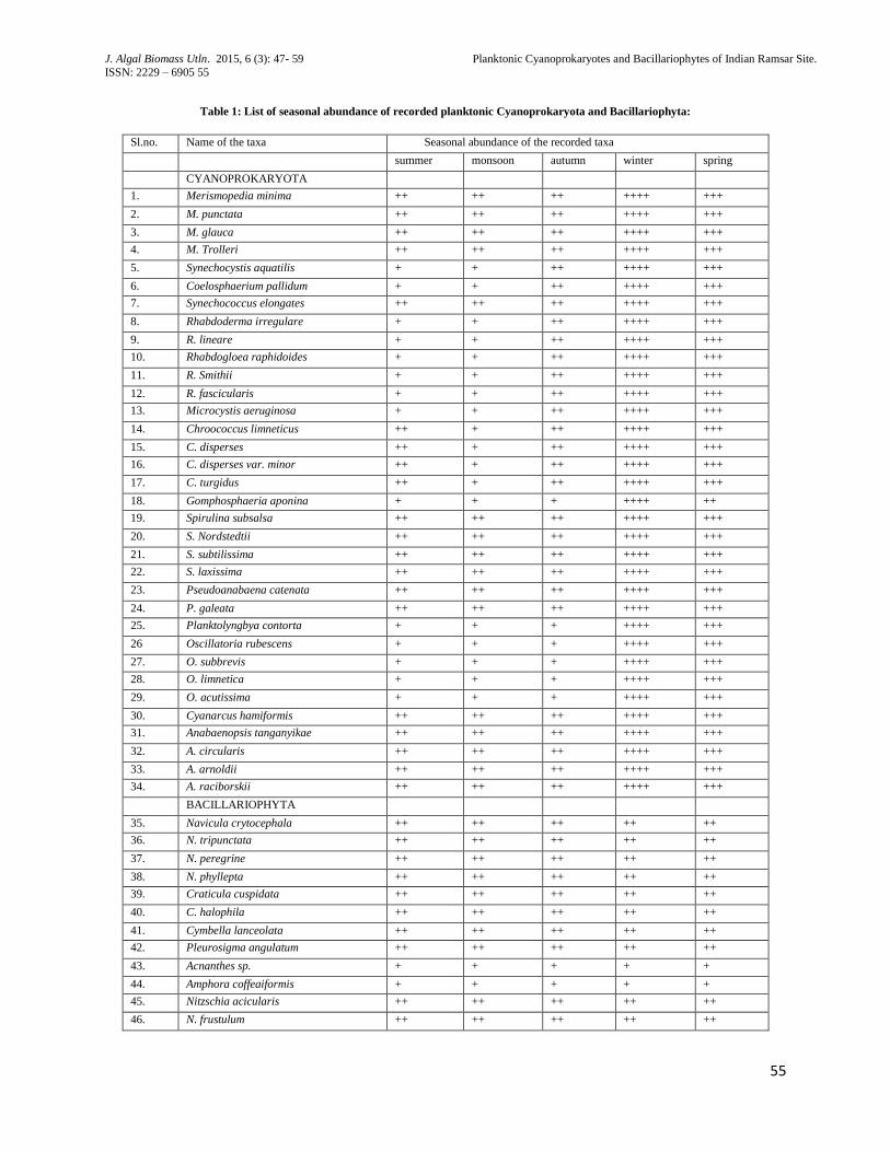
Table 1: List of seasonal abundance of recorded planktonic Cyanoprokaryota and Bacillariophyta:
Sl.no. Name of the taxa Seasonal abundance of the recorded taxa
summer monsoon autumn winter spring
CYANOPROKARYOTA
1. Merismopedia minima ++ ++ ++ ++++ +++
2. M. punctata ++ ++ ++ ++++ +++
3. M. glauca ++ ++ ++ ++++ +++
4. M. Trolleri ++ ++ ++ ++++ +++
5. Synechocystis aquatilis + + ++ ++++ +++
6. Coelosphaerium pallidum + + ++ ++++ +++
7. Synechococcus elongates ++ ++ ++ ++++ +++
8. Rhabdoderma irregulare + + ++ ++++ +++
9. R. lineare + + ++ ++++ +++
10. Rhabdogloea raphidoides + + ++ ++++ +++
11. R. Smithii + + ++ ++++ +++
12. R. fascicularis + + ++ ++++ +++
13. Microcystis aeruginosa + + ++ ++++ +++
14. Chroococcus limneticus ++ + ++ ++++ +++
15. C. disperses ++ + ++ ++++ +++
16. C. disperses var. minor ++ + ++ ++++ +++
17. C. turgidus ++ + ++ ++++ +++
18. Gomphosphaeria aponina + + + ++++ ++
19. Spirulina subsalsa ++ ++ ++ ++++ +++
20. S. Nordstedtii ++ ++ ++ ++++ +++
21. S. subtilissima ++ ++ ++ ++++ +++
22. S. laxissima ++ ++ ++ ++++ +++
23. Pseudoanabaena catenata ++ ++ ++ ++++ +++
24. P. galeata ++ ++ ++ ++++ +++
25. Planktolyngbya contorta + + + ++++ +++
26 Oscillatoria rubescens + + + ++++ +++
27. O. subbrevis + + + ++++ +++
28. O. limnetica + + + ++++ +++
29. O. acutissima + + + ++++ +++
30. Cyanarcus hamiformis ++ ++ ++ ++++ +++
31. Anabaenopsis tanganyikae ++ ++ ++ ++++ +++
32. A. circularis ++ ++ ++ ++++ +++
33. A. arnoldii ++ ++ ++ ++++ +++
34. A. raciborskii ++ ++ ++ ++++ +++
BACILLARIOPHYTA
35. Navicula crytocephala ++ ++ ++ ++ ++
36. N. tripunctata ++ ++ ++ ++ ++
37. N. peregrine ++ ++ ++ ++ ++
38. N. phyllepta ++ ++ ++ ++ ++
39. Craticula cuspidata ++ ++ ++ ++ ++
40. C. halophila ++ ++ ++ ++ ++
41. Cymbella lanceolata ++ ++ ++ ++ ++
42. Pleurosigma angulatum ++ ++ ++ ++ ++
43. Acnanthes sp. + + + + +
44. Amphora coffeaiformis + + + + +
45. Nitzschia acicularis ++ ++ ++ ++ ++
46. N. frustulum ++ ++ ++ ++ ++
J. Algal Biomass Utln. 2015, 6 (3): 47- 59 Planktonic Cyanoprokaryotes and Bacillariophytes of Indian Ramsar Site.
ISSN: 2229 – 6905 56
56
47. N. palea ++ ++ ++ ++ ++
48. N. fruticosa ++ ++ ++ ++ ++
49. Leptocylindrus danicus + + + + +
50. Aulacoseira granulata + + + + +
51. Thalassiosira weissflogii + + + + +
52. Cyclotella striata + + + + +
53. Pseudonitzschia sp. + + + + +
(+ - least abundant, ++ - less abundant, +++ - abundant, ++++ - Highly abundant)
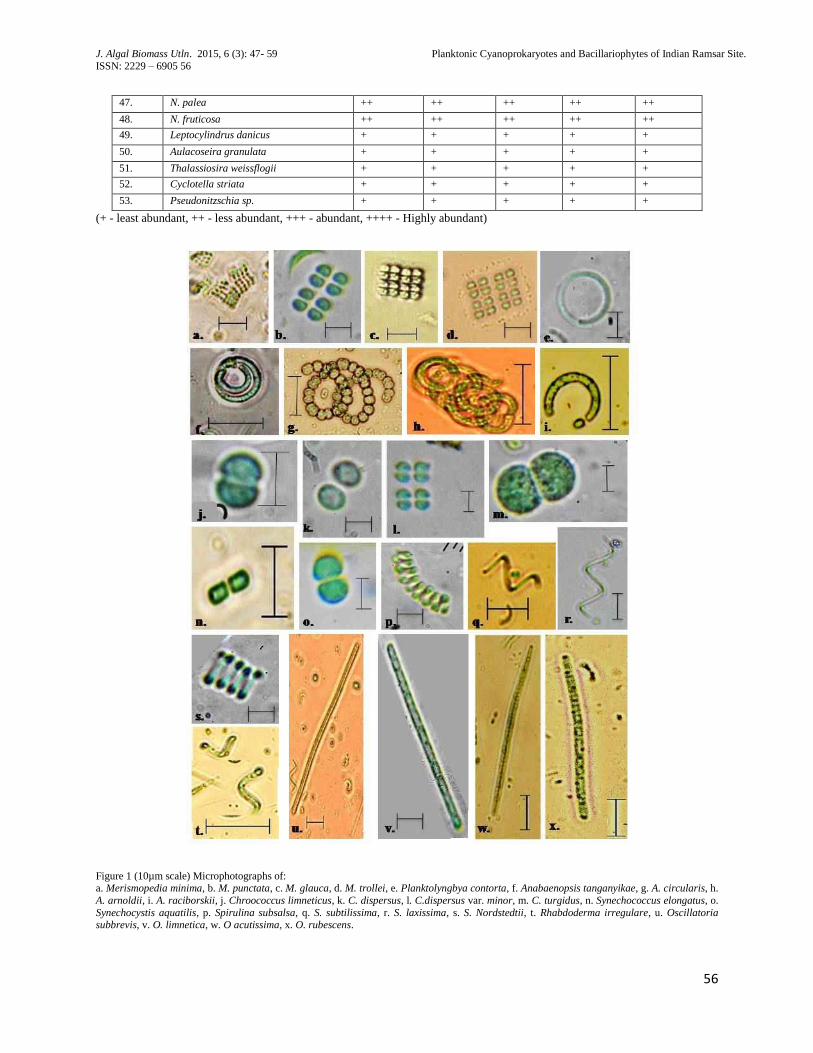
Figure 1 (10µm scale) Microphotographs of: a. Merismopedia minima, b. M. punctata, c. M. glauca, d. M. trollei, e. Planktolyngbya contorta, f. Anabaenopsis tanganyikae, g. A. circularis, h.
A. arnoldii, i. A. raciborskii, j. Chroococcus limneticus, k. C. dispersus, l. C.dispersus var. minor, m. C. turgidus, n. Synechococcus elongatus, o.
Synechocystis aquatilis, p. Spirulina subsalsa, q. S. subtilissima, r. S. laxissima, s. S. Nordstedtii, t. Rhabdoderma irregulare, u. Oscillatoria subbrevis, v. O. limnetica, w. O acutissima, x. O. rubescens.
J. Algal Biomass Utln. 2015, 6 (3): 47- 59 Planktonic Cyanoprokaryotes and Bacillariophytes of Indian Ramsar Site.
ISSN: 2229 – 6905 57
57
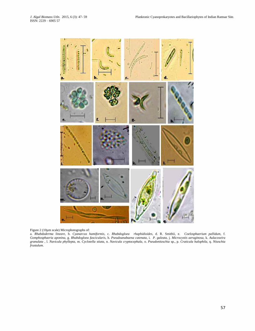
Figure 2 (10µm scale) Microphotographs of: a. Rhabdoderma lineare, b. Cyanarcus hamiformis, c. Rhabdogloea rhaphidioides, d. R. Smithii, e. Coelosphaerium pallidum, f.
Gomphosphaeria aponina, g. Rhabdogloea fascicularis, h. Pseudoanabaena catenata, i. P. galeata, j. Microcystis aeruginosa, k. Aulacoseira
granulata , l. Navicula phyllepta, m. Cyclotella stiata, n. Navicula cryptocephala, o. Pseudonitzschia sp., p. Craticula halophila, q. Nitzschia
frustulum.
J. Algal Biomass Utln. 2015, 6 (3): 47- 59 Planktonic Cyanoprokaryotes and Bacillariophytes of Indian Ramsar Site.
ISSN: 2229 – 6905 58
58
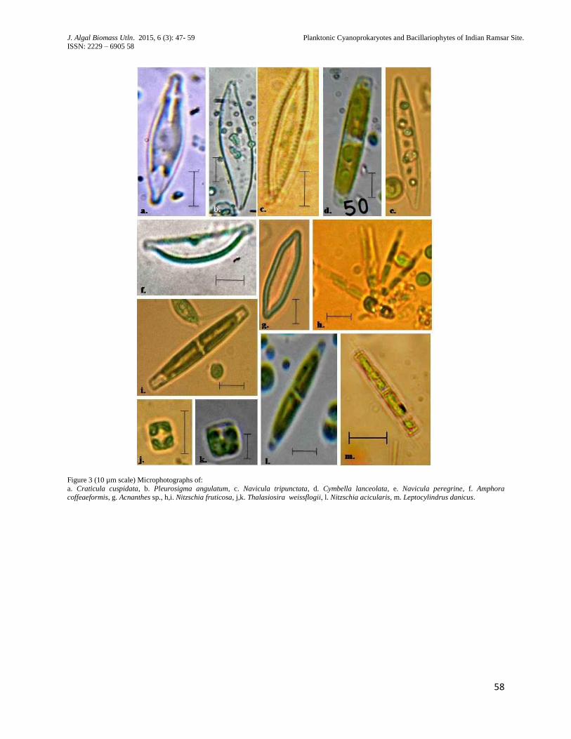
Figure 3 (10 µm scale) Microphotographs of:
a. Craticula cuspidata, b. Pleurosigma angulatum, c. Navicula tripunctata, d. Cymbella lanceolata, e. Navicula peregrine, f. Amphora
coffeaeformis, g. Acnanthes sp., h,i. Nitzschia fruticosa, j,k. Thalasiosira weissflogii, l. Nitzschia acicularis, m. Leptocylindrus danicus.
J. Algal Biomass Utln. 2015, 6 (3): 47- 59 Planktonic Cyanoprokaryotes and Bacillariophytes of Indian Ramsar Site.
ISSN: 2229 – 6905 59
59
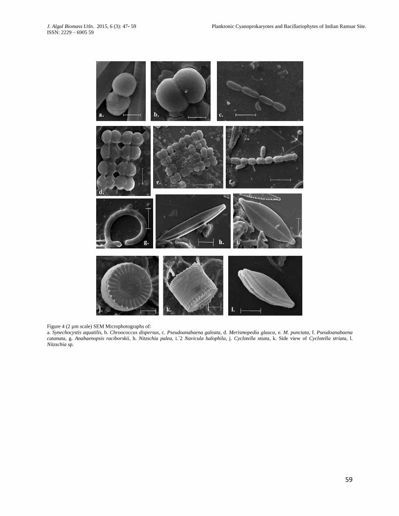
Figure 4 (2 µm scale) SEM Microphotographs of:
a. Synechocystis aquatilis, b. Chroococcus dispersus, c. Pseudoanabaena galeata, d. Merismopedia glauca, e. M. punctata, f. Pseudoanabaena catanata, g. Anabaenopsis raciborskii, h. Nitzschia palea, i.`2 Navicula halophila, j. Cyclotella stiata, k. Side view of Cyclotella striata, l.
Nitzschia sp.