1
GFAP Reveals an abundance of reactive astrocytes. Pick’s Disease Alzheimer’s Disease CSAD The Pick’s brain exhibited far less plaque staining compared to the AD brain. 4X 4X 20X 20X 20X 10X A-beta 1-42 Very little amyloid staining in the Pick’s brain while the Alzheimer’s brain shows an abundance of amyloid staining. 4X 4X 4X 4X Silver Protein (modified Bodian’s Stain Protocol) Pick’s brain shows few Pick’s bodies and neurofibrillary tangles while the Alzheimer’s brain pathology reveals an abundance of these features. Thioflavin-S Far less amyloid plaque staining in the Pick’s versus Alzheimer’s. 4X 4X AT8 The Picks brain shows a significant amount of neurofibrillary tangles as well as hyperphosphorylated tau proteins in the frontal cortex, however, not as much as the AD brain to the right. Ubiquitin stains positive in FTLD-TDP brain. Gallyas The PiD brain reveals some cells with tangles in the frontal cortex, but not as much as the AD brain to the right. 20X 20X 10X 10X H&E Amino CuAg (deOlmos) 10X 10X Lateral view of a normal brain. The brain has normal gross pathology. Photo taken from: http://www.massgeneral.org/radiology/news/radrounds/april_2006/ (for demonstration purpose only). MRI images of normal, Alzheimer’s, and Pick’s brain. Note the difference between AD and PiD brain, with the loss frontal lobe neuronal activity in PiD brain while occipital lobe function is preserved, with the opposite observed in AD brain MRI. Lateral view of diagnosed patient who died from complications of PiD. Note the loss of volume in the frontal cortex and temporal lobe, and the atrophy of the sulci and gyri of those areas compared to the Occipital cortex. Photo taken from: http://www.alzheimer-adna.com/Gb/Diseases/ Pick_disease.html (for demonstration purpose only). Rostral and caudal view of the frontal pole of our donor brain. Note the atrophy of the sulci/gyri and the swollen ventricles. All of the histology presented here was taken from this sample of our donor brain. Areas sampled include striatum, corpus callosum, hippocampus, frontal cortex, and temporal lobe. Pick’s Disease Alzheimer’s Disease Thionine 20X 20X 20X 20X Ubiquitin TDP-43 Ballooned Cells Normal Cells The Hematoxylin and Eosin stain (top left) and Thionine Nissl (bottom left) reveal the nucleus inside the ballooned shaped neurons (Pick bodies). The top right and bottom right images reveal normal stained cells in the same brain. H&E Thionine Iba1 There is a significant amount of hypertrophied microglia present in the hippocampus region. Left image: Reveals argyrophoilic cell bodies and axons in cortex. Right image: Argyrophoilic cell bodies and processes. (Usually features that are argyrophoilic in the amino cupric silver stain are interpreted as being in a disintegrative state). By using a battery of traditional and IHC stains one can gain an understanding of the unique pathological aspects of a chosen disease. Here we have shown the unique anatomical features that contribute to the symptoms of PiD. Baun, J., Hutchings, S., Ballard, J., Tipton, B., Kieffer, J., Zurhellen, C., Switzer III, R.C. NeuroScience Associates, 10915 Lake Ridge Drive, Knoxville, TN 37934 Gaining an understanding of these features and how they present during the course of the disease can lead to better knowledge of the behavioral symptoms and possibly point improved course of treatment and management of symptoms. TDP-43 showed positive staining in the hippocampus area. Pick’s Disease: Characterization of Pathology with Diverse Stains Pick’s Disease (PiD) is a neurodegenerative disease characterized by neuronal cell death in the frontal and temporal lobes of the brain, classifying it as a Frontotemporal dementia (FTD). Symptoms include dementia, changes and difficulty in speech patterns, and many behavioral indicators including anxiety, confusion, attenuated social abilities and disengagement from friends and family. Onset of the disease usually occurs between the ages of 40 and 60, with the average age of onset being 54. We evaluated the pathology postmortem in the brain of a 68 year old female using various histological and antibody stains. One indicator of PiD is a buildup of amyloid and tau proteins, which was evaluated using Ab 1-42 and AT8 (respectively) as well as other amyloid and tau markers. Several silver stains were also used to detect abnormal pathology. The Campbell-Switzer method revealed amyloid plaques and deposits, which resembled Alzheimer’s (AD) pathology but the plaques were far fewer in number in the PiD brain than an average AD brain. A Silver Protein protocol (Wako, cat # 283-90241), a modified Bodian stain, revealed both amyloid plaques and neurofibrillary tangles. Thionine Nissl and H&E stains showed swollen, “ballooned” neurons, one of the hallmark indicators of PiD. A hypertrophied glial state was observed with both GFAP (astrocytes) and Iba1 (microglia). This battery of stains on near adjacent tissue sections further characterizes the unique pathology of PiD. Amyloid Proteins Tau Proteins Amino Cupric Silver “Ballooned Cells” Hypertrophied Glial State Abstract Overview Conclusion PICK’S TDP-43 and Ubiquitin Expression ALZHEIMER’S NORMAL AGING

Pick’s Disease: Characterization of Pathology with Diverse

  • Upload
    others

  • View
    3

  • Download
    0

Embed Size (px)

Citation preview

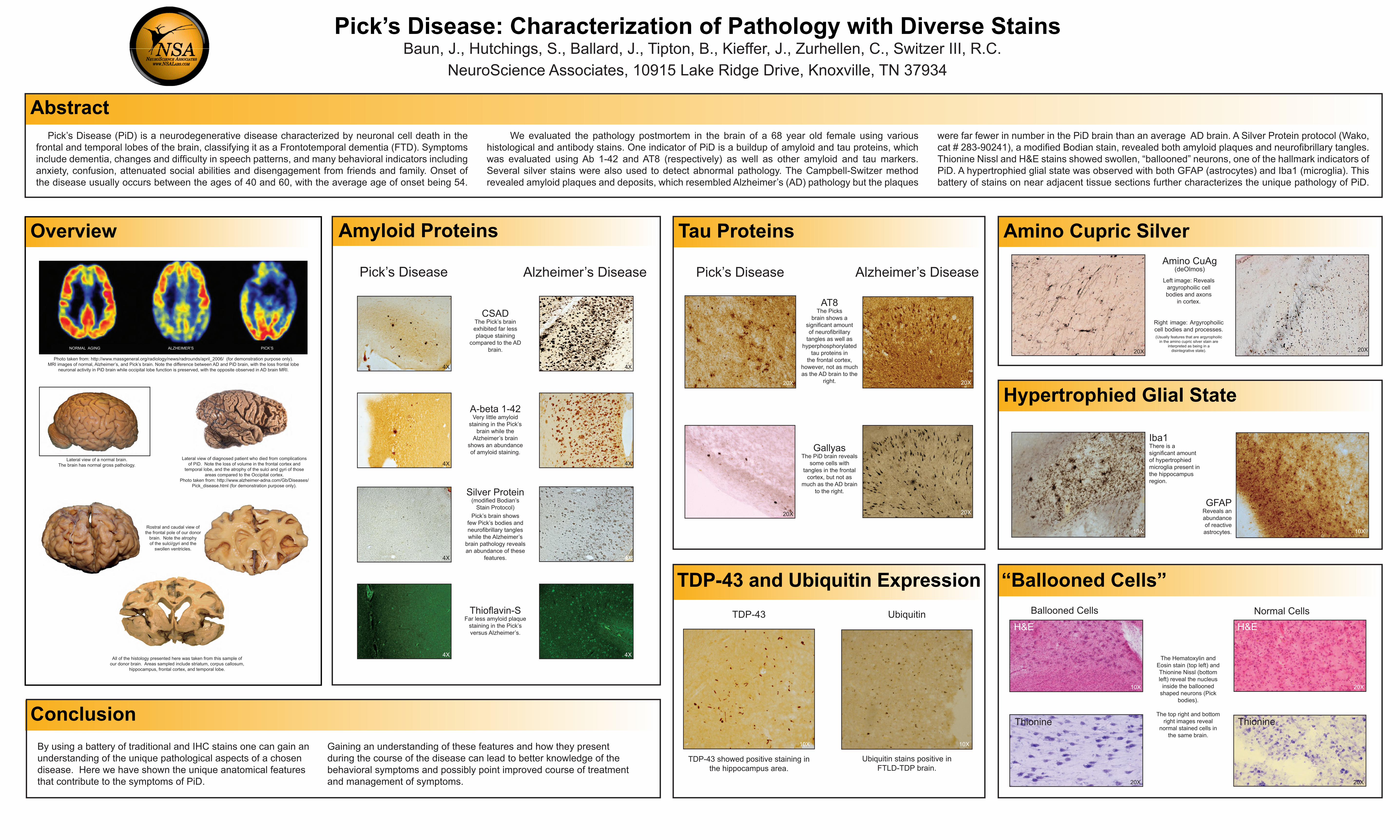
GFAPReveals an abundance of reactive astrocytes.

Pick’s Disease Alzheimer’s Disease

CSADThe Pick’s brain exhibited far less plaque staining

compared to the AD brain.

4X 4X

20X 20X

20X

10X

A-beta 1-42Very little amyloid

staining in the Pick’s brain while the

Alzheimer’s brain shows an abundance of amyloid staining.

4X 4X

4X 4X

Silver Protein(modified Bodian’s

Stain Protocol)Pick’s brain shows

few Pick’s bodies and neurofibrillary tangles while the Alzheimer’s

brain pathology reveals an abundance of these

features.

Thioflavin-SFar less amyloid plaque

staining in the Pick’s versus Alzheimer’s.

4X 4X

AT8The Picks

brain shows a significant amount of neurofibrillary

tangles as well as hyperphosphorylated

tau proteins in the frontal cortex,

however, not as much as the AD brain to the

right.

Ubiquitin stains positive in FTLD-TDP brain.

GallyasThe PiD brain reveals

some cells with tangles in the frontal

cortex, but not as much as the AD brain

to the right.

20X

20X

10X 10X

H&E

Amino CuAg(deOlmos)

10X10X

Lateral view of a normal brain. The brain has normal gross pathology.

Photo taken from: http://www.massgeneral.org/radiology/news/radrounds/april_2006/ (for demonstration purpose only). MRI images of normal, Alzheimer’s, and Pick’s brain. Note the difference between AD and PiD brain, with the loss frontal lobe

neuronal activity in PiD brain while occipital lobe function is preserved, with the opposite observed in AD brain MRI.

Lateral view of diagnosed patient who died from complications of PiD. Note the loss of volume in the frontal cortex and

temporal lobe, and the atrophy of the sulci and gyri of those areas compared to the Occipital cortex.

Photo taken from: http://www.alzheimer-adna.com/Gb/Diseases/Pick_disease.html (for demonstration purpose only).

Rostral and caudal view of the frontal pole of our donor

brain. Note the atrophy of the sulci/gyri and the

swollen ventricles.

All of the histology presented here was taken from this sample of our donor brain. Areas sampled include striatum, corpus callosum,

hippocampus, frontal cortex, and temporal lobe.

Pick’s Disease Alzheimer’s Disease

Thionine

20X

20X

20X

20X

UbiquitinTDP-43 Ballooned Cells Normal Cells

The Hematoxylin and Eosin stain (top left) and Thionine Nissl (bottom left) reveal the nucleus

inside the ballooned shaped neurons (Pick

bodies).

The top right and bottom right images reveal

normal stained cells in the same brain.

H&E

Thionine

Iba1There is a significant amount of hypertrophied microglia present in the hippocampus region.

Left image: Reveals argyrophoilic cell bodies and axons

in cortex.

Right image: Argyrophoilic cell bodies and processes.(Usually features that are argyrophoilic

in the amino cupric silver stain are interpreted as being in a

disintegrative state).

By using a battery of traditional and IHC stains one can gain an understanding of the unique pathological aspects of a chosen disease. Here we have shown the unique anatomical features that contribute to the symptoms of PiD.

Baun, J., Hutchings, S., Ballard, J., Tipton, B., Kieffer, J., Zurhellen, C., Switzer III, R.C.NeuroScience Associates, 10915 Lake Ridge Drive, Knoxville, TN 37934

Gaining an understanding of these features and how they present during the course of the disease can lead to better knowledge of the behavioral symptoms and possibly point improved course of treatment and management of symptoms.

TDP-43 showed positive staining in the hippocampus area.

Pick’s Disease: Characterization of Pathology with Diverse Stains

Pick’s Disease (PiD) is a neurodegenerative disease characterized by neuronal cell death in the frontal and temporal lobes of the brain, classifying it as a Frontotemporal dementia (FTD). Symptoms include dementia, changes and difficulty in speech patterns, and many behavioral indicators including anxiety, confusion, attenuated social abilities and disengagement from friends and family. Onset of the disease usually occurs between the ages of 40 and 60, with the average age of onset being 54.

We evaluated the pathology postmortem in the brain of a 68 year old female using various histological and antibody stains. One indicator of PiD is a buildup of amyloid and tau proteins, which was evaluated using Ab 1-42 and AT8 (respectively) as well as other amyloid and tau markers. Several silver stains were also used to detect abnormal pathology. The Campbell-Switzer method revealed amyloid plaques and deposits, which resembled Alzheimer’s (AD) pathology but the plaques

were far fewer in number in the PiD brain than an average AD brain. A Silver Protein protocol (Wako, cat # 283-90241), a modified Bodian stain, revealed both amyloid plaques and neurofibrillary tangles. Thionine Nissl and H&E stains showed swollen, “ballooned” neurons, one of the hallmark indicators of PiD. A hypertrophied glial state was observed with both GFAP (astrocytes) and Iba1 (microglia). This battery of stains on near adjacent tissue sections further characterizes the unique pathology of PiD.

Amyloid Proteins Tau Proteins Amino Cupric Silver

“Ballooned Cells”

Hypertrophied Glial State

Abstract

Overview

Conclusion

PICK’S

TDP-43 and Ubiquitin Expression

ALZHEIMER’SNORMAL AGING