Embed Size (px)
Citation preview
APPLIED AND ENVIRONMENTAL MICROBIOLOGY, June 1990, p. 1977-19800099-2240/90/061977-04$02.00/0Copyright C) 1990, American Society for Microbiology
Attachment of Vibrio cholerae Serogroup 01 to Zooplankton andPhytoplankton of Bangladesh Waters
MARK L. TAMPLIN,lt* ANNE L. GAUZENS,2 ANWARUL HUQ,3 DAVID A. SACK,3'4AND RITA R. COLWELL"5
Center of Marine Biotechnology, Maryland Biotechnology Institute, University of Maryland, 600 East Lombard Street,Baltimore, Maryland 212021; Horn Point Environmental Laboratories, University of Maryland, Cambridge,Maryland 216132; International Centre for Diarrhoeal Disease Research, Bangladesh, Dhaka, Bangladesh3;
Division of Geographic Medicine, The Johns Hopkins University School of Medicine, Baltimore, Maryland 212244;and Department of Microbiology, University of Maryland, College Park, Maryland 207425
Received 15 November 1989/Accepted 10 March 1990
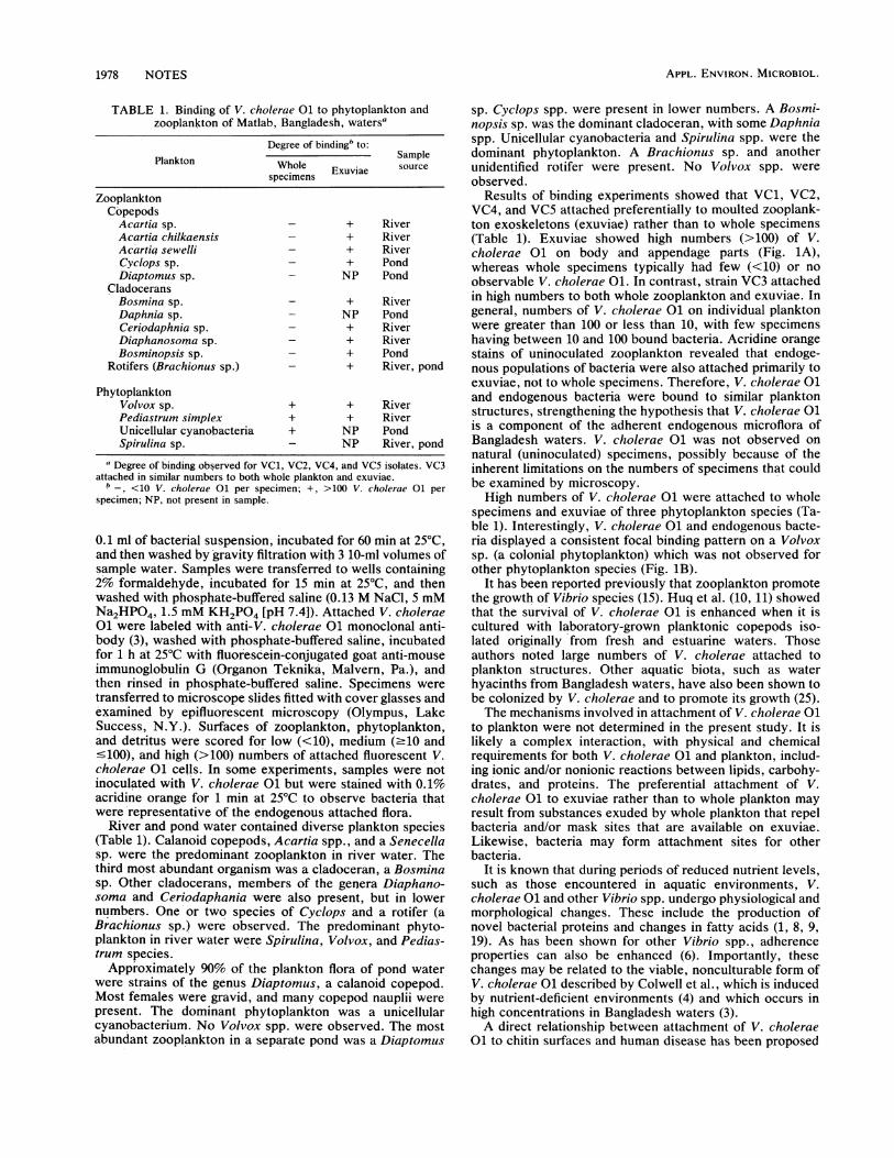
Vibrio cholerae serogroup 01, the causative agent of cholera, is capable of surviving in aquatic environmentsfor extended periods and is considered an autochthonous species in estuarine and brackish waters. Theseenvironments contain numerous elements that may affect its ecology. The studies reported here examinedphysical interactions between V. cholerae 01 and natural plankton populations of a geographical region inBangladesh where cholera is an endemic disease. Results showed that four of five clinical V. cholerae 01 strainsand endogenous bacterial flora were attached preferentially to zooplankton molts (exuviae) rather than to
whole specimens. One strain attached in approximately equal numbers to both exuviae and whole specimens.V. cholerae 01 also attached to several phytoplankton species. The results show that V. cholerae 01 can bindto diverse plankton species collected from an area where cholera is an endemic disease, with potentiallysignificant effects on its ecology.
Vibrio cholerae serogroup 01 causes human enteropatho-genic disease (cholera) in temperate and tropical climates (2,7, 27). In regions of Bangladesh, cholera is an endemicdisease and occurs in a seasonal pattern (7). In such epidem-ics, the aquatic environment appears to be an importantvector in transmission of cholera. The incidence of V.cholerae 01, coupled with epidemiological data, shows thatcontaminated surface and household waters are associatedwith human infections (24). These observations have stimu-lated research to define the effect(s) of aquatic habitats on
the ecology and pathogenicity of V. cholerae 01 and itssurvival under various physicochemical conditions (3, 5, 13,16, 21, 22, 26, 28). However, very little is known of interac-tions between V. cholerae 01 and biotic components ofwater.A variety of biological surfaces in water can bind bacteria.
Bacteria associated with surfaces have been shown to sur-
vive in aquatic environments for longer times than sus-
pended forms (14, 20), possibly as an adaptation to thestressful effects of low nutrient levels (6). Surfaces com-
monly encountered by aquatic bacteria are those of plank-ton, microscopic plants and animals that dominate the mi-crofloras of aquatic ecosystems. In many instances,bacteria, including Vibrio spp., are found attached to theirsurfaces (10, 12, 17, 23) and as part of their gut floras (23).We hypothesize that an important aspect of the ecology of V.cholerae 01 in cholera-endemic regions of Bangladesh mayinvolve a relationship with plankton, supporting previoushypotheses that interepidemic reservoirs of V. cholerae 01in Bangladesh are influenced by seasonal plankton bloomsthat accompany cholera epidemics (11).The present study investigated attachment of V. cholerae
01 to endogenous zooplankton and phytoplankton of Bang-
* Corresponding author.t Present address: U.S. Food and Drug Administration, Fishery
Research Branch, P.O. Box 158, Dauphin Island, AL 36528.
ladesh waters and whether attachment occurred with spe-cific plankton species and their anatomical structures.
Plankton specimens were collected from a river adjacentto a sewage outfall near the Matlab (Bangladesh) hospitaland from two local ponds near Matlab in April 1987. Plank-ton were collected during daylight hours, with the exceptionof one night collection of river water. Samples were obtainedby towing a 64-,um-mesh nitex plankton net, fitted with a
250-ml bucket, through the top 1 to 2 m of surface water.Specimens were rinsed from the collection bucket, sus-
pended in approximately 250 ml of homologous water (pre-filtered through 64-,um-mesh nitex), and transferred to thelaboratory at ambient temperature. In some experiments,surface waters were collected in a 20-liter carboy andplankton were separated in the laboratory.
Five V. cholerae 01 isolates (VC1, VC2, VC3, VC4, VC5)were cultured from diarrheal stools of Bangladesh patientsby the Microbiology Branch of the International Centre forDiarrhoeal Disease Research, Bangladesh. The attachmentproperties of these isolates were tested following culture inhigh-nutrient medium (tryptic soy broth), since V. cholerae01 is rapidly multiplying in human feces when it entersMatlab waters. VC1, VC2, and VC3 were classical biotypes;VC4 and VC5 were El Tor. V. cholerae 01 isolates were
grown at 35°C on tryptic soy agar (Difco Laboratories,Detroit, Mich.) containing 1% NaCl for 18 h, incubated untilmid-exponential-growth phase in tryptic soy broth (Difco)containing 1% NaCl, washed with 3 20-ml volumes offilter-sterilized homologous water at 3,000 x g, and thenadjusted to approximately 107 CFU/ml of water.For attachment assays, plankton samples were added to
polypropylene vials (inner diameter, 1 cm) fitted at one endwith 64-,um-mesh nitex. Plankton were washed three timesby gravity filtration with filter-sterilized homologous water.
Vials containing washed plankton were transferred to 24-well sterile cluster plates (catalog no. 3524; Costar, Cam-bridge, Mass.) containing 0.9 ml of filter-sterilized water and
1977
Vol. 56, No. 6
APPL. ENVIRON. MICROBIOL.
TABLE 1. Binding of V. cholerae 01 to phytoplankton andzooplankton of Matlab, Bangladesh, watersa
Degree of bindingb to:Plankton Whole Sample
specimens Exuvia s
ZooplanktonCopepods
Acartia sp. - + RiverAcartia chilkaensis - + RiverAcartia sewelli - + RiverCyclops sp. - + PondDiaptomus sp. - NP Pond
CladoceransBosmina sp. - + RiverDaphnia sp. - NP PondCeriodaphnia sp. - + RiverDiaphanosoma sp. - + RiverBosminopsis sp. - + Pond
Rotifers (Brachionus sp.) - + River, pond
PhytoplanktonVolvox sp. + + RiverPediastrum simplex + + RiverUnicellular cyanobacteria + NP PondSpirulina sp. - NP River, pond
aDegree of binding observed for VC1, VC2, VC4, and VC5 isolates. VC3attached in similar numbers to both whole plankton and exuviae.b_, <10 V. cholerae 01 per specimen; +, >100 V. cholerae 01 per
specimen; NP, not present in sample.
0.1 ml of bacterial suspension, incubated for 60 min at 25°C,and then washed by gravity filtration with 3 10-ml volumes ofsample water. Samples were transferred to wells containing2% formaldehyde, incubated for 15 min at 25°C, and thenwashed with phosphate-buffered saline (0.13 M NaCl, 5 mMNa2HPO4, 1.5 mM KH2PO4 [pH 7.4]). Attached V. cholerae01 were labeled with anti-V. cholerae 01 monoclonal anti-body (3), washed with phosphate-buffered saline, incubatedfor 1 h at 25°C with fluorescein-conjugated goat anti-mouseimmunoglobulin G (Organon Teknika, Malvern, Pa.), andthen rinsed in phosphate-buffered saline. Specimens weretransferred to microscope slides fitted with cover glasses andexamined by epifluorescent microscopy (Olympus, LakeSuccess, N.Y.). Surfaces of zooplankton, phytoplankton,and detritus were scored for low (<10), medium (.10 and'100), and high (>100) numbers of attached fluorescent V.cholerae 01 cells. In some experiments, sarnples were notinoculated with V. cholerae 01 but were stained with 0.1%acridine orange for 1 min at 25°C to observe bacteria thatwere representative of the endogenous attached flora.
River and pond water contained diverse plankton species(Table 1). Calanoid copepods, Acartia spp., and a Senecellasp. were the predominant zooplankton in river water. Thethird most abundant organism was a cladoceran, a Bosminasp. Other cladocerans, members of the genera Diaphano-soma and Ceriodaphania were also present, but in lowernumbers. One or two species of Cyclops and a rotifer (aBrachionus sp.) were observed. The predominant phyto-plankton in river water were Spirulina, Volvox, and Pedias-trum species.Approximately 90% of the plankton flora of pond water
were strains of the genus Diaptomus, a calanoid copepod.Most females were gravid, and many copepod nauplii werepresent. The dominant phytoplankton was a unicellularcyanobacterium. No Volvox spp. were observed. The mostabundant zooplankton in a separate pond was a Diaptomus
sp. Cyclops spp. were present in lower numbers. A Bosmi-nopsis sp. was the dominant cladoceran, with some Daphniaspp. Unicellular cyanobacteria and Spirulina spp. were thedominant phytoplankton. A Brachionus sp. and anotherunidentified rotifer were present. No Volvox spp. wereobserved.
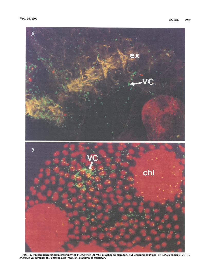
Results of binding experiments showed that VC1, VC2,VC4, and VC5 attached preferentially to moulted zooplank-ton exoskeletons (exuviae) rather than to whole specimens(Table 1). Exuviae showed high numbers (>100) of V.cholerae 01 on body and appendage parts (Fig. 1A),whereas whole specimens typically had few (<10) or noobservable V. cholerae 01. In contrast, strain VC3 attachedin high numbers to both whole zooplankton and exuviae. Ingeneral, numbers of V. cholerae 01 on individual planktonwere greater than 100 or less than 10, with few specimenshaving between 10 and 100 bound bacteria. Acridine orangestains of uninoculated zooplankton revealed that endoge-nous populations of bacteria were also attached primarily toexuviae, not to whole specimens. Therefore, V. cholerae 01and endogenous bacteria were bound to similar planktonstructures, strengthening the hypothesis that V. cholerae 01is a component of the adherent endogenous microflora ofBangladesh waters. V. cholerae 01 was not observed onnatural (uninoculated) specimens, possibly because of theinherent limitations on the numbers of specimens that couldbe examined by microscopy.High numbers of V. cholerae 01 were attached to whole
specimens and exuviae of three phytoplankton species (Ta-ble 1). Interestingly, V. cholerae 01 and endogenous bacte-ria displayed a consistent focal binding pattern on a Volvoxsp. (a colonial phytoplankton) which was not observed forother phytoplankton species (Fig. 1B).
It has been reported previously that zooplankton promotethe growth of Vibrio species (15). Huq et al. (10, 11) showedthat the survival of V. cholerae 01 is enhanced when it iscultured with laboratory-grown planktonic copepods iso-lated originally from fresh and estuarine waters. Thoseauthors noted large numbers of V. cholerae attached toplankton structures. Other aquatic biota, such as waterhyacinths from Bangladesh waters, have also been shown tobe colonized by V. cholerae and to promote its growth (25).The mechanisms involved in attachment of V. cholerae 01
to plankton were not determined in the present study. It islikely a complex interaction, with physical and chemicalrequirements for both V. cholerae 01 and plankton, includ-ing ionic and/or nonionic reactions between lipids, carbohy-drates, and proteins. The preferential attachment of V.cholerae 01 to exuviae rather than to whole plankton mayresult from substances exuded by whole plankton that repelbacteria and/or mask sites that are available on exuviae.Likewise, bacteria may form attachment sites for otherbacteria.
It is known that during periods of reduced nutrient levels,such as those encountered in aquatic environments, V.cholerae 01 and other Vibrio spp. undergo physiological andmorphological changes. These include the production ofnovel bacterial proteins and changes in fatty acids (1, 8, 9,19). As has been shown for other Vibrio spp., adherenceproperties can also be enhanced (6). Importantly, thesechanges may be related to the viable, nonculturable form ofV. cholerae 01 described by Colwell et al., which is inducedby nutrient-deficient environments (4) and which occurs inhigh concentrations in Bangladesh waters (3).A direct relationship between attachment of V. cholerae
01 to chitin surfaces and human disease has been proposed
1978 NOTES
VOL. 56, 1990
FIG. 1. Fluorescence photomicrography of V. cholerae 01 VC1 attached to plankton. (A) Copepod exuviae; (B) Volvox species. VC, V.cholerae 01 (green); chi, chloroplasts (red); ex, plankton exoskeleton.
NOTES 1979
APPL. ENVIRON. MICROBIOL.
by Nalin et al. (18), who showed that chitin protects V.cholerae 01 from the lethal effect of low pH. They suggestthat chitin may promote pathogenicity of V. cholerae 01 byprotecting it from the acidic environment of the humangastrointestinal tract. Our experiments show that chitinoussurfaces of plankton concentrate V. cholerae 01 and mayincrease the number of V. cholerae in a given unit of water.Future experiments will determine if these levels reach aninfective dose and if attachment affects the physiology andpathogenicity of V. cholerae 01.
This research was sponsored in part by World Health Organiza-tion grant C6/181/70(A), Agency for International Developmentgrant DPE-5542-G-55-4060-00, and Public Health Service grantR22,A1-14242 from the National Institutes of Health.
LITERATURE CITED1. Baker, R. M., F. L. Singleton, and M. A. Hood. 1983. Effects of
nutrient deprivation on Vibrio cholerae. Appl. Environ. Micro-biol. 46:930-940.
2. Blake, P. A., D. T. Allegra, J. D. Snyder, T. J. Barrett, L.McFarland, C. T. Caraway, J. C. Feeley, J. P. Craig, J. V. Lee,N. D. Puhr, and R. A. Feldman. 1980. Cholera-a possibleendemic focus in the United States. N. Engl. J. Med. 302:305-309.
3. Brayton, P. R., M. L. Tamplin, A. Huq, and R. R. Colwell. 1987.Enumeration of Vibrio cholerae O1 in Bangladesh waters byfluorescent-antibody direct viable count. Appl. Environ. Micro-biol. 53:2862-2865.
4. Colwell, R. R., P. R. Brayton, D. J. Grimes, D. B. Roszak, S. A.Huq, and L. M. Palmer. 1985. Viable but nonculturable Vibriocholerae and related pathogens in the environment: implicationsfor release of genetically engineered microorganisms. Bio/Tech-nology 3:817-820.
5. Colwell, R. R., R. J. Seidler, J. Kaper, S. W. Joseph, S. Garges,H. Lockman, D. Maneval, H. Bradford, N. Roberts, E. Rem-mers, I. Huq, and A. Huq. 1981. Occurrence of Vibrio choleraeserotype O1 in Maryland and Louisiana estuaries. Appl. Envi-ron. Microbiol. 41:555-558.
6. Dawson, M. P., B. A. Humphrey, and K. C. Marshall. 1981.Adhesion, a tactic in the survival strategy of a marine vibrioduring starvation. Curr. Microbiol. 6:195-198.
7. Glass, R. I., M. I. Huq, B. J. Stoll, M. U. Khan, M. H. Merson,J. V. Lee, and R. E. Black. 1982. Endemic cholera in ruralBangladesh, 1966-1980. J. Epidemiol. Community Health 116:959-970.
8. Guckert, J. B., M. A. Hood, and D. C. White. 1986. Phospho-lipid ester-linked fatty acid profile changes during nutrientdeprivation of Vibrio cholerae: increases in the translcis ratioand proportions of cyclopropyl fatty acids. Appl. Environ.Microbiol. 52:794-801.
9. Hood, M. A., J. B. Guckert, D. C. White, and F. Deck. 1986.Effect of nutrient deprivation on lipid, carbohydrate, DNA,RNA, and protein levels in Vibrio cholerae. Appl. Environ.Microbiol. 52:788-793.
10. Huq, A., E. B. Small, P. A. West, M. I. Huq, R. Rahman, and R.R. Colwell. 1983. Ecological relationships between Vibrio chol-erae and planktonic crustacean copepods. Appl. Environ. Mi-crobiol. 45:275-283.
11. Huq, A., P. A. West, E. B. Small, M. I. Huq, and R. R. Colwell.1984. Influence of water temperature, salinity, and pH on
survival and growth of toxigenic Vibrio cholerae serovar 01associated with live copepods in laboratory microcosms. Appl.Environ. Microbiol. 48:420-424.
12. Kaneko, T., and R. R. Colwell. 1975. Adsorption of Vibrioparahaemolyticus onto chitin and copepods. Appl. Environ.Microbiol. 29:269-274.
13. Kaper, J., H. Lockman, R. R. Colwell, and S. W. Joseph. 1979.Ecology, serology, and enterotoxin production of Vibrio chol-erae in Chesapeake Bay. Appl. Environ. Microbiol. 37:91-103.
14. Kirchman, D., and R. Mitchell. 1982. Contribution of particle-bound bacteria to total microheterotrophic activity in five pondsand two marshes. Appl. Environ. Microbiol. 43:200-209.
15. Kogure, K., U. Simidu, and N. Taga. 1980. Effect of phyto- andzooplankton on the growth of marine bacteria in filtered seawa-ter. Bull. Jpn. Soc. Sci. Fish. 46:323-326.
16. Miller, C. J., B. S. Drasar, and R. G. Feachem. 1984. Responseof toxigenic Vibrio cholerae 01 to physico-chemical stresses inaquatic environments. J. Hyg. 93:475-495.
17. Nagasawa, S., U. Simidu, and T. Nemoto. 1985. Scanningelectron microscopy investigation of bacterial colonization ofthe marine copepod Acartia clausi. Mar. Biol. (Berlin) 87:61-66.
18. Nalin, D. R., V. Daya, A. Reid, M. M. Levine, and L. Cisneros.1979. Adsorption and growth of Vibrio cholerae on chitin.Infect. Immun. 25:768-770.
19. Oliver, J. D., and W. F. Stringer. 1984. Lipid composition ofpsychrophilic marine Vibrio sp. during starvation-induced mor-phogenesis. Appl. Environ. Microbiol. 47:461-466.
20. Pedros, A. C., and T. D. Brock. 1983. The importance ofattachment to particles for planktonic bacteria. Arch. Hydro-biol. 98:354-379.
21. Singleton, F. L., R. Attwell, S. Jangi, and R. R. Colwell. 1982.Effects of temperature and salinity on Vibrio cholerae growth.Appl. Environ. Microbiol. 44:1047-1058.
22. Singleton, F. L., R. W. Attwell, M. S. Jangi, and R. R. Colwell.1982. Influence of salinity and organic nutrient concentration onsurvival and growth of Vibrio cholerae in aquatic microcosms.Appl. Environ. Microbiol. 43:1080-1085.
23. Sochard, M. R., D. F. Wilson, B. Austin, and R. R. Colwell.1979. Bacteria associated with the surface and gut of marinecopepods. Appl. Environ. Microbiol. 37:750-759.
24. Spira, W. M. 1981. Environmental factors in diarrhea transmis-sion: the ecology of Vibrio cholerae 01 and cholera, p. 273-288.In T. Holme, J. Holmgren, M. H. Merson, and R. Mollby (ed.),Acute enteric infections in children. New prospects for treat-ment and prevention. Elsevier/North-Holland BiomedicalPress, Amsterdam.
25. Spira, W. M., A. Huq, Q. S. Ahmed, and Y. A. Saeed. 1981.Uptake of Vibrio cholerae biotype eltor from contaminatedwater by water hyacinth (Eichornia crassipes). Appl. Environ.Microbiol. 42:550-553.
26. Tamplin, M. L., and R. R. Colwell. 1986. Effects of microcosmsalinity and organic substrate concentration on production ofVibrio cholerae enterotoxin. Appl. Environ. Microbiol. 52:297-301.
27. World Health Organization Scientific Working Group. 1980.Cholera and other vibrio-associated diarrhoeas. Bull. W.H.O.58:353-374.
28. Xu, H., N. Roberts, F. L. Singleton, R. W. Attwell, D. J. Grimes,and R. R. Colwell. 1982. Survival and viability of nonculturableEscherichia coli and Vibrio cholerae in the estuarine and marineenvironment. Microb. Ecol. 8:313-323.
1980 NOTES