Embed Size (px)
Citation preview
Phyllosticta capitalensis, a widespread endophyte of plants
Saowanee Wikee & Lorenzo Lombard & Pedro W. Crous &
Chiharu Nakashima & Keiichi Motohashi & Ekachai Chukeatirote &
Siti A. Alias & Eric H. C. McKenzie & Kevin D. Hyde
Received: 21 February 2013 /Accepted: 9 April 2013# Mushroom Research Foundation 2013
Abstract Phyllosticta capitalensis is an endophyte and weakplant pathogen with a worldwide distribution presently knownfrom 70 plant families. This study isolated P. capitalensis fromdifferent host plants in northern Thailand, and determined theirdifferent life modes. Thirty strains ofP. capitalensiswere isolatedas endophytes from 20 hosts. An additional 30 strains of P.capitalensis from other hosts and geographic locations were alsoobtained from established culture collections. Phylogenetic anal-ysis using ITS,ACTand TEF gene data confirmed the identity ofall isolates. Pathogenicity tests with five strains of P. capitalensisoriginating from different hosts were completed on their respec-tive host plants. In all cases there was no infection of healthyleaves, indicating that this endophyte does not cause disease onhealthy, unstressed host plants. That P. capitalensis is oftenisolated as an endophyte has important implications in fungalbiology and plant health. Due to its endophytic nature, P.
capitalensis is commonly found associatedwith lesions of plants,and often incorrectly identified as a species of quarantine impor-tance, which again has implications for trade in agricultural andforestry production.
Keywords Guignardia . Leaf spot . Morphology .
Molecular phylogeny . Quarantine
Introduction
Species in the genus Phyllosticta are mostly plant pathogensof a wide range of hosts and are responsible for diseasesincluding leaf spots and black spots on fruits (Wulandari etal. 2009; Glienke et al. 2011; Wang et al. 2012). There areabout 3,200 names listed for the genus Phyllosticta in Index
S. Wikee : E. Chukeatirote :K. D. HydeSchool of Science, Mae Fah Luang University,Chiangrai 57100, Thailand
S. Wikee (*) : E. Chukeatirote :K. D. HydeInstitute of Excellence in Fungal Research,Mae Fah Luang University, Chiangrai 57100, Thailande-mail: [email protected]
L. Lombard : P. W. CrousCBS-KNAW Fungal Biodiversity Centre, Uppsalalaan 8,3584 CT Utrecht, The Netherlands
P. W. CrousMicrobiology, Department of Biology, Utrecht University,Padualaan 8, 3584 CH Utrecht, The Netherlands
P. W. CrousLaboratory of Phytopathology, Wageningen University andResearch Centre (WUR), Droevendaalsesteeg 1,6708 PB Wageningen, The Netherlands
C. NakashimaGraduate School of Bioresources, Mie University,Kurima-machiya 1577,Tsu, Mie 514-8507, Japan
K. MotohashiElectron Microscope Center, Tokyo University of Agriculture,Sakuraoka 1-1-1, Setagaya,Tokyo 156-8502, Japan
S. A. AliasInstitute Ocean and Earth Sciences, Institute for PostgraduateStudies, University Malaya, Kuala Lumpur 50603, Malaysia
E. H. C. McKenzieLandcare Research, Private Bag 92170, Auckland Mail Centre,Auckland 1142, New Zealand
Fungal DiversityDOI 10.1007/s13225-013-0235-8
Fungorum (http://www.indexfungorum.org/; accessed February2013) and 3 ,340 names in MycoBank (h t tp : / /www.mycobank.org/; accessed February 2013). The USDAFungal Database lists 78 Phyllosticta records associated withplant hosts (http://nt.ars-grin.gov/fungaldatabases/; accessedFebruary 2013).
Phyllosticta species may be associated with a“Guignardia-like” sexual state (van der Aa 1973; Wikee etal. 2011). For example, the sexual state of Phyllostictaampelicida (Engelm.) Aa, the black rot pathogen of grape-vine is Guignardia bidwellii (Ellis) Viala & Ravaz (van derAa 1973; Ullrich et al. 2009). Leaf spots on Morindacitrifolia (Rubiaceae) commonly have both ascomata andpycnidia of P. morindae (Petr. & Syd.) Aa (Wulandari et al.2010a, b). Likewise, both ascomata and pycnidia of P.maculataM.H. Wong & Crous are present on banana leaveswith freckle disease (Wong et al. 2012).
Guignardia citricarpa Kiely (synonym of P. citricarpa(McAlpine) Aa), which causes black spot of citrus (e.g. or-anges), is of quarantine concern in Europe (Baayen et al. 2002;Agostini et al. 2006), but P. citriasiana Wulandari, Crous &Gruyter, which causes brown spot of pomelo fruit (Citrusmaxima) is not of quarantine concern as this fruit is not grownin Europe (Wulandari et al. 2009). A few species have also beenreported as endophytes and saprobes (Van Der Aa et al. 2002;Baayen et al. 2002; Glienke et al. 2011). Phyllosticta maculatathe cause of banana leaf freckle has also been isolated as anendophyte from healthy grapevine leaves (Kuo and Hoch1996). Phyllosticta capitalensis Henn. is commonly isolatedas an endophyte and is a widespread species (Glienke-Blanco etal. 2002; Silva and Pereira 2007; Silva et al. 2008).
Phyllosticta capitalensis was described by Hennings(1908) who found it associated with necrotic leaves ofStanhopea sp. (Orchidaceae) collected in Brazil. The sup-posed sexual morph, G. mangiferae A.J. Roy was later de-scribed from Mangifera indica L. (Anacardiaceae) in India(Roy 1968). However, there has been confusion with theidentification and naming of the P. capitalensis sexual morph.Okane et al. (2003) stated that the teleomorph of P. capitalensisdifferedmorphologically fromG.mangiferae and that it was, infact, G. endophyllicola Okane, Nakagiri & Tad. Ito. The latterfunguswas described as a pathogen of several ericaceous plantsbyOkane et al. (2001). In the past there has also been confusionbetween G. endophyllicola and G. citricarpa. Both sexualnames have been used for this fungus, for example, G.endophyllicola (Okane et al. 2003; Pandey et al. 2003) andG. mangiferae (Baayen et al. 2002; Glienke-Blanco et al. 2002;Guo et al. 2003; Suryanarayanan et al. 2004; Devarajan andSuryanarayanan 2006; Shaw et al. 2006). However, G.citricarpa is a distinct species and the cause of citrus blackspot (Paul et al. 2005; Baayen et al. 2002).
Fungal endophytes colonise healthy plant host tissues butmay become pathogenic when the plant host is stressed
through environmental or biological factors (Petrini 1991;Hyde and Soytong 2008; Purahong and Hyde 2011) that inducethe fungus to change from one life mode to another (Fisher andPetrini 1992). As with Phyllosticta, some species of other com-mon genera such as Bipolaris, Cladosporium, Colletotrichum,Curvularia, Diaporthe, Fusarium, Pestalotiopsis, Phoma andVerticillium have been isolated as endophytes (Photita et al.2001, 2004; Anderson et al. 2011; Bensch et al. 2012; Dammet al. 2012a, b; de Gruyter et al. 2013; Lima et al. 2012;Orlandelli et al. 2012), and some of these are also seriouspathogens (Photita et al. 2004; Slippers and Wingfield 2007).
The present study provides an overview of the distribu-tion and host range of P. capitalensis worldwide, throughthe application of multi-gene phylogeny to illustrate itswidespread nature. Generally, Phyllosticta species are con-sidered plant pathogens but it is still unclear whether theyare generalists or host-specific. The distinction between apathogen and a latent pathogen with endophytic nature isalso unclear. In this study we isolated Phyllosticta speciesfrom northern Thailand, both as endophytes and as patho-gens associated with leaf spots of various hosts. We alsoobtained a range of geographically diverse isolates of P.capitalensis from the CBS-KNAW Fungal BiodiversityCentre. All isolates were sequenced compared with se-quences downloaded from GenBank.
Material and methods
Isolates
Thirty strains of Phyllosticta were isolated from leafspots or as endophytes from healthy leaves of ornamen-tal plants (Table 1). If pycnidia were present on dis-eased tissue then a single spore isolation procedure asdescribed by Chomnunti et al. (2011) was used toobtain cultures. To obtain isolates of Phyllosticta fromdiseased leaves of host plants when fruit bodies werenot present, the leaf was surface disinfected by wipingwith 70 % ethanol. Small pieces of leaf were then cutfrom the interface between healthy and diseased tissue.These were surface sterilised in 70 % ethanol, andplated onto ½ strength potato dextrose agar (½PDA;Crous et al. 2009). For isolation of endophytes, healthyleaves were washed in tap water and surface disinfectedwith 70 % ethanol. They were then cut into smallpieces (about 1×1 cm), suspended in 70 % ethanol (3times for 15 min each) and washed in distilled water (3times) before placing on ½PDA. All dishes were incu-bated at 27 °C for 1 week and observed daily. Thegrowing tips of hyphae of Phyllosticta colonies thatdeveloped were cut out and transferred to fresh PDAdishes. Isolates are deposited in Mae Fah Luang
Fungal Diversity
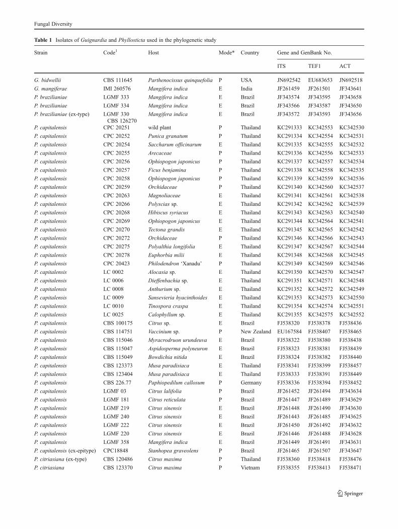
Table 1 Isolates of Guignardia and Phyllosticta used in the phylogenetic study
Strain Code1 Host Mode* Country Gene and GenBank No.
ITS TEF1 ACT
G. bidwellii CBS 111645 Parthenocissus quinquefolia P USA JN692542 EU683653 JN692518
G. mangiferae IMI 260576 Mangifera indica E India JF261459 JF261501 JF343641
P. brazilianiae LGMF 333 Mangifera indica E Brazil JF343574 JF343595 JF343658
P. brazilianiae LGMF 334 Mangifera indica E Brazil JF343566 JF343587 JF343650
P. brazilianiae (ex-type) LGMF 330CBS 126270
Mangifera indica E Brazil JF343572 JF343593 JF343656
P. capitalensis CPC 20251 wild plant P Thailand KC291333 KC342553 KC342530
P. capitalensis CPC 20252 Punica granatum P Thailand KC291334 KC342554 KC342531
P. capitalensis CPC 20254 Saccharum officinarum E Thailand KC291335 KC342555 KC342532
P. capitalensis CPC 20255 Arecaceae P Thailand KC291336 KC342556 KC342533
P. capitalensis CPC 20256 Ophiopogon japonicus P Thailand KC291337 KC342557 KC342534
P. capitalensis CPC 20257 Ficus benjamina P Thailand KC291338 KC342558 KC342535
P. capitalensis CPC 20258 Ophiopogon japonicus P Thailand KC291339 KC342559 KC342536
P. capitalensis CPC 20259 Orchidaceae P Thailand KC291340 KC342560 KC342537
P. capitalensis CPC 20263 Magnoliaceae E Thailand KC291341 KC342561 KC342538
P. capitalensis CPC 20266 Polyscias sp. E Thailand KC291342 KC342562 KC342539
P. capitalensis CPC 20268 Hibiscus syriacus E Thailand KC291343 KC342563 KC342540
P. capitalensis CPC 20269 Ophiopogon japonicus E Thailand KC291344 KC342564 KC342541
P. capitalensis CPC 20270 Tectona grandis E Thailand KC291345 KC342565 KC342542
P. capitalensis CPC 20272 Orchidaceae P Thailand KC291346 KC342566 KC342543
P. capitalensis CPC 20275 Polyalthia longifolia E Thailand KC291347 KC342567 KC342544
P. capitalensis CPC 20278 Euphorbia milii E Thailand KC291348 KC342568 KC342545
P. capitalensis CPC 20423 Philodendron ‘Xanadu’ P Thailand KC291349 KC342569 KC342546
P. capitalensis LC 0002 Alocasia sp. E Thailand KC291350 KC342570 KC342547
P. capitalensis LC 0006 Dieffenbachia sp. E Thailand KC291351 KC342571 KC342548
P. capitalensis LC 0008 Anthurium sp. E Thailand KC291352 KC342572 KC342549
P. capitalensis LC 0009 Sansevieria hyacinthoides E Thailand KC291353 KC342573 KC342550
P. capitalensis LC 0010 Tinospora craspa E Thailand KC291354 KC342574 KC342551
P. capitalensis LC 0025 Calophyllum sp. E Thailand KC291355 KC342575 KC342552
P. capitalensis CBS 100175 Citrus sp. E Brazil FJ538320 FJ538378 FJ538436
P. capitalensis CBS 114751 Vaccinium sp. P New Zealand EU167584 FJ538407 FJ538465
P. capitalensis CBS 115046 Myracrodruon urundeuva E Brazil FJ538322 FJ538380 FJ538438
P. capitalensis CBS 115047 Aspidosperma polyneuron E Brazil FJ538323 FJ538381 FJ538439
P. capitalensis CBS 115049 Bowdichia nitida E Brazil FJ538324 FJ538382 FJ538440
P. capitalensis CBS 123373 Musa paradisiaca E Thailand FJ538341 FJ538399 FJ538457
P. capitalensis CBS 123404 Musa paradisiaca E Thailand FJ538333 FJ538391 FJ538449
P. capitalensis CBS 226.77 Paphiopedilum callosum P Germany FJ538336 FJ538394 FJ538452
P. capitalensis LGMF 03 Citrus lalifolia P Brazil JF261452 JF261494 JF343634
P. capitalensis LGMF 181 Citrus reticulata P Brazil JF261447 JF261489 JF343629
P. capitalensis LGMF 219 Citrus sinensis E Brazil JF261448 JF261490 JF343630
P. capitalensis LGMF 240 Citrus sinensis E Brazil JF261443 JF261485 JF343625
P. capitalensis LGMF 222 Citrus sinensis E Brazil JF261450 JF261492 JF343632
P. capitalensis LGMF 220 Citrus sinensis E Brazil JF261446 JF261488 JF343628
P. capitalensis LGMF 358 Mangifera indica E Brazil JF261449 JF261491 JF343631
P. capitalensis (ex-epitype) CPC18848 Stanhopea graveolens P Brazil JF261465 JF261507 JF343647
P. citriasiana (ex-type) CBS 120486 Citrus maxima P Thailand FJ538360 FJ538418 FJ538476
P. citriasiana CBS 123370 Citrus maxima P Vietnam FJ538355 FJ538413 FJ538471
Fungal Diversity
University Culture Collection (MFLUCC) and in theworking collection of Pedro Crous (CPC) housed atthe CBS-KNAW Fungal Biodiversity Centre (CBS),Utrecht, The Netherlands (CBS). Other fungal isolatesof representative Phyllosticta spp. were obtained fromthe CBS (Table 1).
Morphology
Growth rates, cultural characteristics and morphology of theisolates were determined on culture media preparedaccording to Crous et al. (2009). All isolates were grownat 27 °C. To induce sporulation, isolates were grown on pineneedle agar (PNA) and synthetic nutrient-poor agar (SNA),and incubated under near UV-light. Colony colour andgrowth rate were established on PDA, malt extract agar(MEA) and oatmeal agar (OA). Culture characteristics wereassessed, and the colour of upper and lower surface ofcultures was recorded after 14 days in the dark at 27 °C.Colony colour on MEA, OA and PDA were determinedusing the colour charts of Rayner (1970).
Molecular phylogeny
DNA extraction, amplification, and sequencing
Strains were grown on MEA at room temperature for 2–3 days, after which the mycelium was harvested. DNA wasisolated using Ultraclean™ Microbial DNA kit (Mo Bio,Calsbad, CA, USA) following the manufacturer’s protocol.Transcribed spacer-polymerase chain reaction (ITS-PCR)was performed with primers V9G (De Hoog and Gerritsvan den Ende 1998) and ITS4 as described by White et al.(1990). Part of elongation factor 1-α gene (TEF) was am-plified with forward primer EF1 and reverse primer EF2.The primers ACT-512F and ACT-783R were used to ampli-fy part of the actin gene (ACT). Cycle sequencing of PCRproducts were performed in PCR condition. PCR productswere separated by gel electrophoresis at 130 V for 20 min in1 % agarose gel in 1× TAE running buffer and visualizedunder UV light using a GeneGenius Gel Documentation andAnalysis System (Syngene, Cambridge, UK). Purified PCRproducts were sequenced using both PCR primers with a
Table 1 (continued)
Strain Code1 Host Mode* Country Gene and GenBank No.
ITS TEF1 ACT
P. citriasiana CBS 123371 Citrus maxima P Vietnam FJ538356 FJ538414 FJ538472
P. citriasiana CBS 123372 Citrus maxima P Vietnam FJ538357 FJ538415 FJ538473
P. citribraziliensis (ex-type) CBS 100098 Citrus sp. H Brazil FJ538352 FJ538410 FJ538468
P. citribraziliensis LGMF09 Citrus sp. H Brazil JF261436 JF261478 JF343618
P. citricarpa CBS 102374 Citrus aurantium P Brazil FJ538313 GU349053 FJ538429
P. citricarpa CBS 120489 Citrus sinensis P Zimbabwe FJ538315 FJ538373 FJ538431
P. citricarpa (ex-epitype) CBS 127454 Citrus limon P Australia JF343583 JF343604 JF343667
P. citricarpa CBS 127452 Citrus reticulata P Australia JF343581 JF343602 JF343665
P. citricarpa CBS 127455 Citrus sinensis P Australia JF343584 JF343605 JF343668
P. citrichinaensis ZJUCC 200956 Citrus reticulata P China JN791664 JN791515 JN791589
P. citrichinaensis ZJUCC 200964 Citrus maxima P China JN791662 JN791514 JN791582
P. citrichinaensis ZJUCC 2010150 Citrus maxima P China JN791620 JN791459 JN791533
P. citrichinaensis ZJUCC 2010152 Citrus sinensis P China JN791611 JN791461 JN791535
P. kerriae (ex-holotype) MUCC 17 Kerria japonica P Japan AB454266 KC342576 AB704209
P. hypoglossi CBS 101.72 Ruscus aculeatus P Italy FJ538365 FJ538423 FJ538481
P. hypoglossi CBS 434.92 Ruscus aculeatus P Italy FJ538367 FJ538425 FJ538483
P. hypoglossi CBS 167.85 Ruscus hypoglossum P Italy FJ538366 FJ538424 FJ538482
P. owaniana CBS 776.97 Brabejum stellatifolium P South Africa FJ538368 FJ538426 FJ538484
P. owaniana CPC 14901 Brabejum stellatifolium P South Africa JF261462 JF261504 JF343644
P. spinarum CBS 292.90 Chamaecyparis pisifera P France JF343585 JF343606 JF343669
P. podocarpi CBS 111646 Podocarpus falcatus P South Africa AF312013 KC357671 KC357670
1 CBS: CBS-KNAW Fungal Biodiverstiy Centre, Utrecht, The Netherlands; CPC: working collection of Pedro Crous housed at CBS; IMI:International Mycological Institute, CABI-Bioscience, Egham, Bakeham Lane, LC: culture collection of Nilam F. Wulandari, Chiangmai, Thailand.LGMF: culture collection of Laboratory of Genetics of Microorganisms, Federal University of Parana, Curitiba, Brazil, ZJUCC: ZhejiangUniversity Culture Collection, Zhejiang, China
*P pathogen, E endophyte
Fungal Diversity
BigDye Terminator Cycle Sequencing Kit v3.1 (AppliedBiosystems, Foster City, CA, USA) containing AmpliTagDNA Polymerase. The amplified products were analyzed onan automatic DNA sequencer (Perkin-Elmer, Norwalk, CN)and aligned using MEGA v5 software. Phylogenetic analyz-ing was executed by Phylogenetic analyses using parsimo-ny; PAUP v4.0b10 (Swofford 2003) and MrBayes v. 3.0b4(Huelsenbeck and Ronquist 2001) for Bayesian analyses.Guignardia bidwellii was chosen as outgroup for the phy-logenetic tree. Representative sequences were deposited inGenBank.
Pathogenicity testing
Attached, young healthy leaves of five plant species(Cordyline fruticosa, Dendrobium lindleyi, Ficus sp.,Ophiopogon japonica, Punica granatum) were washedwith distilled water, wiped with 70 % ethanol and driedwith sterile tissue paper. To complete the Koch’s postu-lated the inoculation methods followed Than et al.(2008). Two to five leaves of each plant were woundedwith a total of ten wounds. The leaves were woundedby pricking them with a pin. Both wounded andunwounded leaves were inoculated with plugs (0.7 mmdiam) taken from the edge of 14 day-old colonies oftest fungi growing on PDA; sterile agar plugs served asa control. All leaves were kept individually in moistchambers for 1 week and observed for symptom expres-sion every other day. After 7 days, if positive, thefungus was reisolated from any tissue showing lesionsand this isolate was considered to be pathogenic; ab-sence of symptoms on leaves classified the isolate asnon-pathogenic.
Results
Collection of Phyllosticta species
Thirty strains of Phyllosticta capitalensis were isolated from20 host plants growing in the north of Thailand (Table 1, seealso Fig. 1). No other species of Phyllosticta were isolated.
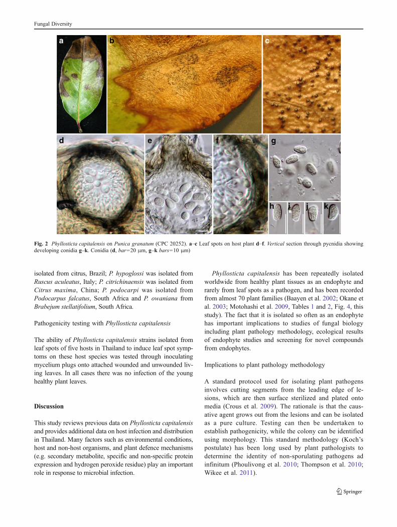
Morphological description of Phyllosticta capitalensis(Fig. 2)
On Punica granatum Pycnidia epiphyllous, globose, brownor black, 120–125 μm high, 135–140 μm wide, wall 12–15 μm thick. Conidiogenous cells lining wall of pycnidium,phialidic, cylindrical, hyaline, 2–2.2×2.2–3 μm. Conidiaellipsoidal, hyaline, 1-celled, smooth-walled, 8–11×5–6 μm, surrounded by a mucilaginous sheath, bearing asingle apical appendage, usually 5–8 μm long.
In culture On SNA, ascomata forming on and under mediain 3 days, black, globose, 69–74×104–119 μm (x ¼ 73� 2�109� 5; n=10), wall composed of a single layer, 7–9 μmthick (x ¼ 8� 1; n=10), brown. Asci bitunicate, containing6–8 ascospores, irregularly biseriate, clavate, 36–80×7–15 μm (x ¼ 51� 1� 11� 2, n=10). Ascospores ellipsoidto broadly fusoid, widest in the middle, hyaline, smooth,thin-walled, 12–22×5–10 μm (x ¼ 16� 2� 7� 1, n=50),1-celled, surrounded by mucilaginous sheath. On OA, col-onies appear flat with an irregular margin, initially hyalinewith abundant mycelium, gradually becoming greenish after3–4 days. On MEA, colonies appear woolly, flat, irregular,initially white with abundant mycelium, gradually becominggreenish to dark green after 2–3 days with white hyphae on theundulate margin, eventually turning black; reverse dark greento black. At 27 °C, in the dark, mycelium reached the edge ofthe Petri-dish in 20 days with a growth rate of 0.4 cm per day.On PDA, colonies appear woolly, initially white with abun-dant mycelium, gradually becoming greenish to dark greenafter 2–3 days with white hyphae on the undulate margin,eventually turning dark green to black; reverse black. After10 days in the dark at 27 °C, mycelium reached the edge of thePetri-dish with a growth rate of 0.9 cm per day.
Material examined All CPC collected by Saowanee Wikeeand LC by Nilam F. Wulandari, from June 2010 toNovember 2011, Chiang Rai, Thailand. From leaf spots ofunknown wild plant CPC 20251; from leaf spots Punicagranatum, CPC 20252; from healthy leaf of Saccharumofficinarum CPC 20254; from leaf spots of ArecaceaeCPC 20255; from leaf spots of Ophiopogon japonica CPC20256, CPC 20258 and CPC 20269; from leaf spots ofFicus benjamina CPC 20257; from leaf spots ofOrchidaceae CPC 20259 and CPC 20272; from healthy leafof Magnoliaceae CPC 20263; from healthy leaf ofCodiaeum variegatum CPC 20265; from healthy leaf ofPolyscias sp. CPC 20266; from healthy leaf of Hibiscussyriacus CPC 20268; from healthy leaf of Tectona grandisCPC 20270; from healthy leaf of Poloalthia longifolia CPC20275; from healthy leaf of Euphorbia milli CPC 20278;from healthy leaf of Philodendron sp. CPC 20423; fromhealthy leaf of Alocasia sp. LC 0002; from healthy leaf ofDieffenbachia sp. LC 0006; from healthy leaf of Anthuriumsp. LC 0008; from healthy leaf of Sansevieria hyacinthodesLC 0009; from healthy leaf of Tinospora craspa LC0010;from healthy leaf of Calophyllum sp. LC 0025.
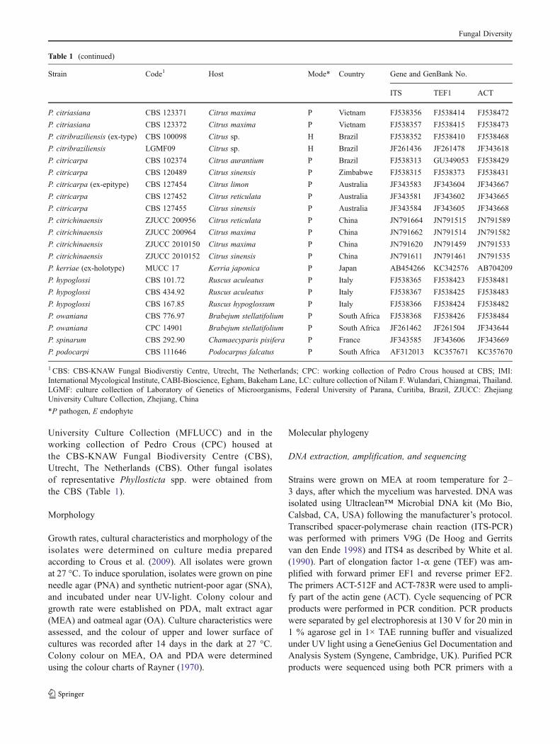
Phylogenetic analysis
Phylogenetic relationships among the Phyllosticta capitalensisisolates from various hosts and locations were investigated inthis study using MP and Bayesian phylogenetic analyses. Theanalysis of combined ITS, TEF and ACT genes of the
Fungal Diversity
Phyllosticta strains newly sequenced in this study and 67strains of Phyllosticta obtained from GenBank and MeiUniversity, Japan (Table 1) were aligned and used to constructtheir phylogeny. The combined dataset of 64 strains (includingthe out-group) consisted of 974 characters, of which 483characters were constant, and 148 characters were variableand parsimony-uninformative. Parsimony analysis generated48 trees, of which the best one is shown in Fig. 3 (TL = 873,CI = 0.804, RI = 0.963, RC = 0.774). In the parsimony tree(Fig. 3) bootstrap values and Bayesian analysis of combineddata are given at the nodes.
In the phylogenetic tree 12 clades representing variousPhyllosticta species are evident. Guignardia bidwellii waschosen as out-group. The representative strain of G.mangiferae (IMI 260576) fell outside the P. capitalensis s.str. clade. The isolates in the P. capitalensis s. str. clade werefrom different hosts and different continents. Phyllostictabrazilianiae was isolated from an orchid in Brazil; P.citricarpa was isolated from Citrus sp. and P. citriasianawas isolated from Citrus maxima, Vietnam; P. spinarum wasisolated from Chamaecyparis pisifera, France; P. kerriae wasisolated from Kerria japonica, Japan; P. citribraziliensis was
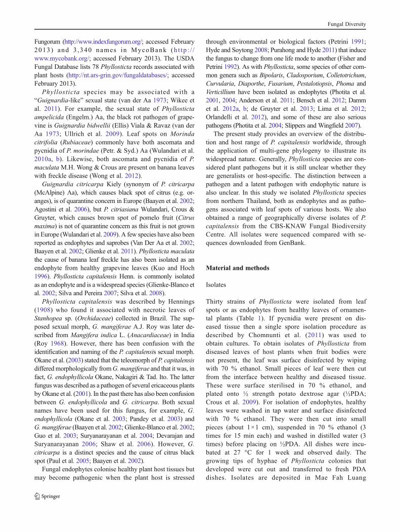
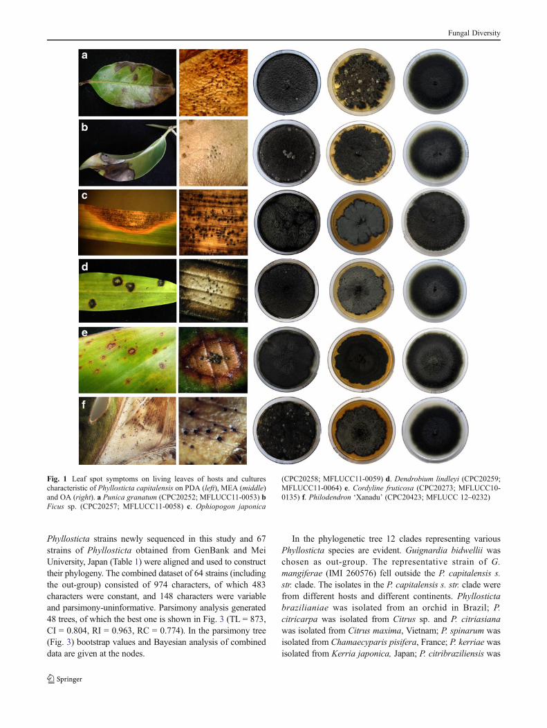
Fig. 1 Leaf spot symptoms on living leaves of hosts and culturescharacteristic of Phyllosticta capitalensis on PDA (left), MEA (middle)and OA (right). a Punica granatum (CPC20252; MFLUCC11-0053) bFicus sp. (CPC20257; MFLUCC11-0058) c. Ophiopogon japonica
(CPC20258; MFLUCC11-0059) d. Dendrobium lindleyi (CPC20259;MFLUCC11-0064) e. Cordyline fruticosa (CPC20273; MFLUCC10-0135) f. Philodendron ‘Xanadu’ (CPC20423; MFLUCC 12–0232)
Fungal Diversity
isolated from citrus, Brazil; P. hypoglossi was isolated fromRuscus aculeatus, Italy; P. citrichinaensis was isolated fromCitrus maxima, China; P. podocarpi was isolated fromPodocarpus falcatus, South Africa and P. owaniana fromBrabejum stellatifolium, South Africa.
Pathogenicity testing with Phyllosticta capitalensis
The ability of Phyllosticta capitalensis strains isolated fromleaf spots of five hosts in Thailand to induce leaf spot symp-toms on these host species was tested through inoculatingmycelium plugs onto attached wounded and unwounded liv-ing leaves. In all cases there was no infection of the younghealthy plant leaves.
Discussion
This study reviews previous data on Phyllosticta capitalensisand provides additional data on host infection and distributionin Thailand. Many factors such as environmental conditions,host and non-host organisms, and plant defence mechanisms(e.g. secondary metabolite, specific and non-specific proteinexpression and hydrogen peroxide residue) play an importantrole in response to microbial infection.
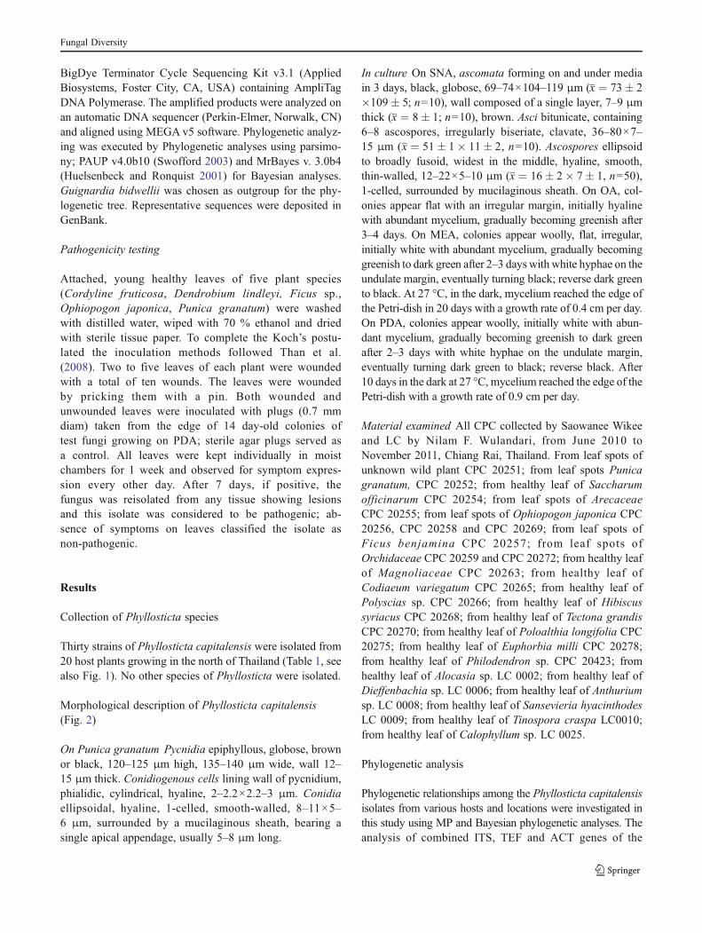
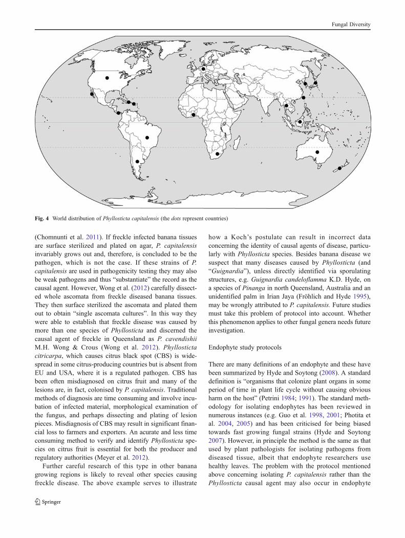
Phyllosticta capitalensis has been repeatedly isolatedworldwide from healthy plant tissues as an endophyte andrarely from leaf spots as a pathogen, and has been recordedfrom almost 70 plant families (Baayen et al. 2002; Okane etal. 2003; Motohashi et al. 2009, Tables 1 and 2, Fig. 4, thisstudy). The fact that it is isolated so often as an endophytehas important implications to studies of fungal biologyincluding plant pathology methodology, ecological resultsof endophyte studies and screening for novel compoundsfrom endophytes.
Implications to plant pathology methodology
A standard protocol used for isolating plant pathogensinvolves cutting segments from the leading edge of le-sions, which are then surface sterilized and plated ontomedia (Crous et al. 2009). The rationale is that the caus-ative agent grows out from the lesions and can be isolatedas a pure culture. Testing can then be undertaken toestablish pathogenicity, while the colony can be identifiedusing morphology. This standard methodology (Koch’spostulate) has been long used by plant pathologists todetermine the identity of non-sporulating pathogens adinfinitum (Phoulivong et al. 2010; Thompson et al. 2010;Wikee et al. 2011).
Fig. 2 Phyllosticta capitalensis on Punica granatum (CPC 20252). a–c Leaf spots on host plant d–f. Vertical section through pycnidia showingdeveloping conidia g–k. Conidia (d, bar=20 μm, g–k bars=10 μm)
Fungal Diversity
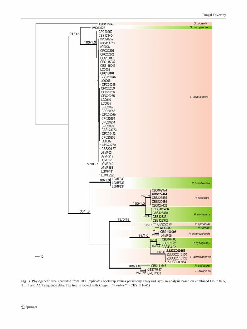
Fig. 3 Phylogenetic tree generated from 1000 replicates bootstrap values parsimony analysis/Bayesian analysis based on combined ITS rDNA,TEF1 and ACT sequence data. The tree is rooted with Guignardia bidwellii (CBS 111645)
Fungal Diversity
Table 2 Hosts and countries from which Phyllosticta capitalensis has been isolated, usually as an endophyte, rarely as a pathogen (P) (See alsoFig. 1)
Plant family Plant genus Country Reference
Acanthaceae Mackaya South Africa
Anacardiaceae Anacardium Brazil Glienke et al. 2011
Comocladia Puerto Rico
Loxostylis South Africa
Mangifera Brazil
Ghana Baayen et al. 2002
Myracrodruon Brazil Glienke et al. 2011
Rhus South Africa Baayen et al. 2002
Sclerocarya South Africa
Spondias Brazil
Annonaceae Monanathotaxis South Africa
Polyalthia Thailand Present study
Apocynaceae Aspidosperma Brazil Glienke et al. 2011
Secamone South Africa
Cerbera Japan Okane et al. 2003
Nerium Japan Motohashi et al. 2009
Aquifoliaceae Ilex USA
Japan Okane et al. 2003
Cerbera Japan Okane et al. 2003
Araliaceae Cussonia South Africa
Hedera South Africa
Polyscias Puerto Rico
Schefflera Costa Rica Baayen et al. 2002
Polyscias Thailand Present study
Araceae Alocasia Thailand Present study
Anthurium Thailand Present study
Dieffenbachia Thailand Present study
Livistona Thailand Present study
Spathiphyllum Japan Motohashi et al. 2009
Philodendron Thailand Present study
Asparagaceae Sansevieria Thailand Present study
Ophiopogon (P)* Thailand Present study
Boraginaceae Cordia South Africa
Calophyllaceae Calophyllum Thailand Present study
Capparaceae Maerua South Africa
Chrysobalanaceae Parinari South Africa
Combretaceae Combretum South Africa
Convolvulaceae Ipomoea Malaysia Present study
Cornaceae (Nyssaceae) Curtisia South Africa Baayen et al. 2002
Davidia Japan Motohashi et al. 2009
Celastraceae Putterlickia South Africa Baayen et al. 2002
Cercidiphyllaceae Cercidiphyllum Japan Motohashi et al. 2009
Ebenaceae Diospyros South Africa
Euclea South Africa
Ericaceae Rhododendron Japan Okane et al. 2003
Enkianthus Japan Okane et al. 2001
Vaccinium New Zealand Glienke et al. 2011
Fabaceae Bowdichia Brazil Glienke et al. 2011
Fungal Diversity
Table 2 (continued)
Plant family Plant genus Country Reference
Cercis Japan Motohashi et al. 2009
Fagaceae Lithocarpus Japan Motohashi et al. 2009
Ginkgoaceae Ginkgo Japan Motohashi et al. 2009
Lamiaceae Vitex Malaysia Present study
Lauraceae Cinnamomum Japan Okane et al. 2003
Ocotea South Africa
Lecythidaceae Barringtonia South Africa Baayen et al. 2002
Leguminosae Caesalpinia Japan Okane et al. 2003
Loganiaceae Stychnos South Africa
Anthocleista South Africa
Lythraceae Punica (P) Thailand Present study
Malvaceae Hibiscus Thailand Present study
Meliaceae Ekebergia South Africa
Trichilia South Africa Baayen et al. 2002
Menispermaceae Cocculus USA
Moraceae Artocarpus Thailand Baayen et al. 2002
Ficus (P) Thailand Present study
Morus Thailand
Magnoliaceae Michelia Thailand Present study
Magnolia Thailand Glienke et al. 2011
USA
Menispermaceae Tinospora Thailand Present study
Euphorbiaceae Clutia South Africa Baayen et al. 2002
Croton South Africa
Codiaeum Thailand Present study
Ctenomeria South Africa
Euphorbia Thailand Present study
Flacourtiaceae Dovyalis South Africa
Iteaceae Itea USA
Lamiaceae Tectona Thailand Present study
Musaceae Musa Thailand Okane et al. 2003
Indonesia, USA Glienke et al. 2011
Myrtaceae Eucalyptus Brazil, South Africa Glienke et al. 2011
Psidium Brazil Baayen et al. 2002
Oleaceae Ligustrum Japan Motohashi et al. 2009
Schrebera South Africa
Ophioglossaceae Botrychium USA
Orchidaceae Arundina Japan Okane et al. 2003
Coelogyne Thailand
Dendrobium Thailand Present study
Paphiopedilium Germany Okane et al. 2001
Orchidaceae Rhynchostylis sp. Malaysia Williams & Liu 1976, Singh 1980
Stanhopea Brazil Glienke et al. 2011
Pittosporaceae Pittosporum Hawaii Baayen et al. 2002
Poaceae Saccharum Thailand Present study
Podocarpaceae Podocarpus South Africa
Proteaceae Leucospermum Hawaii
Protea Hawaii
Telopea Australia
Fungal Diversity
Recent studies on Phyllosticta causing freckle disease ofbanana and disease of other hosts have shown that extremecaution must be applied when using the above standard
plant pathology approach (Wong et al. 2012). Conidia ofPhyllosticta rarely germinate in culture and thus with manyspecies it is impossible to obtain single spore cultures
Table 2 (continued)
Plant family Plant genus Country Reference
Pittosporaceae Pittosporum Japan Motohashi et al. 2009
Pteridophta Pteridophytes Japan Okane et al. 2003
Rhamanaceae Scutia South Africa
Zizyphus South Africa
Rhizophoraceae Kandelia Japan Okane et al. 2003
Rosaceae Cliffortia South Africa
Rubus Japan Okane et al. 2003
Prunus Japan Okane et al. 2003
Eriobotrya Japan Motohashi et al. 2009
Rubiaceae Canthium South Africa
Coprosma Hawaii Baayen et al. 2002
Gardenia South Africa
Pavetta South Africa
Rauvolfia South Africa
Rothmannia South Africa
Rutaceae Zanthoxylum Japan Okane et al. 2003
Citrus (P) Argentina, Australia, Brazil,China, Hong Kong, New Zealand,South Africa, Taiwan, Thailand, USA
Glienke et al. 2011;Wang et al. 2012
Fortunella USA
Vitex South Africa
Zanthoxylum Pueto Rico Baayen et al. 2002
Sapindaceae Allophylus South Africa
Dodonaea Hawaii
Litchi South Africa
Nephelium USA Glienke et al. 2011
Paullinia cupana Brazil Baayen et al. 2002
Smilacaceae Smilax South Africa Glienke et al. 2011
Solanaceae Capsicum Dominican Glienke et al. 2011
Stangeriaceae Stangeria South Africa Baayen et al. 2002
Sterculiaceae Sterculia Puerto Rico
Theaceae Camellia USA Baayen et al. 2002
Tiliaceae Grewia South Africa
Trimeniaceae Xymalos South Africa
Ulmaceae Trema South Africa
Veronicaceae Hebe (Veronica) South Africa
Viscaceae Viscum South Africa
Vitaceae Ampelopsis USA Baayen et al. 2002
Cryphostemma South Africa
Rhoicissus South Africa
Zamiaceae Encephalartos South Africa
Zingiberaceae Amomum Thailand Okane et al. 2003
Zingiber Thailand Okane et al. 2003
*(P) = Leaf spot
Fungal Diversity
(Chomnunti et al. 2011). If freckle infected banana tissuesare surface sterilized and plated on agar, P. capitalensisinvariably grows out and, therefore, is concluded to be thepathogen, which is not the case. If these strains of P.capitalensis are used in pathogenicity testing they may alsobe weak pathogens and thus “substantiate” the record as thecausal agent. However, Wong et al. (2012) carefully dissect-ed whole ascomata from freckle diseased banana tissues.They then surface sterilized the ascomata and plated themout to obtain “single ascomata cultures”. In this way theywere able to establish that freckle disease was caused bymore than one species of Phyllosticta and discerned thecausal agent of freckle in Queensland as P. cavendishiiM.H. Wong & Crous (Wong et al. 2012). Phyllostictacitricarpa, which causes citrus black spot (CBS) is wide-spread in some citrus-producing countries but is absent fromEU and USA, where it is a regulated pathogen. CBS hasbeen often misdiagnosed on citrus fruit and many of thelesions are, in fact, colonised by P. capitalensis. Traditionalmethods of diagnosis are time consuming and involve incu-bation of infected material, morphological examination ofthe fungus, and perhaps dissecting and plating of lesionpieces. Misdiagnosis of CBS may result in significant finan-cial loss to farmers and exporters. An acurate and less timeconsuming method to verify and identify Phyllosticta spe-cies on citrus fruit is essential for both the producer andregulatory authorities (Meyer et al. 2012).
Further careful research of this type in other bananagrowing regions is likely to reveal other species causingfreckle disease. The above example serves to illustrate
how a Koch’s postulate can result in incorrect dataconcerning the identity of causal agents of disease, particu-larly with Phyllosticta species. Besides banana disease wesuspect that many diseases caused by Phyllosticta (and“Guignardia”), unless directly identified via sporulatingstructures, e.g. Guignardia candeloflamma K.D. Hyde, ona species of Pinanga in north Queensland, Australia and anunidentified palm in Irian Jaya (Fröhlich and Hyde 1995),may be wrongly attributed to P. capitalensis. Future studiesmust take this problem of protocol into account. Whetherthis phenomenon applies to other fungal genera needs futureinvestigation.
Endophyte study protocols
There are many definitions of an endophyte and these havebeen summarized by Hyde and Soytong (2008). A standarddefinition is “organisms that colonize plant organs in someperiod of time in plant life cycle without causing obviousharm on the host” (Petrini 1984; 1991). The standard meth-odology for isolating endophytes has been reviewed innumerous instances (e.g. Guo et al. 1998, 2001; Photita etal. 2004, 2005) and has been criticised for being biasedtowards fast growing fungal strains (Hyde and Soytong2007). However, in principle the method is the same as thatused by plant pathologists for isolating pathogens fromdiseased tissue, albeit that endophyte researchers usehealthy leaves. The problem with the protocol mentionedabove concerning isolating P. capitalensis rather than thePhyllosticta causal agent may also occur in endophyte
Fig. 4 World distribution of Phyllosticta capitalensis (the dots represent countries)
Fungal Diversity
studies. Phyllosticta capitalensis is a quick growing species;in culture the colony covers a 9 cm Petri-dish in 10 days.Other species grow more slowly, e.g. P. yuccae reaches 3–5 cm diam in 15 days (Bissett 1986), while growth of P.vaccinii can be as low as 0.4 mm/day. Four species ofPhyllosticta (P. citriasiana, P. capitalensis, P. citricarpa andP. citrichinaensis) were recently isolated from Citrus in China(Wang et al. 2012) and P. citrichinaensis grew at 3.8±0.34 mm per day at 24 °C on PDA. Therefore, it is highlylikely thatP. capitalensiswill be isolated in endophyte studies,while others species which are probably also endophytes, willnot be isolated. This will skew the results considerably and theresulting endophyte lists, percentages and statistics may havelittle scientific meaning.
If this phenomenon of isolating P. capitalensis for the rea-sonsmentioned above is happening in the case ofPhyllosticta itmay also be happening in other genera such as Colletotrichum,Diaporthe, Fusarium or Pestalotiopsis (Promputtha et al.2005; Udayanga et al. 2011; Summerell et al. 2010;Maharachchikumbura et al. 2011; Damm et al. 2012a, b). Todetermine this fact we took the common ubiquitous endophytesColletotrichum siamense Prihastuti, L. Cai & K.D. Hyde,Diaporthe phaseolorum (Cooke & Ellis) Sacc., andPestalotiopsis adusta (Ellis & Everh.) Steyaert and blasted theITS sequence data from the epitype strains against GenBankaccessions and established the percentage of them that wereisolated as endophytes. Twelve strains of Colletotrichum inGenBank had 100 % similarity with the ITS sequence data ofC. siamense (Prihastuti et al. 2009) and 50 % of these strainswere isolated as endophytes. The ITS sequence of ex-isotype ofD. phoenicicola (CBS161.64, Udayanga et al. 2012) wassubjected to a standard BLAST search in GenBank to analyzethe homology of sequences. Among the first 10 results ofhighly similar sequences (100 or 99 % similarity) of retrieveddata, eight were isolated as endophytes from a wide range ofhosts. This is not surprising as Diaporthe is a commonlyisolated genus of fungal endophytes (Botella and Diez 2011;Sun et al. 2011; Hofstetter et al. 2012). Eleven strains ofPestalotiopsis in GenBank had 100 % similarity with the ITSsequence data of P. adusta (Maharachchikumbura et al. 2012)and 73 % were endophytes. Again this is not surprising asPestalotiopsis species are often isolated as endophytes (Aly etal. 2010; Debbab et al. 2011, 2012; Maharachchikumbura et al.2011). Therefore, it seems that certain taxa in these genera arewidespread endophytes and this needs further study.
Screening endophytes for novel compounds
It has been common practice to isolate endophytes frommedicinal plants using the premise that strains will be isolatedthat can produce bioactive compounds similar to those pro-duced by the plant (Krohn et al. 2007; Huang et al. 2008;Kumaran et al. 2008; Xu et al. 2010; Zhao et al. 2010). The
fungi are thought to have obtained the mechanisms of pro-duction of natural products from the plant by so called hori-zontal gene transfer (Strobel et al. 2004); whether this premiseis correct or pure speculation is open to debate (Schulz et al.2002; Selim et al. 2012) and in fact may be false (Heinig et al.2013). The isolation of endophytes may provide a large diver-sity of highly creative fungi for screening (Aly et al. 2010; Xuet al. 2011; Debbab et al. 2011; 2012). The findings of thepresent study indicate that there are problems with the aboveapproach. It is clear in the case of Phyllosticta that P.capitalensis will probably be the only endophyte speciesisolated. Therefore, we recommend that researchers screeningfor novel compounds should study the saprobes and patho-gens as well as the endophytes. This will give a higher fungaldiversity and higher likelihood of isolating rare and unusualspecies, and thus a higher likelihood of discovering greaterchemical diversity.
Acknowledgments We are grateful to Dhanushka Udayanga andSajeewa S.N. Maharachchikumbura for their assistance. This studywas financially supported by the Thailand Research Fund through theRoyal Golden Jubilee Ph. D. Program (Grant No. PHD/0198/2552) toS. Wikee and Kevin D. Hyde. The National Research Council ofThailand is thanked for the award of grant No 54201020004 to studythe genus Phyllosticta in Thailand.
References
Agostini JP, Peres NA, Mackenzie SJ, Adaskaveg JE, Timmer LW(2006) Effect of fungicides and storage conditions on postharvestdevelopment of citrus black spot and survival of Guignardiacitricarpa in fruit tissues. Plant Dis 90:1419–1424
Aly AH, Debbab A, Kjer J, Proksch P (2010) Fungal endophytes fromhigher plants: a prolific source of phytochemicals and other bio-active natural products. Fungal Divers 41:1–16
Anderson CSR, Dominique G, Ana PTU, Rita TOC, Isabela SA,Carlos RRM, Aristóteles GN (2011) Foliar endophytic fungi fromHevea brasiliensis and their antagonism on Microcyclus ulei.Fungal Divers 47:75–84
Baayen R, Bonants P, Verkley G, Carroll G, Van Der Aa H, De WeerdtM, Van Brouwershaven I, Schutte G, Maccheroni W Jr, DeBlanco C (2002) Nonpathogenic isolates of the citrus black spotfungus, Guignardia citricarpa, identified as a cosmopolitan en-dophyte of woody plants, G. mangiferae (Phyllostictacapitalensis). Phytopathology 92(5):464–477
Bensch K, Braun U, Groenewald JZ, Crous PW (2012) The genusCladosporium. Stud Mycol 72:1–401
Bissett J (1986) Discochora yuccae sp. nov. with Phyllosticta andLeptodothiorella synanamorphs. Can J Bot 64:1720–1726
Botella L, Diez JJ (2011) Phylogenetic diversity of fungal endophytesin Spanish stands of Pinus halepensis. Fungal Divers 47:9–18
Chomnunti P, Schoch CL, Aguirre-Hudson B, Ko-Ko TW, Hongsanan S,Jones EBG, Kodsueb R, Phookamsak R, Chukeatirote E, BahkaliAH, Hyde KD (2011) Capnodiaceae. Fungal Divers 51:103–134
Crous PW, Verkleij GJM, Groenewald JZ (2009) In: Samson RA (ed)Fungal biodiversity, vol 1, CBS laboratory manual series.Centraalbureau voor Schimmelcultures, Utrecht
Damm U, Cannon PF, Woudenberg JHC, Crous PW (2012a) TheColletotrichum acutatum species complex. Stud Mycol 73:37–113
Fungal Diversity
Damm U, Cannon PF, Woudenberg JHC, Johnston PR, Weir BS, TanYP, Shivas RG, Crous PW (2012b) The Colletotrichum boninensespecies complex. Stud Mycol 73:1–36
de Gruyter J, Woudenberg JHC, Aveskamp MM, Verkley GJM,Groenewald JZ, Crous PW (2013) Redisposition of Phoma-likeanamorphs in Pleosporales. Stud Mycol 75:1–36
De Hoog GS, Van Den Gerrits EAHG (1998) Molecular diagnostics ofclinical strains of filamentous Basidiomycetes. Mycoses 41:183–189
Debbab A, Aly AH, Proksch P (2011) Bioactive secondary metabolitesfrom endophytes and associated marine derived fungi. FungalDivers 49:1–12
Debbab A, Aly AH, Proksch P (2012) Endophytes and associatedmarine derived fungi—ecological and chemical perspectives.Fungal Divers 57:45–83
Devarajan PT, Suryanarayanan TS (2006) Evidence for the role ofphytophagous insects in dispersal of non-grass fungal endophytes.Fungal Divers 23:111–119
Fisher PJ, Petrini O (1992) Fungal saprobes and pathogens as endo-phytes of rice (Oryza sativa L.). New Phytol 120:137–143
Fröhlich J, Hyde KD (1995) Guignardia candeloflamma sp. nov.causing leaf spots of Pinanga sp. Mycol Res 99:110–112
Glienke C, Pereira O, Stringari D, Fabris J, Kava−Cordeiro V, Galli−Terasawa L, Cunnington J, Shivas R, Groenewald J, Crous PW(2011) Endophytic and pathogenic Phyllosticta species, with refer-ence to those associated with citrus black spot. Persoonia 26:47–56
Glienke-Blanco C, Aguilar-Vildoso CI, Vieira MLC, Barroso PAV,Azevedo JL (2002) Genetic variability in the endophytic fungusGuignardia citricarpa isolated from citrus plants. Genet Mol Biol25:251–255
Guo LD, Hyde KD, Liew ECY (1998) A method to promote sporula-tion in palm endophytic fungi. Fungal Divers 1:109–113
Guo LD, Hyde KD, Liew ECY (2001) Detection and taxonomic place-ment of endophytic fungi within frond tissues of Livistona chinensisbased on rDNA sequences. Mol Phylogenet Evol 20:1–13
Guo LD, Huang GR, Wang Y, He WH, Zheng WH, Hyde KD (2003)Molecular identification of white morphotype strains of endophyticfungi from Pinus tabulaeformis. Mycol Res 107(6):680–688
Heinig U, Scholz S, Jennewein S (2013) Getting to the bottom of taxolbiosynthesis by fungi. Fungal Divers. doi:10.1007/s13225-013-0228-7
Hennings P (1908) Fungi S. Paulenses IV a cl. Puttemans collecti.Hedwigia 48:1–20
Hofstetter V, Buyck B, Croll D, Viret O, Couloux A, Gindro K (2012)What if esca disease of grapevine were not a fungal disease?Fungal Divers 54:51–67
Huang WY, Cai YZ, Hyde KD, Corke H, Sun M (2008) Biodiversity ofendophytic fungi associated with 29 traditional Chinese medicinalplants. Fungal Divers 33:61–75
Huelsenbeck JP, Ronquist FR (2001) MrBayes: Bayesian inference ofphylogenetic trees. Biometrics 17:754–755
Hyde KD, Soytong K (2007) Understanding microfungal diversity-acritique. Cryptog Mycolog 28:281–289
Hyde KD, Soytong K (2008) The fungal endophyte dilemma. FungalDivers 33:163–173
Krohn K, Ullah Z, Hussain H, Flörke U, Schulz B, Draeger S, PescitelliG, Salvadori P, Antus S, Kurtán T (2007) Massarilactones E-G, newmetabolites from the endophytic fungus Coniothyrium sp., associ-ated with the plant Artimisia maritime. Chirality 19:464–470
Kumaran RS, Muthumary J, Hur B (2008) Production of taxol fromPhyllosticta spinarum, an endophytic fungus of Cupressus sp.Eng Life Sci 8:438–446
Kuo K, Hoch HC (1996) The parasitic relationship betweenPhyllosticta ampelicida and Vitis vinifera. Mycologia 88:626–634
Lima JS, Figueiredo JG, Gomes RG, Stringari D, Goulin EH,Adamoski D, Kava-Cordeiro V, Galli-Terasawa LV, Glienke C(2012) Genetic diversity of Colletotrichum spp. an endophytic
fungi in a medicinal plant, Brazilian pepper tree. ISRN Microbiol.doi:10.5402/2012/215716
Maharachchikumbura SSN, Guo LD, Chukeatirote E, Bahkali AH,Hyde KD (2011) Pestalotiopsis—morphology, phylogeny, bio-chemistry and diversity. Fungal Divers 50:167–187
Maharachchikumbura SSN, Guo LD, Cai L, Chukeatirote E, Wu WP,Sun X, Crous PW, Bhat DJ, McKenzie EHC, Bahkali AH, HydeKD (2012) A Multi-locus backbone tree for Pestalotiopsis, with apolyphasic characterization of 14 new species. Fungal Divers56:95–129
Meyer L, Jacobs R, Kotzé JM, Truter M, Korsten L (2012) Detectionand molecular identification protocols for Phyllosticta citricarpafrom citrus matter. S Afr J Sci. doi:10.4102/sajs.v108i3/4.602
Motohashi K, Inaba S, Anzai K, Takamatsu S, Nakashima C (2009)Phylogenetic analyses of Japanese species of Phyllosticta sensustricto. Mycoscience 50:291–302
Okane I, Nakagiri A, Ito T (2001) Identity of Guignardia sp. inhabitingericaceous plants. Can J Bot 79:101–109
Okane I, Lumyong S, Nakagiri A, Ito T (2003) Extensive host range ofan endophytic fungus, Guignardia endophyllicola (anamorph:Phyllosticta capitalensis). Mycoscience 44:353–363
Orlandelli RC, Alberto RN, Rubin Filho CJ, Pamphile JA (2012)Diversity of endophytic fungal community associated with Piperhispidum (Piperaceae) leaves. Genet Mol Res 11:1575–1585
Pandey AK, Reddy M, Sudhakara S, Trichur S (2003) ITS-RFLP andITS sequence analysis of a foliar endophytic Phyllosticta fromdifferent tropical trees. Mycol Res 108:974–978
Paul I, Van Jaarsveld AS, Korsten L, Hattingh V (2005) The potentialglobal geographical distribution of citrus black spot caused byGuignardia citricarpa (Kiely): likelihood of disease establish-ment in the European Union. Crop Prot 24:297–308
Petrini O (1984) Endophytic fungi in British Ericaceae: a preliminarystudy. Trans Br Mycol Soc 83:510–512
Petrini O (1991) Fungal endophytes of tree leaves. In: Fokkema NJ,van den Heuvel (eds) Microbial ecology of leaves. CambridgeUniversity Press, Cambridge, pp 185–187
Photita W, Lumyong S, Lumyong P, Hyde KD (2001) Endophyticfungi of wild banana (Musa acuminata) at Doi Suthep Pui Na-tional Park, in Thailand. Mycol Res 105:1508–1513
Photita W, Lumyong S, Lumyong P, McKenzie EHC, Hyde KD (2004)Are some endophytes of Musa acuminata latent pathogens? Fun-gal Divers 16:131–140
Photita W, Taylor PWJ, Ford R, Hyde KD, Lumyong S (2005) Morpho-logical and molecular characterization of Colletotrichum speciesfrom herbaceous plants in Thailand. Fungal Divers 18:117–133
Phoulivong S, Cai L, Chen H, McKenzie EHC, Abdelsalam K,Chukeatirote E, Hyde KD (2010) Colletotrichum gloeosporioidesis not a common pathogen on tropical fruits. Fungal Divers44:33–43
Prihastuti H, Cai L, Chen H, McKenzie EHC, Hyde KD (2009)Characterization of Colletotrichum species associated with coffeeberries in northern Thailand. Fungal Divers 39:89–109
Promputtha L, Jeewon R, Lumyong S, McKenzie EHC, Hyde KD(2005) Ribosomal DNA fingerprinting in the identification of nonsporulating endophytes from Magnolia liliifera (Magnoliaceae).Fungal Divers 20:167–186
Purahong W, Hyde KD (2011) Effects of fungal endophytes on grassand non-grass litter decomposition rates. Fungal Divers 47:1–7
Rayner RW (1970) A mycological colour chart. Commonwealth My-cological Institute and British Mycological Society, Kew, 34 pp
Roy AJ (1968) Some fungi from Almora. Indian Phytopathol 20:340–348Schulz B, Boyle C, Draeger S, Römmert AK (2002) Endophytic fungi:
a source of novel biologically active secondary metabolites.Mycol Res 106:996–1004
Selim KA, El-Beih AA, Abdel-Rahman TM, El-Diwany AI (2012)Biology of endophytic fungi. CREAM 2:31–82
Fungal Diversity
Shaw BD, Carroll GC, Hoch HC (2006) Generality of the prerequisiteof conidium attachment to a hydrophobic substratum as a signalfor germination among Phyllosticta species. Mycologia 98:186–194
Silva M, Pereira OL (2007) First report of Guignardia endophyllicolaleaf blight on Cymbidium (Orchidaceae) in Brazil. Australas PlantDis 2:31–32
Silva M, Pereira OL, Braga IF, Leli SM (2008) Leaf andpseudobulb diseases on Bifrenaria harrisoniae (Orchidaceae)caused by Phyllosticta capitalensis in Brazil. Australas PlantDis 3:53–56
Singh KG (1980) A check list of host and disease in Malaysia. BullMinist Agric Malays 154:280
Slippers B, Wingfield MJ (2007) Botryosphaeriaceae as endophytesand latent pathogens of woody plants: diversity, ecology andimpact. Fungal Biol Rev 21:90–106
Strobel GA, Daisy B, Castillo U, Harper J (2004) Natural productsfrom endophytic microorganisms. J Nat Prod 67:257–268
Summerell BA, Laurence MH, Liew ECY, Leslie JF (2010) Biogeog-raphy and phylogeography of Fusarium: a review. Fungal Divers44:3–13
Sun X, Guo LD, Hyde KD (2011) Community composition of endo-phytic fungi in Acer truncatum and their role in decomposition.Fungal Divers 47:85–95
Suryanarayanan TS, Ravishankar JP, Venkatesan G, Murali TS (2004)Characterization of the melanin pigment of a cosmopolitan fungalendophyte. Mycol Res 108:974–978
Swofford DL (2003) Paup*: Phylogenetic analysis using parsimony(*and other methods), version 4.0. Sinauer Associates,Sunderland
Than PP, Jeewon R, Hyde KD, Pongsupasamit S, Mongkolporn O,Taylor PWJ (2008) Characterization and pathogenicity ofColletotrichum species associated with anthracnose on chilli(Capsicum spp.) in Thailand. Plant Pathol 57:562–572
Thompson S, Alvarez-Loayza P, Terborgh J, Katul G (2010) Theeffects of plant pathogens on tree recruitment in the WesternAmazon under a projected future climate: a dynamical systemsanalysis. J Ecol 98:1434–1446
Udayanga D, Liu XX, McKenzie EHC, Chukeatirote E, Bahkali AH,Hyde KD (2011) The genus Phomopsis: biology, applications,species concepts and names of common phytopathogens. FungalDivers 50:189–225
Udayanga D, Liu XX, Crous PW, McKenzie EHC, Chukeatirote E,Hyde KD (2012) A multi-locus phylogenetic evaluation ofDiaporthe (Phomopsis). Fungal Divers 56:157–171
Ullrich CI, Kleespies RG, Enders M, Koch E (2009) Biology of theblack rot pathogen, Guignardia bidwellii, its development insusceptible leaves of grapevine Vitis vinifera. J Kult 61:82–90
Van Der Aa HA (1973) Studies in Phyllosticta I. Stud Mycol 5:1–110Van Der Aa H, Vanev S, Aptroot A, Summerbell R, Verkley G (2002) A
revision of the species described in Phyllosticta. Centraalbureauvoor Schimmelcultures, Utrecht
Wang X, Chen G, Huang F, Zhang J, Hyde KD, Li H (2012)Phyllosticta species associated with citrus diseases in China.Fungal Divers 52:209–224
White TJ, Bruns T, Lee S, Taylor J (1990) Amplification and directsequencing of fungal ribosomal RNA genes for phylogenetics. In:Innes MA, Gelfand DH, Sninsky JJ, White TJ (eds) PCR pro-tocols. A guide to methods and applications. Academic, SanDiego, pp 315–322
Wikee S, Udayanga D, Crous PW, Chukeatirote E, McKenzie EHC,Bahkali AH, Dai DQ, Hyde KD (2011) Phyllosticta—an overviewof current status of species recognition. Fungal Divers 46:171–182
Williams TH, Liu PSW (1976) A host list of plant disease in Sabah,Malaysia. Phytopathol Pap 19:1–67
Wong MH, Crous PW, Henderson J, Groenewald JZ, Drenth A (2012)Phyllosticta species associated with freckle disease of banana.Fungal Divers 56:173–187
Wulandari NF, To−Anun C, Hyde KD, Duong L, De Gruyter J, MeffertJ, Groenewald JZ, Crous PW (2009) Phyllosticta citriasiana sp.nov., the cause of Citrus tan spot of Citrus maxima in Asia.Fungal Divers 34:23–39
Wulandari NF, To-Anun C, Hyde KD (2010a) Guignardia morindaefrog eye leaf spotting disease of Morinda citrifolia (Rubiaceae).Mycosphere 1(4):325–331
Wulandari NF, To-Anun C, Lei C, Abd-Elsalam KA, Hyde KD(2010b) Guignardia/Phyllosticta species on banana. CryptogMycol 31(4):403–418
Xu J, Aly AH, Guan HS, Wray V, Proksch P (2010) Pestalotiopsis ahighly creative genus: chemistry and bioactivity of secondarymetabolites. Fungal Divers 44:15–31
Xu YC, Yao DQ, Jian HW, Zheng Z, De LW, Jin DF, Bing CG (2011)Molecular identification of endophytic fungi from medicinal plantHuperzia serrata based on rDNA ITS analysis. World J MicrobiolBiotechnol 27:495–503
Zhao J, Zhou L, Wang J, Shan T, Zhong L, Liu X, Gao X (2010) In:Mendez-Vilas A (ed), Current Research, Technology EducationTopics in Applied Microbiology and Microbial biotechnology:Endophytic fungi for producing bioactive compounds originallyfrom their host plants. p. 567–576
Fungal Diversity