Embed Size (px)
Citation preview

PHOTOSYNTHETICA 42 (1): 7-14, 2004
Photoinhibition of photosynthesis in sun and shade grown leaves
of grapevine (Vitis vinifera L.) M. BERTAMINI*, K. MUTHUCHELIAN**, and N. NEDUNCHEZHIAN*,**,***
Istituto Agrario di San Michele all' Adige, 38010, San Michele all’ Adige, Italy*
School of Energy, Environment and Natural Resources, Madurai Kamaraj University, Madurai - 625 021, India**
Abstract The degree of photoinhibition of sun and shade grown leaves of grapevine was determined by means of the ratio of vari-able to maximum chlorophyll (Chl) fluorescence (Fv/Fm) and electron transport measurements. The potential efficiency of photosystem 2 (PS2), Fv/Fm, markedly declined under high irradiance (HI) in shade leaves with less than 10 % of F0 level. In contrast, Fv/Fm ratio declined with about 20 % increase of F0 level in sun leaves. In isolated thylakoids, the rate of whole chain and PS2 activity in HI shade and sun leaves was decreased by about 60 and 40 %, respectively. A smaller inhibition of photosystem 1 (PS1) activity was also observed in both leaf types. In the subsequent dark incubation, fast recovery was observed in both leaf types that reached maximum PS2 efficiencies similar to non-photoinhibited control leaves. The artificial exogenous electron donors DPC, NH2OH, and Mn2+ failed to restore the HI-induced loss of PS2 activity in sun leaves, while DPC and NH2OH were significantly restored in shade leaves. Hence HI in shade leaves inactivates on the donor side of PS2 whereas it does at the acceptor side in sun leaves, respectively. Quantification of the PS2 reaction centre protein D1 and the 33 kDa protein of water splitting complex following HI-treatment of leaves showed pronounced differences between shade and sun leaves. The marked loss of PS2 activity in HI leaves was due to the marked loss of D1 protein of the PS2 reaction centre protein and the 33 kDa protein of the water splitting complex in sun and shade leaves, respectively. Additional keywords: chlorophyll fluorescence; donor side; D1 protein; electron transport; photoinhibition; photosystems 1 and 2. Introduction Diurnal and seasonal changes of irradiance in the field range from near darkness to levels well in excess of what is required for maximum photosynthesis. Excess radiant energy has the potential to damage the photosynthetic apparatus and plants have evolved strategies to minimise its detrimental effects. Photoinhibition is most commonly equated with photodamage, a long-term depression of quantum efficiency due to damage to the photosynthetic apparatus as a result of excess photosynthetic photon flux density, PPFD (Walters and Horton 1993). Chronic pho-toinhibition may be considered as a depression of photo-synthetic efficiency from which the plant does not reco-ver after 3–4 d in shade (Greer and Laing 1992). Although photoinhibition has been extensively investi-
gated under controlled conditions, there is still some doubt whether it is a significant factor for plants growing in the field. Ögren and Rosenqvist (1992) argue that moderate photoinhibition is common in field-grown plants. Several investigators view photoinhibition of photosynthesis as a process of stress-induced damage to photosystem 2 (PS2). This is based on the fact that, as a consquence of photoinhibition, the D1 protein of PS2 reaction centre becomes degraded (Prášil et al. 1992, Rintamaki et al. 1995). But some recent reports suggest that photoinhibition, first of all, results from the for-mation of photochemically inactive PS2 centres, which convert the excitation energy into heat. This down regu-lation of PS2 and thermal dissipation is considered as
——— Received 29 September 2003, accepted 7 November 2003. ***Author for correspondence. Permanent address: Govt. HSS, Vellimedupettai – 604 207, India; fax: 91-4147-222512, e-mail: [email protected] Abbreviations: Chl – chlorophyll; DCBQ – 2,6-dichloro-p-benzoquinone; DCPIP – 2,6-dichlorophenol indophenol; DPC – diphenyl carbazide; F0 – minimal fluorescence; Fm – maximum fluorescence; HI – high irradiance; MV – methyl viologen; PPFD, photosyn-thetic photon flux density; PS – photosystem; SDS-PAGE – sodium dodecylsulphate-polyacrylamide gel electrophoresis; SiMo – silicomolybdate. Acknowledgements: This work was in part supported by a grant from Provincia Autonoma of Trento and National Council of Research (CNR): project “Analisi e Ricerche per il sistema Agri-Industriale” sub-project “Prometavit”.
7

M. BERTAMINI et al.
a protective mechanism against HI stress (Aro et al. 1993, Gilmore and Björkman 1994). The photoinactivation and impairment of electron transport occur at the acceptor and donor sides of PS2, although inactivation of the acceptor side may be the main mechanism for the impairment of electron transport (Aro et al. 1993).
Shade plants are more prone to photoinhibition of photosynthesis than are sun plants (Powles 1984, Anderson and Osmond 1987). The higher sensitivity of shade plants to photoinhibition is generally ascribed to factors such as HI-absorbing chlorophyll (Chl) antenna for PS2 and lower rates of photon saturated photosynthe-sis. In the cyanobacterium Anacystis nidulans, low irradi-ance grown cells had a lower capacity to recover from photoinhibition than had HI-grown cells (Samuelsson et al. 1987, Lönneborg et al. 1988). This difference in abi-lity to recover from photoinhibition was attributed to different rates of the repair cycle of PS2 with a higher turnover of PS2 in HI-grown cells conferring significant protection against photoinhibition.
The effect of photoinhibition, alone or interacting with other stresses, on the long-term consequences of carbon balance or productivity, is little known. This, as Powles (1984) noted, is in part because the recovery of plants from photoinhibition has received very little atten-tion. A limited number of studies have shown that recovery takes at least several hours to occur. In fresh-water phytoplankton, for instance, recovery of photo-synthesis occurred during 4–20 h, depending on the ex-tent of the inhibition (Belay 1981). Similarly, the reco-very of the photon yield of leaves of Phaseolus vulgaris took between 4 and 8 h (Powles et al. 1983).
The D1 reaction centre protein of PS2 is a target of
HI-induced damage to the PS2 complex; turnover of the D1 protein is accelerated by increasing irradiance (Aro et al. 1993). The hypothesis that degradation of D1 protein may regulate the functioning of the PS2 repair cycle under photoinhibitory conditions has arisen from experi-ments with higher plants acclimated to different growth irradiances. Low irradiance or shade-grown plants are more susceptible to photoinhibition than HI or sun grown species (Powles 1984, Aro et al. 1993, Öquist et al. 1994). This higher susceptibility is accompanied by slow degradation of D1 protein (Tyystjärvi et al. 1992, Aro et al. 1993). In addition to the proteolysis of damaged D1 protein and de-novo synthesis of a new copy of D1, the repair cycle of PS2 involves several other reactions, including post-translation processing and modification of the protein, and ligation of the electron-transfer compo-nents (Aro et al. 1993). Thiele et al. (1996) suggested that, like in cold-acclimated spinach, the D1 protein is stabilised in young and mature canopy leaves and in D1 inactivation, and that turnover takes little part in photo-inhibition and recovery.
The object of our work was to compare the suscepti-bility to photoinhibition and the process of recovery in shade grown and sun grown leaves of Vitis vinifera. The effect of photoinhibition was analysed by fluorescence with respect to photosynthetic oxygen evolution and potential PS2 function. The amounts of D1 and 33 kDa proteins were also analysed in relation to the functional properties of PS2 after photoinhibition. The significance of the capacity of the PS2 repair cycle for the protection against photoinhibition was analysed by establishing rates of recovery from photoinhibition.
Materials and methods Plants: 13-year-old grapevine (Vitis vinifera L.) plants were grown under field conditions in San Michele all’ Adige, Italy (46°12’N, 11°08’E). Vines were trained to a permanent cordon at 1.80×0.80 m spacing, with upright growing shoots. Plants grown at full sunlight (maximum 2 000 µmol m-2 s-1) were designated as sun leaves, while plants grown at 40 % of sunlight (maximum 800 µmol m-2 s-1) were designated as shade leaves. Reduced irra-diances were obtained by screening full sunlight through appropriate nylon green meshes (1 m above the canopy). The fully expanded leaves (40-d-old) were taken as ex-perimental samples. Leaves were sampled early in the morning before they had experienced direct sunlight.
Photoinhibition and recovery under controlled condi-tions: Detached leaves were placed into a controlled-environment chamber equipped with a 24 V/250 W me-tal-halide lamp (H. Walz, Effeltrich, Germany). The up-per leaf surface was exposed to a photosynthetic photon flux density (PPFD) of 1 900 µmol m-2 s-1. Air tempera-ture was 20 ºC and relative humidity was 66±5 %. The
PPFD was measured with a quantum sensor (LI-Cor, Lincoln, NE, USA). Leaf temperatures, recorded with thermocouple attached to the lower surface, were between 27 and 29 ºC. Discs of 1.6 cm2 area were pun-ched from the leaf blades after specified times of HI ex-posure and placed on moist filter paper in petri dishes (temperature 25–27 ºC). The leaf discs were darkened for 10 min before the degree of photoinhibition was determi-ned by fluorescence measurement. For recovery from photoinhibition the leaf discs were kept in complete dark for 60 min.
Modulated Chl fluorescence in leaves was measured on leaf discs using a PAM 2000 fluorometer (H. Walz, Effeltrich, FRG). Before the measurements, the leaves were dark adapted for 10 min. F0 was measured by switching on the modulated radiation of 0.6 kHz; PPFD was less than 0.1 µmol m-2 s-1 at the leaf surface. Fm was measured at 20 kHz with a 1 s pulse of 6 000 µmol m-2 s-1
of “white light”.
8
PHOTOINHIBITION OF PHOTOSYNTHESIS IN SUN AND SHADE GROWN LEAVES
Activities of electron transport: Thylakoid membranes were isolated at 4 ºC, from each sample six to eight disks, as described by Russell et al. (1995). Leaf disks were homogenised in an ice cold grinding medium containing 25 mM Tris-HCl, pH (7.8), 10 mM NaCl, 5 mM MgCl2, and 330 mM sucrose. The homogenate was filtered rapid-ly through four layers of Miracloth and thylakoids were collected by centrifugation at 9 000×g for 5 min.
Whole chain electron transport (H2O→MV) and par-tial reactions of photosynthetic electron transport media-ted by PS2 (H2O→DCBQ; H2O→SiMo) and PS1 (DCPIPH2→MV) were measured as described by Nedunchezhian et al. (1997). Thylakoids were suspended at 10 g(Chl) m-3 in the assay medium containing 20 mM Tris-HCl, pH 7.5, 10 mM NaCl, 5 mM MgCl2, 5 mM NH4Cl, and 100 mM sucrose supplemented with 0.5 mM DCBQ and 0.2 mM SiMo.
DCPIP photoreduction was determined as the decrease in absorbance at 590 nm using a Hitachi 557 spectro-photometer. The reaction mixture (3 cm3) contained 20 mM Tris-HCl, pH 7.5, 5 mM MgCl2, 10 mM NaCl, 100 mM sucrose, 0.1 mM DCPIP, and thylakoid mem-branes equivalent to 20 µg of Chl. Where mentioned, the concentration of MnCl2, DPC, and NH2OH were 5.0, 0.5,
and 5.0 mM, respectively.
Immunological determination of thylakoid proteins: The relative contents of certain thylakoid proteins per Chl unit were determined immunologically by Western blot-ting. Thylakoids were solubilised in 5 % SDS, 15 % glycerine, 50 mM Tris-HCl (pH 6.8), and 2 % mercapto-ethanol at room temperature for 30 min. The polypeptides were separated by SDS-PAGE as described by Laemmli (1970) and proteins were then transferred to nitrocellu-lose by electroblotting for 3 h at 0.4 A after saturation with 10 % milk powder in TBS buffer (pH 7.5). The first antibody in 1 % gelatine was allowed to react overnight at room temperature. After washing with TBS containing 0.05 % Tween-20, the second antibody [Anti-Rabbit IgG (whole molecule) Biotin Conjugate, Sigma] was allowed to react in 1 % gelatine for 2 h. For detection of D1 pro-tein a polyclonal antiserum against spinach D1 protein was used (kindly provided by Prof. I. Ohad, Jerusalum, Israel). The antibody against 33 kDa protein of the water-splitting system was a gift from Dr. Barbato, Padova, Italy. The densitometric analysis of Western blots was performed with a Bio-Image analyser (Millipore Corporation, Michigan, USA).
Results Changes in Chl fluorescence: In order to compare the susceptibility to photoinhibition between sun and shade leaves, leaf samples were subjected to HI of variable duration (Fig. 1) in a controlled-environment chamber and followed by 60 min dark incubation for recovery (Fig. 2). Fig. 1 illustrates that shade leaves responded more sensitively to HI than sun leaves, as indicated by the more pronounced decrease in Fv/Fm ratios of shade lea-ves. The levels of F0, Fm, and Fv/Fm were recovered fast in first 30-min dark incubation and after only small diffe-rences in both sun and shade leaves (Fig. 2). The HI treatment for 60 min lead to a decline of about 38 or 61 % in Fv/Fm and elevation of about 19 or 5 % in F0 in sun and shade leaves, respectively. In the subsequent dark incubation for 60 min, the level of F0 recovered comple-tely in both leaf types while the level of Fv and Fv/Fm ratio did not recover completely (Fig. 2).
Changes in photosynthetic activities: Photosynthetic electron transport activities were measured in thylakoids isolated from HI-treated sun and shade grown leaves (Fig. 3). The rate of PS2 activity was decreased with increase in the time of HI in both sun and shade leaves. After 60 min, photosynthetic electron transports from H2O→DCBQ and H2O→SiMo were reduced by about 38 or 9 % in sun and 10 or 56 % in shade leaves, respec-tively. A significant reduction of PS2 activity was noticed when DCBQ was used as electron acceptor in sun leaves but it was marginally inhibited when SiMo was used
Fig. 1. Changes in the relative fluorescence emitted as minimal fluorescence (F0), maximum fluorescence (Fm), and the ratio of variable to maximum fluorescence (Fv/Fm) of sun and shade grown leaves of Vitis vinifera at different duration of high irra-diance (HI). Data are given in % of untreated controls. Control values for F0, Fm, and Fv/Fm were 2.6, 11.2, 0.811 and 2.6, 10.2, 0.798 in sun and shade grown leaves, respectively (mean ± S.E.; n = 5).
9
M. BERTAMINI et al.
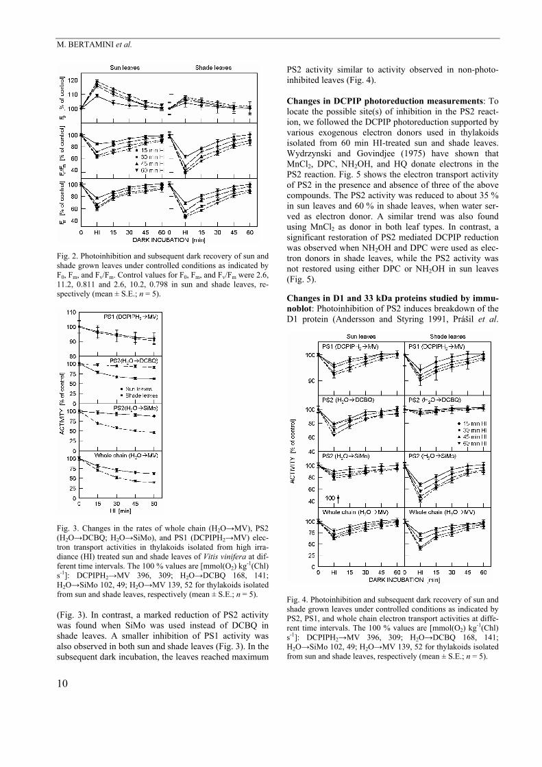
Fig. 2. Photoinhibition and subsequent dark recovery of sun and shade grown leaves under controlled conditions as indicated by F0, Fm, and Fv/Fm. Control values for F0, Fm, and Fv/Fm were 2.6, 11.2, 0.811 and 2.6, 10.2, 0.798 in sun and shade leaves, re-spectively (mean ± S.E.; n = 5).
Fig. 3. Changes in the rates of whole chain (H2O→MV), PS2 (H2O→DCBQ; H2O→SiMo), and PS1 (DCPIPH2→MV) elec-tron transport activities in thylakoids isolated from high irra-diance (HI) treated sun and shade leaves of Vitis vinifera at dif-ferent time intervals. The 100 % values are [mmol(O2) kg-1(Chl) s-1]: DCPIPH2→MV 396, 309; H2O→DCBQ 168, 141; H2O→SiMo 102, 49; H2O→MV 139, 52 for thylakoids isolated from sun and shade leaves, respectively (mean ± S.E.; n = 5). (Fig. 3). In contrast, a marked reduction of PS2 activity was found when SiMo was used instead of DCBQ in shade leaves. A smaller inhibition of PS1 activity was also observed in both sun and shade leaves (Fig. 3). In the subsequent dark incubation, the leaves reached maximum
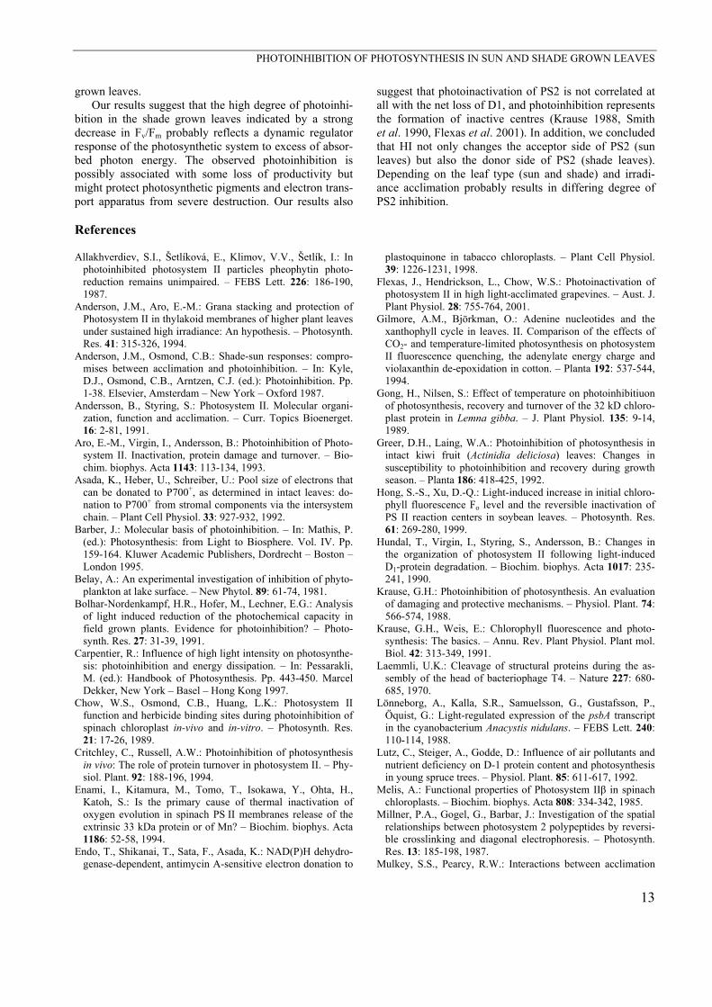
PS2 activity similar to activity observed in non-photo-inhibited leaves (Fig. 4).
Changes in DCPIP photoreduction measurements: To locate the possible site(s) of inhibition in the PS2 react-ion, we followed the DCPIP photoreduction supported by various exogenous electron donors used in thylakoids isolated from 60 min HI-treated sun and shade leaves. Wydrzynski and Govindjee (1975) have shown that MnCl2, DPC, NH2OH, and HQ donate electrons in the PS2 reaction. Fig. 5 shows the electron transport activity of PS2 in the presence and absence of three of the above compounds. The PS2 activity was reduced to about 35 % in sun leaves and 60 % in shade leaves, when water ser-ved as electron donor. A similar trend was also found using MnCl2 as donor in both leaf types. In contrast, a significant restoration of PS2 mediated DCPIP reduction was observed when NH2OH and DPC were used as elec-tron donors in shade leaves, while the PS2 activity was not restored using either DPC or NH2OH in sun leaves (Fig. 5). Changes in D1 and 33 kDa proteins studied by immu-noblot: Photoinhibition of PS2 induces breakdown of the D1 protein (Andersson and Styring 1991, Prášil et al.
Fig. 4. Photoinhibition and subsequent dark recovery of sun and shade grown leaves under controlled conditions as indicated by PS2, PS1, and whole chain electron transport activities at diffe-rent time intervals. The 100 % values are [mmol(O2) kg-1(Chl) s-1]: DCPIPH2→MV 396, 309; H2O→DCBQ 168, 141; H2O→SiMo 102, 49; H2O→MV 139, 52 for thylakoids isolated from sun and shade leaves, respectively (mean ± S.E.; n = 5).
10

PHOTOINHIBITION OF PHOTOSYNTHESIS IN SUN AND SHADE GROWN LEAVES
1992). In systems without protein biosynthesis this can be seen directly as a loss in D1 protein content. In intact plant the correlation between D1 protein content and activity of PS2 is more complex (Smith et al. 1990, Lutz et al. 1992). Photoinhibition induced inhibition of PS2 activity in thylakoids of sun and shade grown leaves was compared with changes in the relative contents of D1 and 33 kDa proteins as determined by Western blotting (Fig. 6) followed by quantification by the Bio-Image ap-paratus (Fig. 6). The relative contents of D1 and 33 kDa proteins decreased to 58 or 3 % and 2 or 68 % in 60 min HI-treated sun and shade leaves, respectively. In the subsequent 60 min of dark incubation, the leaves reached the original contents of D1 in sun leaves and of 33 kDa protein in shade leaves, similar to observation in non-photoinhibited leaves (Fig. 6).
Fig. 5. Effect of various exogenous electron donors on photo-system 2 (PS2) activity (H2O→DCPIP) in thylakoids isolated from 60 min high irradiance (HI) treated sun and shade grown leaves of Vitis vinifera. The 100 % values are [mmol(DCPIP red.) kg-1(Chl) s-1]: H2O→DCPIP 176, 78; DPC→DCPIP 179, 158; NH2OH→DCPIP 180, 154; MnCl2→DCPIP 77, 87 for thylakoids isolated from sun and shade grown leaves, respecti-vely (mean ± S.E.; n = 5).
Fig. 6. Relative contents of D1 and 33 kDa proteins of thylako-ids isolated from high irradiance (HI) treatment followed by dark incubation in sun and shade grown leaves of Vitis vinifera at different time intervals. Lane a, 0 min; lane b, 30 min HI; lane c, 60 min HI; lane d, 30 min dark; lane e, 60 min dark. Each lane was loaded with equal amount of Chl (5 µg). Histogram: Bio-Image densitometric evaluation. Inset: Western-blot.
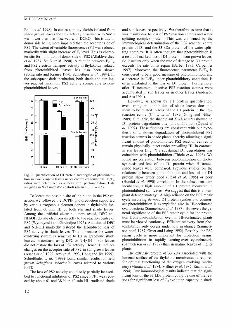
In Fig. 7, relative D1 protein contents and Fv/Fm ratios are compared after photoinhibitory treatments of sun and shade leaves. In the shade leaves, no significant D1 de-gradation could be attributed to the action of photoinhi-bitory radiation, even when Fv/Fm decreased to 58–61 % of the controls. The sun leaves showed a strong decrease in D1 protein (58 %) content together with the decline of Fv/Fm ratio to about 38 % (Fig. 7).
Discussion The present results indicate that exposure of shade and sun grown leaves to HI produces differential loss of pho-tosynthetic activity and potential efficiency of PS2 (Fv/Fm) where the shade leaves are more sensitive to HI than the sun leaves.
The decline in Fv/Fm, used here as a convenient mea-sure of photoinhibition, indicates a reduction in potential PS2 efficiency. In many studies, a close correlation of the Fv/Fm ratio with the quantum yield of photosynthetic O2 evolution or CO2 assimilation under PPFD-limiting con-ditions has been reported (Krause and Weis 1991, Mulkey and Pearcy 1992). The reduction of Fv/Fm in HI-treated sun leaves was mainly caused by marked increase of F0. An increase of F0 may be induced by the inacti-vation of part of PS2 reaction centres (Melis 1985, Critchley and Russell 1994, Yamane et al. 1997). Our experimental results from sun grown leaves are in accor-dance with this idea. The decrease of Fv/Fm in photoinhi-
bited shade leaves is mainly due to the significant decre-ase of Fm and marginal increase of F0. Bolhar-Nordenkampf et al. (1991) observed relatively low Fv/Fm ratios; even small changes of F0 or Fm would result in considerable changes in the Fv/Fm ratio. In the subsequent dark incubation, both shade grown and sun grown leaves reached maximum PS2 photochemistry efficiencies si-milar to those observed in non-photoinhibited leaves. The rate of recovery agrees with other reports on photoinhibi-tion in higher plants (Ögren et al. 1984, Öquist et al. 1992).
As shown by the analysis of electron transport activi-ties in thylakoids isolated from HI-treated sun leaves, the oxygen evolution was inhibited markedly when the electron acceptor used was DCBQ, but not SiMo. This is mainly due to HI-induced changes on the reducing side of PS2, i.e. photoinhibition. Chl fluorescence studies where F0 was markedly increased support it (Asada et al. 1992,
11
M. BERTAMINI et al.
Endo et al. 1998). In contrast, in thylakoids isolated from shade grown leaves the PS2 activity observed with SiMo was lower than that observed with DCBQ. This is due to donor side being more impaired than the acceptor side of PS2. The extent of variable fluorescence (Fv) was reduced markedly with slight increase of F0 level. This is charac-teristic for inhibition of donor side of PS2 (Allakhverdiev et al. 1987, Šetlík et al. 1990). A relation between Fv/Fm and PS2 electron transport activity in thylakoids isolated from photoinhibited leaves has also been shown (Somersalo and Krause 1990, Schnettger et al. 1994). In the subsequent dark incubation, both shade and sun lea-ves reached maximum PS2 activity comparable to non-photoinhibited leaves.
Fig. 7. Quantification of D1 protein and degree of photoinhibi-tion in Vitis vinifera leaves under controlled conditions. Fv/Fm ratios were determined as a measure of photoinhibition. Data are given in % of untreated controls (mean ± S.E.; n = 5).
To locate the possible site of inhibition in the PS2 re-
action, we followed the DCPIP photoreduction supported by various exogenous electron donors in thylakoids iso-lated from 60 min HI of both sun and shade leaves. Among the artificial electron donors tested, DPC and NH2OH donate electrons directly to the reaction centre of PS2 (Wydrzynski and Govindjee 1975). Addition of DPC and NH2OH markedly restored the HI-induced loss of PS2 activity in shade leaves. This is because the water-oxidizing system is sensitive to HI in grapevine shade leaves. In contrast, using DPC or NH2OH in sun leaves did not restore the loss of PS2 activity. Hence HI induces changes on the acceptor side of PS2 in sun-grown leaves (Asada et al. 1992, Aro et al. 1993, Hong and Xu 1999). Schiefthaler et al. (1999) found similar results for field grown Schefflera arboricola leaves adapted to various PPFD.
The loss of PS2 activity could only partially be ascri-bed to functional inhibition of PS2 since Fv/Fm was redu-ced by about 61 and 38 % in 60-min HI-irradiated shade
and sun leaves, respectively. We therefore assume that it was mainly due to loss of PS2 reaction centres and water splitting complex protein. This was confirmed by the immunological determination of the PS2 reaction centre protein of D1 and the 33 kDa protein of the water split-ting complex. It is often thought that photoinhibition is a result of marked loss of D1 protein in sun grown leaves. So it occurs only when the rate of damage to D1 protein exceeds the rate of its repair (Barber 1995, Carpentier 1997). Moreover, the fluorescence parameter Fv/Fm is considered to be a good measure of photoinhibition, and a decrease in Fv/Fm under photoinhibitory conditions is often attributed to the loss of D1 protein. Furthermore, after HI-treatment, inactive PS2 reaction centres were accumulated in sun leaves or in other leaves (Anderson and Aro 1994).
However, as shown by D1 protein quantification, even strong photoinhibition of shade leaves does not seem to be related to loss of the D1 protein in the PS2 reaction centre (Chow et al. 1989, Gong and Nilsen 1989). Similarly, the shade plant Tradescantia showed no D1 protein degradation after photoinhibition (Öquist et al. 1992). These findings are consistent with our hypo-thesis of a slower degradation of photoinhibited PS2 reaction centres in shade plants, thereby allowing a signi-ficant amount of photoinhibited PS2 reaction centres to remain physically intact under prevailing HI. In contrast, in sun leaves (Fig. 7) a substantial D1 degradation was coincident with photoinhibition (Thiele et al. 1996). We found no correlation between photoinhibition of photo-synthesis and loss of the D1 protein when HI-treated shade leaves were compared. Previous studies on the relationship between photoinhibition and loss of the D1 protein show either good (Ohad et al. 1985) or poor (Hundal et al. 1990) correlation. In the subsequent dark incubation, a high amount of D1 protein recovered in photoinhibited sun leaves. We suggest that this is a ‘sun-plant defence strategy’. A high reliance on the PS2 repair cycle involving de-novo D1 protein synthesis to counter-act photoinhibition is exemplified also in HI-acclimated cyanobacteria (Samuelsson et al. 1987). However, the ge-neral significance of the PS2 repair cycle for the protec-tion from photoinhibition even in HI-acclimated plants must be viewed cautiously. Efficient recovery from pho-toinhibition only occurs under low irradiance (Samuels-son et al. 1987, Greer and Laing 1992). Possibly, the PS2 repair cycle is more important for protection against photoinhibition in rapidly turning-over cyanobacteria (Samuelsson et al. 1987) than in mature leaves of higher plants.
The extrinsic protein of 33 kDa associated with the lumenal surface of the thylakoid membranes is required for optimal functioning of the oxygen evolving machi-nery (Murata et al. 1984, Millner et al. 1987, Enami et al. 1994). Our immunological results indicate that the signi-ficant loss of the 33 kDa protein could be one of the rea-sons for significant loss of O2 evolution capacity in shade
12
PHOTOINHIBITION OF PHOTOSYNTHESIS IN SUN AND SHADE GROWN LEAVES
grown leaves. Our results suggest that the high degree of photoinhi-
bition in the shade grown leaves indicated by a strong decrease in Fv/Fm probably reflects a dynamic regulator response of the photosynthetic system to excess of absor-bed photon energy. The observed photoinhibition is possibly associated with some loss of productivity but might protect photosynthetic pigments and electron trans-port apparatus from severe destruction. Our results also
suggest that photoinactivation of PS2 is not correlated at all with the net loss of D1, and photoinhibition represents the formation of inactive centres (Krause 1988, Smith et al. 1990, Flexas et al. 2001). In addition, we concluded that HI not only changes the acceptor side of PS2 (sun leaves) but also the donor side of PS2 (shade leaves). Depending on the leaf type (sun and shade) and irradi-ance acclimation probably results in differing degree of PS2 inhibition.
References Allakhverdiev, S.I., Šetlíková, E., Klimov, V.V., Šetlík, I.: In
photoinhibited photosystem II particles pheophytin photo-reduction remains unimpaired. – FEBS Lett. 226: 186-190, 1987.
Anderson, J.M., Aro, E.-M.: Grana stacking and protection of Photosystem II in thylakoid membranes of higher plant leaves under sustained high irradiance: An hypothesis. – Photosynth. Res. 41: 315-326, 1994.
Anderson, J.M., Osmond, C.B.: Shade-sun responses: compro-mises between acclimation and photoinhibition. – In: Kyle, D.J., Osmond, C.B., Arntzen, C.J. (ed.): Photoinhibition. Pp. 1-38. Elsevier, Amsterdam – New York – Oxford 1987.
Andersson, B., Styring, S.: Photosystem II. Molecular organi-zation, function and acclimation. – Curr. Topics Bioenerget. 16: 2-81, 1991.
Aro, E.-M., Virgin, I., Andersson, B.: Photoinhibition of Photo-system II. Inactivation, protein damage and turnover. – Bio-chim. biophys. Acta 1143: 113-134, 1993.
Asada, K., Heber, U., Schreiber, U.: Pool size of electrons that can be donated to P700+, as determined in intact leaves: do-nation to P700+ from stromal components via the intersystem chain. – Plant Cell Physiol. 33: 927-932, 1992.
Barber, J.: Molecular basis of photoinhibition. – In: Mathis, P. (ed.): Photosynthesis: from Light to Biosphere. Vol. IV. Pp. 159-164. Kluwer Academic Publishers, Dordrecht – Boston – London 1995.
Belay, A.: An experimental investigation of inhibition of phyto-plankton at lake surface. – New Phytol. 89: 61-74, 1981.
Bolhar-Nordenkampf, H.R., Hofer, M., Lechner, E.G.: Analysis of light induced reduction of the photochemical capacity in field grown plants. Evidence for photoinhibition? – Photo-synth. Res. 27: 31-39, 1991.
Carpentier, R.: Influence of high light intensity on photosynthe-sis: photoinhibition and energy dissipation. – In: Pessarakli, M. (ed.): Handbook of Photosynthesis. Pp. 443-450. Marcel Dekker, New York – Basel – Hong Kong 1997.
Chow, W.S., Osmond, C.B., Huang, L.K.: Photosystem II function and herbicide binding sites during photoinhibition of spinach chloroplast in-vivo and in-vitro. – Photosynth. Res. 21: 17-26, 1989.
Critchley, C., Russell, A.W.: Photoinhibition of photosynthesis in vivo: The role of protein turnover in photosystem II. – Phy-siol. Plant. 92: 188-196, 1994.
Enami, I., Kitamura, M., Tomo, T., Isokawa, Y., Ohta, H., Katoh, S.: Is the primary cause of thermal inactivation of oxygen evolution in spinach PS II membranes release of the extrinsic 33 kDa protein or of Mn? – Biochim. biophys. Acta 1186: 52-58, 1994.
Endo, T., Shikanai, T., Sata, F., Asada, K.: NAD(P)H dehydro-genase-dependent, antimycin A-sensitive electron donation to
plastoquinone in tabacco chloroplasts. – Plant Cell Physiol. 39: 1226-1231, 1998.
Flexas, J., Hendrickson, L., Chow, W.S.: Photoinactivation of photosystem II in high light-acclimated grapevines. – Aust. J. Plant Physiol. 28: 755-764, 2001.
Gilmore, A.M., Björkman, O.: Adenine nucleotides and the xanthophyll cycle in leaves. II. Comparison of the effects of CO2- and temperature-limited photosynthesis on photosystem II fluorescence quenching, the adenylate energy charge and violaxanthin de-epoxidation in cotton. – Planta 192: 537-544, 1994.
Gong, H., Nilsen, S.: Effect of temperature on photoinhibitiuon of photosynthesis, recovery and turnover of the 32 kD chloro-plast protein in Lemna gibba. – J. Plant Physiol. 135: 9-14, 1989.
Greer, D.H., Laing, W.A.: Photoinhibition of photosynthesis in intact kiwi fruit (Actinidia deliciosa) leaves: Changes in susceptibility to photoinhibition and recovery during growth season. – Planta 186: 418-425, 1992.
Hong, S.-S., Xu, D.-Q.: Light-induced increase in initial chloro-phyll fluorescence Fo level and the reversible inactivation of PS II reaction centers in soybean leaves. – Photosynth. Res. 61: 269-280, 1999.
Hundal, T., Virgin, I., Styring, S., Andersson, B.: Changes in the organization of photosystem II following light-induced D1-protein degradation. – Biochim. biophys. Acta 1017: 235-241, 1990.
Krause, G.H.: Photoinhibition of photosynthesis. An evaluation of damaging and protective mechanisms. – Physiol. Plant. 74: 566-574, 1988.
Krause, G.H., Weis, E.: Chlorophyll fluorescence and photo-synthesis: The basics. – Annu. Rev. Plant Physiol. Plant mol. Biol. 42: 313-349, 1991.
Laemmli, U.K.: Cleavage of structural proteins during the as-sembly of the head of bacteriophage T4. – Nature 227: 680-685, 1970.
Lönneborg, A., Kalla, S.R., Samuelsson, G., Gustafsson, P., Öquist, G.: Light-regulated expression of the psbA transcript in the cyanobacterium Anacystis nidulans. – FEBS Lett. 240: 110-114, 1988.
Lutz, C., Steiger, A., Godde, D.: Influence of air pollutants and nutrient deficiency on D-1 protein content and photosynthesis in young spruce trees. – Physiol. Plant. 85: 611-617, 1992.
Melis, A.: Functional properties of Photosystem IIβ in spinach chloroplasts. – Biochim. biophys. Acta 808: 334-342, 1985.
Millner, P.A., Gogel, G., Barbar, J.: Investigation of the spatial relationships between photosystem 2 polypeptides by reversi-ble crosslinking and diagonal electrophoresis. – Photosynth. Res. 13: 185-198, 1987.
Mulkey, S.S., Pearcy, R.W.: Interactions between acclimation
13

M. BERTAMINI et al.
and photoinhibition of photosynthesis of a tropical understo-rey herb, Alocasia macrorrhiza, during simulated canopy gap formation. – Funct. Ecol. 6: 719-729, 1992.
Murata, N., Miyao, M., Omata, T., Matsunami, H., Kuwabara, T.: Stoichiometry of components in the photosynthetic oxygen evolution system of photosystem II particles prepared with Triton X-100 from spinach chloroplasts. – Biochim. biophys. Acta 765: 363-369, 1984.
Nedunchezhian, N., Morales, F., Abadia, A., Abadia, J.: Decline in photosynthetic electron transport activity and changes in thylakoid protein pattern in field grown iron deficient peach (Prunus persica L.). – Plant Sci. 129: 29-38, 1997.
Ögren, E., Öquist, G., Hällgren, J.-E.: Photoinhibition of photo-synthesis in Lemna gibba as induced by the interaction be-tween light and temperature. I. Photosynthesis in vivo. – Phy-siol. Plant. 62: 181-186, 1984.
Ögren, E., Rosenqvist, E.: On the significance of photoinhibi-tion of photosynthesis in the field and its generality among species. – Photosynth. Res. 33: 63-71, 1992.
Ohad, I., Kyle, D.J., Hirschberg, J.: Light-dependent degrada-tion of the QB-protein in isolated pea thylakoids. – EMBO J. 4: 1655-1659, 1985.
Öquist, G., Anderson, J.M., McCaffery, S., Chow, W.S.: Mechanistic differences in photoinhibition of sun and shade plants. – Planta 188: 422-431, 1992.
Powles, S.B.: Photoinhibition of photosynthesis induced by visible light. – Annu. Rev. Plant Physiol. 35: 15-44, 1984.
Powles, S.B., Berry, J.A., Björkman, O.: Interaction between light and chilling temperature on the inhibition of photosyn-thesis in chilling sensitive plants. – Plant Cell Environ. 6: 117-123, 1983.
Prášil, O., Adir, N., Ohad, I.: Dynamics of photosystem II: mechanism of photoinhibition and recovery processes. – In: Barber, J. (ed.): The Photosystems: Structure, Function and Molecular Biology. Pp. 295-348. Elsevier, Amsterdam – London – New York – Tokyo 1992.
Rintamaki, E., Salo, R., Lehtonen, E., Aro, E.-M.: Regulation of D1 protein degradation during photoinhibition of photosystem II in vivo: Phosphorylation of D1 protein in various plant groups. – Planta 195: 379-386, 1995.
Russell, A.W., Critchley, C., Robinson, S.A., Franklin, L.A., Seaton, G., Chow, W.S., Anderson, J.M., Osmond, B.: Photo-system II regulation and dynamics of the chloroplast D1 pro-tein in Arabidopsis leaves during photosynthesis and photoinhibition. – Plant Physiol. 107: 943-952, 1995.
Samuelsson, G., Lönneborg, A., Gustafsson, P., Öquist, G.: The susceptibility of photosynthesis to photoinhibition and the capacity of recovery in high and low light grown cyanobacte-ria, Anacystis nidulans. – Plant Physiol. 83: 438-441, 1987.
Schiefthaler, U., Russell, A.W., Bolhàr-Nordenkampf, H.R., Critchley, C.: Photoregulation and photodamage in Scheflera arboricola leaves adapted to different light environments. – Aust. J. Plant Physiol. 26: 485-494, 1999.
Schnettger, B., Critchley, C., Santore, U.J., Graf, M., Krause, G.H.: Relationship between photoinhibition of photosynthe-sis, D1 protein turnover and chloroplast structure: effects of protein synthesis inhibitors. – Plant Cell Physiol. 17: 55-64, 1994.
Šetlík, I., Allakhverdiev, S.I., Nedbal, L., Šetlíkova, E., Klimov, V.V.: Three types of photosystem II photoinactivation. 1. Damaging processes on the acceptor side. – Photosynth. Res. 23: 39-48, 1990.
Smith, B.M., Morrissey, P.J., Guenther, J.E., Nemson, J.A., Harrison, M.A., Allen, J.F., Melis, A.: Response of the photo-synthetic apparatus in Dunaliella salina (green algae) to irra-diance stress. – Plant Physiol. 93: 1433-1440, 1990.
Somersalo, S., Krause, G.H.: Reversible photoinhibition of unhardened and cold-acclimated spinach leaves at chilling temperatures. – Planta 180: 181-187, 1990.
Thiele, A., Schirwitz, K., Winter, K., Krause, G.H.: Increased xanthophyll cycle activity and reduced D1 protein inactivation related to photoinhibition in two plant systems acclimated to excess light. – Plant Sci. 115: 237-250, 1996.
Tyystjärvi, E., Ali-Yrkkö, K., Kettunen, R., Aro, E.-M.: Slow degradation of the D1 protein is related to the susceptibility of low-light-grown pumpkin plants to photoinhibition. – Plant Physiol. 100: 1310-1317, 1992.
Walters, R.G., Horton, P.: Theoretical assessment of alternative mechanisms for non-photochemical quenching of PS II fluo-rescence in barley leaves. – Photosynth. Res. 36: 119-139, 1993.
Wydrzynski, T., Govindjee: A new site of bicarbonate effect in photosystem II of photosynthesis: Evidence from chlorophyll fluorescence transients in spinach chloroplasts. – Biochim. biophys. Acta 387: 403-408, 1975.
Yamane, Y., Kashino, Y., Koike, H., Satoh, K.: Increases in the fluorescence Fo level and reversible inhibition of Photosystem II reaction center by high-temperature treatments in higher plants. – Photosynth. Res. 52: 57-64, 1997.
14