Embed Size (px)
Citation preview
Photochemistry of thylakoid membranes in two peacultivars with different leaf colouration
Selma Mlinaric . Tihomir Cupic . Svetislav Popovic . Vlatka Jurkovic . Lidija Begovic .
Vera Cesar . Hrvoje Lepedus
Received: 23 June 2016 / Accepted: 19 October 2016 / Published online: 23 February 2017
� Brazilian Society of Plant Physiology 2017
Abstract Photochemical features of thylakoid mem-
branes and chlorophyll content were investigated in
two pea (Pisum sativum L.) cultivars with different
leaf colouration: ‘Assas’ (red leaves) and ‘Arvika’
(green leaves). In vivo measured chlorophyll fluores-
cence (OJIP) was used to evaluate overall photosyn-
thetic efficiency (expressed as performance index,
PIABS) as well as for advanced analysis of main
photochemical processes per active reaction centre
(RC) of photosystem II (PSII). To evaluate the
response of PSII driven linear electron transport
(relETR) and non-photochemical quenching (NPQ),
the plants were challenged with short-term high light
(*2000 lmol m-2 s-1). In spite of prevailing red
colour, leaves of ‘Assas’ had identical concentrations
of chlorophylls a and b as green ones. The OJIP
transients showed that red leaves grown in field
(500–800 lmol m-2 s-1) had decreased PIABS and
electron transport per RC beyond primary acceptor
(QA-) compared to green leaves. After high light
exposure red leaves revealed impaired relETR accom-
panied with increased NPQ values. Anthocyanins
located in epidermal cells affected neither chloro-
phylls concentrations nor the light capture features of
PSII. Despite equal concentrations of chlorophylls and
PSII photochemistry further than QA- in both leaf
types, red leaves reduced overall photosynthetic
efficiency due to impaired relETR in thylakoid
membranes.
Keywords Pisum sativum L. � Anthocyanins �Chlorophyll fluorescence � Photosynthesis �Photoprotection
Selma Mlinaric and Hrvoje Lepedus have contributed equally
to this work.
S. Mlinaric � L. Begovic � V. Cesar
Department of Biology, Josip Juraj Strossmayer
University of Osijek, Ulica cara Hadrijana 8/A,
HR-31000 Osijek, Croatia
e-mail: [email protected]
L. Begovic
e-mail: [email protected]
V. Cesar
e-mail: [email protected]
T. Cupic � S. Popovic � V. Jurkovic
Agricultural Institute Osijek, Juzno predgrade 17,
HR-31000 Osijek, Croatia
e-mail: [email protected]
S. Popovic
e-mail: [email protected]
V. Jurkovic
e-mail: [email protected]
H. Lepedus (&)
Faculty of Humanities and Social Sciences, Josip Juraj
Strossmayer University of Osijek, L. Jagera 9,
HR-31000 Osijek, Croatia
e-mail: [email protected]
123
Theor. Exp. Plant Physiol. (2017) 29:13–24
DOI 10.1007/s40626-016-0082-6

1 Introduction
Anthocyanins are the group of water soluble
plant pigments responsible for red, purple and blue
colour during various periods of plant development,
such as juvenile or senescing phases, as well as for
response to various environmental disturbances like
low temperature, high light, drought or salinity
(Cooney et al. 2015; Landi et al. 2015; Ranjan et al.
2014). Anthocyanin biosynthesis is stimulated by
various signals from the environment (temperature,
wounding, nutrient deficiency, etc.), but the most
significant role in biosynthesis of anthocyanins is
attributed to light (Das et al. 2011). Visible and UV-
light are responsible for accumulation of the specific
anthocyanin compounds that absorb fractions of
blue, green and UV wavelengths and, in that way,
contribute to the protection from excessive irradia-
tion, and reduce damage to photosystem II (PSII)
(Gould 2004; Landi et al. 2015).
Red colour of the most vegetative and reproductive
plant organs originates from anthocyanins (Manetas
2006). In leaves, they accumulate in vacuoles of
adaxial (upper) epidermis and mesophyll cell layers
and, less often, in abaxial (lower) epidermis (Hughes
et al. 2014; Landi et al. 2015; Liakopoulos et al. 2006).
Accumulation of anthocyanins in adaxial epidermis is
of great importance in ecological defence and photo-
protection. Ecological defence implies that antho-
cyanins reduce damage caused by potential herbivores
or pathogens (Gould 2004; Hughes and Lev-Yadun
2015). Additionally, the role of anthocyanins is
photoprotection of photosynthetic tissues susceptible
to excessive light. Red leaves serve as filter that
absorbs green light thus diminishing the damage to
photosystems because such filtered light that comes to
the chloroplasts produce less reactive oxygen species
(Gould 2004; Tattini et al. 2014). It was suggested that
photosynthetic cells provide signalling molecules that
are regulated by plastoquinone pool redox state. In that
way, high light conditions may indeed elicit the
accumulation of anthocyanins in leaves what provides
efficient protection for photosynthetic apparatus
against excessive light and, in that way, mitigate
photo inhibition (Das et al. 2011). Beneficial role of
abaxial anthocyanin accumulation was recently sug-
gested by Hughes et al. (2014). They proposed that
abaxial anthocyanins participate in leaves photopro-
tection without reducing the photosynthetic capacity,
which is advantageous in shade conditions with the
occasional appearance of high light sunflecks.
Although physiological role of anthocyanins is the
subject of numerous recent investigations (Dewez and
Perreault 2013; Landi et al. 2015; Solovchenko and
Chivkunova 2011; Tanino et al. 2014; Tsormpatsidis
et al. 2010), their exact role in regulation of photosyn-
thetic processes remain to be elucidated. Photosynthetic
processes are the most sensitive to changes in environ-
mental conditions. Energy absorption and redistribution
between two photosystems needs to be highly balanced
in order to function properly. Under low or moderate
light, it is assumed that the balance between absorption
and utilization of energy is achieved. In cases when
moderate light is combined with some other disturbances
such as low temperature or high light, it can reduce the
utilisation of absorbed light energy (Steyn et al. 2009).
Increased accumulation of anthocyanins in both, juvenile
and mature leaves under excessive light could efficiently
reduce the risk of photoinhibition (Fondom et al. 2009;
Landi et al. 2015). Solovchenko and Chivkunova (2011)
recently showed positive correlation between the elec-
tron transport rate and anthocyanins content at lower light
intensities in common hazel juvenile red leaves. They
also suggested that anthocyanins protect photosynthetic
apparatus from oxidative damage by reducing the
absorption of excessive light that would be normally
absorbed by chlorophylls and carotenoids. On the other
hand, Peng et al. (2006) reported limited electron
transport rate and increased non-photochemical quench-
ing (NPQ) in purple rice leaves suggesting increased
photoprotection in photooxidative conditions. Photopro-
tective influence on PSII was reported by Dewez and
Perreault (2013) supporting the role of anthocyanins in
regulation of PSII energy dissipation processes in red
Tradescantia leaves. Our previous investigation on the
photosynthetic apparatus functioning of red and green
leaves from ‘‘Crimson King’’ Norway maple tree (Stolfa
et al. 2006) revealed fully functional PSII in both leaf
types although red leaves had lower O2 production.
Further, lower Chl a to Chl b ratio and increased number
of thylakoids per granum in red leaves suggested that
they acted as shade ones.
Pea (Pisum sativum L.) cultivars with different leaf
colouration, cv. ‘Assas’ and cv. ‘Arvika’, show good
tolerance to low temperatures and early frost. Spring
pea cultivar ‘Arvika’ has green leaves while winter cv.
‘Assas’ have distinctive red leaves colouration in
14 Theor. Exp. Plant Physiol. (2017) 29:13–24
123

foliar stage of development. Red colouration of leaves
disappears with maturity of the plant. Our preliminary
investigations confirmed differences in leaf colour
using the leaf sections where the presence of antho-
cyanins were detected in red leaves. Histological
localisation of anthocyanins in a number of studies
was accompanied by determination of their content
showing that anthocyanins content to be dominant
over chlorophylls (Landi et al. 2014; Merzlyak et al.
2008; Peng et al. 2006).
Considering the leaf colour of two preliminary
investigated pea cultivars, our aim was to search
differences in primary photosynthesis events connected
with visible presence of anthocyanins. Thus, the chloro-
phylls concentrations, overall photosynthetic activity,
photochemical and non-photochemical processes per
active reaction centre of PSII (ABS, TR0, DI0 and ET0)
were determined. In order to avoid possible photoinhi-
bition, measurements were performed at moderate light
intensity because moderate light intensity provides
optimal conditions for photosynthesis performance
(Dewez and Perreault 2013; Landi et al. 2014; Osorio
et al. 2013; Steyn et al. 2009). However, photosynthetic
processes could be somewhat limited due to the fact that
anthocyanins can attenuate photosynthetically active
radiation. Although it is unfavourable in normal condi-
tions, this feature of red leaves might be beneficial under
stressful high light conditions (Gould et al. 2010). In
order to evaluate green and red pea leaves in response to
high light, the PSII driven electron transport rate
(relETR) and non-photochemical quenching (NPQ)
were investigated.
Here we hypothesized two possible scenarios of
photosynthetic regulatory mechanisms that might
appear as the consequence of anthocyanins accumu-
lation in red leaves in comparison to the green ones:
(1) it might affect chlorophyll accumulation and
consequently, photochemistry of PSII due to changes
in light absorption processes; and (2) it might reduce
photosynthetic efficiency at high light via reducing
thylakoid electron transport.
2 Materials and methods
2.1 Plant material and growth conditions
Two pea (Pisum sativum L.) cultivars, cv. ‘Assas’ and
cv. ‘Arvika’, were grown on euteric cambisol soil in an
experimental field (45�340N, 18�420E). Plants were
investigated at foliar developmental stage (3–4 leaves)
during March 2012, when cv. ‘Assas’ still produced
red leaves. Fully developed leaves (third real leaf from
the top) of both cultivars were sampled between 7:00
and 8:00 h under natural irradiance
(500–800 lmol m-2 s-1) with day/night cycles of
about 8/16 h. Five cm above the ground, average
temperature was 1.6 �C. Fast chlorophyll a fluores-
cence was measured in the field, whereas for other
analyses plants were collected, transported to the
laboratory and immediately used for analyses.
2.2 Experimental strategy
Due to the visible difference in leaf colouration
between cultivars ‘Arvika’ and ‘Assas’, preliminary
investigations using hand cut leaf sections were
performed to confirm the presence of anthocyanins.
After localization of anthocyanins, the fast chlorophyll
a fluorescence was measured in the field at moderate
light intensity to avoid photoinhibition. The same
leaves were used for chlorophyll a fluorescence mea-
surements, determination of photosynthetic pigments
concentration and analysis of acetone pigment extracts
absorption spectral characteristics. In order to acquire
detailed insight into functioning of photosynthetic
apparatus in two pea cultivars, we analysed chloro-
phyll a transients and OJIP test parameters. Further,
same category of leaves were collected in the field and
treated with short-term high irradiation using satura-
tion pulse method in laboratory to estimate the
response of photosynthetic apparatus of two pea
cultivars at high light conditions.
2.3 Anthocyanin localization
The middle parts of a fresh leaves were hand cut with
razor blade and transverse leaf sections were placed in
distilled water. The localization of anthocyanins was
observed using light microscope (Carl Zeiss, Jena,
Germany) and photographed with digital camera
(Olympus FE-115, China).
2.4 Photosynthetic pigments determination
and characteristic of absorption spectra
Both leaf types were sampled to analyse photosyn-
thetic pigments. Six randomly selected leaves from
Theor. Exp. Plant Physiol. (2017) 29:13–24 15
123
different plants were combined in a composite sample
and six replicates were taken for each analysis. After
removal of main veins, leaf tissue was macerated into
fine powder with liquid nitrogen. Powdered plant
material was extracted with the cold absolute acetone
at 4 �C and then re-extracted several times until it was
completely uncoloured. Acetone extracts from both,
red and green leaves were scanned at 330–900 nm by
UV–Vis spectrophotometer (Specord 40, Analytik
Jena, Germany) to obtain the total pigment spectra.
The same acetone extracts were used to determine the
concentrations of chlorophyll a (Chl a) and chloro-
phyll b (Chl b) spectrophotometrically at 661.6 and
644.8 nm. The chlorophylls a and b ratio (Chl a/b) and
total chlorophylls (Chl a ? b) concentration were
calculated according to Licthenthaler (1987).
2.5 Fast chlorophyll a fluorescence
Photosynthetic activity was measured on ten randomly
selected leaves of each cultivar using Plant efficiency
analyzer (PEA, Hansatech, UK). All measurements
were performed on fully dark-adapted leaves in the
field using lightweight leaf clips with shutter. Imme-
diately after 30 min of dark adaptation, the leaves
were exposed to a pulse of saturating red light
(3200 lmol m-2 s-1, peak at 650 nm). OJIP-test
was used to calculate biophysical parameters that
quantify stepwise energy flow through PSII (Strasser
et al. 2000; Strasser et al. 2004). Therefore, the
following recorded data were used: F0 (fluorescence
intensity at 50 ls), Fm (maximal fluorescence inten-
sity), F300 (fluorescence intensity at 300 ls), FI
(fluorescence intensity at 2 ls) and FJ (fluorescence
intensity at 30 ls) to calculate quantum yield of
primary photochemistry (TR0/ABS or Fv/Fm), density
of reaction centers (RC) (QA- reducing RC of PSII; RC/
CS0), variable fluorescence at J and I step (VJ, VI),
energy distribution through PSII per RC (ABS/RC,
TR0/RC, ET0/RC and DI0/RC) as well as performance
index (PIABS) together with three related parameters
(RC/ABS, TR0/DI0 and ET0/(TR0-ET0)) (Strasser
et al. 2000) (Table 1). Further, in order to convert data
into a form that they can be compared, double
normalization between O and P steps was used. The
O-P normalization gives us relative variable fluores-
cence WOP ¼ Ft � F0ð Þ= Fm � F0ð Þ which measures
the fraction of reduced QA at any time (t). Fluores-
cence data were plotted on logarithmic time scale
(from 50 ls to 1 s) and O, J, I and P steps were marked
in plots.
2.6 Saturating pulse method
The effect of light intensity on PSII activity was
determined by measuring chlorophyll a fluorescence
in vivo using amplitude modulated fluorometer (Mini-
PAM, Walz, Germany). Pea leaves were taken in the
field, immediately transferred in laboratory in
portable fridge at ?4 �C, dark-adapted for 30 min and
measured. Minimal (F0) and maximal (Fm) fluorescence
yields were measured using dark-adapted leaves. The F0
and Fm0 parameters were measured at the photosynthet-
ically active photon flux density (PPFD) of 100, 500,
1200 and 2000 lmol m-2 s-1 serving as short-term
treatments. Effective quantum yield of PSII ðDF/Fm0Þ,
relative linear electron transport (relETR) and non-
photochemical quenching (NPQ) were calculated
according to Schreiber et al. (1994).
2.7 Data analysis
Statistical differences between green and red leaves were
analysed using Student’s t testmodified for small samples.
Asterisk (*) indicates significant difference between
compared parameters in red and green leaves. Results
were expressed as arithmetic means of 6 replicates ± s-
tandard error of means (SE) except for measurement of
chlorophyll a fluorescence when 10 replicates were used.
Differences were considered significant at P\0.05. For
all statistical analyses Statistica 8.0 software (StatSoft,
Inc. 2007) was used.
3 Results
3.1 Anthocyanin localization in leaves
Leaf morphology of the two pea cultivars differed
mainly in leaf colouration. Cultivar ‘Arvika’ (Fig. 1a)
showed green leaves without any sign of red coloura-
tion while leaves of cv. ‘Assas’ (Fig. 1b) showed
apparent red colouration. Transverse sections of pea
cv. ‘Arvika’ (Fig. 1c) showed no anthocyanins in
vacuoles while cv. ‘Assas’ (Fig. 1d) revealed that
anthocyanins were mainly located in the vacuole of
upper (adaxial) and lower (abaxial) epidermis.
16 Theor. Exp. Plant Physiol. (2017) 29:13–24
123
3.2 Photosynthetic pigment concentrations
and absorption spectra
Chlorophylls, Chl a (t = 1.337, P = 0.211), Chl
b (t = 1.466, P = 0.173), Chl a ? b (t = 1.454,
P = 0.177) concentration as well as Chl a/b ratio
(t = 1.415, P = 0.187, Table 2) showed no signifi-
cant difference between green and red pea leaves.
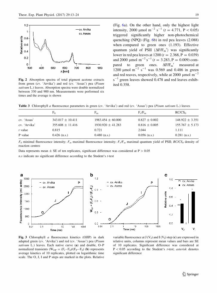
Although the absorption spectra of leaf extracts
revealed some similarities (Fig. 2), there were also
some marked differences between red and green
leaves. These differences can be attributed to the
presence of anthocyanins observed in leaf sections.
The acetone extracts from green leaves showed higher
peaks in blue (430 nm) and red (660 nm) part of the
absorption spectra (scanned at 330–900 nm), while
acetone extracts from red leaves revealed additional
peak at 360 nm (UV–A region) and had slightly lower
absorption in blue and red part of spectra.
3.3 Photosynthetic performance
Minimal, F0 (t = 0.815, P = 0.426) and maximal, Fm
(t = 0.721, P = 0.480) fluorescence intensity, as well
as maximum quantum yield of PSII, Fv/Fm (t = 2.044,
P = 0.056)) and density of active reaction centres per
cross section, RC/CS0 (t = 1.111, P = 0.281) showed
no significant difference between green and red pea
leaves (Table 3).
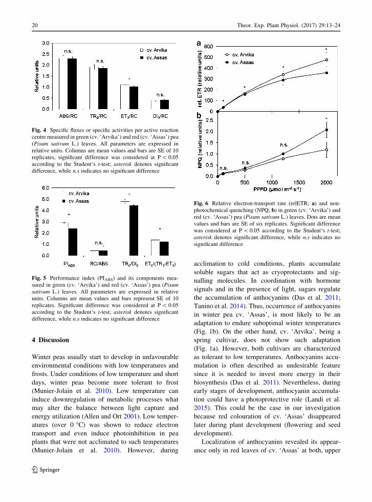
Both green and red pea leaves revealed character-
istic shape of OJIP transients. Significantly higher
variable fluorescence at J step, VJ (t = 2.115,
P = 0.048) and I step, VI (t = 5.515, P\ 0.05) was
observed in red pea leaves (0.450 for VJ and 0.724 for
Table 1 OJIP test parameters and expressions
Recorded and technical parameters Description
F0 Minimal fluorescence intensity (50 ls)
Fm Maximal fluorescence intensity—P step;
F300 Fluorescence intensity at 300 ls
FJ Fluorescence intensity at 2 ms—J step
FI Fluorescence intensity at 30 ms—I step
Fv Maximal variable fluorescence; Fm–F0
Vt Relative variable fluorescence at time t; ðFt�F0Þ= Fm�F0ð ÞVJ Relative variable fluorescence at time J step; FJ�F0ð Þ= Fm�F0ð ÞVI Relative variable fluorescence at time I step; FI�F0ð Þ= Fm�F0ð ÞM0 Approximated initial slope of relative variable fluorescence Fv; (dV/dt)0
TR0/ABS Maximum quantum yield of PSII; 1 � F0=Fmð Þ = Fv=Fm
RC/CS0 Density of active RC per cross section;Fv=Fm� VJ=M0ð Þ� F0
ABS/RC Absorption flux per active RC;
M0� 1=VJð Þ� 1= Fv=Fmð Þ½ �¼ TR0=RCð Þ= TR0=ABSð ÞTR0/RC Trapping flux per active RC; M0 9 (1/VJ)
ET0/RC Electron transport flux per active RC;
M0� 1=VJð Þ� 1 � VJð Þ¼ TR0=RCð Þ� ET0=TR0ð ÞDI0/RC Dissipation flux per active RC; (ABS/RC) - (TR0/RC)
PIABS Performance index on absorption basis;
RC/ABSð Þ� TR0=DI0ð Þ� [ET0= TR0 � ET0ð Þ�RC/ABS Density of RC on chlorophyll a basis;
RC=TR0ð Þ � TR0=ABSð Þ ¼ FJ � F0ð Þ=4 F300ls � F0
� �� �� Fv=Fmð Þ
TR0/DI0 Flux ration trapping per dissipation; Fv/F0
ET0/(TR0-ET0) Electron transport beyond QA-; Fm � FJð Þ= FJ � F0ð Þ
Theor. Exp. Plant Physiol. (2017) 29:13–24 17
123

VI) compared to green ones (0.421 for VJ and 0.676 for
VI, Fig. 3c).
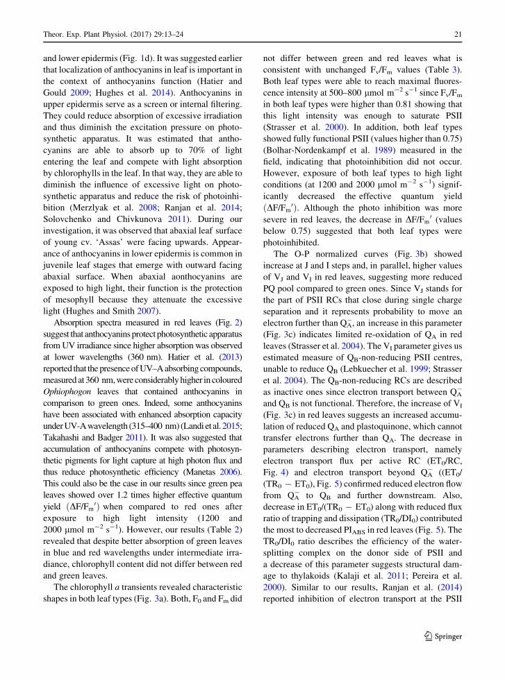
Specific activities (fluxes) per active reaction centre
measured in green and red pea leaves are shown in
Fig. 4. Absorption, ABS/RC (t = 0.286, P = 0.778),
trapping, TR0/RC (t = 0.873, P = 0.394) and dissi-
pation, DI0/RC (t = 1.343, P = 0.196) showed no
significant difference between green and red pea
leaves. On the contrary, electron transport beyond QA-,
ET0/RC (t = 3.420, P = 0.003) in red leaves (1.025)
was significantly decreased in comparison to green
pea leaves (1.100).
Decreased PIABS (t = 2.752, P = 0.013) values
were observed in red (2.915) leaves compared to green
ones (2.390) (Fig. 5). Decreased values of trapping to
dissipation ratio, TR0/DI0 (t = 2.248, P = 0.037) and
efficiency of excitation energy conversion to electron
transport, ET0/(TR0 - ET0) (t = 2.178, P = 0.043)
contributed to lower PIABS in red leaves because
density of reaction centres on Chl basis, RC/ABS
(t = 0.251, P = 0.805) in red leaves showed no
significant difference compared to green pea
leaves (Fig. 5).
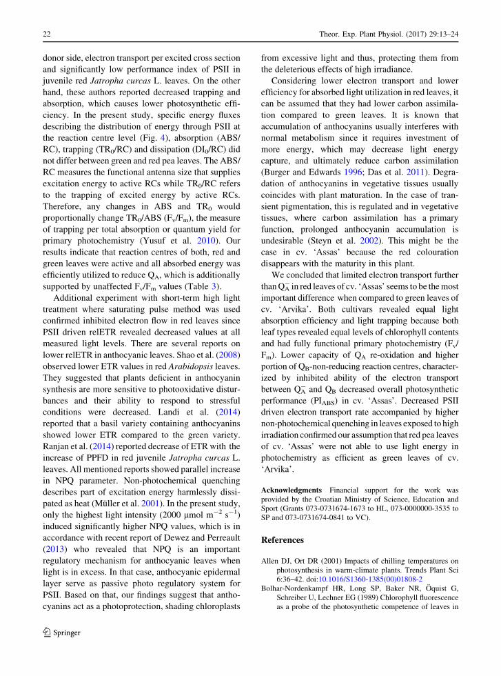
Red pea leaves showed significantly lower PSII
driven relETR values measured at all four PPFDs [100
(t = 2.540, P = 0.029), 500 (t = 3.887, P = 0.003),
1200 (t = 2.368, P = 0.039) and 2000 lmol m-2 s-1
(t = 3.283, P = 0.009)] compared to green leaves
Fig. 1 Morphology and
leaf anatomy of two pea
(Pisum sativum L.)
cultivars: ‘Arvika’ (a, c) and
‘Assas’ (b, d). Photographs
of the intact pea plants were
taken in the field before
measurements (a, b); bars
2 cm. Anthocyanins
localization in transverse
sections of pea leaves (c, d);
bars 100 lm
Table 2 Photosynthetic pigment content in green (cv. ‘Arvika’) and red (cv. ‘Assas’) pea (Pisum sativum L.) leaves
Chl a (mg/g-1 FW) Chl b (mg/g-1 FW) Chl a ? b (mg/g-1 FW) Chl a/b
cv. ‘Arvika’ 1.311 ± 0.064 0.687 ± 0.076 1.997 ± 0.135 1.977 ± 0.139
cv. ‘Assas’ 1.214 ± 0.034 0.553 ± 0.051 1.766 ± 0.085 2.261 ± 0.145
t-value 1.337 1.466 1.454 1.415
P-value 0.211 (n.s.) 0.173 (n.s.) 0.177 (n.s.) 0.187 (n.s.)
Data represents mean ± SE of six replicates, significant difference was considered at P[ 0.05
Chl a chlorophyll a, Chl b chlorophyll b, Chl a ? b total chlorophyll content, Chl a/b chlorophyll a to chlorophyll b ratio
n.s indicate no significant difference according to the Student’s t-test
18 Theor. Exp. Plant Physiol. (2017) 29:13–24
123
(Fig. 6a). On the other hand, only the highest light
intensity, 2000 lmol m-2 s-1 (t = 4.771, P\ 0.05)
triggered significantly higher non-photochemical
quenching (NPQ) (Fig. 6b) in red pea leaves (2.088)
when compared to green ones (1.193). Effective
quantum yield of PSII ðDF/Fm0Þ was significantly
lower in red pea leaves at 1200 (t = 2.368, P = 0.039)
and 2000 lmol m-2 s-1 (t = 3.283, P = 0.009) com-
pared to green ones. DF/Fm0 measured at
1200 lmol m-2 s-1 was 0.569 and 0.486 in green
and red reaves, respectively, while at 2000 lmol m-2
s-1 green leaves showed 0.478 and red leaves exhib-
ited 0.358.
Fig. 2 Absorption spectra of total pigment acetone extracts
from green (cv. ‘Arvika’) and red (cv. ‘Assas’) pea (Pisum
sativum L.) leaves. Absorption spectra were double normalized
between 330 and 900 nm. Measurements were performed six
times and the average is shown
Table 3 Chlorophyll a fluorescence parameters in green (cv. ‘Arvika’) and red (cv. ‘Assas’) pea (Pisum sativum L.) leaves
F0 Fm Fv/Fm RC/CS0
cv. ‘Assas’ 343.017 ± 10.411 1983.454 ± 60.000 0.827 ± 0.002 148.922 ± 3.351
cv. ‘Arvika’ 355.608 ± 11.416 1930.920 ± 41.283 0.816 ± 0.005 155.767 ± 5.173
t value 0.815 0.721 2.044 1.111
P value 0.426 (n.s.) 0.480 (n.s.) 0.056 (n.s.) 0.281 (n.s.)
F0 minimal fluorescence intensity; Fm maximal fluorescence intensity; Fv/Fm maximal quantum yield of PSII; RC/CS0 density of
reaction centres
Data represents mean ± SE of ten replicates, significant difference was considered at P[ 0.05
n.s indicate no significant difference according to the Student‘s t-test
Fig. 3 Chlorophyll a fluorescence kinetics (OJIP) in dark
adapted green (cv. ‘Arvika’) and red (cv. ‘Assas’) pea (Pisum
sativum L.) leaves. Each native curve (a) and double, O–P
normalized transients (WOP = (Ft-F0)/(FP-F0) (b) represents
average kinetics of 10 replicates, plotted on logarithmic time
scale. The O, J, I and P steps are marked in the plots. Relative
variable fluorescence at J (VJ) and I (VI) step (c) are expressed in
relative units, columns represent mean values and bars are SE
of 10 replicates. Significant difference was considered at
P\ 0.05 according to the Student‘s t-test; asterisk denotes
significant difference
Theor. Exp. Plant Physiol. (2017) 29:13–24 19
123
4 Discussion
Winter peas usually start to develop in unfavourable
environmental conditions with low temperatures and
frosts. Under conditions of low temperature and short
days, winter peas become more tolerant to frost
(Munier-Jolain et al. 2010). Low temperature can
induce downregulation of metabolic processes what
may alter the balance between light capture and
energy utilization (Allen and Ort 2001). Low temper-
atures (over 0 �C) was shown to reduce electron
transport and even induce photoinhibition in pea
plants that were not acclimated to such temperatures
(Munier-Jolain et al. 2010). However, during
acclimation to cold conditions, plants accumulate
soluble sugars that act as cryoprotectants and sig-
nalling molecules. In coordination with hormone
signals and in the presence of light, sugars regulate
the accumulation of anthocyanins (Das et al. 2011;
Tanino et al. 2014). Thus, occurrence of anthocyanins
in winter pea cv. ‘Assas’, is most likely to be an
adaptation to endure suboptimal winter temperatures
(Fig. 1b). On the other hand, cv. ‘Arvika’, being a
spring cultivar, does not show such adaptation
(Fig. 1a). However, both cultivars are characterized
as tolerant to low temperatures. Anthocyanins accu-
mulation is often described as undesirable feature
since it is needed to invest more energy in their
biosynthesis (Das et al. 2011). Nevertheless, during
early stages of development, anthocyanin accumula-
tion could have a photoprotective role (Landi et al.
2015). This could be the case in our investigation
because red colouration of cv. ‘Assas’ disappeared
later during plant development (flowering and seed
development).
Localization of anthocyanins revealed its appear-
ance only in red leaves of cv. ‘Assas’ at both, upper
Fig. 4 Specific fluxes or specific activities per active reaction
centre measured in green (cv. ‘Arvika’) and red (cv. ‘Assas’) pea
(Pisum sativum L.) leaves. All parameters are expressed in
relative units. Columns are mean values and bars are SE of 10
replicates, significant difference was considered at P\ 0.05
according to the Student‘s t-test; asterisk denotes significant
difference, while n.s indicates no significant difference
Fig. 5 Performance index (PIABS) and its components mea-
sured in green (cv. ‘Arvika’) and red (cv. ‘Assas’) pea (Pisum
sativum L.) leaves. All parameters are expressed in relative
units. Columns are mean values and bars represent SE of 10
replicates. Significant difference was considered at P\ 0.05
according to the Student‘s t-test; asterisk denotes significant
difference, while n.s indicates no significant difference
Fig. 6 Relative electron-transport rate (relETR; a) and non-
photochemical quenching (NPQ; b) in green (cv. ‘Arvika’) and
red (cv. ‘Assas’) pea (Pisum sativum L.) leaves. Dots are mean
values and bars are SE of six replicates. Significant difference
was considered at P\ 0.05 according to the Student‘s t-test;
asterisk denotes significant difference, while n.s indicates no
significant difference
20 Theor. Exp. Plant Physiol. (2017) 29:13–24
123
and lower epidermis (Fig. 1d). It was suggested earlier
that localization of anthocyanins in leaf is important in
the context of anthocyanins function (Hatier and
Gould 2009; Hughes et al. 2014). Anthocyanins in
upper epidermis serve as a screen or internal filtering.
They could reduce absorption of excessive irradiation
and thus diminish the excitation pressure on photo-
synthetic apparatus. It was estimated that antho-
cyanins are able to absorb up to 70% of light
entering the leaf and compete with light absorption
by chlorophylls in the leaf. In that way, they are able to
diminish the influence of excessive light on photo-
synthetic apparatus and reduce the risk of photoinhi-
bition (Merzlyak et al. 2008; Ranjan et al. 2014;
Solovchenko and Chivkunova 2011). During our
investigation, it was observed that abaxial leaf surface
of young cv. ‘Assas’ were facing upwards. Appear-
ance of anthocyanins in lower epidermis is common in
juvenile leaf stages that emerge with outward facing
abaxial surface. When abaxial aonthocyanins are
exposed to high light, their function is the protection
of mesophyll because they attenuate the excessive
light (Hughes and Smith 2007).
Absorption spectra measured in red leaves (Fig. 2)
suggest that anthocyanins protect photosynthetic apparatus
from UV irradiance since higher absorption was observed
at lower wavelengths (360 nm). Hatier et al. (2013)
reported that the presence of UV–A absorbing compounds,
measured at 360 nm, were considerably higher in coloured
Ophiophogon leaves that contained anthocyanins in
comparison to green ones. Indeed, some anthocyanins
have been associated with enhanced absorption capacity
under UV-Awavelength (315–400 nm) (Landiet al. 2015;
Takahashi and Badger 2011). It was also suggested that
accumulation of anthocyanins compete with photosyn-
thetic pigments for light capture at high photon flux and
thus reduce photosynthetic efficiency (Manetas 2006).
This could also be the case in our results since green pea
leaves showed over 1.2 times higher effective quantum
yield ðDF/Fm0Þ when compared to red ones after
exposure to high light intensity (1200 and
2000 lmol m-2 s-1). However, our results (Table 2)
revealed that despite better absorption of green leaves
in blue and red wavelengths under intermediate irra-
diance, chlorophyll content did not differ between red
and green leaves.
The chlorophyll a transients revealed characteristic
shapes in both leaf types (Fig. 3a). Both, F0 and Fm did
not differ between green and red leaves what is
consistent with unchanged Fv/Fm values (Table 3).
Both leaf types were able to reach maximal fluores-
cence intensity at 500–800 lmol m-2 s-1 since Fv/Fm
in both leaf types were higher than 0.81 showing that
this light intensity was enough to saturate PSII
(Strasser et al. 2000). In addition, both leaf types
showed fully functional PSII (values higher than 0.75)
(Bolhar-Nordenkampf et al. 1989) measured in the
field, indicating that photoinhibition did not occur.
However, exposure of both leaf types to high light
conditions (at 1200 and 2000 lmol m-2 s-1) signif-
icantly decreased the effective quantum yield
ðDF/Fm0Þ. Although the photo inhibition was more
severe in red leaves, the decrease in DF/Fm0 (values
below 0.75) suggested that both leaf types were
photoinhibited.
The O-P normalized curves (Fig. 3b) showed
increase at J and I steps and, in parallel, higher values
of VJ and VI in red leaves, suggesting more reduced
PQ pool compared to green ones. Since VJ stands for
the part of PSII RCs that close during single charge
separation and it represents probability to move an
electron further than QA-, an increase in this parameter
(Fig. 3c) indicates limited re-oxidation of QA in red
leaves (Strasser et al. 2004). The VI parameter gives us
estimated measure of QB-non-reducing PSII centres,
unable to reduce QB (Lebkuecher et al. 1999; Strasser
et al. 2004). The QB-non-reducing RCs are described
as inactive ones since electron transport between QA-
and QB is not functional. Therefore, the increase of VI
(Fig. 3c) in red leaves suggests an increased accumu-
lation of reduced QA and plastoquinone, which cannot
transfer electrons further than QA. The decrease in
parameters describing electron transport, namely
electron transport flux per active RC (ET0/RC,
Fig. 4) and electron transport beyond QA- ((ET0/
(TR0 - ET0), Fig. 5) confirmed reduced electron flow
from QA- to QB and further downstream. Also,
decrease in ET0/(TR0 - ET0) along with reduced flux
ratio of trapping and dissipation (TR0/DI0) contributed
the most to decreased PIABS in red leaves (Fig. 5). The
TR0/DI0 ratio describes the efficiency of the water-
splitting complex on the donor side of PSII and
a decrease of this parameter suggests structural dam-
age to thylakoids (Kalaji et al. 2011; Pereira et al.
2000). Similar to our results, Ranjan et al. (2014)
reported inhibition of electron transport at the PSII
Theor. Exp. Plant Physiol. (2017) 29:13–24 21
123
donor side, electron transport per excited cross section
and significantly low performance index of PSII in
juvenile red Jatropha curcas L. leaves. On the other
hand, these authors reported decreased trapping and
absorption, which causes lower photosynthetic effi-
ciency. In the present study, specific energy fluxes
describing the distribution of energy through PSII at
the reaction centre level (Fig. 4), absorption (ABS/
RC), trapping (TR0/RC) and dissipation (DI0/RC) did
not differ between green and red pea leaves. The ABS/
RC measures the functional antenna size that supplies
excitation energy to active RCs while TR0/RC refers
to the trapping of excited energy by active RCs.
Therefore, any changes in ABS and TR0 would
proportionally change TR0/ABS (Fv/Fm), the measure
of trapping per total absorption or quantum yield for
primary photochemistry (Yusuf et al. 2010). Our
results indicate that reaction centres of both, red and
green leaves were active and all absorbed energy was
efficiently utilized to reduce QA, which is additionally
supported by unaffected Fv/Fm values (Table 3).
Additional experiment with short-term high light
treatment where saturating pulse method was used
confirmed inhibited electron flow in red leaves since
PSII driven relETR revealed decreased values at all
measured light levels. There are several reports on
lower relETR in anthocyanic leaves. Shao et al. (2008)
observed lower ETR values in red Arabidopsis leaves.
They suggested that plants deficient in anthocyanin
synthesis are more sensitive to photooxidative distur-
bances and their ability to respond to stressful
conditions were decreased. Landi et al. (2014)
reported that a basil variety containing anthocyanins
showed lower ETR compared to the green variety.
Ranjan et al. (2014) reported decrease of ETR with the
increase of PPFD in red juvenile Jatropha curcas L.
leaves. All mentioned reports showed parallel increase
in NPQ parameter. Non-photochemical quenching
describes part of excitation energy harmlessly dissi-
pated as heat (Muller et al. 2001). In the present study,
only the highest light intensity (2000 lmol m-2 s-1)
induced significantly higher NPQ values, which is in
accordance with recent report of Dewez and Perreault
(2013) who revealed that NPQ is an important
regulatory mechanism for anthocyanic leaves when
light is in excess. In that case, anthocyanic epidermal
layer serve as passive photo regulatory system for
PSII. Based on that, our findings suggest that antho-
cyanins act as a photoprotection, shading chloroplasts
from excessive light and thus, protecting them from
the deleterious effects of high irradiance.
Considering lower electron transport and lower
efficiency for absorbed light utilization in red leaves, it
can be assumed that they had lower carbon assimila-
tion compared to green leaves. It is known that
accumulation of anthocyanins usually interferes with
normal metabolism since it requires investment of
more energy, which may decrease light energy
capture, and ultimately reduce carbon assimilation
(Burger and Edwards 1996; Das et al. 2011). Degra-
dation of anthocyanins in vegetative tissues usually
coincides with plant maturation. In the case of tran-
sient pigmentation, this is regulated and in vegetative
tissues, where carbon assimilation has a primary
function, prolonged anthocyanin accumulation is
undesirable (Steyn et al. 2002). This might be the
case in cv. ‘Assas’ because the red colouration
disappears with the maturity in this plant.
We concluded that limited electron transport further
than QA- in red leaves of cv. ‘Assas’ seems to be the most
important difference when compared to green leaves of
cv. ‘Arvika’. Both cultivars revealed equal light
absorption efficiency and light trapping because both
leaf types revealed equal levels of chlorophyll contents
and had fully functional primary photochemistry (Fv/
Fm). Lower capacity of QA re-oxidation and higher
portion of QB-non-reducing reaction centres, character-
ized by inhibited ability of the electron transport
between QA- and QB decreased overall photosynthetic
performance (PIABS) in cv. ‘Assas’. Decreased PSII
driven electron transport rate accompanied by higher
non-photochemical quenching in leaves exposed to high
irradiation confirmed our assumption that red pea leaves
of cv. ‘Assas’ were not able to use light energy in
photochemistry as efficient as green leaves of cv.
‘Arvika’.
Acknowledgments Financial support for the work was
provided by the Croatian Ministry of Science, Education and
Sport (Grants 073-0731674-1673 to HL, 073-0000000-3535 to
SP and 073-0731674-0841 to VC).
References
Allen DJ, Ort DR (2001) Impacts of chilling temperatures on
photosynthesis in warm-climate plants. Trends Plant Sci
6:36–42. doi:10.1016/S1360-1385(00)01808-2
Bolhar-Nordenkampf HR, Long SP, Baker NR, Oquist G,
Schreiber U, Lechner EG (1989) Chlorophyll fluorescence
as a probe of the photosynthetic competence of leaves in
22 Theor. Exp. Plant Physiol. (2017) 29:13–24
123

the field: a review of current instrumentation. Funct Ecol
3:497–514. doi:10.2307/2389624
Burger J, Edwards GE (1996) Photosynthetic efficiency, and
photodamage by UV and visible radiation, in red versus
green leaf coleus varieties. Plant Cell Physiol 37:395–399
Cooney LJ, Schaefer HM, Logan BA, Cox B, Gould KS (2015)
Functional significance of anthocyanins in peduncles of
Sambucus nigra. Environ Exp Bot 119:18–26. doi:10.
1016/j.envexpbot.2015.03.001
Das PK, Geul B, Choi S-B, Yoo S-D, Park Y-I (2011) Photo-
synthesis-dependent anthocyanin pigmentation in Ara-
bidopsis. Plant Signal Behav 6:23–25. doi:10.4161/psb.6.
1.14082
Dewez D, Perreault F (2013) Effect of the anthocyanic epider-
mal layer on Photosystem II and I energy dissipation pro-
cesses in Tradescantia pallida (Rose) Hunt. Acta Physiol
Plant 35:463–472. doi:10.1007/s11738-012-1089-5
Fondom NY, Castro-Nava S, Huerta AJ (2009) Photoprotective
mechanisms during leaf ontogeny: cuticular development
and anthocyanin deposition in two morphs of Agave striata
that differ in leaf coloration. Botany 87:1186–1197. doi:10.
1139/B09-076
Gould KS (2004) Nature’s Swiss army knife: the diverse pro-
tective roles of anthocyanins in leaves. J Biomed
Biotechnol 5:314–320. doi:10.1155/S1110724304406147
Gould KS, Dudle DA, Neufeld HS (2010) Why some stems are
red: cauline anthocyanins shield photosystem II against
high light stress. J Exp Bot 61:2707–2717. doi:10.1093/
jxb/erq106
Hatier J-HB, Gould KS (2009) Anthocyanins function in veg-
etative organs. In: Gould K, Davies K, Winefield C (eds)
Anthocyanins. Biosynthesis, functions, and applications.
Springer Science ? Business Media, New York, pp 1–21.
doi:10.1007/978-0-387-77335-3
Hatier JHB, Clearwater MJ, Gould KS (2013) The functional
significance of black-pigmented leaves: photosynthesis,
photoprotection and productivity in Ophiopogon planis-
capus ‘Nigrescens’. PLoS ONE 8:e67850. doi:10.1371/
journal.pone.0067850
Hughes NM, Lev-Yadun S (2015) Red/purple leaf margin col-
oration: potential ecological and physiological functions.
Environ Exp Bot 119:27–39. doi:10.1016/j.envexpbot.
2015.05.015
Hughes NM, Smith WK (2007) Attenuation of incident light in
Galax urceolata (Diapensiaceae): concerted influence of
adaxial and abaxial anthocyanic layers on photoprotection.
Am J Bot 94:784–790. doi:10.3732/ajb.94.5.784
Hughes NM, Carpenter KL, Keidel TS, Miller CN, Waters MN,
Smith WK (2014) Photosynthetic costs and benefits of
abaxial versus adaxial anthocyanins in Colocasia esculenta
‘Mojito’. Planta 240:971–981. doi:10.1007/s00425-014-
2090-6
Kalaji HM, Govindjee Bosa K, Koscielniak J, Zuk-Golaszewska
K (2011) Effects of salt stress on photosystem II efficiency
and CO2 assimilation of two Syrian barley landraces.
Environ Exp Bot 73:64–72. doi:10.1016/j.envexpbot.2010.
10.009
Landi M, Guidi L, Pardossi A, Tattini M, Gould K (2014)
Photoprotection by foliar anthocyanins mitigates effects of
boron toxicity in sweet basil (Ocimum basilicum). Planta
240:941–953. doi:10.1007/s00425-014-2087-1
Landi M, Tattini M, Gould KS (2015) Multiple functional roles
of anthocyanins in plant-environment interactions. Environ
Exp Bot 119:4–17. doi:10.1016/j.envexpbot.2015.05.012
Lebkuecher JG, Haldeman KA, Harris CE, Holz SL, Joudah SA,
Minton DA (1999) Development of photosystem-II activ-
ity during irradiance of etiolated Helianthus (Asteraceae)
seedlings. Am J Bot 86:1087–1092
Liakopoulos G, Nikolopoulos D, Klouvatou A, Vekkos KA,
Manetas Y, Karabourniotis G (2006) The photoprotective
role of epidermal anthocyanins and surface pubescence in
young leaves of grapevine (Vitis vinifera). Ann Bot
98:257–265. doi:10.1093/aob/mcl097
Lichtenthaler HK (1987) Chlorophylls and carotenoids: pig-
ments of photosynthetic biomembranes. Method Enzymol
148:350–382. doi:10.1016/0076-6879(87)48036-1
Manetas Y (2006) Why some leaves are anthocyanic and why
most anthocyanic leaves are red? Flora (Jena)
201:163–177. doi:10.1016/j.flora.2005.06.010
Merzlyak MN, Chivkunova OB, Solovchenko AE, Naqvi KR
(2008) Light absorption by anthocyanins in juvenile,
stressed, and senescing leaves. J Exp Bot 59:3903–3911.
doi:10.1093/jxb/ern230
Muller P, Li X-P, Niyogi KK (2001) Non-photochemical
quenching. A response to excess light energy. Plant Physiol
125:1558–1566. doi:10.1104/pp.125.4.1558
Munier-Jolain N, Biarnes V, Chaillet I, Lecoeur J, Jeuffroy M-H
(2010) Physiology of the pea crop. CRC Press, Boca Raton
Osorio ML, Osorio J, Romano A (2013) Photosynthesis, energy
partitioning, and metabolic adjustments of the endangered
Cistaceae species Tuberaria major under high temperature
and drought. Photosynthetica 51:75–84
Peng CL, Lin ZF, Lin GH, Chen SW (2006) The anti-pho-
tooxidation of anthocyanins-rich leaves of a purple rice
cultivar. Sci China Ser C 49:543–551. doi:10.1007/s11427-
006-2022-1
Pereira WE, de Siqueira DL, Martınez CA, Puiatti M (2000) Gas
exchange and chlorophyll fluorescence in four citrus
rootstocks under aluminium stress. J Plant Physiol
157:513–520. doi:10.1016/S0176-1617(00)80106-6
Ranjan S, Singh R, Singh M, Pathre UV, Shirke PA (2014)
Characterizing photoinhibition and photosynthesis in
juvenile-red versus mature-green leaves of Jatropha cur-
cas L. Plant Physiol Biochem 79:48–59. doi:10.1016/j.
plaphy.2014.03.007
Schreiber U, Bilger W, Neubauer C (1994) Chlorophyll fluo-
rescence as a nonintrusive indicator of rapid assessment of
in vivo photosynthesis. In: Schulze ED, Caldwell MM
(eds) Ecophysiology of photosynthesis: ecological studies.
Springer, Berlin, pp 49–70
Shao L, Shu Z, Peng C, Lin Z, Yang C, Gu Q (2008) Enhanced
sensitivity of Arabidopsis anthocyanin mutants to pho-
tooxidation: a study with fluorescence imaging. Funct Plant
Biol 35:714–724. doi:10.1071/FP08069
Solovchenko AE, Chivkunova OB (2011) Physiological role of
anthocyanin accumulation in common hazel juvenile
leaves. Russ J Plant Physiol 58:674–680. doi:10.1134/
S1021443711040157
Steyn WJ, Wand SJE, Holcroft DM, Jacobs G (2002) Antho-
cyanins in vegetative tissues: a proposed unified function in
photoprotection. New Phytol 155:349–361. doi:10.1046/j.
1469-8137.2002.00482.x
Theor. Exp. Plant Physiol. (2017) 29:13–24 23
123
Steyn WJ, Wand SJE, Jacobs G, Rosecrance RC, Roberts SC
(2009) Evidence for a photoprotective function of low-
temperature-induced anthocyanin accumulation in apple
and pear peel. Physiol Plant 136:461–472
Stolfa I, Lepedus H, Cesar V, Ljubesic N (2006) Leaf anatomy
and photosynthetic apparatus functioning of red and green
laeves from single tre of ‘‘Crimson King’’ Norway maple.
In: Gajovic S (ed) Proceedings of 2nd Croatian Congress
on Microscopy with International Participation, Topusko,
Croatia, 18–21 May 2006. Hrvatsko drustvo za elektronsku
mikroskopiju, Zagreb, pp 212–213
Strasser RJ, Srivastava A, Tsimilli-Michael M (2000) The flu-
orescence transient as a tool to characterize and screen
photosynthetic samples. In: Yunus M, Pathre U, Mohanty P
(eds) Probing photosynthesis: mechanism, regulation &
adaptation, 1st edn. CRC, New York, pp 445–483
Strasser RJ, Tsimilli-Michael M, Srivastava A (2004) Analysis
of the chlorophyll a fluorescence transient. In: Papageor-
giou GC, Govinjee (eds) Chlorophyll a fluorescence: a
signature of photosynthesis, vol 19. Springer, Dordrecht,
pp 321–362
Takahashi S, Badger MR (2011) Photoprotection in plants: a
new light on photosystem II damage. Trends Plant Sci
16:53–60. doi:10.1016/j.tplants.2010.10.001
Tanino KK, Cherry KM, Kriger JN, Hrycan W, Marufu G,
Thomas JD, Gray GR (2014) Photosynthetic responses to
temperature-mediated dormancy induction in contrasting
ecotypes of red-osier dogwood (Cornus sericea L.).
Environ Exp Bot 106:221–230. doi:10.1016/j.envexpbot.
2014.02.015
Tattini M, Landi M, Brunetti C, Giordano C, Remorini D, Gould
KS, Guidi L (2014) Epidermal coumaroyl anthocyanins
protect sweet basil against excess light stress: multiple
consequences of light attenuation. Physiol Plant
152:585–598. doi:10.1111/ppl.12201
Tsormpatsidis E, Henbest RGC, Battey NH, Hadley P (2010)
The influence of ultraviolet radiation on growth, photo-
synthesis and phenolic levels of green and red lettuce:
potential for exploiting effects of ultraviolet radiation in a
production system. Ann Appl Biol 156:357–366. doi:10.
1111/j.1744-7348.2010.00393.x
Yusuf MA, Kumar D, Rajwanshi R, Strasser RJ, Tsimilli-
Michael M, Govindjee Sarin NB (2010) Overexpression of
c-tocopherol methyl transferase gene in transgenic Bras-
sica juncea plants alleviates abiotic stress: physiological
and chlorophyll a fluorescence measurements. Biochim
Biophys Acta 1797:1428–1438. doi:10.1016/j.bbabio.
2010.02.002
24 Theor. Exp. Plant Physiol. (2017) 29:13–24
123