Embed Size (px)
DESCRIPTION
explains the role of phosphatidyl ethanolamine in autopahgy and longevity
Citation preview
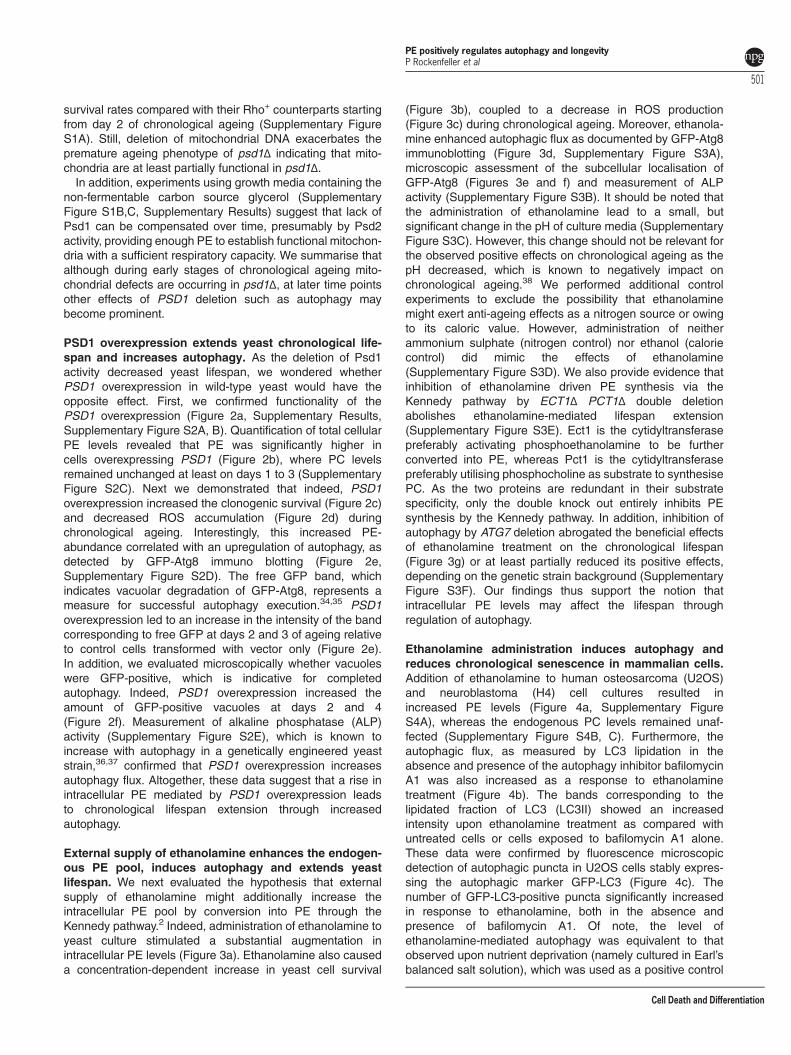
OPEN
Phosphatidylethanolamine positively regulatesautophagy and longevity
P Rockenfeller1, M Koska1, F Pietrocola2, N Minois3, O Knittelfelder1, V Sica2, J Franz1, D Carmona-Gutierrez1, G Kroemer*,2,4,5,6,7
and F Madeo*,1,8
Autophagy is a cellular recycling program that retards ageing by efficiently eliminating damaged and potentially harmful organellesand intracellular protein aggregates. Here, we show that the abundance of phosphatidylethanolamine (PE) positively regulatesautophagy. Reduction of intracellular PE levels by knocking out either of the two yeast phosphatidylserine decarboxylases (PSD)accelerated chronological ageing-associated production of reactive oxygen species and death. Conversely, the artificial increaseof intracellular PE levels, by provision of its precursor ethanolamine or by overexpression of the PE-generating enzyme Psd1,significantly increased autophagic flux, both in yeast and in mammalian cell culture. Importantly administration of ethanolaminewas sufficient to extend the lifespan of yeast (Saccharomyces cerevisiae), mammalian cells (U2OS, H4) and flies (Drosophilamelanogaster). We thus postulate that the availability of PE may constitute a bottleneck for functional autophagy and thatorganismal life or healthspan could be positively influenced by the consumption of ethanolamine-rich food.Cell Death and Differentiation (2015) 22, 499–508; doi:10.1038/cdd.2014.219; published online 9 January 2015
Phosphatidylethanolamine (PE) is a phospholipid found in allliving organisms. Together with phosphatidylcholine (PC),phosphatidylserine (PS) and phosphatidylinositol (PI), PErepresents the backbone of most biological membranes. PE isthe second-most abundant phospholipid in mammalianmembranes ranging from 20 to 50%.1 In yeast, PE is essentialfor growth and is generated through four different enzymaticpathways:2 PE can be produced by decarboxylationof PS, as a first option at the mitochondrial membrane viaphosphatidylserine decarboxylase 1 (Psd1)3,4 or, as a second,option at the Golgi and vacuolar membranes throughphosphatidylserine decarboxylase 2 (Psd2).5 As a thirdpossibility, PE can be produced from actively retrievedextracellular ethanolamine,6,7 which is cytidine 5'-dipho-sphate-activated8 and then coupled to diacylglycerol togenerate PE.9 The fourth, scarcely employed PE-generatingpathway is based on the lysophospholipid acylation oflyso-PE. Importantly, PE does not spontaneously assemblein bilayers and rather incorporates into curved structures, suchas the inverted hexagonal phase.10 The physiological functionof non-bilayer lipids in membranes is considered to reside intheir interaction with membrane proteins via the membranelateral pressure10 and membrane tethering and fusionprocesses, which are relevant for autophagy.11
The term ‘autophagy’ describes a degradation processaffecting intracellular components (for a review see, 12, 13)which as an important cytoprotective mechanism, is closely
linked to ageing. Autophagy mainly differs from the proteaso-mal pathway, the other major cellular degradation mechanism,in two aspects. First, autophagy can degrade large particles orwhole organelles and second, the final degradation occurs inthe lysosome/vacuole and not at the proteasome. Prior to theactual degradation, the cargo is gathered in autophagicparticles, which are surrounded by a characteristic double-membrane. However, the origin of these autophagosomalmembranes is still controversial and might actually depend onthe mode of autophagy induction.14,15 Among the discussedmembrane sources are the Golgi apparatus, the endosplamicreticulum (ER) or the mitochondrion-associated membrane,which is formed at the interface between the ER.16 In highereukaryotes autophagic membranes are enriched in PE with ahigh degree of unsaturation,17 similarly to the PE species foundin mitochondria.14,18 Moreover, the pre-autophagosomal struc-ture or phagophore assembly site (PAS), which appears at thevery beginning of autophagosome formation, already harboursAtg9, an autophagy-related transmembrane protein that shuttlesbetween mitochondria and the PAS structure in yeast.19
Importantly, PE also functions as an anchor to autophago-somal membranes for the autophagy-related protein Atg8 inyeast20 and its mammalian orthologue LC3.21,22 This PEanchor is provided to LC3/Atg8 post-translationally in aprocess called lipidation. First, LC3/Atg8 is carboxy-terminally cleaved by proteases from the Atg4 family.23,24
Subsequently, the remaining C-terminal glycine is coupled to
1Institute of Molecular Biosciences, NAWI Graz, University of Graz, Humboldtstr. 50, 8010 Graz, Austria; 2INSERM U848, Villejuif, Paris, France; 3Biomedical SciencesResearch Complex, University of St Andrews, St Andrews, UK; 4Metabolomics Platform, Institut Gustave Roussy, Villejuif, Paris, France; 5Centre de Recherche desCordeliers, Paris, France; 6Pôle de Biologie; Hôpital Européen Georges Pompidou, AP – HP, Paris, France; 7Université Paris Descartes, Sorbonne Paris Cité, Paris, Franceand 8BioTechMed-Graz, Humboldtstr. 50, 8010 Graz, Austria*Corresponding author: G Kroemer or F Madeo, Institute of Molecular Biosciences, Karl-Franzens-University of Graz, 8010 Graz, Austria. Tel: +43 316 380 1507;Fax: +43 316 380 8878; E-mail: [email protected] or [email protected]
Received 30.6.14; revised 17.11.14; accepted 19.11.14; Edited by M Piacentini; published online 09.1.15
Abbreviations: DHE, Dihydroethidium; ER, endoplasmic reticulum; PE, phosphatidylethanolamine; PC, phosphatidylcholine; PI, phosphatidylinositol; PS,phosphatidylserine; Psd/Pisd, phosphatidylserine decarboxylase; PAS, pre-autophagosomal structure/phagophore assembly site; ROS, reactive oxygen species; PI,propidium iodide
Cell Death and Differentiation (2015) 22, 499–508& 2015 Macmillan Publishers Limited All rights reserved 1350-9047/15
www.nature.com/cdd

PE in a series of ubiquitination-like reactions involving diverseAtg-proteins.20,25–27 In vitro, Atg8-PE causes hemifusion ofvesicles, which argues for its potential role in autophagosomalphagophore expansion.11,28 Consistently, semisynthetic LC3-PE has recently been described to stimulate membranetethering and fusion.29 We thus reasoned that the overallabundance of PEmight be critical for PE-lipidation of LC3/Atg8and could thus regulate autophagosomal membrane forma-tion. Therefore, we tested whether increasing cellular PElevels might have an impact on autophagy and lifespanregulation.Here, we report that knock-out of PSD1 or PSD2 shortens
the chronological lifespan of S. cerevisiae, whereas PSD1-overexpression enhances the autophagic capacity andincreases longevity. Furthermore, external administration ofethanolamine increases endogenous PE levels, enhancesautophagic flux and extends the lifespan of yeast, mammaliancells in culture and flies (Drosophila melanogaster).
Results
A genetic screen identifies psd1Δ and psd2Δ as proger-oid yeast strains. We designed a screen to identify genesinvolved in phospholipid anabolism and catabolism that mighthave an impact on ageing. For this purpose, we performedchronological ageing experiments with a subset of yeaststrains deleted for non-essential genes known to be involvedin phospholipid metabolism. At day 3 of the chronologicalageing experiment, we measured the levels of reactiveoxygen species (ROS) by assessing the ROS-driven oxida-tion of non-fluorescent dihydroethidium (DHE) to fluorescentethidium by cytofluorometry. This screening procedure led tothe identification of three genes whose deletion caused anageing-dependent raise in ROS generation (Figure 1a):isc1Δ, psd1Δ and psd2Δ. ISC1 encodes an inositol phospho-sphingolipid phospholipase, which produces ceramide. Inter-estingly, deletion of ISC1 has been previously reported todecrease chronological lifespan,30 thus validating our screenresults. PSD1 and PSD2 encode phosphatidylserine dec-arboxylases, which similarly convert PS to PE, and arelocated in distinct cellular organelles (see Introduction). Asthese two enzymes have not been associated with yeastaging, we decided to focus our study on these enzymes andtheir products.We confirmed the premature ageing phenotype of thePSD1
and PSD2 knock outs by clonogenic survival plating(Figure 1b) and assessed the PE- and PC-abundance at day3 of ageing by HPLC-assisted analyses of lipid extracts(Figure 1c). PSD1 deletion had a more pronounced inhibitoreffect on both PE synthesis and yeast ageing. This isconsistent with a previous report underscoring that, at leastin standard culture conditions, Psd1 is the predominantphosphatidylserine decarboxylase, the abrogation of whichdisturbs homeostasis of PE and a number of phospholipidspecies including PS and PI which can be overcome by theadministration of ethanolamine.31
As previous works have also shown that PSD1 deletion hasa strong impact on mitochondrial function,32,33 we examinedto which extent the reduction in survival upon chronological
ageing might be a consequence of general mitochondrialdysfunction or specific to the deletion of PSD activity. We thusgenerated psd1Δ and psd2Δ strains, which are additionallyrendered defective for respiration owing to loss of mitochon-drial DNA (Rho0). These Rho0 strains showed reduced
Figure 1 Identification of psd1Δ and psd2Δ as progeroid yeast strains. (a) TheROS-driven conversion of DHE to ethidium was measured at day three ofchronological ageing in eleven yeast strains deficient for different enzymes involved inphospholipid synthesis. (b) The chronological ageing curve, which is based on theclonogenic survival, is shown for psd1Δ and psd2Δ in comparison with the wild type.(c) Phosphatidylethanolamine (PE) and phosphatidyl choline (PC) were quantified byHPLC-ELSD of lipid extracts from wild-type psd1Δ and psd2Δ cells at day three ofchronological ageing
PE positively regulates autophagy and longevityP Rockenfeller et al
500
Cell Death and Differentiation
survival rates compared with their Rho+ counterparts startingfrom day 2 of chronological ageing (Supplementary FigureS1A). Still, deletion of mitochondrial DNA exacerbates thepremature ageing phenotype of psd1Δ indicating that mito-chondria are at least partially functional in psd1Δ.In addition, experiments using growth media containing the
non-fermentable carbon source glycerol (SupplementaryFigure S1B,C, Supplementary Results) suggest that lack ofPsd1 can be compensated over time, presumably by Psd2activity, providing enough PE to establish functional mitochon-dria with a sufficient respiratory capacity. We summarise thatalthough during early stages of chronological ageing mito-chondrial defects are occurring in psd1Δ, at later time pointsother effects of PSD1 deletion such as autophagy maybecome prominent.
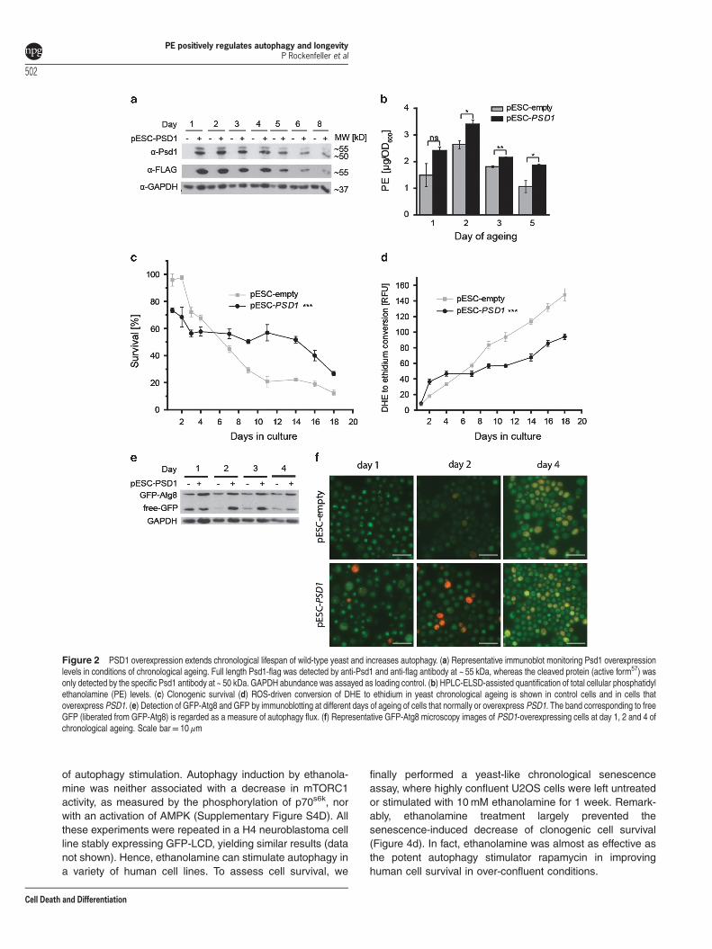
PSD1 overexpression extends yeast chronological life-span and increases autophagy. As the deletion of Psd1activity decreased yeast lifespan, we wondered whetherPSD1 overexpression in wild-type yeast would have theopposite effect. First, we confirmed functionality of thePSD1 overexpression (Figure 2a, Supplementary Results,Supplementary Figure S2A, B). Quantification of total cellularPE levels revealed that PE was significantly higher incells overexpressing PSD1 (Figure 2b), where PC levelsremained unchanged at least on days 1 to 3 (SupplementaryFigure S2C). Next we demonstrated that indeed, PSD1overexpression increased the clonogenic survival (Figure 2c)and decreased ROS accumulation (Figure 2d) duringchronological ageing. Interestingly, this increased PE-abundance correlated with an upregulation of autophagy, asdetected by GFP-Atg8 immuno blotting (Figure 2e,Supplementary Figure S2D). The free GFP band, whichindicates vacuolar degradation of GFP-Atg8, represents ameasure for successful autophagy execution.34,35 PSD1overexpression led to an increase in the intensity of the bandcorresponding to free GFP at days 2 and 3 of ageing relativeto control cells transformed with vector only (Figure 2e).In addition, we evaluated microscopically whether vacuoleswere GFP-positive, which is indicative for completedautophagy. Indeed, PSD1 overexpression increased theamount of GFP-positive vacuoles at days 2 and 4(Figure 2f). Measurement of alkaline phosphatase (ALP)activity (Supplementary Figure S2E), which is known toincrease with autophagy in a genetically engineered yeaststrain,36,37 confirmed that PSD1 overexpression increasesautophagy flux. Altogether, these data suggest that a rise inintracellular PE mediated by PSD1 overexpression leadsto chronological lifespan extension through increasedautophagy.
External supply of ethanolamine enhances the endogen-ous PE pool, induces autophagy and extends yeastlifespan. We next evaluated the hypothesis that externalsupply of ethanolamine might additionally increase theintracellular PE pool by conversion into PE through theKennedy pathway.2 Indeed, administration of ethanolamine toyeast culture stimulated a substantial augmentation inintracellular PE levels (Figure 3a). Ethanolamine also causeda concentration-dependent increase in yeast cell survival
(Figure 3b), coupled to a decrease in ROS production(Figure 3c) during chronological ageing. Moreover, ethanola-mine enhanced autophagic flux as documented by GFP-Atg8immunoblotting (Figure 3d, Supplementary Figure S3A),microscopic assessment of the subcellular localisation ofGFP-Atg8 (Figures 3e and f) and measurement of ALPactivity (Supplementary Figure S3B). It should be noted thatthe administration of ethanolamine lead to a small, butsignificant change in the pH of culture media (SupplementaryFigure S3C). However, this change should not be relevant forthe observed positive effects on chronological ageing as thepH decreased, which is known to negatively impact onchronological ageing.38 We performed additional controlexperiments to exclude the possibility that ethanolaminemight exert anti-ageing effects as a nitrogen source or owingto its caloric value. However, administration of neitherammonium sulphate (nitrogen control) nor ethanol (caloriecontrol) did mimic the effects of ethanolamine(Supplementary Figure S3D). We also provide evidence thatinhibition of ethanolamine driven PE synthesis via theKennedy pathway by ECT1Δ PCT1Δ double deletionabolishes ethanolamine-mediated lifespan extension(Supplementary Figure S3E). Ect1 is the cytidyltransferasepreferably activating phosphoethanolamine to be furtherconverted into PE, whereas Pct1 is the cytidyltransferasepreferably utilising phosphocholine as substrate to synthesisePC. As the two proteins are redundant in their substratespecificity, only the double knock out entirely inhibits PEsynthesis by the Kennedy pathway. In addition, inhibition ofautophagy by ATG7 deletion abrogated the beneficial effectsof ethanolamine treatment on the chronological lifespan(Figure 3g) or at least partially reduced its positive effects,depending on the genetic strain background (SupplementaryFigure S3F). Our findings thus support the notion thatintracellular PE levels may affect the lifespan throughregulation of autophagy.
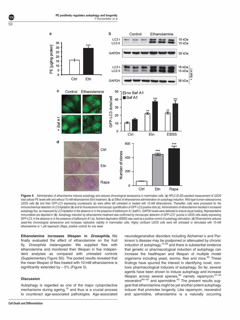
Ethanolamine administration induces autophagy andreduces chronological senescence in mammalian cells.Addition of ethanolamine to human osteosarcoma (U2OS)and neuroblastoma (H4) cell cultures resulted inincreased PE levels (Figure 4a, Supplementary FigureS4A), whereas the endogenous PC levels remained unaf-fected (Supplementary Figure S4B, C). Furthermore, theautophagic flux, as measured by LC3 lipidation in theabsence and presence of the autophagy inhibitor bafilomycinA1 was also increased as a response to ethanolaminetreatment (Figure 4b). The bands corresponding to thelipidated fraction of LC3 (LC3II) showed an increasedintensity upon ethanolamine treatment as compared withuntreated cells or cells exposed to bafilomycin A1 alone.These data were confirmed by fluorescence microscopicdetection of autophagic puncta in U2OS cells stably expres-sing the autophagic marker GFP-LC3 (Figure 4c). Thenumber of GFP-LC3-positive puncta significantly increasedin response to ethanolamine, both in the absence andpresence of bafilomycin A1. Of note, the level ofethanolamine-mediated autophagy was equivalent to thatobserved upon nutrient deprivation (namely cultured in Earl’sbalanced salt solution), which was used as a positive control
PE positively regulates autophagy and longevityP Rockenfeller et al
501
Cell Death and Differentiation
of autophagy stimulation. Autophagy induction by ethanola-mine was neither associated with a decrease in mTORC1activity, as measured by the phosphorylation of p70s6k, norwith an activation of AMPK (Supplementary Figure S4D). Allthese experiments were repeated in a H4 neuroblastoma cellline stably expressing GFP-LCD, yielding similar results (datanot shown). Hence, ethanolamine can stimulate autophagy ina variety of human cell lines. To assess cell survival, we
finally performed a yeast-like chronological senescenceassay, where highly confluent U2OS cells were left untreatedor stimulated with 10mM ethanolamine for 1 week. Remark-ably, ethanolamine treatment largely prevented thesenescence-induced decrease of clonogenic cell survival(Figure 4d). In fact, ethanolamine was almost as effective asthe potent autophagy stimulator rapamycin in improvinghuman cell survival in over-confluent conditions.
Figure 2 PSD1 overexpression extends chronological lifespan of wild-type yeast and increases autophagy. (a) Representative immunoblot monitoring Psd1 overexpressionlevels in conditions of chronological ageing. Full length Psd1-flag was detected by anti-Psd1 and anti-flag antibody at ~ 55 kDa, whereas the cleaved protein (active form57) wasonly detected by the specific Psd1 antibody at ~ 50 kDa. GAPDH abundance was assayed as loading control. (b) HPLC-ELSD-assisted quantification of total cellular phosphatidylethanolamine (PE) levels. (c) Clonogenic survival (d) ROS-driven conversion of DHE to ethidium in yeast chronological ageing is shown in control cells and in cells thatoverexpress PSD1. (e) Detection of GFP-Atg8 and GFP by immunoblotting at different days of ageing of cells that normally or overexpress PSD1. The band corresponding to freeGFP (liberated from GFP-Atg8) is regarded as a measure of autophagy flux. (f) Representative GFP-Atg8 microscopy images of PSD1-overexpressing cells at day 1, 2 and 4 ofchronological ageing. Scale bar= 10 μm
PE positively regulates autophagy and longevityP Rockenfeller et al
502
Cell Death and Differentiation
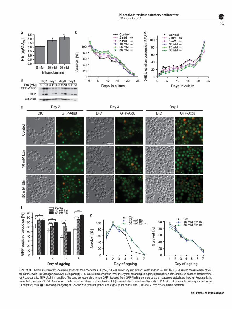
Figure 3 Administration of ethanolamine enhances the endogenous PE pool, induces autophagy and extends yeast lifespan. (a) HPLC-ELSD-assisted measurement of totalcellular PE levels. (b) Clonogenic survival plating and (c) DHE to ethidium conversion throughout yeast chronological ageing upon addition of the indicated doses of ethanolamine.(d) Representative GFP-Atg8 immunoblot. The band corresponding to free GFP (liberated from GFP-Atg8) is considered as a measure of autophagic flux. (e) Representativemicrophotographs of GFP-Atg8-expressing cells under conditions of ethanolamine (Etn) administration. Scale bar=5 μm. (f) GFP-Atg8 positive vacuoles were quantified in live(PI-negative) cells. (g) Chronological ageing of BY4742 wild type (left panel) and atg7Δ (right panel) with 0, 10 and 50 mM ethanolamine treatment
PE positively regulates autophagy and longevityP Rockenfeller et al
503
Cell Death and Differentiation
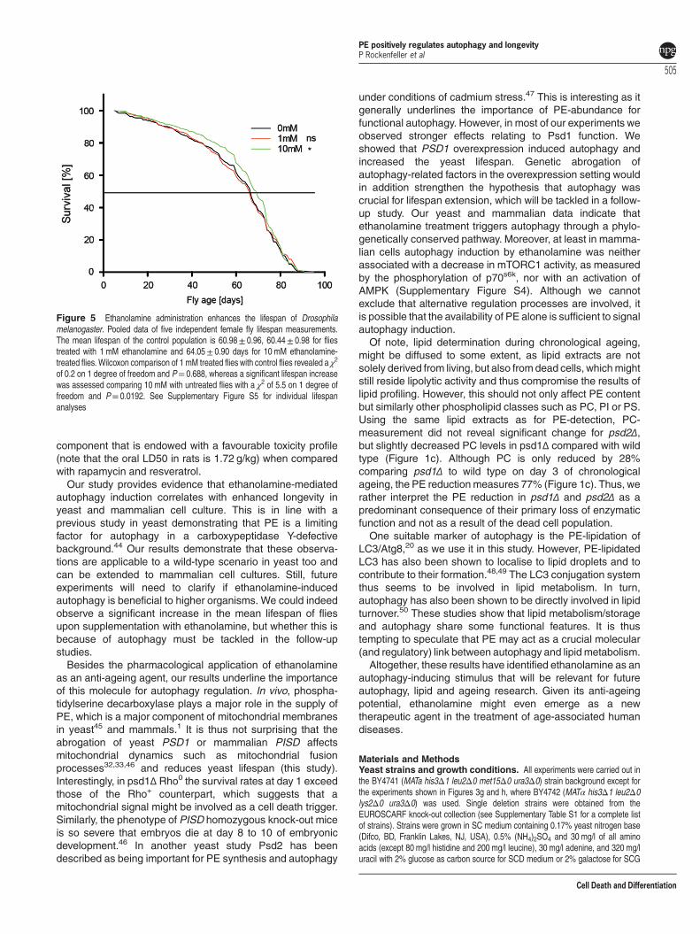
Ethanolamine increases lifespan in Drosophila. Wefinally evaluated the effect of ethanolamine on the fruitfly, Drosophila melanogaster. We supplied flies withethanolamine and monitored their lifespan in five indepen-dent analyses as compared with untreated controls(Supplementary Figure S5). The pooled results revealed thatthe mean lifespan of flies treated with 10mM ethanolamine issignificantly extended by ~ 5% (Figure 5).
Discussion
Autophagy is regarded as one of the major cytoprotectivemechanisms during ageing,12 and thus is a crucial processto counteract age-associated pathologies. Age-associated
neurodegenerative disorders including Alzheimer´s and Par-kinson´s disease may be postponed or attenuated by chronicinduction of autophagy,12,39 and there is substantial evidencethat genetic or pharmacological induction of autophagy canincrease the healthspan and lifespan of multiple modelorganisms including yeast, worms, flies and mice.40 Thesefindings have spurred the interest in identifying novel, non-toxic pharmacological inducers of autophagy. So far, severalagents have been shown to induce autophagy and increaselifespan across several species,40 namely rapamycin,27,40
resveratrol40–42 and spermidine.43 The present results sug-gest that ethanolamine might be yet another potent autophagyinducer that promotes longevity. Like rapamycin, resveratroland spermidine, ethanolamine is a naturally occurring
Figure 4 Administration of ethanolamine induces autophagy and reduces chronological senescence in mammalian cells. (a) HPLC-ELSD-assisted measurement of U2OStotal cellular PE levels with and without 10 mM ethanolamine (Etn) treatment. (b, c) Effect of ethanolamine administration on autophagy induction. Wild-type human osteosarcomaU2OS cells (b) and their GFP-LC3 expressing counterparts (c) were either left untreated or treated with 10 mM ethanolamine. Thereafter, cells were processed for theimmunochemical detection of LC3 lipidation (b) and for fluorescence microscopic quantification of GFP-LC3 positive dots (c). Administration of ethanolamine resulted in increasedautophagy flux, as measured by LC3 lipidation in the absence or in the presence of bafilomycin A1 (bafA1). GAPDH levels were detected to ensure equal loading. Representativeimmunoblots are depicted in (b). Autophagy induction by ethanolamine treatment was confirmed by microscopic detection of GFP-LC3+ puncta in U2OS cells stably expressingGFP-LC3, in the absence or in the presence of bafilomycin A1 (c). Nutrient deprivation (EBSS) was used as a positive control of autophagy stimulation. (d) Ethanolamine reducesyeast-like chronological senescence and increases replicative viability in mammalian cells. Highly confluent U2OS cells were left untreated or stimulated with 10 mMethanolamine or 1 μM rapamycin (Rapa, positive control) for one week
PE positively regulates autophagy and longevityP Rockenfeller et al
504
Cell Death and Differentiation
component that is endowed with a favourable toxicity profile(note that the oral LD50 in rats is 1.72 g/kg) when comparedwith rapamycin and resveratrol.Our study provides evidence that ethanolamine-mediated
autophagy induction correlates with enhanced longevity inyeast and mammalian cell culture. This is in line with aprevious study in yeast demonstrating that PE is a limitingfactor for autophagy in a carboxypeptidase Y-defectivebackground.44 Our results demonstrate that these observa-tions are applicable to a wild-type scenario in yeast too andcan be extended to mammalian cell cultures. Still, futureexperiments will need to clarify if ethanolamine-inducedautophagy is beneficial to higher organisms. We could indeedobserve a significant increase in the mean lifespan of fliesupon supplementation with ethanolamine, but whether this isbecause of autophagy must be tackled in the follow-upstudies.Besides the pharmacological application of ethanolamine
as an anti-ageing agent, our results underline the importanceof this molecule for autophagy regulation. In vivo, phospha-tidylserine decarboxylase plays a major role in the supply ofPE, which is a major component of mitochondrial membranesin yeast45 and mammals.1 It is thus not surprising that theabrogation of yeast PSD1 or mammalian PISD affectsmitochondrial dynamics such as mitochondrial fusionprocesses32,33,46 and reduces yeast lifespan (this study).Interestingly, in psd1Δ Rho0 the survival rates at day 1 exceedthose of the Rho+ counterpart, which suggests that amitochondrial signal might be involved as a cell death trigger.Similarly, the phenotype of PISD homozygous knock-out miceis so severe that embryos die at day 8 to 10 of embryonicdevelopment.46 In another yeast study Psd2 has beendescribed as being important for PE synthesis and autophagy
under conditions of cadmium stress.47 This is interesting as itgenerally underlines the importance of PE-abundance forfunctional autophagy. However, in most of our experiments weobserved stronger effects relating to Psd1 function. Weshowed that PSD1 overexpression induced autophagy andincreased the yeast lifespan. Genetic abrogation ofautophagy-related factors in the overexpression setting wouldin addition strengthen the hypothesis that autophagy wascrucial for lifespan extension, which will be tackled in a follow-up study. Our yeast and mammalian data indicate thatethanolamine treatment triggers autophagy through a phylo-genetically conserved pathway. Moreover, at least in mamma-lian cells autophagy induction by ethanolamine was neitherassociated with a decrease in mTORC1 activity, as measuredby the phosphorylation of p70s6k, nor with an activation ofAMPK (Supplementary Figure S4). Although we cannotexclude that alternative regulation processes are involved, itis possible that the availability of PE alone is sufficient to signalautophagy induction.Of note, lipid determination during chronological ageing,
might be diffused to some extent, as lipid extracts are notsolely derived from living, but also from dead cells, whichmightstill reside lipolytic activity and thus compromise the results oflipid profiling. However, this should not only affect PE contentbut similarly other phospholipid classes such as PC, PI or PS.Using the same lipid extracts as for PE-detection, PC-measurement did not reveal significant change for psd2Δ,but slightly decreased PC levels in psd1Δ compared with wildtype (Figure 1c). Although PC is only reduced by 28%comparing psd1Δ to wild type on day 3 of chronologicalageing, the PE reduction measures 77% (Figure 1c). Thus, werather interpret the PE reduction in psd1Δ and psd2Δ as apredominant consequence of their primary loss of enzymaticfunction and not as a result of the dead cell population.One suitable marker of autophagy is the PE-lipidation of
LC3/Atg8,20 as we use it in this study. However, PE-lipidatedLC3 has also been shown to localise to lipid droplets and tocontribute to their formation.48,49 The LC3 conjugation systemthus seems to be involved in lipid metabolism. In turn,autophagy has also been shown to be directly involved in lipidturnover.50 These studies show that lipid metabolism/storageand autophagy share some functional features. It is thustempting to speculate that PE may act as a crucial molecular(and regulatory) link between autophagy and lipid metabolism.Altogether, these results have identified ethanolamine as an
autophagy-inducing stimulus that will be relevant for futureautophagy, lipid and ageing research. Given its anti-ageingpotential, ethanolamine might even emerge as a newtherapeutic agent in the treatment of age-associated humandiseases.
Materials and MethodsYeast strains and growth conditions. All experiments were carried out inthe BY4741 (MATa his3Δ1 leu2Δ0 met15Δ0 ura3Δ0) strain background except forthe experiments shown in Figures 3g and h, where BY4742 (MATα his3Δ1 leu2Δ0lys2Δ0 ura3Δ0) was used. Single deletion strains were obtained from theEUROSCARF knock-out collection (see Supplementary Table S1 for a complete listof strains). Strains were grown in SC medium containing 0.17% yeast nitrogen base(Difco, BD, Franklin Lakes, NJ, USA), 0.5% (NH4)2SO4 and 30 mg/l of all aminoacids (except 80 mg/l histidine and 200 mg/l leucine), 30 mg/l adenine, and 320 mg/luracil with 2% glucose as carbon source for SCD medium or 2% galactose for SCG
Figure 5 Ethanolamine administration enhances the lifespan of Drosophilamelanogaster. Pooled data of five independent female fly lifespan measurements.The mean lifespan of the control population is 60.98± 0.96, 60.44± 0.98 for fliestreated with 1 mM ethanolamine and 64.05± 0.90 days for 10 mM ethanolamine-treated flies. Wilcoxon comparison of 1 mM treated flies with control flies revealed a χ2
of 0.2 on 1 degree of freedom and P= 0.688, whereas a significant lifespan increasewas assessed comparing 10 mM with untreated flies with a χ2 of 5.5 on 1 degree offreedom and P= 0.0192. See Supplementary Figure S5 for individual lifespananalyses
PE positively regulates autophagy and longevityP Rockenfeller et al
505
Cell Death and Differentiation
medium, respectively. All yeast cultures were inoculated from a stationary overnightculture to an OD of 0.1 at 600 nm (OD600= 0.1) and then grown at 28 °C and 145 r.p.m. shaking for the indicated time periods. For induction of the Gal10 promoter todrive PSD1 expression, pESC-PSD1 containing cells were shifted to SCG mediumat an OD600= 0.35. Ethanolamine chloride from Sigma (St. Louis, MO, USA,E6133) was administered to the cultures from a 1 M stock in ddH2O or from a 5 Mstock for the 50 mM final concentration.
Cloning and molecular biology. PSD1 was cloned into the multiplecloning site 2 of the plasmid pESC His (Agilent Technologies, Santa Clara, CA,USA) by homologous recombination in yeast using the forward primer5ʹ-TTCGAATTCAACCCTCACTAAAGGGCGGCC_ATGTCAATTATGCCAGTTAAGAAC-3ʹand as backward primer 5ʹ-GATCTTATCGTCGTCATCCTTGTAATCCAT_TTTTAAATCATTCTTTCCAATTATGCCTAATTTC-3ʹ.
Survival plating and ROS determination. For survival plating, the cellconcentrations of culture dilutions were determined with a CASY cell counter(Roche Diagnostics, Mannheim, Germany) and aliquots containing 500 cells wereplated on YPD plates. The number of colonies formed was determined after 2 daysat 28 °C. For dihydroethidium staining, 5 × 10⁶ cells were harvested bycentrifugation, resuspended in 250 μl of 2.5 μg/ml DHE in PBS and incubated inthe dark for 5 min. Relative fluorescence units were determined using a TecanGeniusPRO fluorescence reader (Tecan Group, Maennedorf, Switzerland) and thennormalised to OD600. For ROS analysis on the basis of individual cells, flowcytometry was used to count the positive cells.
Yeast autophagy measurement. Autophagy was measured by monitoringthe cytosol to vacuole translocation of Atg8 using fluorescence microscopy orimmunoblotting (GFP liberation assay) of cells/cell extracts from strains carrying aGFP-Atg8 fusion protein34,35 expressed under its endogenous promoter and at itsnatural chromosomal locus. Quantification of micrographs was performed fromblinded pictures with 150–300 cells per micrograph and replicate. Autophagic cellswere defined as cells exhibiting clear vacuolar GFP fluorescence and expressed asfraction of viable (PI-negative) cells. Immunoblotting followed standard proceduresusing anti-GFP (Roche Diagnostics, #11814460001), anti-Psd1 or anti-glyceraldehyde-3-phosphate dehydrogenase (GAPDH) antibodies (both gifts fromDr. Gunther Daum).
Yeast lipid extraction. Total lipids were extracted from exponentially growingyeast cultures at indicated days of ageing with chloroform/methanol 2:1 (v/v)according to Folch et al.51 The organic phase was dried under a stream of nitrogenand dissolved in 500 μl of chloroform/methanol (2:1, v/v).
Lipid analysis. PE was quantified by a normal phase HPLC—evaporative lightscattering detector (ELSD) method as described in Guerfal et al.52 (the method isaccepted for publication in Cold Spring Harbor Protocols: ‘Analyzing andunderstanding lipids of yeast: a challenging endeavour’). In brief, the chromato-graphic separation of lipids was achieved on a Betasil Diol column (100 × 4.6 mm,particle size 5 μm, Thermo Fisher Scientific Inc., Waltham, MA, USA) with a ternarygradient (modified from 53 and described in detail in Cold Spring Harbor Protocols).PE 34:1 standard (Avanti Polar Lipids, Inc., Alabaster, AL, USA) was prepared as1 mg/ml stock solution in chloroform/methanol 2:1 (v/v). Calibration curves(triplicates) were measured from 2.7 to 350 μg/ml, 10 μl sample was injected foreach measurement.
Cell cultureChemicals, cell lines and culture conditions: Unless otherwise specified,chemicals were purchased from Sigma-Aldrich (St. Louis, MO, USA), culture mediaand supplements for cell culture from Gibco-Invitrogen (Carlsbad, CA, USA) andplasticware from Corning (Corning, NY, USA). Human osteosarcoma U2OS cells,their GFP-LC3-expressing derivatives, human neuroblastoma H4 GFP-LC3 (giftfrom Y. Juan) cells were cultured in DMEM medium containing 10% foetal bovineserum, 100 mg/l sodium pyruvate, 10 mM HEPES buffer, 100 units/ml penicillin Gsodium and 100 μg/ml streptomycin sulphate (37 °C, 5% CO2). Lipid extractionswere performed after 12 h of 10 mM ethanolamine (Sigma, E9508) treatment. Forautophagy induction, cells were treated for 12 h with 10 mM ethanolamine orincubated in absence of nutrients. For the yeast-like chronological senescenceassay, cells were treated for 1 week with 10 mM ethanolamine or 1 μM rapamycin(R&D Systems, Minneapolis, MN, USA).
Lipid extraction from U2OS and H4 cells: Cells were harvested in PBS pH7.4 and pelleted before shock freezing in liquid nitrogen. Cell disruption wasperformed by sonication in PBS pH 7.4. Protein concentration was determinedusing the Bio-Rad protein assay kit (Bio-Rad Laboratories, Hercules, CA, USA) andthe results were used for normalisation after HPLC measurement. The raw extractswere extracted with chloroform/methanol (2:1) after Folch.51 The organic phase wascollected and combined with the organic phase obtained from the re-extraction ofthe aqueous phase. Organic solvents were evaporated under a stream of nitrogenand the lipids were dissolved in chloroform/ methanol (2:1). Lipid analysis wasperformed as described above.
Immunoblotting. For immunoblotting, 25 μg of proteins were separated on4–12% bis-tris acrylamide (Thermo Fisher Scientific Inc.) and electrotransferred toImmobilon membranes (Merck Millipore, Darmstadt, Germany). Membranes werethen sliced horizontally in different parts according to the molecular weight of theprotein of interest to allow simultaneous detection of different antigens within the sameexperiment.54,55 Unspecific binding sites were saturated by incubating membranes for1 h in 0.05% Tween 20 (v:v in TBS) supplemented with 5% non-fat powdered milk (w:vin TBS), followed by an overnight incubation with primary antibodies specific for LC3B,phospho-AMPK (Thr172), AMPK, phospho-ribosomal protein S6 kinase (Thr421/Ser424), ribosomal protein S6 kinase (Cell Signalling Technology Inc., Danvers, MA,USA). Development was performed with appropriate horseradish peroxidase (HRP)-labelled secondary antibodies (Southern Biotech, Birmingham, AL, USA) plus theSuperSignal West Pico chemoluminescent substrate (Thermo Fisher Scientific Inc.).An anti-glyceraldehyde-3-phosphate dehydrogenase antibody (Chemicon InternationalInc., Temecula, CA, USA) was used to control equal loading of lanes.
Automated microscopy. U2OS or H4 cells stably expressing GFP-LC3 wereseeded in 96-well imaging plates (BD Falcon, Sparks, USA) 24 h before stimulation.Cells were treated with the indicated agents for 4 h. Subsequently, cells were fixedwith 4% PFA and counterstained with 10 μM Hoechst 33342. Images were acquiredusing a BD pathway 855 automated microscope (BD Imaging Systems, San José,USA) equipped with a 40X objective (Olympus, Center Valley, USA) coupled to arobotised Twister II plate handler (Caliper Life Sciences, Hopkinton, USA). Imageswere analyzed for the presence of GFP-LC3 puncta in the cytoplasm by means ofthe BD Attovision software (BD Imaging Systems). Cell surfaces were segmentedand divided into cytoplasmic and nuclear regions according to manufacturerstandard proceedings. RB 2x2 and Marr–Hildreth algorithms were used to recognizecytoplasmic GFP-LC3 positive dots.
Yeast-like chronological senescence assay. The assay was per-formed as described in Leontieva et al.56 Briefly, 80 000 cells were seeded into96-well plates and left untreated or treated with 10 mM ethanolamine or 1 μMrapamycin. After 8 days, dead cells and conditioning media were removed, cellswere trypsinized and a 10% aliquot was plated in fresh medium-filled six-well plates.After 1 week, clones were marked trough crystal violet staining and counted.
Drosophila lifespan experiments. Female flies from an isogenised w1118
strain were used. They were kept in a 25 °C, 70% humidity, 12 h light/12 h darkincubator on a standard cornmeal-sugar-yeast diet. Flies were collected atemergence and 20 females were kept per vial with an average of 87 flies per groupand replicate. The food was changed twice a week and supplemented or not withethanolamine hydrochloride (Sigma, E6133) at 1 mM or 10 mM final concentrations.The number of dead flies were recorded every weekday until all flies were dead.Five independent replicates were performed. The independent replicates, as well asthe pooled data from all replicates were analysed by a Wilcoxon survival analysistest in R (script available on request).
Statistical analysis. Statistical analyses were calculated in Origin8. Forassessment of significance one-way ANOVA followed by Bonferroni post hoc test wasperformed, except for Figure 1b, Figures 2a and b and Figures 3b, c, g and h whichwere processed using a two-factor ANOVA with strain and time as independentfactors. Data in Figure 5 were assessed for significant difference by Wilcoxon analysis.Error bars indicate standard error of the mean (SEM) and asterisks in the figuresindicate significant differences, *Po0.05, **Po0.01, ***Po0.001.
Conflict of InterestThe authors declare no conflict of interest.
PE positively regulates autophagy and longevityP Rockenfeller et al
506
Cell Death and Differentiation
Acknowledgements. This work was supported by the Austrian Science FundFWF (grants LIPOTOX, I1000-B20, P23490-B12, and P24381-B20) to FM. Weacknowledge support from NAWI Graz. OK is a member of the PhD program‘Molecular Enzymology’, funded by the FWF (project W901-B12). GK is supported bythe Ligue contre le Cancer (équipe labelisée); Agence National de la Recherche(ANR); Association pour la recherche sur le cancer (ARC); Cancéropôle Ile-de-France; Institut National du Cancer (INCa); Fondation Bettencourt-Schueller;Fondation de France; Fondation pour la Recherche Médicale (FRM); the EuropeanCommission (ArtForce); the European Research Council (ERC); the LabEx Immuno-Oncology; the SIRIC Stratified Oncology Cell DNA Repair and Tumour ImmuneElimination (SOCRATE); the SIRIC Cancer Research and Personalized Medicine(CARPEM); and the Paris Alliance of Cancer Research Institutes (PACRI). We thankGünther Daum for the kind gift of the specific Psd1 antibody.
1. Vance JE. Phosphatidylserine and phosphatidylethanolamine in mammalian cells: twometabolically related aminophospholipids. J Lipid Res 2008; 49: 1377–1387.
2. Schuiki I, Schnabl M, Czabany T, Hrastnik C, Daum G. Phosphatidylethanolaminesynthesized by four different pathways is supplied to the plasma membrane of the yeastSaccharomyces cerevisiae. Biochim Biophys Acta 2010; 1801: 480–486.
3. Clancey CJ, Chang SC, Dowhan W. Cloning of a gene (PSD1) encoding phosphatidylserinedecarboxylase from Saccharomyces cerevisiae by complementation of an Escherichiacoli mutant. J Biol Chem 1993; 268: 24580–24590.
4. Zinser E, Sperka-Gottlieb CD, Fasch EV, Kohlwein SD, Paltauf F, Daum G. Phospholipidsynthesis and lipid composition of subcellular membranes in the unicellular eukaryoteSaccharomyces cerevisiae. J Bacteriol 1991; 173: 2026–2034.
5. Trotter PJ, Voelker DR. Identification of a non-mitochondrial phosphatidylserinedecarboxylase activity (PSD2) in the yeast Saccharomyces cerevisiae. J Biol Chem 1995;270: 6062–6070.
6. Nikawa J, Tsukagoshi Y, Yamashita S. Cloning of a gene encoding choline transport inSaccharomyces cerevisiae. J Bacteriol 1986; 166: 328–330.
7. Kim K, Kim KH, Storey MK, Voelker DR, Carman GM. Isolation and characterization of theSaccharomyces cerevisiae EKI1 gene encoding ethanolamine kinase. J Biol Chem 1999;274: 14857–14866.
8. Min-Seok R, Kawamata Y, Nakamura H, Ohta A, Takagi M. Isolation and characterization ofECT1 gene encoding CTP: phosphoethanolamine cytidylyltransferase of Saccharomycescerevisiae. J Biochem 1996; 120: 1040–1047.
9. Hjelmstad RH, Bell RM. The sn-1,2-diacylglycerol ethanolaminephosphotransferase activityof Saccharomyces cerevisiae. Isolation of mutants and cloning of the EPT1 gene. J BiolChem 1988; 263: 19748–19757.
10. van den Brink-van der Laan E, Killian JA, de Kruijff B. Nonbilayer lipids affect peripheral andintegral membrane proteins via changes in the lateral pressure profile. Biochim Biophys Acta2004; 1666: 275–288.
11. Nakatogawa H, Ichimura Y, Ohsumi Y. Atg8, a ubiquitin-like protein required forautophagosome formation, mediates membrane tethering and hemifusion. Cell 2007; 130:165–178.
12. Feng Y, He D, Yao Z, Klionsky DJ. The machinery of macroautophagy. Cell Res 2014; 24:24–41.
13. Inoue Y, Klionsky DJ. Regulation of macroautophagy in Saccharomyces cerevisiae. SeminCell Dev Biol 2010; 21: 664–670.
14. Luo S, Chen Q, Cebollero E, Xing D. Mitochondria: one of the origins for autophagosomalmembranes? Mitochondrion 2009; 9: 227–231.
15. Yang Z, Klionsky DJ. Eaten alive: a history of macroautophagy. Nat Cell Biol 2010; 12:814–822.
16. Hailey DW, Rambold AS, Satpute-Krishnan P, Mitra K, Sougrat R, Kim PK et al.Mitochondriasupply membranes for autophagosome biogenesis during starvation. Cell 2010; 141:656–667.
17. Reunanen H, Punnonen EL, Hirsimaki P. Studies on vinblastine-induced autophagocytosisin mouse liver. V. A cytochemical study on the origin of membranes. Histochemistry 1985; 83:513–517.
18. Bleijerveld OB, Brouwers JF, Vaandrager AB, Helms JB, Houweling M. The CDP-ethanolamine pathway and phosphatidylserine decarboxylation generate different phospha-tidylethanolamine molecular species. J Biol Chem 2007; 282: 28362–28372.
19. Reggiori F, Shintani T, Nair U, Klionsky DJ. Atg9 cycles between mitochondria and the pre-autophagosomal structure in yeasts. Autophagy 2005; 1: 101–109.
20. Ichimura Y, Kirisako T, Takao T, Satomi Y, Shimonishi Y, Ishihara N et al. A ubiquitin-likesystem mediates protein lipidation. Nature 2000; 408: 488–492.
21. Kabeya Y, Mizushima N, Ueno T, Yamamoto A, Kirisako T, Noda T et al. LC3, a mammalianhomologue of yeast Apg8p, is localized in autophagosome membranes after processing.EMBO J 2000; 19: 5720–5728.
22. Tanida I, Sou YS, Ezaki J, Minematsu-Ikeguchi N, Ueno T, Kominami E. HsAtg4B/HsApg4B/autophagin-1 cleaves the carboxyl termini of three human Atg8 homologuesand delipidates microtubule-associated protein light chain 3- and GABAAreceptor-associated protein-phospholipid conjugates. J Biol Chem 2004; 279:36268–36276.
23. Fujita N, Hayashi-Nishino M, Fukumoto H, Omori H, Yamamoto A, Noda T et al. An Atg4Bmutant hampers the lipidation of LC3 paralogues and causes defects in autophagosomeclosure. Mol Biol Cell 2008; 19: 4651–4659.
24. Kirisako T, Ichimura Y, Okada H, Kabeya Y, Mizushima N, Yoshimori T et al. The reversiblemodification regulates the membrane-binding state of Apg8/Aut7 essential for autophagy andthe cytoplasm to vacuole targeting pathway. J Cell Biol 2000; 151: 263–276.
25. Fujita N, Itoh T, Omori H, Fukuda M, Noda T, Yoshimori T. The Atg16L complex specifies thesite of LC3 lipidation for membrane biogenesis in autophagy. Mol Biol Cell 2008; 19:2092–2100.
26. Hanada T, Noda NN, Satomi Y, Ichimura Y, Fujioka Y, Takao T et al. The Atg12-Atg5conjugate has a novel E3-like activity for protein lipidation in autophagy. J Biol Chem 2007;282: 37298–37302.
27. Noda T, Ohsumi Y. Tor, a phosphatidylinositol kinase homologue, controls autophagyin yeast. J Biol Chem 1998; 273: 3963–3966.
28. Xie Z, Nair U, Klionsky DJ. Atg8 controls phagophore expansion during autophagosomeformation. Mol Biol Cell 2008; 19: 3290–3298.
29. Yang A, Li Y, Pantoom S, Triola G, Wu YW. Semisynthetic lipidated LC3 protein mediatesmembrane fusion. Chembiochem 2013; 14: 1296–1300.
30. Almeida T, Marques M, Mojzita D, Amorim MA, Silva RD, Almeida B et al. Isc1p plays a keyrole in hydrogen peroxide resistance and chronological lifespan through modulation of ironlevels and apoptosis. Mol Biol Cell 2008; 19: 865–876.
31. Burgermeister M, Birner-Grunberger R, Nebauer R, Daum G. Contribution of differentpathways to the supply of phosphatidylethanolamine and phosphatidylcholine tomitochondrial membranes of the yeast Saccharomyces cerevisiae. Biochim Biophys Acta2004; 1686: 161–168.
32. Birner R, Burgermeister M, Schneiter R, Daum G. Roles of phosphatidylethanolamine and ofits several biosynthetic pathways in Saccharomyces cerevisiae. Mol Biol Cell 2001; 12:997–1007.
33. Chan EY, McQuibban GA. Phosphatidylserine decarboxylase 1 (Psd1) promotesmitochondrial fusion by regulating the biophysical properties of the mitochondrial membraneand alternative topogenesis of mitochondrial genome maintenance protein 1 (Mgm1). J BiolChem 2012; 287: 40131–40139.
34. Kirisako T, Baba M, Ishihara N, Miyazawa K, Ohsumi M, Yoshimori T et al. Formationprocess of autophagosome is traced with Apg8/Aut7p in yeast. J Cell Biol 1999; 147:435–446.
35. Klionsky DJ, Cuervo AM, Seglen PO. Methods for monitoring autophagy from yeastto human. Autophagy 2007; 3: 181–206.
36. Noda T, Klionsky DJ. The quantitative Pho8Delta60 assay of nonspecific autophagy.Methods Enzymol 2008; 451: 33–42.
37. Noda T, Matsuura A, Wada Y, Ohsumi Y. Novel system for monitoring autophagy in the yeastSaccharomyces cerevisiae. Biochem Biophys Res Commun 1995; 210: 126–132.
38. Burtner CR, Murakami CJ, Kennedy BK, Kaeberlein M. A molecular mechanism ofchronological aging in yeast. Cell Cycle 2009; 8: 1256–1270.
39. Rubinsztein DC, DiFiglia M, Heintz N, Nixon RA, Qin ZH, Ravikumar B et al. Autophagy andits possible roles in nervous system diseases, damage and repair. Autophagy 2005; 1:11–22.
40. Madeo F, Tavernarakis N, Kroemer G. Can autophagy promote longevity? Nat Cell Biol2010; 12: 842–846.
41. Howitz KT, Bitterman KJ, Cohen HY, Lamming DW, Lavu S, Wood JG et al. Smallmolecule activators of sirtuins extend Saccharomyces cerevisiae lifespan. Nature 2003; 425:191–196.
42. Wood JG, Rogina B, Lavu S, Howitz K, Helfand SL, Tatar M et al. Sirtuin activators mimiccaloric restriction and delay ageing in metazoans. Nature 2004; 430: 686–689.
43. Eisenberg T, Knauer H, Schauer A, Buttner S, Ruckenstuhl C, Carmona-Gutierrez D et al.Induction of autophagy by spermidine promotes longevity. Nat Cell Biol 2009; 11: 1305–1314.
44. Nebauer R, Rosenberger S, Daum G. Phosphatidylethanolamine, a limiting factor ofautophagy in yeast strains bearing a defect in the carboxypeptidase Y pathway of vacuolartargeting. J Biol Chem 2007; 282: 16736–16743.
45. Tuller G, Nemec T, Hrastnik C, Daum G. Lipid composition of subcellular membranes of anFY1679-derived haploid yeast wild-type strain grown on different carbon sources. Yeast1999; 15: 1555–1564.
46. Steenbergen R, Nanowski TS, Beigneux A, Kulinski A, Young SG, Vance JE. Disruption ofthe phosphatidylserine decarboxylase gene in mice causes embryonic lethality andmitochondrial defects. J Biol Chem 2005; 280: 40032–40040.
47. Muthukumar K, Nachiappan V. Phosphatidylethanolamine from phosphatidylserinedecarboxylase2 is essential for autophagy under cadmium stress in Saccharomycescerevisiae. Cell Biochem Biophys 2013; 67: 1353–1363.
48. Shibata M, Yoshimura K, Furuya N, Koike M, Ueno T, Komatsu M et al. The MAP1-LC3conjugation system is involved in lipid droplet formation. Biochem Biophys Res Commun2009; 382: 419–423.
49. Shibata M, Yoshimura K, Tamura H, Ueno T, Nishimura T, Inoue T et al. LC3, a microtubule-associated protein1A/B light chain3, is involved in cytoplasmic lipid droplet formation.Biochem Biophys Res Commun 2010; 393: 274–279.
50. Singh R, Kaushik S, Wang Y, Xiang Y, Novak I, Komatsu M et al. Autophagy regulates lipidmetabolism. Nature 2009; 458: 1131–1135.
51. Folch J, Lees M, Sloane Stanley GH. A simple method for the isolation and purification oftotal lipides from animal tissues. J Biol Chem 1957; 226: 497–509.
PE positively regulates autophagy and longevityP Rockenfeller et al
507
Cell Death and Differentiation
52. Guerfal M, Claes K, Knittelfelder O, De Rycke R, Kohlwein SD, Callewaert N. Enhancedmembrane protein expression by engineering increased intracellular membrane production.Microb Cell Fact 2013; 12: 122.
53. Graeve M, Janssen D. Improved separation and quantification of neutral andpolar lipid classes by HPLC-ELSD using a monolithic silica phase: application toexceptional marine lipids. J Chromatogr B Analyt Technol Biomed Life Sci 2009; 877:1815–1819.
54. Criollo A, Niso-Santano M, Malik SA, Michaud M, Morselli E, Marino G et al. Inhibition ofautophagy by TAB2 and TAB3. EMBO J 2011; 30: 4908–4920.
55. Shen S, Niso-Santano M, Adjemian S, Takehara T, Malik SA, Minoux H et al.Cytoplasmic STAT3 represses autophagy by inhibiting PKR activity. Mol Cell 2012; 48:667–680.
56. Leontieva OV, Blagosklonny MV. Yeast-like chronological senescence in mammaliancells: phenomenon, mechanism and pharmacological suppression. Aging 2011; 3:1078–1091.
57. Horvath SE, Bottinger L, Vogtle FN, Wiedemann N, Meisinger C, Becker T et al. Processingand topology of the yeast mitochondrial phosphatidylserine decarboxylase 1. J Biol Chem2012; 287: 36744–36755.
This work is licensed under a Creative CommonsAttribution 3.0 Unported License. The images or other
third party material in this article are included in the article’s CreativeCommons license, unless indicated otherwise in the credit line; if thematerial is not included under the Creative Commons license, users willneed to obtain permission from the license holder to reproduce thematerial. Toview a copyof this license, visit http://creativecommons.org/licenses/by/3.0/
Supplementary Information accompanies this paper on Cell Death and Differentiation website (http://www.nature.com/cdd)
PE positively regulates autophagy and longevityP Rockenfeller et al
508
Cell Death and Differentiation
![Autophagy Precedes Apoptosis in Angiotensin II-Induced ... · apoptosis [10, 11]. Many stimuli can cause simultaneous apoptosis and autophagy. Ang II induces autophagy, which is further](https://img.dokumen.tips/doc/110x75/5f027da77e708231d4048618/autophagy-precedes-apoptosis-in-angiotensin-ii-induced-apoptosis-10-11-many.jpg)