Embed Size (px)
Citation preview
Philippe Lagacé‐WiensMD, DTM&H, FRCPC
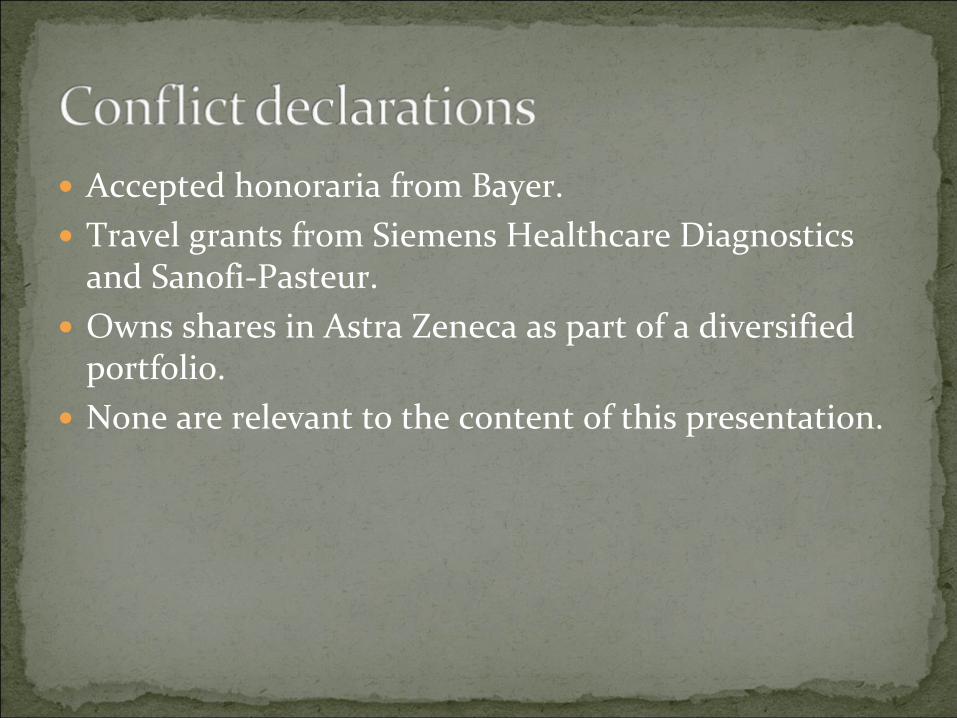
Accepted honoraria from Bayer.Travel grants from Siemens Healthcare Diagnostics and Sanofi‐Pasteur. Owns shares in Astra Zeneca as part of a diversified portfolio.None are relevant to the content of this presentation.
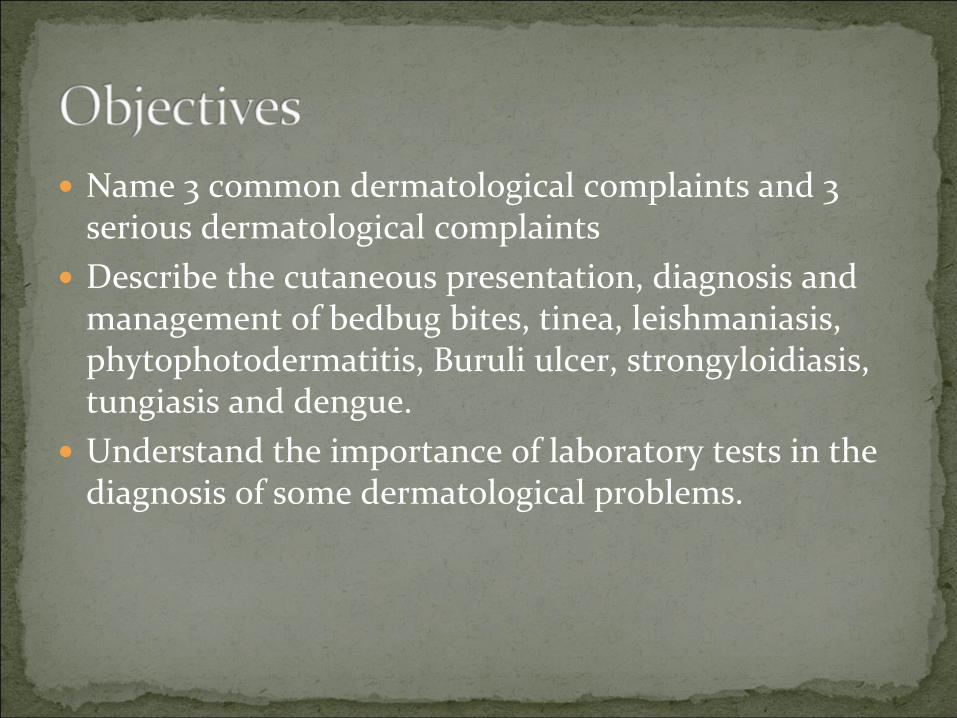
Name 3 common dermatological complaints and 3 serious dermatological complaintsDescribe the cutaneous presentation, diagnosis and management of bedbug bites, tinea, leishmaniasis, phytophotodermatitis, Buruli ulcer, strongyloidiasis, tungiasis and dengue.Understand the importance of laboratory tests in the diagnosis of some dermatological problems.
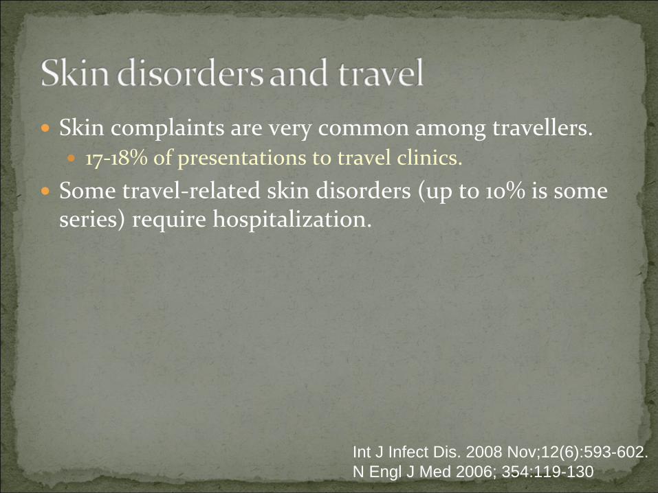
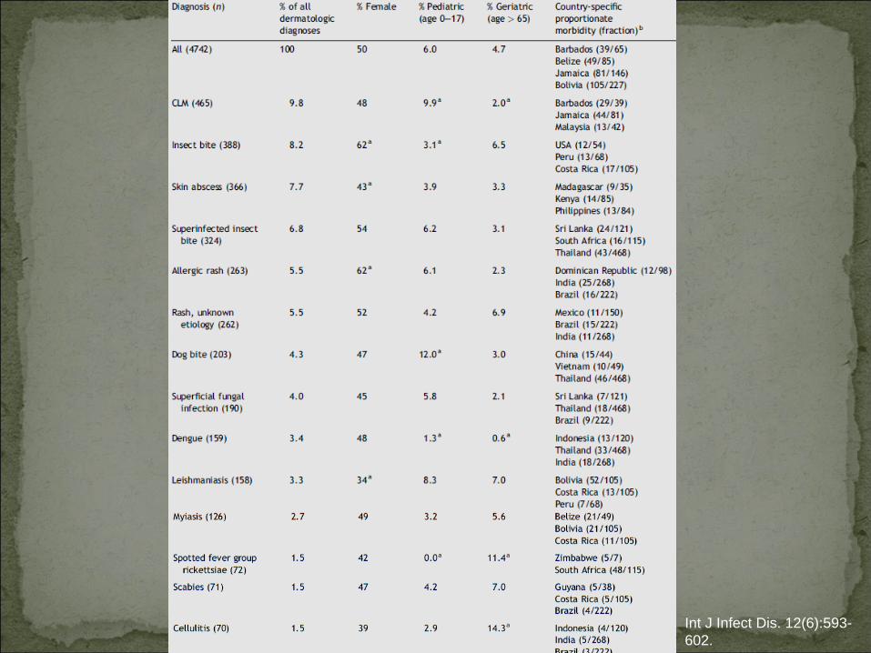
Skin complaints are very common among travellers.17‐18% of presentations to travel clinics.
Some travel‐related skin disorders (up to 10% is some series) require hospitalization.
Int J Infect Dis. 2008 Nov;12(6):593-602.N Engl J Med 2006; 354:119-130
Evaluation of skin complaints in the returned traveller (or immigrant).
Onset, timing, durationGeographic, physical and environmental exposures
Includes plants, animals, medications, drugsDuration of stay, reason, sexual historyLocation and appearance of lesionsAssociated symptoms (pruritus, numbness, pain, fever)Previous treatments and outcome
“Cutaneous Lesions” in Tropical Infectious Diseases: Principles, Pathogens & Practice 2nd Ed., Guerrant RL (ed.), 2005.Travel Med Infect Dis. 2009 May;7(3):125-46.
Int J Infect Dis. 12(6):593- 602.
• Most dermatological problems are diagnosed by history and clinical examination.
• Some cases require lab studies for definitive diagnosis.– Pathology– Microbiology– Serology– Haematology
• Especially important to secure a definitive diagnosis when potentially toxic therapy is needed or response to
treatment is poor.• Important when diagnosis is in question or response to
empiric treatment is poor.
“Cutaneous Lesions” in Tropical Infectious Diseases: Principles, Pathogens & Practice 2nd Ed., Guerrant RL (ed.), 2005.Travel Med Infect Dis. 2009 May;7(3):125-46.

26y previously healthy plastic surgery resident.Presented with a non‐healing nodule of 4 weeks duration on his medial right heel.Returned from Peru three weeks ago where he engaged in adventure travel, including the Andes, Lake Titicaca, the Amazon jungle and Lima.Frequently walked with open sandals.
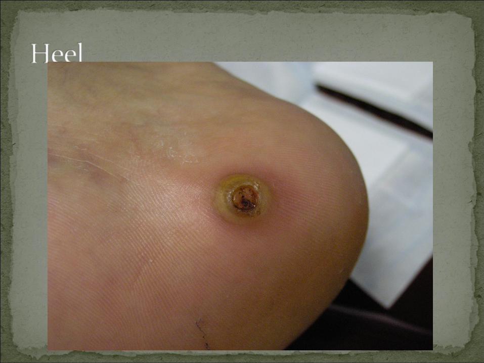
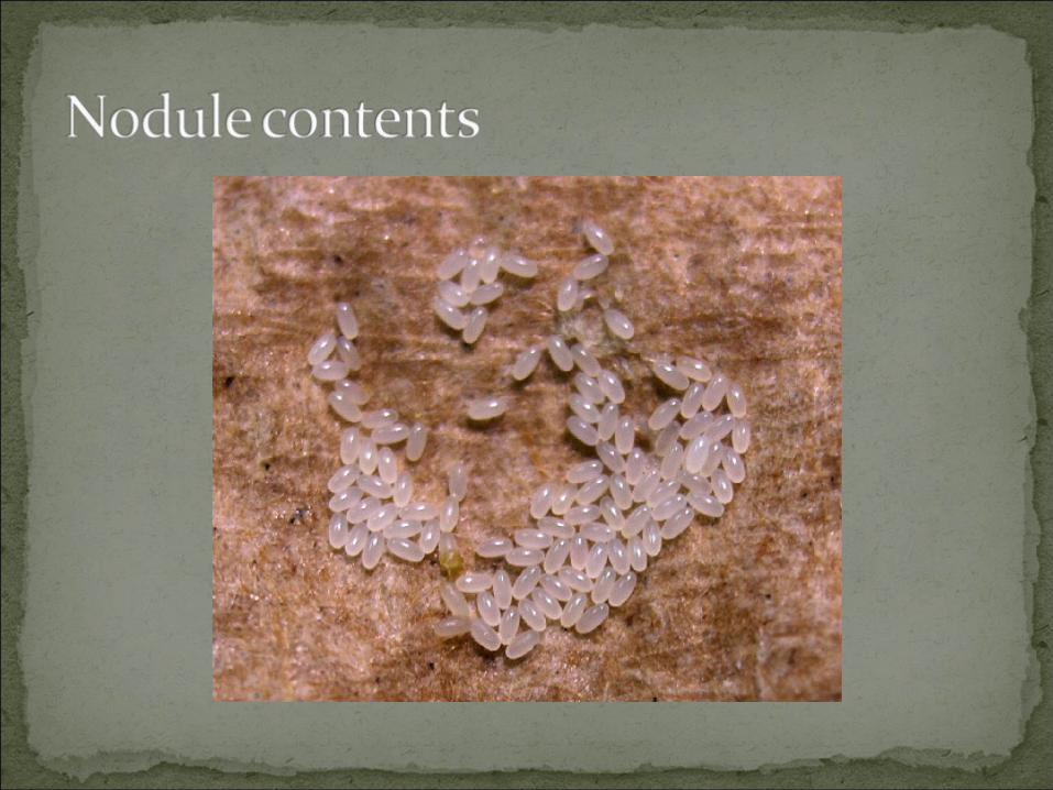
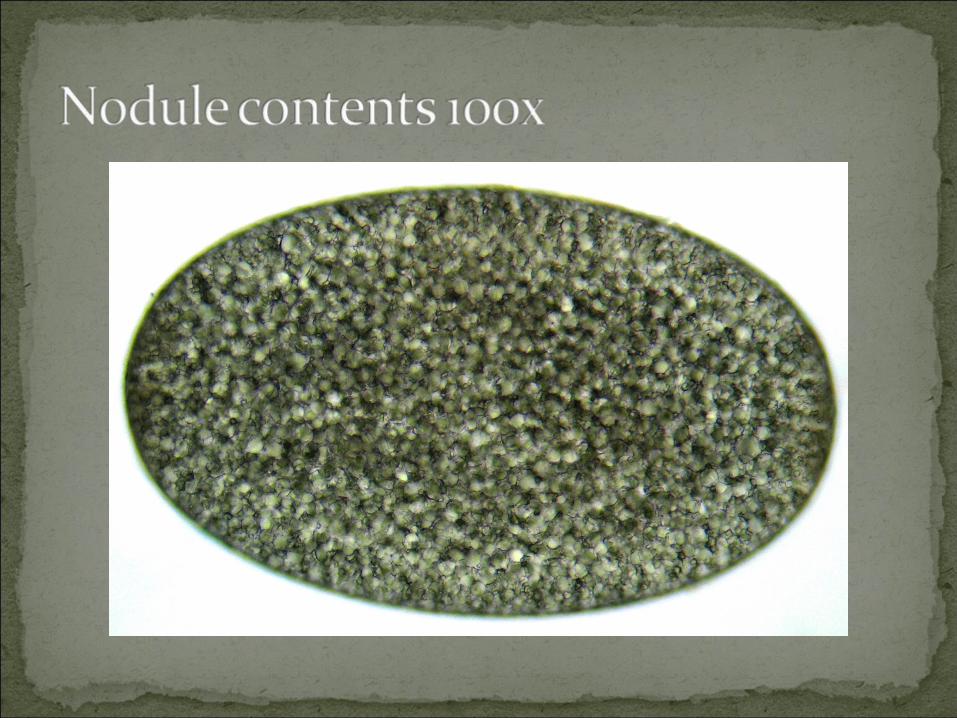
Nodule with opening/ulcer.Small egg‐like structures found inside nodule.Barefoot exposure in South America (Peru).
Ectoparasitic infection caused by the sand flea Tunga penetrans. Originally endemic to South America.
Also found in Central America, the Caribbean, Asia and Africa.
Prevalence amongst local inhabitants of some regions reaches 50%. Infestation typically occurs on the feet. The fertilized female burrows into the patient’s skin, with its anal‐genital opening near the surface.
Clin Dermatol. 2007 Mar-Apr;25(2):158-64.
Feeds on blood and enlarges to a pea‐size, producing a whitish nodule with a central black dot corresponding to the anal‐genital opening. One to three weeks after penetration, the flea expels eggs from the central opening. Approximately five weeks following penetration, the flea dies and is sloughed off, leaving an ulcer that heals slowly.Secondary infections (sometimes severe) can occur.
Clin Dermatol. 2007 Mar-Apr;25(2):158-64.
Surgical removal of flea with subsequent antiseptic washes.
Alternative is to surgically remove the whole nodule.Antibiotic for secondary infections if present.
Consider tetanus prophylaxis if indicated.Lesion heals slowly over weeks.Anti‐parasitic agents (Ivermectin, Thiabendazole) may be effective but are not indicated unless there are numerous lesions.
Clin Dermatol. 2007 Mar-Apr;25(2):158-64.
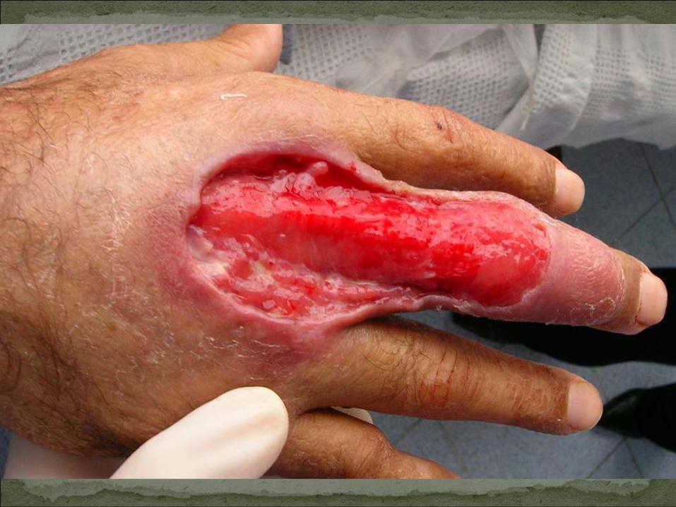
57 year old man from Peru presents with ulcerating lesion to his right hand.Slowly progressive over a year.Not painful, minimal drainage, no fever, chills, night sweats, cough or constitutional symptoms.Unresponsive to multiple antibiotics.
Lesion is deeply undermined, but is not purulent or foul smelling.Culture from GP grew coagulase negative staph and diphtheroids.
On hand, slowly progressive, ulcerative, non‐inflammatory.Residence in jungle area of Peru.Differential diagnosis is broad.
Includes infectious (parasitic, fungal, mycobacterial) and non‐infectious (malignancy – especially BCC)
Biopsy and culture is recommended to direct therapy.
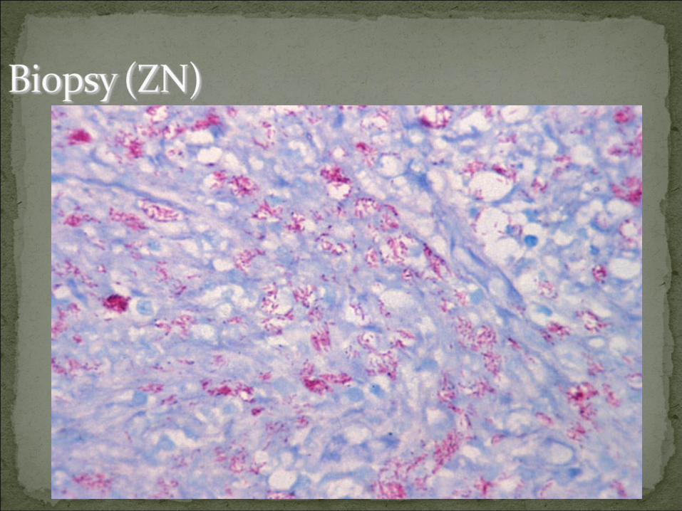
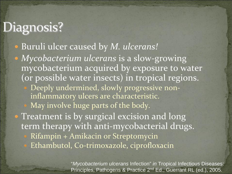
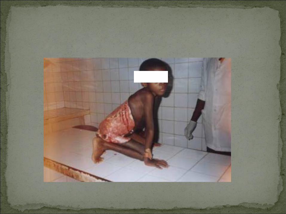
Buruli ulcer caused by M. ulcerans!Mycobacterium ulcerans is a slow‐growing mycobacterium acquired by exposure to water (or possible water insects) in tropical regions.
Deeply undermined, slowly progressive non‐inflammatory ulcers are characteristic.May involve huge parts of the body.
Treatment is by surgical excision and long term therapy with anti‐mycobacterial drugs.
Rifampin + Amikacin or StreptomycinEthambutol, Co‐trimoxazole, ciprofloxacin
“Mycobacterium ulcerans Infection” in Tropical Infectious Diseases: Principles, Pathogens & Practice 2nd Ed., Guerrant RL (ed.), 2005.

A 54 year old woman from Guyana presents for evaluation of eosinophilia.
Absolute count 1.8 x 10⁸/LRelative 20%
Emigrated to Canada 28 years prior. No return to Guyana, no travel except occasional resort areas in Mexico. Denies any symptoms until specifically asked about rash.
“Well…I do get this itchy rash on my bum every few months. GP gave me antihistamines because it was so itchy…”
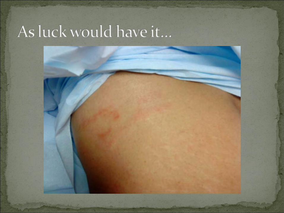
Recurrent, several days duration, then disappears. Happening for >20 years.Geographic, physical and environmental exposures: Guyana, >20 years ago.Location of lesions: Buttocks, sometimes lower abdomen.Associated symptoms: Intense pruritus, eosinophilia.
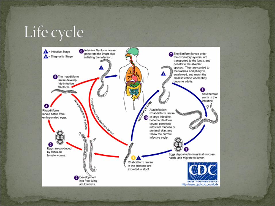
Caused by the nematode Strongyloides stercoralisCommon and persistent infection (last for decades)
30–100 million people worldwideEndemic in Africa, Asia, Southeast Asia, and Central and South AmericaCan result in fulminant dissemination with case‐fatality rates of over 70% in the setting of compromised cellular immunity.Rash and eosinophilia basically diagnostic, but serology also ordered and very positive.
CMAJ. 2004 Aug 31;171(5):479-84.
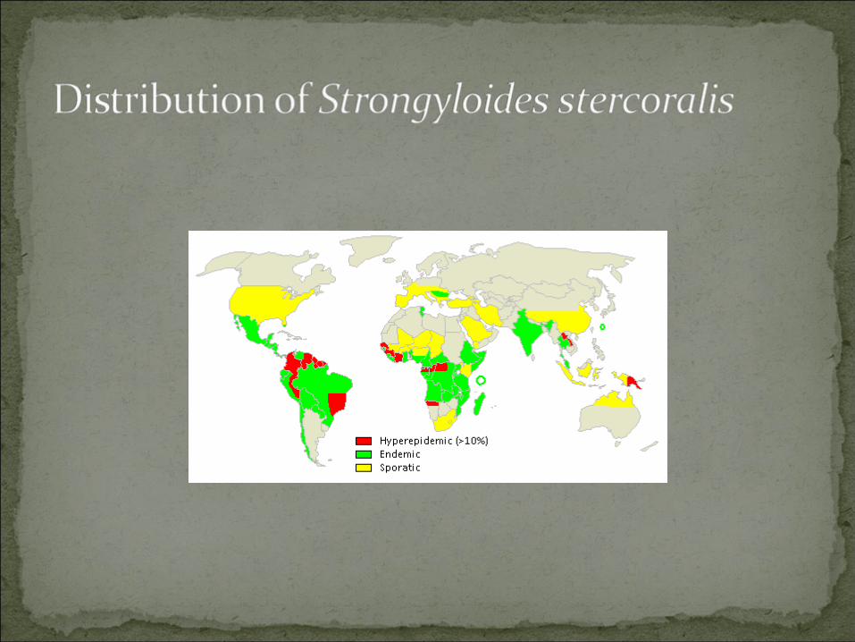
Data is sporadic and few systematic studies have been done.One study showed that Southeast Asian arriving Canada had seroprevalence rates between 11.8% (Vietnamese) and 76.6% (Cambodians).In 2002, a series of 10 consecutive cases of disseminated or fatal Strongyloides infection were identified in 2 academic hospitals in Toronto over a 7‐month periodAll were immigrants: 3 (Asia), 6 (Caribbean), 1 (Africa)One patient had lived in Canada for 56 yrs before symptoms developed.
Am J Epidemiol. 1990 Aug;132(2):257-64. CMAJ. 2004 Aug 31;171(5):479-84.
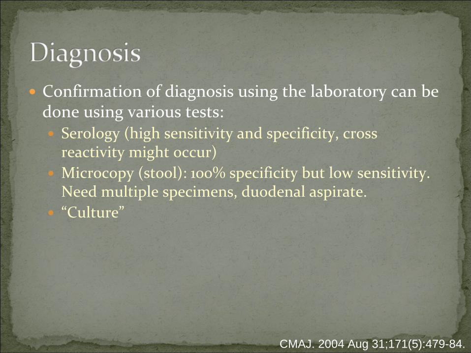
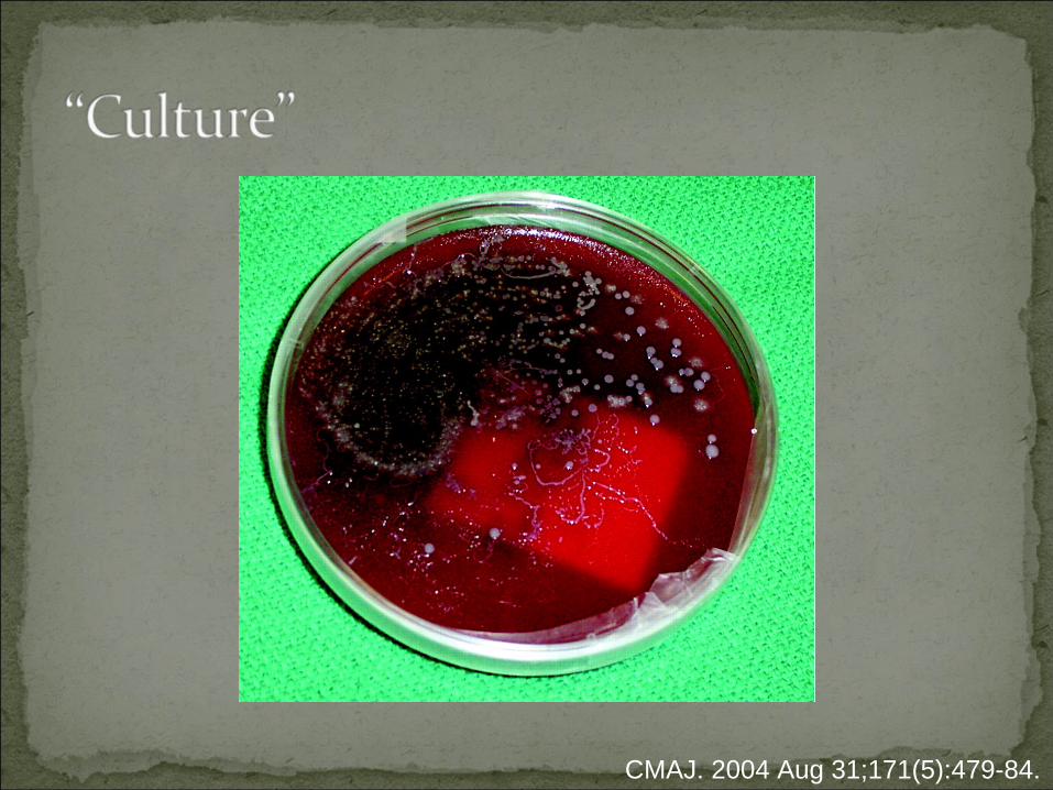
Confirmation of diagnosis using the laboratory can be done using various tests:
Serology (high sensitivity and specificity, cross reactivity might occur)Microcopy (stool): 100% specificity but low sensitivity. Need multiple specimens, duodenal aspirate.“Culture”
CMAJ. 2004 Aug 31;171(5):479-84.
CMAJ. 2004 Aug 31;171(5):479-84.
Immunocompromise can lead to hyperinfection syndrome.
Disseminated infection with larvae in lungs, CSF, bone marrow…High mortality due to polymicrobial sepsis.
HTLV‐1, prednisone, cytotoxic agents and malignancy are common associations.
Good idea to screen patients from endemic areas before immunocompromising regimen started.Good idea to screen patients with Strongyloides for HTLV‐1 if from HTLV‐1 endemic country.
CMAJ. 2004 Aug 31;171(5):479-84.CMAJ. 2007 Aug 28;177(5):451-3
Normal immune system:Single drug: albendazole x 7d OR ivermectin 200 μg/kg daily x 1‐2 d
Immunosuppressed:Combination therapy:
albendazole 400 mg twice daily x 7d AND ivermectin 200 μg/kg daily x 1‐2 d
In cases of disseminated strongyloidiasis, albendazole and ivermectin are continued until there is evidence that the parasite is cleared
Follow‐up serologyFollow‐up serology should be ordered at 6 month intervals until serological cure is documented.Reversion to negative or post‐treatment/pre‐treatment OD ratio of <0.6.
CMAJ. 2007 Aug 28;177(5):451-3Am J Trop Med Hyg. 2009 May;80(5):788-91Am J Trop Med Hyg. 2002 Jun;66(6):749-52.

7 year old boy with itchy progressive lesions over trunk of body for 6 weeks. Returned from Cancun 2 months ago.
All inclusive trip in “luxury resort”.No tick bites, a few mosquito bites.Swam in the ocean only.Exposure to monkeys (on “Jungle tour”), horses (Horse back riding excursion) and dog (the resort’s mascot Beagle).
http://dermatlas.med.jhmi.edu
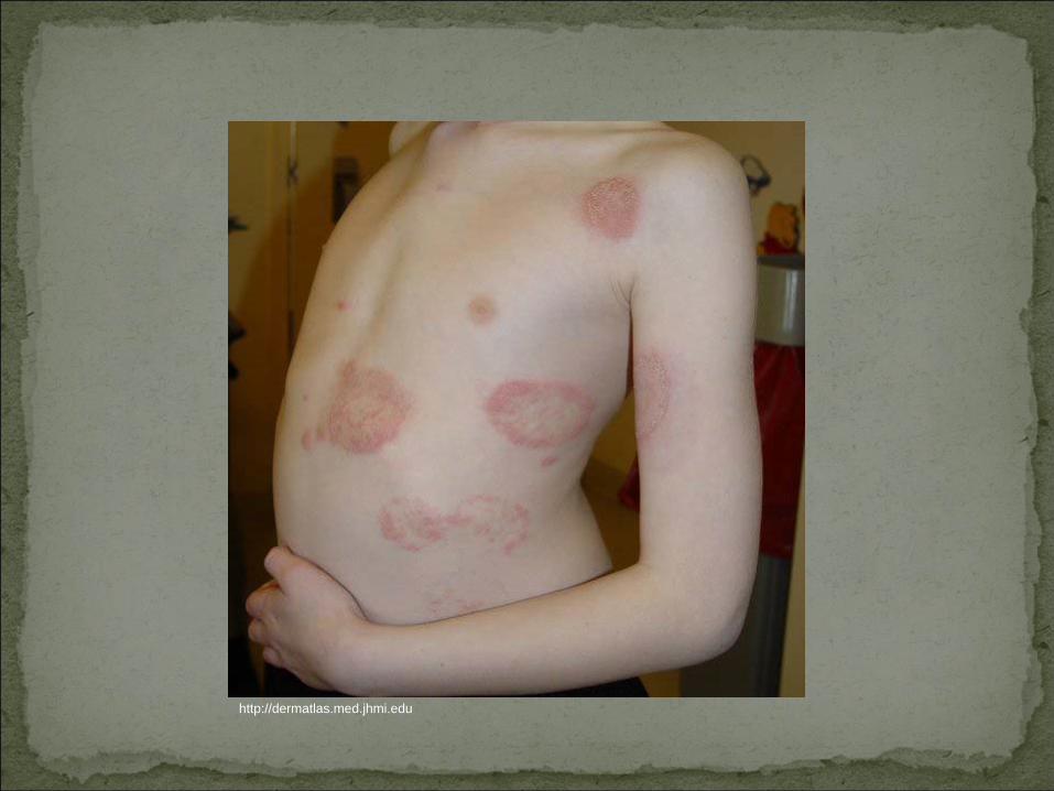
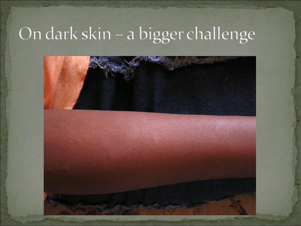
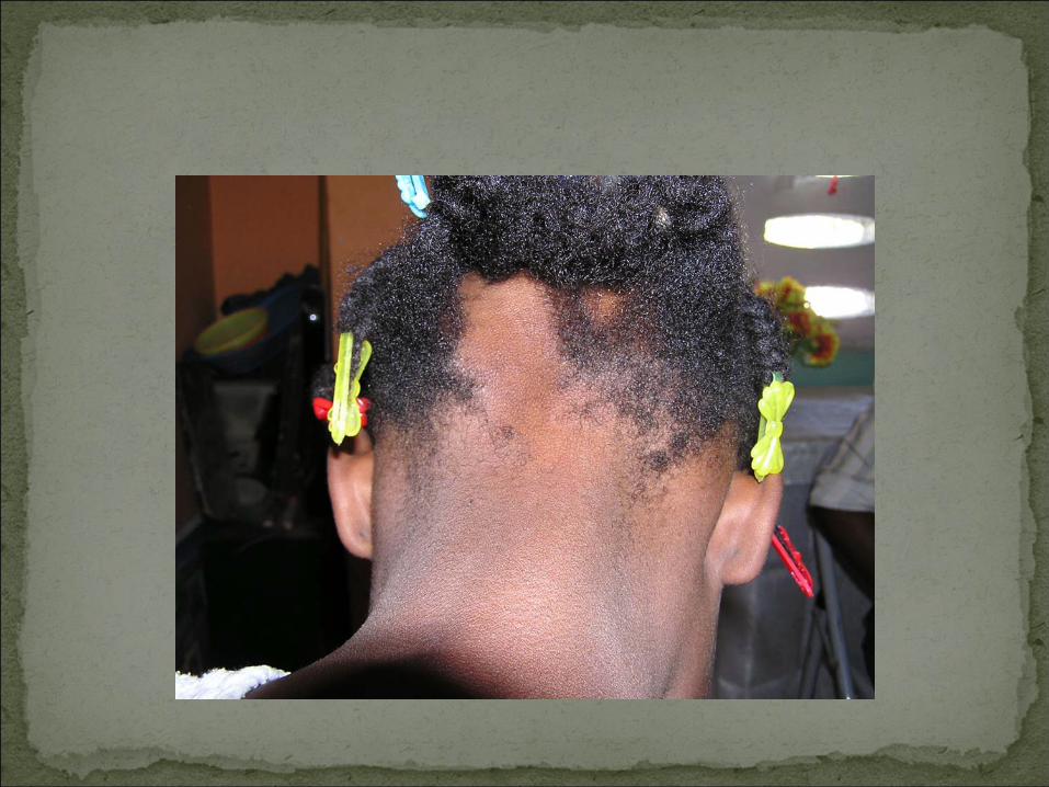
Progressive, red, raised with central clearing.Travelled to Mexico, exposed to horses, monkey, dogsLocation of lesions: TrunkAssociated symptoms: Pruritus, alopecia
Caused by a variety of fungi collectively known as dermatophytes.
Trichophyton rubrum, T. mentagorphytes, T. tonsuransare common, and anthropophilic (usually spread from person to person)Microsporum canis is a common cause, associated with domestic animal exposure (dogs and cats)
Also a common cause of tinea capitis.Microsporum nanum (pigs) and Microsporum gypseum(soil, thorns, wood) also seen.
“Dermatophytosis” in Tropical Infectious Diseases: Principles, Pathogens & Practice 2nd Ed., Guerrant RL (ed.), 2005.
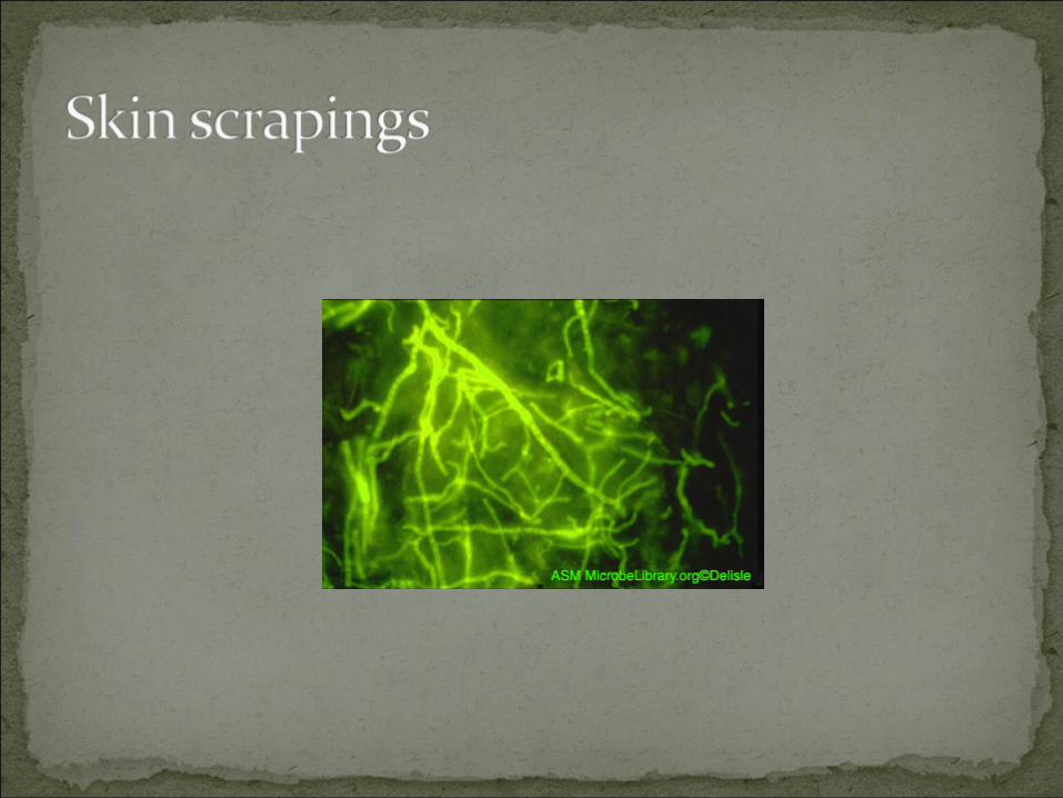
Presentation is fairly typical and trial of therapy is often curative and diagnostic.Fluorescence under UV light can help:
Microsporum species fluoresce green, blue‐green or yellow‐green.
If atypical, recalcitrant to therapy, progressive despite therapy, consider lab studies:
Scrapings or hair for microscopy and culture is usually sufficient.Biopsies should be considered if lesions unusual or atypia, actinic changes, malignancy, or alternative diagnosis is suspected.
Many topical therapies exist:CiclopiroxTopical azolesTurbinafineApply twice daily for 3 – 6 weeks. Compound with 0.5 – 1% hydrocortisone for symptomatic relief.
If there are multiple lesions, hard to reach areas, poor response to topical therapy, involvement of nails, hair or scalp, use oral therapy.
Oral azoles: Weekly Fluconazole or daily intraconazole or ketoconazole.Turbinafine dailyGriseofulvin
Oral treatment should be 4‐6 weeks for tinea corporis, 8 weeks for tinea capitis and 3 – 6 months for tinea unguium.
“Dermatophytosis” in Tropical Infectious Diseases: Principles, Pathogens & Practice 2nd Ed., Guerrant RL (ed.), 2005.

36 year old woman presents complaining of itchy bumps on her legs, arms and shoulders.Started last week while travelling in Egypt. Returned home 3 days ago.Lesions are intensely pruritic and have a red border and raised centre.
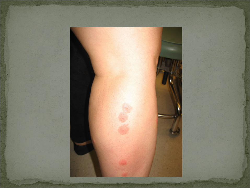
Acute presentation, red, raised, wheal‐like with central vesicle.Travel to Egypt. No other significant exposures elicited.Location of lesions: Arms, shoulders, legs (frequently exposed areas)Associated symptoms: Pruritus
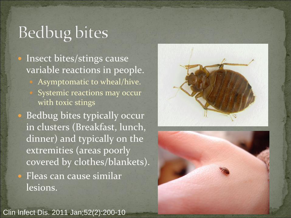
Insect bites/stings cause variable reactions in people.
Asymptomatic to wheal/hive.Systemic reactions may occur with toxic stings
Bedbug bites typically occur in clusters (Breakfast, lunch, dinner) and typically on the extremities (areas poorly covered by clothes/blankets).Fleas can cause similar lesions.
Clin Infect Dis. 2011 Jan;52(2):200-10
Bed bugs get a bad rap…Little more than a nuisance.
Currently no evidence that they are vectors for disease (unlike mosquitoes)Do not have toxins, but do have allergens, sensitizers.Generally keep out of sight from guests!
Rare cases of anaphylactic reactionsLesions can become superinfected.Heavy infestations can lead to anaemia
Especially in children.Social stigma
Clin Infect Dis. 2011 Jan;52(2):200-10
If the infestation is in the home (as opposed to travel related) – insecticides are required.
Pyrethrins are generally used. Possible increasing tolerance.
Bugs live in walls, furniture (beds, couches) and carpets.
Can live ~6 months without food.
Bugs travel in suitcases.It travel‐related cases, it is wise to freeze/heat clothes on arrival.
Clin Infect Dis. 2011 Jan;52(2):200-10
30 year old male.Works as a wildlife photographer, most recently in the area of Madidi National Park (Bolivia) for a 2 month photography assignment.
Returned two months ago.
Noticed a raised lesion on his leg 3 months ago.Treated with steroid creams, antibiotics (cephalexin) and antifungals (clotrimazole)
No improvement, progressive.
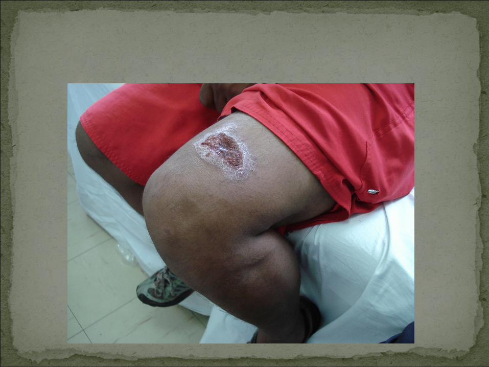
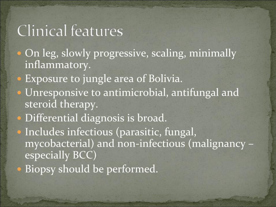
On leg, slowly progressive, scaling, minimally inflammatory.Exposure to jungle area of Bolivia.Unresponsive to antimicrobial, antifungal and steroid therapy.Differential diagnosis is broad.Includes infectious (parasitic, fungal, mycobacterial) and non‐infectious (malignancy –especially BCC)Biopsy should be performed.
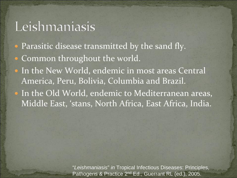
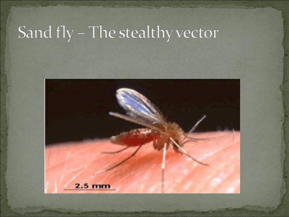
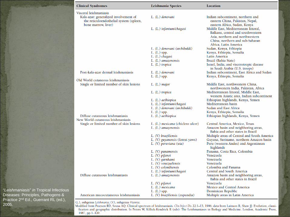
Parasitic disease transmitted by the sand fly.Common throughout the world.In the New World, endemic in most areas Central America, Peru, Bolivia, Columbia and Brazil.In the Old World, endemic to Mediterranean areas, Middle East, ‘stans, North Africa, East Africa, India.
“Leishmaniasis” in Tropical Infectious Diseases: Principles, Pathogens & Practice 2nd Ed., Guerrant RL (ed.), 2005.
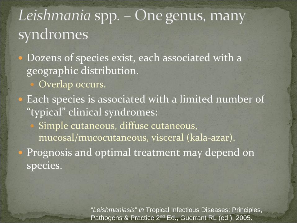
Dozens of species exist, each associated with a geographic distribution.
Overlap occurs.Each species is associated with a limited number of “typical” clinical syndromes:
Simple cutaneous, diffuse cutaneous, mucosal/mucocutaneous, visceral (kala‐azar).
Prognosis and optimal treatment may depend on species.
“Leishmaniasis” in Tropical Infectious Diseases: Principles, Pathogens & Practice 2nd Ed., Guerrant RL (ed.), 2005.
“Leishmaniasis” in Tropical Infectious Diseases: Principles, Pathogens & Practice 2nd Ed., Guerrant RL (ed.), 2005.
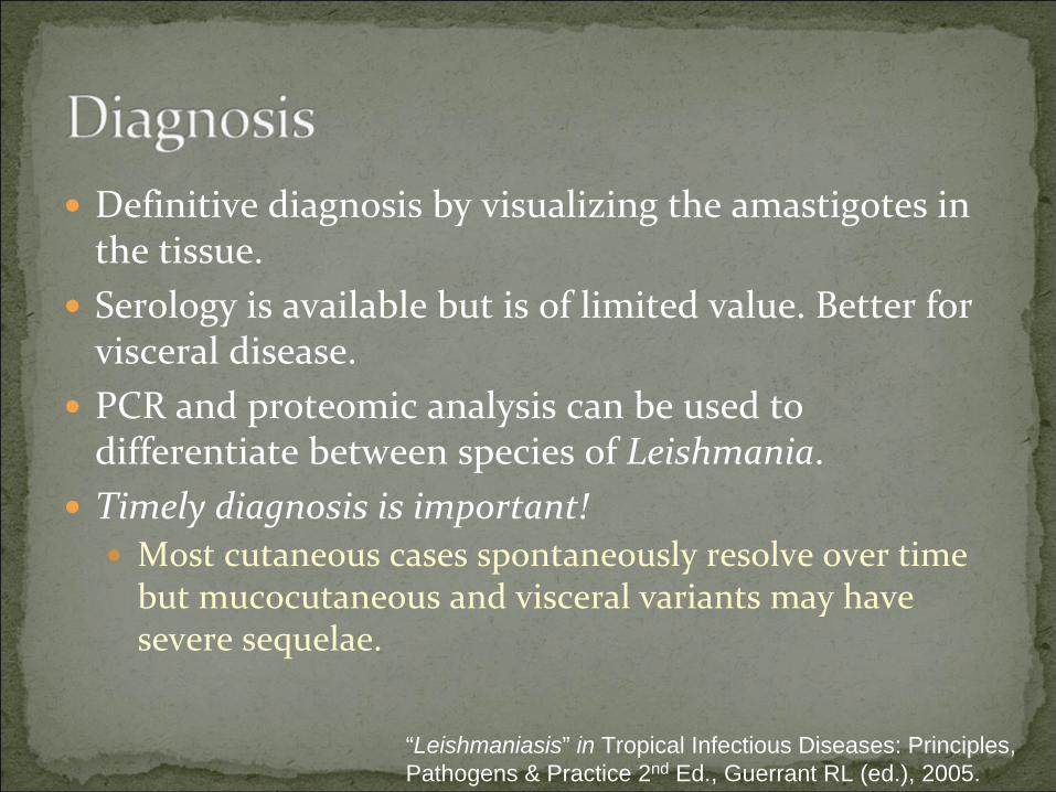
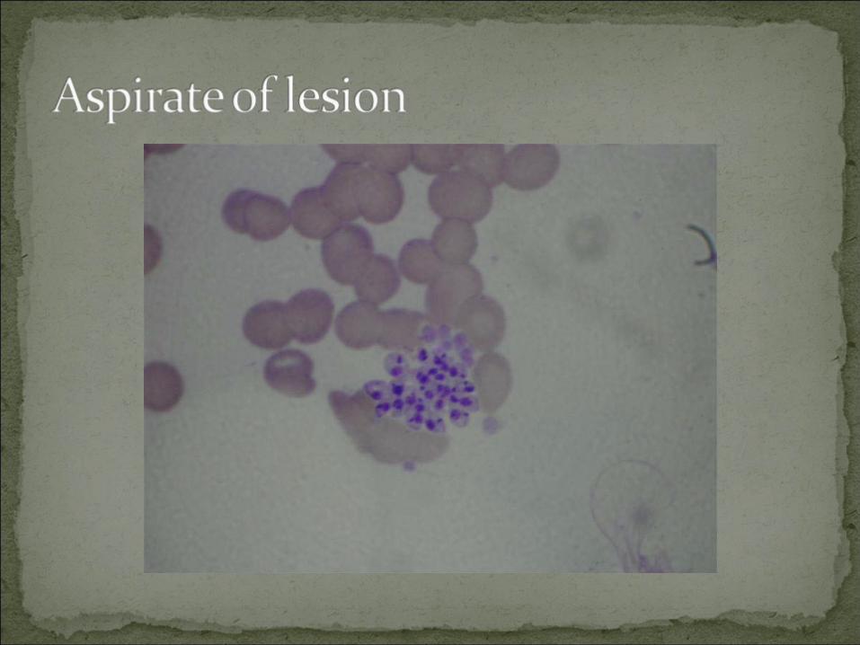
Definitive diagnosis by visualizing the amastigotes in the tissue.Serology is available but is of limited value. Better for visceral disease.PCR and proteomic analysis can be used to differentiate between species of Leishmania. Timely diagnosis is important!
Most cutaneous cases spontaneously resolve over time but mucocutaneous and visceral variants may have severe sequelae.
“Leishmaniasis” in Tropical Infectious Diseases: Principles, Pathogens & Practice 2nd Ed., Guerrant RL (ed.), 2005.
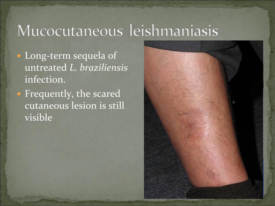
Long‐term sequela of untreated L. braziliensisinfection.Frequently, the scared cutaneous lesion is still visible
Evolving field…! Effective treatment also depends somewhat on species.Pentavalent antimonials are generally effective for cutaneous diseases and is some areas for visceral disease. Amphotericin B (liposomal) is a better choice for visceral forms.
More effective, less resistance.Also consider for mucocutaneous variants – less relapses.
Miltefosine is a relatively new oral addition to treatment options for visceral leishmaniasis
97% cure rate in an Indian series of visceral disease.
“Leishmaniasis” in Tropical Infectious Diseases: Principles, Pathogens & Practice 2nd Ed., Guerrant RL (ed.), 2005.
Thermal therapy, cryotherapy, imiquimod, pentamidine and intra‐lesional antimonials have been used for cutaneous variants.
Success rate is variable.Difficulty in speciation, risk of geographic overlap in species associated with sequelae (mucocutaneous, visceral, diffuse cutaneous) limits use of topical treatment.
“Leishmaniasis” in Tropical Infectious Diseases: Principles, Pathogens & Practice 2nd Ed., Guerrant RL (ed.), 2005.


9 year old girl returns from a Christmas vacation in Mexico with “a weird sunburn” on her face.Started off as “sunburn”, on both cheeks, pigmentation present since return. Stayed at all‐inclusive resort, only went to beach, poolside and hotel restaurants.
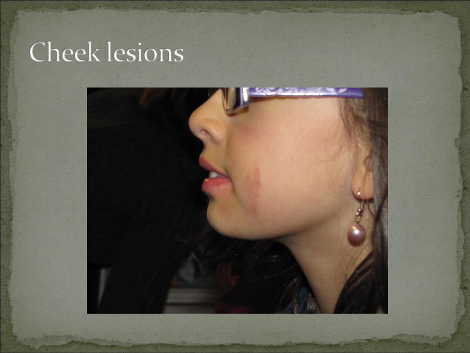
On face, bilateral, sudden onset, initially sun‐burn like quality.Exposure resorts in Mexico.Localized, linear pattern, streaks.

Did you eat or drink Did you eat or drink anything with lime or anything with lime or
lemon juice?lemon juice?

“I ate lemons all day!”
Cutaneous phototoxic inflammatory eruption resulting from contact with light‐sensitizing botanical substances and long‐wave ultraviolet (UV‐A 320‐380 nm) radiation.Usually begins approximately 24 hours after exposure and peaks at 48‐72 hours.
Following initial symptoms, persistent hyper or hypo pigmentation of variable duration occurs.
Am J Contact Dermat. 1999 Jun;10(2):89-93.
Exposure to plant psoralens and related compounds and UV light is critical.Psoralens are activated by UV light bind to DNA and create cross‐links in the DNA structure.
Leads to cell death and a clinical syndrome similar to sunburn (erythema, blistering, inflammation).
Many plants contain psoralens and related compounds.
Common culprits are citrus (limes, lemons), parsnips, celery, carrots, fennel, fig leaves, hogweed, Queen Anne’s Lace.
Am J Contact Dermat. 1999 Jun;10(2):89-93.
Reassurance is typically all that is needed.Phytophotodermatitis is a self‐limited problem.Avoid the agent if possible, especially if exposed to UV light.Sunscreens helpful to prevent cases.Topical steroids or anti‐inflammatories may be prescribed during the painful initial phase if required.
Am J Contact Dermat. 1999 Jun;10(2):89-93.
24 year old female on a trip to Thailand with girlfriends.4 days after return to Winnipeg from “Phuket” leg, develops sudden onset fever, rash, headache and back pain.
Friend from Australia with similar symptoms
Clinically stable, rash distributed on whole body, but prominent on chest, abdomen.Took malaria prophylaxis where appropriate and used bed nets at night when appropriate.
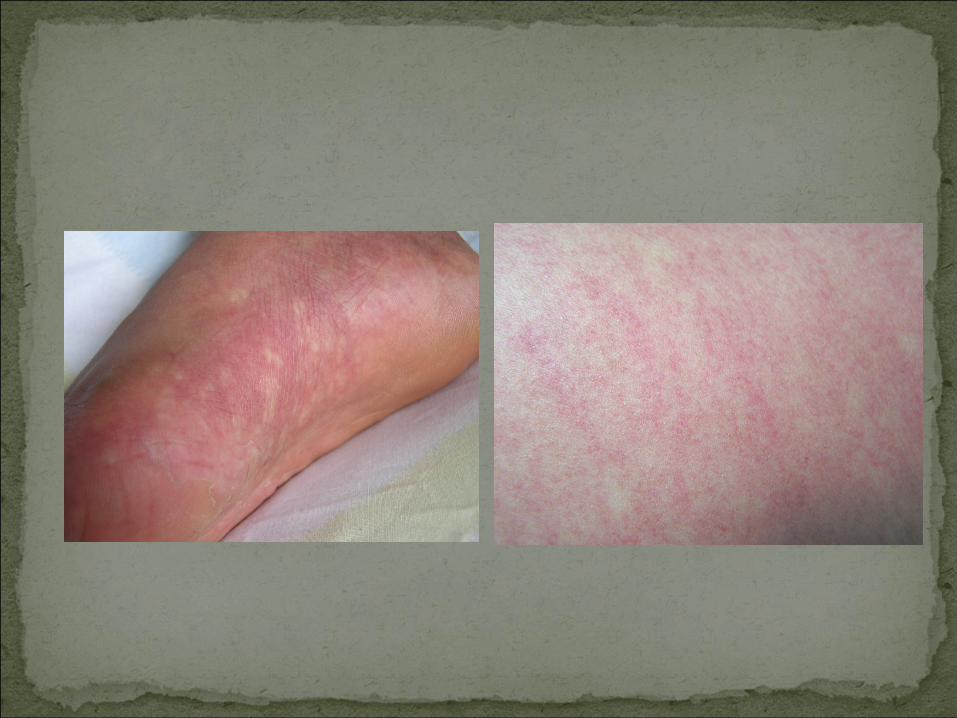
Rapid onset, acute, diffuse rash, erythematous with white patches, fever, headache, back pain. Exposure to Thailand in past week.No coagulopathy, clinically stable.
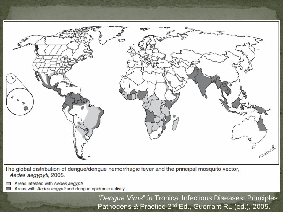
Classic symptoms and exposures.Highly prevalent in Thailand (#1 diagnosed caused of fever in travellers to SEA).Most mild cases (no features of haemorrhage, coagulopathy, shock) can be managed expectantly.Always consider malaria and typhoid and order appropriate investigations if needed.This patient had negative serology at the time of presentation and IgG and IgM positive 6 weeks later.
N Engl J Med 2006; 354:119-130“Dengue Virus” in Tropical Infectious Diseases: Principles, Pathogens & Practice 2nd Ed., Guerrant RL (ed.), 2005.
Flavivirus transmitted by the day‐biting mosquito Aedes aegyptii and A. albopictus.Disease varies from asymptomatic infection (more common in young children), Dengue fever (“Break bone fever”) to Dengue Haemorrhagic Fever.Generally diagnosed clinically and by excluding other conditions. Serology is helpful, and PCR diagnosis is possible early in disease.Common laboratory findings are lymphocytosis, neutropenia, elevated liver enzymes and thromobocytopenia.
“Dengue Virus” in Tropical Infectious Diseases: Principles, Pathogens & Practice 2nd Ed., Guerrant RL (ed.), 2005.
Obvious mucosal haemorrhage, petechiae, easy bruising.JaundiceHaemodynamic labilityPositive tourniquet test
>20 petechiae per square inch (3/cm2) after inflating a BP cuff to the midway point between diastolic and systolic for 5 minutes.
Restlessness, lethargy, cyanosis…Should prompt immediate referral to hospital.
“Dengue Virus” in Tropical Infectious Diseases: Principles, Pathogens & Practice 2nd Ed., Guerrant RL (ed.), 2005.
“Dengue Virus” in Tropical Infectious Diseases: Principles, Pathogens & Practice 2nd Ed., Guerrant RL (ed.), 2005.
Supportive for Dengue fever.Analgesics, antipyretics.Convalescence may be long (4 – 6 weeks) and associated with weakness, depression, pruritus.
Dengue Haemorrhagic Fever/ShockRapid fluid resuscitation, blood products as needed, supportive care.Steroids not helpful.No effective anti‐virals exist.
“Dengue Virus” in Tropical Infectious Diseases: Principles, Pathogens & Practice 2nd Ed., Guerrant RL (ed.), 2005.