Embed Size (px)
Citation preview
University of Kentucky University of Kentucky
UKnowledge UKnowledge
Theses and Dissertations--Pharmacy College of Pharmacy
2017
EFFECTS OF CORE AND SHELL MODIFICATION TO TETHERED EFFECTS OF CORE AND SHELL MODIFICATION TO TETHERED
NANOASSEMBLIES ON SIRNA THERAPY NANOASSEMBLIES ON SIRNA THERAPY
Steven Rheiner University of Kentucky, [email protected] Digital Object Identifier: https://doi.org/10.13023/ETD.2017.272
Right click to open a feedback form in a new tab to let us know how this document benefits you. Right click to open a feedback form in a new tab to let us know how this document benefits you.
Recommended Citation Recommended Citation Rheiner, Steven, "EFFECTS OF CORE AND SHELL MODIFICATION TO TETHERED NANOASSEMBLIES ON SIRNA THERAPY" (2017). Theses and Dissertations--Pharmacy. 73. https://uknowledge.uky.edu/pharmacy_etds/73
This Doctoral Dissertation is brought to you for free and open access by the College of Pharmacy at UKnowledge. It has been accepted for inclusion in Theses and Dissertations--Pharmacy by an authorized administrator of UKnowledge. For more information, please contact [email protected].

STUDENT AGREEMENT: STUDENT AGREEMENT:
I represent that my thesis or dissertation and abstract are my original work. Proper attribution
has been given to all outside sources. I understand that I am solely responsible for obtaining
any needed copyright permissions. I have obtained needed written permission statement(s)
from the owner(s) of each third-party copyrighted matter to be included in my work, allowing
electronic distribution (if such use is not permitted by the fair use doctrine) which will be
submitted to UKnowledge as Additional File.
I hereby grant to The University of Kentucky and its agents the irrevocable, non-exclusive, and
royalty-free license to archive and make accessible my work in whole or in part in all forms of
media, now or hereafter known. I agree that the document mentioned above may be made
available immediately for worldwide access unless an embargo applies.
I retain all other ownership rights to the copyright of my work. I also retain the right to use in
future works (such as articles or books) all or part of my work. I understand that I am free to
register the copyright to my work.
REVIEW, APPROVAL AND ACCEPTANCE REVIEW, APPROVAL AND ACCEPTANCE
The document mentioned above has been reviewed and accepted by the student’s advisor, on
behalf of the advisory committee, and by the Director of Graduate Studies (DGS), on behalf of
the program; we verify that this is the final, approved version of the student’s thesis including all
changes required by the advisory committee. The undersigned agree to abide by the statements
above.
Steven Rheiner, Student
Dr. Younsoo Bae, Major Professor
Dr. David Feola, Director of Graduate Studies
Title Page
EFFECTS OF CORE AND SHELL MODIFICATION TO TETHERED
NANOASSEMBLIES ON SIRNA THERAPY
DISSERTATION
A dissertation submitted in partial fulfillment of the
requirements for the degree of Doctor of Philosophy in the
College of Pharmacy
at the University of Kentucky
By
Steven Neil Rheiner
Lexington, Kentucky
Director: Dr. Younsoo Bae, Associate Professor of Pharmaceutical Sciences
Lexington, Kentucky
Copyright © Steven Rheiner 2017
Abstract
ABSTRACT OF DISSERTATION
EFFECTS OF CORE AND SHELL MODIFICATION TO TETHERED
NANOASSEMBLIES ON SIRNA THERAPY
siRNA therapy is an emerging technique that reduces protein expression in cells by
degrading their mRNAs via the RNA interference pathway (RNAi). Diseases such as
cancer often proliferate due to increased protein expression and siRNA therapy offers a
new method of treatment for those diseases. Although siRNA therapy has shown success
in vitro, it often fails in vivo due to instability in the blood stream. To overcome this
limitation, delivery vehicles are necessary for successful transfection of siRNA into target
cells and cationic polymers have been widely studied for this purpose. However,
complexes between siRNA and delivery vehicles made from cationic polymers exhibit
stability issues in the blood stream which results in toxicity and low transfection. This
work hypothesizes that improvement of vehicle/siRNA complex stability will improve
siRNA transfection efficiency. To test this, the contributions and outcomes of
poly(ethylene glycol) [PEG] shell and hydrophobic core modification to a
polyethylenimine (PEI) based tethered nanoassemblies (TNAs) were examined. Initially,
hydrophobic modification of palmitate (PAL) to the core of the TNA yielded improved
transfection efficiency due to an enhanced endosomal escape capability. However, this
modification also reduced the TNA/siRNA complex stability. This indicated that the core
hydrophobicity must be balanced in order increase stability while increasing transfection
efficiency. Additionally, TNAs made from PEG and PEI did not cause transfection in our
initial study. The PEG shell density was found to be too great and thereby reduced
transfection efficiency. Reducing the PEG density by lowering PEG molecular weight,
reducing attachment percentage, and removing small PEI impurities from the synthesis
stock increased overall transfection efficiency and unimolecularity of the TNA
complexes. This indicated that the shell composition of the TNA must be tuned in order
to improve particle design. Further study of the hydrophobically modification to TNAs
yielded unintended effects on the transfection efficiency evaluation assay. These particles
exhibited an siRNA independent reduction in the reporter protein used to observe
transfection, or a false positive effect, that was not previously observed. It was found that
this false positive was influence mainly by the hydrophobic group rather than the cationic
polymer backbone. Cellular stress was observed in cells dosed with the hydrophobically
modified TNAs which lead to over ubiquitination and rapid degradation of the luciferase
protein. This demonstrated that core components of TNAs could cause cellular stress and
influence interaction outside of the TNA. Overall, this work demonstrates that
hydrophobic core and PEG shell modification require balancing and consideration to
improve properties of future cationic polymer based siRNA delivery vehicle design.
Keywords: Tethered nanoassemblies, siRNA therapy, gene delivery, cationic polymer,
chemical modification, transfection
Steven Rheiner
Date
7/13/17

Approval Page
EFFECTS OF CORE AND SHELL MODIFICATION TO TETHERED
NANOASSEMBLIES ON SIRNA THERAPY
By
Steven Neil Rheiner
Director of Dissertation
Director of Graduate Studies
Dr. Younsoo Bae
Dr. David Feola
7/13/17
Date

Dedication
To my wife and family, without your love and support I would not have been able to
accomplish this.
iii
Acknowledgments
ACKNOWLEDGMENTS
Firstly, I would to thank my thesis advisor, Dr. Younsoo Bae, whose support and
guidance has been instrumental to my success in my PhD training. I would like to thank
Dr. Bae for the opportunity to learn from him and his lab in order to forge a path of my
own in such an interesting research field. I sincerely appreciate Dr. Bae’s mentoring and
invaluable advice throughout my research.
Secondly, I would also like to thank my committee members, Dr. Patrick McNamara,
Dr. Daniel Pack, and Dr. Edith Glazer, for their guidance through my research, qualifying
exam, and defense. Their advice and help throughout my graduate studies has been
invaluable to my development as a scientist and furthering my research.
Further, I would like to thank the Cancer Nanotechnology Training Center
(CNTC) of the University of Kentucky for the opportunity to participate in such a
wonderful program. The CNTC granted me the opportunity to gain insight into my
research project from a diverse group in a collaborative setting. This knowledge and
opportunity was valuable to my research and scientific career. Special thanks to Dr. Piotr
Rychahou, Dr. Brad Anderson, and Dr. Robert Yokel for their insight into my research
project and Tonya Vance for her help throughout the traineeship.
I would also like to thank all current and past lab members of the Bae laboratory
that have shared their experience and knowledge to assist with research and growth as a
scientist. Their advice has proven invaluable to my scientific development. I would like
to extend special thanks to Derek Reichel whose critical analysis of my writing and

iv
experimentation has been a valuable source of improvement. Additionally, I would like to
thank all past lab members, Dr. Andrei Ponta, Dr. Pengxiu Cao, Dr. Geunwoo Jin, Dr.
Matthew Dickerson, and Amber Jerke, for their assistance and support.
I would like to acknowledge and give thanks to all other faculty and staff members
throughout my graduate career, especially graduate coordinator Catina Rossoll and
director of graduate studies Dr. Jim Pauly at the College of Pharmacy at University of
Kentucky. They have given me guidance and advice throughout my graduate career that
has helped keep me on track even in difficult times.
I would like to thank and show appreciation to my family. My wife, Faith Rheiner,
has always been by myside to help me through all the ups and downs my graduate career
has brought about. Her love and support has been crucial to my success. I would like to
thank my parents Richard and Cathryn Rheiner, sister Rebecca Rheiner, aunt and uncle
Dr. Mike Cotta and Patti Cotta, and all my other family members that have shown me
love and support my entire life. They have provided a strong support system that I needed
in order to be successful. I would also like to thank my friends, Greg Laver, Liam Flavin,
Danny Jasinski, Dan Binzel, Rob Wensing, Kevin Chen, Matt McErlean, and Ryan
Hughes, as well as many others who have also provided an invaluable support system and
advice throughout the years.
Finally, I would also like to give special thanks to Richard Rheiner, Dr. Mike Cotta,
and Dr. Matthew Wheeler. They have helped to foster and guide my interest in science as
well as provide wisdom in difficult times during my education. Their advice has guided
many of my decisions and helped me to realize my dreams.
v
Table of Contents
Acknowledgments.............................................................................................................. iii
List of Figures ..................................................................................................................... x
List of Tables .................................................................................................................... xii
1. Chapter 1: Tethered Nanoassemblies for siRNA Therapy .......................................... 1
1.1. Genetic Disease Treatment and siRNA Therapy ................................................. 1
1.2. Tethered Nanoassembly (TNA) as siRNA Delivery Vehicles ............................. 5
1.3. Chemical Modifications to Improve siRNA Therapy Efficacy ........................... 7
2. Chapter 2: Effects of Hydrophobic Core Modification on TNA stability and siRNA
transfection ........................................................................................................................ 12
2.1. Introduction ........................................................................................................ 13
2.2. Materials and Methods ....................................................................................... 18
2.2.1. Materials and Cells ............................................................................................. 18
2.2.2. Synthesis of stabilized TNAs ............................................................................ 18
2.2.3. Characterization of stabilized TNAs ................................................................ 19
2.2.4. Analysis of TNAs and siRNA Interactions ..................................................... 20
2.2.5. In vitro transfection efficiency of TNAs.......................................................... 21
2.2.6. Toxicity of TNAs in vitro .................................................................................. 22
2.2.7. In vitro intracellular uptake and trafficking of fluorescent siRNA in TNAs ..
............................................................................................................................... 22
vi
2.3. Results ................................................................................................................ 24
2.3.1. The hydrophobicity of the TNA core reduces interactions between siRNA
and TNA ............................................................................................................................... 24
2.3.2. Increased hydrophobicity of the TNA core increases TNA transfection
efficiency .............................................................................................................................. 30
2.3.3. Hydrophobic modification of nanoassembly core increases intracellular
siRNA delivery and endosomal escape ............................................................................ 33
2.3.4. Combined dosage of hydrophobic modified and unmodified TNA
decreases colocalization of siRNA in endosomes .......................................................... 38
2.3.5. Modulating the hydrophobic substitution of TNA core increases
transfection efficiency while decreasing siRNA/particle interactions ......................... 38
2.4. Discussion .......................................................................................................... 41
2.5. Conclusions ........................................................................................................ 45
3. Chapter 3: Effects of TNA shell modification on siRNA transfection ..................... 46
3.1. Introduction ........................................................................................................ 47
3.2. Materials and Methods ....................................................................................... 51
3.2.1. Materials and Cells ............................................................................................. 51
3.2.2. Synthesis of TNAs of varying PEG substitutions and PEI backbones ........ 51
3.2.3. Quantification of size and surface charge of TNAs and complexes ............ 52
3.2.4. Complex formation of TNAs with siRNA ...................................................... 52
3.2.5. In vitro transfection efficiency and toxicity of TNAs .................................... 53
vii
3.3. Results ................................................................................................................ 54
3.3.1. Increasing PEG corona density increases particle size and decreases surface
charge ... ..................................................................................................................... 54
3.3.2. Increased polymer homogeneity of TNAs increases particle size and
decrease number of polymer chains used in complexation ........................................... 57
3.3.3. Increased PEG corona density increases particle/siRNA complexation ratio
............................................................................................................................... 59
3.3.4. PEG corona density decreases siRNA transfection efficiency ..................... 62
3.3.5. Increased polymer homogeneity decreases necessary complexation ratio to
achieve maximum siRNA transfection............................................................................. 64
3.4. Discussion .......................................................................................................... 66
3.5. Conclusions ........................................................................................................ 70
4. Chapter 4: Hydrophobic Modifications to TNAs and Non-Specific Reduction of
Reporter Protein Concentrations. ...................................................................................... 71
4.1. Introduction ........................................................................................................ 72
4.2. Materials and Methods ....................................................................................... 76
4.2.1. Materials and Cells ............................................................................................. 76
4.2.2. Synthesis of TNAs with different backbones and hydrophobic moieties ... 77
4.2.3. Determination of minimum complexation ratios of TNAs and siRNA ....... 78
4.2.4. In vitro transfection and toxicity efficiency of TNAs .................................... 78
4.2.5. Analysis of Luciferase Protein Expression Levels in vitro ........................... 80
viii
4.2.6. Activity of luciferase protein after pre-incubation with TNAs ..................... 81
4.2.7. Observed interactions of TNAs and components with siRNA through
Raman spectroscopy ........................................................................................................... 81
4.3. Results ................................................................................................................ 82
4.3.1. Hydrophobic moiety in the TNA core has greater influence on luciferase
expression reduction than TNA condensation ................................................................. 82
4.3.2. Luciferase activity is unaffected by TNA interactions with either luciferase
or luciferin ............................................................................................................................ 87
4.3.3. Raman spectroscopy indicates that hydrophobic moieties contribute to
TNA’s interaction with siRNA .......................................................................................... 89
4.3.4. Hydrophobically modified TNAs reduce protein expression, membrane
integrity, and ATP concentration but retained mitochondrial activity ......................... 92
4.3.5. TNAs increased ubiquitination and degradation of luciferase ...................... 94
4.4. Discussion .......................................................................................................... 96
4.5. Conclusions ...................................................................................................... 102
4.6. Limitations of Observations ............................................................................. 102
5. Chapter 5: Conclusions............................................................................................ 104
5.1. Core components of TNAs influence siRNA/TNA complex stability and
transfection efficiency ................................................................................................. 105
5.2. PEG shell density effects transfection efficiency and complex stability ......... 106
5.3. Future Directions .............................................................................................. 107

ix
6. Supplemental Figures .............................................................................................. 109
7. References ............................................................................................................... 115
8. Vita .......................................................................................................................... 131
x
List of Figures
List of Figures
Figure 1.1: RNA Interference Pathway .............................................................................. 4
Figure 2.1. Synthesis of tethered nanoassemblies (TNAs) for siRNA delivery. .............. 17
Figure 2.2. Characterization of TNAs............................................................................... 27
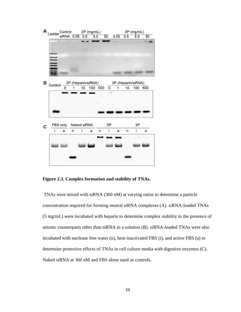
Figure 2.3. Complex formation and stability of TNAs. .................................................... 29
Figure 2.4. Transfection efficiency and toxicity of TNAs. ............................................... 32
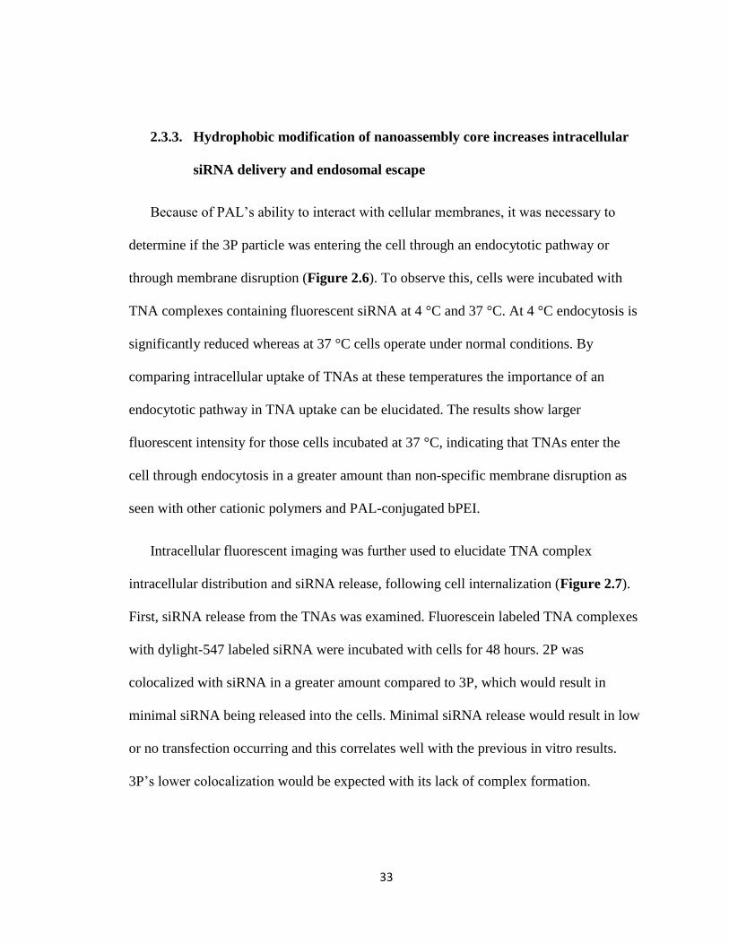
Figure 2.5. Fluorescent microscopy. ................................................................................. 35
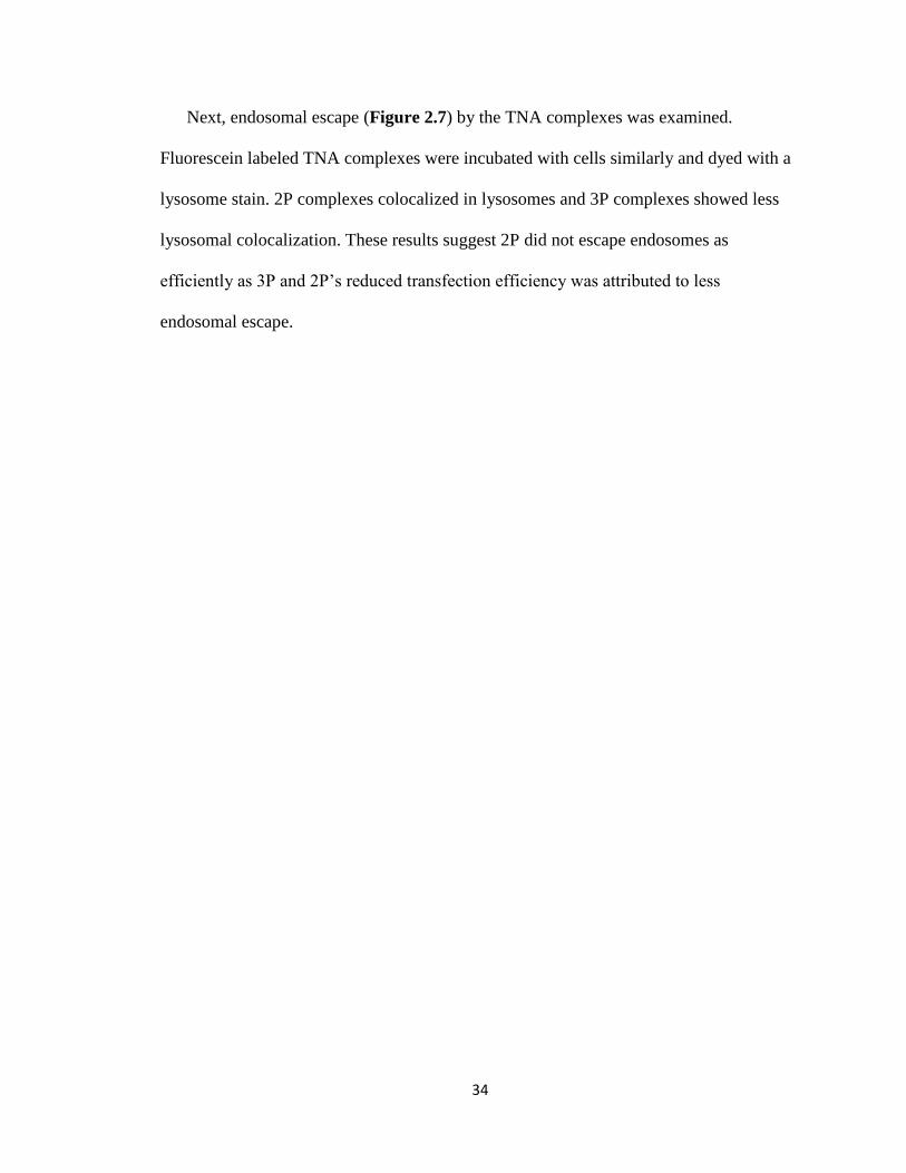
Figure 2.6. Elucidation of siRNA transfection mechanisms for TNAs. ........................... 36
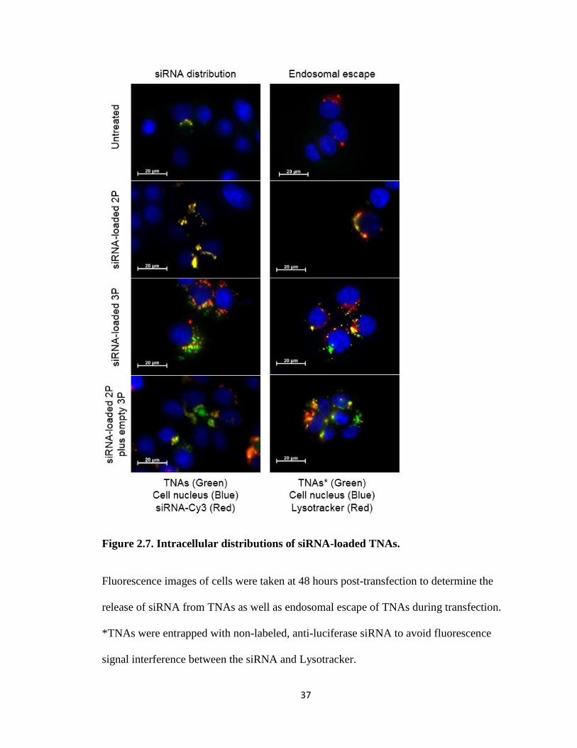
Figure 2.7. Intracellular distributions of siRNA-loaded TNAs. ....................................... 37
Figure 2.8. Complex formation and siRNA transfection of TNAs with varying PAL
contents in the core. .......................................................................................................... 40
Figure 3.1. PEG density analysis. ..................................................................................... 50
Figure 3.2. Gel permeation chromatograms of PEG-PEI particles. .................................. 56
Figure 3.3 Gel permeation chromatography of PEG-PEI-d particles. .............................. 58
Figure 3.4. siRNA complexation of PEG-PEI particles. .................................................. 60
Figure 3.5. siRNA complexation of PEG-PEI-d particles. ............................................... 61
Figure 3.6. Effect of PEG density on transfection efficacy of siRNA/PEG-PEI complexes.
........................................................................................................................................... 63
Figure 3.7. Effect of PEG density on transfection efficacy of siRNA/PEG-PEI-d
Complexes......................................................................................................................... 65
xi
Figure 4.1: TNA Scheme .................................................................................................. 75
Figure 4.2: Characterization of TNAs .............................................................................. 84
Figure 4.3: TNA in vitro Luciferase Reduction ................................................................ 85
Figure 4.4: PLL Based TNA in vitro Luciferase Reduction ............................................. 86
Figure 4.5: Direct TNA/Protein and TNA/Substrate Interactions .................................... 88
Figure 4.6: Raman Spectroscopy of TNAs and Their Components ................................. 91
Figure 4.7: Alternative Cell Health Markers After Incubation with 3P ........................... 93
Figure 4.8: TNA Induced Ubiquitination of Luciferase ................................................... 95
Figure S1: Characterization of 2PD TNA ....................................................................... 110
Figure S2: Raman spectra of siRNA ............................................................................... 111
Figure S3: Raman spectra comparison of multiple PEG samples .................................. 112
Figure S4: Normalized Raman spectra comparison of multiple PEG samples .............. 113
Figure S5: Multiple Raman spectra comparison of single PEG sample ......................... 114
xii
List of Tables
List of Tables
Table 2.1. Particle diameter, polydispersity index (PDI), and zeta potential of 2P and 3P
particles ............................................................................................................................. 28
Table 3.1. Characterization of PEG-PEI and PEG-PEI-d Library .................................... 55
1
1. Chapter 1: Tethered Nanoassemblies for siRNA Therapy
1
1.1. Genetic Disease Treatment and siRNA Therapy
Genetic diseases, such as cancer, are caused by damaged cells with abnormal protein
expressions that allows these cells to grow rapidly and without checks1-2. Current small
molecule therapies to target these diseases only target a small subset of proteins involved
in their proliferation. Currently, small molecule drugs cannot target many of these
abnormally expressed proteins. Instead, the drugs target proteins and pathways that are
not necessarily specific to the proteins involved in the disease which can kill both healthy
and diseased cells3-4. A more specific and tailored therapy would involve targeting the
specific proteins known to cause the disease. Nucleic acids that alter the expression of
specific proteins, known as gene therapy, has been widely studied since its discovery
nearly 40 years ago5.
Gene therapy has the potential to upregulate or downregulate protein expression in
diseased cells. Gene therapy can upregulate specific protein expression in diseased cells
by introducing exogenous DNA containing the code for the targeted protein6-7. This DNA
will be translated and replicated in the nucleus just as other genes providing multiple
copies of the targeted protein8. Though many genetic diseases can benefit from an
increase in protein expression, it is often a long-term or permanent solution that may not
be desirable. To achieve the desired effect in cells, DNA is either delivered via plasmid
or injected in to the genome. Though plasmid activity can be long term but not
permanent, gene insertion is permanent and if not inserted correctly will cause genetic
damage leading to more cell issues9. Conversely, Gene therapy achieves protein
downregulation using a less permanent method which is inherently safer10-11. Introducing
2
a short length of RNA complimentary to an endogenous mRNA or DNA that is
complimentary to an endogenous DNA gene sequence, referred to as “antisense”
nucleotides, will form pairs with the endogenous genetic material to prevent their
transcription or translation. This is a reversible process that will inhibit specific protein
production, called antisense therapy12. Currently, antisense therapy is widely studied due
to its increased safety compared to protein upregulation methods.
Antisense therapy uses two mechanisms to reduce protein expression: physically
blocking translation and transcription or inducing degradation of complimentary mRNA.
Initially, it was discovered that complimentary RNA and DNA could physically block
translation and transcription by tightly binding to regions that produce the targeted
protein13. Complimentary DNA was found to prevent translation but it was a reversible
process which shortened the time of the antisense effect. Further investigation in to
complimentary RNA discovered that it could promote cleavage of the mRNA by RNases
in the cell14. This improved upon physically blocking mRNA translation by degrading the
mRNA and improving the therapy. In 1994, an endogenous pathway was discovered that
could promote mRNA degradation using specific lengths of double stranded RNA, called
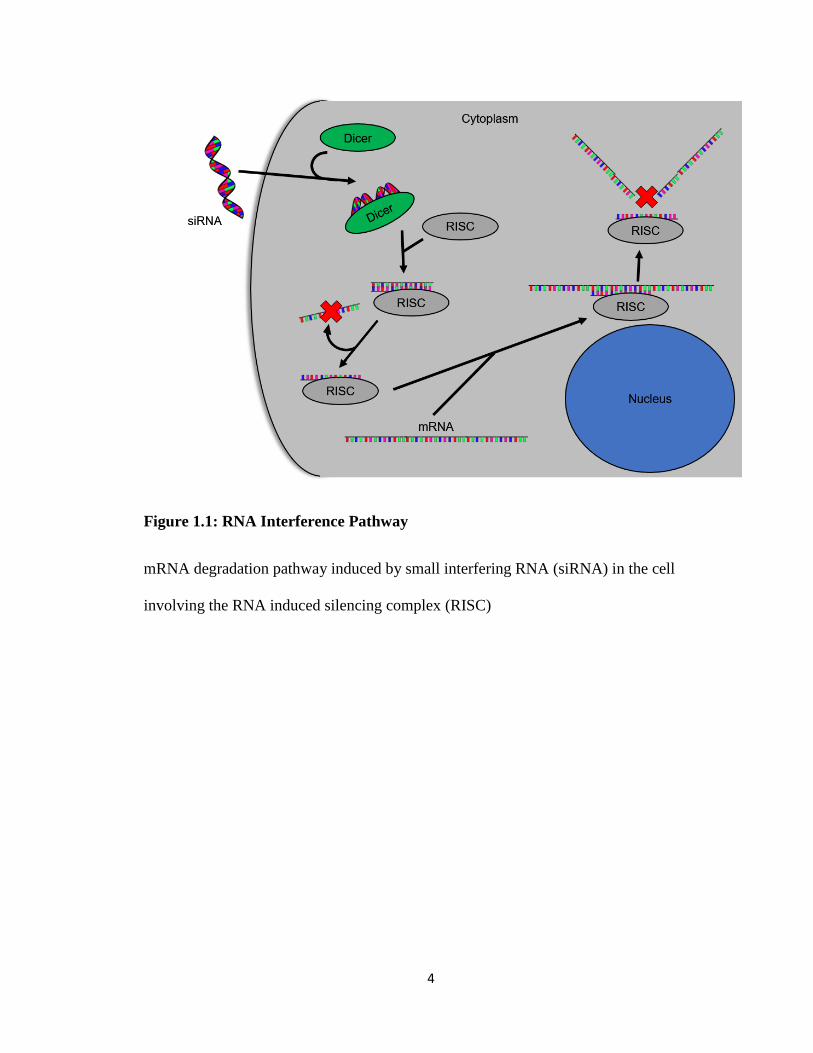
the RNA interference pathway (RNAi)15-16 (Figure 1.1).
RNAi is an endogenous pathway that uses 21-25 base pairs of small interfering RNA
(siRNA) complimentary to a portion of the mRNA of a targeted protein17. Once siRNA
enters the cytoplasm of the cell, a dicer complex takes up the siRNA to load it in to the
RNA induced silencing complex (RISC). RISC then unwinds and removes one strand of
the siRNA while keeping the other as a guide strand. The siRNA loaded RISC screens
mRNA present in the cytoplasm until it reaches a region complimentary to the siRNA
3
guide strand. RISC’s RNase activity then cleaves the mRNA. RISC may then release the
siRNA guide strand to pick up another piece of siRNA or reclaim the previous guide
strand to seek out more mRNA. This effect is continuous until the siRNA is degraded
within the cell18.
The concept of RNAi has been proven in vitro using multiple diseased cell lines and
many different protein targets19-22 but very little siRNA has succeeded in in vivo testing.
At the in vivo level, siRNA is introduced to clearance mechanisms, blood proteins, and
immune responses that quickly degrade and remove naked siRNA from the blood stream.
RNases present in blood serum will rapidly degrade siRNA generating a short half-life
ranging from 5 minutes to 1 hour23-24. Additionally, siRNA is cleared due to its small
size overcome these issues, further reducing its half-life in the blood stream. This short
half-life is a large problem for siRNA circulation which prevents it from reaching its
active site and hinders its clinical potential25. To improve the efficacy of siRNA, current
studies are focused on increasing stability of siRNA in the bloodstream by different
means.
4
Figure 1.1: RNA Interference Pathway
mRNA degradation pathway induced by small interfering RNA (siRNA) in the cell
involving the RNA induced silencing complex (RISC)
5
1.2. Tethered Nanoassembly (TNA) as siRNA Delivery Vehicles
As mentioned in the previous section, siRNA is in need of a means to improve its
stability in the blood stream. Many methods have been investigated to increase siRNAs
stability, including chemical modification of siRNA26-28, but research in to siRNA
delivery vehicles has been at the forefront of this field. siRNA delivery vehicles are
designed to protect siRNA by encapsulating them which prevents RNase degradation.
Further, these vehicles should also help usher the siRNA to the location of the targeted
cells and in to their cytoplasm, either through endosomal escape or another uptake
method29. These factors will allow delivery vehicles to enhance siRNA therapy.
Initially viral delivery systems were developed as gene therapy delivery vehicles for
both DNA and siRNA30-31. Viral delivery vehicles are viruses, such as retroviruses or
adeno-associated viruses, that have part of their genetic code altered to prevent
replication and include a copy of the targeted gene32. The viral vectors will inject genetic
material in to the cell using the viral mechanism. These can be used in siRNA therapy by
employing small hairpin RNA (shRNA) which will be replicated inside the cell to
consistently produce the desired siRNAs33. However, viral delivery presents safety issues
including risk of the virus replicating or random insertion of genetic material into the
cellular genome which can interfere with normal cellular processes34-35. These safety
issues have led to increased research in to non-viral delivery systems.
Non-viral delivery systems include a wide range of vectors including polymer and
lipid based systems36-38. Lipid based systems, such as liposomes39-40 and lipid
nanoparticles41-42, have been investigated for siRNA delivery as they were already a well-
studied system for drug delivery. These lipid based vehicles usually include lipids
6
modified with cationic head groups in order to form an ionic complex with the siRNA43.
The cationic charge density of the lipid vehicle can be tuned by including non-cationic
lipids along with the cationic lipids44. However, these vehicles often have lower
efficiency when forming complexes compared to cationic polymers. Cationic polymers
are synthetic and natural based polymers, such as poly-l-lysine (PLL)45 and
polyethylenimine (PEI)46-47, that have a high cationic charge density48. Again, these
polymers form ionic complexes with siRNA and offer base for modification to tune the
vehicle for desired delivery characteristics.
Although both lipid and polymer based systems have value to deliver siRNA, this
work focuses on cationic polymer based delivery vehicles because of their intrinsic
properties which offer advantages to siRNA delivery. These properties include: high
cationic charge density49, endosomal escape capability50, ease of modification51, and
control of particle size52. Cationic polymers include repeating units containing primary,
secondary, and sometimes tertiary amines. The pKas of these amines determine that they
are usually protonated at physiological pH which increases binding of siRNA to the
polymer. Some polyamine compounds, including PEI53, have multiple pKas for their
amines due to their close proximity to other amines leading to a unique endosomal escape
ability called the proton sponge effect54-55. The proton sponge effect occurs when the
polymer/siRNA complex enters the endosome. As the endosome decreases its pH to
become a lysosome, the polymer increases the protonation of its amines. This removes
hydrogens from the inside of the endosome, forcing the endosome pump in more ions and
swell in order to lower its pH. Eventually, the endosome will burst and release the vehicle
to the cytoplasm of the cell. This creates one mechanism of endosomal escape for these
7
polymers. Additionally, the cationic groups can interact with membranes which causes
disruption and frees the polymer/siRNA complex56-57. These cationic groups also offer
sites for modification of the polymer by relatively simple chemistry, such as n-
hydoxysuccinimide (NHS) coupling58-59. This can tune the vehicle for its intended
purpose. Further tuning can be done by altering the chain length of the polymer to change
the size of the polymer/siRNA complexes. Size of the complex is important to avoid
immune response (diameter > 100 nm)60 and renal clearance (diameter < 15 nm)61.
Based on these advantages, this work uses a model cationic polymer based delivery
system called a tethered nanoassembly (TNA). TNAs are a delivery vehicle consisting of
the cationic backbone, such as PEI or PLL, which poly(ethylene glycol) [PEG] and other
chemical modifications which are chemically linked, or “tethered”. TNAs were designed
to help elucidate the contribution of polymer/siRNA complex stability to transfection
efficiency and overall efficacy of siRNA delivery. While increased stability can be
achieved through modification, discussed further in section 1.3, the inherent design of the
vehicle also contributes to complex stability. A long chain cationic polymer was used as a
backbone so that a single polymer can form complexes with siRNA rather than multiple
polymers, which is common for these delivery vehicles. The resulting unimolecular
system can increase the uniformity of the vehicles and siRNA entrapment, thereby
increasing complex stability.
1.3. Chemical Modifications to Improve siRNA Therapy Efficacy
Cationic polymer based delivery vehicles are widely studied because of their
advantageous properties for delivering siRNA but these vehicles have difficulty
achieving success in in vivo models. This is often due to a lack of siRNA/vehicle
8
complex stability that can result in increased toxicity, low siRNA uptake in to the cell,
and increased siRNA clearance62-63. Poor complex stability results in polymers breaking
free from the complex which will increase non-specific interaction and toxicity, both in
the blood stream64 and inside the cell65. As more polymers break free, the siRNA is less
protected from degradation and doesn’t reach its target site. This indicates that complex
stability is crucial to delivery vehicle efficacy. In order to overcome these issues, research
has been focused on modification of the polymers making up the delivery vehicle to
improve complex stability and efficacy.
Chemical modification to siRNA delivery vehicles can alter physical and chemical
properties of the complex in order to improve its stability and efficacy66-67. When
considering chemical modifications, two properties play a key role: cationic surface
charge68 and secondary structure formation69. Increased cationic surface charge can cause
both toxicity and accelerated blood clearance due to non-specific interactions with
cellular membranes and blood proteins70-72, respectively. Therefore, reducing the surface
charge will benefit the overall efficacy of the complex. Surface charge reduction is often
done by covalent modification of PEG to the vehicle, or pegylation. Additionally,
increasing the types of forces, i.e. ionic interaction or hydrophobic interaction, used by a
vehicle to form its secondary structure can increase stability of the siRNA/vehicle
complex and reduce the amount of free polymer in formulation73-74. This reduces toxicity
and improves delivery of siRNA to its target site75-77. There are multiple ways to increase
secondary structure formation78-79, however inclusion of hydrophobic moieties80-81 on
cationic polymers is common. Based on this, this work focuses on pegylation and
hydrophobic modification of cationic polymer delivery vehicles.
9
Pegylation siRNA/vehicle complexes can enhance complex stability by reducing
surface charge and preventing off-target effects. Modification of vehicles with PEG
produces a field of PEG around the outside of the vehicle, also known as a PEG shell82.
This PEG shell has been shown to reduce surface charge by hiding the charged moieties
toward the center of the vehicle, or core83. This gives the vehicle what has been referred
to as “stealth” properties including reduced ionic interaction with blood proteins and
cellular membranes, reduced opsinization of the vehicle and removal by the immune
system, and avoiding renal clearance by increasing vehicle diameter84. It should be noted
that reduced cellular membrane interaction reduces toxicity caused by membrane
disruption, the main mechanism of toxicity for cationic polymers, but it is also required to
activate endocytosis85-87, which is a main mechanism of cellular uptake for cationic
polymer delivery vehicles. Additionally, pegylation can also benefit secondary structure
formation by pushing the polymer towards a core/shell structure73. These actions of
pegylation have proven to benefit vehicles by increasing circulation time and vehicle
stability which increases the efficacy of the vehicle88-89.
Hydrophobic modification to cationic polymers can improve complex stability and
enhance intracellular uptake by increasing the secondary structure formation80.
Hydrophobic modification is the introduction of a hydrophobic group to the cationic
polymer. These groups give the polymer a mechanism other than ionic forces to form a
secondary structure with itself or other polymers90. This prevents unwanted release of
polymer and siRNA from the complex. It has also been shown that genetic materials may
use the hydrophobic groups in complex formation91-92, therefore increasing the binding
efficiency of the polymer to siRNA and further stabilizing the siRNA/vehicle complex.
10
Hydrophobic modification also provides additional benefits such as increasing cellular
membrane interactions93-94. Increasing membrane interactions can increase endocytosis
by increasing activation of this pathway as well as increasing endosomal escape through
interaction and disruption of the endosomal membrane.
Understanding the effects of chemical modification on cationic polymers can often be
difficult due to variance in properties of different cationic polymers. Modifications, such
as with PEG and hydrophobic groups, can often have similar effects across polymers but
it becomes difficult to attribute specifics effects to modifications. The TNA model system
used in this work offers a uniform system to pinpoint effects from modifications. TNAs
offer a backbone to which many different modifications can be tethered. The design of
these particles allows for sequential modification and analysis where differences between
TNAs can be attributed to specific modifications. These findings can likely translate to
other cationic polymers. This system will bring more focus on the effects of chemical
modification to cationic polymer delivery vehicles.
Based on this background, this work hypothesizes that increasing the stability of the
delivery vehicle through chemical modification will increase its siRNA transfection
efficiency. In the subsequent studies, the contributions of hydrophobic core modification
and pegylation of the shell on transfection efficiency and complex stability are elucidated
using a model colon cancer cell line (HT29). This work will modify TNAs with
hydrophobic moieties and PEG as a model system for these modifications in order to
gather information that is relevant to both PEI based siRNA delivery vehicles as well as
other cationic polymer based vehicles. This will increase our understanding of these

11
modifications in order to improve future particle design of both TNAs and other cationic
polymer delivery vehicles.
12
2. Chapter 2: Effects of Hydrophobic Core Modification on TNA stability and
siRNA transfection
13
This section was adapted with permission from work published by the author in
AIMS Biophysics on July 30th, 201595. I would like to extend a special acknowledgment
to Dr. Piotr Rychahou who assisted with the cell line and transfection methods.
2.1. Introduction
As mentioned in the previous chapter, gene therapy using siRNA, siRNA therapy, has
been investigated to treat different genetic diseases, including cancer96-97. Currently,
siRNA therapy has shown great promise in treating undruggable proteins and pathways in
an in vitro setting. However, siRNA’s shows poor stability, rapid degradation, and low
circulation time in the bloodstream98-99. This makes progression from in vitro to in vivo
evaluation difficult. In order for siRNA therapy to be viable in a clinical setting, the
siRNA must be protected during delivery to its target site. Therefore, there has been a
push to develop siRNA delivery carriers that overcome these hurdles.
Though multiple types of carriers have been studied for delivery of siRNA98, 100, this
work focuses on cationic polymer based delivery vehicles. Delivery vehicles typically
form ionic complexes between the cationic portions of the vehicle and the anionic
phosphate backbone of siRNA. In this regard, the poly-amine nature of cationic polymers
enhances their abilities as delivery vehicles because of their high cationic charge density
which improves ionic complexation with siRNA101-103. One such example of a cationic
polymer is branched poly(ethylene imine) (bPEI)64. bPEI contains primary, secondary,
and tertiary amines, which are protonated at physiological pH, that may be used in
combination to complex with siRNA in a high efficiency104, resulting in bPEI offering a
highly charged, nanosized delivery carrier for siRNA. Although PEI can be toxic to cells,
bPEI is considered a safer alternative to linear PEI because it often shows comparatively
14
lower cytotoxicity105. However, issues of stability and off-target effects in the presence of
negatively charged serum proteins and other anions in the bloodstream prevent
bPEI/siRNA polyplexes from success in vivo74.
Typically to increase the stability of the polyplexes, the ratio of polymer to siRNA is
increased which introduces excess polymer in to the formulation. However, the polymers
that fail to interact with the siRNA, or free polymers, are left separate from other
polyplexes which often cause various adverse effects due to the increased availability of
their cationic groups106. Polymers that weakly interact with the complexed siRNA can
dissociate from the polyplex in the presence of the competing anion. This can further
reduce the stability of the polyplex and further dissociation until the siRNA is no longer
protected107. Additionally, cationic polymers used in excess can also create a large
positive surface charge detrimental to the polyplexes safety and stability108-109. High
surface charge and free polymers are known to reduce the particle circulation time in
vivo, cause cytotoxicity, decrease stability of the formulation, and fail to protect siRNA
before delivery to target sites73, 110. These all culminate in a lack of transfection efficiency
and stability of the siRNA formulation but modifications to the existing polymers could
help to return the lost efficacy.
Modification to both the core and shell of cationic polymer based delivery vehicles
can address these unwanted aspects of formulation111. Excess cationic surface charge can
be attenuated through pegylation, or covalent modification of the polymer with
[poly(ethylene glycol): PEG]112. Pegylation creates a hydrophilic shell around the particle
that shields and increases the circulation time of the polyplexes by reducing interactions
with its environment and neutralizing its surface charge113. Free polymer in the
15
formulation can be decreased by increasing the attractive forces between the polymers in
the polyplex core. Hydrophobic interaction has been shown to enhance particle stability
by allowing the polymer chain to interact with itself as well as the siRNA114-116. Nucleic
acid strands have shown to interact with hydrophobic groups once their anionic charge
has been neutralized which would further stabilize the complex117. Additionally, if
siRNA dissociates from the complex, the particle can stay together based on the
hydrophobic interactions and prevent free polymer generation118. Alternatively, linking
all free cationic polymer together, effectively forming a unimolecular system, will reduce
free polymer in formulation because it would remove free polymer from the system all
together. By modifying a single cationic polymer to covalently link with multiple other
moieties including PEG and hydrophobic groups, a single polymer could form a complex
with siRNA creating a unimolecular particle with no ability to dissociate its components
from itself yet siRNA would be free to associate and disassociate. By reducing the free
polymer and surface charge of the cationic polymer delivery vehicle, efficacy and
stability should increase resulting in increased transfection efficiency.
Based on this background, we hypothesized that siRNA transfection efficiency will
improve by protecting siRNA in the core of a unimolecular cationic nanoassembly with
improved complex stability. To test this, the work in this chapter set out to create
polymer nanoassemblies stabilized with a lipophilic core and examine the effects of
complex stability on transfection efficiency, toxicity, and intracellular siRNA delivery.
Polymer nanoassemblies were synthesized by tethering hydrophilic polymer chains, PEG,
onto a single polymer backbone (branched PEI: PEI) while modifying the core of the
nanoassemblies with lipophilic pendant groups (palmitate: PAL). Figure 2.1 illustrates
16
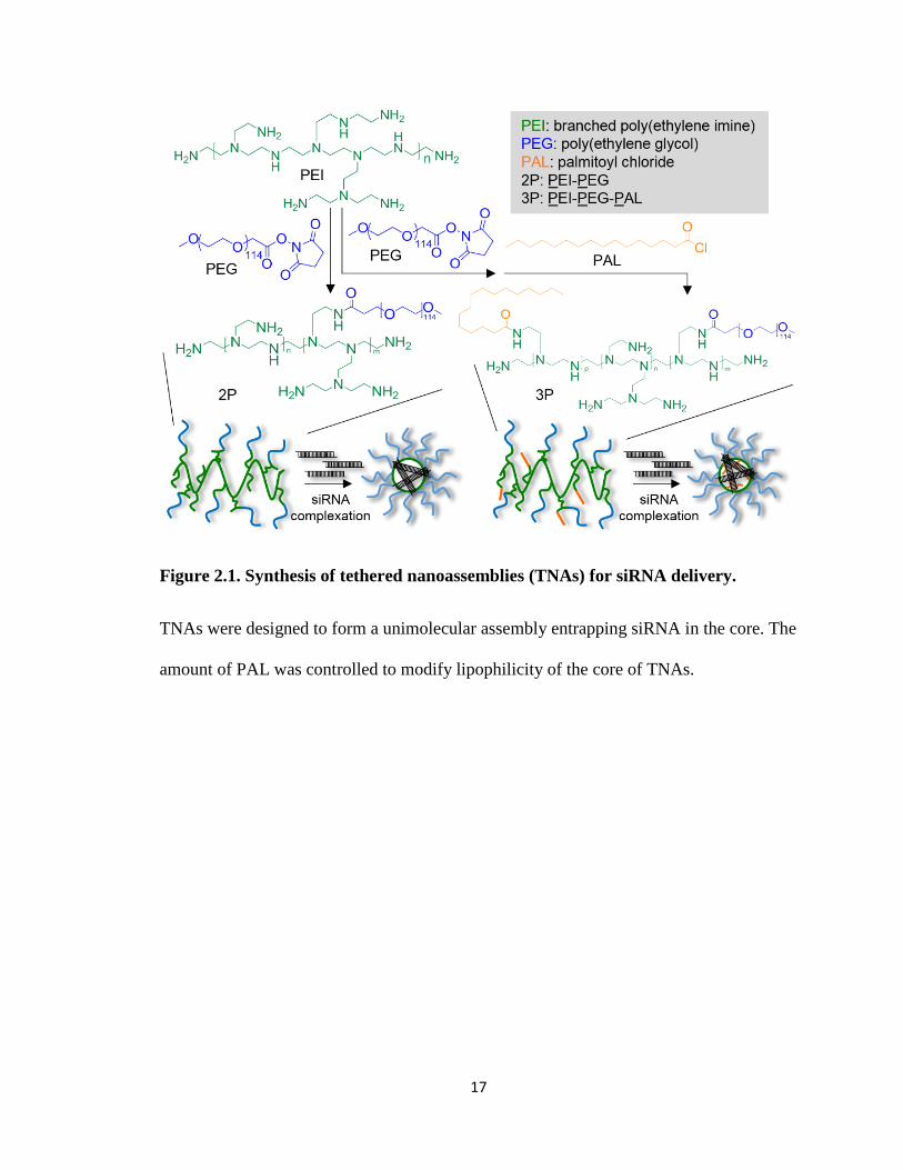
two types of polymer tethered nanoassemblies (TNAs) used in this chapter, PEG-PEI
(2P) and PEG-PEI-PAL (3P). 2P has cationic moieties in the core and thus attract siRNA
to prepare polyionic complexes while 3P has a hydrophobic core modified with PAL to
increase stability of the complexes through ionic and hydrophobic interactions between
the core and siRNA payload. The PAL content in 3P was also modulated at varying ratios
to prepare TNAs that behave between 2P and 3P. For this chapter, the siRNA-loaded
TNAs were designed to reduce luciferase, an exogenous bioluminescent protein,
expression level within a cell so that its efficacy in vitro could be examined through a
facile method.
17
Figure 2.1. Synthesis of tethered nanoassemblies (TNAs) for siRNA delivery.
TNAs were designed to form a unimolecular assembly entrapping siRNA in the core. The
amount of PAL was controlled to modify lipophilicity of the core of TNAs.
18
2.2. Materials and Methods
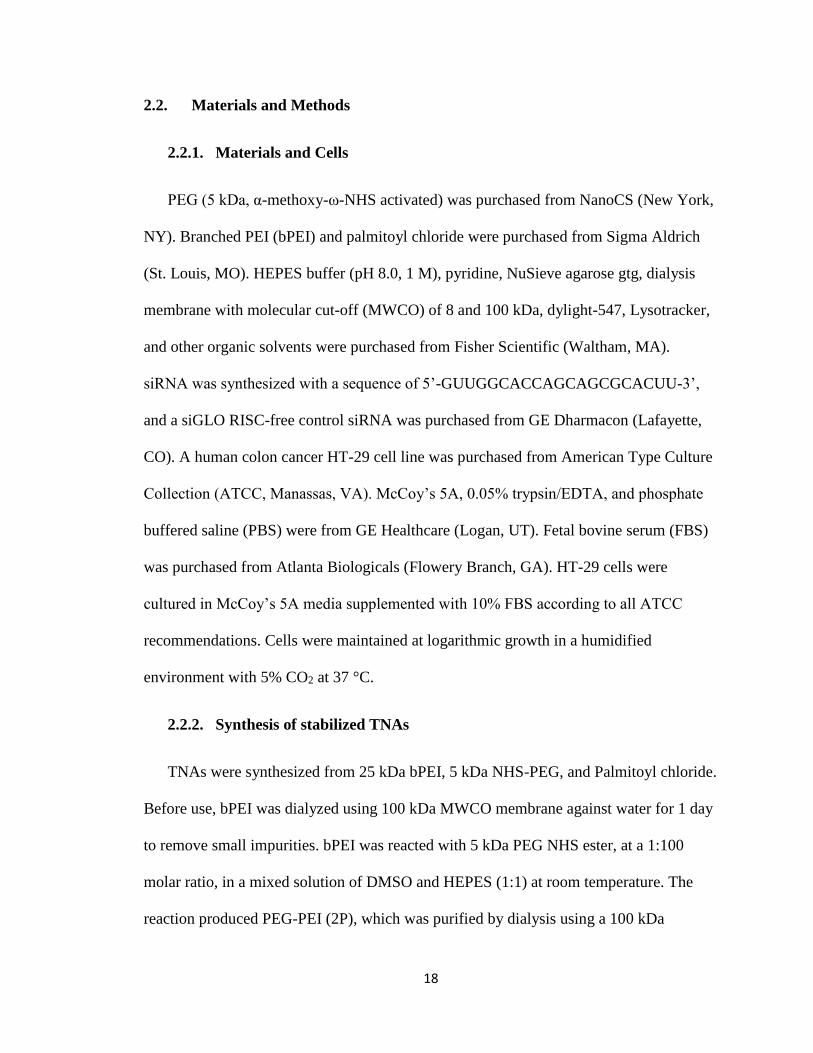
2.2.1. Materials and Cells
PEG (5 kDa, α-methoxy-ω-NHS activated) was purchased from NanoCS (New York,
NY). Branched PEI (bPEI) and palmitoyl chloride were purchased from Sigma Aldrich
(St. Louis, MO). HEPES buffer (pH 8.0, 1 M), pyridine, NuSieve agarose gtg, dialysis
membrane with molecular cut-off (MWCO) of 8 and 100 kDa, dylight-547, Lysotracker,
and other organic solvents were purchased from Fisher Scientific (Waltham, MA).
siRNA was synthesized with a sequence of 5’-GUUGGCACCAGCAGCGCACUU-3’,
and a siGLO RISC-free control siRNA was purchased from GE Dharmacon (Lafayette,
CO). A human colon cancer HT-29 cell line was purchased from American Type Culture
Collection (ATCC, Manassas, VA). McCoy’s 5A, 0.05% trypsin/EDTA, and phosphate
buffered saline (PBS) were from GE Healthcare (Logan, UT). Fetal bovine serum (FBS)
was purchased from Atlanta Biologicals (Flowery Branch, GA). HT-29 cells were
cultured in McCoy’s 5A media supplemented with 10% FBS according to all ATCC
recommendations. Cells were maintained at logarithmic growth in a humidified
environment with 5% CO2 at 37 °C.
2.2.2. Synthesis of stabilized TNAs
TNAs were synthesized from 25 kDa bPEI, 5 kDa NHS-PEG, and Palmitoyl chloride.
Before use, bPEI was dialyzed using 100 kDa MWCO membrane against water for 1 day
to remove small impurities. bPEI was reacted with 5 kDa PEG NHS ester, at a 1:100
molar ratio, in a mixed solution of DMSO and HEPES (1:1) at room temperature. The
reaction produced PEG-PEI (2P), which was purified by dialysis using a 100 kDa
19
MWCO membrane for 5 days in water, and collected by freeze drying. PEG-PEI was
further reacted with palmitoyl chloride at 1:100, 1:50, and 1:30 molar ratios in THF at 40
°C for 2 hours in the presence of pyridine as a scavenger of a hydrochloric acid byproduct
to create PEG-PEI-PAL (3P), 3P mid, and 3P low respectively. The reaction solutions
were precipitated in diethyl ether and subsequently dialyzed in water prior to freeze
drying.
2.2.3. Characterization of stabilized TNAs
The purity and uniformity of TNAs were determined by gel permeation
chromatography (GPC) (Asahipak GF-7M column, 2 mg/mL, PBS, 0.5 mL/min, 40 °C)
Molecular weights were determined by comparing peak retention time to PEG standards.
The diameter and surface charge of TNAs were determined by dynamic light scattering
and zeta potential measurements (Zetasizer Nano, Malvern, UK). Particle solutions of 2
mg/ml were loaded into disposable zeta cuvettes and read for particle size and then zeta
potential in the usage.
Extent of palmitoylation was examined by fluorescamine assay. Fluorescamine
powder was dissolved in DMSO to 10 mg/ml. Particles were dissolved in a 50/50 mixture
of DMSO/water to a concentration of 1 mg/ml. 100 μL of each particle solution and
blank DMSO/water was added to a clear 96 well plate and then 10 μL of the
fluorescamine solution was added immediately before reading on a fluorescent plate
reader at 390/460 excitation/emission (SpectraMax M5, Molecular Devices). The
fluorescence intensity of the blank was subtracted from the experimental wells and then
experimental wells compared with each other.
20
2.2.4. Analysis of TNAs and siRNA Interactions
To determine siRNA/TNA complex formation ratios, TNA complexes were formed
by mixing solutions of particles at several concentrations from 0.1 and 100 mg/ml in
Optimem with 720 nM siRNA in Optimem. Solutions were mixed at a 1:1 ratio and
allowed to equilibrate for 30 minutes at room temperature. 20 μL of each solution and 5
μL of low range DNA ladder were loaded to 4% agarose gel and run at 100 volts for 80
minutes at room temperature. The gel was stained in 200 ml of 100 ng/ml ethidium
bromide in TAE (Tris-Acetate 0.04 M, EDTA 0.001 M) buffer, rinsed 3 times in TAE
buffer, and imaged via Typhoon GLA 9500 (GE Healthcare, Logan, UT) fluorescent
imager under the ethidium bromide filter set.
siRNA release from TNAs was determined using a competing anion assay.
Complexes were formed by mixing solutions of particles, concentrated in optimem media
at 10 mg/ml, with 720 nM fluorescently-labeled siRNA optimem solution at a 1:1 ratio to
a final concentration of 5 mg/ml of particle and 360 nM of siRNA for 30 minutes at room
temperature. 20 μL of the complex solutions were added to 10 μL solutions of varying
heparin concentration. 0, 10, 100, 1000, and 5000 μg/ml concentrations were used to
create weight ratios of siRNA/heparin between 0 and 500. 10 minutes later, 20 μL of each
solution was loaded onto an agarose gel and run at 100 V for 80 minutes. The gel was
imaged using Typhoon equipped with a cy3 filter set (dylight-547 compatible).
siRNA protection by TNAs was determined by TNA/siRNA complex incubation in
media containing RNases. Complexes were prepared for particle-siRNA release study
above were also used for siRNA protection study. Complex solutions (20 μL) were
incubated with 20 μL of active FBS or heat-inactivated FBS. A control was created by
21
adding complex solution to RNase free water (20 μL, respectively) and then frozen. 20
μL of sample solution was loaded and run on 4% agarose gel in TAE buffer at 100 V for
80 minutes. After the gel was run, the gel was imaged with Typhoon through the cy3
filter set.
2.2.5. In vitro transfection efficiency of TNAs
Cells were seeded at 5,000 cells per well into a white opaque 96 well plate for 24
hours. After 24 hours, Complex solutions were created by mixing 100 μL of 10 mg/ml
particle solutions with 100 μL of 720 nM anti-luciferase siRNA, all solution in optimem.
These were incubated at room temp for 30 minutes. Control Solutions were created by
setting aside 200 μL of optimem for a blank control and mixing 100 μL of 720 nM anti-
luciferase siRNA solution with 100 μL optimem or 100 μL of optimem containing 5 μL
of RNAiMAX agent to create a naked siRNA control and RNAiMAX control,
respectively. These were incubated at room temp for 30 minutes except the RNAiMAX
control which was incubated for 20 minutes, per manufacturer’s instructions.180 μL of
each solution was added to 720 μL of McCoy’s 5A supplemented with 10% FBS creating
a final concentration of 1 mg/ml particle concentration, 72 nM siRNA, and 0.5 μL well of
RNAiMAX concentration. Media was removed from each well and 100 μL of complex
solutions and controls were to the wells, n=8. The plates were incubated until their
endpoints (24, 48, or 72 hours) and then assayed for bioluminescence. Cells were injected
with 100 μL of 0.1 mg/ml luciferin solution in PBS via a GloMax luminometer
(Promega). Bioluminescence intensity was integrated over a 10 second period and
recorded by the luminometer. Blank control wells were used to compare normal
bioluminescence intensity to the experimental wells and data is reported as percentage
22
luciferase activity. This data was then normalized based on a viability assay described
later to account for cell death in the reduction of luciferase signal. For the 2P/3P
combination experiments after 24 hours incubation, 20 μL of media was removed from
each well. 20 μL of 10 mg/ml 3P optimem solution was added to wells containing 2P and
20 μL of optimem was added to all other wells. Cells were then incubated for a further 48
hours, and subjected to viability and bioluminescence assays as described previously.
2.2.6. Toxicity of TNAs in vitro
After the transfection assay was completed, each plate underwent a resazurin assay.
10 μL of a 100 mM solution of resazurin in PBS was added to each well of the 96 well
plate. The plate was returned to the incubator for 3 hours and then read on a SpectraMax
M5 (Molecular Devices) fluorescent plate reader at an excitation/emission of 560/590. A
control of blank media with resazurin was used to subtract out background fluorescence
and then fluorescence intensity was compared between each experimental well and the
blank control wells to give a percentage of viable cells.
2.2.7. In vitro intracellular uptake and trafficking of fluorescent siRNA in TNAs
8 well glass slides were plated with 10,000 cells per well in McCoy’s 5A media
supplemented with 10% FBS. 24 hours later, complex solutions were created based on
the endpoint of the imaging study. To image siRNA within the cell, 30 μL of 10 mg/ml
unlabeled particle solutions and 30 μL of 720 nM dylight-547 labeled siRNA were
mixed. To image siRNA/particle colocalization, 30 μL of 10 mg/ml fluorescein labeled
particle solutions and 30 μL of 720 nM dylight-547 labeled siRNA were mixed. To
image particle/lysosome colocalization, 30 μL of 10 mg/ml fluorescein labeled particle
23
solutions with 30 μL of 720 nM unlabeled siRNA were mixed. To image combination of
2P/3P, 30 μL of 10 mg/ml fluorescein labeled 2P solutions with 30 μL of 720 nM
unlabeled siRNA were mixed. Controls of naked siRNA and blanks were created by
mixing 30 μL of 720 nM dylight-547 labeled siRNA with 30 μL of optimem and 30 μL
of optimem with 30 μL of optimem, respectively. All solutions were incubated at room
temp for 30 minutes. Afterwards, 60 μL of each solution was added to 240 μL of
McCoy’s 5A supplemented with 10% FBS creating a final concentration of 1 mg/ml
particle concentration and 72 nM siRNA. Media was then removed from each well and
200 μL of complex solutions were added to the wells and incubated for 48 hours.
Additionally, for the 2P/3P combination study, 24 hours after dosage 30 μL of media was
removed from the well and 30 μL of 10 mg/ml fluorescein labeled 3P was added. 48
hours after dosage, cells were rinsed with PBS 3 times, fixed with formalin for 20
minutes, stained with Hoechst 33342 and Lysotracker red (if applicable), and rinsed
another 3 times. Cells were imaged at 100X on a Zeiss axiovert 200M fluorescent
microscope using dapi, texas red, and fluorescein filter sets. Images were captured for the
fluorescein and Texas red filter for fluorescein particles and siRNA, respectively, while
using the same exposure between images to compare fluorescent intensity between them.
Cells (5,000 cells/well) were seeded into a white 96 well plate. After 24 hours,
Complex solutions and controls were created by mixing 100 μL of 720 nM dylight-547
labeled siRNA optimem solution with 100 μL of 10 mg/ml 3P optimem solution or 100
μL of optimem, respectively. These were incubated at room temp for 30 minutes. One
plate was stored in 4C for 30 minutes along with particle and control solutions to be used
with the plate. Media was removed from the wells and 100 μL of control and complex
24
solutions were added to the plate (n=4). The plated was returned to 4 ⁰C. Another plate
was treated identically but kept at 37 ⁰C. After 4 hours, the plates were rinsed with cold
PBS three times and read on a fluorescent plate reader at excitation/emission of 557/570.
The fluorescence intensity was compared with an untreated control well at each
temperature.
2.3. Results
2.3.1. The hydrophobicity of the TNA core reduces interactions between siRNA
and TNA
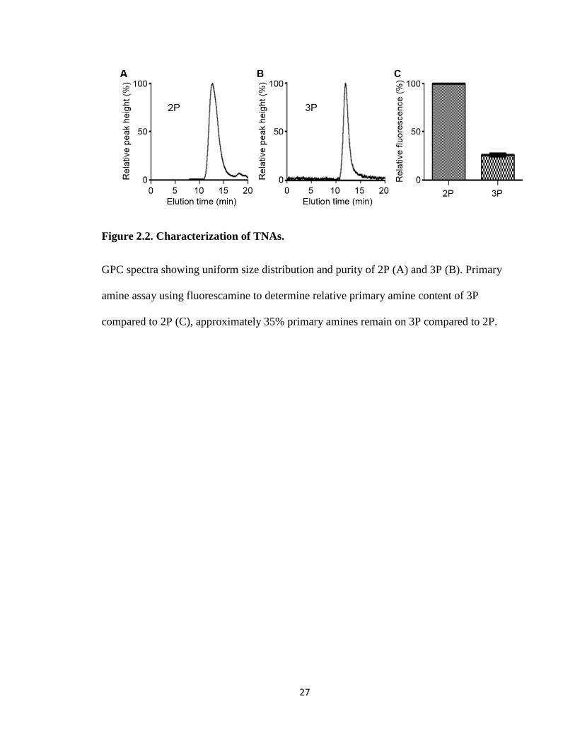
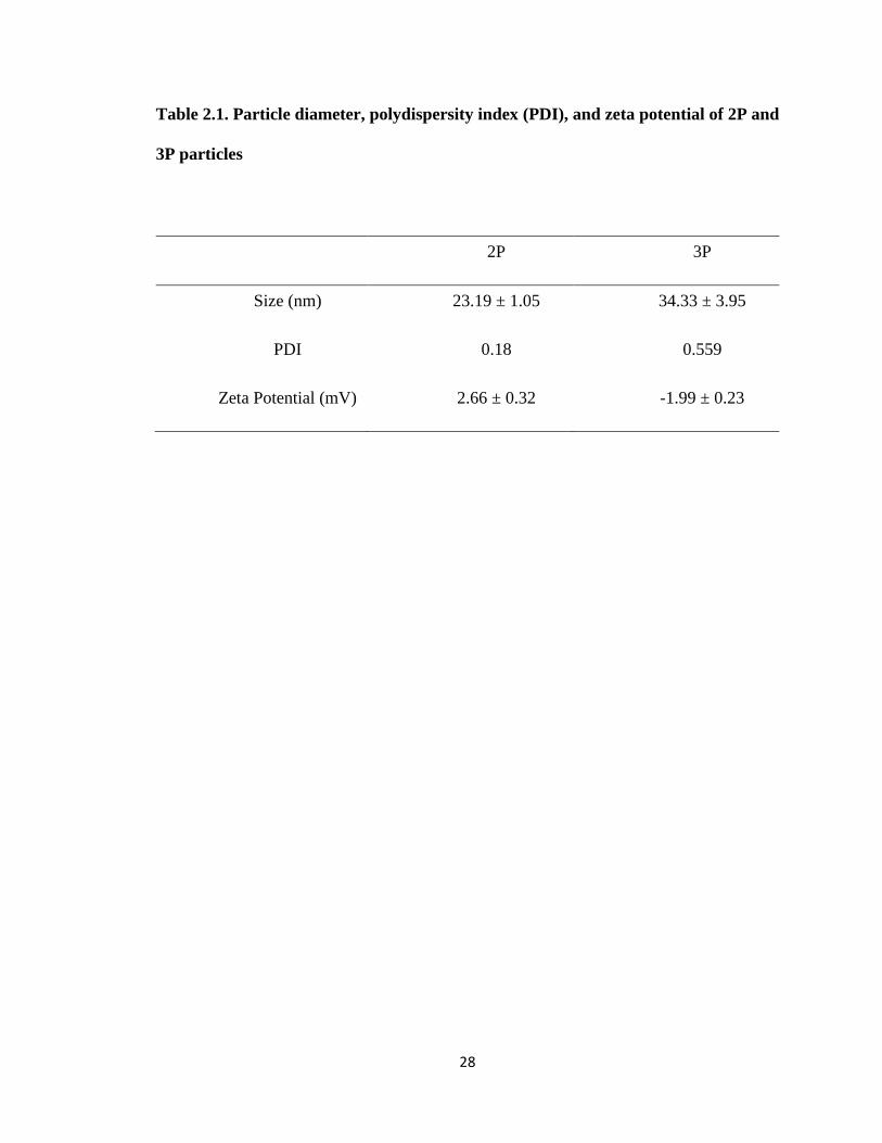
TNAs were synthesized as shown in Figure 2.1. All TNAs were uniform and
contained no impurities as confirmed by a single peak shown on GPC (Figure 2.2). DLS
and zeta-potential measurements confirmed that 2P and 3P were less than 40 nm in
diameter and had a neutral surface charge regardless of PAL modification (Table 2.1).
These results indicate that PAL is mostly present in the core of TNAs and the PEG shell
efficiently shields the charge and hydrophobic groups. However, there is a discrepancy
between the DLS measured polydispersity index (PDI) and the sharpness of the GPC
peak from 3P.
Modification of the core with PAL groups did not seem to affect the surface
properties of TNAs although it altered lipophilicity and molecular conformation of the
core. GPC revealed that the molecular weights of 2P and 3P were 128 and 158 kDa,
respectively. Although the exact number of primary amines on bPEI is unknown, our
estimation based on the molecular weights of bPEI (25 kDa) and TNAs suggests that
approximately 10% and 55% of binding sites on bPEI were conjugated with PEG and
25
PAL, respectively. To further confirm the reaction, a fluorescamine assay was used to
compare the amounts of remaining primary amines between 2P and 3P. 3P contained
fewer primary amines per particle than 2P, corresponding with the GPC estimation.
Interactions between siRNA and TNAs were characterized in 3 different ways:
siRNA uptake by TNAs, binding strength of TNA to siRNA, and protection of siRNA
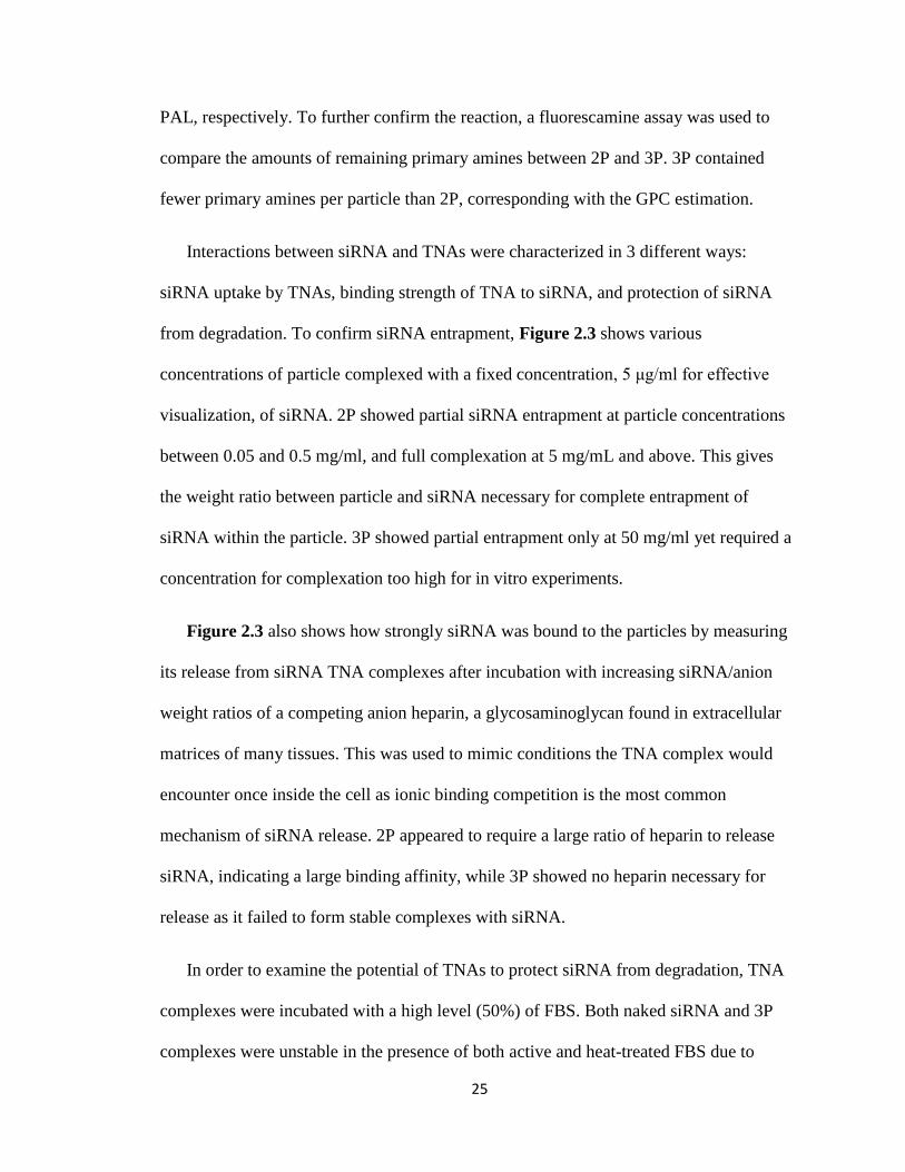
from degradation. To confirm siRNA entrapment, Figure 2.3 shows various
concentrations of particle complexed with a fixed concentration, 5 μg/ml for effective
visualization, of siRNA. 2P showed partial siRNA entrapment at particle concentrations
between 0.05 and 0.5 mg/ml, and full complexation at 5 mg/mL and above. This gives
the weight ratio between particle and siRNA necessary for complete entrapment of
siRNA within the particle. 3P showed partial entrapment only at 50 mg/ml yet required a
concentration for complexation too high for in vitro experiments.
Figure 2.3 also shows how strongly siRNA was bound to the particles by measuring
its release from siRNA TNA complexes after incubation with increasing siRNA/anion
weight ratios of a competing anion heparin, a glycosaminoglycan found in extracellular
matrices of many tissues. This was used to mimic conditions the TNA complex would
encounter once inside the cell as ionic binding competition is the most common
mechanism of siRNA release. 2P appeared to require a large ratio of heparin to release
siRNA, indicating a large binding affinity, while 3P showed no heparin necessary for
release as it failed to form stable complexes with siRNA.
In order to examine the potential of TNAs to protect siRNA from degradation, TNA
complexes were incubated with a high level (50%) of FBS. Both naked siRNA and 3P
complexes were unstable in the presence of both active and heat-treated FBS due to
26
nuclease degradation, whereas 2P protected siRNA for the duration. This was expected as
3P had not previously shown complexation with siRNA but 2P had shown tight binding
to the siRNA. These results would give 2P greater potential as an in vivo formulation
compared with 3P because of 2Ps ability to form complexes and protect the siRNA.
27
Figure 2.2. Characterization of TNAs.
GPC spectra showing uniform size distribution and purity of 2P (A) and 3P (B). Primary
amine assay using fluorescamine to determine relative primary amine content of 3P
compared to 2P (C), approximately 35% primary amines remain on 3P compared to 2P.
28
Table 2.1. Particle diameter, polydispersity index (PDI), and zeta potential of 2P and
3P particles
2P 3P
Size (nm) 23.19 ± 1.05 34.33 ± 3.95
PDI 0.18 0.559
Zeta Potential (mV) 2.66 ± 0.32 -1.99 ± 0.23
29
Figure 2.3. Complex formation and stability of TNAs.
TNAs were mixed with siRNA (360 nM) at varying ratios to determine a particle
concentration required for forming neutral siRNA complexes (A). siRNA-loaded TNAs
(5 mg/mL) were incubated with heparin to determine complex stability in the presence of
anionic counterparts other than siRNA in a solution (B). siRNA-loaded TNAs were also
incubated with nuclease free water (n), heat-inactivated FBS (i), and active FBS (a) to
determine protective effects of TNAs in cell culture media with digestive enzymes (C).
Naked siRNA at 360 nM and FBS alone used as controls.
30
2.3.2. Increased hydrophobicity of the TNA core increases TNA transfection
efficiency
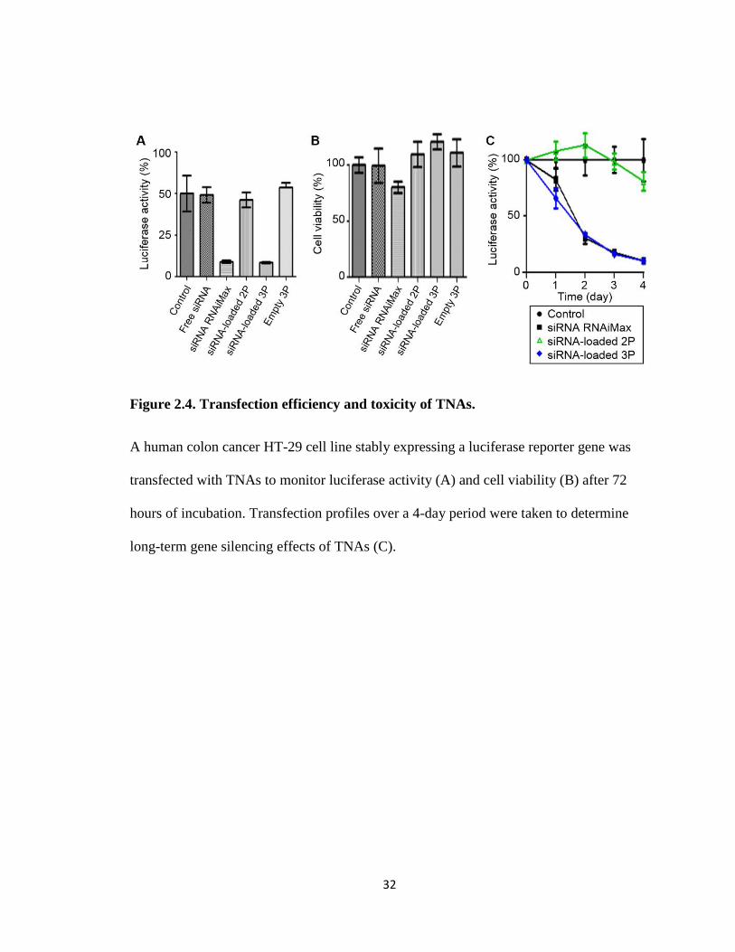
In order to assess the delivery efficacy of the TNA complexes, bioluminescence was
used as a facile method of protein reduction quantification. Luciferase protein was
introduced to HT-29 colorectal adenocarcinoma cells so that it was stably expressed at all
times (HT-29-LUC). Anti-luciferase siRNA uses the RNAi pathway to degrade luciferase
mRNA, attenuating protein production. This allows for easy detection of remaining
protein by addition of luciferin substrate. The amount of protein remaining correlates to
siRNA delivery efficacy18.
HT-29-LUC cells were incubated with siRNA-TNA complexes and 72 hours later the
cells were examined for luminescence and cell viability (Figure 2.4). Particle
concentrations used were 1 mg/ml or 20% that of what was found to be full complexation
in previous experiments (5 mg/ml) due to 20% the amount of siRNA (1 μg/ml) to be used
in vitro. Initially, 2P showed no transfection but 3P showed approximately 70% reduction
of the luciferase activity, attributed to siRNA delivery. The transfection efficiency of 3P
was comparable to that of Lipofectamine RNAiMax, a commercially available
transfection reagent. To further examine the transfection efficiency of both particles,
transfection was monitored daily for total 4 days past dosage. 3P and RNAiMax showed
a similar transfection profile, reducing luciferase expression continuously for 4 days, but
2P induced no transfection in the same period of time. Additionally, neither 2P nor 3P
exhibited any noticeable toxicity under our experimental condition while RNAiMAX
showed mild toxicity with approximately 20% reduction in cell viability.
31
Further confirmation of siRNA delivery was shown by fluorescent imaging with
fluorescently labeled siRNA. This allowed for viewing of the amount of siRNA used
within the cells for each formulation (Figure 2.5). Compared with the non-treatment and
free siRNA incubated cells, both TNA formulations showed siRNA within the cell.
However, cells incubated with 3P complexes showed a much larger fluorescent intensity
through confocal microscopy than those incubated with 2P indicating that 3P was able to
increase the amount of siRNA that would be trafficked into the cell.
32
Figure 2.4. Transfection efficiency and toxicity of TNAs.
A human colon cancer HT-29 cell line stably expressing a luciferase reporter gene was
transfected with TNAs to monitor luciferase activity (A) and cell viability (B) after 72
hours of incubation. Transfection profiles over a 4-day period were taken to determine
long-term gene silencing effects of TNAs (C).
33
2.3.3. Hydrophobic modification of nanoassembly core increases intracellular
siRNA delivery and endosomal escape
Because of PAL’s ability to interact with cellular membranes, it was necessary to
determine if the 3P particle was entering the cell through an endocytotic pathway or
through membrane disruption (Figure 2.6). To observe this, cells were incubated with
TNA complexes containing fluorescent siRNA at 4 °C and 37 °C. At 4 °C endocytosis is
significantly reduced whereas at 37 °C cells operate under normal conditions. By
comparing intracellular uptake of TNAs at these temperatures the importance of an
endocytotic pathway in TNA uptake can be elucidated. The results show larger
fluorescent intensity for those cells incubated at 37 °C, indicating that TNAs enter the
cell through endocytosis in a greater amount than non-specific membrane disruption as
seen with other cationic polymers and PAL-conjugated bPEI.
Intracellular fluorescent imaging was further used to elucidate TNA complex
intracellular distribution and siRNA release, following cell internalization (Figure 2.7).
First, siRNA release from the TNAs was examined. Fluorescein labeled TNA complexes
with dylight-547 labeled siRNA were incubated with cells for 48 hours. 2P was
colocalized with siRNA in a greater amount compared to 3P, which would result in
minimal siRNA being released into the cells. Minimal siRNA release would result in low
or no transfection occurring and this correlates well with the previous in vitro results.
3P’s lower colocalization would be expected with its lack of complex formation.
34
Next, endosomal escape (Figure 2.7) by the TNA complexes was examined.
Fluorescein labeled TNA complexes were incubated with cells similarly and dyed with a
lysosome stain. 2P complexes colocalized in lysosomes and 3P complexes showed less
lysosomal colocalization. These results suggest 2P did not escape endosomes as
efficiently as 3P and 2P’s reduced transfection efficiency was attributed to less
endosomal escape.
35
Figure 2.5. Fluorescent microscopy.
HT29 cells were incubated with TNAs (1 mg/mL) containing dylight-547 labeled siRNA
(72 nM) to confirm intracellular delivery of siRNA at 48 hours post-transfection,
following the treatment of cells with PBS (A), naked siRNA (B), 2P complexes (C), and
3P complexes (D).
36
Figure 2.6. Elucidation of siRNA transfection mechanisms for TNAs.
Cells were treated with siRNA-loaded TNAs at an incubation temperature that
endocytosis is active (37 °C) or suppressed (4 °C) to determine the intracellular uptake
mechanism for TNAs (A). Cancer cells treated with siRNA-loaded 2P were incubated
with empty 3P at 24 hours post transfection to demonstrate a unique property of 3P that
enhances siRNA transfection alone or in combination with 2P (B).
37
Figure 2.7. Intracellular distributions of siRNA-loaded TNAs.
Fluorescence images of cells were taken at 48 hours post-transfection to determine the
release of siRNA from TNAs as well as endosomal escape of TNAs during transfection.
*TNAs were entrapped with non-labeled, anti-luciferase siRNA to avoid fluorescence
signal interference between the siRNA and Lysotracker.
38
2.3.4. Combined dosage of hydrophobic modified and unmodified TNA
decreases colocalization of siRNA in endosomes
A combinatorial approach to TNA transfection was examined to help elucidate 3P’s
role in transfection efficacy as well as attempt to protect siRNA while achieving
transfection. 2P-siRNA complexes were incubated with cells for 24 hours and then empty
3P particles introduced (Figure 2.6). It was found that the addition of empty 3P did
increase the transfection efficiency of 2P-siRNA complexes from 0% to 60%.
A closer look at the intracellular trafficking of this dosage was achieved through
fluorescent microscopy. Colocalization between siRNA and particle was relatively less
compared with 2P as shown in Figure 2.7. The combinatorial dosage also showed a
relative reduction in colocalization between endosome and particle, indicating that
particles and siRNA were escaping endosomes, previously not seen with 2P particles
alone. This gave a greater indication that 3P has a greater endosomal escape capability
compared with 2P and that it may lend it to other particles taken up by the cell
concurrently.
2.3.5. Modulating the hydrophobic substitution of TNA core increases
transfection efficiency while decreasing siRNA/particle interactions
Because of the success of the 2P/3P combination approach, the ratio of PAL
substitution on 3P was examined to find an optimal ratio where 3P could form complexes
with siRNA. This would enable 3P to protect cells so that a less complicated approach
could be taken compared to the combination 2P/3P described in the previous section.
Two additional TNAs were synthesized by aiming for 15% and 25% palmitoylation of
39
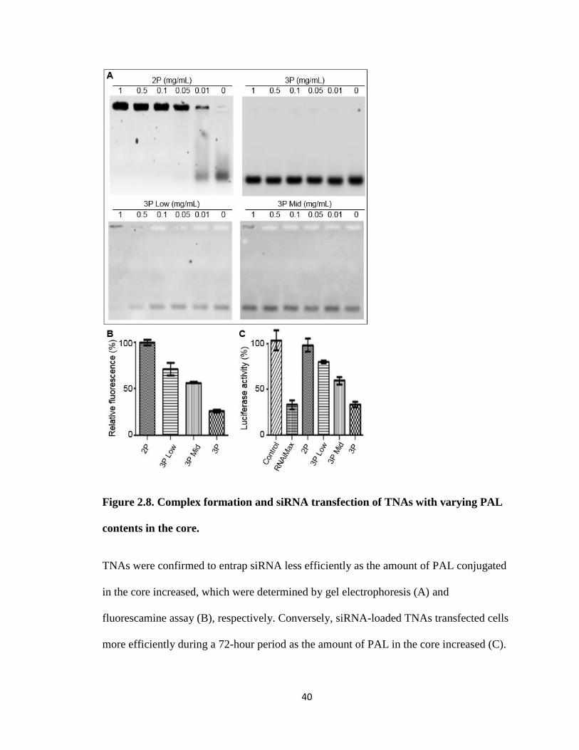
2P, named 3P low and 3P mid, respectively. The amounts of primary amines remaining
in the core of 3P low and 3P mid were determined by a fluorescamine assay (Figure 2.8).
The assay indicated that palmitoylation was achieved at differing levels compared with
the initial 3P particle. Gel electrophoresis was performed to confirm siRNA complexation
with 3P low and 3P mid. 3P low achieved complexation between 0.5 mg/ml and 1.0
mg/ml, significantly lower than the original 3P at 72 nM siRNA concentration. 3P mid
began to form partial complexes at 1 mg/ml but required a higher polymer concentration
for complete complexation.
To examine the effect of palmitoylation on transfection efficiency, cells were
incubated with each form of 3P complexes with siRNA for 72 hours. Though 3P low was
able to maintain a complex with siRNA, it did not find more than 20% transfection
efficiency but the partial complexes formed by 3P mid increased transfection efficiency
further. This showed a positive correlation between PAL content on 3P and transfection
efficiency as well as provided insight into PAL’s role on transfection.
40
Figure 2.8. Complex formation and siRNA transfection of TNAs with varying PAL
contents in the core.
TNAs were confirmed to entrap siRNA less efficiently as the amount of PAL conjugated
in the core increased, which were determined by gel electrophoresis (A) and
fluorescamine assay (B), respectively. Conversely, siRNA-loaded TNAs transfected cells
more efficiently during a 72-hour period as the amount of PAL in the core increased (C).
41
2.4. Discussion
siRNA therapy has shown much promise in the treatment of genetic diseases, such as
cancer, at the in vitro level by reducing expression levels of proteins currently unable to
be target by drugs. However, few formulations evaluated in vitro have moved to the in
vivo level or clinical trials. Most of the issues encountered in moving to in vivo involve
lack of stability in the bloodstream which ultimately lead to low transfection efficiency.
Many formulations have attempted to address these issues by using a linked, or
crosslinked119-120, polymer system or a hydrophobic core121 to stabilize both the particle
and its complex with siRNA. Here we have taken tethered nanoassemblies (TNAs) which
consist of a unimolecular pegylated PEI system (2P) and modified its core to include a
hydrophobic region (3P) to examine its effects on stability and transfection efficiency as
well as elucidate its effect on improving the system as a siRNA delivery vehicle.
Delivery vehicles for siRNA therapy must focus on 3 factors effected by complex
stability: delivery of the siRNA to its target site, protection of the siRNA from
degradation, and release of the siRNA into the cytoplasm of the cell. Both physical
properties of the particle and thermodynamic properties of the TNA/siRNA complex can
determine if it will meet these criteria and improve its chance at becoming a working in
vivo system122. In terms of physicochemical properties, both particles have beneficial
surface charge and size for in vivo delivery. Pegylation reduced surface charge to a
neutral state and increased the empty particle diameter was between 20-40 nm which is
beneficial for enhanced circulation time and decreasing off-target interactions122-123.
Although both particles had similar physical properties, only 2P was able to form a
stable complex with siRNA. 2P was shown to bind strongly to siRNA and protect it from
42
degradation. Tight binding, as indicated by the complexes resistance to competing anions,
would typically indicate an effective particle124. Resistance to competing anions is
indicative of the vehicle being capable of maintaining it’s complex with siRNA in the
blood stream when presented with other negatively charged moieties107. However, if the
complexes are too stable then it is troubling from a therapeutic perspective as the
complexes may have difficulty releasing siRNA inside targeted cells125. In contrast, 3P
was unable to form complexes at concentrations lower than 50 mg/ml or protect siRNA
from degradation. This result was surprising because 3P was found to have active
primary amines present in formulation, as indicated by the fluorescamine assay, but it
required more than 100 times the amount of particle to show even slight complexation.
The ratio of active amines to siRNA for complexation differed between 2P and 3P,
indicating that the addition of PAL to the nanoparticle core can interfere with free
primary amines possibly by condensing in the core around active amines to block their
interaction with siRNA. This indicates hydrophobic modification to the core of TNAs
causes a decrease in complex stability.
The result contradicted our initial hypothesis as PAL was expected to increase
stability of the TNA/siRNA complex rather than reduce it. Though the primary amines
may be blocked from interacting with siRNA by the hydrophobic group, it would not
affect the fluorescamine interaction since the hydrophobic nature of fluorescamine, its
size, and the presence of organic solvent may allow for fluorescamine to reach the amine
groups in 3Ps core. It is unclear if the addition of organic solvent during siRNA
complexation would allow stable complex formation with 3P. Despite its lack of complex
formation, 3P was able to transfect cells with much greater efficiency than 2P. Again, this
43
would appear to contradict traditional thinking that a strong complex is necessary for
effective transfection. This must mean that 3P is moving siRNA into the cell more
effectively than 2P, as we see a greater amount of siRNA within the cell after dosages.
Two pathways are common for cellular internalization of nanoparticles: endosomal
pathway and membrane disruption126. Membrane disruption occurs when a lipid fuses
with a membrane to create holes or help push through them. This has been seen with
other lipid containing nanoparticles, such as liposomes and micelles127-128. As the lipid
portion of 3P should be localized to the core and protected by a PEG shell, an endosomal
pathway is the most likely of these two pathways. After experimentation, it was
confirmed that endocytosis was the major pathway of TNA uptake (Figure 2.6).
Considering that the TNAs enter the cell via endocytosis, endosomal escape of the
TNAs would be crucial to delivering the siRNA to the cytoplasm of the cell. Without a
mechanism to remove the TNA/siRNA complex from the endosome, transfection
efficiency would be greatly reduced. While there are many mechanisms for this, bPEI is
hypothesized to exhibit the proton sponge effect129. The proton sponge effect is a type of
hydrogen ion buffering where the hydrogen ions and chloride ions entering the endosome
are absorbed by the amines within bPEI. This causes more and more ions to be pumped
in the endosome in order to lower the pH, to become a lysosome. Eventually the
endosome swells and bursts, releasing the particle130. This is considered to be the main
mechanism of endosomal escape for many cationic polymer systems131-132. Another
mechanism is cellular membrane disruption through direct interaction between cationic
bPEI and anionic lipid layers125. In this case, the mechanism of endosomal escape is
unclear but PAL addition seemed to be key in assisting disruption of the endosome. The
44
colocalization images (Figure 2.7) showed that 2P remained within the lysosomes with
its siRNA payload but 3P had little colocalization with either. This indicates that 2P has
lost some of its natural endosomal escape property but addition of PAL returned an
endosomal escape method, either by proton sponge or membrane disruption via PAL.
This would show that PAL must play a major role in the 3P’s transfection ability possible
through endosomal escape enhancement. However, it is important to note that based on
design PAL is presumed to be within the core of 3P but the discrepancy between the PDI
value and GPC peak could indicate some weak hydrophobic interaction between
particles. Aggregates between small numbers of particles would account for large PDI
seen via DLS measurement (Table 2.1) and indicate a mild interaction with PAL between
particles. This would not necessarily be seen in the GPC data due to shear stress and
column interaction during measurement. Therefore, palmitate may be present in low
amounts near the surface of the nanoparticle. Further study of how 3P delivered siRNA
into the cell is necessary.
It must be noted that combination of both 2P and 3P nanoparticles would provide an
effective siRNA delivery system where 2P complexes would protect the siRNA and
empty 3P would assist in endosomal escape. Colocalization images provided evidence for
this, showing less particles within the endosome and less siRNA within the particles. This
also shows that 2P is able to release its siRNA once it is within the cytoplasm of the cell.
However, the success of this ‘combined’ formulation may depend on the co-delivery
efficiency of both particles, and therefore developing a single particulate system might be
still a viable option. In this chapter, we have found that increasing lipophilicity of the
core of siRNA TNA complexes with PAL would be beneficial to enhance siRNA
45
transfection, as others have seen. However, PAL’s hindrance to siRNA loading still
remains a major challenge to developing polymer nanoassemblies with a lipophilic core
for future clinical applications. It may be possible to optimize transfection efficiency and
siRNA loading in polymer nanoassemblies by utilizing alternate moieties as the effect of
the hydrophobic group in enhancing siRNA transfection appeared evident.
2.5. Conclusions
In this chapter, TNAs were used to evaluate the effects of hydrophobic modification
of the TNA core on TNA/siRNA complex stability and transfection of siRNA. The
results showed that nanoassemblies with a hydrophilic, cationic core (2P) either bind
siRNA too tightly to release their siRNA payload once inside the cell or are unable to
escape the endosome. Both of these factors are crucial for an efficacious delivery system.
However, TNAs with a hydrophobic, partially cationic core (3P) appeared to fail to form
stable siRNA complexes but enhance target gene silencing presumably due to enhanced
intracellular uptake and endosomal escape of siRNA. Further investigation demonstrated
that a combined formulation of 2P and 3P was effective to protect siRNA and achieve
effective transfection, which revealed that PAL conjugated in the core of nanoassemblies
played an important role in determining the fate of siRNA-loaded nanoassemblies in the
cell. Taken together, we conclude from this chapter that ionic and hydrophobic
interactions should be considered concurrently in the design of siRNA delivery carriers to
guarantee stability and transfection efficiency.
46
3. Chapter 3: Effects of TNA shell modification on siRNA transfection
47
This work was adapted with permission from work published by the author in AIMS
Bioengineering on November 16th, 2016133.
3.1. Introduction
siRNA therapy holds promise as a method to treat genetic diseases through
degradation of specific protein mRNAs, thereby reducing their expression96, 134. Reducing
the expression of these targeted proteins allows for treatment of many different diseases,
including those with few therapeutic options135-137. However, this therapy is hindered by a
lack of stability in the bloodstream98-99, 138 and incurs the need for a delivery vehicle in
order to improve the therapy. Cationic polymers, such as polyethylenimine (PEI), have
intrinsic properties that aid in their development as delivery vehicles by enhancing
protection of siRNA and escape of endosomes resulting in high transfection
effeciencies102. Cationic PEI interacts with anionic siRNA to form ionic complexes,
offering protection from RNase degradation and renal clearance105. These siRNA/PEI
complexes have a highly cationic surface charge which can be used to interact with
cellular membranes, promoting uptake through endocytosis, and endosomal escape139-140.
However, the surface charge of siRNA/PEI complexes can limit their applications in
vivo. The cationic surface charge of siRNA complexes has induced toxicity and reduced
stability of the particle in the bloodstream73. Additionally, the cationic surface charge can
promote other anionic compounds to interact with the siRNA/PEI complexes which
increases dissociation due to counter ions. This causes decreased complex stability
resulting in the release of polymer and free siRNA before reaching their target site.
Released polymers may disrupt cellular membranes and cause further toxicity, while
siRNA would leave the body and not have therapeutic effects. To address these issues,
48
chemical modifications to cationic siRNA/PEI complexes are often made to neutralize
the surface charge. A common modification to shield surface charge and stability of
complexes is to chemically conjugate poly(ethylene glycol) (PEG), a biocompatible and
non-ionic polymer, to cationic PEI chains111. In previous studies, chemical conjugation of
PEG significantly reduced toxicity of PEI by avoiding uncontrolled
aggregation/dissociation of the PEG-conjugated PEI and genetic cargos141-142. PEG
prevents interactions of siRNA complexes with anionic materials in the bloodstream and
cellular membranes by forming a neutral, hydrophilic shell around siRNA/PEI
complexes143. Preventing interactions can lessen the toxicity and enhance the stability of
PEI complexes compared to unmodified PEI complexes.
In the previous chapter, PEG-PEI-based nanoparticles (TNAs) for siRNA delivery
were introduced95. The TNAs were < 30 nm in diameter with improved particle
uniformity and stability. TNAs exhibited siRNA transfection comparable to a commercial
reagent (RNAiMAX)95. However, it was found that the PEG-PEI (2P) TNA did not cause
transfection which is not consistent with other groups findings136, 144. Covalent
modification is common among PEI formulations as well as other cationic polymers
however the size and attachment percentage varies considerably145. This variation of PEG
density may alter the transfection efficiency of the PEI. This is evidenced by other reports
that PEG decreases endosomal escape146, which was observed in the previous chapter.
Additionally, particle uniformity also effects PEG density. Commercially purchased PEI
often contains small impurities (small molecule amine compounds) that are produced by
hydrolysis and oxidation during storage. These PEI contaminants can form a mixture of
PEG-PEI particles and polymer chains varying in PEG density across particles.
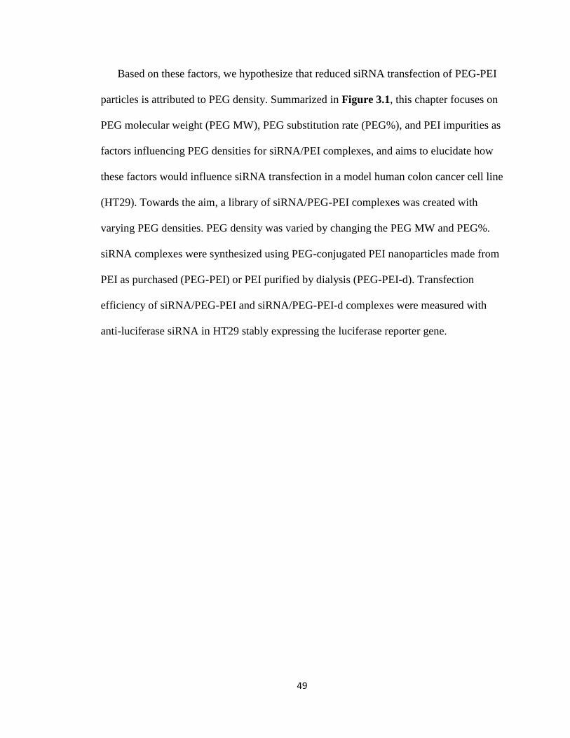
49
Based on these factors, we hypothesize that reduced siRNA transfection of PEG-PEI
particles is attributed to PEG density. Summarized in Figure 3.1, this chapter focuses on
PEG molecular weight (PEG MW), PEG substitution rate (PEG%), and PEI impurities as
factors influencing PEG densities for siRNA/PEI complexes, and aims to elucidate how
these factors would influence siRNA transfection in a model human colon cancer cell line
(HT29). Towards the aim, a library of siRNA/PEG-PEI complexes was created with
varying PEG densities. PEG density was varied by changing the PEG MW and PEG%.
siRNA complexes were synthesized using PEG-conjugated PEI nanoparticles made from
PEI as purchased (PEG-PEI) or PEI purified by dialysis (PEG-PEI-d). Transfection
efficiency of siRNA/PEG-PEI and siRNA/PEG-PEI-d complexes were measured with
anti-luciferase siRNA in HT29 stably expressing the luciferase reporter gene.
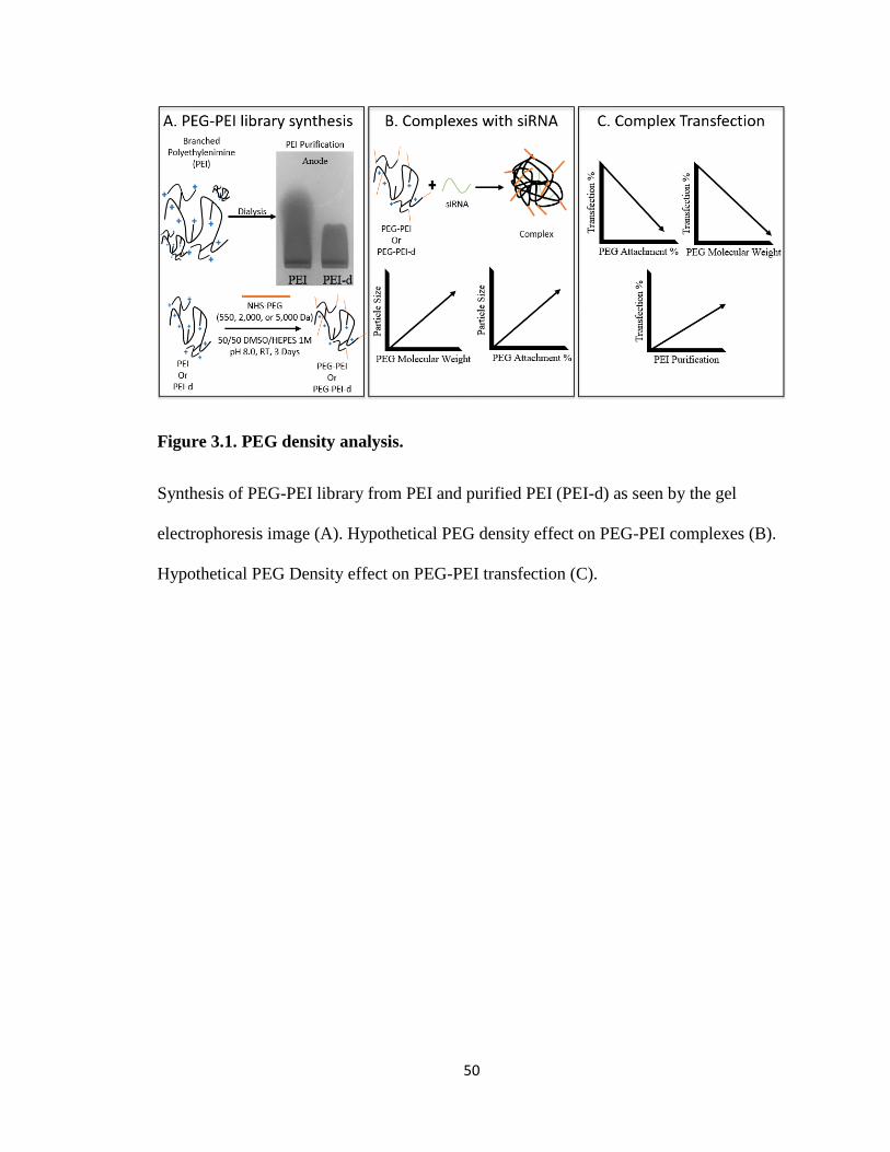
50
Figure 3.1. PEG density analysis.
Synthesis of PEG-PEI library from PEI and purified PEI (PEI-d) as seen by the gel
electrophoresis image (A). Hypothetical PEG density effect on PEG-PEI complexes (B).
Hypothetical PEG Density effect on PEG-PEI transfection (C).
51
3.2. Materials and Methods
3.2.1. Materials and Cells
PEI (25 kDa, branched) was purchased from Sigma Aldrich. PEGs (550 Da, 2 kDa,
and 5 kDa α-methoxy-ω-NHS activated, NHS-PEG) were purchased from NOF America.
HEPES buffer (pH 8.0, 1 M), NuSieve agarose GTG, dialysis membrane with molecular
weight cutoff (MWCO) of 100 kDa, deuterated DMSO, Opti-MEM, SimplyBlue
SafeStain, and phosphate buffered saline (PBS 1X) were purchased from Fisher Scientific
(Waltham, MA). Anti-Luciferase siRNA was synthesized with the sequence of 5’-
GUUGGCACCAGCAGCGCACUU-3. A human colon cancer cell line (HT29) was from
American Type Culture Collection (ATCC). HT29 cells were grown using McCoy’s 5A
with 10% fetal bovine serum (FBS) according to ATCC recommendations. Cells were
cultured in a humidified environment with 5% CO2 at 37 °C
3.2.2. Synthesis of TNAs of varying PEG substitutions and PEI backbones
We first purified PEI by dialysis (100 kDa MWCO) against water for 2 days. The
dialyzed PEI (PEI-d) was collected by lyophilization. PEI and PEI-d (20 µL of a 10
mg/mL) were run on a 2% agarose gel at 120 V for 1 hour to verify the removal of small
impurities. The gel was stained with SimplyBlue SafeStain overnight and destained in
water for 6 hours. PEG-PEI (or PEG-PEI-d) particles were then synthesized by reacting
NHS-PEG with PEI (or PEI-d) in a 1:1 mixture of DMSO:HEPES buffer (1 M) for 3 days
at room temperature. Molar ratios of NHS-PEG:PEI were 3:1, 15:1, or 40:1 for 1%, 5%,
and 10% (denoted as PEG% of PEI). The reactants were dialyzed against water (100K
52
MWCO) to remove free PEG and other impurities. PEG-PEI and PEG-PEI-d particles
were collected by lyophilization.
PEG-PEI and PEG-PEI-d particles were analyzed via 1H-NMR to obtain the PEG%.
Each PEG-PEI (5 mg) was added to deuterated DMSO (500 μL) and run in an NMR (400
MHz Varian NMR). To determine the PEG%, peak areas were compared between the
PEI main chain (CH2, 2.6 ~ 3.4 ppm) and PEG conjugated to PEI (CH2, 3.6 ppm). The
peak area comparisons determined the molar ratios between PEG chains and primary
amines of PEI (214 in average), which were converted to the percent substitution of PEG
on PEI. The PEG-PEI and PEG-PEI-d forming unimolecular nanoparticles were analyzed
for purity and uniformity by gel permeation chromatography (GPC, Asahipak GF-7M
column, 0.5 mL/min, 40 °C, PBS mobile phase).
3.2.3. Quantification of size and surface charge of TNAs and complexes
The surface charge and diameter of the particles were determined by measuring zeta
potential and dynamic light scattering (DLS, Zetasizer Nano, Malvern, UK). Aqueous
solutions of PEG-PEI particles were mixed with siRNA according to their minimum
complexation ratio, found in section 3.2.4, for 30 minutes at room temperature. PEG-PEI
complexes and empty polymer at a concentration of 1 mg/ml (500 μL) were loaded into
disposable zeta cuvettes and analyzed for particle diameter via DLS measurement
(Zetasizer Nano, Malvern, UK) and then zeta potential in the same cuvette.
3.2.4. Complex formation of TNAs with siRNA
A solution of PEG-PEI particle (1 mg/mL) was mixed with a solution of siRNA in
PBS to create a final concentration of particle ranging from 0 to 100 μg/mL polymer
53
mixed with 72 nM siRNA. PEG-PEI particles and siRNA were mixed at room
temperature for 30 minutes. The mixing ratio is described as the N/P ratio, where N is
amines on PEI and P is the phosphate groups on siRNA. siRNA/PEG-PEI complexes at
varying N/P ratios were analyzed by gel electrophoresis (1% agarose gel, 20 μL loading
per well, 120V, 1 hour). The gels were stained with ethidium bromide and imaged with
Typhoon GLA 9500 (GE Healthcare, Logan, UT).
3.2.5. In vitro transfection efficiency and toxicity of TNAs
Cells were seeded in a white 96 well cell culture plate (5,000 cells/well, 100 μL Opti-
MEM) and incubated for 24 hours prior to experiments. A solution of each polymer was
solubilized in Opti-MEM at 1000X the concentration at which the polymer formed
complexes with siRNA (determined by gel electrophoresis). One hundred microliters of
polymer dilutions (500X, 200X, and 10X) were added to 100 μL of 720 nM siRNA
solution. The mixtures were incubated at room temperature for 30 minutes. Cell culture
media (20 μL) in each well was replaced with 20 μL PEG-PEI/siRNA or PEI/siRNA
complexes (n = 4). The N/P ratios were 1, 20, 50, and 100 for each well. The plates were
incubated for 72 hours, and luciferin (100 μL, 0.1 mg/ml in PBS) was added to each well
for luminescence measurement via a GloMax luminometer (Promega). To measure cell
viability, a resazurin solution (10 μL, 1 mM) was added to each well. The plates were
incubated for 3 hours and live cells were counted on a fluorescent plate reader
(SpectraMax M5, Molecular Devices, 560Ex/590Em). The luciferase readings were
normalized to cell viability to obtain the percentage of luciferase activity remaining after
treatment.
54
3.3. Results
3.3.1. Increasing PEG corona density increases particle size and decreases surface
charge
PEG-PEI nanoparticles were successfully synthesized by using 25 kDa PEI and PEG
with varying molecular weights. A library of PEG-PEI particles with varying PEG
densities. NMR confirmed successful modification of two parameters for the particles:
PEG MW (550, 2,000, and 5,000 Da) and PEG substitution percent (1, 5, 10%). Table
3.1 summarizes the synthesis conditions including PEG% aimed and obtained. PEG-PEI
particles were characterized by GPC (Figure 3.2) and DLS (Table 3.1). The GPC shows
the removal of free PEG from the PEG-PEI particles. DLS revealed that the diameters of
empty particles ranged from 4 nm to over 30 nm. PEG MW and PEG% were considered
collectively to determine PEG density on PEG-PEI particles.
PEG MW and PEG substitution rate influenced PEG density directly, and particle
diameter increased as PEG density increased. Particles increased their diameter as PEG
MW and PEG% increased. A similar trend was observed with the surface charge of the
particles. PEG-PEI with low PEG MW and PEG% (e.g., 550 Da or 2 kDa with 1% PEG
substitution) showed positive surface charge. The surface charge decreased as PEG MW
and PEG% increased, while the larger molecular weight PEGs shielded surface charge
better at lower PEG%. Table 3.1 summarizes particle diameters of PEG-PEI particles
forming complexes with siRNA at their minimum complexation ratios (found in section
3.3.3). PEG-PEI particles increased diameter after complexation but less so as PEG
corona density increased.
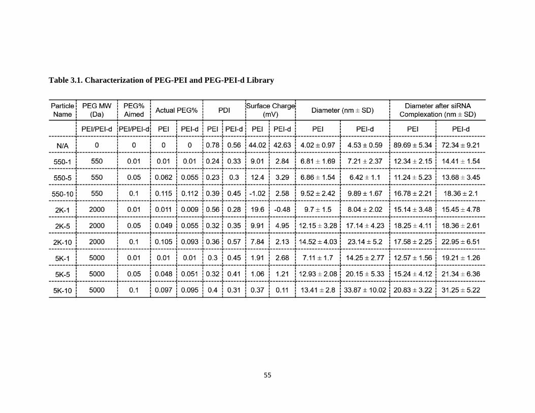
55
Table 3.1. Characterization of PEG-PEI and PEG-PEI-d Library
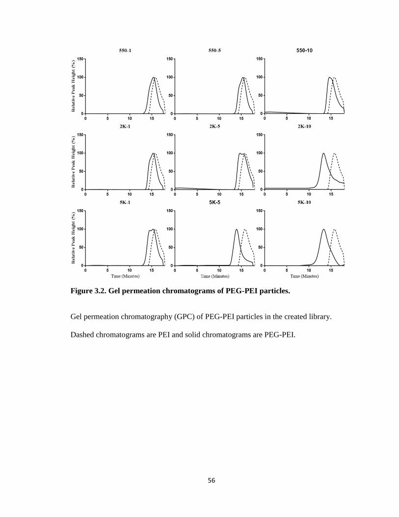
56
Figure 3.2. Gel permeation chromatograms of PEG-PEI particles.
Gel permeation chromatography (GPC) of PEG-PEI particles in the created library.
Dashed chromatograms are PEI and solid chromatograms are PEG-PEI.
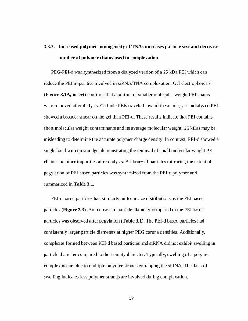
57
3.3.2. Increased polymer homogeneity of TNAs increases particle size and decrease
number of polymer chains used in complexation
PEG-PEI-d was synthesized from a dialyzed version of a 25 kDa PEI which can
reduce the PEI impurities involved in siRNA/TNA complexation. Gel electrophoresis
(Figure 3.1A, insert) confirms that a portion of smaller molecular weight PEI chains
were removed after dialysis. Cationic PEIs traveled toward the anode, yet undialyzed PEI
showed a broader smear on the gel than PEI-d. These results indicate that PEI contains
short molecular weight contaminants and its average molecular weight (25 kDa) may be
misleading to determine the accurate polymer charge density. In contrast, PEI-d showed a
single band with no smudge, demonstrating the removal of small molecular weight PEI
chains and other impurities after dialysis. A library of particles mirroring the extent of
pegylation of PEI based particles was synthesized from the PEI-d polymer and
summarized in Table 3.1.
PEI-d based particles had similarly uniform size distributions as the PEI based
particles (Figure 3.3). An increase in particle diameter compared to the PEI based
particles was observed after pegylation (Table 3.1). The PEI-d based particles had
consistently larger particle diameters at higher PEG corona densities. Additionally,
complexes formed between PEI-d based particles and siRNA did not exhibit swelling in
particle diameter compared to their empty diameter. Typically, swelling of a polymer
complex occurs due to multiple polymer strands entrapping the siRNA. This lack of
swelling indicates less polymer strands are involved during complexation.
58
Figure 3.3 Gel permeation chromatography of PEG-PEI-d particles.
Gel permeation chromatography (GPC) of PEG-PEI-d particles created. Dashed
chromatograms are PEI and solid chromatograms are PEG-PEI-d.
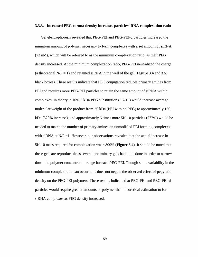
59
3.3.3. Increased PEG corona density increases particle/siRNA complexation ratio
Gel electrophoresis revealed that PEG-PEI and PEG-PEI-d particles increased the
minimum amount of polymer necessary to form complexes with a set amount of siRNA
(72 nM), which will be referred to as the minimum complexation ratio, as their PEG
density increased. At the minimum complexation ratio, PEG-PEI neutralized the charge
(a theoretical N/P = 1) and retained siRNA in the well of the gel (Figure 3.4 and 3.5,
black boxes). These results indicate that PEG conjugation reduces primary amines from
PEI and requires more PEG-PEI particles to retain the same amount of siRNA within
complexes. In theory, a 10% 5 kDa PEG substitution (5K-10) would increase average
molecular weight of the product from 25 kDa (PEI with no PEG) to approximately 130
kDa (520% increase), and approximately 6 times more 5K-10 particles (572%) would be
needed to match the number of primary amines on unmodified PEI forming complexes
with siRNA at N/P =1. However, our observations revealed that the actual increase in
5K-10 mass required for complexation was ~800% (Figure 3.4). It should be noted that
these gels are reproducible as several preliminary gels had to be done in order to narrow
down the polymer concentration range for each PEG-PEI. Though some variability in the
minimum complex ratio can occur, this does not negate the observed effect of pegylation
density on the PEG-PEI polymers. These results indicate that PEG-PEI and PEG-PEI-d
particles would require greater amounts of polymer than theoretical estimation to form
siRNA complexes as PEG density increased.
60
Figure 3.4. siRNA complexation of PEG-PEI particles.
Gel electrophoresis images of PEG-PEI run on a 1% agarose gel. siRNA at 72 nM was
allowed to complex for 30 minutes with varying concentrations of PEG-PEI. The mixture
was run on the gel and stained with ethidium bromide to image. The boxes indicate at
which point the PEG-PEI forms a complete complex with siRNA.
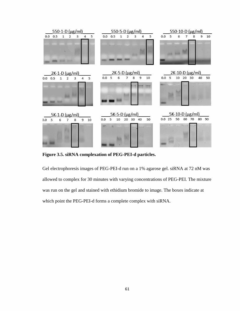
61
Figure 3.5. siRNA complexation of PEG-PEI-d particles.
Gel electrophoresis images of PEG-PEI-d run on a 1% agarose gel. siRNA at 72 nM was
allowed to complex for 30 minutes with varying concentrations of PEG-PEI. The mixture
was run on the gel and stained with ethidium bromide to image. The boxes indicate at
which point the PEG-PEI-d forms a complete complex with siRNA.
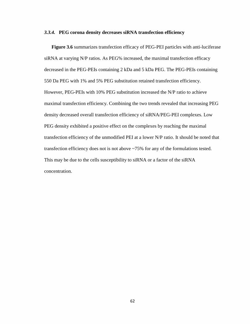
62
3.3.4. PEG corona density decreases siRNA transfection efficiency
Figure 3.6 summarizes transfection efficacy of PEG-PEI particles with anti-luciferase
siRNA at varying N/P ratios. As PEG% increased, the maximal transfection efficacy
decreased in the PEG-PEIs containing 2 kDa and 5 kDa PEG. The PEG-PEIs containing
550 Da PEG with 1% and 5% PEG substitution retained transfection efficiency.
However, PEG-PEIs with 10% PEG substitution increased the N/P ratio to achieve
maximal transfection efficiency. Combining the two trends revealed that increasing PEG
density decreased overall transfection efficiency of siRNA/PEG-PEI complexes. Low
PEG density exhibited a positive effect on the complexes by reaching the maximal
transfection efficiency of the unmodified PEI at a lower N/P ratio. It should be noted that
transfection efficiency does not is not above ~75% for any of the formulations tested.
This may be due to the cells susceptibility to siRNA or a factor of the siRNA
concentration.
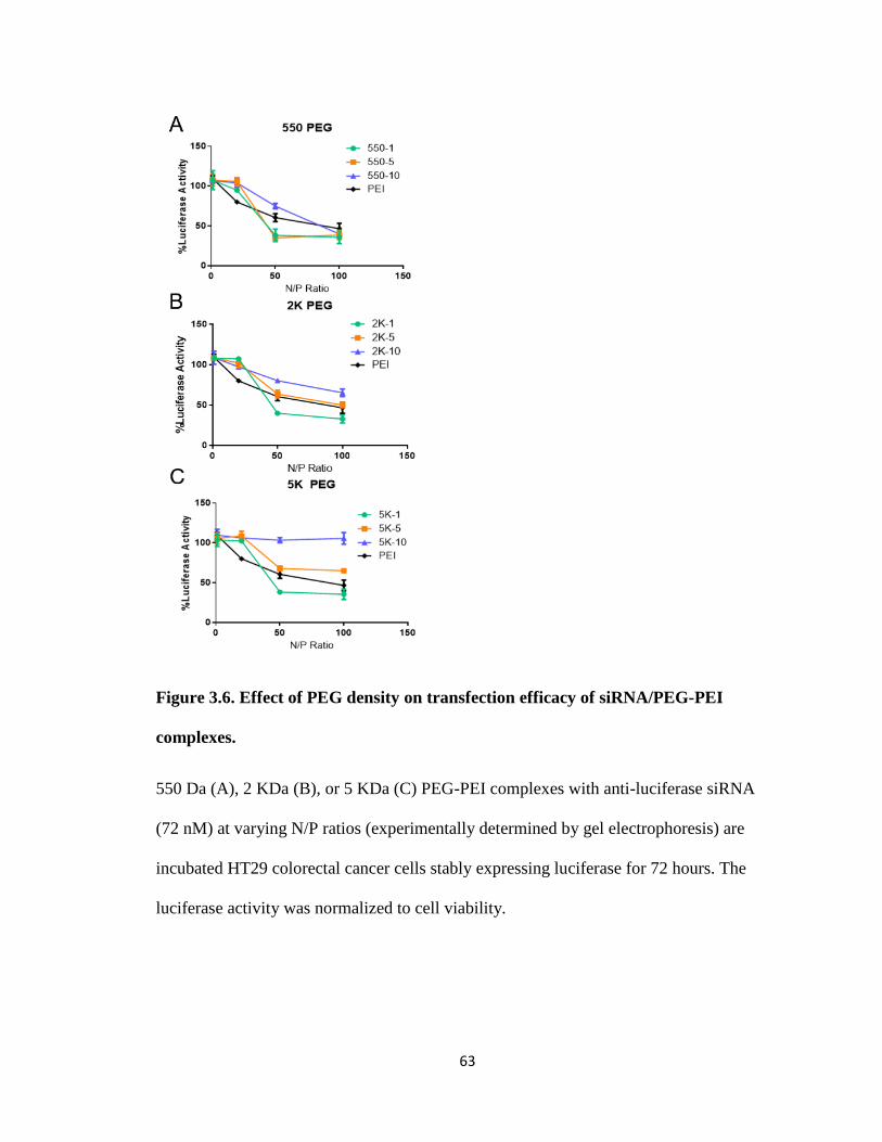
63
Figure 3.6. Effect of PEG density on transfection efficacy of siRNA/PEG-PEI
complexes.
550 Da (A), 2 KDa (B), or 5 KDa (C) PEG-PEI complexes with anti-luciferase siRNA
(72 nM) at varying N/P ratios (experimentally determined by gel electrophoresis) are
incubated HT29 colorectal cancer cells stably expressing luciferase for 72 hours. The
luciferase activity was normalized to cell viability.
64
3.3.5. Increased polymer homogeneity decreases necessary complexation ratio to
achieve maximum siRNA transfection
Figure 3.7 shows the effects of the PEG-PEI-d on transfection efficiency at varying
N/P ratios. Similar to PEG-PEI particles, PEG-PEI-d decreased the maximal transfection
efficacy as PEG% increased, while PEG MW showed no negative effects on transfection
at low PEG%. However, PEG-PEI-d showed maximal transfection at a lower N/P ratio
than PEG-PEI (20 vs 50). This effect was reduced as the PEG density was increased.

65
Figure 3.7. Effect of PEG density on transfection efficacy of siRNA/PEG-PEI-d
Complexes.
550 Da (A), 2 KDa (B), or 5 KDa (C) PEG-PEI-d complexes with anti-luciferase siRNA
(72 nM) at varying N/P ratios (experimentally determined by gel electrophoresis) are
incubated HT29 colorectal cancer cells stably expressing luciferase for 72 hours. The
luciferase activity was normalized to cell viability.
66
3.4. Discussion
PEI is a well-studied cationic polymer used in gene delivery, including siRNA
delivery, because it can readily form ionic complexes with genetic material to improve
stability of the genetic cargo in the blood stream. However, clinical applications of the
siRNA/PEI complexes are often held back by complex instability and toxicity147. The
toxicity of siRNA/PEI complexes is mainly attributed to the cationic surface charge
disrupting anionic cellular membranes148. A common method of reducing surface charge
of PEI/siRNA complexes is covalently attaching PEG to PEI149, forming a PEG shell.
PEG is available in many different molecular weights and thus can create PEIs with a
variety of PEG densities150. In the previous chapter, we prepared nanoparticles from
PEG-conjugated PEI (PEG-PEI) for siRNA delivery95 and found that their transfection
efficiency was significantly reduced, presumably due to variations of PEG density.
However, the effects of PEG density on physicochemical properties and transfection
efficiency of siRNA/PEG-PEI complexes were elusive. Therefore, this chapter elucidates
how PEG density affects transfection efficiency of siRNA/PEG-PEI complexes by
investigating three factors that influence PEG density on PEI (PEG MW, PEG% and PEI
impurities).
Increased PEG MW was found to weaken siRNA/PEG-PEI complex stability and
lower transfection efficacy. These results indicated that increasing PEG MW increased
the amount of PEG-PEI required to form complexes (Figure 3.4 and 3.5). Our initial
speculation was that the increased amount of PEG-PEI was due to increased molecular
weight of the polymer after PEG attachment. PEG conjugation reduces the weight
percentage of primary amines on PEI, and thus requires more polymer to form complexes
67
with siRNA and neutralize the charge. Therefore, we used the reduced weight percentage
of primary amines to predict the increase in the weight ratio between PEG-PEI and
siRNA to form complexes. For example, if PEI’s primary amines are reduced by 20%,
20% additional polymer is needed to form complexes. However, our data shows that
PEG-PEIs containing 2 kDa and 5 kDa PEG need more polymer than predicted to form
these complexes. This may be due to the PEG chains blocking siRNA from interacting
with the amines of PEI, similarly to how PAL may act when attached to 3P proposed in
chapter 2.
Larger PEG chains (2 kDa or 5 kDa) would block cationic sites on PEI more
effectively than shorter chains due to increased flexibility, as evidenced by the reduction
of surface charge as the PEG chain length was increased (Table 3.1). Since both PEI and
PEG are hydrophilic, the polymers may entangle rather than form a distinct core and shell
system like nanoparticles containing hydrophobic polymers113, 151. This entanglement
would block the cationic sites from interacting with the siRNA, but the effect would be
lessened by smaller chain lengths of PEG, as confirmed by lower molecular weight PEG-
PEIs (Figure 3.4 and 3.5). Additionally, blockage of amine groups would reduce
interaction with cellular membranes and endosomes. This may result in lowered
transfection efficiency responsible for unsuccessful transfection of the PEG-PEI used in
the previous chapter. PEG/PEI entanglement could further decrease complex stability as
the interact with water at the surface of the nanoparticle. This interaction would cause the
TNA to constantly alter its conformation, which would change the conformation of the
core and may release siRNA prematurely.
68
Lowering PEG% increased PEG-PEI/siRNA interactions and retained transfection
efficacy of the complex. Reducing the PEG% reduced the minimum amount of polymer
required for complex formation regardless of PEG MW (Figure 3.4 and 3.5). Lower
PEG% would reduce the number of PEG chains blocking cationic moieties from
interaction with both siRNA and cellular membranes. This was supported by observing
increased surface charge after reduction of PEG% (Table 3.1). Complexes with lower
PEG% achieved greater transfection efficiency than those with higher PEG% (Figure 3.6
and 3.7). In fact, complexes with relatively low PEG% and low PEG MW increased
transfection beyond the unmodified PEI control. This result agrees with others findings
that PEG-PEI can be more efficacious than PEI152. It should be noted that as PEG MW
increases, the disparity in transfection efficiency between PEG% also increases. This is
likely the result of PEG composing a larger weight percentage of the complex and further
blocking interactions with cellular membranes. For example, when complexes are
synthesized with 10 % attachment of 5 KDa PEG, their weight percentage of PEG is
~81%. The PEG weight percentage will be ~30% if the complexes have 1% attachment
of the same PEG. This increase in weight percentage of PEG on the polymer correlates
with a larger PEG shell. These trends show the importance of PEG% to influence the
interactions between siRNA and PEI. While the exact mechanism by which PEG chain
length reduces transfection is unknown, it is speculated that either the complex is less
stable145, 153 or unable to escape the endosome86, 154. Additionally, this gives further
evidence as to why the 2P from the previous chapter did not cause transfection.
Impurities in PEI were confirmed to alter the physicochemical properties of
complexes and reduce transfection efficacy at low N/P ratios and thereby alter PEG shell
69
density. PEI stocks that are purchased commercially often include smaller molecular
weight PEIs that are the result of polymer degradation and synthesis byproducts. Dialysis
of the PEI stock used in the synthesis of these PEG-PEIs was able to successfully remove
a portion of these impurities (Figure 3.1A). Removing these residual impurities increased
the proportional molecular weight of the PEI and the homogeneity of the stock (PEI-d).
After synthesis of PEG-PEIs using the PEI-d stock (PEG-PEI-d), physicochemical
properties of PEG-PEI-d differed from PEG-PEI mainly in particle diameter before and
after complexation with siRNA (Table 3.1). The increased particle diameter is likely due
to the increased proportion of larger cationic polymer chains in PEG-PEI-d. Particle
diameter swelling after complexation was also reduced which indicates fewer PEG-PEI-d
chains were involved complex formation. Fewer chains involved in complex formation
increases the homogeneity of the PEG shell surrounding the particle as well as the overall
homogeneity of the complexes. In PEG-PEI particles, the small PEI impurities can take
part in siRNA complexation and increase the amount of PEG-PEI needed to neutralize
the charge of siRNA and increase weight percentage of PEG. Therefore, PEG-PEI
complexes would have greater PEG content, which is beneficial to reduce the surface
charge yet disadvantageous to improving transfection. This speculation is supported by
PEG-PEI-d complexes that showed transfection efficacy similar to PEG-PEI complexes
yet transfected cells at a lower N/P ratio.
70
3.5. Conclusions
In this chapter, pegylation density of TNA/siRNA complexes, influenced by factors
including PEG molecular weight, PEG attachment percentage, and PEI impurities,
significantly alters transfection efficiency of these complexes. PEG density correlated
negatively with transfection efficiency of TNAs as low PEG density increased
transfection efficiency and high PEG density removed transfection ability from the
particles. This effect also demonstrates why the PEG-PEI (2P) from chapter 2 was unable
to transfect cells. Additionally, the removal of PEI impurities increased the overall
transfection efficiency of all complexes at lower N/P ratios. Our findings demonstrate the
importance of PEG length, PEG attachment percentage, and removal of PEI impurities in
improving transfection efficacy of siRNA carriers using PEG-PEI. These results may be
applicable to other cationic polymers forming complexes with siRNA and interacting
with cellular membranes in a similar way to PEI, such as poly(lysine) and chitosan
derivatives, and thus provide valuable insights for future development of more effective
and much safer siRNA carriers.
71
4. Chapter 4: Hydrophobic Modifications to TNAs and Non-Specific Reduction of
Reporter Protein Concentrations.
72
This chapter is adapted from work published in the International Journal of
Pharmaceutics on June 15th, 2017155. I would like to extend a special acknowledgement
to Dr. Tadahide Izumi who helped with western blotting of the high molecular weight
luciferase. I would also like to acknowledge Dr. Piotr Rychahou who helped with cell
lines and luciferase western blotting methods.
4.1. Introduction
Small interfering RNA (siRNA) can suppress mutated genes generating proteins
which are currently unable to be targeted by small molecule drugs and thus provide a new
therapy for genetic diseases including cancer 25, 156-157. However, siRNA often shows low
transfection in vivo due to poor delivery efficiency158. To improve delivery of siRNA to
target sites, polymer-based non-viral gene vectors have been developed, which include
ionic complexes between anionic siRNA and cationic polyethyleneimine (PEI)75, 147.
siRNA/PEI complexes have achieved successful gene silencing in various human cells in
vitro, but they are unstable in the body105. Polyethyleneglycol (PEG) is frequently used to
stabilize siRNA/PEI complexes121, 159. The hydrophilic PEG chains surrounding
siRNA/PEI complexes can shield the particle charge and prevent protein adsorption. The
siRNA/PEI complexes can be further modified with hydrophobic excipients and other
additives for improving stability and fine-tuning release of siRNA111.
Based on this background, we developed siRNA delivery vectors in the previous
chapters by using tethered nanoassemblies (TNAs) made from PEG, PEI, and
hydrophobic pendant groups such as palmitate (PAL)95. TNAs are unimolecular
assemblies of 30 nm diameter particles having cationic polymer backbone to which PEG
and a hydrophobic moiety can be covalently conjugated. In previous chapters, we
73
reported that these TNAs enhanced siRNA transfection in a human colorectal cancer
HT29 cell line as functions of PEG composition and PAL modification. These TNAs
displayed siRNA transfection efficiency comparable to PEG-PEI formulations developed
by other research groups136, 160 as well as commercially available transfection reagents
such as RNAiMAX. Upon further investigation of hydrophobically modified TNAs,
subsequent batches of PEG-PEI appeared to substantially change siRNA transfection
efficiency although the mechanism remained elusive95. However, further study of the
hydrophobic modification to PEG-PEI also revealed that the resulting PEG-PEI-PAL
TNA (3P) unexpectedly reduced luciferase expression in cells even in the absence of
siRNA, called the false positive effect.
The false positive effect on siRNA transfection increases the difficulty in evaluating
and predicting in vivo performance of PEG-PEI based non-viral gene vectors for future
studies and clinical applications. Protein reporter assays are commonly used to evaluate
the transfection efficiency of the siRNA/PEG-PEI complex, and luciferase is a widely-
used reporter protein that offer a quick method for measuring siRNA-mediated gene
silencing in living cells18, 161-162. Luciferase protein activity is correlated to the amount of
luciferase protein in the cell following siRNA transfection by measuring luminescence
using luminogenic substrate 163. Luciferase has a short half-life in live cells, less than 3
hours164, which makes it ideal for determining long-term siRNA transfection efficiency.
Any protein left within the cell after incubation with siRNA/PEG-PEI complexes would
be produced after the siRNA has had a chance to take effect. However, this would not
account for protein that has been denatured or had its expression reduced by other means.
If PEG-PEI can interfere with the luciferase reporter assay this way, it would explain
74
siRNA transfection efficiency varying among product batches. Another possible reason
for inconsistent siRNA transfection of PEG-PEI is the cellular stress response triggered
by siRNA/PEG-PEI complexes. Off target effects of siRNA have been demonstrated63, 73,
yet little is known if and how PEG-PEI would induce non-specific gene silencing during
siRNA transfection.
Our previous findings suggest that the addition of hydrophobic pendant groups to
TNAs would influence intracellular luciferase expression by either directly interacting
with cells or indirectly disrupting the protein synthesis process95, 133. Therefore, this
chapter aims to elucidate the false positive effect of hydrophobically modified TNAs on
siRNA transfection by using TNAs made from combinations of PEG, PEI, poly(L-lysine)
(PLL), palmitate (PAL), and deoxycholate (DOC): PEG-PEI (2P), PEG-PEI-PAL (3P),
PEG-PLL (2P’), PEG-PLL-PAL (3P’), and PEG-PEI-DOC (2PD) as shown in Figure
4.1. These PEG-PEI TNAs (+/- siRNA) are characterized by in vitro siRNA transfection,
cell viability, toxicity and immunoblotting in a human colorectal cancer cell line stably
overexpressing luciferase (HT29/Luc). Raman spectroscopy is also employed to
investigate interactions between hydrophobic pendant groups conjugated to the TNA core
and compounds outside the particles. To investigate the cellular stress caused by TNAs,
assays determining total protein count, ATP concentration, and cellular membrane
porosity are used. Data obtained from these experiments are analyzed to determine the
effects of hydrophobic groups and polymer scaffold condensation on false transfection of
siRNA/PEG-PEI complexes. Understanding these unexpected outcomes of covalent
modification to cationic polymers is crucial for future design of siRNA delivery vehicle
development.
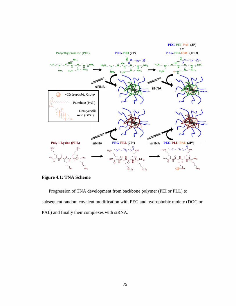
75
Figure 4.1: TNA Scheme
Progression of TNA development from backbone polymer (PEI or PLL) to
subsequent random covalent modification with PEG and hydrophobic moiety (DOC or
PAL) and finally their complexes with siRNA.
76
4.2. Materials and Methods
4.2.1. Materials and Cells
PEG (5 kDa, α-methoxy-ω-NHS ester activated) was purchased from NanoCS (New
York, NY). Branched PEI (bPEI), deoxycholate (DOC), N-hydroxy succinimide, 4-
dimethylaminopyridine, 1-ethyl-3-(3-dimethyl aminopropyl) carbodiimide hydrochloride
(EDC HCl), palmitoyl chloride, resazurin sodium salt, sulforhodamine B based in vitro
toxicology assay kit (TOX6), and poly(L-lysine) (PLL, 30-70 kDa) were purchased from
Sigma Aldrich (St. Louis, MO). RNAiMAX, 4-12% NuPAGE Bis-Tris precast gels,
HEPES buffer (pH 8.0, 1 M), MES buffer (0.1 M, pH 5.0), RIPA buffer, pyridine,
NuSieve GTG agarose, dialysis membrane with molecular cut-off (MWCO) of 6-8 and
100 kDa, and other organic solvents were purchased from Fisher Scientific (Waltham,
MA). siRNA (5’-GUUGGCACCAGCAGCGCACUU-3’) was purchased from GE
Dharmacon (Lafayette, CO). Opti-MEM was purchased from Life Technologies
(Carlsbad, CA). Mitochondrial ToxGlo™ assay kit and Bradford assay kit were
purchased from Promega (Madison, WI). McCoy’s 5A, 0.05% trypsin/EDTA, and
phosphate buffered saline (PBS) were from GE Healthcare (Logan, UT). Fetal bovine
serum (FBS) was purchased from Atlanta Biologicals (Flowery Branch, GA).
HT29 human colon cancer cell line was purchased from American Type Culture
Collection (ATCC, Manassas, VA), and subsequently transfected to establish a cell-line
stably expressing firefly luciferase (HT29/Luc). Cells were cultured at logarithmic
growth in a humidified environment with 5% CO2 at 37 °C in McCoy’s 5A media
supplemented with 10% FBS according to ATCC recommendations.
77
4.2.2. Synthesis of TNAs with different backbones and hydrophobic moieties
TNAs with a PEI backbone (2P and 3P) were synthesized from 25 kDa bPEI, 5 kDa
NHS-activated PEG, and palmitoyl chloride as previously reported95. In this chapter, 2P
was further modified with DOC through covalent conjugation to the PEI backbone.
Briefly, DOC was mixed with NHS, EDC, and DMAP in a 5:5:0.2 molar ratio in MES
buffer (0.1 M, pH 5.0). 2P was then added to the mixture in a 1:100 molar ratio of
2P:DOC at room temperature. After 48 hours, the product was purified by dialysis and
freeze drying to collect 2PD.
TNAs with a PLL backbone (2P’ and 3P’) were prepared from 30-70 kDa PLL with 5
kDa NHS-activated PEG and palmitoyl chloride as previously reported165. 2P’ was
synthesized by reacting PLL with NHS-activated PEG in water:DMSO:pyridine mixture
at ratios of 2:1:1. The solution was mixed for 72 hours at room temperature. 2P’ was
further modified with palmitoyl chloride to create 3P’. 2P’ was dissolved in THF at 40 °C
and palmitoyl chloride was added in a 1.1:1 molar ratio palmitate:2P’. After 15 minutes
of mixing, pyridine in a 2:1 molar ratio of pyridine:palmitate was added as a HCl
scavenger. The solution was reacted for additional 2 hours and purified by ether
precipitation and dialysis. The product was collected by freeze drying.
Purity and molecular weight uniformity of TNAs were determined by gel
permeation chromatography (GPC, Asahipak GF-7M column, 2 mg/mL, DMF mobile
phase, 0.5 mL/min, 40 °C). Diameter and surface charge were determined by dynamic
light scattering (DLS) and zeta potential measurements using Zetasizer Nano (Malvern,
UK).
78
4.2.3. Determination of minimum complexation ratios of TNAs and siRNA
TNAs at varying concentrations (0-1 mg/mL) were mixed with 2 µg/mL siRNA
solutions in Opti-MEM at a 1:1 ratio and allowed to equilibrate for 30 minutes. Each
mixture (20 µL) was then loaded on to a 1% agarose gel and run at 100 volts for 60
minutes at room temperature in TAE (Tris-Acetate 0.04 M, EDTA 0.001M) buffer. The
gel was stained with 100 ng/mL ethidium bromide in a TAE buffer and rinsed 3 times
with deionized water. The gel was imaged using Typhoon GLA 9500 (GE Healthcare,
Logan, UT) fluorescent imager with an ethidium bromide filter set.
4.2.4. In vitro transfection and toxicity efficiency of TNAs
Cells were plated at 5,000 cells per well into white opaque 96 well plates and
incubated for 24 hours. After 24 hours, TNA solutions were prepared by adding 100 µL
of 10 mg/mL TNA solutions to 100 µL of 10 µg/mL siRNA solution or 100 µL of Opti-
MEM. Naked siRNA controls were created with 200 µL of siRNA solutions at 5 µg/mL.
Opti-MEM (200 µL) was used as the blank control. RNAiMAX controls were created by
mixing 5 µL of RNAiMAX with 95 µL of Opti-MEM and then either 100 µL of Opti-
MEM or 100 µL of 10 µg/mL siRNA solution for the empty RNAiMAX control or
siRNA-loaded RNAiMAX control, respectively. Complexes were formed by incubating
the mixtures for 30 minutes at room temperature, except the RNAiMAX control that was
incubated for 5 minutes as instructed by the manufacturer. From each well, 20 µL of
solutions were replaced with their respective complex solutions or controls (n = 6) to
yield 1 mg/mL of TNA and 1 µg/mL siRNA in the well. The plate was incubated for 72
hours and then read for bioluminescent intensity using GloMax luminometer. The
substrate was 100 µL of 0.1 mg/mL luciferin solution in PBS. Luminescent intensity was
79
collected for 10 seconds and reported as percentage of luciferase activity with respect to
control wells. The data was normalized to cell viability (described below) to account for
cell death in the reduction of the luciferase signal.
Three cell viability assays were used in this chapter to consider different aspects of
cell health. The first assay was a resazurin assay, which measures mitochondrial activity
in live cells. 10 µL of a 1 mM resazurin solution was added to each well and incubated
for 3 hours. The plate was read on SpectraMax M5 (Molecular Devices) fluorescent plate
reader at excitation/emission of 560/590 nm. The readings were subtracted from blank
controls and reported as percentage of viable cells.
The second assay was a sulforhodamine B assay for the quantification of cellular
proteins in live cells. Briefly, cells were fixed with 30 µL/well of provided TCA solution
at 4 °C for 1 hour. After media removal and air drying, 20 µL/well of sulforhodamine B
solution was added and allowed to stain for 30 minutes. The stain was then removed,
washed with 1% acetic acid solution, and air dried. 100 µL/well of 10 mM Tris base
solution was added and incubated for 5 minutes at room temperature. Absorbance was
measured at 565 nm excitation and 690 nm emission with a SpectraMax M5 (Molecular
Devices) fluorescent plate reader. The absorbance at 690 nm is subtracted from the
absorbance at 565 nm to determine percent viable cells with respect to controls.
The third assay was for monitoring cell membrane integrity and mitochondrial
activity of cultured cells, which were measured with the Mitochondrial ToxGlo assay kit
per manufacturer’s instruction. Briefly, 20 µL/well of the provided bis-AAF-R110
solution was added to the plate and incubated at 37 °C for 30 minutes. Fluorescence was
measured at excitation/emission of 495/520 nm using a SpectraMax M5 (Molecular
80
Devices) fluorescent plate reader. Then, 100 µL/well of the provided ATP detection
reagent was added to the plate and read for luminescence with GloMax luminometer. The
values were compared to their respective controls (untreated wells) and reported as
percentages of the control.
4.2.5. Analysis of Luciferase Protein Expression Levels in vitro
Cells were plated at 50,000 cells/well in a 12 well clear plate. 24 hours later, 200 µL
of media were replaced with 200 µL of Opti-MEM, empty TNAs, or siRNA/TNA
complexes. After a 72-hour incubation at 37 °C, cells were lysed with a RIPA buffer and
assayed for protein content with a Bradford assay kit (Promega). 100 µg of protein for
each sample were subjected to electrophoresis through a 4-12% NuPAGE Bis-Tris
precast gel. Afterwards, the proteins were transferred to a nitrocellulose membrane.
Primary antibodies used to treat the membranes were anti-luciferase and anti-beta-actin
(Cell Signaling Technologies). After washing, the membranes were exposed to IgG-
horseradish peroxidase conjugate secondary antibodies for 1 hour and developed with an
ECL Western blotting substrate (Promega and SCBT). To detect the slow-migrating
firefly luciferase protein in SDS/PAGE, cells were incubated with the 2P, 2P’, 3P, and
3P’ for 72 h as described above, and further incubated in the presence of MG132 (40 µM)
for 6 h. The protein extracts were run in SDS/PAGE and transferred to PVDF
membranes, which were cut above the intact firefly luciferase protein to exclude the
intact protein in the membrane, and blotted with the rabbit polyclonal anti-firefly
luciferase antibody (Promega). The blot was developed with SuperSignal West Femto
(Pierce, Thermofisher), and the signals were analyzed in ChemiDoc Imaging Systems
(Bio-Rad).
81
4.2.6. Activity of luciferase protein after pre-incubation with TNAs
20 µL of 10 mg/mL TNA solutions or RNAiMAX solution were incubated for 10
minutes in wells of a white 96 well plate (n = 6). Each well contained 80 µL/well of PBS
with luciferase proteins to obtain 1 mg/mL TNA and 5 ng/mL of luciferase. 100 µL of
0.1 mg/mL luciferin solution in PBS were added to each well and read using GloMax
luminometer with an integration time of 10 seconds. Similarly, 2 mL of 10 mg/mL TNA
solutions or RNAiMAX solution were mixed for 30 minutes with 8 mL of 0.125 mg/mL
luciferin solution. 100 µL of this solution was then injected using GloMax luminometer
into each well of a white 96 well plate containing 100 µL/well of 5 ng/mL luciferase in
PBS. Luminescence was measured and integrated for 10 seconds. Data is reported as
RLU values.
4.2.7. Observed interactions of TNAs and components with siRNA through Raman
spectroscopy
Raman spectroscopy was conducted in the solid state using a Nicolet iS 50 FTIR with
a Raman module (Thermo Fisher Scientific, Waltham, MA). The samples were prepared
on a 48 well metal plate. 100 µL of a 10 mg/mL solution of each sample was added to a
well. To analyze individual components of 3P and TNAs, 100 μL of water was added to
each well. For physical mixtures of components, 100 μL of a 10 mg/mL solution of the
respective component was added to the well. 1 µL of a 1 mg/mL siRNA solution or 1 μL
of water was added to each well. The loaded metal plate was put at room temp for 30
minutes to acclimate all samples. The plate was then transferred to dry ice and the
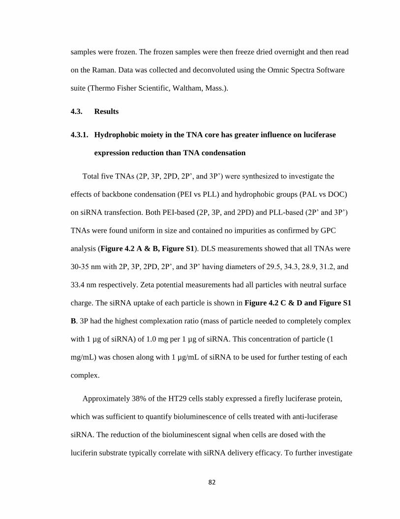
82
samples were frozen. The frozen samples were then freeze dried overnight and then read
on the Raman. Data was collected and deconvoluted using the Omnic Spectra Software
suite (Thermo Fisher Scientific, Waltham, Mass.).
4.3. Results
4.3.1. Hydrophobic moiety in the TNA core has greater influence on luciferase
expression reduction than TNA condensation
Total five TNAs (2P, 3P, 2PD, 2P’, and 3P’) were synthesized to investigate the
effects of backbone condensation (PEI vs PLL) and hydrophobic groups (PAL vs DOC)
on siRNA transfection. Both PEI-based (2P, 3P, and 2PD) and PLL-based (2P’ and 3P’)
TNAs were found uniform in size and contained no impurities as confirmed by GPC
analysis (Figure 4.2 A & B, Figure S1). DLS measurements showed that all TNAs were
30-35 nm with 2P, 3P, 2PD, 2P’, and 3P’ having diameters of 29.5, 34.3, 28.9, 31.2, and
33.4 nm respectively. Zeta potential measurements had all particles with neutral surface
charge. The siRNA uptake of each particle is shown in Figure 4.2 C & D and Figure S1
B. 3P had the highest complexation ratio (mass of particle needed to completely complex
with 1 µg of siRNA) of 1.0 mg per 1 µg of siRNA. This concentration of particle (1
mg/mL) was chosen along with 1 µg/mL of siRNA to be used for further testing of each
complex.
Approximately 38% of the HT29 cells stably expressed a firefly luciferase protein,
which was sufficient to quantify bioluminescence of cells treated with anti-luciferase
siRNA. The reduction of the bioluminescent signal when cells are dosed with the
luciferin substrate typically correlate with siRNA delivery efficacy. To further investigate
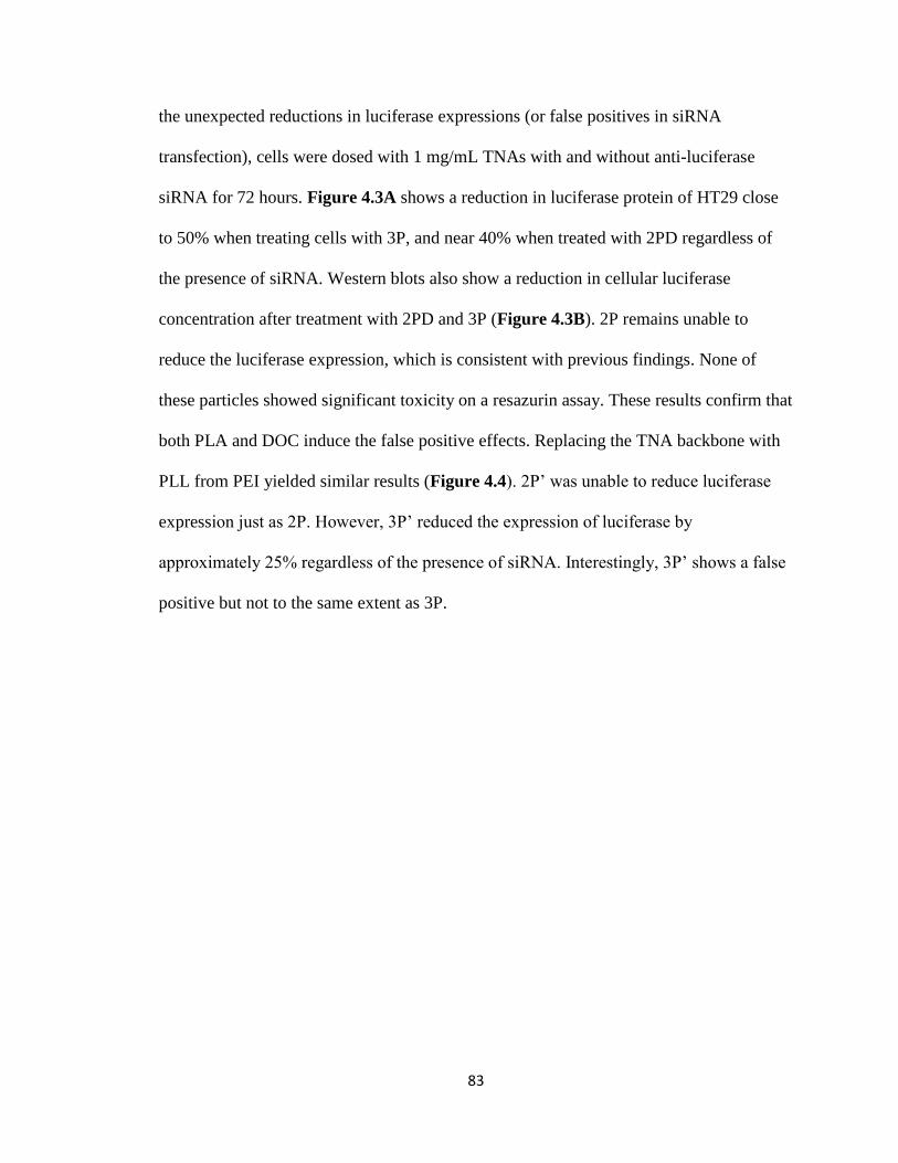
83
the unexpected reductions in luciferase expressions (or false positives in siRNA
transfection), cells were dosed with 1 mg/mL TNAs with and without anti-luciferase
siRNA for 72 hours. Figure 4.3A shows a reduction in luciferase protein of HT29 close
to 50% when treating cells with 3P, and near 40% when treated with 2PD regardless of
the presence of siRNA. Western blots also show a reduction in cellular luciferase
concentration after treatment with 2PD and 3P (Figure 4.3B). 2P remains unable to
reduce the luciferase expression, which is consistent with previous findings. None of
these particles showed significant toxicity on a resazurin assay. These results confirm that
both PLA and DOC induce the false positive effects. Replacing the TNA backbone with
PLL from PEI yielded similar results (Figure 4.4). 2P’ was unable to reduce luciferase
expression just as 2P. However, 3P’ reduced the expression of luciferase by
approximately 25% regardless of the presence of siRNA. Interestingly, 3P’ shows a false
positive but not to the same extent as 3P.
84
Figure 4.2: Characterization of TNAs
Gel permeation chromatography (GPC) spectra shows particle uniformity and purity
of PEI based TNAs (A) where black chromatograph represents 2P and green represents
3P and PLL based TNAs (B) where black chromatograph represents 2P’ and green
represents 3P’. Gel electrophoresis of PEI based TNA/siRNA mixtures (C) and PLL
based TNA/siRNA mixtures (D) with 72 nM siRNA and varying concentrations of TNA
to determine the ratio of TNA to siRNA needed to form complexes (black boxes).
85
Figure 4.3: TNA in vitro Luciferase Reduction
A: luminometric assay of luciferase (left) and viability assay (right) after 72-hour
incubation with each particle. Dark grey bars represent each condition without siRNA
and light grey bars represent condition with 1 µg/mL anti-luciferase siRNA. B: Western
blotting results after 72-hour incubation with 3P and 2PD including an untreated control.
siRNA in each condition is represented by N (no siRNA present) or L (anti-luciferase
siRNA, 1 µg/mL). * denotes that the column is significantly different from the control
condition (p<0.01)
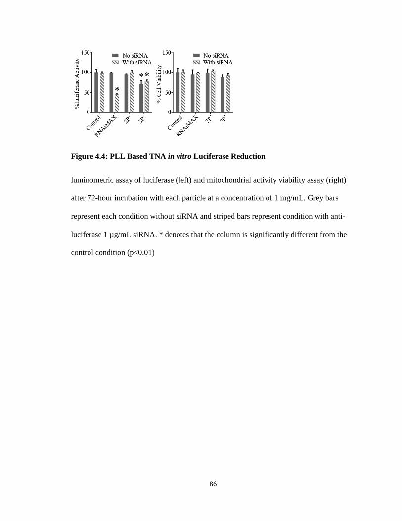
86
Figure 4.4: PLL Based TNA in vitro Luciferase Reduction
luminometric assay of luciferase (left) and mitochondrial activity viability assay (right)
after 72-hour incubation with each particle at a concentration of 1 mg/mL. Grey bars
represent each condition without siRNA and striped bars represent condition with anti-
luciferase 1 µg/mL siRNA. * denotes that the column is significantly different from the
control condition (p<0.01)
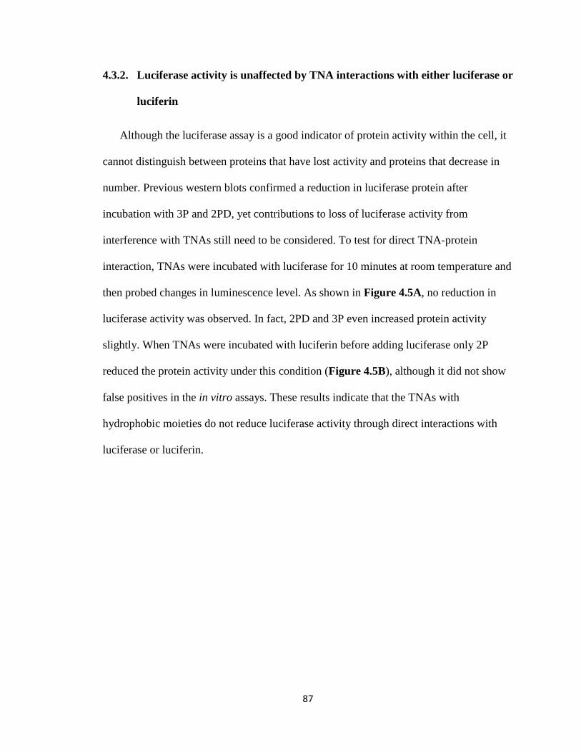
87
4.3.2. Luciferase activity is unaffected by TNA interactions with either luciferase or
luciferin
Although the luciferase assay is a good indicator of protein activity within the cell, it
cannot distinguish between proteins that have lost activity and proteins that decrease in
number. Previous western blots confirmed a reduction in luciferase protein after
incubation with 3P and 2PD, yet contributions to loss of luciferase activity from
interference with TNAs still need to be considered. To test for direct TNA-protein
interaction, TNAs were incubated with luciferase for 10 minutes at room temperature and
then probed changes in luminescence level. As shown in Figure 4.5A, no reduction in
luciferase activity was observed. In fact, 2PD and 3P even increased protein activity
slightly. When TNAs were incubated with luciferin before adding luciferase only 2P
reduced the protein activity under this condition (Figure 4.5B), although it did not show
false positives in the in vitro assays. These results indicate that the TNAs with
hydrophobic moieties do not reduce luciferase activity through direct interactions with
luciferase or luciferin.
88
Figure 4.5: Direct TNA/Protein and TNA/Substrate Interactions
A: Interaction between TNAs and Luciferase protein was observed by incubating the
protein and TNA together for 10 minutes. Then luciferin solution was added to the well
and a luminescence reading was taken. The readings were normalized to the control well
of luciferase protein alone. B: TNA and Luciferin (substrate to luciferase) direct
interaction was observed by 30-minute incubation of TNA and Luciferin before an
injection of luciferase solution was added to the well. A luminescence reading was taken
and the values normalized to the control well which did not contain TNAs.
89
4.3.3. Raman spectroscopy indicates that hydrophobic moieties contribute to
TNA’s interaction with siRNA
To better understand how palmitate could interact with other cellular components and
induce cell stress, Raman spectroscopy of 2P, 3P, and each of their individual
components was conducted (Figure 4.6). Each component was allowed to interact with
siRNA, which does not appear on the Raman spectra (Figure S2), while their spectra
were compared for change in vibrational intensity. Interactions between siRNA with PEG
or PAL were minor but the interaction between PEI and siRNA was more pronounced.
However, physical mixtures of PAL/PEI and PAL/PEG showed unexpected interactions
with siRNA. PEG/PAL showed a large change in vibrational intensity although neither
component showed significant interaction with siRNA on their own. PEI/PAL increased
its vibrational intensity, which indicates that the polymers are more mobile in the
presence of siRNA. Raman spectra of 2P and 3P with siRNA slightly changed after the
addition of siRNA although the exact influence of PAL in these interactions could not be
determined.
To account for any variance in measurement of the polymers, PEG polymer was
analyzed under a number of conditions to determine what effect sample conditions would
have on the measurements taken. Figure S3 indicates that samples of PEG taken from the
same stock and analyzed in succession would have different peak intensities but the
number and location of peaks would be the same. Referencing peak intensity to the
largest peak of the of the spectra determined that these peaks were all in the same ratio
(Figure S4), which supports why the Raman spectra in Figure 4.6 use normalized
intensity. Figure S5 indicates that Raman spectra of the same sample over a few hours

90
have very minor changes. Though the Raman spectra of the polymer samples were all
taken in succession, no experiment took longer than 2 hours to perform and therefore
peak changes between samples should not have occurred.
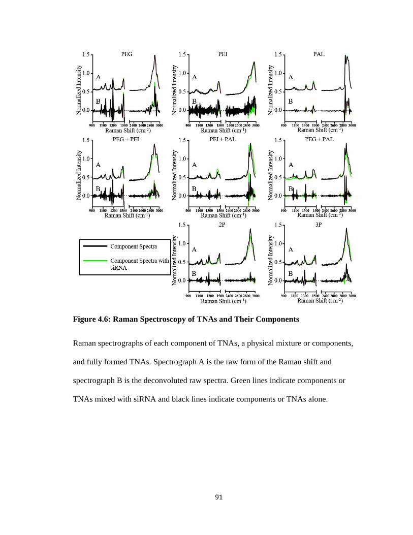
91
Figure 4.6: Raman Spectroscopy of TNAs and Their Components
Raman spectrographs of each component of TNAs, a physical mixture or components,
and fully formed TNAs. Spectrograph A is the raw form of the Raman shift and
spectrograph B is the deconvoluted raw spectra. Green lines indicate components or
TNAs mixed with siRNA and black lines indicate components or TNAs alone.
92
4.3.4. Hydrophobically modified TNAs reduce protein expression, membrane
integrity, and ATP concentration but retained mitochondrial activity
Resazurin cell viability assay was used to determine levels of live cells after
incubation of TNAs. The resazurin assay measures redox activity within the cell, but this
process does not represent a complete picture of cell health. Changes in ATP content,
total protein amount, and cell membrane porosity were also investigated to determine the
presence of cellular stress. Figure 4.7 shows differences among these cytotoxicity assays.
3P did not have a significant impact on redox activity in the cell but ATP and total
protein levels in the cells greatly decreased. Additionally, membrane porosity decreased
with incubation of 3P.
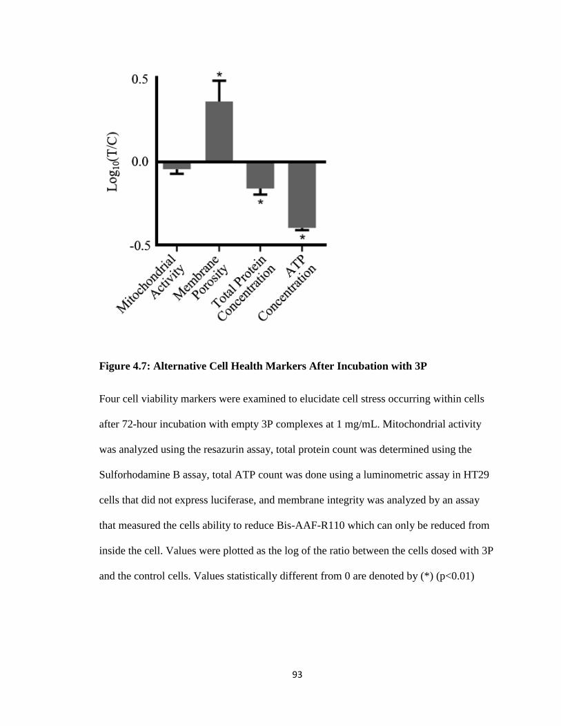
93
Figure 4.7: Alternative Cell Health Markers After Incubation with 3P
Four cell viability markers were examined to elucidate cell stress occurring within cells
after 72-hour incubation with empty 3P complexes at 1 mg/mL. Mitochondrial activity
was analyzed using the resazurin assay, total protein count was determined using the
Sulforhodamine B assay, total ATP count was done using a luminometric assay in HT29
cells that did not express luciferase, and membrane integrity was analyzed by an assay
that measured the cells ability to reduce Bis-AAF-R110 which can only be reduced from
inside the cell. Values were plotted as the log of the ratio between the cells dosed with 3P
and the control cells. Values statistically different from 0 are denoted by (*) (p<0.01)
94
4.3.5. TNAs increased ubiquitination and degradation of luciferase
Western blotting determined the extent of ubiquitination of luciferase to gain a better
understanding of the fate of the luciferase protein after TNA treatment. Western blot
analysis of cells dosed for 72 hours with 2P, 2P’, 3P, and 3P’ and an additional 6 hours
with MG132 proteasome inhibitor confirmed the presence of both intact luciferase and
ubiquitinated luciferase (Figure 4.8). The proteasome inhibitor prevents the degradation
of ubiquitinated luciferase so that it can be visualized on the blot. Figure 4.8 A shows the
reductions in luciferase concentration previously seen with 3P and 3P’ and displays little
effect by the proteasome inhibitor MG132. Initially ubiquitinated luciferase decreased in
cells dosed with 3P and 3P’, but its corresponding bands increased upon addition of the
proteasome inhibitor (Figure 4.8B). All TNAs induced an increase in ubiquitination of
luciferase while 3P and 3P’ caused the highest increase in this band comparatively. This
indicates that 3P and 3P’ increase ubiquitination of luciferase and they may also increase
proteasome activity.
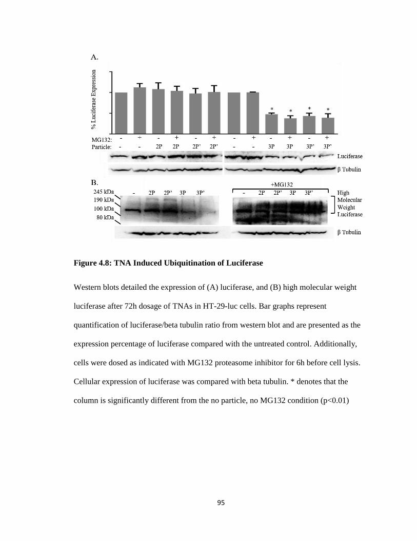
95
Figure 4.8: TNA Induced Ubiquitination of Luciferase
Western blots detailed the expression of (A) luciferase, and (B) high molecular weight
luciferase after 72h dosage of TNAs in HT-29-luc cells. Bar graphs represent
quantification of luciferase/beta tubulin ratio from western blot and are presented as the
expression percentage of luciferase compared with the untreated control. Additionally,
cells were dosed as indicated with MG132 proteasome inhibitor for 6h before cell lysis.
Cellular expression of luciferase was compared with beta tubulin. * denotes that the
column is significantly different from the no particle, no MG132 condition (p<0.01)
96
4.4. Discussion
In chapter 2, PEG-PEI TNAs were investigated to improve siRNA delivery by
modulating complex stability through addition of palmitate, a hydrophobic moiety95.
Although those TNAs modified with a hydrophobic group PAL (3P) caused increased
transfection, their complex stability was lowered. In further investigation of how these
TNAs were able to accomplish this effect, an siRNA-independent reduction of luciferase
in cells treated 3P was observed in luciferase assays for transfection efficiency (Figure
4.3), referred to as a false positive effect. These false positives can be detrimental in
developing siRNA-based therapeutic options that require specific gene silencing for
targeted gene therapy. Therefore, this chapter further investigated potential causes for the
unexpected protein reduction by using TNAs made from PEG-PEI with different
hydrophobic pendant groups (PAL vs DOC) and polymer backbone (PEI vs PLL).
Structural differences in hydrophobic groups may influence false positives caused by
TNAs differently. TNAs attached with rigid bile acid DOC (2PD) and the flexible fatty
acid PAL (3P) pendant groups were investigated as these hydrophobic groups are
structurally different. 2PD observed a reduction of luciferase activity slightly less than
3P, indicating that the false positive effect is not unique to palmitate. However, it is
unknown if the decreased protein activity is due to inactivated protein or reduced cellular
concentration of luciferase because the assay does not differentiate between the two
conditions. To observe any loss of activity of luciferase due to TNA interaction, TNAs
were incubated with free luciferase protein or free luciferin substrate before addition of
the other component. Since neither 3P nor 2PD reduced the activity of luciferase, the
protein concentration should be lower in the cell. Western blotting supported this
97
speculation, indicating that 3P and 2PD reduced luciferase protein concentrations. This
determined that the false positive transfection is due to reduced cellular protein
concentration brought on by the hydrophobic moiety.
Another factor that may affect the false positive effect is composition of the cationic
backbone which influences the TNA’s secondary structure formation. Secondary
structure refers to the folding or condensing of the polymer structure either on its own or
in the presence of anionic groups. This can influence the location of components within
the TNA’s core. Linear PLL, another well studied cationic polymer for gene transfection,
replaced branched PEI as the backbone for TNAs, creating 2P’ and 3P’. These particles
were similar in size and surface charge to PEI based TNAs. The false positive effect was
observed in 3P’ but initially to a lesser extent than 3P. While changing the backbone
structure mildly decreased the false positive effect, it did not eliminate it. This indicates
that the contribution of the backbone structure to the false positive effect is significantly
less than that of the hydrophobic moiety.
Raman spectroscopy offers another method to better understand PALs influence on
false positives by examining PAL’s influence on TNA interaction. Raman spectroscopy
measures the vibrational energy of chemical bonds in a compound which can determine
the influence of TNA components on interactions with other compounds166-168. These
bond vibrations are unique and will change by either shifting their energy spectrum or
reducing signal intensity depending on interactions or conformation of TNA components.
Raman spectroscopy showed the differences in vibrational energies after the interaction
between siRNA and components of 3P, their physical mixtures, and finally the complete
TNAs. Initially, the Raman spectra detected minor intensity reductions when siRNA
98
interacted with each separate component of 3P (PEG, PEI, and PAL). However, physical
mixtures of components containing PAL (PAL/PEI and PAL/PEG) showed larger
vibrational intensity changes than the components. The changes indicated that the PAL
had influence over the conformation or interaction of the components. When each
component is tethered in a TNA, the vibrational changes are more intense in some areas
as siRNA interacts with TNAs. Even if the PAL and siRNA are not directly interacting,
PAL may influence the way 3P condenses around the siRNA, which would further alter
the particle conformation and disrupt vibrational energies. While this is not a strong
correlation, it gives evidence that the inclusion of PAL in the TNA does influence the
TNA condensation when it comes in to contact with other compounds.
PAL influence over TNA interaction with cellular components could induce cell
stress without significant cytotoxicity. Cell stress is a condition where cell metabolism,
protein concentration, and gene expression may be altered to combat cellular damage or
sudden changes in environment169. Though cell stress often leads to cytotoxicity, it can
still be present without inducing significant toxicity and can be overlooked by some
cellular viability assays. The viability assay used in conjunction with the transfection
assays was a resazurin based assay which is a widely-used method for determining cell
viability in mammalian cells170. Resazurin is reduced to a fluorescent molecule, resorufin,
in the cell indicating redox activity which correlates with cell viability. This assay
focuses on cell metabolism and does not account for other indicators of cell health. Three
other markers of cell health were examined to obtain a more complete picture of cell
health: total protein concentration in cells, ATP production, and membrane porosity.
Cells dosed with 3P exhibited less total protein, lower ATP concentration, and more
99
porous membranes than those of the control group. Lowered total protein production does
indicate cell death has occurred but the amount of cell death indicated in Figure 4.7
would not completely account for the reduced luciferase concentration. More porous
membranes and lowered ATP production171 are significant signs of cell stress and adds
further evidence that 3P is not completely harmless to the cell. These factors support that
3P has an adverse effect on the cells that leads to cell stress but the specific cause of the
stress is uncertain.
Core components of hydrophobically modified TNAs have been known to cause cell
stress in other formulations which may give possible mechanisms to cell stress induced
by 3P. Free PAL can cause cell stress and autophagy through endoplasmic reticulum
stress172, free radical formation173, toll like receptor activation174, caspase mediated
apoptosis175, and glucose metabolism reduction176. Lipids attached to cationic head
groups have been shown to increase enzyme inhibition inside cells, a result of both
cationic and lipophilic regions177. Additionally, cationic polymers and some lipids can
reduce protein expression and increased protein degradation due to cell stress178-181.
However, it should be noted that these cell stress mechanisms are due to free components
either alone or removed from their respective nanoparticles. The purification methods
used during TNA synthesis removed all free hydrophobic groups and the hydrophobic
moieties to the PEI backbone are stable so that no free hydrophobic groups would be
present in formulation. However, the chemically conjugated hydrophobic moieties may
be able to interact with cellular components in a similar way to these free hydrophobic
groups. Although these formulations differ from TNAs, they present possible
100
mechanisms by which hydrophobic moieties can cause cellular stress through interaction
with the cell.
3P induced cell stress may produce false positives by causing fluctuation of cellular
protein concentration, including luciferase. One mechanism of protein reduction in the
cell is increased ubiquitination of proteins, which increases their proteasome degradation.
Protein ubiquitination occurs normally in the cell as part of the ubiquitination-proteasome
pathway but is increased in times of stress or when certain pathways are activated, such
as autophagy182. Western blotting confirmed an increase in the molecular weight of
luciferase inside cells dosed with hydrophobically modified TNAs. Although this is not
direct evidence of luciferase ubiquitination, an increase in molecular weight of a protein
can often be the result of ubiquitination. Additionally, hydrophobic groups may cause
oxidative and endoplasmic reticulum stress which has been shown to increase protein
ubiquitination183-184. These stresses can also lead to regulated cell death which would
increase protein ubiquitination185 as well. Although the exact cellular mechanism
inducing the luciferase reduction is unknown, this gives a plausible mechanism for the
interactions hydrophobically modified TNAs are generating in the cell.
Based on the results of this chapter, we propose that the hydrophobically modified
TNAs can interact with cellular components by allowing hydrophobic groups to influence
TNA interactions with compounds outside of the nanoparticle. The data suggests that the
hydrophobic group is most influential, compared with backbone structure, in generating
the false positive. To achieve this, the hydrophobic component would be required to
interact near the surface of the TNA. TNAs exist as a single polymer, core/shell systems
regardless of the presence of siRNA, as discussed in chapter 3, and empty TNAs should

101
behave similarly to complexed TNAs. Hydrophobic moieties are housed in the core of the
TNA and the PEG shell should prevent their interaction while the TNA maintains its
secondary structure, or folded state. However, TNAs could be in a state of equilibrium
between the folded and unfolded states. The addition of PEG and PAL will drive the
equilibrium toward the folded state, forming a core/shell environment, but a small
percentage of the unfolded state will persist. This unfolded state may allow the PAL to
interact with cellular components, possibly causing endoplasmic reticulum stress.
While this mechanism accounts for interactions outside the TNA, the TNA may
additionally interact with cellular components reaching its core. TNAs may also capture
molecules needed for cellular metabolism, such as ATP or other important cellular
process cofactors, which are small enough to enter the particle core. Entrapping these
important cellular molecules would have induced cellular stress causing non-specific
reduction in luciferase. In particular, 3P has both cationic and hydrophobic regions, and
thus they can attract coenzymes like ATP through both ionic and hydrophobic
interactions. After the coenzyme is charge neutralized, they may be entrapped in TNAs
further strongly by hydrophobic interactions. We previously confirmed that TNAs altered
drug release patterns in the presence of hydrophobic excipients outside of the TNAs, yet
these hydrophobic excipients were not found to enter the TNA but weakly bound the
surface of the TNA186. Therefore, binding of cofactors to the surface or inside the TNA is
possible and can cause cell stress. Further investigation into these mechanisms is ongoing
and will be reported in future studies.
102
4.5. Conclusions
This chapter demonstrates a false positive outcome of the luciferase based
transfection efficiency assay induced by siRNA/TNA complexes modified with
hydrophobic pendant groups. This effect was not specific to any hydrophobic group
tested and may be triggered by other groups and factors including polymer composition,
conformation, and complex stability. Altering the condensation mechanism of the TNA
based on backbone linearity appeared to reduce this effect but was unable to eliminate it.
Additionally, the hydrophobic moieties of the TNA appeared to induce some mechanism
of cell stress. This stress likely caused the over-ubiquitination and degradation of
luciferase. The exact cellular mechanism which induces this effect remains elusive but
these results clearly indicate that the hydrophobic core components of the TNAs can
induce false positive results in the luciferase based transfection efficiency assay. These
findings provide a valuable insight into designing non-viral gene vectors made from
PEG-PEI and potentially other types of cationic polymers.
4.6. Limitations of Observations
It should be noted that while the data in this chapter provides evidence for the
conclusions being made, there are limitations to what the data can concretely tell us. We
concluded that cellular stress is likely inducing an over ubiquitination of the luciferase
protein as shown by the increased luciferase molecular weight. While the western blots
and the viability assays point in the direction of this conclusion, it is more suggestive than
conclusive. Future experimentation can elaborate on these findings in order to strengthen
these conclusions. The viability assays indicate declining cell health but cellular stress
can be more concretely measured through reactive oxidative species detection or
103
generalized oxidative stress assays. Comparing these results and the luciferase
concentrations to a total cell count would also yield a more concrete characterization of
normalization of cellular viability assays and cellular luciferase activity. Additionally, the
western blot data shows an increase in high molecular weight luciferase which may
indicate increased ubiquitination but overall ubiquitination can be measured on a western
blot. Proteasome activity can be assayed separately which would further indicate any
upregulation of the ubiquitin-proteasome pathway. Overall, the data in this chapter
supports our conclusions but key control experiments would strengthen our findings.

104
5. Chapter 5: Conclusions
105
5.1. Core components of TNAs influence siRNA/TNA complex stability and
transfection efficiency
In this work, the effects of hydrophobic core modification on transfection efficiency
and TNA/siRNA complex stability were examined. It was found that increasing
hydrophobic content of the core can increase transfection efficiency but at the cost of
reduced complex stability. The hydrophobically modified TNAs were unable to protect
siRNA from degradation as the siRNA’s interaction with the amines present in the core
of the TNA is likely blocked by the hydrophobic moiety. These hydrophobic groups had
significant effects outside the core by increasing the endosomal escape ability of the
TNA.
The hydrophobic moiety used in the TNA did not initially interfere with the
transfection evaluation assays but upon subsequent batches it began to cause false
positives in the assay. A luciferase based assay was used in all parts of this work to
evaluate transfection efficiency of the TNAs. However, subsequent batches of
hydrophobically modified TNAs caused a reduction in luciferase activity relative to the
control without siRNA being present. This effect was not specific to the type of
hydrophobic moiety used and it was determined that the hydrophobic moiety was causing
cellular stress that wasn’t observed using the mitochondrial activity assay. The reduction
of luciferase was likely caused by over-ubiquitination and subsequent rapid degradation
of the protein brought on by cellular stress caused by the hydrophobically modified
TNAs. This work reinforces the necessity of proper controls to account for unexpected
results such as this and calls attention to the inclusion of hydrophobic groups in
unimolecular nanoparticle systems.
106
Although hydrophobic core modification of TNAs shows beneficial properties, such
as increased endosomal escape and transfection efficiency, proper selection of these
groups is crucial to siRNA delivery vehicle design. In this work, it was demonstrated that
the core components of TNAs can have negative effects outside of the TNA. Proper
selection of core components will help to reduce their negative effects and improve
overall efficacy of the TNA. Though these effects were observed using a unimolecular
PEI based nanoparticle, these results should be applicable to other cationic polymer based
delivery vehicles that interact with siRNA as a single polymer and improve delivery
vehicle design.
5.2. PEG shell density effects transfection efficiency and complex stability
This work demonstrated that PEG shell density influenced the overall transfection
efficiency of the TNAs. In chapter 2, PEG-PEI (2P) was not found to transfect cells
though other groups have had success with this formulation. Decreasing the pegylation
density of the 2P increased its transfection efficiency past that of unmodified PEI. This
was observed by altering all factors effecting shell composition including PEG molecular
weight, PEG attachment percentage, and reduced PEI impurities in the synthesis stock.
Additionally, reducing the PEI impurities also decreased the number of polymers
involved in siRNA/TNA complex formation. This further increased the unimolecularity
and stability of the complex.
The effect of pegylation density on transfection efficiency and stability of the TNA
complexes indicates that future particle design should limit the shell density in order to
delivery vehicle efficacy. The results of this work indicated that some pegylation density
was beneficial to the transfection efficiency of the TNA. Additionally, complexes were
107
more unimolecular after a moderate amount of pegylation and removal of PEI impurities.
To obtain these beneficial effects, particle design needs to include a consistent and
balanced shell density for the most efficacious delivery vehicle. Considering that
pegylation is one of the most common shell modifications nanoparticles, these findings
may also be applicable to other cationic polymer based delivery vehicles.
5.3. Future Directions
The data presented in this document shows a number of the complexities and
problems with developing a cationic nanoparticle based siRNA delivery system. While
the effects of modifications to the TNA on siRNA transfection were determined by this
work, siRNA transfection issues made it difficult to examine the effects further. The
TNA in its current form must reduce its pegylation percentage in order to deliver siRNA
more efficaciously in vitro and control the location of the hydrophobic moiety in the core.
Additionally, issues with hydrophobic modification of TNAs needs more elucidation.
Therefore, the design of the TNA needs to be altered in order to examine the effects of
improve the delivery of siRNA and
TNAs can be redesigned while being true to their underlying values: a stable system
that can form unimolecular complexes with siRNA. A unimolecular system can be
formed using a single large polymer linked to itself or many smaller polymers linked
together. Linking multiple smaller PEI polymers through crosslinking would create a
more rigid core where hydrophobic component would be more tightly sequestered. This
would help control or even eliminate the issues seen in this work from the hydrophobic
group. The crosslinked TNA could then be pegylated at a lower percentage compared to
the original TNAs. The resulting TNA could then be used to investigate the effect of
108
different hydrophobic moieties and attachment percentages on siRNA/TNA stability and
transfection efficacy. Ideally, the resulting TNAs could be examined for siRNA/TNA
stability using isothermal calorimetry in order to obtain a more succinct measure of
binding stability. The results of these studies would generate more information on the
effects of TNA/siRNA binding stability on transfection efficiency to produce a more
efficacious siRNA delivery vehicle and further their design.
The issues seen with hydrophobic modification also warrant further study and
elucidation. While this work provides evidence that the hydrophobic groups increase
ubiquitination of our reporter protein, it is unknown exactly what effect the hydrophobic
group has on the cell. Multiple cell lines should be investigated to determine how broad
this effect is as well as other protein concentration assays to determine the overall
ubiquitination of proteins in the cell and proteasome activity. The viability assays should
be compared to total cell counts in order to give a more succinct term of specific cell
health factors per cell. Further elucidation of the pathways that the hydrophobic groups
activate, through western blotting or RT-PCR, would help identify the cause of the
cellular stress. This direction would allow for better choices of TNA core components
and control or reporter proteins. Understanding this effect will be valuable for the future
of siRNA delivery vehicle design.

109
6. Supplemental Figures
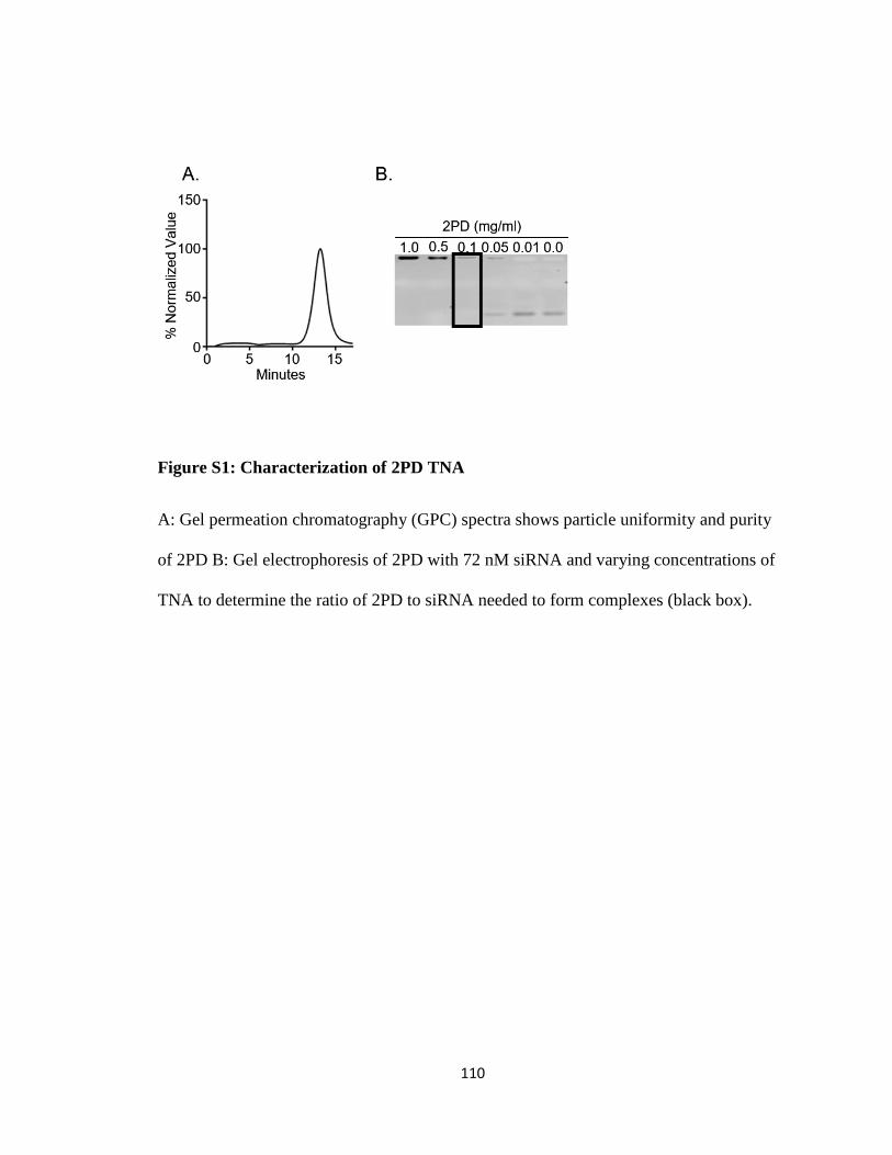
110
Figure S1: Characterization of 2PD TNA
A: Gel permeation chromatography (GPC) spectra shows particle uniformity and purity
of 2PD B: Gel electrophoresis of 2PD with 72 nM siRNA and varying concentrations of
TNA to determine the ratio of 2PD to siRNA needed to form complexes (black box).
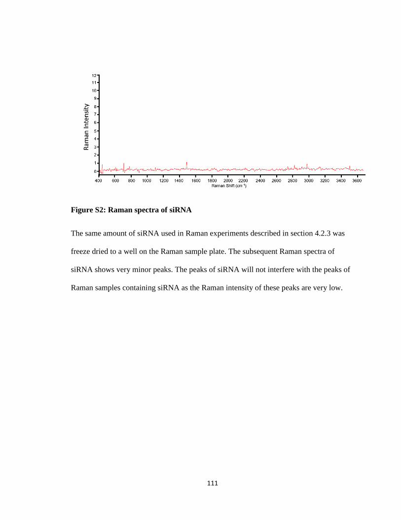
111
Figure S2: Raman spectra of siRNA
The same amount of siRNA used in Raman experiments described in section 4.2.3 was
freeze dried to a well on the Raman sample plate. The subsequent Raman spectra of
siRNA shows very minor peaks. The peaks of siRNA will not interfere with the peaks of
Raman samples containing siRNA as the Raman intensity of these peaks are very low.
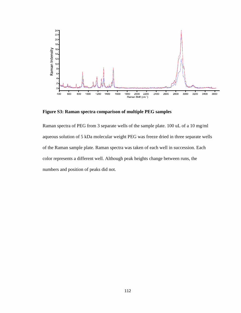
112
Figure S3: Raman spectra comparison of multiple PEG samples
Raman spectra of PEG from 3 separate wells of the sample plate. 100 uL of a 10 mg/ml
aqueous solution of 5 kDa molecular weight PEG was freeze dried in three separate wells
of the Raman sample plate. Raman spectra was taken of each well in succession. Each
color represents a different well. Although peak heights change between runs, the
numbers and position of peaks did not.
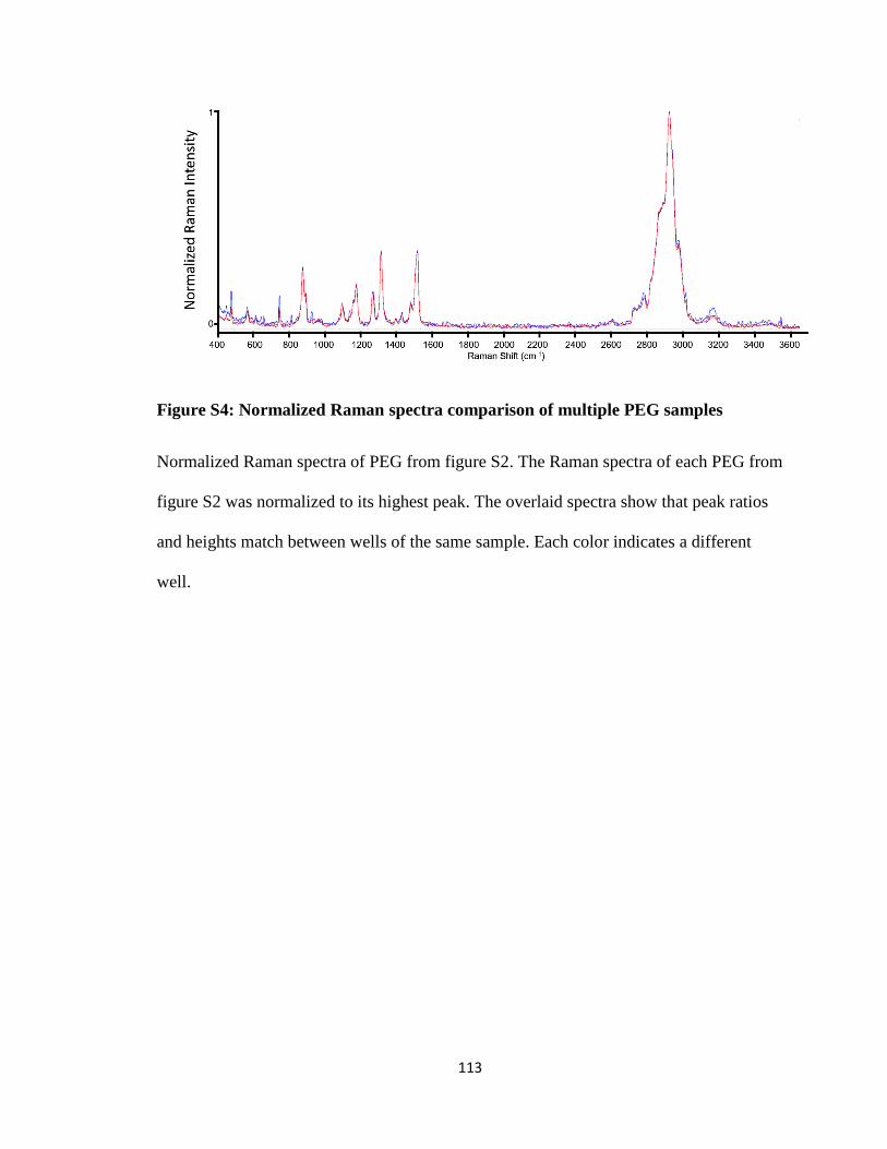
113
Figure S4: Normalized Raman spectra comparison of multiple PEG samples
Normalized Raman spectra of PEG from figure S2. The Raman spectra of each PEG from
figure S2 was normalized to its highest peak. The overlaid spectra show that peak ratios
and heights match between wells of the same sample. Each color indicates a different
well.
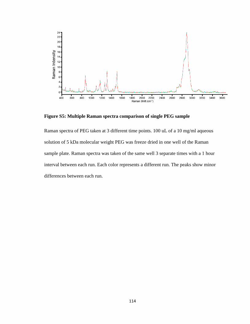
114
Figure S5: Multiple Raman spectra comparison of single PEG sample
Raman spectra of PEG taken at 3 different time points. 100 uL of a 10 mg/ml aqueous
solution of 5 kDa molecular weight PEG was freeze dried in one well of the Raman
sample plate. Raman spectra was taken of the same well 3 separate times with a 1 hour
interval between each run. Each color represents a different run. The peaks show minor
differences between each run.
115
7. References
1. Hanahan, D.; Weinberg, R. A., The hallmarks of cancer. Cell 2000, 100 (1), 57-
70.
2. Hanahan, D.; Weinberg, R. A., Hallmarks of cancer: the next generation. Cell
2011, 144, 646-74.
3. Pecot, C. V.; Calin, G. A.; Coleman, R. L.; Lopez-Berestein, G.; Sood, A. K.,
RNA interference in the clinic: challenges and future directions. Nat Rev Cancer 2011,
11 (1), 59-67.
4. Patil, S. D.; Rhodes, D. G.; Burgess, D. J., DNA-based therapeutics and DNA
delivery systems: a comprehensive review. AAPS J 2005, 7 (1), E61-77.
5. Friedmann, T. R., R., Gene therapy for human genetic disease? Science 1973,
949-55.
6. Brewster, L. P.; Brey, E. M.; Greisler, H. P., Cardiovascular gene delivery: The
good road is awaiting. Adv Drug Deliv Rev 2006, 58 (4), 604-29.
7. Ohlfest, J. R.; Demorest, Z. L.; Motooka, Y.; Vengco, I.; Oh, S.; Chen, E.;
Scappaticci, F. A.; Saplis, R. J.; Ekker, S. C.; Low, W. C.; Freese, A. B.; Largaespada, D.
A., Combinatorial antiangiogenic gene therapy by nonviral gene transfer using the
sleeping beauty transposon causes tumor regression and improves survival in mice
bearing intracranial human glioblastoma. Mol Ther 2005, 12 (5), 778-88.
8. Uherek, C.; Wels, W., DNA-carrier proteins for targeted gene delivery. Adv Drug
Deliv Rev 2000, 44 (2-3), 153-66.
9. Hackett, P. B.; Ekker, S. C.; Largaespada, D. A.; McIvor, R. S., Sleeping beauty
transposon-mediated gene therapy for prolonged expression. Adv Genet 2005, 54, 189-
232.
10. Kormann, M. S.; Hasenpusch, G.; Aneja, M. K.; Nica, G.; Flemmer, A. W.;
Herber-Jonat, S.; Huppmann, M.; Mays, L. E.; Illenyi, M.; Schams, A.; Griese, M.;
Bittmann, I.; Handgretinger, R.; Hartl, D.; Rosenecker, J.; Rudolph, C., Expression of
therapeutic proteins after delivery of chemically modified mRNA in mice. Nat Biotechnol
2011, 29 (2), 154-7.
11. Bangel-Ruland, N.; Tomczak, K.; Fernandez Fernandez, E.; Leier, G.;
Leciejewski, B.; Rudolph, C.; Rosenecker, J.; Weber, W. M., Cystic fibrosis
transmembrane conductance regulator-mRNA delivery: a novel alternative for cystic
fibrosis gene therapy. J Gene Med 2013, 15 (11-12), 414-26.
12. Lundin, K. E.; Gissberg, O.; Smith, C. I., Oligonucleotide Therapies: The Past
and the Present. Hum Gene Ther 2015, 26 (8), 475-85.
116
13. Stephenson, M. L.; Zamecnik, P. C., Inhibition of Rous sarcoma viral RNA
translation by a specific oligodeoxyribonucleotide. Proc Natl Acad Sci U S A 1978, 75
(1), 285-8.
14. Kole, R.; Krainer, A. R.; Altman, S., RNA therapeutics: beyond RNA interference
and antisense oligonucleotides. Nat Rev Drug Discov 2012, 11 (2), 125-40.
15. Fire, A., RNA-triggered gene silencing. Trends in Genetics 1999, 15, 358-363.
16. Fire, A.; Xu, S.; Montgomery, M.; Kostas, S., Potent and specific genetic
interference by double-stranded RNA in Caenorhabditis elegans. nature 1998, 391, 806-
811.
17. Hannon, G. J., RNA interference. Nature 2002, 418 (6894), 244-51.
18. Bartlett, D. W.; Davis, M. E., Insights into the kinetics of siRNA-mediated gene
silencing from live-cell and live-animal bioluminescent imaging. Nucleic acids research
2006, 34, 322-33.
19. Sunaga, N.; Shames, D. S.; Girard, L.; Peyton, M.; Larsen, J. E.; Imai, H.; Soh, J.;
Sato, M.; Yanagitani, N.; Kaira, K.; Xie, Y.; Gazdar, A. F.; Mori, M.; Minna, J. D.,
Knockdown of oncogenic KRAS in non-small cell lung cancers suppresses tumor growth
and sensitizes tumor cells to targeted therapy. Mol Cancer Ther 2011, 10 (2), 336-46.
20. Freire, J. M.; Rego de Figueiredo, I.; Valle, J.; Veiga, A. S.; Andreu, D.; Enguita,
F. J.; Castanho, M. A., siRNA-cell-penetrating peptides complexes as a combinatorial
therapy against chronic myeloid leukemia using BV173 cell line as model. J Control
Release 2017, 245, 127-136.
21. Bochicchio, S.; Dapas, B.; Russo, I.; Ciacci, C.; Piazza, O.; De Smedt, S.; Pottie,
E.; Barba, A. A.; Grassi, G., In vitro and ex vivo delivery of tailored siRNA-
nanoliposomes for E2F1 silencing as a potential therapy for colorectal cancer. Int J
Pharm 2017.
22. Rengaswamy, V.; Zimmer, D.; Suss, R.; Rossler, J., RGD liposome-protamine-
siRNA (LPR) nanoparticles targeting PAX3-FOXO1 for alveolar rhabdomyosarcoma
therapy. J Control Release 2016, 235, 319-27.
23. Layzer, J. M.; McCaffrey, A. P.; Tanner, A. K.; Huang, Z.; Kay, M. A.;
Sullenger, B. A., In vivo activity of nuclease-resistant siRNAs. RNA 2004, 10 (5), 766-
71.
24. Xu, C.-f.; Wang, J., Delivery systems for siRNA drug development in cancer
therapy. Asian Journal of Pharmaceutical Sciences 2015, 10 (1), 1-12.
25. Wittrup, A.; Lieberman, J., Knocking down disease: a progress report on siRNA
therapeutics. Nat Rev Genet 2015, 16 (9), 543-52.
117
26. Chiu, Y. L.; Rana, T. M., siRNA function in RNAi: a chemical modification
analysis. RNA 2003, 9 (9), 1034-48.
27. Harborth, J.; Elbashir, S. M.; Vandenburgh, K.; Manninga, H.; Scaringe, S. A.;
Weber, K.; Tuschl, T., Sequence, chemical, and structural variation of small interfering
RNAs and short hairpin RNAs and the effect on mammalian gene silencing. Antisense
Nucleic Acid Drug Dev 2003, 13 (2), 83-105.
28. Czauderna, F.; Fechtner, M.; Dames, S.; Aygun, H.; Klippel, A.; Pronk, G. J.;
Giese, K.; Kaufmann, J., Structural variations and stabilising modifications of synthetic
siRNAs in mammalian cells. Nucleic Acids Res 2003, 31 (11), 2705-16.
29. Whitehead, K. A.; Langer, R.; Anderson, D. G., Knocking down barriers:
advances in siRNA delivery. Nat Rev Drug Discov 2009, 8 (2), 129-38.
30. Anderson, W. F., Prospects for human gene therapy in the born and unborn
patient. Clin Obstet Gynecol 1986, 29 (3), 586-94.
31. McManus, M. T.; Sharp, P. A., Gene silencing in mammals by small interfering
RNAs. Nat Rev Genet 2002, 3 (10), 737-47.
32. Mali, S., Delivery systems for gene therapy. Indian J Hum Genet 2013, 19 (1), 3-
8.
33. Yu, J. Y.; DeRuiter, S. L.; Turner, D. L., RNA interference by expression of
short-interfering RNAs and hairpin RNAs in mammalian cells. Proc Natl Acad Sci U S A
2002, 99 (9), 6047-52.
34. Sibbald, B., Death but one unintended consequence of gene-therapy trial. CMAJ
2001, 164 (11), 1612.
35. Thomas, C. E.; Ehrhardt, A.; Kay, M. A., Progress and problems with the use of
viral vectors for gene therapy. Nat Rev Genet 2003, 4 (5), 346-58.
36. Kim, H. J.; Kim, A.; Miyata, K.; Kataoka, K., Recent progress in development of
siRNA delivery vehicles for cancer therapy. Adv Drug Deliv Rev 2016, 104, 61-77.
37. Sarett, S. M.; Nelson, C. E.; Duvall, C. L., Technologies for controlled, local
delivery of siRNA. J Control Release 2015, 218, 94-113.
38. Zhang, S.; Zhao, Y.; Zhi, D.; Zhang, S., Non-viral vectors for the mediation of
RNAi. Bioorganic chemistry 2012, 40, 10-8.
39. Singh, Y.; Tomar, S.; Khan, S.; Meher, J. G.; Pawar, V. K.; Raval, K.; Sharma,
K.; Singh, P. K.; Chaurasia, M.; Surendar Reddy, B.; Chourasia, M. K., Bridging small
interfering RNA with giant therapeutic outcomes using nanometric liposomes. J Control
Release 2015, 220 (Pt A), 368-87.
118
40. Ozpolat, B.; Sood, A. K.; Lopez-Berestein, G., Liposomal siRNA nanocarriers for
cancer therapy. Advanced drug delivery reviews 2014, 66, 110-6.
41. Leung, A. K.; Tam, Y. Y.; Cullis, P. R., Lipid nanoparticles for short interfering
RNA delivery. Adv Genet 2014, 88, 71-110.
42. Hope, M. J., Enhancing siRNA delivery by employing lipid nanoparticles. Ther
Deliv 2014, 5 (6), 663-73.
43. Semple, S. C.; Akinc, A.; Chen, J.; Sandhu, A. P.; Mui, B. L.; Cho, C. K.; Sah, D.
W.; Stebbing, D.; Crosley, E. J.; Yaworski, E.; Hafez, I. M.; Dorkin, J. R.; Qin, J.; Lam,
K.; Rajeev, K. G.; Wong, K. F.; Jeffs, L. B.; Nechev, L.; Eisenhardt, M. L.; Jayaraman,
M.; Kazem, M.; Maier, M. A.; Srinivasulu, M.; Weinstein, M. J.; Chen, Q.; Alvarez, R.;
Barros, S. A.; De, S.; Klimuk, S. K.; Borland, T.; Kosovrasti, V.; Cantley, W. L.; Tam,
Y. K.; Manoharan, M.; Ciufolini, M. A.; Tracy, M. A.; de Fougerolles, A.; MacLachlan,
I.; Cullis, P. R.; Madden, T. D.; Hope, M. J., Rational design of cationic lipids for siRNA
delivery. Nat Biotechnol 2010, 28 (2), 172-6.
44. Cheng, X.; Lee, R. J., The role of helper lipids in lipid nanoparticles (LNPs)
designed for oligonucleotide delivery. Adv Drug Deliv Rev 2016, 99 (Pt A), 129-37.
45. Watanabe, K.; Harada-Shiba, M.; Suzuki, A.; Gokuden, R.; Kurihara, R.; Sugao,
Y.; Mori, T.; Katayama, Y.; Niidome, T., In vivo siRNA delivery with dendritic poly(L-
lysine) for the treatment of hypercholesterolemia. Mol Biosyst 2009, 5 (11), 1306-10.
46. Hobel, S.; Aigner, A., Polyethylenimine (PEI)/siRNA-mediated gene knockdown
in vitro and in vivo. Methods Mol Biol 2010, 623, 283-97.
47. Boussif, O.; Lezoualc'h, F.; Zanta, M. A.; Mergny, M. D.; Scherman, D.;
Demeneix, B.; Behr, J. P., A versatile vector for gene and oligonucleotide transfer into
cells in culture and in vivo: polyethylenimine. Proc Natl Acad Sci U S A 1995, 92 (16),
7297-301.
48. Singha, K.; Namgung, R.; Kim, W. J., Polymers in small-interfering RNA
delivery. Nucleic Acid Ther 2011, 21 (3), 133-47.
49. Rezvani Amin, Z.; Rahimizadeh, M.; Eshghi, H.; Dehshahri, A.; Ramezani, M.,
The effect of cationic charge density change on transfection efficiency of
polyethylenimine. Iran J Basic Med Sci 2013, 16 (2), 150-6.
50. Ma, D., Enhancing endosomal escape for nanoparticle mediated siRNA delivery.
Nanoscale 2014, 6 (12), 6415-25.
51. Sun, X.; Zhang, N., Cationic polymer optimization for efficient gene delivery.
Mini Rev Med Chem 2010, 10 (2), 108-25.
119
52. Gary, D. J.; Puri, N.; Won, Y. Y., Polymer-based siRNA delivery: perspectives on
the fundamental and phenomenological distinctions from polymer-based DNA delivery. J
Control Release 2007, 121 (1-2), 64-73.
53. Ziebarth, J. D.; Wang, Y., Understanding the protonation behavior of linear
polyethylenimine in solutions through Monte Carlo simulations. Biomacromolecules
2010, 11 (1), 29-38.
54. Ding, Y.; Jiang, Z.; Saha, K.; Kim, C. S.; Kim, S. T.; Landis, R. F.; Rotello, V.
M., Gold nanoparticles for nucleic acid delivery. Mol Ther 2014, 22 (6), 1075-83.
55. Yang, S.; May, S., Release of cationic polymer-DNA complexes from the
endosome: A theoretical investigation of the proton sponge hypothesis. J Chem Phys
2008, 129 (18), 185105.
56. Hong, S.; Leroueil, P. R.; Janus, E. K.; Peters, J. L.; Kober, M. M.; Islam, M. T.;
Orr, B. G.; Baker, J. R., Jr.; Banaszak Holl, M. M., Interaction of polycationic polymers
with supported lipid bilayers and cells: nanoscale hole formation and enhanced
membrane permeability. Bioconjug Chem 2006, 17 (3), 728-34.
57. Benjaminsen, R. V.; Mattebjerg, M. A.; Henriksen, J. R.; Moghimi, S. M.;
Andresen, T. L., The possible "proton sponge " effect of polyethylenimine (PEI) does not
include change in lysosomal pH. Mol Ther 2013, 21 (1), 149-57.
58. Gauthier, M. A.; Gibson, M. I.; Klok, H. A., Synthesis of functional polymers by
post-polymerization modification. Angew Chem Int Ed Engl 2009, 48 (1), 48-58.
59. Figueroa, E. R.; Lin, A. Y.; Yan, J.; Luo, L.; Foster, A. E.; Drezek, R. A.,
Optimization of PAMAM-gold nanoparticle conjugation for gene therapy. Biomaterials
2014, 35 (5), 1725-34.
60. Kievit, F. M.; Zhang, M., Cancer nanotheranostics: improving imaging and
therapy by targeted delivery across biological barriers. Adv Mater 2011, 23 (36), H217-
47.
61. Choi, H. S.; Liu, W.; Misra, P.; Tanaka, E.; Zimmer, J. P.; Itty Ipe, B.; Bawendi,
M. G.; Frangioni, J. V., Renal clearance of quantum dots. Nat Biotechnol 2007, 25 (10),
1165-70.
62. Ward, C. M.; Read, M. L.; Seymour, L. W., Systemic circulation of poly(L-
lysine)/DNA vectors is influenced by polycation molecular weight and type of DNA:
differential circulation in mice and rats and the implications for human gene therapy.
Blood 2001, 97 (8), 2221-9.
63. Xue, H. Y.; Liu, S.; Wong, H. L., Nanotoxicity: a key obstacle to clinical
translation of siRNA-based nanomedicine. Nanomedicine (Lond) 2014, 9 (2), 295-312.
120
64. Fischer, D.; Bieber, T.; Li, Y.; Elsasser, H. P.; Kissel, T., A novel non-viral vector
for DNA delivery based on low molecular weight, branched polyethylenimine: effect of
molecular weight on transfection efficiency and cytotoxicity. Pharm Res 1999, 16 (8),
1273-9.
65. Godbey, W. T.; Wu, K. K.; Mikos, A. G., Poly(ethylenimine)-mediated gene
delivery affects endothelial cell function and viability. Biomaterials 2001, 22 (5), 471-80.
66. Park, T. G.; Jeong, J. H.; Kim, S. W., Current status of polymeric gene delivery
systems. Adv Drug Deliv Rev 2006, 58 (4), 467-86.
67. Jeong, J. H.; Kim, S. W.; Park, T. G., Molecular design of functional polymers for
gene therapy. Progress in Polymer Science 2007, 32 (11), 1239-1274.
68. Rehman, Z.; Zuhorn, I. S.; Hoekstra, D., How cationic lipids transfer nucleic acids
into cells and across cellular membranes: recent advances. J Control Release 2013, 166
(1), 46-56.
69. Toy, R.; Roy, K., Engineering nanoparticles to overcome barriers to
immunotherapy. Bioengineering & Translational Medicine 2016, 1 (1), 47-62.
70. Burke, R. S.; Pun, S. H., Extracellular barriers to in Vivo PEI and PEGylated PEI
polyplex-mediated gene delivery to the liver. Bioconjug Chem 2008, 19 (3), 693-704.
71. Plank, C.; Mechtler, K.; Szoka, F. C., Jr.; Wagner, E., Activation of the
complement system by synthetic DNA complexes: a potential barrier for intravenous
gene delivery. Hum Gene Ther 1996, 7 (12), 1437-46.
72. Chollet, P.; Favrot, M. C.; Hurbin, A.; Coll, J. L., Side-effects of a systemic
injection of linear polyethylenimine-DNA complexes. J Gene Med 2002, 4 (1), 84-91.
73. Lv, H.; Zhang, S.; Wang, B.; Cui, S.; Yan, J., Toxicity of cationic lipids and
cationic polymers in gene delivery. Journal of controlled release : official journal of the
Controlled Release Society 2006, 114, 100-9.
74. Merkel, O. M.; Beyerle, A.; Beckmann, B. M.; Zheng, M.; Hartmann, R. K.;
Stöger, T.; Kissel, T. H., Polymer-related off-target effects in non-viral siRNA delivery.
Biomaterials 2011, 32, 2388-98.
75. Patil, Y.; Panyam, J., Polymeric nanoparticles for siRNA delivery and gene
silencing. International journal of pharmaceutics 2009, 367, 195-203.
76. Moghimi, S. M.; Symonds, P.; Murray, J. C.; Hunter, A. C.; Debska, G.;
Szewczyk, A., A two-stage poly(ethylenimine)-mediated cytotoxicity: implications for
gene transfer/therapy. Mol Ther 2005, 11 (6), 990-5.
121
77. Kircheis, R.; Schuller, S.; Brunner, S.; Ogris, M.; Heider, K. H.; Zauner, W.;
Wagner, E., Polycation-based DNA complexes for tumor-targeted gene delivery in vivo.
J Gene Med 1999, 1 (2), 111-20.
78. Patnaik, S.; Aggarwal, A.; Nimesh, S.; Goel, A.; Ganguli, M.; Saini, N.; Singh,
Y.; Gupta, K. C., PEI-alginate nanocomposites as efficient in vitro gene transfection
agents. J Control Release 2006, 114 (3), 398-409.
79. Maheshwari, A.; Mahato, R. I.; McGregor, J.; Han, S.; Samlowski, W. E.; Park, J.
S.; Kim, S. W., Soluble biodegradable polymer-based cytokine gene delivery for cancer
treatment. Mol Ther 2000, 2 (2), 121-30.
80. Liu, Z.; Zhang, Z.; Zhou, C.; Jiao, Y., Hydrophobic modifications of cationic
polymers for gene delivery. Progress in Polymer Science 2010, 35, 1144-1162.
81. Incani, V.; Lavasanifar, A.; Uludag, H., Lipid and hydrophobic modification of
cationic carriers on route to superior gene vectors. Soft Matter 2010, 6 (10), 2124-2138.
82. Kataoka, K.; Harada, A.; Nagasaki, Y., Block copolymer micelles for drug
delivery: design, characterization and biological significance. Adv Drug Deliv Rev 2001,
47 (1), 113-31.
83. Suk, J. S.; Xu, Q.; Kim, N.; Hanes, J.; Ensign, L. M., PEGylation as a strategy for
improving nanoparticle-based drug and gene delivery. Adv Drug Deliv Rev 2016, 99 (Pt
A), 28-51.
84. Knop, K.; Hoogenboom, R.; Fischer, D.; Schubert, U. S., Poly(ethylene glycol) in
drug delivery: pros and cons as well as potential alternatives. Angewandte Chemie
(International ed. in English) 2010, 49, 6288-308.
85. Xie, Z.; Ji, Z.; Zhang, Z.; Gong, T.; Sun, X., Adenoviral vectors coated with
cationic PEG derivatives for intravaginal vaccination against HIV-1. Biomaterials 2014,
35 (27), 7896-908.
86. Mishra, S.; Webster, P.; Davis, M. E., PEGylation significantly affects cellular
uptake and intracellular trafficking of non-viral gene delivery particles. Eur J Cell Biol
2004, 83 (3), 97-111.
87. Hatakeyama, H.; Akita, H.; Harashima, H., A multifunctional envelope type nano
device (MEND) for gene delivery to tumours based on the EPR effect: a strategy for
overcoming the PEG dilemma. Adv Drug Deliv Rev 2011, 63 (3), 152-60.
88. Abuchowski, A.; McCoy, J. R.; Palczuk, N. C.; van Es, T.; Davis, F. F., Effect of
covalent attachment of polyethylene glycol on immunogenicity and circulating life of
bovine liver catalase. J Biol Chem 1977, 252 (11), 3582-6.
89. Merdan, T.; Kunath, K.; Petersen, H.; Bakowsky, U.; Voigt, K. H.; Kopecek, J.;
Kissel, T., PEGylation of poly(ethylene imine) affects stability of complexes with
122
plasmid DNA under in vivo conditions in a dose-dependent manner after intravenous
injection into mice. Bioconjugate chemistry 2005, 16, 785-92.
90. Masotti, A.; Moretti, F.; Mancini, F.; Russo, G.; Di Lauro, N.; Checchia, P.;
Marianecci, C.; Carafa, M.; Santucci, E.; Ortaggi, G., Physicochemical and biological
study of selected hydrophobic polyethylenimine-based polycationic liposomes and their
complexes with DNA. Bioorganic & medicinal chemistry 2007, 15 (3), 1504-15.
91. Kuhn, P. S.; Levin, Y.; Barbosa, M. C., Charge inversion in DNA–amphiphile
complexes: possible application to gene therapy. Physica A: Statistical Mechanics and its
Applications 1999, 274 (1–2), 8-18.
92. Alshamsan, A.; Haddadi, A.; Incani, V.; Samuel, J.; Lavasanifar, A.; Uludag, H.,
Formulation and delivery of siRNA by oleic acid and stearic acid modified
polyethylenimine. Mol Pharm 2009, 6 (1), 121-33.
93. Wong, S. Y.; Pelet, J. M.; Putnam, D., Polymer systems for gene delivery—Past,
present, and future. Progress in Polymer Science 2007, 32 (8-9), 799-837.
94. Takigawa, D. Y.; Tirrell, D. A., Interactions of synthetic polymers with cell
membranes and model membrane systems. Part 6. Disruption of phospholipid packing by
branched poly(ethylenimine) derivatives. Macromolecules 1985, 18 (3), 338-342.
95. Rheiner, S.; Rychahou, P.; Bae, Y., Effects of the Lipophilic Core of Polymer
Nanoassemblies on Intracellular Delivery and Transfection of siRNA. AIMS Biophysics
2015, 2 (3), 284-302.
96. Resnier, P.; Montier, T.; Mathieu, V.; Benoit, J.-P.; Passirani, C., A review of the
current status of siRNA nanomedicines in the treatment of cancer. Biomaterials 2013, 34,
6429-43.
97. Oh, Y.-K.; Park, T. G., siRNA delivery systems for cancer treatment. Advanced
drug delivery reviews 2009, 61, 850-62.
98. Aliabadi, H. M.; Landry, B.; Sun, C.; Tang, T.; Uludag, H., Supramolecular
assemblies in functional siRNA delivery: where do we stand? Biomaterials 2012, 33 (8),
2546-69.
99. Guo, P.; Coban, O.; Snead, N. M.; Trebley, J.; Hoeprich, S.; Guo, S.; Shu, Y.,
Engineering RNA for targeted siRNA delivery and medical application. Advanced drug
delivery reviews 2010, 62, 650-66.
100. Sheikhi Mehrabadi, F.; Fischer, W.; Haag, R., Dendritic and lipid-based carriers
for gene/siRNA delivery (a review). Current Opinion in Solid State and Materials
Science 2012, 16, 310-322.
101. Gonzalez, H.; Hwang, S. J.; Davis, M. E., New class of polymers for the delivery
of macromolecular therapeutics. Bioconjugate chemistry 1999, 10, 1068-74.
123
102. Zhang, S.; Zhao, B.; Jiang, H.; Wang, B.; Ma, B., Cationic lipids and polymers
mediated vectors for delivery of siRNA. Journal of controlled release : official journal of
the Controlled Release Society 2007, 123, 1-10.
103. Ballarín-González, B.; Ebbesen, M. F.; Howard, K. A., Polycation-based
nanoparticles for RNAi-mediated cancer treatment. Cancer letters 2013.
104. Grayson, A. C.; Doody, A. M.; Putnam, D., Biophysical and structural
characterization of polyethylenimine-mediated siRNA delivery in vitro. Pharm Res 2006,
23 (8), 1868-76.
105. Wightman, L.; Kircheis, R.; Rössler, V.; Carotta, S.; Ruzicka, R.; Kursa, M.;
Wagner, E., Different behavior of branched and linear polyethylenimine for gene delivery
in vitro and in vivo. The journal of gene medicine 2001, 3, 362-72.
106. Akhtar, S.; Benter, I., Toxicogenomics of non-viral drug delivery systems for
RNAi: potential impact on siRNA-mediated gene silencing activity and specificity.
Advanced drug delivery reviews 2007, 59, 164-82.
107. Zuckerman, J. E.; Choi, C. H. J.; Han, H.; Davis, M. E., Polycation-siRNA
nanoparticles can disassemble at the kidney glomerular basement membrane.
Proceedings of the National Academy of Sciences of the United States of America 2012,
109, 3137-42.
108. Nel, A. E.; Madler, L.; Velegol, D.; Xia, T.; Hoek, E. M.; Somasundaran, P.;
Klaessig, F.; Castranova, V.; Thompson, M., Understanding biophysicochemical
interactions at the nano-bio interface. Nat Mater 2009, 8 (7), 543-57.
109. Albanese, A.; Tang, P. S.; Chan, W. C. W., The effect of nanoparticle size, shape,
and surface chemistry on biological systems. Annual review of biomedical engineering
2012, 14, 1-16.
110. He, C.; Hu, Y.; Yin, L.; Tang, C.; Yin, C., Effects of particle size and surface
charge on cellular uptake and biodistribution of polymeric nanoparticles. Biomaterials
2010, 31, 3657-66.
111. Zintchenko, A.; Philipp, A.; Dehshahri, A.; Wagner, E., Simple modifications of
branched PEI lead to highly efficient siRNA carriers with low toxicity. Bioconjugate
chemistry 2008, 19, 1448-55.
112. Pasche, S.; Voros, J.; Griesser, H. J.; Spencer, N. D.; Textor, M., Effects of ionic
strength and surface charge on protein adsorption at PEGylated surfaces. J Phys Chem B
2005, 109 (37), 17545-52.
113. Miteva, M.; Kirkbride, K. C.; Kilchrist, K. V.; Werfel, T. A.; Li, H.; Nelson, C.
E.; Gupta, M. K.; Giorgio, T. D.; Duvall, C. L., Tuning PEGylation of mixed micelles to
overcome intracellular and systemic siRNA delivery barriers. Biomaterials 2014.
124
114. Zhu, C.; Jung, S.; Luo, S.; Meng, F.; Zhu, X.; Gwan, T.; Zhong, Z., Biomaterials
Co-delivery of siRNA and paclitaxel into cancer cells by biodegradable cationic micelles
based on PDMAEMA – PCL – PDMAEMA triblock copolymers. Biomaterials 2010, 31,
2408-2416.
115. Kim, D.; Lee, D.; Jang, Y. L.; Chae, S. Y.; Choi, D.; Jeong, J. H.; Kim, S. H.,
Facial amphipathic deoxycholic acid-modified polyethyleneimine for efficient MMP-2
siRNA delivery in vascular smooth muscle cells. Eur J Pharm Biopharm 2012, 81 (1),
14-23.
116. Hong, J.; Ku, S. H.; Lee, M. S.; Jeong, J. H.; Mok, H.; Choi, D.; Kim, S. H.,
Cardiac RNAi therapy using RAGE siRNA/deoxycholic acid-modified polyethylenimine
complexes for myocardial infarction. Biomaterials 2014, 35, 7562-73.
117. Patel, M. M.; Anchordoquy, T. J., Contribution of hydrophobicity to
thermodynamics of ligand-DNA binding and DNA collapse. Biophys J 2005, 88 (3),
2089-103.
118. Kim, H. J.; Miyata, K.; Nomoto, T.; Zheng, M.; Kim, A.; Liu, X.; Cabral, H.;
Christie, R. J.; Nishiyama, N.; Kataoka, K., siRNA delivery from triblock copolymer
micelles with spatially-ordered compartments of PEG shell, siRNA-loaded intermediate
layer, and hydrophobic core. Biomaterials 2014, 35 (15), 4548-56.
119. Matsumoto, S.; Christie, R. J.; Nishiyama, N.; Miyata, K.; Ishii, A.; Oba, M.;
Koyama, H.; Yamasaki, Y.; Kataoka, K., Environment-responsive block copolymer
micelles with a disulfide cross-linked core for enhanced siRNA delivery.
Biomacromolecules 2009, 10 (1), 119-27.
120. Xia, W.; Wang, P.; Lin, C.; Li, Z.; Gao, X.; Wang, G.; Zhao, X., Bioreducible
polyethylenimine-delivered siRNA targeting human telomerase reverse transcriptase
inhibits HepG2 cell growth in vitro and in vivo. Journal of controlled release : official
journal of the Controlled Release Society 2012, 157, 427-36.
121. Nelson, C. E.; Kintzing, J. R.; Hanna, A.; Shannon, J. M.; Gupta, M. K.; Duvall,
C. L.; Al, N. E. T., Balancing cationic and hydrophobic content of PEGylated siRNA
polyplexes enhances endosome escape, stability, blood circulation time, and bioactivity
in vivo. ACS nano 2013, 7, 8870-80.
122. Petros, R. a.; DeSimone, J. M., Strategies in the design of nanoparticles for
therapeutic applications. Nature reviews. Drug discovery 2010, 9, 615-27.
123. Maeda, H.; Wu, J.; Sawa, T.; Matsumura, Y.; Hori, K., Tumor vascular
permeability and the EPR effect in macromolecular therapeutics: a review. J Control
Release 2000, 65 (1-2), 271-84.
124. Holzerny, P.; Ajdini, B.; Heusermann, W.; Bruno, K.; Schuleit, M.; Meinel, L.;
Keller, M., Biophysical properties of chitosan/siRNA polyplexes: profiling the
125
polymer/siRNA interactions and bioactivity. Journal of controlled release : official
journal of the Controlled Release Society 2012, 157, 297-304.
125. Guo, G.; Zhou, L.; Chen, Z.; Chi, W.; Yang, X.; Wang, W.; Zhang, B., Alkane-
modified low-molecular-weight polyethylenimine with enhanced gene silencing for
siRNA delivery. International journal of pharmaceutics 2013, 450, 44-52.
126. Canton, I.; Battaglia, G., Endocytosis at the nanoscale. Chemical Society Reviews
2012.
127. Noguchi, A.; Furuno, T.; Kawaura, C.; Nakanishi, M., Membrane fusion plays an
important role in gene transfection mediated by cationic liposomes. FEBS Lett 1998, 433
(1-2), 169-73.
128. Cevc, G.; Richardsen, H., Lipid vesicles and membrane fusion. Advanced Drug
Delivery Reviews 1999, 38, 207-232.
129. Akinc, A.; Thomas, M.; Klibanov, A. M.; Langer, R., Exploring
polyethylenimine-mediated DNA transfection and the proton sponge hypothesis. J Gene
Med 2005, 7 (5), 657-63.
130. Pack, D. W.; Hoffman, A. S.; Pun, S.; Stayton, P. S., Design and development of
polymers for gene delivery. Nat Rev Drug Discov 2005, 4 (7), 581-93.
131. Kurtulus, I.; Yilmaz, G.; Ucuncu, M.; Emrullahoglu, M.; Becer, C. R.; Bulmus,
V., A new proton sponge polymer synthesized by RAFT polymerization for intracellular
delivery of biotherapeutics. Polymer Chemistry 2014, 5 (5), 1593-1604.
132. Richard, I.; Thibault, M.; De Crescenzo, G.; Buschmann, M. D.; Lavertu, M.,
Ionization behavior of chitosan and chitosan-DNA polyplexes indicate that chitosan has a
similar capability to induce a proton-sponge effect as PEI. Biomacromolecules 2013, 14
(6), 1732-40.
133. Rheiner, S.; Bae, Y., Increased poly(ethylene glycol) density decreases
transfection efficacy of siRNA/poly(ethylene imine) complexes. AIMS Bioengineering
2016, 3 (4), 454-467.
134. Lachelt, U.; Wagner, E., Nucleic Acid Therapeutics Using Polyplexes: A Journey
of 50 Years (and Beyond). Chem Rev 2015, 115 (19), 11043-78.
135. Choudhury, S. R.; Hudry, E.; Maguire, C. A.; Sena-Esteves, M.; Breakefield, X.
O.; Grandi, P., Viral vectors for therapy of neurologic diseases. Neuropharmacology
2016.
136. Liu, Y.; Liu, Z.; Wang, Y.; Liang, Y. R.; Wen, X.; Hu, J.; Yang, X.; Liu, J.; Xiao,
S.; Cheng, D., Investigation of the performance of PEG-PEI/ROCK-II-siRNA complexes
for Alzheimer's disease in vitro. Brain Res 2013, 1490, 43-51.
126
137. Aliabadi, H. M.; Maranchuk, R.; Kucharski, C.; Mahdipoor, P.; Hugh, J.; Uludağ,
H., Effective response of doxorubicin-sensitive and -resistant breast cancer cells to
combinational siRNA therapy. Journal of controlled release : official journal of the
Controlled Release Society 2013, 172, 219-28.
138. Dominska, M.; Dykxhoorn, D. M., Breaking down the barriers: siRNA delivery
and endosome escape. J Cell Sci 2010, 123 (Pt 8), 1183-9.
139. Lee, S. J. S.-Y. S.; Huh, M. S.; Lee, S. J. S.-Y. S.; Lee, S. J. S.-Y. S.; Chung, H.;
Park, J. H.; Oh, Y.-K.; Choi, K.; Kim, K.; Kwon, I. C., Stability and cellular uptake of
polymerized siRNA (poly-siRNA)/polyethylenimine (PEI) complexes for efficient gene
silencing. Journal of controlled release : official journal of the Controlled Release
Society 2010, 141, 339-46.
140. Varkouhi, A. K.; Scholte, M.; Storm, G.; Haisma, H. J., Endosomal escape
pathways for delivery of biologicals. Journal of controlled release : official journal of the
Controlled Release Society 2011, 151, 220-8.
141. Wen, S.; Zheng, F.; Shen, M.; Shi, X., Surface modification and PEGylation of
branched polyethyleneimine for improved biocompatibility. Journal of Applied Polymer
Science 2013, 128 (6), 3807-3813.
142. Ogris, M.; Steinlein, P.; Carotta, S.; Brunner, S.; Wagner, E.,
DNA/polyethylenimine transfection particles: influence of ligands, polymer size, and
PEGylation on internalization and gene expression. AAPS PharmSci 2001, 3 (3), E21.
143. Alexis, F.; Pridgen, E.; Molnar, L. K.; Farokhzad, O. C., Factors affecting the
clearance and biodistribution of polymeric nanoparticles. Mol Pharm 2008, 5 (4), 505-15.
144. Merkel, O. M.; Beyerle, A.; Librizzi, D.; Pfestroff, A.; Behr, T. M.; Sproat, B.;
Barth, P. J.; Kissel, T., Nonviral siRNA delivery to the lung: investigation of PEG-PEI
polyplexes and their in vivo performance. Mol Pharm 2009, 6 (4), 1246-60.
145. Mao, S.; Neu, M.; Germershaus, O.; Merkel, O.; Sitterberg, J.; Bakowsky, U.;
Kissel, T., Influence of polyethylene glycol chain length on the physicochemical and
biological properties of poly(ethylene imine)-graft-poly(ethylene glycol) block
copolymer/SiRNA polyplexes. Bioconjugate chemistry 2006, 17, 1209-18.
146. Walker, G. F.; Fella, C.; Pelisek, J.; Fahrmeir, J.; Boeckle, S.; Ogris, M.; Wagner,
E., Toward synthetic viruses: endosomal pH-triggered deshielding of targeted polyplexes
greatly enhances gene transfer in vitro and in vivo. Mol Ther 2005, 11 (3), 418-25.
147. Pandey, A. P.; Sawant, K. K., Polyethylenimine: A versatile, multifunctional non-
viral vector for nucleic acid delivery. Mater Sci Eng C Mater Biol Appl 2016, 68, 904-18.
148. Forsbach, A.; Müller, C.; Montino, C.; Kritzler, A.; Curdt, R.; Benahmed, A.;
Jurk, M.; Vollmer, J., Impact of delivery systems on siRNA immune activation and RNA
interference. Immunology Letters 2011, 141, 169-180.
127
149. Huang, F. W.; Wang, H. Y.; Li, C.; Wang, H. F.; Sun, Y. X.; Feng, J.; Zhang, X.
Z.; Zhuo, R. X., PEGylated PEI-based biodegradable polymers as non-viral gene vectors.
Acta Biomater 2010, 6 (11), 4285-95.
150. Fitzsimmons, R. E.; Uludag, H., Specific effects of PEGylation on gene delivery
efficacy of polyethylenimine: interplay between PEG substitution and N/P ratio. Acta
Biomater 2012, 8 (11), 3941-55.
151. Milla, P.; Dosio, F.; Cattel, L., PEGylation of proteins and liposomes: a powerful
and flexible strategy to improve the drug delivery. Curr Drug Metab 2012, 13 (1), 105-
19.
152. Tang, G. P.; Zeng, J. M.; Gao, S. J.; Ma, Y. X.; Shi, L.; Li, Y.; Too, H. P.; Wang,
S., Polyethylene glycol modified polyethylenimine for improved CNS gene transfer:
effects of PEGylation extent. Biomaterials 2003, 24 (13), 2351-62.
153. Petersen, H.; Fechner, P. M.; Martin, A. L.; Kunath, K.; Stolnik, S.; Roberts, C.
J.; Fischer, D.; Davies, M. C.; Kissel, T., Polyethylenimine-graft-poly(ethylene glycol)
copolymers: influence of copolymer block structure on DNA complexation and
biological activities as gene delivery system. Bioconjug Chem 2002, 13 (4), 845-54.
154. Holland, J. W.; Hui, C.; Cullis, P. R.; Madden, T. D., Poly(ethylene glycol)--lipid
conjugates regulate the calcium-induced fusion of liposomes composed of
phosphatidylethanolamine and phosphatidylserine. Biochemistry 1996, 35 (8), 2618-24.
155. Rheiner, S.; Reichel, D.; Rychahou, P.; Izumi, T.; Yang, H.-S.; Bae, Y., Polymer
nanoassemblies with hydrophobic pendant groups in the core induce false positive siRNA
transfection in luciferase reporter assays. International Journal of Pharmaceutics 2017,
528 (1–2), 536-546.
156. Lee, S. J.; Kim, M. J.; Kwon, I. C.; Roberts, T. M., Delivery strategies and
potential targets for siRNA in major cancer types. Adv Drug Deliv Rev 2016, 104, 2-15.
157. Davis, M. E.; Zuckerman, J. E.; Choi, C. H.; Seligson, D.; Tolcher, A.; Alabi, C.
A.; Yen, Y.; Heidel, J. D.; Ribas, A., Evidence of RNAi in humans from systemically
administered siRNA via targeted nanoparticles. Nature 2010, 464 (7291), 1067-70.
158. Kim, D. H.; Rossi, J. J., Strategies for silencing human disease using RNA
interference. Nat Rev Genet 2007, 8 (3), 173-84.
159. Malek, A.; Czubayko, F.; Aigner, A., PEG grafting of polyethylenimine (PEI)
exerts different effects on DNA transfection and siRNA-induced gene targeting efficacy.
J Drug Target 2008, 16 (2), 124-39.
160. Fella, C.; Walker, G. F.; Ogris, M.; Wagner, E., Amine-reactive
pyridylhydrazone-based PEG reagents for pH-reversible PEI polyplex shielding.
European journal of pharmaceutical sciences : official journal of the European
Federation for Pharmaceutical Sciences 2008, 34 (4-5), 309-20.
128
161. Helmfors, H.; Eriksson, J.; Langel, U., Optimized luciferase assay for cell-
penetrating peptide-mediated delivery of short oligonucleotides. Anal Biochem 2015,
484, 136-42.
162. Auld, D. S.; Thorne, N.; Nguyen, D. T.; Inglese, J., A specific mechanism for
nonspecific activation in reporter-gene assays. ACS Chem Biol 2008, 3 (8), 463-70.
163. Choy, G.; O'Connor, S.; Diehn, F. E.; Costouros, N.; Alexander, H. R.; Choyke,
P.; Libutti, S. K., Comparison of noninvasive fluorescent and bioluminescent small
animal optical imaging. BioTechniques 2003, 35, 1022-6, 1028-30.
164. Thorne, N.; Inglese, J.; Auld, D. S., Illuminating insights into firefly luciferase
and other bioluminescent reporters used in chemical biology. Chem Biol 2010, 17 (6),
646-57.
165. Reichel, D.; Lee, M. J.; Lee, W.; Kim, K. B.; Bae, Y., Tethered polymer
nanoassemblies for sustained carfilzomib release and prolonged suppression of
proteasome activity. Ther Deliv 2016, 7 (10), 665-681.
166. Dootz, R.; Nie, J.; Du, B.; Herminghaus, S.; Pfohl, T., Raman and surface
enhanced Raman microscopy of microstructured polyethylenimine/DNA multilayers.
Langmuir 2006, 22 (4), 1735-41.
167. York, S. S.; Boesch, S. E.; Wheeler, R. A.; Frech, R., Vibrational Assignments for
High Molecular Weight Linear Polyethylenimine (LPEI) Based on Monomeric and
Tetrameric Model Compounds. Macromolecules 2003, 36 (19), 7348-7351.
168. Romero, G.; Estrela-Lopis, I.; Zhou, J.; Rojas, E.; Franco, A.; Espinel, C. S.;
Fernández, A. G.; Gao, C.; Donath, E.; Moya, S. E., Surface Engineered Poly(lactide-co-
glycolide) Nanoparticles for Intracellular Delivery: Uptake and Cytotoxicity—A
Confocal Raman Microscopic Study. Biomacromolecules 2010, 11 (11), 2993-2999.
169. Spriggs, K. A.; Bushell, M.; Willis, A. E., Translational regulation of gene
expression during conditions of cell stress. Mol Cell 2010, 40 (2), 228-37.
170. O'Brien, J.; Wilson, I.; Orton, T.; Pognan, F., Investigation of the Alamar Blue
(resazurin) fluorescent dye for the assessment of mammalian cell cytotoxicity. European
Journal of Biochemistry 2000, 267 (17), 5421-5426.
171. Tiwari, B. S.; Belenghi, B.; Levine, A., Oxidative stress increased respiration and
generation of reactive oxygen species, resulting in ATP depletion, opening of
mitochondrial permeability transition, and programmed cell death. Plant Physiol 2002,
128 (4), 1271-81.
172. Cunha, D. A.; Hekerman, P.; Ladrière, L.; Bazarra-Castro, A.; Ortis, F.;
Wakeham, M. C.; Moore, F.; Rasschaert, J.; Cardozo, A. K.; Bellomo, E.; Overbergh, L.;
Mathieu, C.; Lupi, R.; Hai, T.; Herchuelz, A.; Marchetti, P.; Rutter, G. A.; Eizirik, D. L.;
129
Cnop, M., Initiation and execution of lipotoxic ER stress in pancreatic β-cells. Journal of
Cell Science 2008, 121 (14), 2308.
173. Koyama, T.; Kume, S.; Koya, D.; Araki, S.-i.; Isshiki, K.; Chin-Kanasaki, M.;
Sugimoto, T.; Haneda, M.; Sugaya, T.; Kashiwagi, A.; Maegawa, H.; Uzu, T., SIRT3
attenuates palmitate-induced ROS production and inflammation in proximal tubular cells.
Free Radical Biology and Medicine 2011, 51 (6), 1258-1267.
174. Suganami, T.; Tanimoto-Koyama, K.; Nishida, J.; Itoh, M.; Yuan, X.; Mizuarai,
S.; Kotani, H.; Yamaoka, S.; Miyake, K.; Aoe, S.; Kamei, Y.; Ogawa, Y., Role of the
Toll-like Receptor 4/NF-κB Pathway in Saturated Fatty Acid–Induced Inflammatory
Changes in the Interaction Between Adipocytes and Macrophages. Arteriosclerosis,
Thrombosis, and Vascular Biology 2006, 27 (1), 84.
175. Listenberger, L. L.; Ory, D. S.; Schaffer, J. E., Palmitate-induced apoptosis can
occur through a ceramide-independent pathway. J Biol Chem 2001, 276 (18), 14890-5.
176. Alcazar, O.; Qiu-yue, Z.; Gine, E.; Tamarit-Rodriguez, J., Stimulation of islet
protein kinase C translocation by palmitate requires metabolism of the fatty acid.
Diabetes 1997, 46 (7), 1153-8.
177. Aberle, A. M.; Tablin, F.; Zhu, J.; Walker, N. J.; Gruenert, D. C.; Nantz, M. H., A
novel tetraester construct that reduces cationic lipid-associated cytotoxicity. Implications
for the onset of cytotoxicity. Biochemistry 1998, 37 (18), 6533-40.
178. Hollins, A. J.; Omidi, Y.; Benter, I. F.; Akhtar, S., Toxicogenomics of drug
delivery systems: Exploiting delivery system-induced changes in target gene expression
to enhance siRNA activity. J Drug Target 2007, 15 (1), 83-8.
179. Omidi, Y.; Hollins, A. J.; Benboubetra, M.; Drayton, R.; Benter, I. F.; Akhtar, S.,
Toxicogenomics of non-viral vectors for gene therapy: a microarray study of lipofectin-
and oligofectamine-induced gene expression changes in human epithelial cells. J Drug
Target 2003, 11 (6), 311-23.
180. Omidi, Y.; Hollins, A. J.; Drayton, R. M.; Akhtar, S., Polypropylenimine
dendrimer-induced gene expression changes: the effect of complexation with DNA,
dendrimer generation and cell type. J Drug Target 2005, 13 (7), 431-43.
181. Ishii, M.; Maeda, A.; Tani, S.; Akagawa, M., Palmitate induces insulin resistance
in human HepG2 hepatocytes by enhancing ubiquitination and proteasomal degradation
of key insulin signaling molecules. Arch Biochem Biophys 2015, 566, 26-35.
182. Kirkin, V.; McEwan, D. G.; Novak, I.; Dikic, I., A role for ubiquitin in selective
autophagy. Mol Cell 2009, 34 (3), 259-69.
183. Yoshida, H., ER stress and diseases. FEBS J 2007, 274 (3), 630-58.
130
184. Shang, F.; Taylor, A., Ubiquitin-proteasome pathway and cellular responses to
oxidative stress. Free Radic Biol Med 2011, 51 (1), 5-16.
185. Orlowski, R. Z., The role of the ubiquitin-proteasome pathway in apoptosis. Cell
Death Differ 1999, 6 (4), 303-13.
186. Reichel, D.; Bae, Y., Comparison of Dialysis- and Solvatofluorochromism-Based
Methods to Determine Drug Release Rates from Polymer Nanoassemblies.
Pharmaceutical Research 2016, 1-14.
131
8. Vita
EDUCATION
B.S.E. in Chemical Engineering August 2007 - May 2011
University of Iowa, Iowa City, Iowa
Graduate Student in Pharmaceutical Sciences August 2011 - Current
University of Kentucky, Lexington, KY
Advisor: Dr. Younsoo Bae
PROFFESIONAL EXPERIENCE
USDA NCAUR Internship, Peoria, IL June 2010 – July 2010
Alcon Laboratories Internship, Ft. Worth, TX, June 2009 - July 2009
FELLOWSHIPS
• Cancer and Nanotechnology Training Center (CNTC) Fellowship, University of
Kentucky
o February 2015 – September 2015
PUBLICATIONS
• Jackson, M.A., Appell, M.D., Blackburn, J.A., Rheiner, S.N., Berhow, M.A.
2011. The acrylation of glycerol: A precursor to functionalized lipids. Journal of
the American Oil Chemists' Society. 89(4):713-719. DOI: 10.1007/s11746-011-
1950-5.
• Rheiner, S., Rychahou, P., Bae, Y., 2015. Effects of the Lipophilic Core of
Polymer Nanoassemblies on Intracellular Delivery and Transfection of siRNA,
AIMS Biophysics 2(3): 284-302, DOI: 10.3934/biophy.2015.3.284
• Yuhang Jiang, Phonepasong Arounleut, Steven Rheiner, Younsoo Bae,
Alexander V. Kabanov, Carol Milligan, Devika S. Manickam, SOD1 nanozyme
with reduced toxicity and MPS accumulation, Journal of Controlled Release,
Volume 231, 10 June 2016, Pages 38-49, ISSN 0168-3659,
http://dx.doi.org/10.1016/j.jconrel.2016.02.038.
132
• Rheiner, S., Bae, Y., 2016. Increased poly(ethylene glycol) density decreases
transfection efficacy of siRNA/poly(ethylene imine) complexes, AIMS
Bioengineering 3(4):454-467, DOI: 10.3934/bioeng.2016.4.454
• Steven Rheiner, Derek Reichel, Piotr Rychahou, Tadahide Izumi, Hsin-Sheng
Yang, Younsoo Bae, Polymer nanoassemblies with hydrophobic pendant groups
in the core induce false positive siRNA transfection in luciferase reporter assays,
International Journal of Pharmaceutics, Volume 528, Issues 1–2, 7 August 2017,
Pages 536-546, ISSN 0378-5173, https://doi.org/10.1016/j.ijpharm.2017.06.056.
PRESENTATIONS
• S. Rheiner, Y. Bae (2012) Cross-linked Nanoassemblies for Controlled siRNA
Delivery. University of Kentucky PS Seminar Series, Lexington, KY.
• S. Rheiner, Y. Bae (2013) Crosslinked Nanoassemblies for Delivery of siRNA in
Tumors. University of Kentucky PS Seminar Series, Lexington, KY.
• S. Rheiner, Y. Bae (2014) Polymer Tethered Nano-assemblies for siRNA
Delivery. University of Kentucky PS Seminar Series, Lexington, KY.
• S. Rheiner, Y. Bae (2015) Controlled siRNA Delivery for Treatment of Cancer.
University of Kentucky PS Seminar Series, Lexington, KY.
• S. Rheiner, Y. Bae (2015) Tethered Nanoassemblies for Enhanced siRNA
Delivery in vivo. Cancer and Nanotechnology Training Center Seminar Series,
Lexington, KY.
• S. Rheiner, Y. Bae (2015) Tethered Nanoassemblies for Enhanced Delivery and
Transfection of siRNA. Drug Discovery and Development Symposium,
Lexington, KY.
POSTERS
• S. Rheiner, Y. Bae (2012) Cross-linked Nano Assemblies for siRNA Delivery.
Pharmaceutics Graduate Student Research Meeting (PGSRM), Omaha, NB.
• S. Rheiner, Y. Bae (2012) Disulfide Cross-linked Nanoassemblies for siRNA
Delivery. American Association of Pharmaceutical Scientists (AAPS), Chicago,
IL.
• S. Rheiner, Y. Bae (2013) Crosslinked Nanoassemblies for In Vivo siRNA
Delivery to Solid Tumors. 2nd International Conference of RNA Nanotechnology
and Therapeutics, Lexington, KY.
• S. Rheiner, Y. Bae (2013) Crosslinked Nanoassemblies for siRNA Therapy.
Markey Cancer Center Research Day, Lexington, KY.
• S. Rheiner, Y. Bae (2013) Crosslinked Nanoassemblies for Delivery of siRNA.
Pharmaceutics Graduate Student Research Meeting (PGSRM), Iowa City, IA.
• S. Rheiner, Y. Bae (2014) Polymer Tethered Nano-assemblies for siRNA
Delivery. Markey Center Research Day, Lexington, KY.
133
• S. Rheiner, Y. Bae (2014) Modified Polymer Tethered Nano-assemblies for
siRNA DeliveryPharmaceutics Graduate Student Research Meeting (PGSRM),
Chicago, IL.
• S. Rheiner, Y. Bae (2014) Polymer Tethered Nano-assemblies for siRNA
Delivery. Drug Development and Discovery Symposium. Lexington, KY.
• S. Rheiner, Y. Bae (2014) Polymer Tethered Nano-assemblies for siRNA
Delivery. Biomaterials Day. Lexington, KY.
• M. Takahashi, S. Rheiner, Y. Bae (2015) Chemosensitization of triple negative
breast cancer by survivin protein reduction, Markey Cancer Center Research Day,
Lexington, KY.
• S. Rheiner, P. Rychahou, Y. Bae (2015) Lipophilic Core Modification of Polymer
Nanoassemblies for siRNA Delivery Markey Cancer Center Research Day,
Lexington, KY.
• S. Rheiner, Y. Bae (2015) Nanoassemblies for Enhanced In Vivo siRNA Delivery
and Transfection, 47th Pharmaceutics Graduate Student Research meeting,
Lexington, KY.
PROFESSIONAL SOCIETIES
AAPS (2012-Current)
American Association of Pharmaceutical Sciences
AAPS, University of Kentucky Chapter (2013- 2016)
Officer Position as Graduate Program Committee Representative. Helped in recruitment
of new students. Participated in events, such as E-Day and others where AAPS had a
presence, to represent AAPS. Attends Conferences for AAPS.
AICHE (2007-2011)
American Institute of Chemical Engineers
AICHE University of Iowa Student Chapter (2007-2011)
Officer Position (2009-2011) as Leader of the University of Iowa’s ChemE Car team.
Assisted in E Day. Helped AICHE Student Chapter at multiple events. Attended AICHE
Regional and National Conferences as an Officer.
ORGANIZATIONAL_COMMITTEES________________________________________
Pharmaceutics Graduate Student Research Meeting (PGSRM) 2015 Planning
Committee (2012-2015)
Co-Head Chair with responsibility of planning the 47th annual conference hosted in
Lexington, KY by The University of Kentucky. Responsible for choosing speakers,
organizing activities, booking venues, and managing day of events.
134
TEACHING EXPERIENCE_________________________________________________
University of Iowa (2010-2011)
Teaching Assistantship Material Science Lab (057-171)
University of Kentucky (2011-2012)
Teaching Assistantship Introduction to Pharmaceutical Sciences (PHS-914/924)
University of Kentucky (2012-2013)
Teaching Assistantship Introduction to Pharmaceutical Sciences (PHS-914/924)
University of Kentucky (2013-2014)
Proctor for College of Pharmacy Classes
University of Kentucky (2014-2015)
Proctor for College of Pharmacy Classes
AWARDS & FELLOWSHIPS_______________________________________________
• University of Kentucky College of Pharmacy Travel Award
o To Attend AAPS Conference (2012)
o To Attend PGSRM Conference (2013)
o To Attend PGSRM Conference (2014)
• Fellowship to attend 2nd International Conference of RNA Nanotechnology and
Therapeutics (2014)