Embed Size (px)
Citation preview
JPET #163444
1
Pharmacology and Antitumor Activity of ABC294640, a
Selective Inhibitor of Sphingosine Kinase-2
Kevin J. French, Yan Zhuang, Lynn W. Maines, Peng Gao, Wenxue Wang, Vladimir Beljanski,
John J. Upson, Cecelia L. Green, Staci N. Keller and Charles D. Smith
Apogee Biotechnology Corporation, Hummelstown, Pennsylvania (KJF1, YZ, LWM, JJU1, CLG,
SNK and CDS)
Department of Pharmaceutical and Biomedical Sciences, Medical University of South Carolina,
Charleston, SC (PG, WW, VB and CDS)
JPET Fast Forward. Published on January 8, 2010 as DOI:10.1124/jpet.109.163444
Copyright 2010 by the American Society for Pharmacology and Experimental Therapeutics.
This article has not been copyedited and formatted. The final version may differ from this version.JPET Fast Forward. Published on January 8, 2010 as DOI: 10.1124/jpet.109.163444
at ASPE
T Journals on February 12, 2018
jpet.aspetjournals.orgD
ownloaded from
JPET #163444
2
Running title: Antitumor Sphingosine Kinase Inhibitor
Correspondence: Charles D. Smith, Apogee Biotechnology Corporation, 1214 Research Blvd,
Suite 1016, Hummelstown, PA 17036. Phone: (843) 792-3240; Fax: (843)792-9588; E-mail:
Number of text pages: 34
Number of tables: 5
Number of figures: 8
Number of references: 40
Number of words in Abstract: 249
Number of words in Introduction: 509
Number of words in Discussion: 1433
Abbreviations: ABC294649, 3-(4-chlorophenyl)-adamantane-1-carboxylic acid (pyridin-4-
ylmethyl)amide; BSA, bovine serum albumin; DMS, N,N-dimethylsphingosine; IC50,
concentration that inhibits by 50%; NBD-Sph, omega(7-nitro-2-1,3-benzoxadiazol-4-
yl)(2S,3R,4E)-2-aminooctadec-4-ene-1,3-diol; PBS, phosphate-buffered saline; PEG,
polyethylene glycol; S1P, sphingosine 1-phosphate; SK1, sphingosine kinase-1; SK2,
sphingosine kinase-2.
Recommended Section Assignment: Chemotherapy, Antibiotics and Gene Therapy
This article has not been copyedited and formatted. The final version may differ from this version.JPET Fast Forward. Published on January 8, 2010 as DOI: 10.1124/jpet.109.163444
at ASPE
T Journals on February 12, 2018
jpet.aspetjournals.orgD
ownloaded from
JPET #163444
3
Abstract
Sphingolipid-metabolizing enzymes control the dynamic balance of the cellular levels of
important bioactive lipids, including the apoptotic compound ceramide and the proliferative
compound sphingosine 1-phosphate (S1P). Many growth factors and inflammatory cytokines
promote the cleavage of sphingomyelin and ceramide leading to rapid elevation of S1P levels
through the action of sphingosine kinases (SK1 and SK2). SK1 and SK2 are overexpressed in
a variety of human cancers, making these enzymes potential molecular targets for cancer
therapy. We have identified an aryladamantane compound, termed ABC294640, that
selectively inhibits SK2 activity in vitro, acting as a competitive inhibitor with respect to
sphingosine with a Ki of 9.8 µM, and attenuates S1P formation in intact cells. In tissue culture,
ABC294640 suppresses the proliferation of a broad panel of tumor cell lines, and inhibits tumor
cell migration concomitant with loss of microfilaments. In vivo, ABC294640 has excellent oral
bioavailability, and demonstrates a plasma half-time of clearance of 4.5 hr in mice. Acute and
chronic toxicology studies indicate that ABC294640 induces a transient minor decrease in the
hematocrit of rats and mice; however, this normalizes by 28 days of treatment. No other
changes in hematology parameters, or gross or microscopic tissue pathology result from
treatment with ABC294640. Oral administration of ABC294640 to mice bearing mammary
adenocarcinoma xenografts results in dose-dependent antitumor activity associated with
depletion of S1P levels in the tumors and progressive tumor cell apoptosis. Therefore, this
newly developed SK2 inhibitor provides an orally-available drug candidate for the treatment of
cancer and other diseases.
This article has not been copyedited and formatted. The final version may differ from this version.JPET Fast Forward. Published on January 8, 2010 as DOI: 10.1124/jpet.109.163444
at ASPE
T Journals on February 12, 2018
jpet.aspetjournals.orgD
ownloaded from
JPET #163444
4
Introduction
Sphingolipids have become a focal point in biological research, with excellent rationale
for their manipulation for the treatment of diseases, including cancer (reviewed in (Ogretmen,
2006; Cuvillier, 2007; Huwiler and Zangemeister-Wittke, 2007)). The parent lipid sphingomyelin
is a structural component of cellular membranes, but also serves as the precursor for potent
second messengers that have profound cellular effects. Stimulus-induced metabolism of these
lipids is critically involved in cancer cell biology and inflammatory diseases, and so this
metabolic pathway offers exciting new molecular targets for drug development.
In response to stimuli including growth factors and inflammatory cytokines,
sphingomyelin is enzymatically hydrolyzed to ceramide, which can be further hydrolyzed by the
action of ceramidase to produce sphingosine. Ceramide and sphingosine induce apoptosis in
cancer cells by mechanisms that remain to be elucidated. Sphingosine is rapidly
phosphorylated by sphingosine kinase (SK) to produce sphingosine 1-phosphate (S1P), which
is mitogenic and anti-apoptotic. Through these conversions, a critical balance, i.e. a ceramide /
S1P rheostat, has been hypothesized to determine the fate of the cell (Cuvillier et al., 1996). In
this model, the balance between the cellular concentrations of ceramide and S1P determines
whether a cell proliferates or undergoes apoptosis. Upon exposure to mitogens or activation of
intracellular oncoproteins, the cells experience an increase in the intracellular levels of S1P and
depletion of ceramide levels, and this situation promotes cell survival and proliferation. In
contrast, activation of sphingomyelinase in the absence of activation of ceramidase and/or SK
results in the accumulation of ceramide and subsequent apoptosis.
In spite of the high level of interest in sphingolipid-mediated signaling, there are very few
known inhibitors of the enzymes of this pathway. In particular, the field suffers from a lack of
potent and selective inhibitors of SK. Most pharmacological studies to date have used three
compounds to inhibit SK activity: N,N-dimethylsphingosine (DMS), D,L-threo-
dihydrosphingosine and N,N,N-trimethylsphingosine. However, these compounds are not
This article has not been copyedited and formatted. The final version may differ from this version.JPET Fast Forward. Published on January 8, 2010 as DOI: 10.1124/jpet.109.163444
at ASPE
T Journals on February 12, 2018
jpet.aspetjournals.orgD
ownloaded from
JPET #163444
5
specific inhibitors of SK as they have been shown to affect protein kinase C (Igarashi et al.,
1989), 3-phosphoinositide-dependent kinase (King et al., 2000), and casein kinase II (McDonald
et al., 1991). Additionally, the compound phenoxodiol has been described as an SK inhibitor
(Gamble et al., 2006); however, this isoflavone also inhibits several other enzymes. A few
natural product inhibitors of SK have been isolated (Kono et al., 2001), but their selectivities
remain unknown and their amenability to large-scale production is doubtful.
Clearly, inhibitors of SK that can be easily synthesized would be highly desirable for
evaluating this enzyme as a therapeutic target. To address this problem, we previously
identified and characterized structurally novel inhibitors of SK (French et al., 2003; French et al.,
2006). These compounds, called SKI-I, -II and –V, inhibited S1P formation in intact cells,
induced apoptosis, and demonstrated antitumor activity upon intraperitoneal administration
(French et al., 2006), reinforcing the approach of targeting SK in cancer. We report here the
pharmacologic characterization of a new orally-available SK inhibitor with in vivo activity.
Importantly, this compound is selective for SK2, thereby providing the first pharmacologic probe
to evaluate the biological roles of this SK isozyme.
This article has not been copyedited and formatted. The final version may differ from this version.JPET Fast Forward. Published on January 8, 2010 as DOI: 10.1124/jpet.109.163444
at ASPE
T Journals on February 12, 2018
jpet.aspetjournals.orgD
ownloaded from

JPET #163444
6
Methods
Materials. Unless otherwise noted, all chemicals and reagents were purchased from Sigma-
Aldrich (St. Louis, MO). Phalloidin -FITC were purchased from Invitrogen (Carlsbad, CA).
Fibronectin and cell culture inserts were purchased from BD Biosciences (San Jose, CA).
Recombinant human SK1 and SK2 were purchased from BPS Biosciences (San Diego, CA).
C17-sphingosine and C17-S1P were purchased from Avanti Polar Lipids, Inc. (Alabaster, AL).
All cell lines were obtained from the American Type Culture Collection and grown in either
DMEM or RPMI 1640 medium containing 10% fetal bovine serum and 50 µg/ml gentamycin
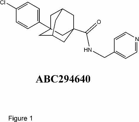
sulfate. ABC294640, 3-(4-chlorophenyl)-adamantane-1-carboxylic acid (pyridin-4-
ylmethyl)amide (Figure 1), was synthesized as described previously (Maines et al., 2008). The
hydrochloride salt, ABC294640 • HCl, was prepared by dissolving 9.6 g of ABC294640 in 50 mL
of CH2Cl2, and slowly adding an equimolar amount of 1M HCl in ether. After filtration and
washing, 9.6 g (93%) of ABC294640 • HCl was recovered as fine crystals with a melting point of
204 - 206 ºC.
Sphingosine kinase assays. The IC50s for ABC294640 and DMS were determined by a
newly-developed HPLC-based SK activity assay. Briefly, the test compounds were incubated
with recombinant SK1 or SK2 and omega(7-nitro-2-1,3-benzoxadiazol-4-yl)(2S,3R,4E)-2-
aminooctadec-4-ene-1,3-diol (NBD-Sph) in the isozyme-selective assay buffers detailed below
with 1 mg/ml fatty acid-free BSA, 100 µM ATP and 400 µM MgCl2. The product, i.e. NBD-S1P,
was separated from NBD-Sph by HPLC as follows: Waters 2795 HPLC system with a Waters
2495 fluorescence detector, C8 Chromolith RP-8e column (100 × 4.6 mm, Merck KGaA,
Germany), 1 ml/min mobile phase (acetonitrile : 20 mM (pH2.5) sodium phosphate buffer at 45
: 55). Fluorescence was monitored with excitation at 465 nm and emission at 531 nm. The ratio
of NBD-S1P / (NBD-Sph + NBD-S1P) was used as a measure of SK activity. SK-isozyme
selective assay buffers each contained 20 mM Tris pH7.4, 5 mM EDTA, 5 mM EGTA, 3 mM
This article has not been copyedited and formatted. The final version may differ from this version.JPET Fast Forward. Published on January 8, 2010 as DOI: 10.1124/jpet.109.163444
at ASPE
T Journals on February 12, 2018
jpet.aspetjournals.orgD
ownloaded from
JPET #163444
7
beta-mercaptoethanol, 5% glycerol, 1X protease inhibitors (Sigma, USA) and 1X phosphatase
inhibitors (Roche, USA). For the SK1 assay buffer, 0.25% (final) Triton X-100 was added; and
for the SK2 buffer, 1M (final) KCl was added. Assays were run for 2 hr at room temperature, and
then a 1.5 volume of methanol was added to terminate the kinase reaction. After 10 min, the
samples were centrifuged at 20,000 x g to pellet the precipitated protein, and the supernatants
were analyzed by HPLC. In experiments to determine the Ki for inhibition of SK2 by
ABC294640, the ADP Quest assay system (DiscoveRx Corporation, Fremont CA) was used to
measure kinase activity in the presence of varying concentrations of sphingosine and
ABC294640.
To determine the effects of ABC294640 on cellular SK activity, near-confluent MDA-MB-
231 cells were serum-starved overnight, and then treated with varying concentrations of
ABC294640. The cells were then incubated with [3H]sphingosine at a final concentration of 1
μM as previously described (French et al., 2006). The cells take up the exogenous sphingosine,
which is converted to S1P via SK activity, and [3H]S1P is separated from [3H]sphingosine by
extraction and quantified by scintillation counting.
Sphingolipid analyses. Biochemical analyses of ceramide species, sphingoid bases and their
phosphates were performed by the Lipidomics Shared Resource at the Medical University of
South Carolina on a Thermo Finnigan TSQ 7000, triple-stage quadurpole mass spectrometer
operating in a Multiple Reaction Monitoring positive ionization mode. Quantitative analyses of
the cellular sphingolipids were based on the calibration curves generated by spiking an artificial
matrix with known amounts of target standards and an equal amount of the internal standard.
The target analyte to internal standard peak area ratios from the samples were similarly
normalized to their respective internal standards and compared to the calibration curves by
linear regression. Final results were expressed as the level of the particular sphingolipid
This article has not been copyedited and formatted. The final version may differ from this version.JPET Fast Forward. Published on January 8, 2010 as DOI: 10.1124/jpet.109.163444
at ASPE
T Journals on February 12, 2018
jpet.aspetjournals.orgD
ownloaded from
JPET #163444
8
normalized the total phospholipids phosphate levels determined from the Bligh & Dyer lipid
extract (Bielawski et al., 2006).
Cytotoxicity assays. To determine the effects of the test compounds on proliferation, cells
were plated into 96-well microtiter plates and allowed to attach for 24 h. Varying concentrations
of ABC294640 were added to individual wells and the cells were incubated for an additional 72
h. At the end of this period, the number of viable cells was determined using the
sulforhodamine-binding assay. The percentage of cells killed was calculated as the percentage
decrease in sulforhodamine-binding compared with control cultures. Regression analyses of
inhibition curves were performed using GraphPad Prism (San Diego, CA).
Chemotaxis and cytoskeletal assays. Chemotaxis assays were performed as follows:
Transwell inserts (8 µm pore size) were pre-coated with fibronectin (5 µg/ml) for 1 h at room
temperature. A-498 cells were trypsinized and resuspended in serum-free media at a
concentration of 105 cells/ml. A total volume of 500 µl of this cell suspension was added to the
top of the insert and medium containing 10 % fetal bovine serum was placed at the bottom to
act as a chemoattractant. Equal concentrations of ABC294640 were added to the top and the
bottom of the chamber. After 4 h at 37 oC, cells that migrated through the membrane were fixed
in 4 % formaldehyde and stained with crystal violet. The number of migrating cells was
established by counting ten random microscope fields. Experiments were performed in
duplicate and repeated three times.
Microfilaments were stained with FITC-conjugated phalloidin (Invitrogen), according to
the manufacturer’s directions. Briefly, cells in chamber slides were exposed to the vehicle,
ABC294640 or cytochalasin B for various times, fixed in 3.7 % paraformaldehide, permeabilized
with 0.1% Triton-X 100, blocked with 2% BSA in TBST, and stained with 20 µl of phalloidin
This article has not been copyedited and formatted. The final version may differ from this version.JPET Fast Forward. Published on January 8, 2010 as DOI: 10.1124/jpet.109.163444
at ASPE
T Journals on February 12, 2018
jpet.aspetjournals.orgD
ownloaded from
JPET #163444
9
methanol solution per ml of blocking solution. After 12-16 h at 4 oC, the excess phalloidin was
removed by washing in TBST, cells were mounted and observed with a Zeiss LSM 510 laser
scanning confocal microscope. Cells were randomly selected and at least five different images
with multiple cells were taken per experimental point.
The G (globular) to F (fibrous) actin ratio was quantified to assess the status of
microfilaments. Briefly, cells were exposed to the drug for various times and actin was extracted
by washing the cells with 1% Triton X-100 solution in PBS. The two forms of actin were
separated by centrifugation at 14,000 x g. The supernatant (containing G actin) was mixed 1:1
with Laemmli buffer and the pellet (containing F actin) was dissolved in Laemmli buffer.
Samples were subjected to SDS-PAGE and the actin content of each fraction was quantified by
immunoblotting with anti-actin antibody and analyses using the ImageJ (NIH) program.
Quantification of ABC294640 in plasma and tumors. Plasma samples were prepared by
centrifugation (5000 x g, 5 min at 4 ºC) of whole blood that was collected into syringes
containing EDTA as an anticoagulant. Samples were spiked with 10 μg of an internal standard
(3-(4-chlorophenyl)adamantane-1-carboxylic acid [2-(3,4-dihydroxyphenyl)ethyl]amide), brought
to 1 mL with water and extracted three times with 2 mL of ethyl acetate. Extracts were dried
over nitrogen at 35 ºC, reconstituted in 0.2 mL of 0.1% formic acid in water/methanol (50:50,
Phase A), filtered and transferred to vials. Analyses were performed using an Agilent 1100
binary pump HPLC system coupled to a Finnigan LCQ Classic ion trap quadrupole mass
spectrometer running in ESI positive ion mode. Sample (10 μL) was injected and resolved
using a Supelco Discovery C18 column (2.1 x 20 mm, 5 mm particle size) connected to a
Zorbax precolumn (Agilent) with a mobile phase consisting of 0.1% formic acid in
water/methanol (50:50). The flow rate was 0.3 mL/min, and samples were eluted by a linear
gradient increasing from 50% to 100% methanol over 3 min. ABC294640 and the internal
standard were detected at 5.1 min and 5.5 min, respectively, using selected ion mode (m/z =
This article has not been copyedited and formatted. The final version may differ from this version.JPET Fast Forward. Published on January 8, 2010 as DOI: 10.1124/jpet.109.163444
at ASPE
T Journals on February 12, 2018
jpet.aspetjournals.orgD
ownloaded from
JPET #163444
10
381 and 426, respectively). Peak areas were integrated using Xcalibur software, and
ABC294640 concentrations determined from a standard curve, which was linear in range of all
plasma levels observed in these studies.
Oral bioavailability and pharmacokinetic studies. Formulations of ABC294640 • HCl were
administered orally or intravenously to fasted female Swiss-Webster mice at a dose of 100
mg/kg in 0.1 mL of the indicated solvents. Blood samples were removed at 1 and 7 hr after
dosing, and the plasma concentration of ABC294640 was determined by reverse-phase LC/MS
running in SIM mode as described above. For pharmacokinetic studies, female Swiss-Webster
mice (6-8 weeks old) were fasted overnight and administered a bolus dose of 0.1 mL of
ABC294640 • HCl either orally or intravenously. After dosing, mice were anesthetized with
halothane and blood was removed via intracardiac puncture at the indicated times. Plasma
samples were processed and ABC294640 levels were determined as described above.
Noncompartmental pharmacokinetic analyses were performed using WINNONLIN (Pharsight).
Toxicology studies. Acute (7-day) and chronic (28-day) toxicology studies were conducted
with ABC294640 • HCl. In the first study (which was conducted by Eurofins | Product Safety
Laboratories, Dayton, NJ), Sprague-Dawley male rats (7-8 weeks old) were orally dosed with 0,
100 or 250 mg of ABC294640 • HCl /kg in 0.375% Polysorbate-80 in PBS daily for 7 days. The
animals were observed daily for viability, signs of gross toxicity, and behavioral changes, and a
battery of detailed observations were performed on study Days 1 and 7. Blood was sampled
from all animals on Day 8 of the study for hematology, clinical biochemistry and serology
assessments, and the animals were sacrificed. Gross necropsies were performed on all study
rats, and selected organs and tissues were evaluated in the control and high-dose level groups.
In the second study, C57BL/6 mice were orally dosed with 0, 100 or 250 mg of ABC294640 •
This article has not been copyedited and formatted. The final version may differ from this version.JPET Fast Forward. Published on January 8, 2010 as DOI: 10.1124/jpet.109.163444
at ASPE
T Journals on February 12, 2018
jpet.aspetjournals.orgD
ownloaded from
JPET #163444
11
HCl /kg daily exactly as indicated above, and sacrificed at either Day 7 or Day 28 for
hematology studies.
Antitumor evaluation. A syngeneic mouse tumor model that uses a transformed murine
mammary adenocarcinoma cell line (JC, ATCC Number CRL-2116) and Balb/C mice (Charles
River, Wilmington, MA) was performed as previously described (Lee et al., 2003). Animal care
and procedures were in accordance with guidelines and regulations of the IACUC of the Penn
State College of Medicine. Animals were housed under 12 hr light/dark cycles, with food and
water provided ad libitum. Tumor cells (1 x 10⁶) were implanted subcutaneously, and tumor
volume was calculated using the equation: (L x W2)/2. Upon detection of tumors, mice were
randomized into treatment groups. Treatment was then administered every other day thereafter
consisting of oral doses of 3.5, 10, 35 or 100 mg ABC294640 • HCl / kg body weight or vehicle
(0.375% Polysorbate-80). Whole body weights and tumor volume measurements were
performed each day of treatment. On Day 15, mice were dosed, euthanized 1 hr later and
tumors were excised and immediately frozen. P-values were determined using one-way
ANOVA using GraphPad InStat (San Diego, CA).
Pharmacodynamic studies and tumor accumulation of ABC294640. Apoptosis was
measured in sections from tumors treated with ABC294640 • HCl using a TUNEL detection kit
according to the manufacturer’s instructions (In situ cell death detection kit, Roche, Germany).
Briefly, tumor sections were incubated with permeabilization solution (0.1% Triton X–100, 0.1%
sodium citrate, freshly prepared) for 8 min at room temperature and then washed twice with
PBS. Sections were incubated with TUNEL reaction mixture in a humid atmosphere at 37 °C for
60 min and mounted with crystal mounting medium. The amount of apoptosis was calculated
for an average of 10 microscopic fields in each sample (magnification ×100) and expressed as
the percentage of cells that were TUNEL-positive. For the analyses of sphingolipids, frozen
This article has not been copyedited and formatted. The final version may differ from this version.JPET Fast Forward. Published on January 8, 2010 as DOI: 10.1124/jpet.109.163444
at ASPE
T Journals on February 12, 2018
jpet.aspetjournals.orgD
ownloaded from
JPET #163444
12
tumor slices were homogenized in ice-cold PBS to a final concentration of 10 mg/mL. A 0.5 mL
aliquot of the homogenate was combined with 0.5 mL of methanol, 0.25 mL of chloroform, and
375 pmol each of internal standards C17-sphingosine and C17-S1P. Blank samples spiked with
known amounts of sphingosine, S1P and the internal standards were processed in parallel to
provide a standard curve for quantification. After sonication, samples were incubated overnight
at 48 ºC, followed by addition of 75 μL of 1N potassium hydroxide in methanol. The samples
were then sonicated and incubated at 37 ºC for 2 hr. A portion (0.4 mL) of each sample was
then transferred to a new tube, dried, reconstituted in 0.25 mL of Phase A, filtered and
transferred to a vial. HPLC was performed as described above. Elution was performed at 0.45
mL/min with 65% Phase B for 2 min followed by a linear gradient to 100% Phase B over 5 min.
Ions for C17-sphingosine, sphingosine, C17-S1P and S1P were monitored at m/z 286 (parent ion)
� 268 (daughter ion), 300 � 282, 366 � 250 and 380 � 264, respectively. Similarly, extracts
of tumors from ABC294640-treated mice were prepared and levels of ABC294640 were
quantified by LC/MS as described above.
This article has not been copyedited and formatted. The final version may differ from this version.JPET Fast Forward. Published on January 8, 2010 as DOI: 10.1124/jpet.109.163444
at ASPE
T Journals on February 12, 2018
jpet.aspetjournals.orgD
ownloaded from
JPET #163444
13
Results
Inhibition of sphingosine kinase activity by ABC294640. Screening of a chemical library
identified SK inhibitors containing an aryladamantane scaffold, and approximately 140
congeners were synthesized and tested for their ability inhibit a recombinant fusion protein of
GST and human SK1 (Smith et al., 2008). The structure-activity properties of this series will be
reported elsewhere. Although ABC294640 is not the most potent SK inhibitor in this series, its
biologic and pharmacologic properties were characterized because of its excellent solubility and
oral absorption (described below), and in vivo activity in models of diabetic retinopathy (Maines
et al., 2006) and ulcerative colitis (Maines et al., 2008).
In evaluating the potencies of these compounds, we developed an HPLC-based assay
for SK activity that avoids the use of radiolabeled substrate as we have previously described
(French et al., 2003). Using recombinant human SK1 and SK2, ABC294640 demonstrated
dose-dependent inhibition of SK2 with an IC50 of approximately 60 µM without affecting the
activity of SK1 at concentrations up to at least 100 µM (Figure 2A). In contrast, DMS inhibited
both SK1 and SK2 with IC50s of approximately 60 and 20 µM, respectively. Additional studies
demonstrated that both ABC294640 and DMS act as competitive inhibitors with respect to
sphingosine, making the IC50 strongly affected by the concentration of sphingosine used in the
assay. Kinetic analyses of varying concentrations of ABC294640 in the presence of 2.5 – 25
µM sphingosine indicated a Ki of 9.8 ± 1.4 µM for the inhibition of SK2 (Figure 2B).
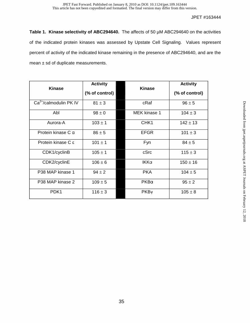
To assess its selectivity, ABC294640 was tested against a panel of serine / threonine
and tyrosine protein kinases, several of which are regulated by interactions with lipids. Each
kinase was incubated with its appropriate peptide substrate, ATP and 50 μM ABC294640.
Activities were normalized to vehicle-treated (DMSO) control reactions. As indicated in Table 1,
none of the other 20 diverse kinases tested were significantly inhibited by the compound.
These data suggest that the biological effects of this compound are not mediated by off-target
inhibition of protein kinases, consistent with its targeting of the sphingosine binding site of SK2.
This article has not been copyedited and formatted. The final version may differ from this version.JPET Fast Forward. Published on January 8, 2010 as DOI: 10.1124/jpet.109.163444
at ASPE
T Journals on February 12, 2018
jpet.aspetjournals.orgD
ownloaded from
JPET #163444
14
It was also important to determine the ability of ABC294640 to inhibit endogenous SK
activity in an intact cell model. Therefore, we used our previously described method in which
MDA-MB-231 cells are incubated with [3H]sphingosine at a final concentration of 1 μM (French
et al., 2003). The cells take up the exogenous sphingosine, which is converted to S1P via SK
activity, and the cells are harvested and [3H]S1P is separated from [3H]sphingosine by
extraction and quantified by scintillation counting. In this assay, ABC294640 decreased
[3H]S1P formation in a dose-dependent fashion with an IC50 value of 26 μM. To confirm the
affects of ABC294640 on endogenous SK activity, the time-dependence of alteration of the
sphingolipid profile in JC murine adenocarcinoma cells was assessed by lipidomic profiling. As
demonstrated in Figure 3, the predominant molecular species of ceramide in untreated cells
included C16-ceramide, C22-ceramide, C24-ceramide and C24:1-ceramide. Each of these
molecular species of ceramide was elevated after 12 and 24 hr of ABC294640-treatment, but
these changes were largely normalized by 48 hr of treatment. Interestingly, the cellular levels of
dihydro-C16-ceramide were dramatically and persistently elevated by treatment with
ABC294640, as were levels of dihydro-sphingosine. Importantly, cellular S1P levels were
decreased at all times of ABC294640-treatment, particularly in cells treated for 48 hr.
Therefore, ABC294640 markedly alters the ratio of ceramide : S1P consistent with inhibition of
SK activity.
Inhibition of tumor cell proliferation and migration by ABC294640. The effects of
ABC294640 on the proliferation of human tumor cell lines representing major tumor types were
determined using the sulforhodamine B assay for quantifying cell number. As indicated in Table
2, ABC294640 inhibited tumor cell proliferation with IC50s ranging from approximately 6 – 48 μM
with Hep-G2 and HT-29 cells being the most and least sensitive, respectively. It is notable that
the IC50 for inhibition of the proliferation of MDA-MB-231 closely matches the IC50 for
suppression of SK activity in this cell line, i.e. 29 and 26 μM respectively, supporting the
This article has not been copyedited and formatted. The final version may differ from this version.JPET Fast Forward. Published on January 8, 2010 as DOI: 10.1124/jpet.109.163444
at ASPE
T Journals on February 12, 2018
jpet.aspetjournals.orgD
ownloaded from

JPET #163444
15
hypotheses that the antiproliferative effects of ABC294640 are mediated by inhibition of SK
activity, and that SK2 in particular is important for cell proliferation.
Binding of S1P to G protein-coupled S1P receptors induces a plethora of biological
effects, including rapid reorganization of the actin cytoskeleton and stimulated migration of the
cells (Hait et al., 2006). Therefore, we hypothesized that inhibition of SK activity by ABC294640
will affect microfilament structure and the ability of cells to migrate. A-498 cells are highly
metastatic and mobile, which allowed us to perform migration assays using Boyden chambers
for a short time (4 hr). As shown in Figure 4A, even short exposure to ABC294640 affected the
ability of A-498 cells to migrate through a filter. This was confirmed using the “scratch assay”,
which measures haptokinetic (random) cell movement (data not shown). Since cell migration is
dependent on the microfilament cytoskeleton, A-498 cells were treated with ABC294640 and
actin fibers were stained with FITC-phalloidin and observed by confocal microscopy.
Representative images from those experiments are presented in Figure 4C, in which actin fibers
are stained green, and nuclei are stained blue with DAPI. Cytochalasin D was used as a
positive control for microfilament depolymerization. At the 24 h time point and concentrations of
50 µM or higher, the most noticeable phenotypes were a decrease in the number of stress fibers
in the cells and a decrease in the number of lamellipodia protruding from the cell surface.
Reorganization of actin structure was confirmed by separating and measuring the globular (G)
and fibrous (F) actin levels. As shown on Figure 4B, the increase in G/F actin ratio is consistent
with the changes in actin structure observed by confocal microscopy.
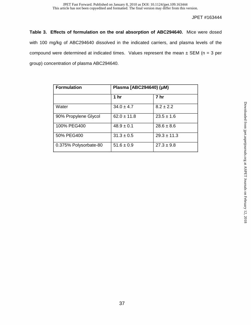
Absorption of ABC294640. The HCl salt of ABC294640 (ABC294640 • HCl) has been
synthesized in multigram amounts for characterizations of its toxicity, pharmacokinetics and in
vivo efficacy. Formulation analyses were conducted to identify a suitable pharmaceutical
composition for in vivo studies. We chose five different oral formulations from the Division of
Drug Information Resources’ Inactive Ingredient Guide, a compendium of all inactive ingredients
This article has not been copyedited and formatted. The final version may differ from this version.JPET Fast Forward. Published on January 8, 2010 as DOI: 10.1124/jpet.109.163444
at ASPE
T Journals on February 12, 2018
jpet.aspetjournals.orgD
ownloaded from
JPET #163444
16
in approved drug products currently marketed for human use, to assess their ability to support
oral absorption of ABC294640. Solutions of ABC294640 • HCl in water, 90% propylene glycol
(PG), 100% polyethylene glycol 400 (PEG400), 50% PEG400 or 0.375% Polysorbate-80 did not
precipitate (measured as turbidity at 590 nm), and so were administered to fasted female Swiss-
Webster mice at a dose of 100 mg/kg. Blood samples were removed at 1 and 7 hr, and plasma
levels of ABC294640 were determined using an internal standard and reverse-phase HPLC
coupled to an ion-trap quadrupole mass spectrometer running in positive SIM detection mode.
As shown in Table 3, substantial amounts of ABC294640 were detected in the blood 1 hr after
oral dosing, with the highest levels attained in the samples formulated in 90% PG. It should be
noted that these ABC294640 concentrations are sufficient to inhibit SK activity and proliferation
of tumor cells. By 7 hr, the plasma concentrations decreased by approximately 50% in most
cases. Effective absorption was observed in the sample formulated in 0.375% Polysorbate-80,
and this solvent for ABC294640 • HCl was used in further pharmacokinetic and efficacy
analyses because of its low toxicity.
To further understand the absorption properties of ABC294640 • HCl, the relationship
between plasma concentration and dose was examined. Mice were orally dosed with 10, 35 or
100 mg/kg of ABC294640, and the plasma levels were determined at 30 min. As shown in
Figure 5, the plasma ABC294640 values demonstrated a good linear response with doses up to
at least 100 mg/kg.
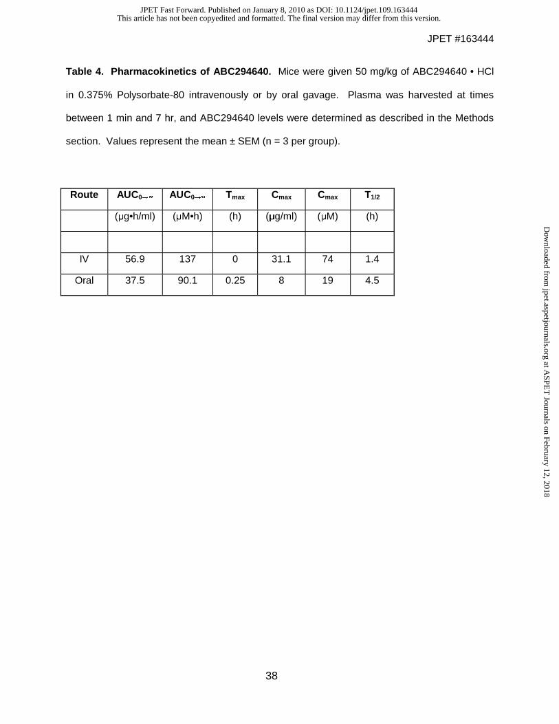
Pharmacokinetics of ABC294640. Detailed pharmacokinetic studies were performed on
ABC294640 • HCl in 0.375% Polysorbate-80. Female Swiss-Webster mice were dosed with 50
mg/kg ABC294640 either intravenously or orally. Groups of mice (3 per group) were
anesthetized and blood removed via cardiac puncture at time points ranging from 1 min to 7 hr.
Plasma concentrations of ABC294640 were determined by LC/MS and pharmacokinetic
parameters were calculated using the WINNONLIN software package (Table 4). Intravenous
This article has not been copyedited and formatted. The final version may differ from this version.JPET Fast Forward. Published on January 8, 2010 as DOI: 10.1124/jpet.109.163444
at ASPE
T Journals on February 12, 2018
jpet.aspetjournals.orgD
ownloaded from
JPET #163444
17
administration of ABC294640 resulted in high plasma concentrations that were eliminated with a
half-time of clearance of 1.4 hr. Although the peak plasma level of ABC294640 was lower when
the compound was administered by oral gavage, the compound was much more persistent,
likely reflecting continued absorption from the gastrointestinal tract, such that the calculated
half-time for clearance was 4.5 hr. Importantly, comparison of the oral versus the intravenous
pharmacokinetics of ABC294640 indicated an excellent oral bioavailability of 66% (F= AUCoral /
AUCiv).
Toxicity of ABC294640. Preliminary toxicity studies were performed to determine the
appropriate dose for in vivo efficacy testing. No immediate or delayed toxicity was observed in
female Swiss-Webster mice treated with intraperitoneal doses of ABC294640 • HCl up to at
least 250 mg/kg. Repeated injections in the same mice every other day over 15 days showed a
similar lack of toxicity at doses up to at least 250 mg/kg. Dose-escalation toxicity testing was
performed via oral gavage with ABC294640 • HCl dissolved in 0.375% Polysorbate-80, and no
toxic effects were observed after administration of doses up to 1000 mg/kg. Therefore, the
compound was considered to be suitable for more detailed in vivo studies.
Non-GLP acute toxicology studies were contracted to Eurofins │ Product Safety
Laboratories, in which ABC294640 • HCl was given orally in 0.375% Polysorbate-80 to rats at
doses of 0, 100 or 250 mg/kg daily for 7 days. There were no clinical observations or gross
findings that were considered to be the result of ABC294640 • HCl administration or otherwise.
There were no significant changes in total body weight of the treated animals, although there
was a slight decrease in body weight gain, consistent with a small decrease in food
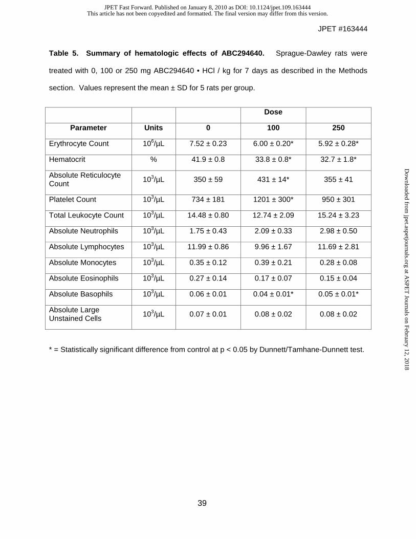
consumption, in the high-dose rats. Hematology studies (Table 5) indicated decreases in red
blood cell number and hematocrit of approximately 20% in animals given either 100 or 250
mg/kg/day; and a slight increase in neutrophils and decrease in basophils in the treated rats.
These changes would be scored as grade 0 toxicities on the standard NCI scale for evaluating
This article has not been copyedited and formatted. The final version may differ from this version.JPET Fast Forward. Published on January 8, 2010 as DOI: 10.1124/jpet.109.163444
at ASPE
T Journals on February 12, 2018
jpet.aspetjournals.orgD
ownloaded from
JPET #163444
18
toxicity in clinical trials. Importantly, no decreases in lymphocyte, platelet or granulocyte counts
were observed, indicating that the compound does not induce immunologic toxicities that are
common with other anticancer drugs. Similarly, there were no drug-induced alterations of a
broad panel of clinical chemistry or coagulation parameters. No gross abnormalities were noted
for any of the euthanized animals when necropsied at the end of the 7-day observation period.
Similarly, there were no treatment-related microscopic changes in any organ examined in the
high-dose group, except for a slight decrease in the background level of extrameduallary
hematopoiesis in the spleen which may underlie the small decreases in the hematocrit.
To further characterize the hematologic changes observed in the acute study, mice were
treated with 0, 100 or 250 mg ABC294640 • HCl / kg daily for 28 days. As indicated in Figure
6A, mice treated with 250 mg/kg experienced a 20% decrease in red blood cell count and
hematocrit, and a modest increase in the number of circulating neutrophils on Day 7, essentially
identical to the previous study with rats. However, after 28 days of treatment (Figure 6B), these
parameters were restored to normal levels, indicating that the animals had fully recovered from
any transient impairment of hematopoiesis. Additionally, there were no changes in the brain or
spleen weights of treated mice, whereas a slight decrease (12%) in liver weight was observed in
mice treated with 250 mg/kg.
Antitumor activity of ABC294640. The antitumor activity of ABC294640 • HCl was tested in a
syngeneic tumor model that uses the mouse JC mammary adenocarcimona cell line growing
subcutaneously in immunocompetent Balb/c mice (Lee et al., 2003). Because of the excellent
oral absorption described above, we determined the ability of orally-delivered ABC294640 • HCl
to reduce tumor growth in vivo. The SK inhibitor was administered to fasted mice on odd days
at doses ranging from 3.5 to 100 mg/kg. Body weights and tumor volumes were monitored
daily. As demonstrated in Figure 7, ABC294640 • HCl caused dose-dependent reductions in
the growth of the mammary adenocarcinoma xenografts. Body weights in each treatment group
This article has not been copyedited and formatted. The final version may differ from this version.JPET Fast Forward. Published on January 8, 2010 as DOI: 10.1124/jpet.109.163444
at ASPE
T Journals on February 12, 2018
jpet.aspetjournals.orgD
ownloaded from
JPET #163444
19
remained unchanged from vehicle-treated mice during the course of the study (data not shown).
Comparison with the potencies in the tumor studies with the toxicity data described above
reveals that ABC294640 • HCl has a therapeutic index of greater than 7 (250 mg/kg nontoxic
dose / 35 mg/kg antitumor activity). Thus, this SK2 inhibitor has an excellent therapeutic index.
To ensure that the antitumor effect of ABC294640 • HCl administration is mediated by
the compound, its accumulation in tumors was quantified by LC/MS. In these experiments,
mice bearing JC tumor xenografts were treated with 100 mg/kg of ABC294640 • HCl by
intraperitoneal injection, and the concentrations of the compound in the plasma and the tumor
were measured at 2 and 5 hr. As indicated in Figure 8A, approximately 75 μg/mL (197 μM)
ABC294640 was present in the plasma at 2 hr, and this decreased to 56 μg/mL (147 μM) at 5
hr. The amounts of ABC294640 in the tumor at 2 and 5 hr were determined to be 36 and 54
μg/g wet weight, respectively, corresponding to approximately 94 and 140 μM (assuming that 1
g approximately equals 1 mL). Therefore, amounts of ABC294640 well above those needed to
block cell proliferation are accumulated in the tumors of intact mice.
As a global pharmacodynamic endpoint for ABC294640 • HCl treatment, tumors from
mice given a single dose of the compound were scored for their apoptotic indices using the
TUNEL method. As shown in Figure 8B, untreated tumors had focal areas of apoptosis such
that the overall tumor apoptosis index was approximately 12%. Treatment of the mice with 100
mg/kg ABC294640 • HCl enhanced tumor cell apoptosis in a time-dependent manner, such that
nearly 50% of the tumor cells expressed fragmented DNA by 5 days.
To determine a mechanistic pharmacodynamic endpoint for ABC294640 • HCl, we
quantified sphingolipid metabolites in the tumors by LC-MS/MS. Samples of frozen tumors
removed at the end of the study in Figure 7 were homogenized in cold PBS, spiked with internal
standards and processed by liquid-liquid extraction. Ratios of sphingosine and S1P to internal
standards were determined and compared with vehicle-treated tumor samples. As shown in
Figure 8C, treatment of the mice with ABC294640 • HCl at the 100 mg/kg had no effect on
This article has not been copyedited and formatted. The final version may differ from this version.JPET Fast Forward. Published on January 8, 2010 as DOI: 10.1124/jpet.109.163444
at ASPE
T Journals on February 12, 2018
jpet.aspetjournals.orgD
ownloaded from
JPET #163444
20
sphingosine levels in the tumors. This is likely due to rapid conversion of sphingosine to
ceramide in the presence of the SK inhibitor. Importantly, S1P levels were significantly reduced
in the tumors of mice treated with ABC294640 • HCl. These findings provide further evidence
that the antitumor activity of this compound is linked to SK inhibition and decreased S1P
formation.
This article has not been copyedited and formatted. The final version may differ from this version.JPET Fast Forward. Published on January 8, 2010 as DOI: 10.1124/jpet.109.163444
at ASPE
T Journals on February 12, 2018
jpet.aspetjournals.orgD
ownloaded from
JPET #163444
21
Discussion
Accumulating evidence demonstrates that S1P is a critical second messenger that
exerts proliferative, antiapoptotic and inflammatory actions. An oncogenic role of SK has been
directly demonstrated since transfection into NIH/3T3 fibroblasts was sufficient to promote foci
formation and cell growth in soft-agar, and to allow these cells to form tumors in NOD/SCID
mice (Xia et al., 2000). Additionally, inhibition of SK by transfection with a dominant-negative
SK mutant or by treatment of cells with the nonspecific SK inhibitor DMS blocked transformation
mediated by oncogenic H-Ras. As abnormal activation of Ras frequently occurs in cancer,
these findings suggest a significant role of SK in this disease. SK has also been linked to
estrogen signaling (Sukocheva et al., 2003), and estrogen-dependent tumorigenesis in MCF-7
cells (Nava et al., 2002). Other pathways or targets to which SK activity has been linked in
cancer include VEGF signaling via the Ras and MAP kinase pathway (Shu et al., 2002), protein
kinase C (Nakade et al., 2003), TNFα (Vann et al., 2002) and caspase activation (Edsall et al.,
2001). Angiogenic factors and processes, such as cell motility, mitogenesis in smooth muscle
cells, endothelial cell differentiation and growth factor signaling are also affected by SK and S1P
(Lee et al., 1999). While the elucidation of downstream targets of S1P remains an interesting
problem in cell biology, sufficient validation of these pathways has been established to justify
their evaluation as targets for new types of anticancer drugs. As S1P appears to be the most
direct mitogenic messenger, inhibition of its production should have useful antiproliferative
effects on tumor cells.
Two isozymes of SK (termed SK1 and SK2) exist (Kohama et al., 1998), with SK2 being
cloned in 2000 (Liu et al., 2000). However, the roles of these SK isozymes remain to be defined
as they demonstrate different kinetic parameters and expression patterns in normal tissue. The
majority of studies linking SK activity to cancer and growth control have focused on SK1 (Nava
et al., 2002; Johnson et al., 2005; Le Scolan et al., 2005; Pchejetski et al., 2005; Sarkar et al.,
2005; Kawamori et al., 2006). For example, EGF rapidly induces the expression of SK1, but not
This article has not been copyedited and formatted. The final version may differ from this version.JPET Fast Forward. Published on January 8, 2010 as DOI: 10.1124/jpet.109.163444
at ASPE
T Journals on February 12, 2018
jpet.aspetjournals.orgD
ownloaded from
JPET #163444
22
SK2, in MCF-7 cells (Doll et al., 2005); however, it has also been reported that EGF activates
SK2 in MDA-MB-453 cells (Hait et al., 2005). Overexpression of SK1 has been shown to be
oncogenic in a variety of cells (Xia et al., 2000; French et al., 2003; Le Scolan et al., 2005);
whereas overexpression of SK2 is reported to inhibit cell growth and to induce apoptosis
(Maceyka et al., 2005). However, these effects of SK2 are only partially dependent on its
catalytic activity, suggesting that the antiproliferative effects may be mediated by its BH3
domain (Maceyka et al., 2005). Consistent with this, are the observations that physiological
levels of SK2 do not inhibit DNA synthesis (Okada et al., 2005). SK1-deleted transgenic mice
have normal phenotypes, and serum concentrations of S1P are reduced by 50%. However,
levels of S1P within a variety of tissues are not different from those of control mice, indicating
functional replacement of SK1 by SK2 in normal tissues (Allende et al., 2004). Similarly, genetic
ablation of SK2 results in mice with normal fertility and life-span, and 25% reduced serum S1P
levels (Kharel et al., 2005). Our data (French et al., 2003), and that of others (Johnson et al.,
2005; Van Brocklyn et al., 2005; Kawamori et al., 2006), demonstrate that SK1 is marked
overexpressed in a variety of human cancers, while SK2 is typically not (Van Brocklyn et al.,
2005), although several types of cancer do have elevated levels of SK2 message (unpublished
observations). Therefore, it was unclear if selective targeting of SK1 or SK2 by SK inhibitors will
be sufficient to inhibit tumor growth and induce apoptosis, or whether simultaneous inhibition of
SK1 and SK2 will be necessary to prevent the functional redundancy of these isozymes.
Based upon this growing body of knowledge implicating SKs in the abnormal growth of
cancer cells, we sought to identify novel inhibitors of SK1 and/or SK2 that may serve as
effective cancer therapeutics. Screening a chemically diverse small molecule library using a
recombinant human SK1-fusion protein resulted in the identification of multiple chemotypes
(French et al., 2003). Follow up hit-to-lead efforts resulted in the discovery of ABC294640, an
aryladamantane compound with SK inhibitory activity (Smith et al., 2008). The potency of
ABC294640 is similar to other SK inhibitors described in the literature (Kono et al., 2001);
This article has not been copyedited and formatted. The final version may differ from this version.JPET Fast Forward. Published on January 8, 2010 as DOI: 10.1124/jpet.109.163444
at ASPE
T Journals on February 12, 2018
jpet.aspetjournals.orgD
ownloaded from
JPET #163444
23
however, these natural products have unknown selectivities and potential for development.
Furthermore, analyses of the kinetics of inhibition of SK2 by ABC294640 clearly indicate that
this compound acts as a competitive inhibitor with respect to sphingosine, with a Ki of
approximately 10 µM (Figure 2B). Since the levels of sphingosine in a cell are very low, e.g. in
comparison with ceramide levels (Figure 3), inhibition of SK2 by ABC294640 is likely to be very
efficient. The selective targeting of SK2 was unexpected; however, our studies using siRNAs
directed at either SK1 or SK2 demonstrate that ablation of SK2 from tumor cells has greater
antiproliferative and anti-migratory affects than does depletion of SK1 2. Since many kinase
inhibitors lack specificity, we tested the ability of ABC294640 to inhibit several other kinases.
These assays indicated that ABC294640 is highly specific for SK versus protein kinases,
making it a viable candidate chemotherapeutic agent.
Our studies on the chemicophysical properties of ABC294640 further support its
development. For example, the presence of the pyridine moiety permits protonation of the
nitrogen atom, and the formation of a hydrochloride salt. This greatly improves the solubility,
such that ABC294640 • HCl was quite soluble in almost all tested carriers. These broad
solubility properties enabled testing multiple formulations to evaluate their respective in vivo
absorption properties. While all formulations showed encouraging results, we chose 0.375%
Polysorbate-80 due to its ease of formation and lower viscosity, which facilitates oral dosing.
Additionally, long term stability studies revealed that ABC294640 • HCl was highly stable in
0.375% Polysorbate-80 versus the other formulations (data not shown).
Identification of an optimal oral formulation permitted acute and chronic toxicology
studies, where we observed excellent toxicity profiles for ABC294640 • HCl. A dose of 100
mg/kg, which was determined to be at least 10-fold below than the lowest observed adverse
effect level, resulted in ABC294640 plasma levels that exceeded the IC50 towards human SK2
and are sufficient for suppression of tumor cell proliferation and migration. Biodistribution
This article has not been copyedited and formatted. The final version may differ from this version.JPET Fast Forward. Published on January 8, 2010 as DOI: 10.1124/jpet.109.163444
at ASPE
T Journals on February 12, 2018
jpet.aspetjournals.orgD
ownloaded from
JPET #163444
24
studies demonstrated that high concentrations of ABC294640 accumulate in the tumors of intact
mice, indicating that the drug does effectively reach its target tissue.
To determine the therapeutic efficacy of the compound, we evaluated ABC294640 • HCl
in a syngeneic murine breast cancer model and demonstrated significant dose-dependent
inhibition of tumor growth. Previous studies have demonstrated this model is sensitive to SK
inhibitors (French et al., 2003). Furthermore, tumor growth inhibition correlated with progressive
tumor cell apoptosis and decreases in S1P levels in the tumors when compared to vehicle-
treated mice. This is the first pharmacodynamic evidence of S1P modulation linked to antitumor
activity. We believe that this finding, in conjunction with low toxicity of ABC294640 • HCl,
provides validation of SK2 as a chemotherapeutic target.
Current studies are underway to determine the mechanism of antitumor activity, since
SK activity has been linked to survival, inflammatory and angiogenic pathways. Ideally, SK2
inhibition could lead to modulation of all of these pathways, resulting in tumor inhibition through
multiple mechanisms. We also determined that sphingosine levels remained unchanged
despite decreases in S1P. This likely relates to an increase in synthesis of ceramide from
sphingosine, resulting in a constant sphingosine level. This would be beneficial in a tumor
environment, as increases in ceramide levels cause apoptosis in cancer cells (Kolesnick and
Fuks, 2003), and this does occur in the ABC294640-treated tumor cells. Interestingly, we found
that SK2 inhibition affects actin structure at time points and concentrations that are non-toxic,
suggesting that the compound may also have anti-metastatic activity in vivo.
In conclusion, ABC294640 • HCl is a first-in-class inhibitor of SK with good
pharmacologic properties, low toxicity and anticancer activity. These data support our previous
demonstrations that ABC294640 • HCl has therapeutic activity in inhibiting diabetes-induced
retinal vascular leakage in rats (Maines et al., 2006) and dextran sulfate sodium-induced
ulcerative colitis in mice (Maines et al., 2008). Additional unpublished studies also demonstrate
This article has not been copyedited and formatted. The final version may differ from this version.JPET Fast Forward. Published on January 8, 2010 as DOI: 10.1124/jpet.109.163444
at ASPE
T Journals on February 12, 2018
jpet.aspetjournals.orgD
ownloaded from
JPET #163444
25
that ABC294640 • HCl is efficacious in models of Crohn’s Disease, rheumatoid arthritis and
ischemia-reperfusion injury. In all, the data indicate that ABC294640 • HCl warrants further
developmental efforts to fully determine its potential as an anticancer and anti-inflammatory
drug.
This article has not been copyedited and formatted. The final version may differ from this version.JPET Fast Forward. Published on January 8, 2010 as DOI: 10.1124/jpet.109.163444
at ASPE
T Journals on February 12, 2018
jpet.aspetjournals.orgD
ownloaded from
JPET #163444
26
References
Allende ML, Sasaki T, Kawai H, Olivera A, Mi Y, van Echten-Deckert G, Hajdu R, Rosenbach M,
Keohane CA, Mandala S, Spiegel S and Proia RL (2004) Mice deficient in sphingosine
kinase 1 are rendered lymphopenic by FTY720. J Biol Chem 279:52487-52492.
Bielawski J, Szulc ZM, Hannun YA and Bielawska A (2006) Simultaneous quantitative analysis
of bioactive sphingolipids by high-performance liquid chromatography-tandem mass
spectrometry. Methods 39:82-91.
Cuvillier O (2007) Sphingosine kinase-1--a potential therapeutic target in cancer. Anticancer
Drugs 18:105-110.
Cuvillier O, Pirianov G, Kleuser B, Vanek PG, Coso OA, Gutkind S and Spiegel S (1996)
Suppression of ceramide-mediated programmed cell death by sphingosine-1-phosphate.
Nature 381:800-803.
Doll F, Pfeilschifter J and Huwiler A (2005) The epidermal growth factor stimulates sphingosine
kinase-1 expression and activity in the human mammary carcinoma cell line MCF7.
Biochim Biophys Acta 1738:72-81.
Edsall LC, Cuvillier O, Twitty S, Spiegel S and Milstien S (2001) Sphingosine kinase expression
regulates apoptosis and caspase activation in PC12 cells. J Neurochem 76:1573-1584.
French KJ, Schrecengost RS, Lee BD, Zhuang Y, Smith SN, Eberly JL, Yun JK and Smith CD
(2003) Discovery and evaluation of inhibitors of human sphingosine kinase. Cancer Res
63:5962-5969.
French KJ, Upson JJ, Keller SN, Zhuang Y, Yun JK and Smith CD (2006) Antitumor activity of
sphingosine kinase inhibitors. J Pharmacol Exp Ther 318:596-603.
Gamble JR, Xia P, Hahn CN, Drew JJ, Drogemuller CJ, Brown D and Vadas MA (2006)
Phenoxodiol, an experimental anticancer drug, shows potent antiangiogenic properties
in addition to its antitumour effects. Int J Cancer 118:2412-2420.
This article has not been copyedited and formatted. The final version may differ from this version.JPET Fast Forward. Published on January 8, 2010 as DOI: 10.1124/jpet.109.163444
at ASPE
T Journals on February 12, 2018
jpet.aspetjournals.orgD
ownloaded from
JPET #163444
27
Hait NC, Oskeritzian CA, Paugh SW, Milstien S and Spiegel S (2006) Sphingosine kinases,
sphingosine 1-phosphate, apoptosis and diseases. Biochim Biophys Acta 1758:2016-
2026.
Hait NC, Sarkar S, Le Stunff H, Mikami A, Maceyka M, Milstien S and Spiegel S (2005) Role of
sphingosine kinase 2 in cell migration toward epidermal growth factor. J Biol Chem
280:29462-29469.
Huwiler A and Zangemeister-Wittke U (2007) Targeting the conversion of ceramide to
sphingosine 1-phosphate as a novel strategy for cancer therapy. Crit Rev Oncol Hematol
63:150-159.
Igarashi Y, Hakomori S, Toyokuni T, Dean B, Fujita S, Sugimoto M, Ogawa T, el-Ghendy K and
Racker E (1989) Effect of chemically well-defined sphingosine and its N-methyl
derivatives on protein kinase C and src kinase activities. Biochemistry 28:6796-6800.
Johnson KR, Johnson KY, Crellin HG, Ogretmen B, Boylan AM, Harley RA and Obeid LM
(2005) Immunohistochemical distribution of sphingosine kinase 1 in normal and tumor
lung tissue. J Histochem Cytochem 53:1159-1166.
Kawamori T, Osta W, Johnson KR, Pettus BJ, Bielawski J, Tanaka T, Wargovich MJ, Reddy
BS, Hannun YA, Obeid LM and Zhou D (2006) Sphingosine kinase 1 is up-regulated in
colon carcinogenesis. Faseb J 20:386-388.
Kharel Y, Lee S, Snyder AH, Sheasley-O'neill S L, Morris MA, Setiady Y, Zhu R, Zigler MA,
Burcin TL, Ley K, Tung KS, Engelhard VH, Macdonald TL, Pearson-White S and Lynch
KR (2005) Sphingosine kinase 2 is required for modulation of lymphocyte traffic by
FTY720. J Biol Chem 280:36865-36872.
King CC, Zenke FT, Dawson PE, Dutil EM, Newton AC, Hemmings BA and Bokoch GM (2000)
Sphingosine is a novel activator of 3-phosphoinositide-dependent kinase 1. J Biol Chem
275:18108-18113.
This article has not been copyedited and formatted. The final version may differ from this version.JPET Fast Forward. Published on January 8, 2010 as DOI: 10.1124/jpet.109.163444
at ASPE
T Journals on February 12, 2018
jpet.aspetjournals.orgD
ownloaded from
JPET #163444
28
Kohama T, Olivera A, Edsall L, Nagiec MM, Dickson R and Spiegel S (1998) Molecular cloning
and functional characterization of murine sphingosine kinase. J Biol Chem 273:23722-
23728.
Kolesnick R and Fuks Z (2003) Radiation and ceramide-induced apoptosis. Oncogene 22:5897-
5906.
Kono K, Tanaka M, Ono Y, Hosoya T, Ogita T and Kohama T (2001) S-15183a and b, new
sphingosine kinase inhibitors, produced by a fungus. J Antibiot (Tokyo) 54:415-420.
Le Scolan E, Pchejetski D, Banno Y, Denis N, Mayeux P, Vainchenker W, Levade T and
Moreau-Gachelin F (2005) Overexpression of sphingosine kinase 1 is an oncogenic
event in erythroleukemic progression. Blood 106:1808-1816.
Lee BD, French KJ, Zhuang Y and Smith CD (2003) Development of a syngeneic in vivo tumor
model and its use in evaluating a novel P-glycoprotein modulator, PGP-4008. Oncol Res
14:49-60.
Lee OH, Kim YM, Lee YM, Moon EJ, Lee DJ, Kim JH, Kim KW and Kwon YG (1999)
Sphingosine 1-phosphate induces angiogenesis: its angiogenic action and signaling
mechanism in human umbilical vein endothelial cells. Biochem Biophys Res Commun
264:743-750.
Liu H, Sugiura M, Nava VE, Edsall LC, Kono K, Poulton S, Milstien S, Kohama T and Spiegel S
(2000) Molecular cloning and functional characterization of a novel mammalian
sphingosine kinase type 2 isoform. J Biol Chem 275:19513-19520.
Maceyka M, Sankala H, Hait NC, Le Stunff H, Liu H, Toman R, Collier C, Zhang M, Satin LS,
Merrill AH, Jr., Milstien S and Spiegel S (2005) SphK1 and SphK2, sphingosine kinase
isoenzymes with opposing functions in sphingolipid metabolism. J Biol Chem 280:37118-
37129.
This article has not been copyedited and formatted. The final version may differ from this version.JPET Fast Forward. Published on January 8, 2010 as DOI: 10.1124/jpet.109.163444
at ASPE
T Journals on February 12, 2018
jpet.aspetjournals.orgD
ownloaded from
JPET #163444
29
Maines LW, Fitzpatrick LR, French KJ, Zhuang Y, Xia Z, Keller SN, Upson JJ and Smith CD
(2008) Suppression of ulcerative colitis in mice by orally available inhibitors of
sphingosine kinase. Dig Dis Sci 53:997-1012.
Maines LW, French KJ, Wolpert EB, Antonetti DA and Smith CD (2006) Pharmacologic
manipulation of sphingosine kinase in retinal endothelial cells: implications for
angiogenic ocular diseases. Invest Ophthalmol Vis Sci 47:5022-5031.
McDonald OB, Hannun YA, Reynolds CH and Sahyoun N (1991) Activation of casein kinase II
by sphingosine. J Biol Chem 266:21773-21776.
Nakade Y, Banno Y, K TK, Hagiwara K, Sobue S, Koda M, Suzuki M, Kojima T, Takagi A,
Asano H, Nozawa Y and Murate T (2003) Regulation of sphingosine kinase 1 gene
expression by protein kinase C in a human leukemia cell line, MEG-O1. Biochim Biophys
Acta 1635:104-116.
Nava VE, Hobson JP, Murthy S, Milstien S and Spiegel S (2002) Sphingosine kinase type 1
promotes estrogen-dependent tumorigenesis of breast cancer MCF-7 cells. Exp Cell
Res 281:115-127.
Ogretmen B (2006) Sphingolipids in cancer: regulation of pathogenesis and therapy. FEBS Lett
580:5467-5476.
Okada T, Ding G, Sonoda H, Kajimoto T, Haga Y, Khosrowbeygi A, Gao S, Miwa N, Jahangeer
S and Nakamura S (2005) Involvement of N-terminal-extended form of sphingosine
kinase 2 in serum-dependent regulation of cell proliferation and apoptosis. J Biol Chem
280:36318-36325.
Pchejetski D, Golzio M, Bonhoure E, Calvet C, Doumerc N, Garcia V, Mazerolles C, Rischmann
P, Teissie J, Malavaud B and Cuvillier O (2005) Sphingosine kinase-1 as a
chemotherapy sensor in prostate adenocarcinoma cell and mouse models. Cancer Res
65:11667-11675.
This article has not been copyedited and formatted. The final version may differ from this version.JPET Fast Forward. Published on January 8, 2010 as DOI: 10.1124/jpet.109.163444
at ASPE
T Journals on February 12, 2018
jpet.aspetjournals.orgD
ownloaded from
JPET #163444
30
Sarkar S, Maceyka M, Hait NC, Paugh SW, Sankala H, Milstien S and Spiegel S (2005)
Sphingosine kinase 1 is required for migration, proliferation and survival of MCF-7
human breast cancer cells. FEBS Lett 579:5313-5317.
Shu X, Wu W, Mosteller RD and Broek D (2002) Sphingosine kinase mediates vascular
endothelial growth factor-induced activation of ras and mitogen-activated protein
kinases. Mol Cell Biol 22:7758-7768.
Smith CD, French KJ and Zhuang Y (2008) Sphingosine Kinase Inhibitors, Apogee
Biotechnology Corporation, USPTO 7,338,961.
Sukocheva OA, Wang L, Albanese N, Pitson SM, Vadas MA and Xia P (2003) Sphingosine
kinase transmits estrogen signaling in human breast cancer cells. Mol Endocrinol
17:2002-2012.
Van Brocklyn JR, Jackson CA, Pearl DK, Kotur MS, Snyder PJ and Prior TW (2005)
Sphingosine kinase-1 expression correlates with poor survival of patients with
glioblastoma multiforme: roles of sphingosine kinase isoforms in growth of glioblastoma
cell lines. J Neuropathol Exp Neurol 64:695-705.
Vann LR, Payne SG, Edsall LC, Twitty S, Spiegel S and Milstien S (2002) Involvement of
sphingosine kinase in TNF-alpha-stimulated tetrahydrobiopterin biosynthesis in C6
glioma cells. J Biol Chem 277:12649-12656.
Xia P, Gamble JR, Wang L, Pitson SM, Moretti PA, Wattenberg BW, D'Andrea RJ and Vadas
MA (2000) An oncogenic role of sphingosine kinase. Curr Biol 10:1527-1530.
This article has not been copyedited and formatted. The final version may differ from this version.JPET Fast Forward. Published on January 8, 2010 as DOI: 10.1124/jpet.109.163444
at ASPE
T Journals on February 12, 2018
jpet.aspetjournals.orgD
ownloaded from
JPET #163444
31
Footnotes
This work was supported by the National Institutes of Health National Cancer Institute [Grants
R43 CA097833, R01 CA122226].
Reprint requests: Charles D. Smith, Apogee Biotechnology Corporation, 1214 Research Blvd,
Suite 1016, Hummelstown, PA 17036. Phone: (843) 792-3240; Fax: (843)792-9588; E-mail:
1 Current address: GlaxoSmithKline, King of Prussia, PA
2 Gao, P. and Smith, C. D. Differential Roles of Sphingosine Kinase Isozymes in A498 Kidney
Carcinoma Cells. Manuscript in preparation.
This article has not been copyedited and formatted. The final version may differ from this version.JPET Fast Forward. Published on January 8, 2010 as DOI: 10.1124/jpet.109.163444
at ASPE
T Journals on February 12, 2018
jpet.aspetjournals.orgD
ownloaded from
JPET #163444
32
Legends for Figures
Figure 1. Structure of ABC294640. 3-(4-chlorophenyl)-adamantane-1-carboxylic acid
(pyridin-4-ylmethyl)amide (CAS 915385-81-8).
Figure 2. Inhibition of sphingosine kinases. Panel A. Recombinant SK1 (circles) or SK2
(squares) was incubated with the indicated concentrations of ABC294640 (filled symbols) or
DMS (open symbols), and the kinase activity was measured using the HPLC assay described in
the Methods section. Values represent the mean ± SEM for three experiments. Panel B.
Recombinant SK2 was incubated with the indicated concentration of ABC294640 and kinase
activity was determined in assays containing 2.5 (�), 5 (▲), 10 (▼) or 25 (�) µM sphingosine
using the ADP Quest assay as described in the Methods section. The reciprocal of the velocity
is plotted against the ABC294640 concentration providing a Dixon plot of the results.
Figure 3. Time course of sphingolipid alterations by ABC294640. JC murine
adenocarcinoma cells were exposed to 40 µM ABC294640 for 0 (blue), 12 (red), 24 (yellow) or
48 (green) hr. Cells were harvested and the masses of the indicated sphingolipid species were
quantified by mass spectrometry in the Lipidomics Core Facility as described in the Methods
section. Labels indicate the chain-length and saturation of molecular species of ceramide (Cer).
Other labels refer to dihydro-C16-ceramide (DHC16-Cer), dihydrosphingosine (DHSph),
dihydrosphingosine 1-phosphate (DhSph-1P), sphingosine (Sph) and S1P.
This article has not been copyedited and formatted. The final version may differ from this version.JPET Fast Forward. Published on January 8, 2010 as DOI: 10.1124/jpet.109.163444
at ASPE
T Journals on February 12, 2018
jpet.aspetjournals.orgD
ownloaded from
JPET #163444
33
Figure 4. Disruption of tumor cell migration and microfilament structure by ABC294640.
Panel A. A-498 cells were plated into a Boyden chamber and treated with the indicated
concentration of ABC294640 for 4 hr as described in the Methods section. The number of cells
migrating to the opposite side of the filter was then quantified. Panel B. A-498 cells were
treated with the indicated concentration of ABC294640 for 24 hr, and the amounts of G-actin
and F-actin were determined as described in the Methods section. Cytochalasin D (cch) was
used as a positive control at a concentration of 1 µM. Panels C and D. A-498 cells were
treated with 0 (Panel C) or 50 µM ABC294640 (Panel D) for 24 hr, and then stress fibers were
visualized with FITC-phalloidin as described in the Methods section.
Figure 5. Relationship between dose and plasma ABC294640 level. Mice were orally
dosed with the indicated amounts of ABC294640 • HCl in 0.375% Polysorbate-80, bled at 30
minutes, and the concentration of ABC294640 in the plasma was determined as described in
the Methods section. Values represent mean ± SEM (n =3 mice per group).
Figure 6. Hematologic parameters in ABC294640-treated mice. Mice were orally dosed
with 0 (Veh), 100 or 250 mg/kg of ABC294640 • HCl in 0.375% Polysorbate-80 daily for either 7
(Panel A) or 28 (Panel B) days. Blood was harvested by cardiac puncture and a Complete
Blood Count was performed. Values represent mean ± standard error (n = 3 or 4 mice per
group) for the red blood cells (RBC), hematocrit (HCT), total white blood cell (WBC), and
neutrophil (Neutr) values.
This article has not been copyedited and formatted. The final version may differ from this version.JPET Fast Forward. Published on January 8, 2010 as DOI: 10.1124/jpet.109.163444
at ASPE
T Journals on February 12, 2018
jpet.aspetjournals.orgD
ownloaded from
JPET #163444
34
Figure 7. Antitumor activity of orally-administered ABC294640. Female Balb/C mice were
injected subcutaneously with JC cells suspended in PBS. After palpable tumor growth, animals
were treated every-other day by oral gavage with 0 (�), 3.5 (�), 10 (▼), 35 (▲), or 100 (�)
mg/kg of ABC294640 • HCl in 0.375% Polysorbate-80. Values represent the mean ± SEM (n =
5 mice per group) tumor volume normalized to treatment Day 1 for each mouse. *, p < 0.05; **,
p < 0.01.
Figure 8. Pharmacodynamic effects of ABC294640. Panel A. Accumulation of
ABC294640 in tumors. Mice bearing JC tumor xenografts were treated by intraperitoneal
injection of 100 mg/kg of ABC294640 • HCl and tumors were harvested at the times 2 or 5 hr.
The tumors were homogenized and the amount of ABC294640 was quantified (n = 4 mice per
group). Values represent the mean ± SEM in μg/mL for plasma samples (filled bars) and μg/g
wet weight for tumor samples (open bars). Panel B. Induction of tumor apoptosis by
ABC294640. Mice bearing JC tumor xenografts were treated by intraperitoneal injection of o or
100 mg/kg of ABC294640 • HCl. Tumors were harvested either 3 or 5 days after drug
treatment, fixed and sectioned, and the amount of apoptosis was quantified as TUNEL staining.
Values represent the mean ± SEM percent of tumor cells that were TUNEL-positive (n = 4 mice
per group). Panel C. Alteration of S1P levels by ABC294640. Tumors were harvested at the
end of the experiment described in Figure 7. The levels of sphingosine (filled bars) or S1P
(open bars) were determined by LC/MS/MS. Values represent the mean ± sd levels compared
with the vehicle-treatment group. **, p < 0.01 versus vehicle-treated mice.
This article has not been copyedited and formatted. The final version may differ from this version.JPET Fast Forward. Published on January 8, 2010 as DOI: 10.1124/jpet.109.163444
at ASPE
T Journals on February 12, 2018
jpet.aspetjournals.orgD
ownloaded from
JPET #163444
35
Table 1. Kinase selectivity of ABC294640. The affects of 50 µM ABC294640 on the activities
of the indicated protein kinases was assessed by Upstate Cell Signaling. Values represent
percent of activity of the indicated kinase remaining in the presence of ABC294640, and are the
mean ± sd of duplicate measurements.
Kinase Activity
(% of control) Kinase
Activity
(% of control)
Ca2+/calmodulin PK IV 81 ± 3 cRaf 96 ± 5
Abl 98 ± 0 MEK kinase 1 104 ± 3
Aurora-A 103 ± 1 CHK1 142 ± 13
Protein kinase C α 86 ± 5 EFGR 101 ± 3
Protein kinase C ε 101 ± 1 Fyn 84 ± 5
CDK1/cyclinB 105 ± 1 cSrc 115 ± 3
CDK2/cyclinE 106 ± 6 IKKα 150 ± 16
P38 MAP kinase 1 94 ± 2 PKA 104 ± 5
P38 MAP kinase 2 109 ± 5 PKBα 95 ± 2
PDK1 116 ± 3 PKBγ 105 ± 8
This article has not been copyedited and formatted. The final version may differ from this version.JPET Fast Forward. Published on January 8, 2010 as DOI: 10.1124/jpet.109.163444
at ASPE
T Journals on February 12, 2018
jpet.aspetjournals.orgD
ownloaded from
JPET #163444
36
Table 2. Effects of ABC294640 on tumor cell proliferation. Sparsely plated cells were
treated with ABC294640 for 48 h, and the number of viable cells was determined and compared
to vehicle (DMSO)-treated cells as described in the Methods section. Values are the mean ±
SEM for at least three separate experiments.
Cell Line Tissue IC50 (μM) Cell Line Tissue IC50 (μM)
1025LU melanoma 33.7 ± 2.7 Hep-G2 liver 6.0 ± 2.6
A-498 kidney 12.2 ± 6.0 MCF-7 breast, ER+ 18.4 ± 7.4
Caco-2 colon 11.8 ± 5.6 MDA-MB-231 breast, ER- 29.1 ± 11.1
HT-29 colon 48.1 ± 7.6 Panc-1 pancreas 32.8 ± 0.1
DU145 prostate 21.9 ± 1.5 T24 bladder 39.4 ± 7.4
SK-OV-3 ovary 10.5 ± 2.6
This article has not been copyedited and formatted. The final version may differ from this version.JPET Fast Forward. Published on January 8, 2010 as DOI: 10.1124/jpet.109.163444
at ASPE
T Journals on February 12, 2018
jpet.aspetjournals.orgD
ownloaded from
JPET #163444
37
Table 3. Effects of formulation on the oral absorption of ABC294640. Mice were dosed
with 100 mg/kg of ABC294640 dissolved in the indicated carriers, and plasma levels of the
compound were determined at indicated times. Values represent the mean ± SEM (n = 3 per
group) concentration of plasma ABC294640.
Formulation Plasma [ABC294640) (μM)
1 hr 7 hr
Water 34.0 ± 4.7 8.2 ± 2.2
90% Propylene Glycol 62.0 ± 11.8 23.5 ± 1.6
100% PEG400 48.9 ± 0.1 28.6 ± 8.6
50% PEG400 31.3 ± 0.5 29.3 ± 11.3
0.375% Polysorbate-80 51.6 ± 0.9 27.3 ± 9.8
This article has not been copyedited and formatted. The final version may differ from this version.JPET Fast Forward. Published on January 8, 2010 as DOI: 10.1124/jpet.109.163444
at ASPE
T Journals on February 12, 2018
jpet.aspetjournals.orgD
ownloaded from
JPET #163444
38
Table 4. Pharmacokinetics of ABC294640. Mice were given 50 mg/kg of ABC294640 • HCl
in 0.375% Polysorbate-80 intravenously or by oral gavage. Plasma was harvested at times
between 1 min and 7 hr, and ABC294640 levels were determined as described in the Methods
section. Values represent the mean ± SEM (n = 3 per group).
Route AUC0→∞ AUC0→∞ Tmax Cmax Cmax T1/2
(μg•h/ml) (μM•h) (h) (μg/ml) (μM) (h)
IV 56.9 137 0 31.1 74 1.4
Oral 37.5 90.1 0.25 8 19 4.5
This article has not been copyedited and formatted. The final version may differ from this version.JPET Fast Forward. Published on January 8, 2010 as DOI: 10.1124/jpet.109.163444
at ASPE
T Journals on February 12, 2018
jpet.aspetjournals.orgD
ownloaded from
JPET #163444
39
Table 5. Summary of hematologic effects of ABC294640. Sprague-Dawley rats were
treated with 0, 100 or 250 mg ABC294640 • HCl / kg for 7 days as described in the Methods
section. Values represent the mean ± SD for 5 rats per group.
Dose
Parameter Units 0 100 250
Erythrocyte Count 106/µL 7.52 ± 0.23 6.00 ± 0.20* 5.92 ± 0.28*
Hematocrit % 41.9 ± 0.8 33.8 ± 0.8* 32.7 ± 1.8*
Absolute Reticulocyte Count 103/µL 350 ± 59 431 ± 14* 355 ± 41
Platelet Count 103/µL 734 ± 181 1201 ± 300* 950 ± 301
Total Leukocyte Count 103/µL 14.48 ± 0.80 12.74 ± 2.09 15.24 ± 3.23
Absolute Neutrophils 103/µL 1.75 ± 0.43 2.09 ± 0.33 2.98 ± 0.50
Absolute Lymphocytes 103/µL 11.99 ± 0.86 9.96 ± 1.67 11.69 ± 2.81
Absolute Monocytes 103/µL 0.35 ± 0.12 0.39 ± 0.21 0.28 ± 0.08
Absolute Eosinophils 103/µL 0.27 ± 0.14 0.17 ± 0.07 0.15 ± 0.04
Absolute Basophils 103/µL 0.06 ± 0.01 0.04 ± 0.01* 0.05 ± 0.01*
Absolute Large Unstained Cells 103/µL 0.07 ± 0.01 0.08 ± 0.02 0.08 ± 0.02
* = Statistically significant difference from control at p < 0.05 by Dunnett/Tamhane-Dunnett test.
This article has not been copyedited and formatted. The final version may differ from this version.JPET Fast Forward. Published on January 8, 2010 as DOI: 10.1124/jpet.109.163444
at ASPE
T Journals on February 12, 2018
jpet.aspetjournals.orgD
ownloaded from
This article has not been copyedited and formatted. The final version may differ from this version.JPET Fast Forward. Published on January 8, 2010 as DOI: 10.1124/jpet.109.163444
at ASPE
T Journals on February 12, 2018
jpet.aspetjournals.orgD
ownloaded from
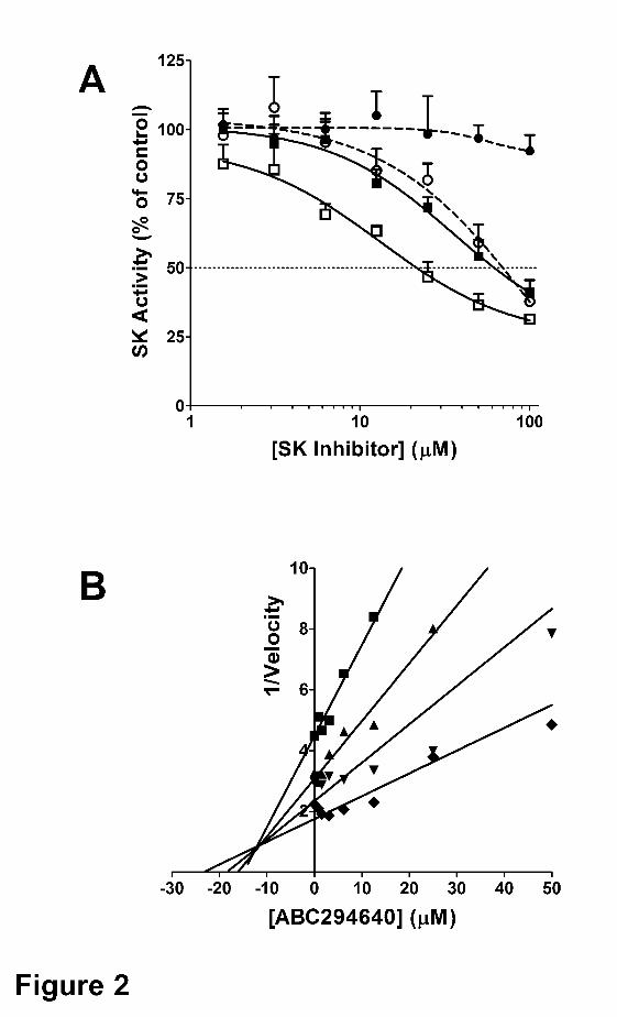
This article has not been copyedited and formatted. The final version may differ from this version.JPET Fast Forward. Published on January 8, 2010 as DOI: 10.1124/jpet.109.163444
at ASPE
T Journals on February 12, 2018
jpet.aspetjournals.orgD
ownloaded from
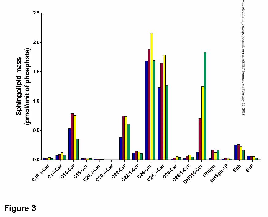
This article has not been copyedited and formatted. The final version may differ from this version.JPET Fast Forward. Published on January 8, 2010 as DOI: 10.1124/jpet.109.163444
at ASPE
T Journals on February 12, 2018
jpet.aspetjournals.orgD
ownloaded from
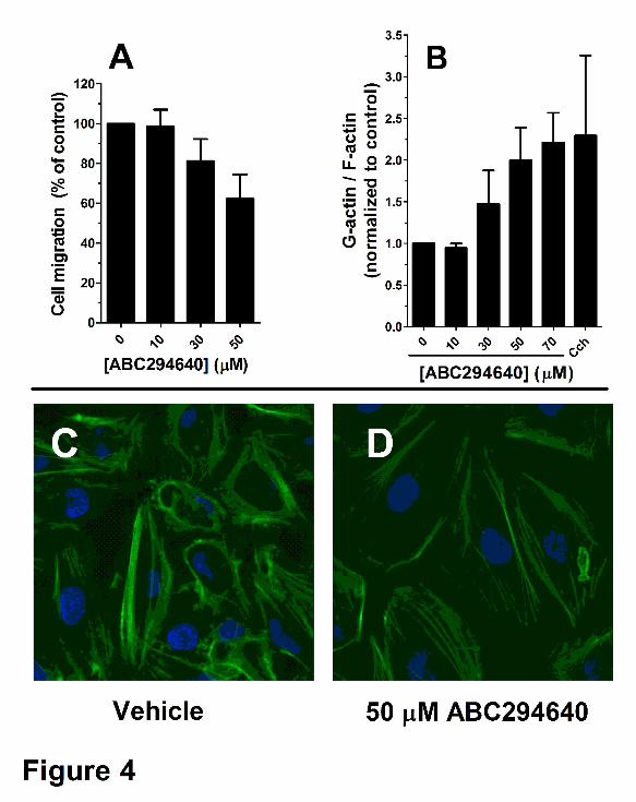
This article has not been copyedited and formatted. The final version may differ from this version.JPET Fast Forward. Published on January 8, 2010 as DOI: 10.1124/jpet.109.163444
at ASPE
T Journals on February 12, 2018
jpet.aspetjournals.orgD
ownloaded from
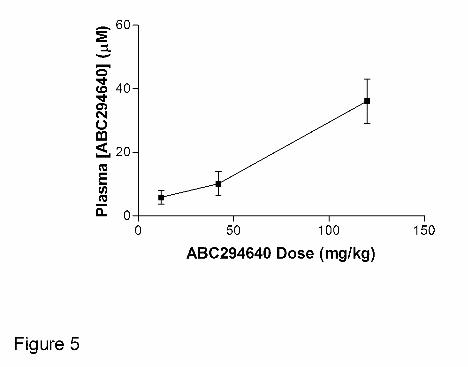
This article has not been copyedited and formatted. The final version may differ from this version.JPET Fast Forward. Published on January 8, 2010 as DOI: 10.1124/jpet.109.163444
at ASPE
T Journals on February 12, 2018
jpet.aspetjournals.orgD
ownloaded from
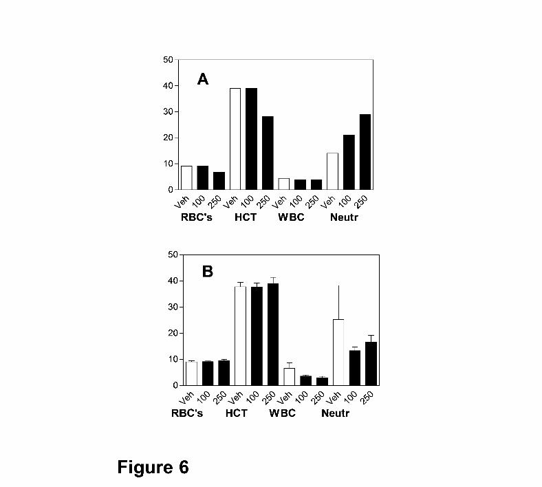
This article has not been copyedited and formatted. The final version may differ from this version.JPET Fast Forward. Published on January 8, 2010 as DOI: 10.1124/jpet.109.163444
at ASPE
T Journals on February 12, 2018
jpet.aspetjournals.orgD
ownloaded from
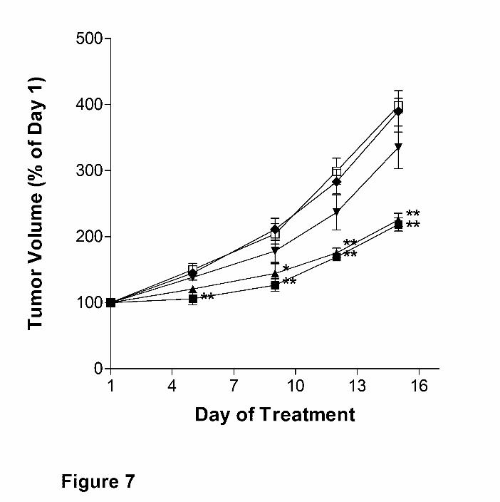
This article has not been copyedited and formatted. The final version may differ from this version.JPET Fast Forward. Published on January 8, 2010 as DOI: 10.1124/jpet.109.163444
at ASPE
T Journals on February 12, 2018
jpet.aspetjournals.orgD
ownloaded from
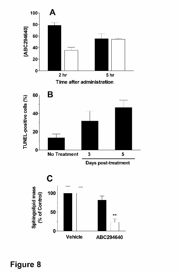
This article has not been copyedited and formatted. The final version may differ from this version.JPET Fast Forward. Published on January 8, 2010 as DOI: 10.1124/jpet.109.163444
at ASPE
T Journals on February 12, 2018
jpet.aspetjournals.orgD
ownloaded from