Embed Size (px)
Citation preview

PET Applications in PET Applications in OncologyOncology
المقبل قصي المقبل الدكتور قصي الدكتور
م م أستاذ والطب- شاركشاركأستاذ األشعة والطب- قسم األشعة قسمالنوويالنووي
- والتكنولوجيا العلوم جامعة الطب - كلية والتكنولوجيا العلوم جامعة الطب كليةاألردنيةاألردنية
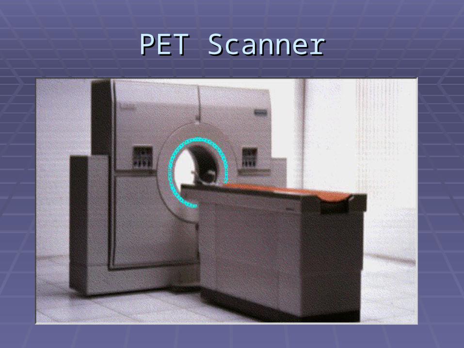
PET ScannerPET Scanner
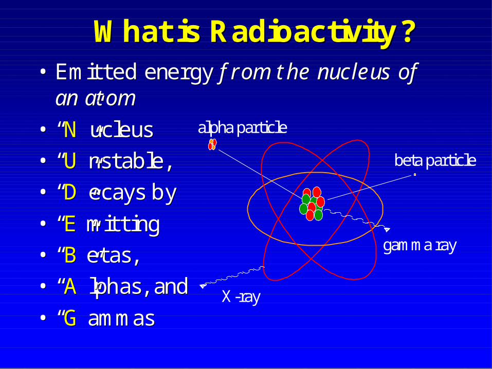
What is Radioactivity?What is Radioactivity?•• Emitted energy Emitted energy f rom the nucleus of f rom the nucleus of
an atom an atom
•• ““NN””ucleusucleus•• ““UU””nstablenstable,,•• ““DD””ecaysecays byby
•• ““EE””mittingmitting•• ““BB””etasetas,,•• ““AA””lphaslphas, and, and
•• ““GG””ammasammas
alpha particle
beta particle
gamma ray
X-ray
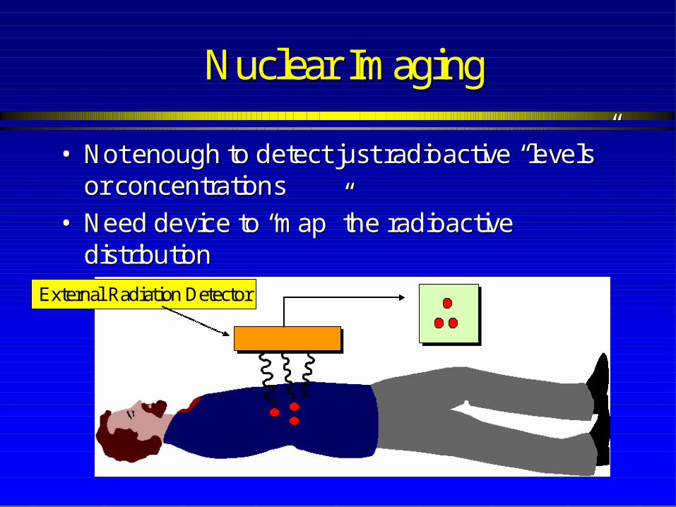
Nuclear ImagingNuclear Imaging
•• Not enough to detect just radioactive “levels” Not enough to detect just radioactive “levels” or concentrationsor concentrations
•• Need device to “map” the radioactive Need device to “map” the radioactive distributiondistribution
External Radiation Detector
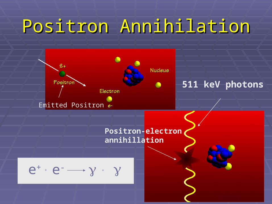
Positron AnnihilationPositron Annihilation
Emitted Positron
511 keV photons
Positron-electronannihillation
e+ + e- +
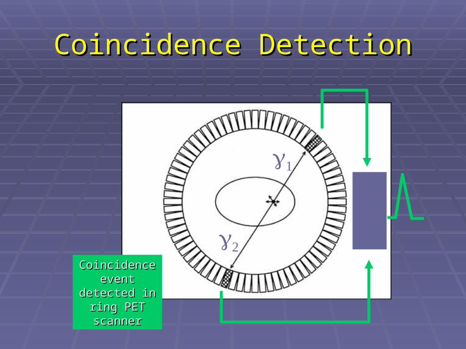
Coincidence DetectionCoincidence Detection
Coincidence Coincidence event event
detected in detected in ring PET ring PET scannerscanner
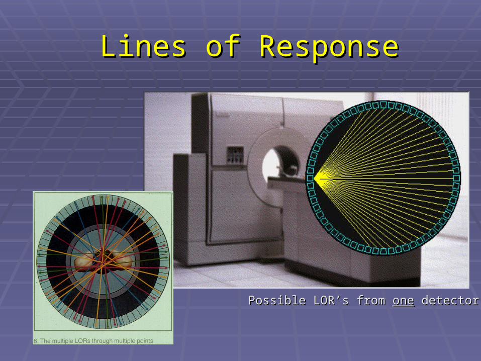
Lines of ResponseLines of Response
Possible LOR’s from Possible LOR’s from oneone detector detector
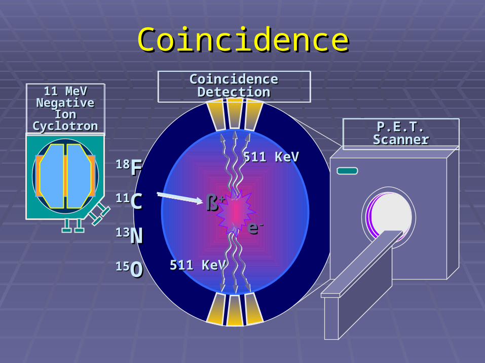
CoincidenceCoincidence
1818FF1111CC1313NN1515OO
11 MeV 11 MeV Negative Negative
Ion Ion CyclotronCyclotron
11 MeV 11 MeV Negative Negative
Ion Ion CyclotronCyclotron
511 KeV511 KeV
511 KeV511 KeV
Coincidence Coincidence DetectionDetection
Coincidence Coincidence DetectionDetection
P.E.T. ScannerP.E.T. ScannerP.E.T. ScannerP.E.T. Scanner
ßß++
ee--
FDGFDG
The main application of PET scanning is in The main application of PET scanning is in staging and restaging of cancer.staging and restaging of cancer.
Theoretically, cancer and its metastatic Theoretically, cancer and its metastatic lesions have high metabolic rate. This lesions have high metabolic rate. This means that glucose consumption is high.means that glucose consumption is high.
Attempts have been made unsuccessfully Attempts have been made unsuccessfully to label glucose with to label glucose with single photon single photon emittersemitters, like Tc99m, gallium (Ga67)., like Tc99m, gallium (Ga67).

FDG-ContFDG-Cont....
Eventually, glucose has been labeled with Eventually, glucose has been labeled with Fluorine-18 (F18) by replacing oxygen Fluorine-18 (F18) by replacing oxygen atom by F18. This radiopharmaceutical is atom by F18. This radiopharmaceutical is called flourodeoxy glucose or F18 FDG.called flourodeoxy glucose or F18 FDG.
But F18 is a positron emitter, where two But F18 is a positron emitter, where two 511 Kev photons are produced at 180˚ 511 Kev photons are produced at 180˚ (coincidence event).(coincidence event).
F18 has short half life of 110 minutes. F18 has short half life of 110 minutes.

FDG (Flourodexyglucose)FDG (Flourodexyglucose)

Biokinetics of Biokinetics of 1818FDGFDG
FDG mimics glucose metabolism
hexokinasehexokinasehexokinasehexokinase
Fructose-Fructose-6-6-
PhosphatePhosphate
Fructose-Fructose-6-6-
PhosphatePhosphate
PET definedPET defined:: PositronPositron
Radionuclide imaging using “unconventional” Radionuclide imaging using “unconventional” positron emitting radiopharmaceuticalspositron emitting radiopharmaceuticals
EmissionEmission Detection of radiation energy emitted from the Detection of radiation energy emitted from the
patient rather than transmitted through the patient rather than transmitted through the patientpatient
TomographyTomography Computer generated 3 dimensional images of Computer generated 3 dimensional images of
the radionuclidic distribution within the patientthe radionuclidic distribution within the patient

Tomographic Tomographic ViewsViews

What Makes PET different than What Makes PET different than conventional nuclear medicineconventional nuclear medicine??
Positron emitter physicsPositron emitter physics BB++ annihilation resulting in two 511KeV annihilation resulting in two 511KeV
photons emitted in opposite directionsphotons emitted in opposite directions Instrumentation differences:Instrumentation differences:
Detect coincidence eventsDetect coincidence events Can stop the energetic 511 Kev photons.Can stop the energetic 511 Kev photons.
Short lived radionuclides requiring near Short lived radionuclides requiring near production site (cyclotron).production site (cyclotron).
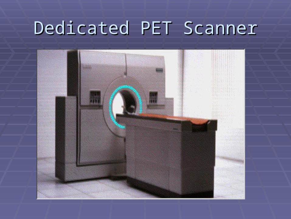
Dedicated PET ScannerDedicated PET Scanner
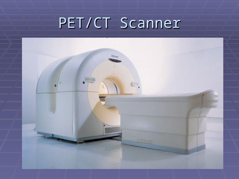
PET/CT ScannerPET/CT Scanner
PET/CT scannerPET/CT scanner
PET scanner is combined with CT scanner PET scanner is combined with CT scanner CT image is taken first followed by PET CT image is taken first followed by PET
image.image. CT image is obtained with low dose x-ray CT image is obtained with low dose x-ray
and no contrast. and no contrast. CT image is fused with PET image for CT image is fused with PET image for
anatomical correlation (lesion localization).anatomical correlation (lesion localization). CT image in PET/CT is a non-diagnostic CT image in PET/CT is a non-diagnostic
image. image.
Normal PET ScanNormal PET Scan
The following structures are somewhat The following structures are somewhat take up FDG: Heart, liver, spleen, kidneys take up FDG: Heart, liver, spleen, kidneys and urinary collecting systems.and urinary collecting systems.
Brain cortex takes significant amount of Brain cortex takes significant amount of FDG which makes it difficult to see FDG which makes it difficult to see abnormally hot lesions. abnormally hot lesions.
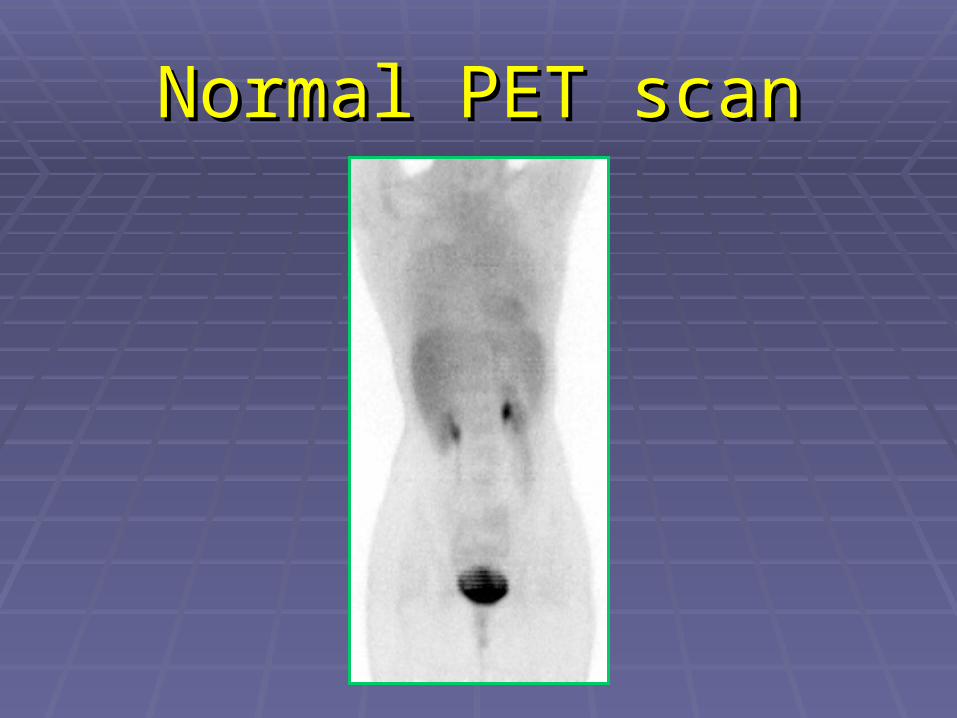
Normal PET scanNormal PET scan
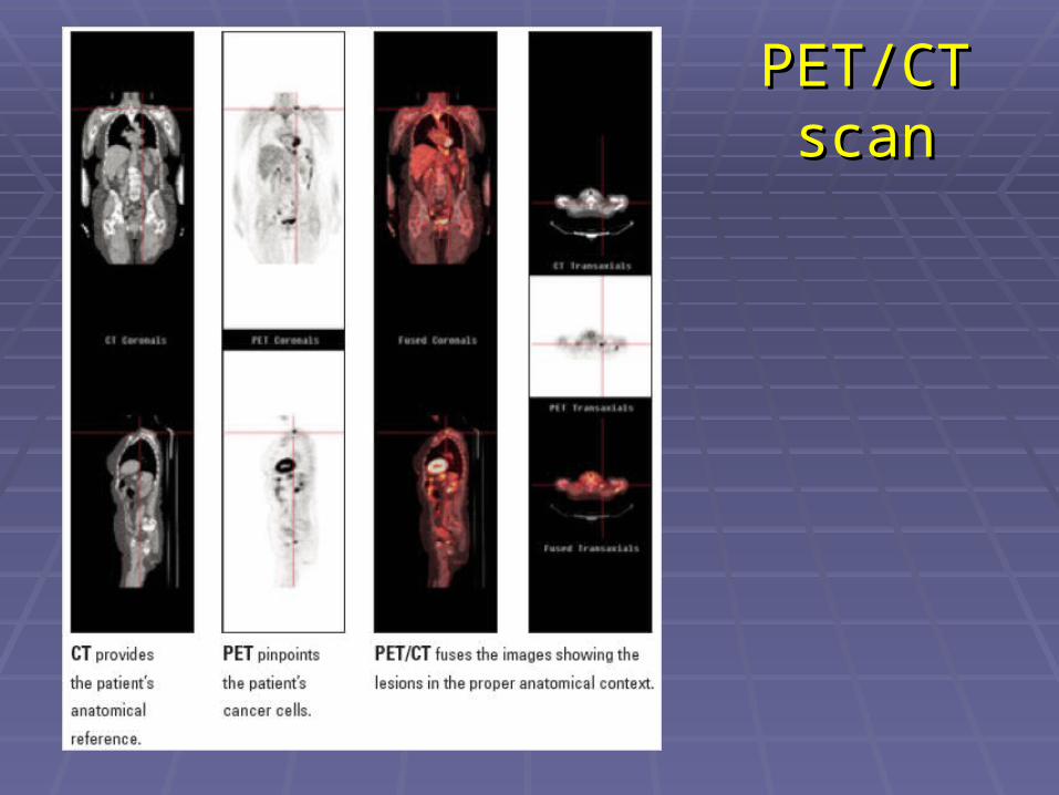
PET/CT PET/CT scanscan
Abnormal PatternsAbnormal Patterns
Hot lesions due to malignancy.Hot lesions due to malignancy. Hot lesions due to inflammatory process.Hot lesions due to inflammatory process. Hot lesions due to acute infection.Hot lesions due to acute infection.
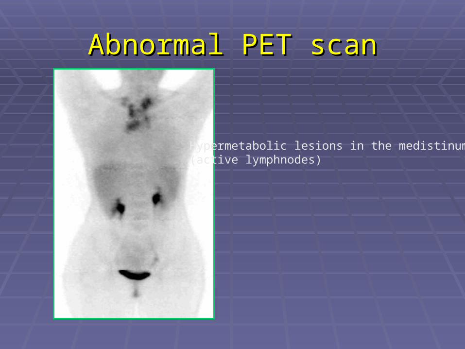
Abnormal PET scanAbnormal PET scan
Hypermetabolic lesions in the medistinum)active lymphnodes(
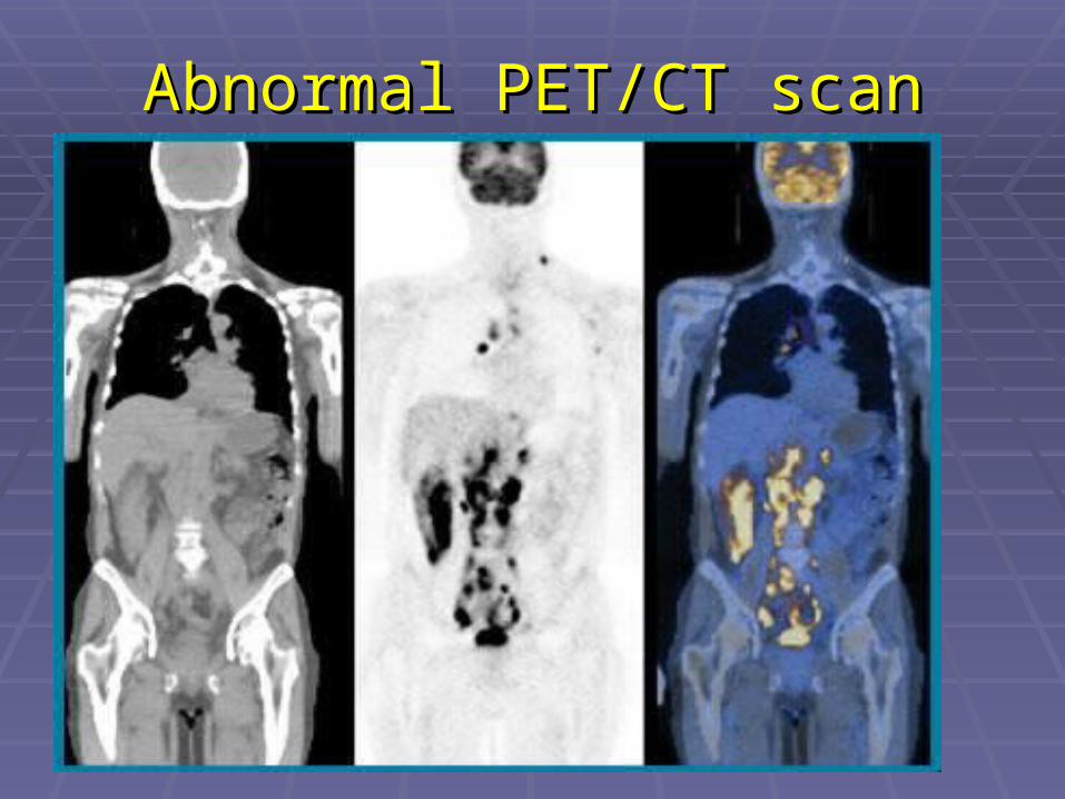
Abnormal PET/CT scanAbnormal PET/CT scan
PET Applications in PET Applications in OncologyOncology
Specific Purpose Specific Purpose
of PET Examinationsof PET Examinations Preoperative staging of cancerPreoperative staging of cancer Differentiation of scar from Differentiation of scar from
residual diseaseresidual disease Demonstration of suspected Demonstration of suspected
recurrent cancersrecurrent cancers Follow-up of therapyFollow-up of therapy
Impact of PET on Solitary Lung Impact of PET on Solitary Lung Nodules ManagementNodules Management
The sensitivity of PET is high enough that The sensitivity of PET is high enough that negative results on PET scan is negative results on PET scan is reasonably considered to rule out reasonably considered to rule out malignancy.malignancy.
False negative in well-differentiated False negative in well-differentiated adenocarcinoma and BronchioAlviolar adenocarcinoma and BronchioAlviolar carcinomacarcinoma

Lung CancerLung Cancer
Staging of non-small cell lung cancer. Staging of non-small cell lung cancer. Assessment of recurrence.Assessment of recurrence. Monitoring therapy.Monitoring therapy. Assessment of pleural malignancy. Assessment of pleural malignancy.

NSCLC Staging-Nodal DiseaseNSCLC Staging-Nodal Disease
Negative predictive value of PET Negative predictive value of PET evaluation for mediastinal lymph nodes is evaluation for mediastinal lymph nodes is greater than 95%. greater than 95%.
Positive predictive value of PET for Positive predictive value of PET for mediastinal disease is lower.mediastinal disease is lower.
PET scan is more accurate than PET scan is more accurate than diagnostic CT scan for mediastinum diagnostic CT scan for mediastinum evaluation. evaluation.
NSCLC Staging-Distant MetsNSCLC Staging-Distant Mets
PET scan is superior to conventional PET scan is superior to conventional imaging techniques for distant imaging techniques for distant metastatic disease.metastatic disease.
PET is limited for brain metastasis.PET is limited for brain metastasis.

NSCLC Staging-Adrenal MetsNSCLC Staging-Adrenal Mets
PET has been shown to help reliably PET has been shown to help reliably differentiate between adrenal differentiate between adrenal metastases and benign adrenal metastases and benign adrenal nodules.nodules.
NSCLC RestagingNSCLC Restaging
PET imaging is accurate in PET imaging is accurate in differentiating postsurgical/post-differentiating postsurgical/post-radiation changes from recurrent radiation changes from recurrent tumor.tumor.
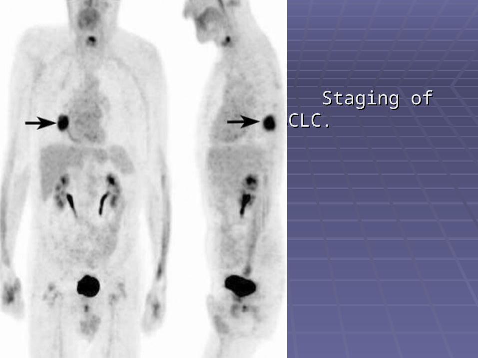
Staging of NSCLC.Staging of NSCLC.

NSCLC with MetsNSCLC with Mets
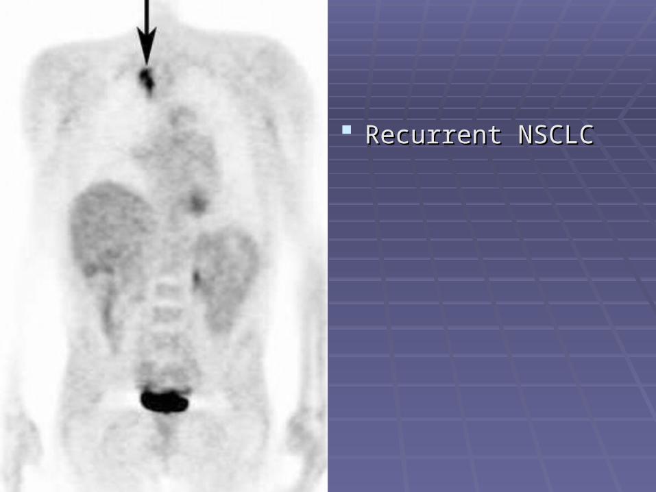
Recurrent NSCLCRecurrent NSCLC

Recurrent NSCLCRecurrent NSCLC. .
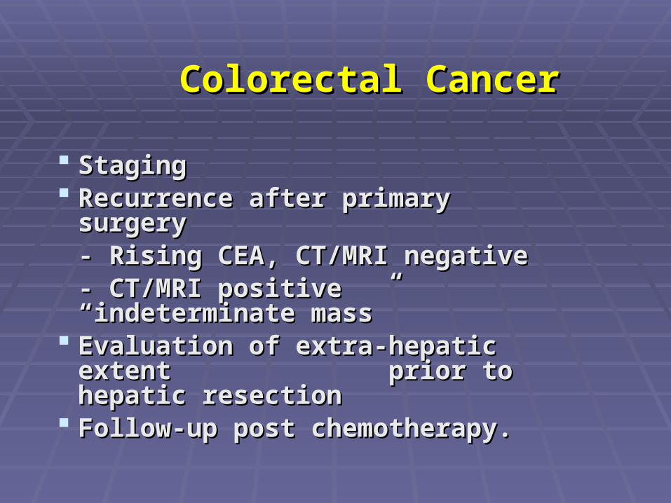
Colorectal CancerColorectal Cancer
StagingStaging Recurrence after primary surgeryRecurrence after primary surgery
- Rising CEA, CT/MRI negative- Rising CEA, CT/MRI negative- CT/MRI positive - CT/MRI positive
“indeterminate mass”“indeterminate mass” Evaluation of extra-hepatic extent Evaluation of extra-hepatic extent
prior to hepatic resection prior to hepatic resection Follow-up post chemotherapy.Follow-up post chemotherapy.

Colorectal cancer staging and re-Colorectal cancer staging and re-stagingstaging
PET was found to be more sensitive PET was found to be more sensitive than diagnostic CT for intra-than diagnostic CT for intra-abdominal metastatic disease.abdominal metastatic disease.
PET was found to be more sensitive PET was found to be more sensitive than both CT and CEA level for than both CT and CEA level for detection of disease recurrence.detection of disease recurrence.
Colorectal cancer Re-stagingColorectal cancer Re-staging
PET is accurate for differentiation PET is accurate for differentiation of benign from malignant of benign from malignant presacral changes and is superior presacral changes and is superior to CT and MR imaging in this to CT and MR imaging in this regardregard..
Campbell, SCampbell, Smr#705830mr#705830
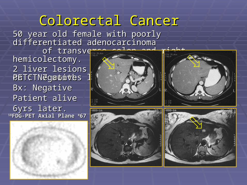
50 year old female with poorly differentiated 50 year old female with poorly differentiated adenocarcinoma of transverse colon and adenocarcinoma of transverse colon and right hemicolectomy. right hemicolectomy. 2 liver lesions seen2 liver lesions seenon CT 7 months later.on CT 7 months later.
Colorectal CancerColorectal Cancer
1818FDG-PET Axial Plane FDG-PET Axial Plane ##6767
PET: NegativePET: NegativeBx: Negative Bx: Negative Patient alive 6yrs Patient alive 6yrs later.later.
Graber, MGraber, Mmr#773593mr#773593
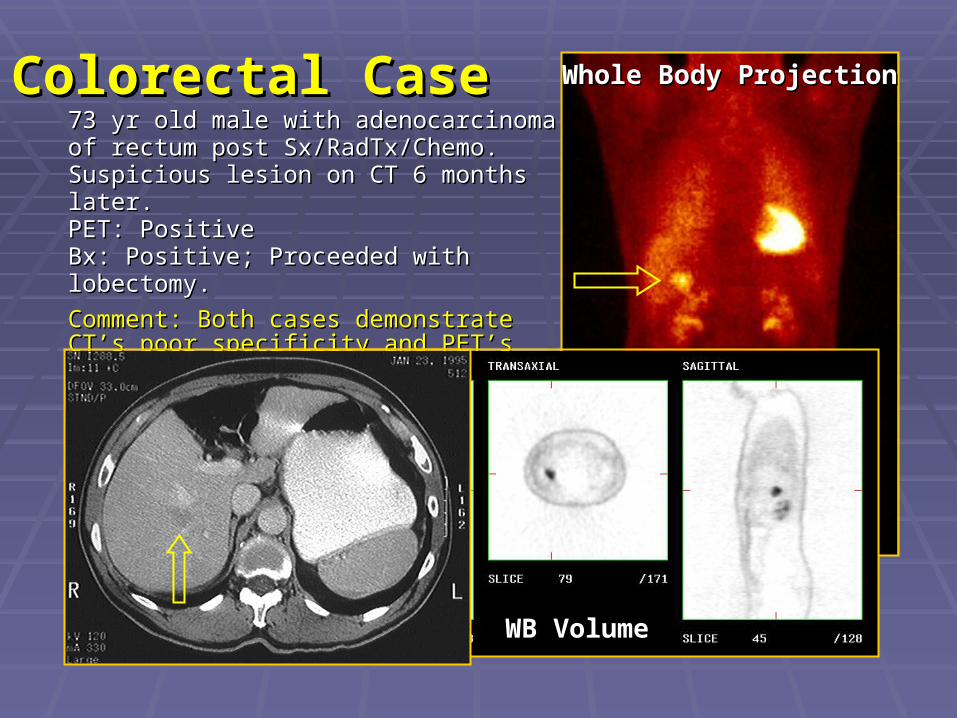
Whole Body ProjectionWhole Body ProjectionColorectal CaseColorectal Case73 yr old male with adenocarcinoma of rectum 73 yr old male with adenocarcinoma of rectum post Sx/RadTx/Chemo.post Sx/RadTx/Chemo.Suspicious lesion on CT 6 months later.Suspicious lesion on CT 6 months later.PET: PositivePET: PositiveBx: Positive; Proceeded with lobectomy. Bx: Positive; Proceeded with lobectomy.
Comment: Both cases demonstrate CT’s poor Comment: Both cases demonstrate CT’s poor specificity and PET’s ability to guide therapy.specificity and PET’s ability to guide therapy.
WB VolumeWB Volume

Recurrent rectal Recurrent rectal cance cancercance cancer
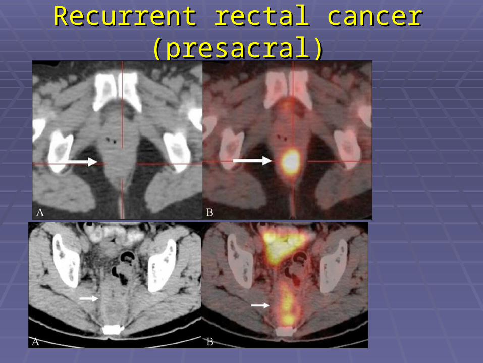
Recurrent rectal cancer (presacral)Recurrent rectal cancer (presacral)
Impact of PET on Impact of PET on Lymphoma ManagementLymphoma Management
Impact on staging: 44% of the patientsImpact on staging: 44% of the patients Impact on management: >60% of the Impact on management: >60% of the
patients.patients.

Lymphoma Staging and RestagingLymphoma Staging and Restaging
HD:HD: PET was found to be 86% sensitive PET was found to be 86% sensitive and 96% specific, compared with 81% and 96% specific, compared with 81% sensitivity and 41% specificity of CT. sensitivity and 41% specificity of CT.
NHL:NHL: the sensitivity and specificity for CT the sensitivity and specificity for CT were the same as for patients with HD, were the same as for patients with HD, whereas PET was found to be 89% whereas PET was found to be 89% sensitive and 100% specific for the sensitive and 100% specific for the presence of disease. presence of disease.
Nodal SitesNodal Sites
Discordant interpretations between PET Discordant interpretations between PET and CT images were almost always and CT images were almost always resolved in favor of the PET interpretation resolved in favor of the PET interpretation when confirmation was obtained.when confirmation was obtained.
The identification of additional sites of The identification of additional sites of disease resulted in an increase in disease disease resulted in an increase in disease stage in more than half of the patients. stage in more than half of the patients.
Extranodal SitesExtranodal Sites
PET has been found to be PET has been found to be accurate for identification of accurate for identification of disease in multiple sites in the disease in multiple sites in the abdomen.abdomen.
Restaging of LymphomaRestaging of Lymphoma
Positive PET after the end of therapy in Positive PET after the end of therapy in HD and NHL patients is a strong predictor HD and NHL patients is a strong predictor of relapse (100% of cases in 2 years).of relapse (100% of cases in 2 years).
A negative PET study is also an excellent A negative PET study is also an excellent predictor of good prognosis. predictor of good prognosis.
The diagnostic accuracy of PET to assess The diagnostic accuracy of PET to assess the presence of residual disease after the presence of residual disease after therapy is superior to that of CT.therapy is superior to that of CT.
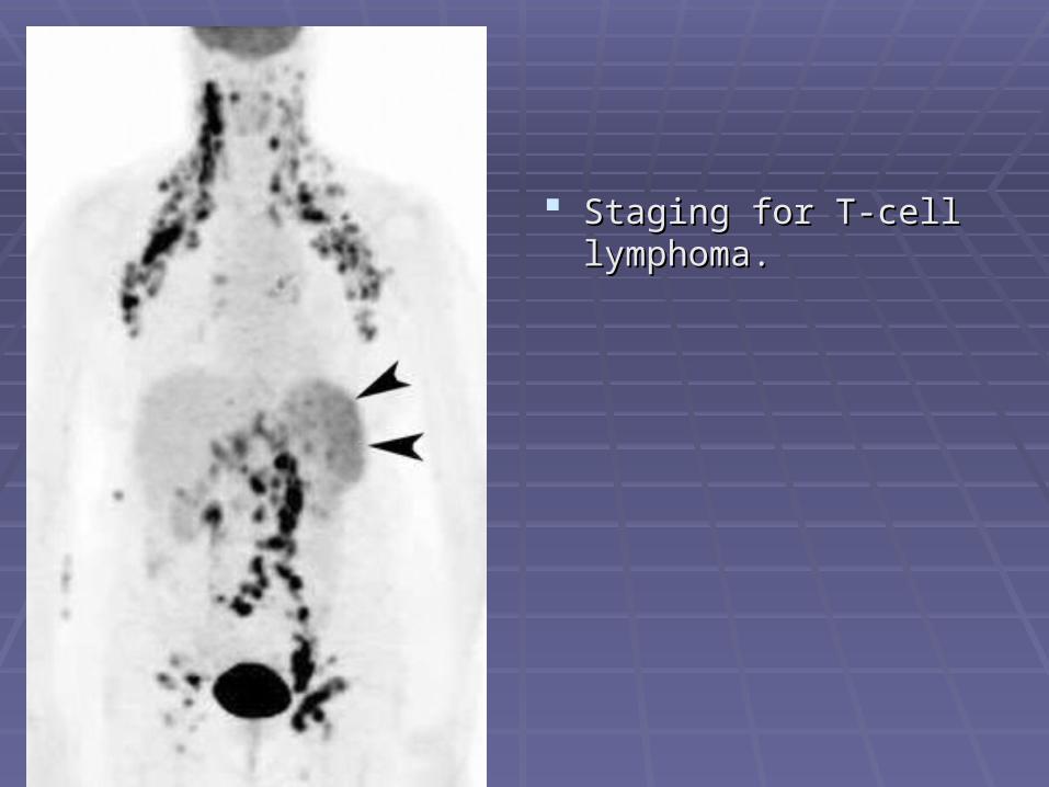
Staging for T-cell Staging for T-cell lymphoma.lymphoma.
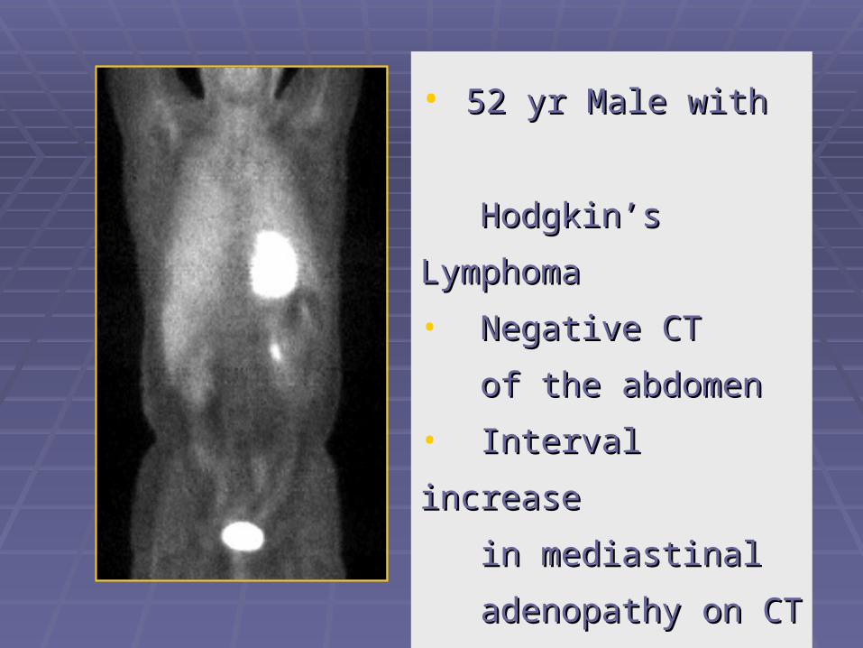
• 52 yr Male with 52 yr Male with
Hodgkin’s LymphomaHodgkin’s Lymphoma
• Negative CT Negative CT
of the abdomenof the abdomen
• Interval increase Interval increase
in mediastinal in mediastinal
adenopathy on CTadenopathy on CT
• FDG-PET: NEDFDG-PET: NED
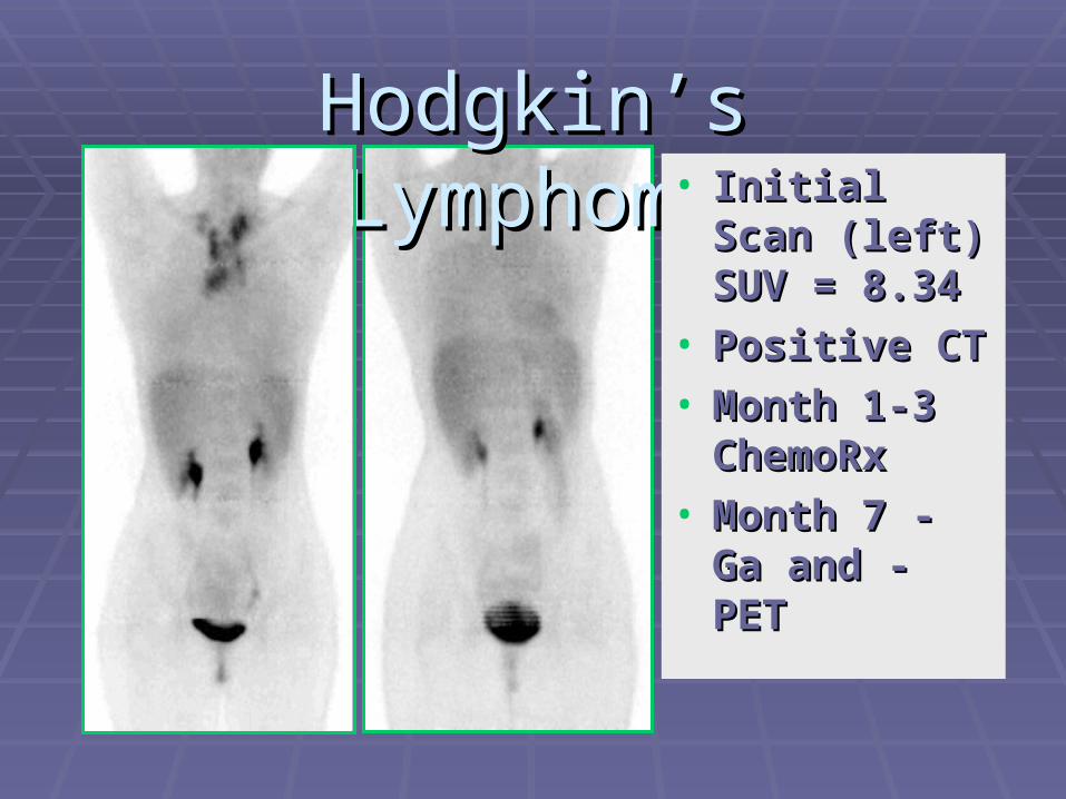
Hodgkin’s LymphomaHodgkin’s Lymphoma• Initial Scan Initial Scan
(left) SUV = (left) SUV = 8.34 8.34
• Positive CTPositive CT• Month 1-3 Month 1-3
ChemoRxChemoRx• Month 7 - Ga Month 7 - Ga
and - PETand - PET
Staging and Restaging of Staging and Restaging of Esophageal CancerEsophageal Cancer
Distant metastasis: PET was 95% : PET was 95% sensitive and 80% specific, compared with sensitive and 80% specific, compared with CT, which was 79% sensitive and 70% CT, which was 79% sensitive and 70% specific. specific.

Metastatic esophageal Metastatic esophageal cancercancer

Staging and re-staging of Head and Staging and re-staging of Head and Neck CancerNeck Cancer
PET is more sensitive and specific for PET is more sensitive and specific for cervical nodal disease than is diagnostic cervical nodal disease than is diagnostic CT.CT.
PET is more accurate for detection of PET is more accurate for detection of recurrent tumor than is diagnostic CT, MR recurrent tumor than is diagnostic CT, MR imaging, or a combination of both (post-imaging, or a combination of both (post-surgical/radiotherapy). surgical/radiotherapy).
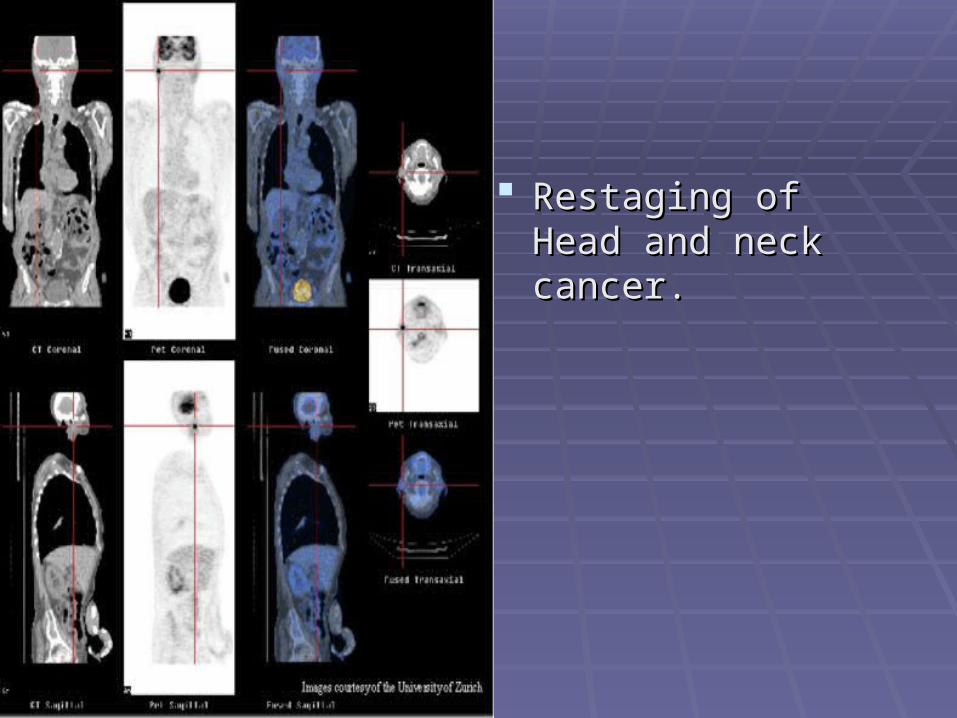
Restaging of Head Restaging of Head and neck cancer. and neck cancer.
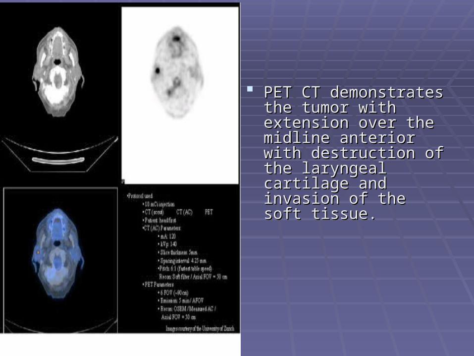
PET CT demonstrates PET CT demonstrates the tumor with extension the tumor with extension over the midline anterior over the midline anterior with destruction of the with destruction of the laryngeal cartilage and laryngeal cartilage and invasion of the soft tissue. invasion of the soft tissue.
Breast Cancer StagingBreast Cancer Staging
Compared with CT, PET is more Compared with CT, PET is more sensitive and specific for osseous and sensitive and specific for osseous and hepatic metastases and equally hepatic metastases and equally sensitive and specific for pulmonary sensitive and specific for pulmonary metastasesmetastases..

Breast Cancer RestagingBreast Cancer Restaging
PET is useful for demonstration of both PET is useful for demonstration of both locally recurrent disease and distant locally recurrent disease and distant metastases and appears to be superior to metastases and appears to be superior to conventional imaging in this regard.conventional imaging in this regard.
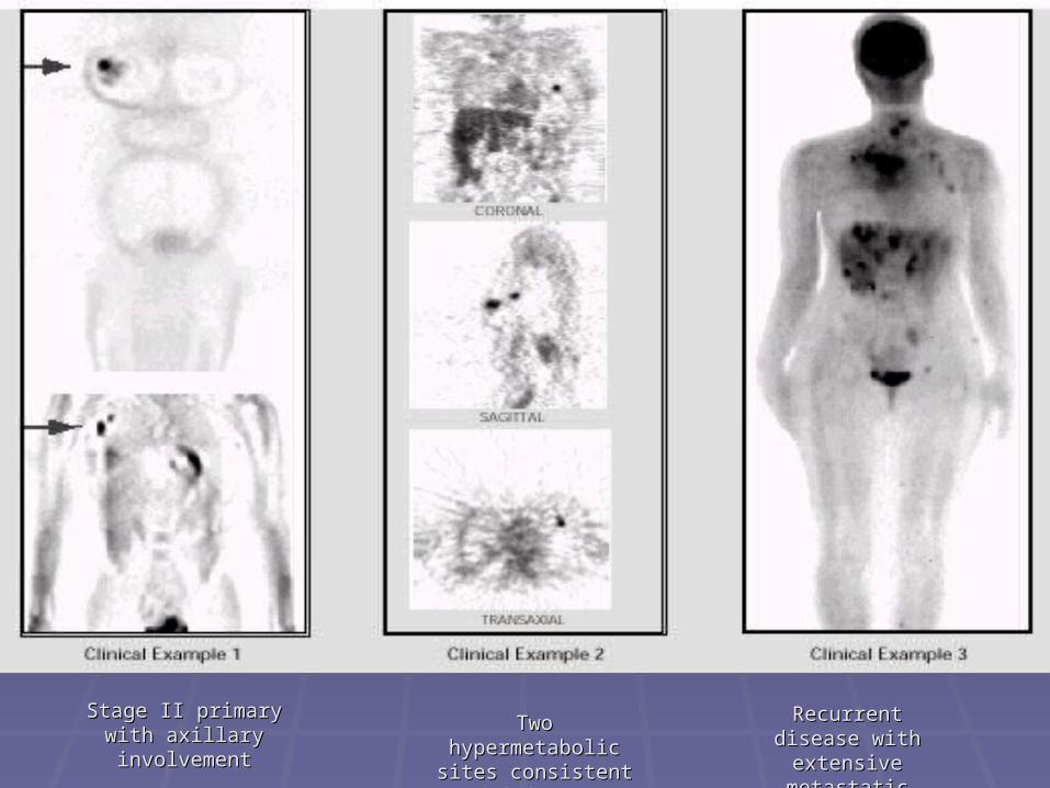
Disease StagingDisease Staging
Stage II primary with Stage II primary with axillary involvementaxillary involvement Two hypermetabolic Two hypermetabolic
sites consistent with sites consistent with breast carcinomabreast carcinoma
Recurrent disease Recurrent disease with extensive with extensive
metastatic metastatic involvementinvolvement

Breast cancerBreast cancer
. .
Unknown Primary TumorUnknown Primary Tumor
PET imaged unknown primary tumors in PET imaged unknown primary tumors in about about one third of all patientsone third of all patients investigated. investigated.

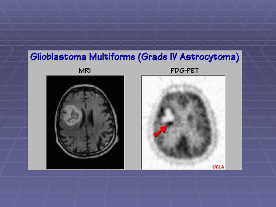
Brain TumorsBrain Tumors
PET can differentiate scar or necrosis from PET can differentiate scar or necrosis from persistent or recurrent tumor after surgery, persistent or recurrent tumor after surgery, chemotherapy or radiotherapy.chemotherapy or radiotherapy.
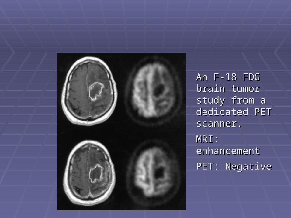
An F-18 FDG An F-18 FDG brain tumor brain tumor study from a study from a dedicated PET dedicated PET scanner.scanner.
MRI: MRI: enhancementenhancement
PET: NegativePET: Negative