Embed Size (px)
Citation preview

Neuropsycholoyio, Vol 25, No IA, PP. 119-133, 1987. Prmkd in Great Britam
0028-3932187 S3.00+0.00 Pergamon Journals Ltd.
PERCEPTUAL AND ATTENTIONAL PROCESSES FOLLOWING CALLOSAL SECTION IN HUMANS*
MICHAEL S. GAZZANIGA
Division of Cognitive Neuroscience, Department of Neurology, Cornell University Medical College, New York, NY, 10021, U.S.A.
(Accepted 7 October 1986)
Abstract-Studies are reviewed that examine how perceptual and attentional systems operate in the cortically disconnected human. The data indicate that even though both simple and complex perceptual information associated with the cognitive activities of each disconnected half brain show virtually no interactions, the attentional system remains largely integrated in the split-brain patient. It also appears that the human brain is subject to a set of finite resources it can allocate to cognitive activities. These resources are not changed following cortical disconnection. Taken together, studies to date support the view that the attentional system is an independently functioning and integrated entity, following brain bisection, that participates in both perceptual and cognitive activities of each hemisphere.
INTRODUCTION
THE INTACT cerebral cortices collaborate to carry out the operations of human conscious experience. The specialized functions of each cerebrum are interconnected via the corpus callosum, giving rise to an integrated system that manages linguistic, memorial, and cognitive functions in a seemingly unified way. While classic neuropsychologic observations following focal lesions assess the consequences of damage on the whole operating system that produces human cognition, split-brain studies have centered on considering what each half brain can accomplish alone and isolated from its partner [6,25]. With a crisp surgical lesion dividing the two cortices, questions have been asked about whether or not the two halfbrains are similar and equally competent in a host of learning and perceptual tasks. In general these studies have suggested that the disconnected cerebral hemispheres are able to work independently and without interference from the other. These findings were consistent with expectations from the animal literature and with the fact that only cortical systems had been disconnected.
The cortically disconnected patient, however, has rich interconnections remaining between the two half brains, structures that could possibly subserve the exchange of some kind of information related to or involved in perceptual, cognitive, or attentional processes. The process that has consistently shown interhemispheric interactions has been the attention system. The picture that has newly emerged is an intricate one that evolved with the appearance of new sophisticated experimentation and the inevitable richness that follows from the opportunity to study more split-brain cases. In much of what follows I will review studies that my late colleague, Jeffrey Holtzman and I carried out over 6 yr. In the history of
*Aided by NINCDS Grants 2POl NS17778 and Javits Neuroscience Investigator Award lRO1 NS22626.
119

neuropsychology, there never was a more ingenious cxperimentalist than Jeff I-loltzman. to whom this article is respectfully dedicated.
GENERAL BACKGROUND
The original human split-brain studies carried out in the early 1960’s suggested that the disconnected right hemisphere was a near equal partner to the left in a wide range of language skills and cognitive activities 16, 10, 751. II; some tests, it was even argued that it had capacities superior to those seen in the left. The main difference was that it could not speak. These results were based largely on the results from two patients who have proven over the years to be unique and uncommon when considering the now expanded split-brain patient pool. While other patients who have similar. if not richer, right hemisphere skills have come along, the vast majority of the cases from all surgical series reveal little cognitive capacity in their right hemispheres [21]. Since it is difficult to establish a commerce with these right hemispheres, studies that attempt to examine what sub-callosal interactions might be acti\c or at least have the capacity to be active in interhemispheric integration of information are carried out on the select patients with responsive right hemispheres. As a consequence, in the present paper 1 will review the studies that examine what perceptual and attentional interactions can occur between two active but disconnected cerebral cortices. To begin with, I will review studies of ours that demonstrate the degree of independence that is evident for both simple and complex perceptually based tasks presented to each half brain. I will then focus on studies carried out on attentional processes, studies that suggest the independence of function seen with perceptual and cognitive processes does not apply for attentional processes. As a possible consequence of this, additional studies are reviewed that reveal the split-brain patient does not have additional resources to call upon for cognitive processing.
SIMPLE PERCEPTUAL INTERACTIONS ARE NOT SEEN FOLLOWING CALLOSAL SECTION
Despite several reports on the failure of split-brain patients to cross-integrate visual information of all kinds [6. 12, 161 there have been recent reports arguing that some perceptual information is integrated at some level. One report suggests that apparent motion can be perceived when two visual stimuli are alternately flashed, one to each half brain [ 191. Another report suggests that a split-brain patient can detect whether two letters arc a vowel or not when one is flashed to one hemisphere and the other to the opposite half brain [22]. Both studies serve as examples on how difticult it is to interpret split,-brain studies examining interhemispheric communication.
In the experiment on apparent motion, two lights were first quickly Hashed to one
hemisphere. The subject was asked tojudge in which direction the light moved by pointing to one of three cards. One card had a lcftward pointing arrow, one a rightward pointing arrow and a third depicted two dots which was to indicate both lights had appeared simultaneously. The subjects performed the task well. At this point in the training sequence. each hemisphere knew two lights were always going to be presented. Subsequently. the two lights appeared ditrerently with one appearing in one visual field and one in the other. On these trials either the left dot appeared for I30 msec, there was a 30 rnsec ISI and the rikrht dot appeared or vice versa. There were also trials where the lights came on simultaneously in each field. Two of the
PtKCEPTlON ANI) ATTENTION FOLLOWING CAL.LOSAL SECTION 121
three subjects were able to point to the correct arrow, thereby suggesting they could make good temporal order judgments and distinguish those trials from the simultaneous trials. However, the evidence that they saw apparent motion between the two hemispheres was inferred by interview. Additionally, with a sufficiently long ISI, subjects claimed only a blinking light had occurred when each hemisphere was tested alone and also in the between hemisphere condition. This observation was interpreted as supporting the idea of a common system active for both conditions. The difficulty with this interpretation is that it is known that split-brain patients are capable of reporting the presence or absence of stimulation of one hemisphere by the other [6, 10, 1 2, 151. With this being the case, it easy for them to assume, especially in the presence of the response cards with printed arrows pointing either to the left or right, that movement could have occurred. Their responses could well be a product of the set established by the examiner. Whether they actually saw motion is a different matter.
One way to examine this issue is to compare the within and between field performance on an apparent motion judgment under conditions where no set is created that encourages cross-cuing. By exposing a subject to a set of all of the different possibilities for the stimulus array, the interpretations of what is seen can more accurately reflect perception. This was done on Case J.W. On each trial the subject either saw two lights blink, with a 30 msec ISI, or the presentation of only one fight. Thus, from the start, the possibility of a blinking light occurring was in their response set. In the two light condition, either both lights appeared in one or the other field (within field condition) or one light appeared in each field (between field condition). There were four possible positions for the lights, two appearing in the left visual field and two in the right. The distance between the two lights for the between field condition was the same for the two lights in the within field condition (approx 2”). On each trial the subject was instructed to fixate and with a variable foreperiod the stimulus sequence was presented. On the single light trials, the stimulus appeared in either the right or left visual field. The subject was required to judge “Do you see movement?” by responding to either a “yes” button or a “no” button. There were 60 trials in a block, 25 between field trials, 10 within field and 25 single field trials. Four blocks were run and the results of a typical block are as follows: within a visual field on the two light condition, there were no errors in judging apparent movement. In the between field two light condition, he made a “no” response on every trial thereby indicating that each hemisphere judged the trial as a single blinking light. He judged all the single light presentations as a “no” response as well.
The results show that a callosum sectioned patient was unable to judge whether or not apparent motion had occurred across the midline. At the same time Case J.W. was perfect at judging apparent motion for all within hemisphere trials. In a separate run, where only between field trials occurred randomly mixed with single blinking lights, he also failed to see movement.
The differences in the two sets of results may reflect different degrees of cortical disconnection or more likely, differences in testing procedures which establish different criteria for the patients’ judgments. Having clearly established in J.W. the reality of single field lights, he may well find it difficult to conclude on partial information that an event had occurred. In a further test where a specific request was made to judge temporal order, J.W. was unable to do so. Had he been given the other procedure, he may well have taken advantage of partial information to carry out the task. It must be remembered how special the split-brain case is and how quickly and easily clever strategies are developed. As a consequence, it remains open whether or not apparent motion can be observed across the midline.
122 MICHAEL s. C;AZ7ANIGA
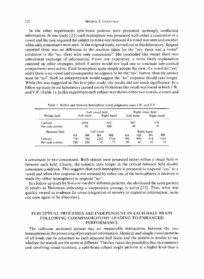
In the other experiments split-brain patients were presented seemingly conflicting information. In one study [22] each hemisphere was presented with either a consonant or a vowel and the task required the subject to make one response if a vowel was seen and another when only consonants were seen. In the original study, carried out in this laboratory, Sergent reported there was no difference in the reaction times for the “yes, there was a vowel” condition vs the “no, there were only consonants”. She concluded this meant there was subcortical exchange of information. From our experience, a more likely explanation centered on other strategies, which if active would not lead one to conclude sub-cortical comparisons were active. Each hemisphere quite simply adopts the view, if a vowel hit “yes” and if there is no vowel and consequently no urgency to hit the “yes” button, then the answer must be “no”. Such an interpretation would suggest the “no” response should take longer. While this was suggested in this first pilot study, the results did not reach significance. In a follow up study in our laboratory carried out by Holtzman this result was found in both J.W. and V.P. (Table 1). In this experiment each subject was shown either two vowels, a vowel and
Table I. Within and between hemisphere vowel judgments cases J.W. and V.P
Within field
Latency Per cent correct
Between field
Latency Per cent correct
Left visual field Right visual field Left hand Right hand Left hand Right hand
1018 843 943 836 96 loo 99 91
Left hand Right hand AA AB BA BB AA AB BA BB 778 784 1242 1710 799 994 814 1464 100 loo 92 7s 98 88 92 74
a consonant or two consonants. Both stimuli were presented either within a visual field or between each field. Clearly, the subjects take longer in the critical between field double consonant condition. This suggests that each hemisphere is prepared to respond “yes” to a vowel and when that response is not initiated by either one of the hemispheres, a decision is made (by either hemisphere) to respond “no”.
In a follow up study by SERGENT on the California patients, she also found the same pattern of results as Holtzman indicating a cooperative strategy is active [23]. Thus, what was quickly viewed as evidence for cross-integration of sensory or cognitive information, turns out once again to be illusionary.
PERCEPTUAL PROCESSES ARE INDEPENDENT IN EACH HALF BRAIN FOLLOWING COMMISSUROTOMY LEADING TO ENHANCED
PERFORMANCE
The callosum sectioned patient has no measurable interactions between the two hemispheres in the processing of perceptual information. Identical and simple visual patterns of all kinds can be presented to each separate half brain and the patient is unable to say whether the stimuli are the same or different. This fact raises the possibility that in a memory task involving visual retention, a split-brain subject might perform at a higher level than a
PERCEPTION AND ATTENTION FOLLOWING CALLOSAL SECTION 123
normal intact control subject if the perceptual information was distributed between the two visual half fields [S, 111. In a recent study on this issue, a complex spatial memory task was administered to both a split-brain patient and two normal controls, where critical information was presented in each visual half field [14]. For the normals, the visual information was automatically combined and perceived as one large problem. For the split- brain patient, each hemisphere perceived a problem that remained separate from the perceptual information presented to the other half brain, thus, each hemisphere perceived a much simpler task.
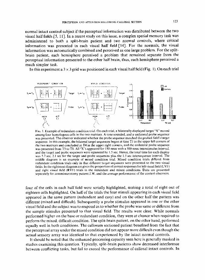
In this experiment a 3 x 3 grid was positioned in each visual half field (Fig. 1). On each trial
FIG. I. Example of redundant condition trial. On each trial, a bilaterally displayed target “x” moved among four homologous cells in the two matrices. A tone sounded, and a unilateral probe sequence was presented. The observer indicated whether the probe sequence matched the probed field’s target sequence. In this example, the bilateral target sequences began at time Tl in the upper left corners of the two matrices and concluded at T4 in the upper right corners, and the unilateral probe sequence was presented from T5 to T8. All “X”s appeared for 150 msec with a 500 msec interstimulus interval, and the target and probe sequences were separated by 1.5 sec. Hence, the total time for each display was 5.7 set, 2.1 set for the target and probe sequences plus the 1.5 set intersequence interval. The middle diagram is an example of mixed condition trial. Mixed condition trials differed from redundant condition trials only in that different target sequences were presented to the two visual fields. In the rightmost diagram are given the proportion ofcorrect responses for left visual field (LVF) and right visual field (RVF) trials in the redundant and mixed conditions. Data are presented separately for commissurotomy patient J.W. and the average performance of the control observers.
four of the cells in each half field were serially highlighted, making a total of eight out of eighteen cells highlighted. On half of the trials the four stimuli appearing in each visual field appeared in the same pattern (redundant and easy) and on the other half the pattern was different (mixed and difficult). Subsequently a probe stimulus appeared in one or the other visual field and the subject was to respond as to whether the probe was same or different from the sample stimulus presented to that visual field. The results were clear. While normals performed higher on the base or redundant condition, they were at chance when required to perform the mixed, difficult condition. The split-brain patient, on the other hand, performed equally well in both conditions. The callosum sectioned patient benefited from the fact that the perceptual array under the mixed condition did not appear more difficult even though the actual sensory array was identical to that experienced by the intact normal controls.
It should be noted that the enhanced processing capacity seen here is generally masked in studies examining this question. Typically, split-brain patients show decreased interference between conflicting tasks, but fail to exceed the performance of callosal intact controls. In
most cases this diminished performance is due to experimental design considerations such as requiring alternating responses between the hemispheres 14, 5, 26. 271, maintaining prolonged activation of one hemisphere [2, 31, or generating bimanual responses [IX]. When these factors are minimized and other task demands are included. a superior performance by split-brain patients relative to normals can be demonstrated.
Disconnection of the cerebral hemispheres clearly allows for a unique cognitive state. In a sense it turns a serial, unified perceptual system into two simpler perceptual systems that do not interact. and therefore do not interfere with each other. It allows for the breaking down of a large perceptual problem into a smaller, more manageable problem that a half brain is capable of solving. From the observer’s point of view. however, it looks like the total information processing capacity of the patient has been increased and is superior to that of normals. Yet. close inspection of the problem along with some follow up studies suggest that the split-brain patient has not increased the amount of resources he can call upon to solve problems. In a series of studies to be reviewed below. it is argued that the human brain has a set number of resources it can allocate to cognitive tasks and that these resources remain constant following commissurotomy.
In carrying out the studies related to these issues, it was first necessary to determine the nature of the remaining interhemispheric interactions. and the way attentional processes work following brain bisection.
CALLOSUM SECTIONED HUMANS TRANSFER ONLY CRUDE SPATIAL. INFORMATION
The original split-brain patients operated on by BOGEN and VOGEI, had both the anterior commissure and corpus callosum sectioned [I). In all other series to date, only the corpus callosum is sectioned [ 151. Earlier reports that transfer of visual information may at times occur with sparing of the anterior commissure seem not to hold. When transfer of visual information is present, it is now thought to reflect inadvertent sparing ofcallosal tibers which can now be easily detected with post-operative MRI [S]. As a result, studies examining the capacity of the two disconnected brains to integrate visual information between the hemispheres in patients with the anterior commissure as well as the callosum sectioned, or with only the callosum sectioned, show no differences [I’]. Visual integration of any perceptual. color, or brightness information is not possible in cithcr case. Studies on more cognitive phenomena, such as semantic priming, have been inconsistent. Original attempts to demonstrate semantic priming between the hemispheres of patients with bilateral language seemed promising, but now have been abandoned [29]. Our own observations on Case J.W. initially suggested that some priming was possible [24]. However. in several follow-up attempts to reproduce and extend the phenomenon, we have failed to see any effects [7, 91. As a result, cross hemisphere studies on both perceptual and cognitive tasks reveal and confirm the remarkable separation that occurs following callosal section.
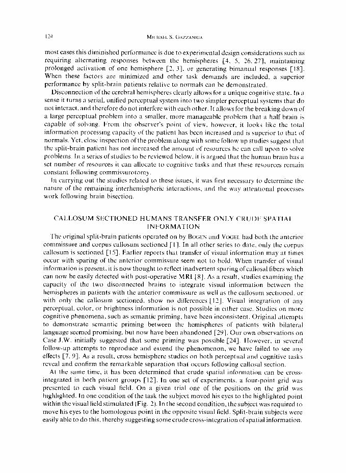
At the same time. it has been determined that crude spatial information can be cross- integrated in both patient groups [12]. In one set of experiments, a four-point grid was presented to each visual field. On a given trial one of the positions on the grid was highlighted. In one condition of the task the subject moved his eyes to the highlighted point within the visual field stimulated (Fig. 2). In the second condition, the subject was required to move his eyes to the homologous point in the opposite visual field. Split-brain subjects were easily able to do this, thereby suggesting some crude cross-integration of spatial information.
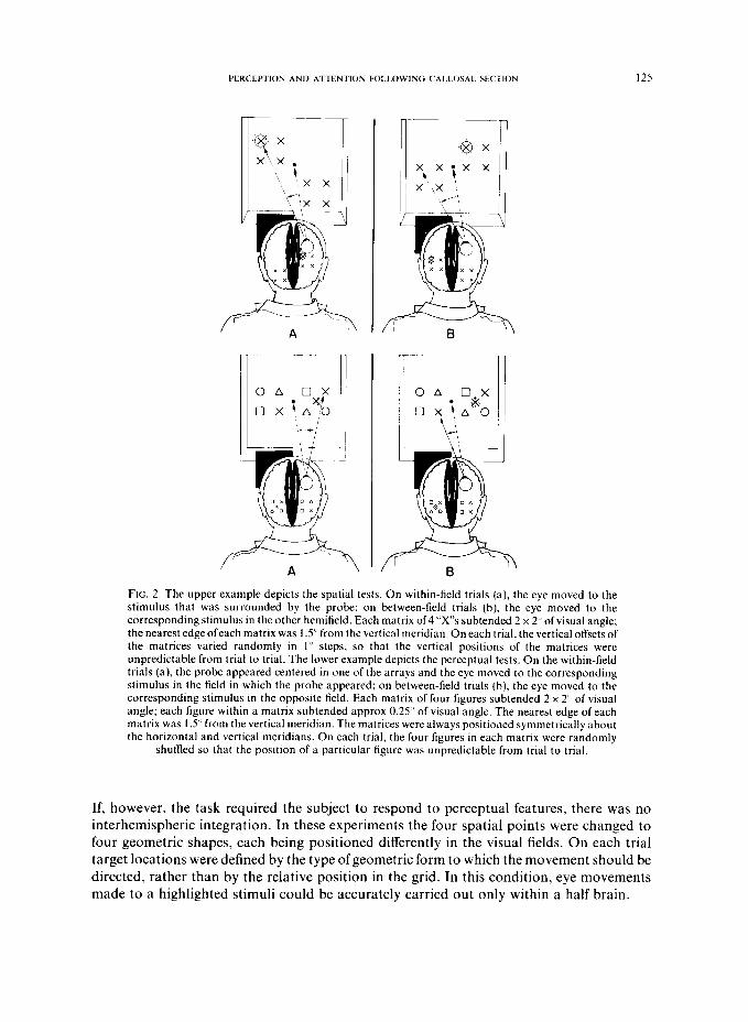
‘u!wq 3p2q t2 u!qi!~ dluo in0 pa!~le~ A~awn33r! aq pIno gnuys paiq%qq%!q e 01 aperu s$uawaAow alCa ‘uoypuo~ sly1 UI ‘p!.~% aql u! uog!sod a,yela~ aql Lq ur?q~ laqlw ‘pa)Da.up aq p]noqs )uauraAoLu ayl qXqM 01 u.1.103 syawoa83o ad& aql dq pauyap aJaM suo!~1?30~ la%.wl ~Z!.IJ qzea uo ‘splay pzns!A aq) u! /CI3ualag!p pauoysod Su!aq qcea ‘sadeqs Dgauroa8 Ino3 01 pafheq3 alaM sw!od Iyvzds rno3 aq3 sluauyadxa asaql UI ‘uoge.GE?a~u! cyaqds!uraqla$u! ou WM alay] ‘sa.wr?a3 IvnldaDlad 01 puodsal 01 IDa[qns aq$ pa+nbal ysel aql ‘IaAaMoq ‘31
‘pl) 01 [eq UIOJJ a[qvta!paJdun SBM ahiy Je[na!lJvd B JO uog!sod aql leq) OS pamnqs
L~LLIO~UEJ aJaM x!Jlsrn qaza ul saJnOy JnoJ aql ‘[~!JJ qasa uo 3ue!p!~arn leagJai\ pue ~e~uoz!~oq aq,
lnoqe i(l[~agaururds paoog!sod sb~41~e aJaM saa!~)vtu aqL .ue!p!~aru pzag~a~ aql luoq ,,s’, SRM x!~lmu pea JO a%pa lsaleau aqL .a@ue [ens!A JO c,sz'o xoldda papualqns x!~lew e uypu alnay qaea :a@?
[ens!A JO -2 x 2 papualqns saJn%y InoJ JO x!llsm qaG3 ‘play al!soddo aql u! sn[nw!ls Bu!puodsaJJoa
aql 01 panow a.ia aql ‘(q) sp?!~i play-uaamlaq uo :paleadde aqold aql qa!qM u! play aq, u! sn[nm!is
Bu!puodsaJJoa aql 01 pahour ai(a aql pw sde~~e aq, JO auo u! paIa)uaa paJeadde aqold aqt ‘(B) s~_?!JI
play-u!q]!M aql uo ‘sisal pznldanlad aqi s]s!dap aldurexa laMo[ aqL ‘IE!J) 01 [B!JI LUOJJ alqela!paldun
aJaM saap]ew aql JO suog!sod [t?a!).~aa aq, )eq) OS ‘sda]s .I u! L[LLIO~W?J pa!JeA saa!-l)sm aql
Jo siasgo pqlai\ aql ‘leg qar?a ug w!p!lam ~!JJ~A aql WOJJ .q L sm+ xysm qavaJ0 a??pa lsa.teau aql
!a@? [ens!AJo ,,i: x 2 papualqns s&.x,, PJO x!~larn qaex ‘play!maq laqlo aql u! snlnm!ls %u!puodsaJJoa
aq) 01 paAotu al(a aql ‘(q) sle!ll play-uaaM)aq uo :aqoJd aq) Lq papunoJlns SBM leq) sn[nm!ls
aql 01 paAom a6a aql ‘(e) sp+ti play-u!ql!M uo ‘slsai [e!)eds aql sla!dap aldwsxa Jaddn aqL ‘z ‘~I.J
-1
126
THERE IS COLLICULAR-CORTICAL INVOLVEMENT IN THE CROSS-INTEGRATION OF SPATIAL INFORMATION FOLLOWING
CALLOSAL SECTION
Although the above reviewed findings on split-brain patients imply that representation of the ipsilateral hemifield is not completely eliminated by callosal transection. they do not necessarily imply that the availability of such information depends on interactions between cortical and subcortical structures. Consistent with the much reported blindsight hypothesis, ipsilateral representation could be provided directly via retino-collicular projections, independent of any cortico-collicular influence. If so, it would be expected that such ipsilateral representation would be unaffected by damage to the occipital cortex, and that the performance of a patient with occipital damage at these tasks would be comparable to that obtained from the commissurotomy patients.
The performance of Case B.H. indicates that this is not the case. B.H. is a 34-year-old woman who had undergone surgery for an AVM in the right occipital lobe. Postoperatively. she had a dense left homonymous hemianopia (Fig. 3). She performed quite accurately when required to localize targets in her perceptually intact RVF, whereas her performance did not exceed chance levels for eye movements into her perceptually blind LVF. Thus. there is no evidence that these targets could be localized in a visual field rendered blind by occipital damage. Therefore, the localization of visual stimuli in the ipsilatcral hemifield would appear to require intact occipital cortices.
This kind of observation raises questions about the generality of the concept of “blindsight” [28], and suggests that simple spatial information can not be managed by an intact collicular (or other mid-brain) system in the absence of normal input from visual cortical areas. Cross-integration of such information is only possible when midbrain structures remained connected to cortical processes. as is the case in the split-brain patient.
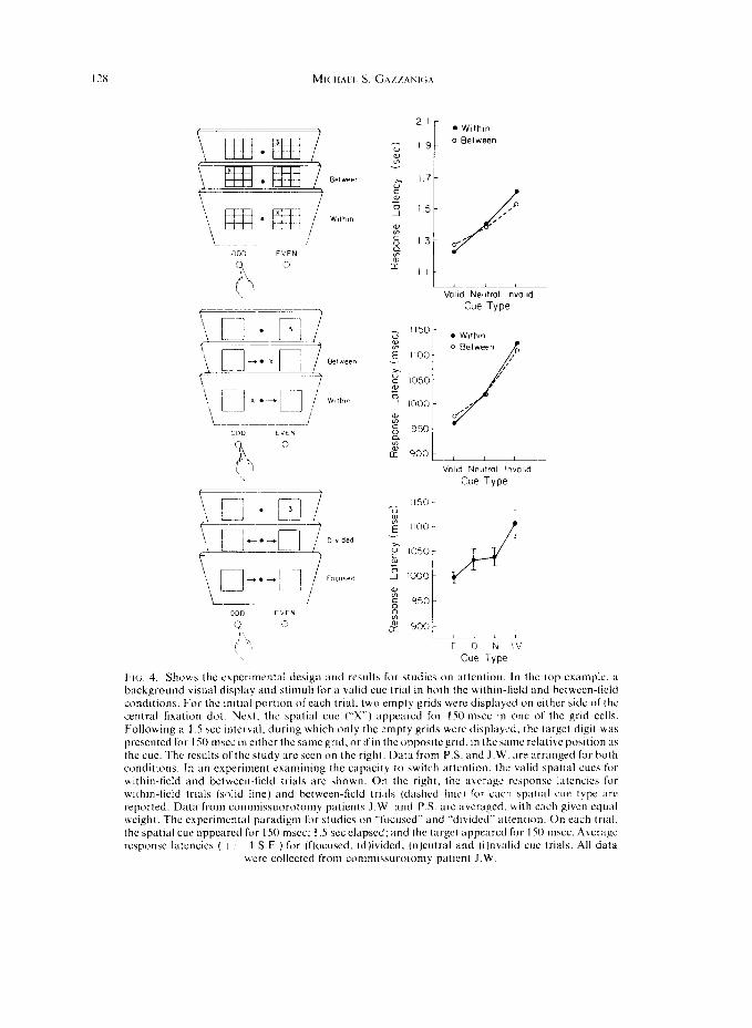
SPATIAL ATTENTION CAN BE DIRECTED BETWEEN THE DISCONNECTED HEMISPHERES
With the foregoing demonstration that some kind of crude spatial information remained integrated between the two half brains, it was natural to also consider whether or not attentional processes associated with spatial information were affected with cortical disconnection surgery. Using a modification of a paradigm developed by POSNE:K [ 171 that capitalizes on priming phenomena. it was shown that either hemisphere was capable of directing attention to any point in the same or opposite visual field [ 161 (Fig. 4, top. middle). In brief, Posner et nl. first showed that the response latency to a peripheral visual target is reduced when observers have prior information regarding its spatial locus, cvcn when eye movements to the cued location are prohibited. Presumably, the spatial cue allows observers to direct their attention to the appropriate location prior to the onset of the target. In the present context. this paradigm was used in split-brain patients to mc;lsurc the extent to which such attentional cues affected performance under a variety ofconditions. In two cxperimcnts, we determined that despite the perceptual segregation ofthc visual fields for explicit stimulus identification in split-brain patients, the separated hemispheres are not strictly indcpendcnt in the control of spatial orientation. They rely on a common orienting system that serves to maintain a single focus of attention, a system that makes use of visual information from both hemifields. Thus. as with normal\, a CLK to direct attention to ;I particular point in the visual field was honored no matter which half way presented with the critical stimulus. Finally-. it is

PERCEPTION AND ATTENTION FOLLOWING CALLOSAL SECTION 127
CORRECT INITIAL SACCADES
lo v BH
t
: I?;“,:
2? 8 - m3,
;
8 .6
FIG. 3. Goldman perimetry for the right eye of subject B.H. B.H. had a dense left homonymous hemianopia following a right occipital lobe excision. She was tested using a visual display as depicted here. It consisted of two 2” square matrices of four l/4” “X”s located 1 S” from a central fixation. Each trial began with a 1.5 set period of fixation, during which the display shown here was presented. A probe circle was then flashed around one of the “X”s for 150 msec. After the probe, a tone sounded and she was required to move her eyes to the “X”. The results, seen on the right, show her performance for both the good and the hemianopic field for both the initial saccade and the final position. Each data point represents a block of 25 trials. 15 blocks of 20 trials for each of the delays 25% correct
reflects chance performance.
clear from the first experiment that the capacity to direct attention between the hemispheres is specific to the cued location.
SPATIAL ATTENTION CANNOT BE DIVIDED WITH EACH HEMISPHERE HAVING INDEPENDENT CONTROL
The discovery that spatial attention could be directed with ease to either visual field raised the question of whether each separate cognitive system in the split-brain patient could, if instructed to do so, direct attention to a particular part of its own visual field independent of the activities of the other half brain [15]. Thus, could the right hemisphere direct attention to a point in the left visual field, while the left brain simultaneously directed attention to a point in the right visual field. Normals cannot so divide their attention, but it was a possibility that the split-brain patient, especially those patients with complex mental control systems for each half brain could do so.
178 MI(.HAI.I. S. C;A/LANK;A
L-.__.. 1
Valid Neutral lnvalld
Cue Type
. Wlthln
_/I
Volld Neutral lnvolid
Cue Type
!I50 z % E 1100
_,
u” E 1050 3 1 1000
% 6 950
%
d 900 I_
2-J FIG. 4. Shows the cxperunrntal design and results for studies on attention. In the lop example. ;I background visual display and stimuli for a valid cue trial in both the within-lield and hctwecn-licld conditions. For the initial port1011 of each trial. two empt? grids were displayed on either side of the central fixation dot. Next, the spatial cue (“X”) appeared for I50 m5cc in one of the grid cells. Following a 1.5 set Interval, during which only the empty grids wcrc‘ displayed, the target digit was presented for 150 msec in either the same grid. or ifin the oppositcgrid. in the same relative position :15 the cue. The results of the study are seen on the right. Data from P.S. and J.W. are arranged for both conditions. In an experiment examining the capacity to hwitch attention. the valid spatial cues for wrthin-field and between-field trials are shown. On the right, the a\~age response latencics for wlchin-lield trials (solid line) and between-field trials (dashed 11~) for each spatial cue type are reported. Data from commissuorotomy patients J.W. :md KS. are averaged. with each given equal weight. The experimental paradigm for studies on -‘li~cuscd” and “divided” attention. On each trial,
the spatial cut appeared for I SO msc: 1.5 set clapscd; and the target appeared for 150 mscc. Average response latcncies ( + I- I SE ) for (flocused, (d)ivided, (n)eutral and (i)n\alid cue trials. All data
were collected from commisurotomy patient J.W.
PERCEPTION AND ATTENTION FOLLOWING CALLOSAL SECTION 129
Results of several studies have shown that the split-brain patient is unable to divide attention between the two half brains. There would appear to be one integrated attention system that remains intact following cortical disconnection. The background visual display for this study, generated by a microprocessor and displayed on a video monitor, is depicted in Fig. 4. It consisted of two 3” square boxes, presented 7” on either side of a central dot that the observer fixated at all times. On each trial a target digit briefly appeared in one of the boxes and the observer indicated with a forced-choice key press whether the digit was even or odd. On each trial one of four spatial cue configurations briefly appeared 1.5 set before the onset ofthe target. On “focused” attention trials two arrows, one in each field, pointed to the box in which the target would appear; on “divided” attention trials, the arrows pointed in opposite directions; on neutral cue trials, two non-informative X’s appeared in place of the arrows; and on invalid cue trials two arrows pointed to the wrong box.
If the separated hemispheres have completely independent attentional systems, response latencies on “focused” and “divided” attention trials should be similarly facilitated. If spatial attention is restricted to one area, then performance on “divided” attention trials should more closely resemble performance on “neutral” cue trials,
It can be seen from Fig. 4 that performance on “divided” attention trials was most similar to performance on neutral cue trials. These results imply that commissurotomy does not result in separate orienting systems that can be manipulated independently and concurrently by each hemisphere. Thus, like neurologically intact observers, split-brain patients are unable to prepare for events in two spatially disparate locations, i.e., their attention is unifocal.
ATTENTIONAL RESOURCES ARE SHARED IN THE SEPARATE COGNITIVE SYSTEMS OF SPLIT-BRAIN PATIENTS
An operational definition of attention has eluded investigators of human cognitive function. Yet few attempts surpass William James observation, “Everyone knows what attention is. It is the taking possession by the mind, in clear and vivid form, of one out of what seems several simultaneously possible objects or trains of thought. Focalization, concentra- tion, of consciousness are of its essence. It implies withdrawal from some things in order to deal effectively with others”. Such a quintessential operation of human cognition might be thought to be so tied to cognitive processes that it exists as part of any separately acting cognitive system. Others would say what we call attention is merely an observer’s description of the operating characteristics of a functioning system. Such views can be directly examined in the split-brain patient where it is established beyond a doubt that each half brain in the select few cases that possess language in each hemisphere can cognate independent of the other hemisphere. From this. one would predict that the cognitive operations of one half brain, no matter what the difficulty, would little influence the cognitive activities of the other. A competing view is that the brain and psychologic system have an important additional parameter usually referred to as “resources”. These are set and limited. They exist in a common pool and represent in some way the fuel for cognitive operations. If they are being applied to task A, there are fewer available for task B. This model would predict that the harder hemisphere A worked on a task, the worse hemisphere B would do on a task of constant complexity. One of the implications of such a finding would be that in fact the attentional system is an independent system serving the cognitive activities of a psychological
system.

Two studies have been carried out on this issue; both confirm the notion that there is a set amount of central resources. In the first experiment, two series of geometric shapes were displayed concurrently to the left and right ofcentral fixation and thus were lateralized to the right and left hemispheres respectively [ 131. A unilateral probe tigure subsequently appeared, and the observer indicated with a forced-choice key press whether it matched any of the probed field’s items (Fig. 5). On half of the trials the same three figures were displayed
Y l N
% !-f&J
FIG. 5. Sequence of events for 3 redundant Scondition trial. Stimuli were sclectcd from 3 cc1 ufccven geometric forms (square, circle, triangle and so forth). each about 3 x 2 of visual angle and displayed 5 directly to the left and right of central fixation. Each stimulus appeared for I SO mxc Hlth an interstimulus interval of 500 msec. A I set d&q followed presentation of the last rtm~ulu~. and the unilateral probe stimulus was presented for IS0 msec. All displays were generated by :I
microprocessor (Apple II) and presented on a video monitor at a viewing distance of0.S m
in the two fields. the hard condition. On the other half, one hemisphere saw three items while the other saw only one stimulus presented three times, the latter being the easy condition. The results clearly showed that when one half brain was working on processing only one repeated stimulus, the opposite hemisphere performed better at recalling whether the probed stimulus was part of the original set of three stimuli. When both hemisphcrcs were trying to process three stimuli, the performance of each was impaired.
In a follow up study, the experiment was extended to include lexical memory [20]. The task required remembering and recognizing lists of high frequency words. While one hemisphere learned a short, medium, or long word list (the primary task), the other hemisphere learned another word list offixed length (the secondary task). If the hemispheres share resources, performance by the hemisphere learning a constant number of words should be influenced by the length of the opposite hemisphere’s list.
Two patients were examined, .l.W. and V.P. The subjects were first exposed to 20 trials of bilaterally presented words. One hemisphere received a constant list of IO words, (repeated twice), while the other reccivcd tither 2, 1Oor 20 words. In the two word condition the words were repeated IO times while in the 10 word condition they were repeated two times. On half
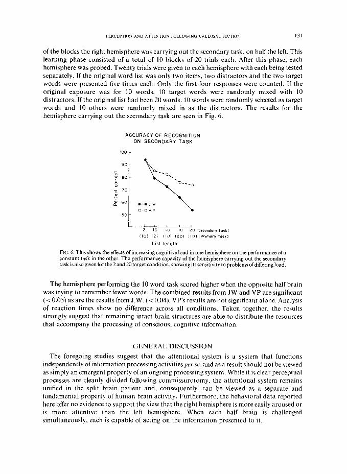
PERCEPTION AND ATTENTION FOLLOWING CALLOSAL SECTION 131
of the blocks the right hemisphere was carrying out the secondary task, on half the left. This learning phase consisted of a total of 10 blocks of 20 trials each. After this phase, each hemisphere was probed. Twenty trials were given to each hemisphere with each being tested separately. If the original word list was only two items, two distracters and the two target words were presented five times each. Only the first four responses were counted. If the original exposure was for 10 words, 10 target words were randomly mixed with 10 distracters. If the original list had been 20 words, 10 words were randomly selected as target words and 10 others were randomly mixed in as the distracters. The results for the hemisphere carrying out the secondary task are seen in Fig. 6.
ACCURACY OF RECOGNITION
ON SECONDARY TASK
HJW
O-O v P \
20 (Secondary Task)
(IO) (2) 110) I201 llO)iPr~mary Task1
Lfst length
Fit;. 6. This shows the effects of increasing cognitive load in one hemisphere on the performance of a constant task in the other. The performance capacity of the hemisphere carrying out the secondary task is also given for the 2 and 20 target condition, showing its sensitivity to problems ofdiffering load.
The hemisphere performing the 10 word task scored higher when the opposite half brain was trying to remember fewer words. The combined results from JW and VP are significant (< 0.05) as are the results from J.W. (< 0.04). VP’s results are not significant alone. Analysis of reaction times show no difference across all conditions. Taken together, the results strongly suggest that remaining intact brain structures are able to distribute the resources that accompany the processing of conscious, cognitive information.
GENERAL DISCUSSION
The foregoing studies suggest that the attentional system is a system that functions independently of information processing activities per se, and as a result should not be viewed as simply an emergent property of an ongoing processing system. While it is clear perceptual processes are cleanly divided following commissurotomy, the attentional system remains unified in the split brain patient and, consequently, can be viewed as a separate and fundamental property of human brain activity. Furthermore, the behavioral data reported here offer no evidence to support the view that the right hemisphere is more easily aroused or is more attentive than the left hemisphere. When each half brain is challenged simultaneously, each is capable of acting on the information presented to it.
132 bfIC’HA1.I. S. C;,277AVIGA
Determining where in the information processing sequence attentional resources are needed has been a difficult task. The foregoing studies suggest that split-brain subjects. like normals, pay the price for total complexity of tasks given to the brain. That is, there appears to be a set amount of resources available for information processing and these remain constant even in the disconnected brains where there is a clear capacity to have separate and independent perceptual and cognitive operations.
Spelling out the exact relation between the various components of the attentional system is not yet possible. In the present review, two related but distinct kinds of attention have been examined. The mechanism of spatial attention and the mechanism of resource allocation have been shown to be closely related in how they operate in the cortically disconnected human. It cannot, however, be assumed that the two processes arc one and the same. lt remains for further experimentation to clarify this issue.
Finally, it is of interest to consider where in the total sequence of a perceptual-motor task, resources are applied. Are they, for example; automatically applied during the early phases of information processing that deal with the complexity of the visual stimulus itself‘? Or arc attentional processes of the type that draw on central resources active at later loci of the information processing sequence that deal with the cognate aspects of the task‘? The present series of experiments suggests the allocation of resources is most involved with the memory or more cognitive aspects of a task. While resources may be applied to the simple perceptual stages of an information processing task, such as the kind used in the dual task performance test described above, they do not appear demanding. It is only when the normal intact subject perceives the perceptual array as a more complex task, that the system discovers it does not have enough resources to carry out the task.
REFERENCES I. BMXN, J. E., FISHFK, E. D. and VOC;I:I,. P. J. Cerebral commissuorotomy: a second case report. J. .dnr. Med
Assoc. 194, 1328-l 329. 1965. 2. DIMOh'D, S. J. Performance by split-brain humans at lateralized vigilance tasks. (‘or~rz 15, 43 50. 1979. 3. EI LEP;HEKG. L. and SPFKKY, R. W. Capacity for holding sustained attention followingcommi~surotomy. C‘r,rfc\
15,43143x. 1979. 4. EI.L.ENBFKG. L. and SPFKKY. R. W. Lateralized division of attention in the commissuorotomiLed and intact
brain. N~uropsycholoUitr IS, 41 I 418. 1980. 5. GAZZANIGA, M. S. Short term memory and brain bisected man. Ps~honon~. SC,;. 12, 161 162, 196X 6. GAZZANIC;A. M. S. The Hisectd Bruin. Appleton Century Crofts, New York. 1970. 7. GAZZANI~~A. M. S.. BAYNES, K. A., HIKST. W. A. and M<CL.~AKY. C. Profiles of right hemisphere language and
speech following brain bisection. Brain Lung. 22, 206 220, 19X4. X. GAZZANK;A. M. S.. HOLTZMAK, J. D.. DLCK, M. D. F. and Lw, B. c‘. P. NMR assessment of human callosal
surgery w,ith neuropsychological correlates. .Yrur~~/~~q~ 35. 1763 1766, 19X5. 9. GAZZANI(;A, M. S.. KUTAS, M.. SMYLII. C. S. and HOLI/.M.AX. J. D. (Unpublished observations).
IO. GAZZANKA, M. S. and Lr:Doux. J. E. The Irlrcyrtrtrd Mind. f’lenum Press. New York. 1978. I I. GA~'ZANI(;A, M. S. and YOKNG. E. D. Effects of commlssurotomy on the processing of incrcastng v~sua)
information. Eupl. Braifr Kea. 3, 36X 371. 1967. 12. HOLTZMAY. J. D. Interactions between cortical and subcortical YI\U;II arcah: evidence from human
commissurotomy patients. Vision Kes. 24, 801 X 13. 19X4. 13. HOLT~MAK, J. D. and GAZIANIGA. M. S. Dual task interactions duecxcluslvely to hmits in processing rcaourceh.
.Scifwr 218, I325 1327, 1982. 14. HOLTZMAX. J. D. and GA~~ANIGA. M. S Enhanced dual task performance following corpus commissurotomy
in humans. Neurops~~holo~ia 236, 3 IS 31 I ( 1945. 15. HOL~UMAN. J. D., VOLPF., B. T. and GA~TANK;A. M. S. Spatial orientation following commissural scctlon. In
I;arirtirs of‘ilttmrion. R. PAHASIIKAMAN and D. R. DA~II,S (Editors), pp. 375 394. Academic Press. New York. 1984.
16. HOI.TZMAL~, J. D., SIIITIS, J. J., VOI 1’1. B T., WILSON, I). H. and GAUANIGA. M S. D~asoc~at~on of hpatial information for stimulus localization and the control of attention. Bruin 104, 861 X72, I98 I.
PEKCEPTlON AN,) ATTENTION FOLLOWING CALLOSAL SECTION 133
17. POSNEK, M. I., SNYDER, C. R. and DAVIDSON, B. J. Attention and the detection of signals. J. Exp. Psycho!. Gen. 21, 160 174, 1980.
18. PREILOWSKI, B. Possible contribution of the anterior forebrain commissures to bilateral motor coordination. Neuropsycholoyia 101, 267 271, 1972.
19. RAMACHANI~RAN, V. S., CRONIN-GOLOMB, A. and MYERS, J. J. PerceptIon of apparent motion by commissurotomy patients. Nature 320, 358-359.
20. REDINGTON, K., HOLTZMAN, J. D. and GAZZANIC;A, M. S. Separate lexical systems ofcommissurotomy patients share common processing resources (submitted).
21. REEVES, A. G. (Editor) Epilepsy and the Corpus Cullosum. Plenum Publishing Corporation, New York, 1985. 22. SERGENT, J. Unified response to bilateral hemispheric stimulation by a split-brain patient. Nufure, Land. 305,
80%302. 1983. 23. SERGENT, J. Subcortical coordination of hemisphere activity in commissurotomized patients. Brain 109,
351-369, 19X6. 24. SIDTIS, J. J. Bilateral language and commissurotomy: interactions between the hemispheres with and without
the corpus callosum. In Epilepsy and the Corpus Callosum, A. G. REEVES (Editor), pp. 369 -380. Plenum Press, New York, 1985.
25. SPERRY, R. W. Mental unity following surgical disconnection of the cerebral hemispheres. The Harvey Lecture Series 62, pp. 293 323. Academic Press, New York, 1968.
26. TENC;, E. L. and SPERRY, R. W. Interhemispheric interaction during simultaneous bilateral presentation of letters and digits in commissurotomized patients. Neuropsycholoyia 11, 131- 140, 1973.
27. TENC;, E. L. and SPERRY, R. W. Interhemispheric rivalry during simultaneous bilateral task presentation in commissurotomized patients. Cortex 10, 11 l-120, 1974.
28. WEISKRANTZ, L., WARRINGTON, E. R., SANDERS, M. D. and MARSHALL, J. Visual capacity in the hemianopic field following a restricted occipital ablation. Bruin 97, 709-728, 1974.
29. ZAIUEL, E. Disconnection syndrome as a model for laterality effects in the normal brain. In Cerebral Hemisphrre Asymmrfry: Method, Throry, and Application, J. B. HELLICE (Editor), pp. 95-151. Praeger Publishers, New York, 1983.