Embed Size (px)
Citation preview
Khan, Harris, Barna, Craig, Bruce, Munro, Moody & Scrutton 1
Pentaerythritol tetranitrate reductase: kinetic and structural
basis of reactivity with NADPH, 2-cyclohexenone,
nitroesters and nitroaromatic explosives.
Huma Khan‡, Richard J. Harris‡, Terez Barna‡, Daniel H. Craig‡*, Neil. C.
Bruce§, Andrew W. Munro‡, Peter C. E. Moody‡ and Nigel S. Scrutton‡
‡ Department of Biochemistry and Centre for Chemical Biology, University of Leicester,
University Road, Leicester LE1 7RH, UK
§ Institute of Biotechnology, University of Cambridge, Tennis Court Road, Cambridge
CB2 1QT, UK
Running title: explosive degradation by PETN reductase
*Present address: Department of Chemistry, University of Edinburgh, West Mains Road,
Edinburgh, Scotland, UK
Corresponding author: Professor N. S. Scrutton. Telephone +44 116 223 1337; Fax,
+44 116 252 3369; email, [email protected].
This work was funded by grants from the Biotechnology and Biological Sciences
Research Council, the Wellcome Trust and the Lister Institute of Preventive Medicine.
N.S.S. is a Lister Institute Research Professor. The first 3 authors contributed equally to
the work.
Copyright 2002 by The American Society for Biochemistry and Molecular Biology, Inc.
JBC Papers in Press. Published on March 28, 2002 as Manuscript M200637200 by guest on A
ugust 18, 2018http://w
ww
.jbc.org/D
ownloaded from

Khan, Harris, Barna, Craig, Bruce, Munro, Moody & Scrutton 2
ABBREVIATIONS AND FOOTNOTES
1Abbreviations: PETN, pentaerythritol tetranitrate; OYE, Old Yellow Enzyme; MR,
morphinone reductase; EBP, estrogen binding protein; TNT, trin itrotoluene; GTN,
glycerol trinitrate; 2,4-DNP, 2,4-dinitrophenol.
by guest on August 18, 2018
http://ww
w.jbc.org/
Dow
nloaded from

Khan, Harris, Barna, Craig, Bruce, Munro, Moody & Scrutton 3
ABSTRACT
The reaction of pentaerythritol tetranitrate reductase with reducing and oxidising
substrates has been studied by stopped-flow spectrophotometry, redox potentiometry and
X-ray crystallography. We show in the reductive half-reaction of PETN reductase that
NADPH binds to form an enzyme -NADPH charge -transfer intermediate prior to hydride
transfer from the nicotinamide coenzyme to FMN. In the oxidative half-reaction, the 2
electron-reduced enzyme reacts with several substrates including nitroester explosives
[glycerol trinitrate (GTN) and pentaerythritol tetranitrate (PETN)], nitroaromatic
explosives [trinitrotoluene (TNT) and picric acid] and α,β unsaturated carbonyl
compounds (2-cyclohexenone). Oxidation of the flavin by the nitroaromatic substrate
TNT is kinetically indistinguishable from formation of its hydride -Meisenheimer
complex, consistent with a mechanism involving direct nucleophilic attack by hydride
from the flavin N5 at the electron-deficient aromatic nucleus of the substrate. Crystal
structures of complexes of the oxidised enzyme bound to picric acid and TNT are
consistent with direct hydride transfer from the reduced flavin to nitroaromatic substrates.
The mode of binding the inhibitor 2,4 dinitrophenol (2,4 DNP) is similar to that observed
with picric acid and TNT. In this position the aromatic nucleus, however, is not activated
for hydride transfer from the flavin N5, thus accounting for the lack of reactivity with 2,4
DNP. Our work with PETN reductase establishes further a close relationship to the Old
Yellow Enzyme family of proteins, but at the same time highlights important differences
compared with the reactivity of OYE. Our studies provide a structural and mechanistic
rationale for the ability of PETN reductase to react with the nitroaromatic explosive
compounds TNT and picric acid, and for the inhibition of enzyme activity with 2,4 DNP.
by guest on August 18, 2018
http://ww
w.jbc.org/
Dow
nloaded from

Khan, Harris, Barna, Craig, Bruce, Munro, Moody & Scrutton 4
INTRODUCTION
A large number of sites worldwide are contaminated with high explosives as a result of
large-scale manufacturing and handling of these compounds. Bioremediation is an
attractive means of decontaminating such sites (1), which has led to a search for enzymes
capable of degrading high explosive compounds. We previously isolated a strain of
Enterobacter cloacae (strain PB2) on the basis of its ability to utilise nitrate ester
explosives such as pentaerythritol tetranitrate (PETN1) and glycerol trinitrate (GTN) as
sole nitrogen source (2). The ability of Enterobacter cloacae PB2 to utilise nitrate esters
as a nitrogen source is conferred by the NADPH-dependent flavoenzyme PETN
reductase (3). Sequence analysis of the cloned gene encoding PETN reductase has
established a close evolutionary relationship with the flavoenzyme Old Yellow Enzyme
(OYE) (4), and related enzymes such as bacterial morphinone reductase (MR) (5) and the
estrogen-binding protein (EBP) of Candida albicans (6). These enzymes bind a variety of
cyclic enones, including 2-cyclohexenone and steroids. Some steroids act as substrates
whilst others are inhibitors of both PETN reductase and OYE. We have demonstrated that
PETN reductase degrades all major classes of explosive including nitroaromatic
compounds [e.g. trinitrotoluene (TNT); (7-9)] and cyclic triazine explosives [e.g. Royal
Demolition Explosive (RDX)], making the enzyme attractive in phytoremediation of
explosive contaminated land (10). Homologues of PETN reductase from strains of
Pseudomonas (11) and Agrobacterium (12) have been isolated and these enzymes also
show reactivity against explosive substrates. In the case of xenobiotic reductase from
Pseudomonas fluorescens I-C, the products of TNT reduction have been identified and
by guest on August 18, 2018
http://ww
w.jbc.org/
Dow
nloaded from

Khan, Harris, Barna, Craig, Bruce, Munro, Moody & Scrutton 5
shown to proceed either by hydride addition to the aromatic nucleus or by nitro group
reduction (13).
The crystal structure of PETN reductase has been solved in both its oxidised and 2
electron-reduced forms (14). The structures of a number of complexed forms with both
steroid substrates and inhibitors are also known (14). The enzyme is a conventional eight-
fold β/α barrel protein that contains a single FMN redox centre, and overall resembles the
structure of OYE (15). However, the mode of steroid binding to oxidized enzyme differs
from that seen with OYE in that the reactive olefinic bond in the steroid is not positioned
over the flavin N5 (14). Reactions performed with ‘A-side’ deuterated nicotinamide
cofactor have shown that in 2 electron-reduced PETN reductase the steroid is ‘flipped’
compared with the mode of binding to oxidized enzyme (14). In this ‘flipped’ binding
mode the reactive olefinic bond is aligned with the flavin N5 atom in a geometry that is
compatible with hydride transfer to the steroid substrate. Deuterium labelling methods
have enabled us to assign the reactive olefinic bond as the C1-C2 bond in
1,4−androstadiene-3,17-dione and prednisone, to elucidate the stereochemistry of bond
reduction and to propose a mechanism for the reduction of cyclic enones (14). Our work
on the stereochemistry of olefinic bond reduction by PETN reductase again establishes a
close relationship with OYE. Vaz et al (16) have shown that reduction of α,β-unsaturated
carbonyl compounds by OYE proceeds by hydride transfer from the flavin N5 to the β
carbon followed by proton uptake at the α carbon — a finding that is consistent with our
more recent determination of the stereochemistry of bond reduction catalysed by PETN
reductase.
by guest on August 18, 2018
http://ww
w.jbc.org/
Dow
nloaded from
Khan, Harris, Barna, Craig, Bruce, Munro, Moody & Scrutton 6
In this paper we report a detailed kinetic analysis of the reaction of PETN
reductase with NADPH and the substrate 2-cyclohexenone, which is used widely as a
‘generic’ substrate of the OYE family of enzymes. We also report studies of enzyme
oxidation by nitroester substrates (GTN and PETN) and the nitroaromatic explosives
TNT and picric acid. The structures of PETN reductase complexed with picric acid, TNT,
2-cyclohexenone and the inhibitor 2,4 dinitrophenol (2,4-DNP) are also presented, which
provide atomic insight into the mechanism of nitroaromatic reduction and the reduction
of α,β unsaturated carbonyl compounds.
by guest on August 18, 2018
http://ww
w.jbc.org/
Dow
nloaded from

Khan, Harris, Barna, Craig, Bruce, Munro, Moody & Scrutton 7
EXPERIMENTAL PROCEDURES
Chemicals and enzymes — Complex bacteriological media were from Unipath and all
media were prepared as described by Sambrook et al (17). Mimetic Orange 2 affinity
chromatography resin was from Affinity Chromatography Ltd. Q-sepharose resin was
from Pharmacia. PETN reductase was prepared from E. coli JM109/pONR1 and purified
as described (3) , but also incorporating a final chromatographic step using Q-sepharose
(14). NADPH, glucose 6-phosphate dehydrogenase, glucose 6-phosphate, benzyl
viologen, methyl viologen, 2-hydroxy-1,4-naphthaquinone, phenazine methosulfate and
2,4 DNP were from Sigma. 2-cyclohexenone was from Acros Organics. Dr S Nicklin
(UK Defence and Evaluation Research Agency) supplied TNT, GTN, PETN and picric
acid. The following extinction coefficients were used to calculate the concentration of
substrates and enzyme: NADPH (ε340 = 6.22 x 10³ M-1 cm-1); PETN reductase (ε464 =
11.3 x 10³ M-1 cm-1); 2-cyclohexenone (ε232 = 11.0 x 10³ M-1 cm-1). Stock solutions of
TNT (600 mM) were made up in acetone. Dilutions were then made into potassium
phosphate buffer, pH 7.0, and the acetone concentration was maintained at 1 % (v/v). The
presence of acetone in buffers at 1 % (v/v) was shown not to affect enzyme activity.
Redox potentiometry — Redox titrations were performed within a Belle Technology
glove box under a nitrogen atmosphere (oxygen maintained at <5 ppm) in 50 mM
potassium phosphate buffer, pH 7.0. Anaerobic titration buffer was prepared by flushing
freshly prepared buffer with oxygen-free nitrogen. PETN reductase admitted to the glove
box was de-oxygenated by passing through a Biorad 10DG column, with final dilution of
the eluted protein to give a concentration of ~60 µM. Solutions of benzyl viologen,
by guest on August 18, 2018
http://ww
w.jbc.org/
Dow
nloaded from
Khan, Harris, Barna, Craig, Bruce, Munro, Moody & Scrutton 8
methyl viologen, 2-hydroxy-1,4-naphthaquinone and phenazine methosulfate were added
to a final concentration of 0.5 µM as redox mediators for the titrations. Absorption
spectra (300 - 750 nm) were recorded on a Varian (Cary 50 probe) UV-visible
spectrophotometer, and the electrochemical potential was monitored using a Hanna
instruments pH/voltmeter coupled to a Russell Pt/calomel electrode. The electrode was
calibrated using the Fe(II)/Fe(III)-EDTA couple (+108 mV) as a standard. The enzyme
solution was titrated electrochemically using sodium dithionite as reductant and
potassium ferricyanide as oxidant, as described by Dutton (18). After the addition of each
aliquot of reductant, and allowing equilibration to occur (stabilization of the observed
potential), the spectrum was recorded and the potential was noted. The process was
repeated at several (typically ~ 40) different potentials. In this way, a set of spectra
representing reductive and oxidative titrations was obtained. Small corrections were
made for any drift in the baseline by correcting the absorbance at 750 nm to zero. The
observed potentials were corrected to those for the standard hydrogen electrode (SHE)
(Pt/calomel + 244 mV). Data manipulation and analysis were performed using Origin
software (Microcal). Absorbance values at wavelengths of 468 nm (close to the oxidized
flavin maximim) were plotted against potential. Data were fitted using Eq. 1, which
represents a concerted 2-electron redox process derived by extension to the Nernst
equation and the Beer-Lambert Law, as described previously (18):
( )
( ) 5.29/
5.29/
46812
12
101)10(
EE
EEbaA
−
−
++
= Eq. 1
where A468 is the absorbance value at 468 nm at the electrode potential E, and a and b are
the absorbance values of the fully oxidized and reduced enzyme, respectively, at 468 nm.
by guest on August 18, 2018
http://ww
w.jbc.org/
Dow
nloaded from
Khan, Harris, Barna, Craig, Bruce, Munro, Moody & Scrutton 9
In using Eq. 1 to fit the absorbance-potential data the variables were unconstrained, and
regression analysis provided values in close agreement to those of the initial estimates.
Throughout the titration the enzyme remained soluble and corrections for turbidity were
not required.
Kinetic measurements— Rapid reaction kinetic experiments were performed using an
Applied Photophysics SF.17MV stopped-flow spectrophotometer contained within an
anaerobic glove box (Belle Technology). Time-dependent reductions of PETN reductase
with NADPH were performed by rapid-scanning stopped-flow spectroscopy using a
photodiode array detector and X-SCAN software (Applied Photophysics). Spectral
deconvolution was performed by global analysis and numerical integration methods using
PROKIN software (Applied Photophysics). For single wavelength studies, data collected
at 464 nm and 560 nm were analysed using nonlinear least squares regression analysis on
an Acorn Risc PC microcomputer using Spectrakinetics software (Applied Photophysics).
Experiments were performed by mixing PETN reductase in the appropriate buffer with an
equal volume of NADPH in the same buffer at the desired concentration. For studies of
the oxidative half-reaction, PETN reductase was titrated with sodium dithionite to the 2-
electron level and then mixed with 2-cyclohexenone. In reductive and oxidative
reactions, the concentration of substrate was always at least 10-fold greater than that of
enzyme, thereby ensuring pseudo-first-order conditions. For each substrate concentration,
at least five replica measurements were collected and averaged. Transients were
generally recorded at 5 °C to maximise data capture for fast reaction rates. For slow
oxidising substrates (i.e. TNT and 2-cyclohexenone), transients were recorded at 25° C.
by guest on August 18, 2018
http://ww
w.jbc.org/
Dow
nloaded from
Khan, Harris, Barna, Craig, Bruce, Munro, Moody & Scrutton 10
Observed rate constants for flavin absorption changes accompanying (i) mixing of
oxidized PETN reductase with NADPH or (ii) oxidation of reduced PETN reductase by
oxidising substrates were obtained from fits of the data to a single exponential
expression. Reductive transients at 464 nm are strictly biphasic (see Results), but the fast
first phase (charge transfer formation) contributes only a very small absorption change,
making analysis using a biphasic expression inappropriate. For this reason fitting using a
single exponential expression was used and analysis was performed on the kinetic
transient in which signal for the first 20 ms after the mixing event was truncated. In the
reductive half -reaction, transients at 560 nm were analysed using Eq. 2:
( ) beeCkk
kA tktk
obsobs
obs obs +−−
= −− 21obs 12
1600 Eq. 2
where kobs1 and kobs2 are observed rate constants for the formation and decay of an
oxidized enzyme-NADPH charge-transfer species, C is the amplitude term and b an off-
set value. Observed rates for the oxidative half-reaction were fitted using the rapid
equilibrium formalism of Strickland et al [(19); Eq. 3] for the kinetic scheme (Eq. 4)
]S[]S[
d
3obs +
=K
kk
Eq. 3
A + B C Dk1
k2
k3
k4 Eq. 4
In Eq. 4, A is 2-electron reduced PETN reductase, B is oxidising substrate, C is the
reduced enzyme-substrate complex and D is the oxidised enzyme-product complex. The
lack of an ordinate intercept in plots of kobs against substrate concentration indicates that
substrate reduction is essentially irreversible (i.e. k4 ~ 0).
by guest on August 18, 2018
http://ww
w.jbc.org/
Dow
nloaded from

Khan, Harris, Barna, Craig, Bruce, Munro, Moody & Scrutton 11
Ligand binding studies— PETN reductase was titrated with stock solutions of picric acid,
2,4 DNP and TNT in 50 mM potassium phosphate buffer, pH 7.0. Spectroscopic
titrations were performed using a Jasco double-beam V-550 spectrophotometer. Spectral
changes resulting from the addition of ligand to PETN reductase indicated a 1:1 binding
stoichiometry and the isosbestic points observed during the titration indicated a single
step process. Absorption changes, ∆A, at 518 nm were plotted against ligand
concentration. Data were fitted using Eq. 5 to obtain dissociation constants, Kd, for the
enzyme-ligand complex:
( ) ( )( )[ ]5.02max )4(2 TTdTTdTT
T
ELKELKELE
AA −++−++
∆=∆ Eq. 5
where ∆Amax is the maximum absorption change at 518 nm, LT is the total ligand
concentration and ET the total enzyme concentration.
Multiple turnover studies of PETN reductase with nitroaromatic substrates— Multiple
turnover studies were performed under anaerobic conditions and the reaction progress
monitored by absorption spectroscopy. The reaction mix (total volume 1 mL) comprised
0.2 µM PETN reductase, 30 µM NADPH and 100 µM TNT contained in 50 mM
potassium phosphate buffer, pH 7.0 and reactions were performed at 25 °C. An NADPH
generating system comprising 10 mM glucose 6-phosphate, and 1 unit of glucose 6-
phosphate dehydrogenase was also included in the reaction mix. UV-visible spectra were
recorded using a Jasco V530 spectrophotometer contained within a Belle Technology
anaerobic glove box.
by guest on August 18, 2018
http://ww
w.jbc.org/
Dow
nloaded from

Khan, Harris, Barna, Craig, Bruce, Munro, Moody & Scrutton 12
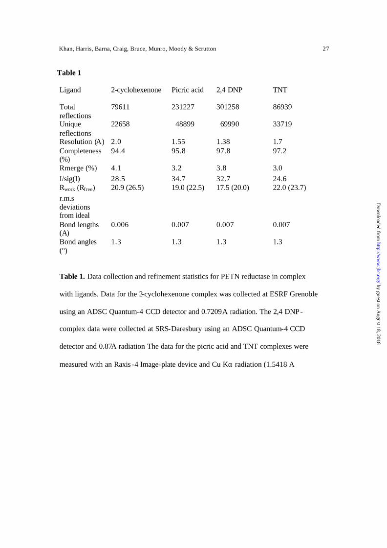
Crystallography — Crystals of PETN reductase-ligand complexes were prepared by co-
crystallization in the manner described previously for PETN reductase-steroid complexes
(14). The crystals have space group P212121 with one molecule per asymmetric unit. Data
were measured and reduced with the HKL suite (20) , and electron density maps were
calculated using the CCP4 suite (21) and displayed using XtalView (22). Refinement
was carried out with CNS (23). Details of data collection and refinement are shown in
Table 1. The data and coordinate files have been deposited with the RCS PDB, accession
codes (1GVO, 1GVQ, 1GVR, and 1GVS).
by guest on August 18, 2018
http://ww
w.jbc.org/
Dow
nloaded from
Khan, Harris, Barna, Craig, Bruce, Munro, Moody & Scrutton 13
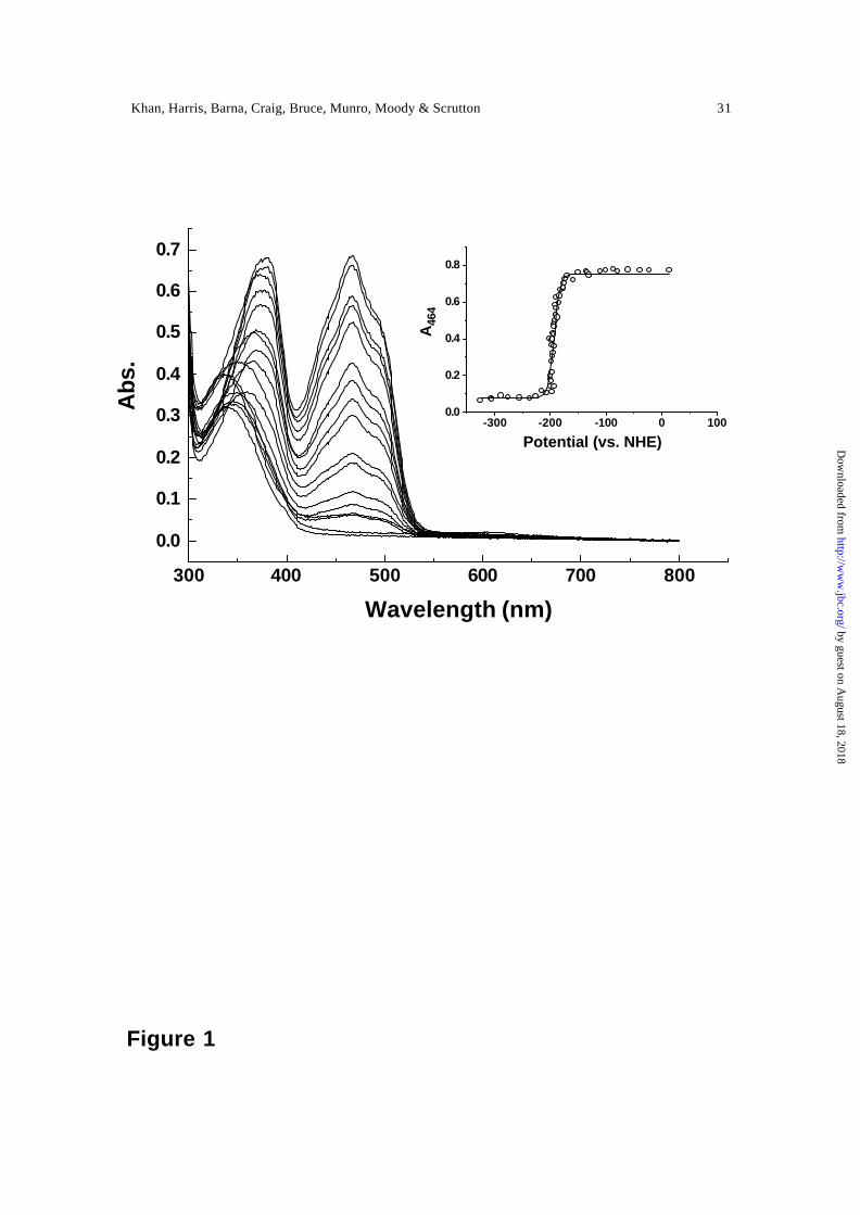
RESULTS
Mid-point redox potential of the FMN — The titrations of enzyme with dithionite were
from fully oxidised enzyme and proceeded gradually to the end-point of the titration by
the addition of small aliquots of reductant, and then back again to oxidised enzyme by the
addition of potassium ferricyanide. The observed spectral changes indicated the lack of
turbidity during the course of titration and no hysteretic effects were observed. Spectra
recorded at similar potentials in the reductive and oxidative phases of the titration were
essentially identical. Representative spectra for the reductive phase are shown in Figure 1
and a plot of the absorbance at 468 nm versus potential is shown in the inset of Figure 1.
Evidence for population of a semiquinone species during reductive and oxidative
titrations was not obtained. A good fit of the data to Eq. 1 was observed. The spectral
changes accompanying reduction of PETN reductase contrast with those seen for the
photoreduction of OYE in which the anionic red semiquinone is populated (24) , but are
similar to comparable titrations performed with bacterial morphinone reductase (25). Eq.
1 describes a concerted 2-electron reduction of the enzyme and fitting the
spectroelectrochemical data for PETN reductase produced a value for E12 of –193 ± 5
mV.
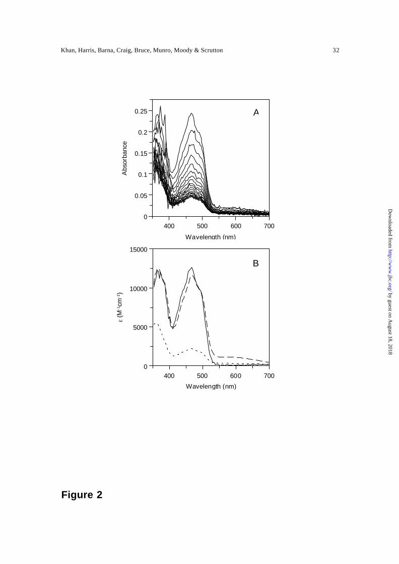
Reductive half-reaction of PETN reductase — The spectral changes accompanying
reduction of PETN reductase by a stoichiometric concentration β-NADPH are illustrated
in Figure 2A. Our previous studies with deuterated NADPD (A-side) have indicated that
hydride transfer is from the A-side of the nicotinamide ring, consistent with the known
stereospecificity of OYE (26). Analysis of the spectral changes accompanying flavin
by guest on August 18, 2018
http://ww
w.jbc.org/
Dow
nloaded from
Khan, Harris, Barna, Craig, Bruce, Munro, Moody & Scrutton 14
reduction by numerical integration methods using a two-step model (A → B → C)
revealed the presence of three enzyme forms. A is oxidized PETN reductase, B is an
enzyme-NADPH charge-transfer intermediate characterised by a long wavelength
absorption (550 to 700 nm) and C is PETN reductase containing the dihydroflavin form
of FMN. Residual absorption at ~460 nm indicates that reduction of the flavin is not
complete, suggesting that hydride transfer is reversible. Reversibility will depend on the
redox potentials of the FMN and NADPH in the enzyme-NADPH charge-transfer
complex, and these may differ from the potentials of NADPH in solution (-320 mV) and
unliganded PETN reductase (-193 mV). The kinetic scheme and observed spectral
changes are similar to those described previously for OYE (26) and bacterial morphinone
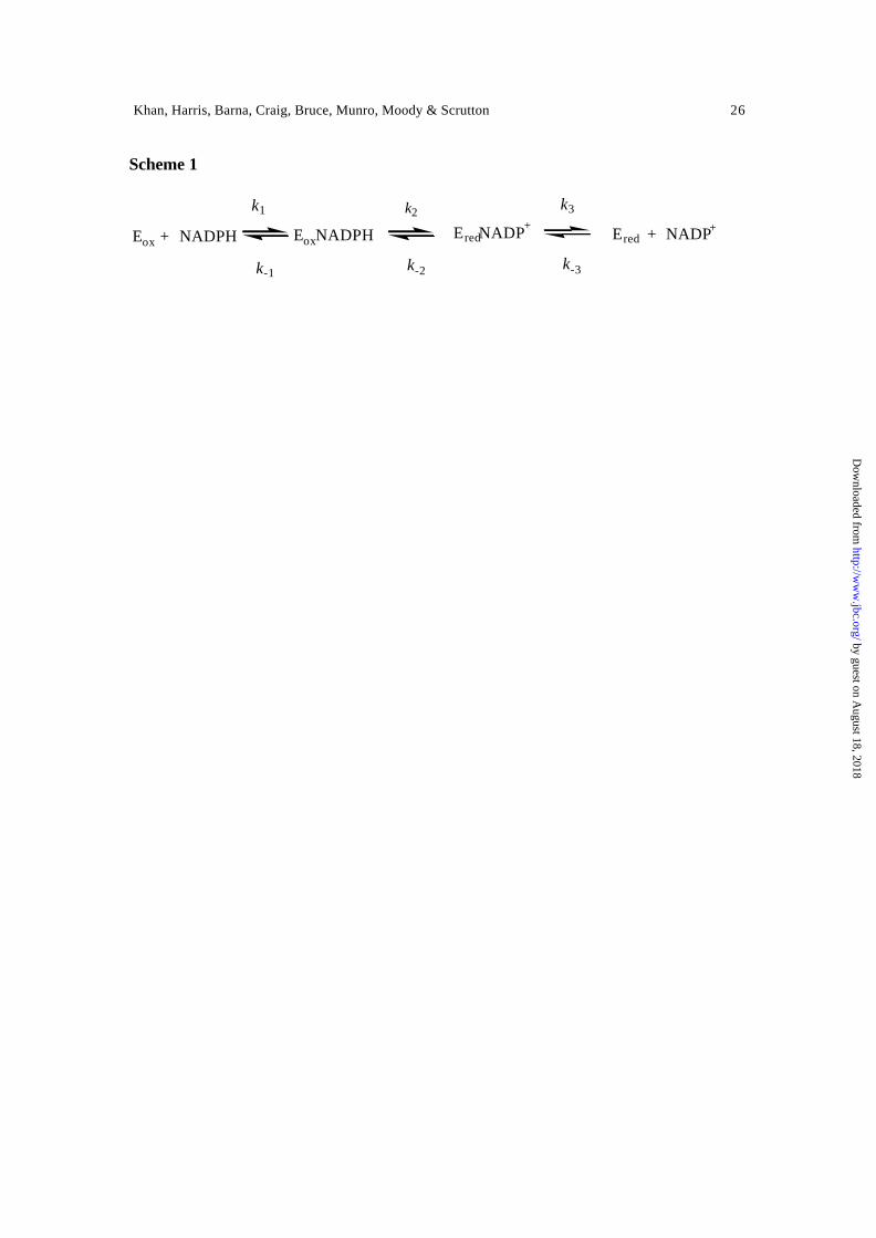
reductase (27) , and is shown as a series of reversible reactions in Scheme 1.
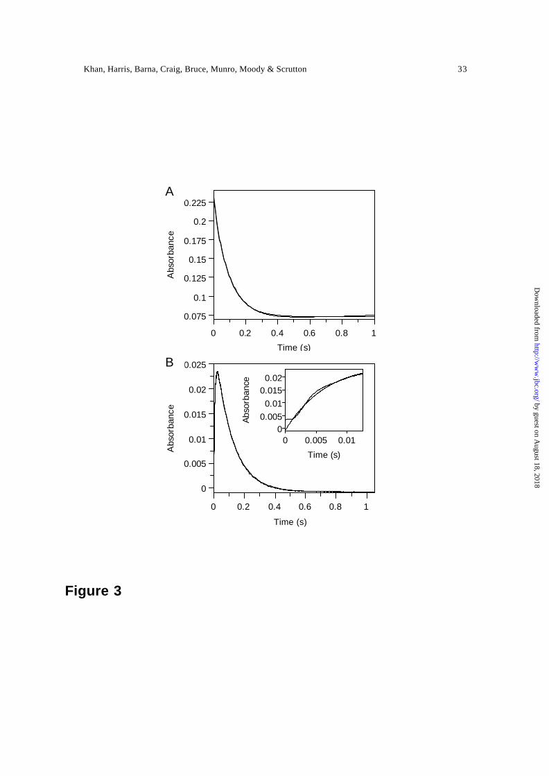
Observed rate constants for the formation and decay of the enzyme -NADPH
charge-transfer complex, and hydride transfer from NADPH to FMN, were obtained by
performing rapid mixing experiments of PETN reductase with NADPH using single
wavelength detection. The large absorption changes at 464 nm are suitable for monitoring
flavin reduction (i.e. step B → C), and a typical reaction transient is shown (Figure 3A).
Charge-transfer formation and decay was monitored at 560 nm (Figure 3B). The rate of
charge-transfer decay (560 nm) is identical to the rate of flavin reduction (464 nm),
indicating that decay of the enzyme-NADPH charge -transfer complex is a direct
consequence of flavin reduction. Formation of the charge -transfer complex is not readily
observed at 464 nm, owing to the small accompanying absorption change and relatively
large absorption change for flavin reduction at the same wavelength. However, a small
deviation from the fit to a single exponential expression is seen in the very early time
by guest on August 18, 2018
http://ww
w.jbc.org/
Dow
nloaded from
Khan, Harris, Barna, Craig, Bruce, Munro, Moody & Scrutton 15
domain of the transient (up to ~20 ms after mixing; not shown), which is likely attributed
to formation of the NADPH-enzyme charge-transfer complex. Formation of the charge-
transfer species is more readily observed at 560 nm (i.e. the ‘up’ phase of the kinetic
transient) (Figure 3B). Eq. 2 describes the early phase of the kinetic transient reasonably
well, but there is a small deviation from the fit perhaps suggesting more than one discrete
charge-transfer species accumulates in the early time domain (Figure 3B, inset). Similar
deviations (but more pronounced) have been seen with our work on the nicotinamide-
dependent flavoprotein human cytochrome P450 reductase (28).
The dependence of the observed rates for formation of the charge-transfer species
and flavin reduction (i.e. charge-transfer decay) on NADPH concentration is illustrated in
Figure 4. Consistent with our kinetic scheme for the reductive half -reaction, the rate of
formation of the charge-transfer species shows a linear dependence on NADPH
concentration. The second order rate constant for formation of the charge-transfer
complex is 0.95 x 10⁶ ± 0.02 x 10⁶ M-1 s-1. For scheme 1, the value of the positive
intercept of the ordinate axis (32 ± 7 s-1) approximates to k2 + k-1. Additionally, the
observed rate of flavin reduction (~12 s-1) measured at 464 nm is independent of NADPH
concentration (Figure 4). An approximate value of 20 s-1 for k -1 can therefore be
estimated which gives rise to a value of about 20 µM for the enzyme-NADPH
dissociation constant. Given that the rates of flavin reduction were measured at NADPH
concentrations of 100 µM and above, this would account for the lack of an apparent
dependence of the flavin reduction rate on NADPH concentration (Figure 4). In studies
performed with EBP (29) and OYE (26) an additional intermediate has been proposed
prior to formation of the charge -transfer complex. Incorporation of such an intermediate
by guest on August 18, 2018
http://ww
w.jbc.org/
Dow
nloaded from

Khan, Harris, Barna, Craig, Bruce, Munro, Moody & Scrutton 16
into Scheme 1 for PETN reductase would still be consistent with the observed kinetic
behaviour, but in the absence of direct evidence for such an intermediate we have omitted
to show the presence of a pre charge-transfer species in the catalytic scheme.
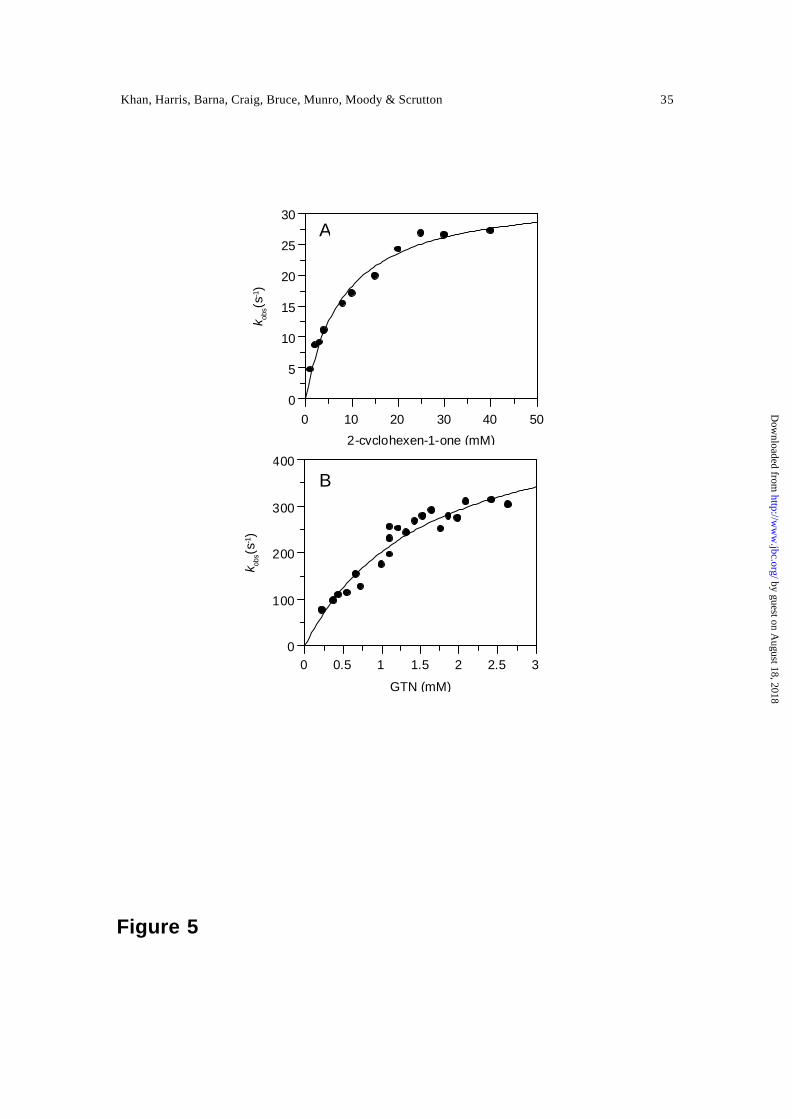
Oxidative half-reaction with 2-cyclohexenone and nitroester explosives — PETN
reductase uses a number of oxidising substrates including 2-cyclohexenone, the
nitroesters GTN and PETN, and nitroaromatics picric acid and TNT. 2-cyclohexenone is
a common oxidising substrate for the OYE family of enzymes (8). Studies of the
oxidative half -reaction with 2-cyclohexenone were initiated by mixing 2-electron reduced
PETN reductase, generated by titration with sodium dithionite, with substrate. Analysis
of multiple wavelength data indicated that oxidation occurred without the development of
visible charge-transfer intermediates or product release steps (not shown). The data were
best described using a single-step model (A → B) in which A is 2-electron reduced
enzyme and B is oxidised enzyme. The rate of flavin oxidation was investigated as a
function of 2-cyclohexenone concentration in single wavelength studies at 464 nm
(Figure 5A). Observed rates were hyperbolically dependent on 2-cyclohexenone
concentration and kinetic parameters were determined by fitting the data to Eq. 4. Fitting
produced a limiting rate constant, k lim, for flavin oxidation of 33.3 ± 1.5 s-1 and an
enzyme-substrate dissociation constant, Kd, of 8.1 ± 1.1 mM.
Oxidation of PETN reductase by the nitroester substrates GTN and PETN was
found to occur rapidly. As with 2-cyclohexenone, multiple wavelength absorption studies
indicated that enzyme oxidaton occurred in a single kinetic phase. With GTN, observed
rates were hyperbolically dependent on GTN concentration and fitting to the rapid
by guest on August 18, 2018
http://ww
w.jbc.org/
Dow
nloaded from
Khan, Harris, Barna, Craig, Bruce, Munro, Moody & Scrutton 17
equilibrium formalism of Strickland et al (Eq. 4) yielded a limiting rate constant, k lim, for
flavin oxidation of 518 ± 51 s-1 and reduced enzyme-GTN dissociation constant, Kd, of
1.5 ± 0.3 mM (Figure 5B). Owing to the extreme insolubility of PETN, we were unable
to analyse with confidence the dependence of the rate of flavin oxidation on PETN
concentration. However, an observed rate of ~25 s-1 was measured at a single
concentration of ~20 µM in reactions additionally containing 10 % ethanol (PETN is
sparingly soluble in 10 % ethanol).
Binding and reaction of PETN reductase with nitroaromatic explosives — The binding of
nitroaromatic compounds to oxidised PETN reductase results in perturbat ion of the
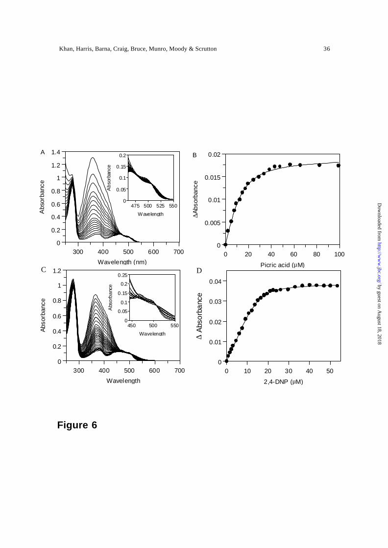
electronic absorption spectrum of the enzyme-bound FMN (Figure 6). Optical titrations
performed with picric acid and 2,4 DNP revealed clear isosbestic points at 506 nm. Plots
of absorption change at 518 nm versus ligand concentration and fitting to Eq. 5 produced
dissociation constants of 5.4 ± 1.1 µM and 1.0 ± 0.1 µM for picric acid and 2,4 DNP,
respectively. Optical titrations performed with TNT over a similar concentration range
failed to elicit perturbations in the flavin spectrum in the range 350 nm to 650 nm,
suggesting relatively weak binding of this ligand and/or lack of electronic interaction
with the flavin.
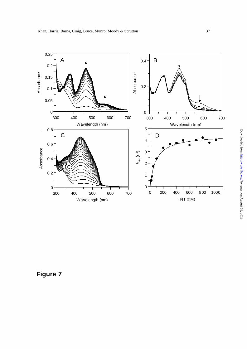
Single turnover stopped-flow studies of the oxidation of 2-electron reduced PETN
reductase with TNT clearly indicate the development of spectral features between 520 nm
and 700 nm) characteristic of the formation of a hydride-Meisenheimer complex [Figure
7A; (30)], and consistent with our previous studies (7). Prolonged incubation of the
solution following formation of the hydride-Meisenheimer complex leads to further
by guest on August 18, 2018
http://ww
w.jbc.org/
Dow
nloaded from

Khan, Harris, Barna, Craig, Bruce, Munro, Moody & Scrutton 18
spectral change, indicating further breakdown of the hydride-Meisenheimer complex
(Figure 7B). The chemical identity of compounds generated by the reactions occurring
after formation of the hydride -Meisenheimer complex have been investigated recently in
studies with the PETN reductase homologue, xenobiotic reductase (13). The
accumulation of different products after initial formation of the hydride -Meisenheimer
complex is also apparent under multiple turnover conditions with PETN reductase. The
spectra of the accumulated products are distinctly different from those observed in single
turnover stopped-flow studies (Figure 7C). Single turnover stopped-flow studies
performed at 464 nm (flavin oxidation and hydride-Meisenheimer complex formation)
and 580 nm (hydride-Meisenheimer complex formation) produced monophasic
absorption transients with identical kinetics, thus suggesting that hydride-Meisenheimer
complex formation is kinetically indistinguishable from flavin oxidation. Plots of the
concentration dependence of the rate of flavin oxidation and hydride-Meisenheimer
complex formation (measured at 464 nm) versus TNT concentration are hyperbolic
(Figure 7D) and fits to Eq. 3 produced values for the limiting rate of flavin oxidation, k lim ,
of 4.5 ± 0.1 s-1 and the reduced enzyme -TNT dissociation constant, Kd, of 88.9 ± 12 µM.
Reduction of picric acid by 2-electron reduced PETN reductase occurs very slowly.
Multiple-turnover studies performed under anaerobic conditions in a conventional
spectrophotometer indicated the development of spectral signature between 430 nm to
600 nm suggesting formation of the picric acid hydride-Meisenheimer complex.
Reduction, however, was very slow taking approximately 15 h and, unlike for TNT, the
long-wavelength signature for the hydride -Mesienheimer was relatively stable over this
period. Detailed chemical analysis of the breakdown products of picric acid and TNT
by guest on August 18, 2018
http://ww
w.jbc.org/
Dow
nloaded from

Khan, Harris, Barna, Craig, Bruce, Munro, Moody & Scrutton 19
after formation of the hydride -Meisenheimer complex by PETN reductase is to be
described elsewhere, and is beyond the scope of the present paper.
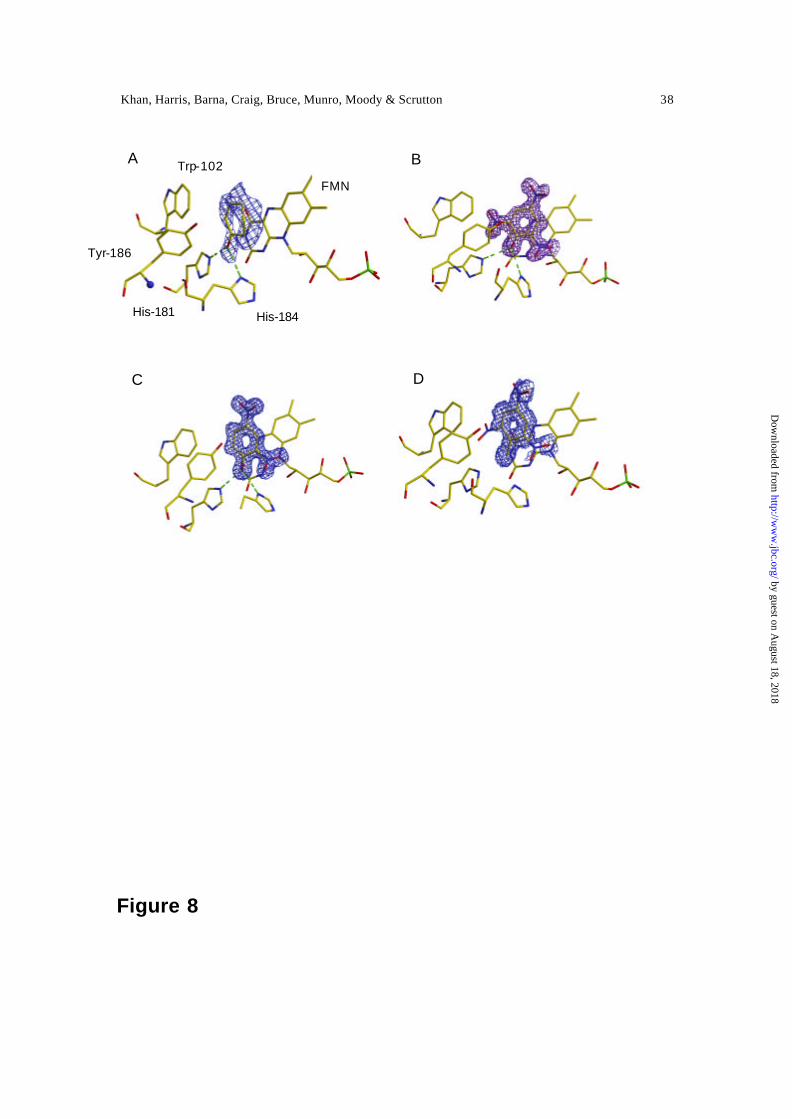
Structures of PETN reductase in complex with nitroaromatic ligands and 2-
cyclohexenone — The structure of PETN reductase in the oxidised and reduced form, and
in complex with steroid substrates and inhibitors, has been described (14). Herein, we
describe the structure of oxid ised PETN reductase in complex with TNT, picric acid, 2,4
DNP and 2-cyclohexenone. Each of these complexes shows positive difference density in
the active site, and the refined interpretation (and electron density) for each enzyme-
ligand complex is shown in Figure 8. The ligands are bound above the si-face of the
isoalloxazine ring. The imidazole side-chains of the histidine pair (His-181 and His-184),
previously shown to coordinate with the electronegative atoms in steroid ligands (14) ,
makes a similar interaction with the carbonyl group of 2-cyclohexenone (Figure 8A).
This binding mode positions the olefinic bond over the reactive flavin N5 atom to enable
hydride transfer; as with steroid substrates (14) , we infer Tyr-186 acts as proton donor
during reduction of the olefinic bond. This role for Tyr -186 is consistent with the results
of recent mutagenesis studies of the equivalent residue (Tyr -196) of OYE in reactions
with α,β unsaturated carbonyl compounds (31). The His-181/His-184 pair also
coordinates the hydroxy group of picric acid and 2,4 DNP (Figures 8B and 8C). Both
nitroaromatics are located with the C5 carbon close to the flavin N5 in a position optimal
for hydride transfer. With picric acid, the C5 position is activated for nucleophilic attack
from the flavin N5 through resonance stabilisation; this is not the case with 2,4 DNP (see
below). Despite the lack of opportunity for good interaction between the C1 methyl of
by guest on August 18, 2018
http://ww
w.jbc.org/
Dow
nloaded from
Khan, Harris, Barna, Craig, Bruce, Munro, Moody & Scrutton 20
TNT and the His-181/His-184 pair, the difference density for TNT indicates that it is
bound in a similar mode to picric acid and 2,4 DNP (Figure 8D). We infer, therefore, that
reduction of the nitroaromatic nucleus of both TNT and picric acid occurs by similar
mechanisms.
The shape of the electron density for 2-cyclohexenone is consistent with a second,
minor, binding mode whereby the ligand is “flipped” by 180° degrees, thus pointing the
carbonyl group of 2-cyclohexenone away from the histidine pair. The structures of
PETN-reductase complexed with picric acid shows an apparent bond with unusual
geometry between the nitro group at C6 and the indole ring of Trp-102. This may be the
result of the superimposition of multiple, partially occupied conformations and is the
subject of a separate high-resolution study. TNT is less than fully occupied and the 6-
nitro group is clearly disordered and therefore does not show any density.
by guest on August 18, 2018
http://ww
w.jbc.org/
Dow
nloaded from
Khan, Harris, Barna, Craig, Bruce, Munro, Moody & Scrutton 21
DISCUSSION
Our recent determination of the crystal structure of PETN reductase established a close
relationship to OYE, confirming inferences drawn from gene sequencing studies. Both
enzymes contain a single FMN cofactor, and the active sites of both enzymes are very
similar. Despite this structural similarity, our solution studies of PETN reductase have
established key differences in the reactivity of OYE and PETN reductase towards
oxidising substrates. Unlike OYE, PETN reductase reduces the nitroaromatic compounds
TNT and picric acid to form a hydride-Meisenheimer complex (8,9). Similar reactivity
towards nitroaromatics has also been reported for the OYE homologue termed
‘xenobiotic reductase’ isolated from Pseudomonas fluorescens I-C (13). Reduction of
nitroesters such as GTN appears to be a common feature of the OYE family of enzymes
and has been demonstrated for the xenobiotic reductases of Pseudomonas fluorescens I-C
and Pseudomonas putida II-B (32) , PETN reductase (this work), E.coli N-ethyl
maleimide reductase (8) and OYE (33). Recently, detailed stopped-flow studies of the
GTN-catalysed re-oxidation of OYE were performed and the reaction was shown to
involve the reductive liberation of nitrite (33). The oxidative half-reaction of both PETN
reductase and OYE can be modelled using the rapid equilibrium formalism of Strickland
et al (19). However, the limiting rate of flavin re-oxidation by GTN in OYE [40 s-1 at 25
° C; (33)] is considerably less than that for PETN reductase (518 s-1 at 5 °C); the reduced
enzyme-GTN dissociation constants are similar (2.7 mM for OYE and 1.3 mM for PETN
reductase). Further differences in the properties of PETN reductase and OYE can also be
identified: OYE stabilise the red anionic semiquinone of FMN, but reductive titration of
PETN reductase proceeds direct to the dihydroquinone. Thus, despite the similarities in
by guest on August 18, 2018
http://ww
w.jbc.org/
Dow
nloaded from

Khan, Harris, Barna, Craig, Bruce, Munro, Moody & Scrutton 22
the overall active site structure of OYE and PETN reductase (14) , key differences in the
reactivity and redox properties of the enzymes are apparent.
Our studies of the reductive half-reaction of PETN reductase have established
mechanistic similarities with OYE (26) , MR (27) and EBP (34). In all cases, enzyme -
NADPH charge-transfer complexes have been observed prior to flavin reduction by the
nicotinamide coenzyme. In contrast, our stopped-flow and spectrophotometric studies of
the oxidative half-reaction have established key differences between different members
of the OYE family of enzymes. The oxidative half -reaction of PETN reductase with TNT
and picric acid generates the hydride-Meisenheimer complexes of these substrates. In the
case of TNT, the hydride -Meisenheimer complex then breaks down to form alternate
products, the chemical identities of which are uncertain but have been studied in reactions
catalysed by xenobiotic reductase from Pseudomonas fluorescens I-C (13). The crystal
structures of the TNT-and picric acid-bound PETN reductase complexes indicate a
plausible mechanism involving direct hydride transfer from the N5 atom of the flavin
isoalloxazine ring to the C5 position of the aromatic nucleus of the substrate. Despite the
lack of electronic interaction with the isoalloxazine ring, the crystal structure of the
enzyme-TNT complex clearly indicates that TNT binds in a similar mode to picric acid.
The loss of the key interactions with His -181 and His-184 seen in the enzyme-picric acid
complex might provide a rationale for weaker binding of TNT in the active site (and thus
loss of electronic interaction with the flavin). The binding mode of 2,4 DNP is also
similar to that observed for picric acid. The question arises, therefore, as to why 2,4 DNP
is an inhibitor, whereas TNT and picric acid (albeit poor) are substrates. The answer
likely lies in the different resonance stabilised forms of 2,4 DNP: resonance stabilisation
by guest on August 18, 2018
http://ww
w.jbc.org/
Dow
nloaded from
Khan, Harris, Barna, Craig, Bruce, Munro, Moody & Scrutton 23
of 2,4 DNP preferentially enhances the electrophilicity of the C3 atom in this inhibitor,
but it is the C5 atom that is located above the flavin N5, and thus this geometry is not
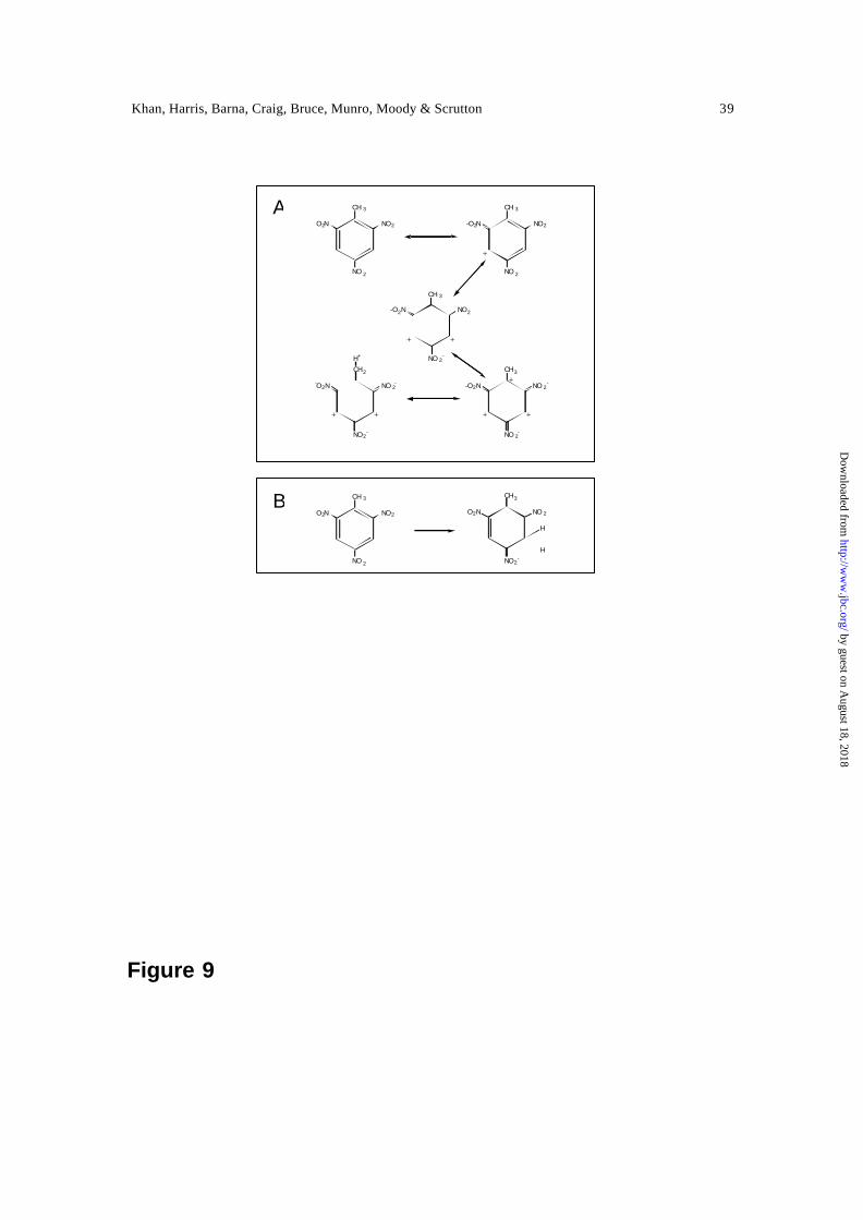
favourable for hydride transfer to the C3 of 2,4 DNP. In TNT and picric acid the
electrophilicity of both the C3 and C5 atoms is enhanced through resonance stabilisation
(see Figure 9 for TNT), thus enabling hydride transfer to the C5 atom. Given the
relatively simple reaction for nitroaromatic reduction, a key question arising from our
work is why OYE, and indeed other members of the OYE family, are not able to reduce
TNT and picric acid to their hydride-Meisenheimer complexes. Careful structural
comparisons coupled with mutagenesis studies should identify those residues that ‘switch
on’ reductive attack of nitroaromatics – a line of inquiry we are currently pursuing.
REFERENCES
1. Hooker, B. S. , and Skeen, R. S. (1999) Nat. Biotechnol. 17 , 428
2. Binks, P. R., French, C. E., Nicklin, S., and Bruce, N. C. (1996) Appl. Environ.
Microbiol. 62 , 1214-1219
3. French, C. E., Nicklin, S., and Bruce, N. C. (1996) J. Bacteriol. 178, 6623-6627
4. Saito, K., Thiele, D. J., Davio, M., Lockridge, O., and Massey, V. (1991) J. Biol.
Chem. 266, 20720-20724
5. French, C. E., and Bruce, N. C. (1995) Biochem. J. 312, 671-678
6. Madani, N. D., Malloy, P. J., Rodriguez-Pombo, P., Krishnan, A. V., and
Feldman, D. (1994) Proc. Natl. Acad. Sci. U S A 91 , 922-926
7. French, C. E., Nicklin, S., and Bruce, N. C. (1998) Appl. Environ. Microbiol. 64,
2864-2868
by guest on August 18, 2018
http://ww
w.jbc.org/
Dow
nloaded from

Khan, Harris, Barna, Craig, Bruce, Munro, Moody & Scrutton 24
8. Williams, R. E., Rathbone, D., Bruce, N. C., Scrutton, N. S., Moody, P. C. E., and
Nicklin, S. (1999) in Flavins and Flavoproteins (Ghisla, S., Kroneck, P.,
Macheroux, P., and Sund, H., eds), pp. 663-666, Rudolf Weber, Berlin
9. Williams, R., Rathbone, D., Moody, P., Scrutton, N., and Bruce, N. (2001)
Biochem. Soc. Symp. 68 , 143-153
10. French, C. E., Rosser, S. J., Davies, G. J., Nicklin, S., and Bruce, N. C. (1999)
Nat. Biotechnol. 17 , 491-494
11. Blehert, D. S., Fox, B. G., and Chambliss, G. H. (1999) J. Bacteriol. 181 , 6254-
6263
12. Snape, J. R., Walkley, N. A., Morby, A. P., Nicklin, S., and White, G. F. (1997) J.
Bacteriol. 179 , 7796-7802
13. Pak, J. W., Knoke, K. L., Noguera, D. R., Fox, B. G., and Chambliss, G. H.
(2000) Appl. Environ. Microbiol. 66, 4742-4750.
14. Barna, T., Khan, H., Bruce, N., Barsukov, I., Scrutton, N., and Moody, P. (2001)
J. Mol. Biol. 310 , 433-447
15. Fox, K. M., and Karplus, P. A. (1994) Structure 2 , 1089-1105
16. Vaz, A. D., Chakraborty, S., and Massey, V. (1995) Biochemistry 34 , 4246-4256
17. Sambrook, J., Fritsch, E., and Maniatis, T. (1989) Molecular cloning: a
laboratory manual, 2nd edition, Cold Spring Harbor Laboratory Press, Cold
Spring Harbor NY
18. Dutton, P. (1978) Methods Enzymol. 54 , 411-435
19. Strickland, S., Palmer, G., and Massey, V. (1975) J. Biol. Chem. 250, 4048-4052
20. Otwinowski, Z., and Minor, W. (1997) Methods Enzymol. 276 , 307-326
by guest on August 18, 2018
http://ww
w.jbc.org/
Dow
nloaded from
Khan, Harris, Barna, Craig, Bruce, Munro, Moody & Scrutton 25
21. Collaborative computational project, n. (1994) Acta Cryst. D50, 760-763
22. McRee, D. (1992) J. Mol. Graphics 10 , 44-46
23. Brunger, A., Adams, P., Clore, G., DeLano, W., Gros, P., Grosse-Kunstleve, R.,
Jiang, J.-S., Kuszewski, J., Nilges, M., Pannu, N., Read, R., Rice, L., Simonson,
T., and Warren, G. (1998) Acta Cryst. D54 , 905-929
24. Massey, V., and Hemmerich, P. (1978) Biochemistry 17, 9-16.
25. Craig, D. H., Barna, T., Moody, P. C., Bruce, N. C., Chapman, S. K., Munro, A.
W., and Scrutton, N. S. (2001) Biochem. J. 359 , 315-323.
26. Massey, V., and Schopfer, L. M. (1986) J. Biol. Chem. 261 , 1215-1222.
27. Craig, D. H., Moody, P. C. E., Bruce, N. C., and Scrutton, N. S. (1998)
Biochemistry 37, 7598-7607
28. Gutierrez, A., Lian, L. Y., Wolf, C. R., Scrutton, N. S., and Roberts, G. C. (2001)
Biochemistry 40, 1964-1975.
29. Buckman, J., and Miller, S. M. (2000) Biochemistry 39 , 10521-10531.
30. Vorbeck, C., Lenke, H., Fischer, P., and Knackmuss, H. J. (1994) J. Bacteriol.
176, 932-934.
31. Kohli, R. M., and Massey, V. (1998) J. Biol. Chem. 273, 32763-32770
32. Blehert, D. S., Knoke, K. L., Fox, B. G., and Chambliss, G. H. (1997) J.
Bacteriol. 179 , 6912-6920
33. Meah, Y., Brown, B. J., Chakraborty, S., and Massey, V. (2001) Proc. Natl. Acad.
Sci. U S A 98 , 8560-8565.
34. Buckman, J., and Miller, S. M. (1998) Biochemistry 37 , 14326-14336
by guest on August 18, 2018
http://ww
w.jbc.org/
Dow
nloaded from
Khan, Harris, Barna, Craig, Bruce, Munro, Moody & Scrutton 26
Scheme 1
Eox + NADPH EoxNADPH EredNADP+ Ered + NADP+
k1
k-1k-3
k3
k-2
k2
by guest on August 18, 2018
http://ww
w.jbc.org/
Dow
nloaded from
Khan, Harris, Barna, Craig, Bruce, Munro, Moody & Scrutton 27
Table 1
Ligand
2-cyclohexenone Picric acid 2,4 DNP TNT
Total reflections
79611 231227 301258 86939
Unique reflections
22658 48899 69990 33719
Resolution (A) 2.0 1.55 1.38 1.7 Completeness (%)
94.4 95.8 97.8 97.2
Rmerge (%) 4.1 3.2 3.8 3.0 I/sig(I) 28.5 34.7 32.7 24.6 Rwork (Rfree) 20.9 (26.5) 19.0 (22.5) 17.5 (20.0) 22.0 (23.7) r.m.s deviations from ideal Bond lengths (A) Bond angles (°)
0.006 1.3
0.007 1.3
0.007 1.3
0.007 1.3
Table 1. Data collection and refinement statistics for PETN reductase in complex
with ligands. Data for the 2-cyclohexenone complex was collected at ESRF Grenoble
using an ADSC Quantum-4 CCD detector and 0.7209A radiation. The 2,4 DNP-
complex data were collected at SRS-Daresbury using an ADSC Quantum-4 CCD
detector and 0.87A radiation The data for the picric acid and TNT complexes were
measured with an Raxis -4 Image-plate device and Cu Kα radiation (1.5418 A)
by guest on August 18, 2018
http://ww
w.jbc.org/
Dow
nloaded from
Khan, Harris, Barna, Craig, Bruce, Munro, Moody & Scrutton 28
FIGURE LEGENDS
Figure 1. Spectral changes accompanying the reductive titration of PETN reductase.
Inset, plot of absorbance versus potential. Data are shown fitted to Eq. 1 (E12 = -193 ± 5
mV).
Figure 2. Spectral changes observed during the reduction of PETN reductase (20 µM)
with NADPH (20 µM). Panel A, time-dependent spectral changes for PETN reductase
mixed with NADPH. The first spectrum was recorded at 1.28 ms after mixing. For
clarity, only selected subsequent spectra are illustrated. Conditions: 50 mM potassium
phosphate buffer, pH 7.0, 5 °C; the time-dependent dataset was recorded over a period of
1 s. Panel B, denconvoluted spectra of initial, intermediate and final forms of the enzyme
obtained by global analysis using ProKin software. Spectrum 1 (solid line), oxidised
enzyme; spectrum 2 (broken line), charge-transfer intermediate; spectrum 3 (dotted line),
two electron-reduced enzyme.
Figure 3. Kinetic transients observed for the reductive half-reaction of PETN reductase.
Panel A, transient observed at 464 nm; panel B, transient observed at 560 nm. The inset
in panel B illustrates the same reaction recorded over 12 ms fitted to Eq. 2. Conditions:
50 mM potassium phosphate buffer, pH 7.0; reactions were performed using 20 µM
PETN reductase and 200 µM NADPH at 5 °C.
by guest on August 18, 2018
http://ww
w.jbc.org/
Dow
nloaded from

Khan, Harris, Barna, Craig, Bruce, Munro, Moody & Scrutton 29
Figure 4. Concentration dependence of the observed rates measured at 560 nm (charge -
transfer formation) and 464 nm (hydride transfer). Conditions: 50 mM potassium
phosphate buffer, pH 7.0 and 5 °C; reactions were performed using 20 µM PETN
reductase. Filled squares, charge-transfer formation; open circles, hydride transfer.
Figure 5. Concentration dependence of the rate of flavin reoxidation measured at 464 nm
for the reaction of dithionite-reduced PETN reductase with 2-cyclohexenone (panel A)
and GTN (Panel B). Conditions: 50 mM potassium phosphate buffer, pH 7.0; reactions
were performed using 20 µM PETN reductase, at 25 °C (2-cyclohexenone) and 5 °C
(GTN). The fits shown are to Eq. 3.
Figure 6. Titrations of PETN reductase with picric acid and 2,4 DNP. Conditions, 50 mM
potassium phosphate, pH 7.0, 25 °C; enzyme concentration 10 µM. Panel A, spectral
changes observed on titrating PETN reductase with picric acid. Inset, detail for the region
500 nm to 525 nm. Panel B, plot of absorbance change versus picric acid concentration.
Solid line indicates the fit to Eq. 5. Panels C and D as for panels A and B, respectively,
but for 2,4, DNP.
Figure 7. Panel A, spectral changes occurring during the oxidation of 2-electron reduced
PETN reductase (16 µM) following rapid mixing with TNT (16 µM). Arrows indicate
direction of spectral change. The first spectrum is shown at 1.28 ms after mixing; for
clarity, not all subsequent spectra are shown. The time period for acquisition of spectra is
5 s. Panel B, spectral changes following prolonged incubation of 2-electron reduced
by guest on August 18, 2018
http://ww
w.jbc.org/
Dow
nloaded from
Khan, Harris, Barna, Craig, Bruce, Munro, Moody & Scrutton 30
PETN reductase with TNT, illustrating the decay of the hydride-Meisenheimer complex.
Following formation of the hydride-Meisenheimer complex, subsequent spectra were
recorded at 1.5 minute intervals. Arrows indicate direction of spectral change. Panel C,
spectra observed during the multiple turnover of PETN reductase (0.2 µM) with TNT
(100 µM). The reaction was performed over 50 minutes (each spectrum recorded after 1.5
minutes). Panel D, plot of observed rate of hydride -Meisenheimer complex formation and
flavin reoxidation versus TNT concentration (data taken from stopped-flow studies
performed at 464 nm). Solid line, fit to Eq. 4.
Figure 8. Difference electron density for each of the PETN reductase-ligand complexes.
The contours are at 3σ. Panel A, the complex of oxidised enzyme and 2-cyclohexenone.
Panel B, the complex of oxidised enzyme and picric acid; panel C, the complex of
oxidised enzyme and 2,4 DNP; panel D, the complex of oxidised enzyme and TNT.
Figure 9. (A) The structure of TNT as the resonance hybrid of several canonical forms,
illustrating the enhancement in the electrophilicity of C3 and C5, and (B) reduction of
TNT to form the Meisenheimer-hydride complex.
by guest on August 18, 2018
http://ww
w.jbc.org/
Dow
nloaded from
Khan, Harris, Barna, Craig, Bruce, Munro, Moody & Scrutton 31
Figure 1
300 400 500 600 700 800
0.0
0.1
0.2
0.3
0.4
0.5
0.6
0.7
Abs
.
Wavelength (nm)
-300 -200 -100 0 1000.0
0.2
0.4
0.6
0.8
A46
4Potential (vs. NHE)
by guest on August 18, 2018
http://ww
w.jbc.org/
Dow
nloaded from
Khan, Harris, Barna, Craig, Bruce, Munro, Moody & Scrutton 32
Figure 2
700600500400
0.25
0.2
0.15
0.1
0.05
0
Wavelength (nm)
Abs
orba
nce
A
700600500400
15000
10000
5000
0
Wavelength (nm)
ε (M
-1cm
-1)
B
A
B
by guest on August 18, 2018
http://ww
w.jbc.org/
Dow
nloaded from
Khan, Harris, Barna, Craig, Bruce, Munro, Moody & Scrutton 33
Figure 3
10.80.60.40.20
0.225
0.2
0.175
0.15
0.125
0.1
0.075
Time (s)
Abs
orba
nce
A
10.80.60.40.20
0.025
0.02
0.015
0.01
0.005
0
Time (sec)
Abs
orba
nce
0.010.0050
0.020.0150.01
0.0050
Time (s)
Abs
orba
nce
B
A
B
Time (s)
by guest on August 18, 2018
http://ww
w.jbc.org/
Dow
nloaded from

Khan, Harris, Barna, Craig, Bruce, Munro, Moody & Scrutton 34
Figure 4
10008006004002000
1000
800
600
400
200
0
NADPH conc (µM)
k ob
s (s-1
)
by guest on August 18, 2018
http://ww
w.jbc.org/
Dow
nloaded from
Khan, Harris, Barna, Craig, Bruce, Munro, Moody & Scrutton 35
Figure 5
32.521.510.50
400
300
200
100
0
GTN (mM)
k obs(
s-1)
B
50403020100
30
25
20
15
10
5
0
2-cyclohexen-1-one (mM)
k obs(
s-1)
A
A
B
by guest on August 18, 2018
http://ww
w.jbc.org/
Dow
nloaded from
Khan, Harris, Barna, Craig, Bruce, Munro, Moody & Scrutton 36
Figure 6
700600500400300
1.4
1.2
1
0.8
0.6
0.4
0.2
0
Wavelength (nm)
Abs
orba
nce
A
550525500475
0.2
0.15
0.1
0.05
0
Wavelength
Abs
orba
nce
100806040200
0.02
0.015
0.01
0.005
0
Picric acid (µM)
∆A
bsor
banc
e
B
50403020100
0.04
0.03
0.02
0.01
0
2,4-DNP (µM)
∆ A
bsor
banc
e
D
700600500400300
1.2
1
0.8
0.6
0.4
0.2
0
Wavelength
Abs
orba
nce
550500450
0.25
0.2
0.15
0.1
0.05
0
Wavelength
Abs
orba
nce
C
by guest on August 18, 2018
http://ww
w.jbc.org/
Dow
nloaded from
Khan, Harris, Barna, Craig, Bruce, Munro, Moody & Scrutton 37
Figure 7
700600500400300
0.25
0.2
0.15
0.1
0.05
0
Wavelength (nm)
Abs
orb
ance
A
10008006004002000
5
4
3
2
1
0
TNT (µM)
k obs
(s-1
)
D
700600500400300
0.8
0.6
0.4
0.2
0
Wavelength (nm)
Ab
sorb
anc
e
C
700600500400300
0.4
0.2
0
Wavelength (nm)
Abs
orba
nce
B
A B
C D
by guest on August 18, 2018
http://ww
w.jbc.org/
Dow
nloaded from
Khan, Harris, Barna, Craig, Bruce, Munro, Moody & Scrutton 38
Figure 8
A B
C D
Trp-102
Tyr-186
FMN
His-184 His-181
by guest on August 18, 2018
http://ww
w.jbc.org/
Dow
nloaded from
Khan, Harris, Barna, Craig, Bruce, Munro, Moody & Scrutton 39
NO2-
NO 2O2N
CH3
NO 2
NO2O2N
CH 3
H
H
NO 2
NO2O2N
CH 3
NO 2
NO2-O2N
CH 3
NO 2-
NO2-O2N
CH 3
NO 2-
NO 2--O2N
CH3
NO2-
NO 2--O2N
CH2
H+
+
+
+
+
+
+
+
+
Figure 9
A
B
by guest on August 18, 2018
http://ww
w.jbc.org/
Dow
nloaded from
Munro, Peter C.E. Moody and Nigel S. ScruttonHuma Khan, Richard J. Harris, Terez Barna, Daniel H. Craig, Neil C. Bruce, Andrew W.
with NADPH, 2-cyclohexenone, nitroesters and nitroaromatic explosivesPentaerythritol tetranitrate reductase: kinetic and structural basis of reactivity
published online March 28, 2002J. Biol. Chem.
10.1074/jbc.M200637200Access the most updated version of this article at doi:
Alerts:
When a correction for this article is posted•
When this article is cited•
to choose from all of JBC's e-mail alertsClick here
by guest on August 18, 2018
http://ww
w.jbc.org/
Dow
nloaded from