Embed Size (px)
Citation preview
573H. Kuwano: Histology of Esophageal CancerSurg TodayJpn J Surg (1998) 28:573–575
and several studies have also suggested its significanceas a precancerous lesion.2 Our serial study revealed 75dysplastic lesions in 32 cases (20.1%) among the 159cases of esophageal cancer without any preoperativetreatment. These results also showed that the less ad-vanced the lesion, the higher the incidence of dysplasia,while in the cases with dysplastic lesions both themultiplicity of squamous cell carcinoma and theintraepithelial spread of the main lesion were morefrequently seen, thus suggesting the multicentric occur-rence of dysplastic lesions and carcinomas.3
Coexistence of Glandular Differentiation withSquamous Cell Carcinoma
The most common histologic type of esophageal canceris squamous cell carcinoma while, in contrast, primaryadenocarcinoma of the esophagus only rarely occurs.Primary adenocarcinoma of the esophagus is thus con-sidered to occur most commonly in the esophagogastricjunction including Barrett’s esophagus, or in the ectopicgastric mucosa. However, they also appear to arise inthe esophageal submucosa, exhibiting a histologicpattern resembling that seen in adenoid cystic ormucoepidermoid tumors of salivary gland origin.4
Our review of 195 patients with carcinoma of theesophagus disclosed 41 cases (21.0%) with glandularand/or mucus-secreting components, in addition to theordinary component of squamous cell carcinoma. Thehistologic features of such glandular differentiationwere also reminiscent of those seen in salivary glandtumors, and they were frequently located in the submu-cosa and lamina propria mucosae, thereby suggestingthat such differentiation had arisen in either the esoph-ageal glands or their ducts. These findings suggestedthat this type of esophageal tumor had originated notonly from the covering squamous epithelium but alsofrom the esophageal mucus-gland or ductal epithelium,
As the most common histologic type of esophageal can-cer is squamous cell carcinoma and most cases consist ofeither advanced cases or preoperatively irradiated casesamong the resected cases with esophageal cancer, lesshistopathologic attention has thus been paid to esoph-ageal cancer than to other gastrointestinal tract cancers,such as gastric and colon cancers. However, owing tothe remarkable development of esophago-fiberscopicendoscopy, esophageal carcinoma can now frequentlybe diagnosed at an early stage, and, as a result, thenumber of patients with early esophageal carcinomahas increased significantly.1 Consequently, serial histo-phathologic investigations of esophageal cancer, as wellas squamous epithelial dysplasia, are now being per-formed more extensively, and several peculiar histo-logic features have thus been demonstrated. In thisarticle, I would like to present some of the distinctivefeatures of esophageal cancer, and will also discuss theclinicophathologic significance of such characteristics.These characteristics include: (1) the frequent coexis-tence of squamous epithelial dysplasia, (2) the occa-sional coexistence of glandular differentiation withsquamous cell carcinoma, (3) the frequent coexistenceof intraepithelial spread, (4) the occasional intra-esophageal multiple occurrence of carcinoma, (5) thesynchronous and metachronous occurrence of carci-noma of other organs, such as head and neck and gastriccancers, and (6) the occasional existence of intramuralmetastasis.
Coexistence of Squamous Epithelial Dysplasia
Squamous epithelial dysplasia is frequently encoun-tered in the esophagus with squamous cell carcinoma,
Reprint requests to: H. Kuwano(Received for publication on Sept. 4, 1997; accepted onNov. 6, 1997)
Editorial
Peculiar Histopathologic Features of Esophageal Cancer
Hiroyuki Kuwano
Department of Surgery II, Faculty of Medicine, Kyushu University, 3-1-1 Maidashi, Higashi-ku, Fukuoka 812-8582, Japan
574 H. Kuwano: Histology of Esophageal Cancer
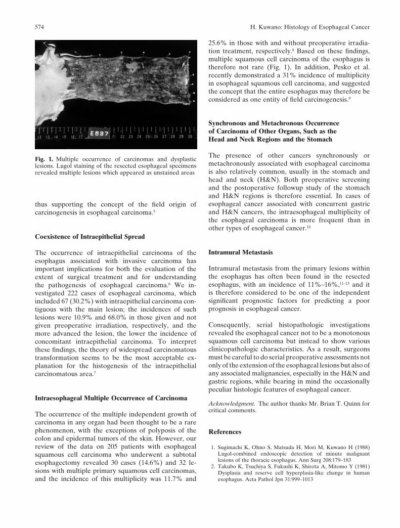
Fig. 1. Multiple occurrence of carcinomas and dysplasticlesions. Lugol staining of the resected esophageal specimensrevealed multiple lesions which appeared as unstained areas
25.6% in those with and without preoperative irradia-tion treatment, respectively.8 Based on these findings,multiple squamous cell carcinoma of the esophagus istherefore not rare (Fig. 1). In addition, Pesko et al.recently demonstrated a 31% incidence of multiplicityin esophageal squamous cell carcinoma, and suggestedthe concept that the entire esophagus may therefore beconsidered as one entity of field carcinogenesis.9
Synchronous and Metachronous Occurrenceof Carcinoma of Other Organs, Such as theHead and Neck Regions and the Stomach
The presence of other cancers synchronously ormetachronously associated with esophageal carcinomais also relatively common, usually in the stomach andhead and neck (H&N). Both preoperative screeningand the postoperative followup study of the stomachand H&N regions is therefore essential. In cases ofesophageal cancer associated with concurrent gastricand H&N cancers, the intraesophageal multiplicity ofthe esophageal carcinoma is more frequent than inother types of esophageal cancer.10
Intramural Metastasis
Intramural metastasis from the primary lesions withinthe esophagus has often been found in the resectedesophagus, with an incidence of 11%–16%,11–13 and itis therefore considered to be one of the independentsignificant prognostic factors for predicting a poorprognosis in esophageal cancer.
Consequently, serial histopathologic investigationsrevealed the esophageal cancer not to be a monotonoussquamous cell carcinoma but instead to show variousclinicopathologic characteristics. As a result, surgeonsmust be careful to do serial preoperative assessments notonly of the extension of the esophageal lesions but also ofany associated malignancies, especially in the H&N andgastric regions, while bearing in mind the occasionallypeculiar histologic features of esophageal cancer.
Acknowledgment. The author thanks Mr. Brian T. Quinn forcritical comments.
References
1. Sugimachi K, Ohno S, Matsuda H, Mori M, Kuwano H (1988)Lugol-combined endoscopic detection of minute malignantlesions of the thoracic esophagus. Ann Surg 208:179–183
2. Takubo K, Tsuchiya S, Fukushi K, Shirota A, Mitomo Y (1981)Dysplasia and reserve cell hyperplasia-like change in humanesophagus. Acta Pathol Jpn 31:999–1013
thus supporting the concept of the field origin ofcarcinogenesis in esophageal carcinoma.5
Coexistence of Intraepithelial Spread
The occurrence of intraepithelial carcinoma of theesophagus associated with invasive carcinoma hasimportant implications for both the evaluation of theextent of surgical treatment and for understandingthe pathogenesis of esophageal carcinoma.6 We in-vestigated 222 cases of esophageal carcinoma, whichincluded 67 (30.2%) with intraepithelial carcinoma con-tiguous with the main lesion; the incidences of suchlesions were 10.9% and 68.0% in those given and notgiven preoperative irradiation, respectively, and themore advanced the lesion, the lower the incidence ofconcomitant intraepithelial carcinoma. To interpretthese findings, the theory of widespread carcinomatoustransformation seems to be the most acceptable ex-planation for the histogenesis of the intraepithelialcarcinomatous area.7
Intraesophageal Multiple Occurrence of Carcinoma
The occurrence of the multiple independent growth ofcarcinoma in any organ had been thought to be a rarephenomenon, with the exceptions of polyposis of thecolon and epidermal tumors of the skin. However, ourreview of the data on 205 patients with esophagealsquamous cell carcinoma who underwent a subtotalesophagectomy revealed 30 cases (14.6%) and 32 le-sions with multiple primary squamous cell carcinomas,and the incidence of this multiplicity was 11.7% and
575H. Kuwano: Histology of Esophageal Cancer
3. Kuwano H, Watanabe M, Sadanaga N, Ikeda M, Mori M,Sugimachi K (1993) Squamous epithelial dysplasia associatedwith squamous cell carcinoma of the esophagus. Cancer Lett72:141–147
4. Bell-Thompson J, Haggitt RC, Ellis FH Jr (1980) Muco-epidermoid and adenoid cystic carcinomas of the esophagus. JThorac Cardiovasc Surg 79:438–446
5. Kuwano H, Ueo H, Sugimachi K, Inokuchi K, Toyoshima S,Enjoji M (1985) Glandular or mucus-secreting components insquamous cell carcinoma of the esophagus. Cancer 56:514–518
6. Soga J, Tanaka O, Sasaki K, Kawaguchi M, Muto T (1982) Super-ficial spreading carcinoma of the esophagus. Cancer 50:1641–1645
7. Kuwano H, Matsuda H, Matsuoka H, Kai H, Okudaira Y,Sugimachi K (1987) Intra-epithelial carcinoma concomitant withesophageal squamous cell carcinoma. Cancer 59:783–787
8. Kuwano H, Ohno S, Matsuda H, Mori M, Sugimachi K (1988)Serial histologic evaluation of multiple primary squamous cellcarcinomas of the esophagus. Cancer 61:1635–1638
9. Pesko P, Rakic S, Miliceric M, Bulajic P, Gerzic Z (1994) Preva-lence and clinicopathologic features of multiple squamous cellcarcinoma of the esophagus. Cancer 73:2687–2690
10. Kuwano H, Morita M, Tsutsui S, Kido Y, Mori M, Sugimachi K(1991) Comparison of characteristics of esophageal squamous cellcarcinoma associated with head and neck cancer and those withgastric cancer. J Surg Oncol 46:107–109
11. Takubo K, Sasajima K, Yamashita K, Tanaka Y, Fujita K (1990)Prognostic significance of intramural metastasis in patients withesophageal carcinoma. Cancer 65:1816–1819
12. Kato H, Tachimori Y, Watanabe H, Itabashi M, Hirota T,Yamaguchi H (1992) Intramural metastasis of thoracic esoph-ageal carcinoma. Int J Cancer 50:49–52
13. Kuwano H, Watanabe M, Sadanaga N, Kamakura T, Nozoe T,Yasuda M, Mimori K, Mori M, Sugimachi K (1994) Univariateand multivariate analyses of the prognostic significance of discon-tinuous intramural metastasis in patients with esophageal cancer.J Surg Oncol 57:17–21