Embed Size (px)
Citation preview

Available online at www.sciencedirect.com
Pectin structure and biosynthesisDebra Mohnen
Pectin is structurally and functionally the most complex
polysaccharide in plant cell walls. Pectin has functions in plant
growth, morphology, development, and plant defense and also
serves as a gelling and stabilizing polymer in diverse food and
specialty products and has positive effects on human health
and multiple biomedical uses. Pectin is a family of galacturonic
acid-rich polysaccharides including homogalacturonan,
rhamnogalacturonan I, and the substituted galacturonans
rhamnogalacturonan II (RG-II), and xylogalacturonan (XGA).
Pectin biosynthesis is estimated to require at least 67
transferases including glycosyl-, methyl-, and
acetyltransferases. New developments in understanding pectin
structure, function, and biosynthesis indicate that these
polysaccharides have roles in both primary and secondary cell
walls. Manipulation of pectin synthesis is expected to impact
diverse plant agronomical properties including plant biomass
characteristics important for biofuel production.
Address
Complex Carbohydrate Research Center, Department of Biochemistry
and Molecular Biology, DOE BioEnergy Science Center (BESC), The
University of Georgia, 315 Riverbend Road, Athens, GA 30602-4712,
United States
Corresponding author: Mohnen, Debra ([email protected])
Current Opinion in Plant Biology 2008, 11:266–277
This review comes from a themed issue on
Physiology and metabolism
Edited by Ken Keegstra and Markus Pauly
1369-5266/$ – see front matter
Published by Elsevier Ltd.
DOI 10.1016/j.pbi.2008.03.006
IntroductionPectin is the most structurally complex family of poly-
saccharides in nature, making up �35% of primary walls
in dicots and non-graminaceous monocots, 2–10% of grass
and other commelinoid primary walls, and up to 5% of
walls in woody tissue [1,2] (Department of Energy,
Energy Efficiency and Renewable Energy, Biomass Pro-
gram: http://www1.eere.energy.gov/biomass/feedstock_
databases.html). Pectin is abundant in walls that
surround growing and dividing cells, walls of cells in
the soft parts of the plant, and in the middle lamella
and cell corners. Pectin is also present in the junction
zone between cells with secondary walls including xylem
and fiber cells in woody tissue. Pectin is a component of
all higher plant walls and of the walls of gymnosperms,
Current Opinion in Plant Biology 2008, 11:266–277
pteridophytes, bryophytes and Chara, a charophycean
algae believed to be the closest extant relative of land
plants [3]. The correlation of increased amounts of the
pectin RG-II in vascular plants, and its appearance as
plants adapted to upright growth on land and developed
lignified secondary walls [4], suggests that pectin has
fundamental roles in both primary and secondary wall
structure and function. An understanding of pectin struc-
ture and synthesis is crucial to understanding, and poten-
tially modifying, wall structure so as to promote efficient
production of biofuel from recalcitrant plant lignocellu-
losic biomass [5,6].
Several reviews on pectin biosynthesis synthesis [2,7,8],
plant wall biosynthesis [9–12], and regulation of cell wall
synthesis [13,14] have recently been published. The goal
of this paper, after a brief review of pectin function, is to
summarize recent developments in our understanding of
pectin structure and biosynthesis, with special attention
to genes that encode pectin biosynthetic transferases. For
a more detailed summary of the enzymology of pectin
synthesis, readers are directed to four comprehensive
reviews [6–8,15].
Overview of pectin functionMultiple lines of evidence indicate a role for pectin in
plant growth, development, morphogenesis, defense,
cell–cell adhesion, wall structure, signaling, cell expan-
sion, wall porosity, binding of ions, growth factors and
enzymes, pollen tube growth, seed hydration, leaf abscis-
sion, and fruit development [2,16]. Pectin is also used as a
gelling and stabilizing agent in the food and cosmetic
industries and has multiple positive effects on human
health including lowering cholesterol and serum glucose
levels, reducing cancer [17], and stimulating the immune
response [18]. Pectin is also used in the production of a
variety of specialty products including edible and biode-
gradable films, adhesives, paper substitutes, foams and
plasticizers, surface modifiers for medical devices,
materials for biomedical implantation, and for drug deliv-
ery. The complexity of pectin structure provides a multi-
plicity of structural epitopes that impart unique functions.
The identification of genes encoding proteins that cata-
lyze or regulate pectin synthesis is instrumental to under-
stand pectin structure/function relationships.
Pectin structurePectins are a family of covalently linked galacturonic
acid-rich plant cell wall polysaccharides [19]. Galacturo-
nic acid comprises approximately 70% of pectin, and all
the pectic polysaccharides contain galacturonic acid
linked at the O-1 and the O-4 position.
www.sciencedirect.com
Pectin structure and biosynthesis Mohnen 267
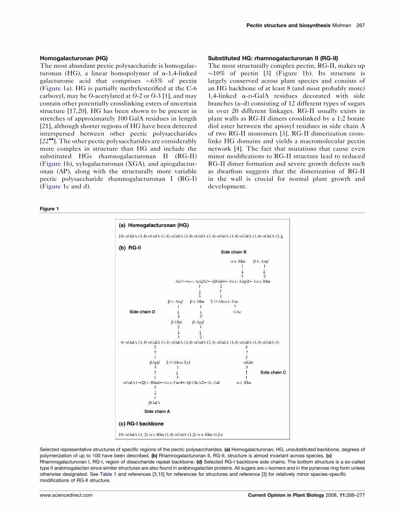
Homogalacturonan (HG)
The most abundant pectic polysaccharide is homogalac-
turonan (HG), a linear homopolymer of a-1,4-linked
galacturonic acid that comprises �65% of pectin
(Figure 1a). HG is partially methylesterified at the C-6
carboxyl, may be O-acetylated at O-2 or O-3 [1], and may
contain other potentially crosslinking esters of uncertain
structure [17,20]. HG has been shown to be present in
stretches of approximately 100 GalA residues in length
[21], although shorter regions of HG have been detected
interspersed between other pectic polysaccharides
[22��]. The other pectic polysaccharides are considerably
more complex in structure than HG and include the
substituted HGs rhamnogalacturonan II (RG-II)
(Figure 1b), xylogalacturonan (XGA), and apiogalactur-
onan (AP), along with the structurally more variable
pectic polysaccharide rhamnogalacturonan I (RG-I)
(Figure 1c and d).
Figure 1
Selected representative structures of specific regions of the pectic polysacch
polymerization of up to 100 have been described. (b) Rhamnogalacturonan
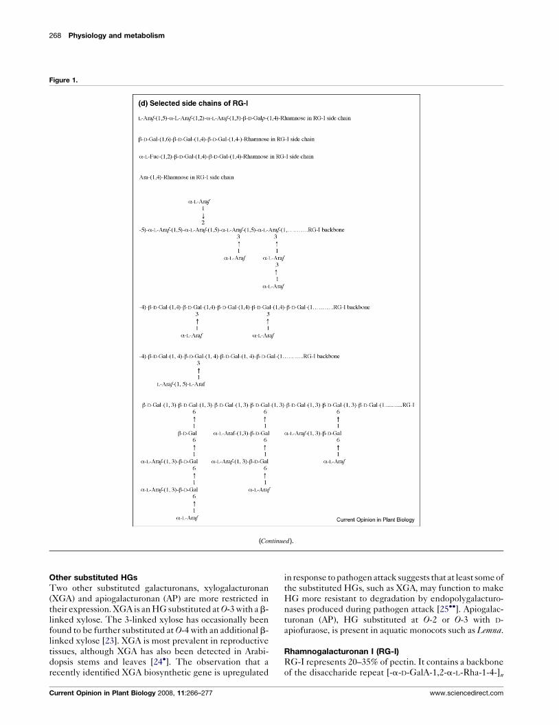
Rhamnogalacturonan I, RG-I, region of disaccharide repeat backbone. (d) S
type II arabinogalactan since similar structures are also found in arabinogalac
otherwise designated. See Table 1 and references [3,15] for references for
modifications of RG-II structure.
www.sciencedirect.com
Substituted HG: rhamnogalacturonan II (RG-II)
The most structurally complex pectin, RG-II, makes up
�10% of pectin [3] (Figure 1b). Its structure is
largely conserved across plant species and consists of
an HG backbone of at least 8 (and most probably more)
1,4-linked a-D-GalA residues decorated with side
branches (a–d) consisting of 12 different types of sugars
in over 20 different linkages. RG-II usually exists in
plant walls as RG-II dimers crosslinked by a 1:2 borate
diol ester between the apiosyl residues in side chain A
of two RG-II monomers [3]. RG-II dimerization cross-
links HG domains and yields a macromolecular pectin
network [4]. The fact that mutations that cause even
minor modifications to RG-II structure lead to reduced
RG-II dimer formation and severe growth defects such
as dwarfism suggests that the dimerization of RG-II
in the wall is crucial for normal plant growth and
development.
arides. (a) Homogalacturonan, HG, unsubstituted backbone, degrees of
II, RG-II, structure is almost invariant across species. (c)
elected RG-I backbone side chains. The bottom structure is a so-called
tan proteins. All sugars are D isomers and in the pyranose ring form unless
structures and reference [3] for relatively minor species-specific
Current Opinion in Plant Biology 2008, 11:266–277
268 Physiology and metabolism
Figure 1.
(Continued ).
Other substituted HGs
Two other substituted galacturonans, xylogalacturonan
(XGA) and apiogalacturonan (AP) are more restricted in
their expression. XGA is an HG substituted at O-3 with a b-
linked xylose. The 3-linked xylose has occasionally been
found to be further substituted at O-4 with an additional b-
linked xylose [23]. XGA is most prevalent in reproductive
tissues, although XGA has also been detected in Arabi-
dopsis stems and leaves [24�]. The observation that a
recently identified XGA biosynthetic gene is upregulated
Current Opinion in Plant Biology 2008, 11:266–277
in response to pathogen attack suggests that at least some of
the substituted HGs, such as XGA, may function to make
HG more resistant to degradation by endopolygalacturo-
nases produced during pathogen attack [25��]. Apiogalac-
turonan (AP), HG substituted at O-2 or O-3 with D-
apiofuraose, is present in aquatic monocots such as Lemna.
Rhamnogalacturonan I (RG-I)
RG-I represents 20–35% of pectin. It contains a backbone
of the disaccharide repeat [-a-D-GalA-1,2-a-L-Rha-1-4-]n
www.sciencedirect.com

Pectin structure and biosynthesis Mohnen 269
(Figure 1c) and exhibits a high degree of cell type and
develop-dependent expression in the type and number of
sugars, oligosaccharides, and branched oligosaccharides
attached to its backbone (Figure 1d) [2,16,26]. The reason
for this level of variation in RG-I structure is not under-
stood but suggests diverse functional specialization. Be-
tween 20 and 80% of the rhamnosyl residues in the RG-I
backbone have side chains containing individual, linear,
or branched a-L-Araf and b-D-Galp residues. The side
chains include a-1,5-linked L-arabinan with 2- and 3-
linked arabinose or arabinan branching, b-1,4-linked D-
galactans with degrees of polymerization (DP) of up to 47
[22��], b-1,4-linked D-galactans with 3-linked L-arabinose
or arabinan branching, and b-1,3-linked D-galactan with
b-6-linked galactan or arabinogalactan branching
(reviewed in references [7,27]. The side chains may also
contain a-L-Fucp, b-D-GlcpA, and 4-O-Me b-D-GlcpA
residues [27].
Model of how the pectic polysaccharides are linked
It is generally believed that the pectic polysaccharides are
covalently crosslinked since harsh chemical treatments or
digestion by pectin-degrading enzymes are required to
isolate HG, RG-I, and RG-II from each other and from
walls. There is currently no consensus as to how the pectic
Figure 2
Schematic structure of pectin showing the four pectic polysaccharides homo
and rhamnogalacturonan II (RG-II) linked to each other. The representative
increased �12.5-fold and RG-I increased �2.5-fold to approximate the amo
[8]. Monosaccharide symbols used are taken partly from the Symbol and Te
Consortium for Functional Glycomics (http://www.functionalglycomics.org/g
www.sciencedirect.com
polysaccharides are linked to each other, or to other
polymers, in the wall; however, the available data
[22��,28,74] support a model with HG, RG-I, and RG-
II linked via their backbones (Figure 2). There are also
indications based on co-elution of pectins with other wall
polymers and mutant phenotype studies that pectins may
be covalently linked to, or tightly associated with, other
types of wall polysaccharides including xyloglucans [29�]and xylans [30]. Analyses of soluble soybean polysacchar-
ides indicate that, at least in soybean, b-1,4-linked xylose
of degrees of polymerization of up to 7 may be attached to
O-3 of HG [30]. Other studies suggest that at least a dimer
of b-1,4-linked xylose may be attached to RG-I in Dios-pyros kaki leaves [31]. Although it remains to be deter-
mined how general such structures are in diverse plant
species, these results open up the possibility that pectins
may serve to hold at least some hemicelluloses in the wall.
The structural data suggesting a link between pectins and
xylans, a class of hemicelluloses abundant in secondary
walls, are intriguing in light of the mutant phenotypes of
several GAUT1-related gene family members [32��], a
family that includes proven and putative pectin biosyn-
thetic glycosyltransferases. For example, the mutant phe-
notypes of qua1/GAUT8 [33,34�], irx8/GAUT12 [35�,36��]and parvus/glz1/GATL1 [37��,38] include effects on both
galacturonan (HG), xylogalacturonan (XGA), rhamnogalacturonan I (RG-I)
pectin structure shown is not quantitatively accurate, HG should be
unts of these polysaccharides in walls. Figure is modified from reference
xt Nomenclature for Representation of Glycan Structure from the
lycomics/molecule/jsp/carbohydrate/carbMoleculeHome.jsp).
Current Opinion in Plant Biology 2008, 11:266–277
270 Physiology and metabolism
xylan and HG content and/or synthesis. Clarification of
the specific enzyme activity affected in these mutants will
probably contribute a clearer understanding of how pec-
tins and hemicelluloses, or their synthesis [39�,40] are
associated.
Pectin biosynthesisLocation of pectin synthesis
Autoradiographic pulse chase experiments, immunocyto-
chemical localization studies with anti-pectin-specific
antibodies, and pectin biosynthetic enzyme subcellullar
fractionation and topology studies demonstrate that pec-
tin is synthesized in the Golgi and transported to the wall
in membrane vesicles. Pectin synthesis occurs simul-
taneously in numerous Golgi stacks in the cell in a process
that appears to include a compartmentalization of specific
biosynthetic enzymes to drive the construction of increas-
ingly complex pectin polysaccharides through the cis,medial and trans Golgi cisternae [41]. The polymers
are targeted to the wall by the movement of Golgi
vesicles, presumably along actin filaments that have
myosin motors [42].
Mechanism of pectin synthesis
All evidence to date suggests that pectin is synthesized in
the Golgi lumen by membrane bound, or associated,
Golgi-localized glycosyltransferases (GTs) that transfer
glycosyl residues from nucleotide-sugars onto oligosac-
charide or polysaccharide acceptors. It is not clear, how-
ever, how synthesis of any of the pectic polysaccharides is
initiated or whether lipid or protein donors are involved
[40,43]. During synthesis some pectic glycosyl residues
are modified by methyltransferase-catalyzed esterifica-
tion or O-methylation, by acetyltransferase-catalyzed
acetylation, and at least in Chenopodiaceae (e.g. spinach),
by feruloylation driven by feruloyltransferases. The sub-
strates for these modifications appear to be S-adenosyl-
methionine (SAM), acetylCoA, and feruloylCoA,
respectively. The recent demonstration that SAM is
transported into pea Golgi lumen via a Golgi mem-
brane-localized transporter with an apparent Km of
30 mM [44�] agrees with previously reported Kms for
HG methyltransferase activity in plant microsomal mem-
brane preparations and is consistent with pectin synthesis
models depicting transport of cytosolic substrates into the
Golgi via Golgi membrane-bound transporters.
HG appears to be inserted into the wall as a highly
methylesterified polymer that is deesterified to varying
degrees by wall-localized pectin methylesterases [45].
The conversion of HG from a methylesterified form to
the negatively charged form has been associated with the
cessation of growth [46], with the binding of positively
charged ions and proteins to the wall, and with the
association of HG molecules to each other via Ca++
binding. The resulting HG:HG salt bridges contribute
to cell–cell adhesion [47]. The spatial partitioning of HG
Current Opinion in Plant Biology 2008, 11:266–277
esterification and deesterification during synthesis, or in
the wall, is not understood, although it has been shown
that the HG in the middle lamella is relatively unester-
ified and that localized regions of esterified HG exist in
some cell walls.
Types of pectin biosynthetic enzymes
Many of the genes encoding nucleotide-sugar biosyn-
thetic enzymes required for pectin synthesis have been
identified [48–51]. Most of these genes exist in gene
families, and it has been proposed that unique gene
family members function in the synthesis of specific
pectic polysaccharides. Definitive evidence for this hy-
pothesis remains to be presented. Approximately 67
glycosyltransferase, methyltransferase and acetyltransfer-
ase activities are predicted to be required for pectin
synthesis [6]. Table 1 lists those transferases for which
enzyme activity has been demonstrated in vitro and/or for
which the encoding gene has been conclusively ident-
ified. Conclusive identification of pectin biosynthetic
genes requires proof of enzyme activity of the encoded
protein. To date, this has only been accomplished for four
glycosyltransferases (Table 1).
Enzymatically proven pectin biosynthetic transferases
Homogalacturonan a1,4-Galacturonosyltransferase
(HG:a1,4GalAT): GAUT1 [32��]
A membrane-bound a1,4-galacturonoslytransferase that
transfers galacturonic acid (GalA) from UDP-a-D-GalA
onto the non-reducing end of homogalacturonan poly-
saccharide and oligosaccharide acceptors, but with a pre-
ference for HG of a degree of polymerizatyion (DP) >9,
has been identified in every higher plant species tested. A
gene encoding an HG:a1,4-GalAT highly expressed in
Arabidopsis cell suspension cultures was identified by
partial purification of the enzyme from detergent-solu-
bilized protein preparations. Out of 20 proteins identified
by tandem MS sequencing, only two contained sequence
domains indicative of glycosyltransferases, and only
one, Galacturonosyltransferase 1 (GAUT1; At3g61130)
yielded HG:GalAT activity when transiently expressed
in human HEK293 cells [32��]. Anti-GAUT1 antibodies
also immunoabsorbed HG:GalAT activity from partially
purified Arabidopsis protein preparations, confirming that
GAUT1 encodes HG:GalAT and indicating that in vitroGAUT1 catalyzes the elongation of HG oligosaccharide
acceptors resulting in the synthesis of polymeric HG. The
other predicted glycosyltransferase in the protein prep-
aration used to identify GAUT1 had 36% sequence
identify (59% similarity) to GAUT1 and was named
GAUT7 (At2g38650) (see below). GAUT7 did not have
GalAT activity when transiently expressed in HEK293
cells [32��], but recent results suggest that GAUT7 may
be part of a GalAT complex (MA Atmodjo et al., unpub-
lished). GAUT1 and GAUT7 both encode proteins
with characteristics consistent with the biochemical prop-
erties of HG:GalAT: a basic PI and an apparent type II
www.sciencedirect.com

Pectin structure and biosynthesis Mohnen 271
Table 1
List of enzymatically confirmed pectin biosynthetic transferases and identified genesa
Type of
transferasebType of GT
or transferase
activity
Parent
polymerc
(side chain)
Enzymed acceptor substrate
Enzyme activity (unless noted:
enzyme adds to the glycosyl
residue on the left*)
Referencee for
structure for GTs
and for enzyme activity
for MTs and ATs
Gene identified
[reference]f
GT D-GalAT HG/RG-II *GalAa1,4-GalA a1,4-GalAT [2] GAUT1 At3g61130
HG:GalAT [32��]
GT D-GalT RG-I L-Rhaa1,4-GalA b1,4-GalT [2]
GT D-GalT RG-I Galb1,4-Rha b1,4-GalT [2,72] –
GT D-GalT RG-I Galb1,4-Gal b1,4-GalT [2] –
GT D-GalT RG-I/AGPg Galb1,3-Gal b1,3-GalT [2] –
GT D-GalT RG-I/AGPg Galb1,3-Gal b1,6-GalT [2] –
GT D-GalT RG-I/AGPg Galb1,6-Galb1,3-Gal b1,6-GalT [2] –
GT L-AraT RG-I L-Arafa1,2-Araf 1,5-L-ArafT [2] –
GT L-AraT RG-I L-Arafa1,5-Araf a1,5-L-ArafT [2] –
GT L-AraT RG-I L-Arafa1,3-Araf a1,3-L-ArafT [2] –
GT L-ArapT RG-I/AGPgL-Arafa1,5-Araf b1,3-L-ArapT [67] –
GT L-ArapT RG-I Galb1,4-Gal a14-L-ArapT [68] –
GT D-ApifT HG/RG-II (A, B) GalAa1,4-GalA b1,2-ApifT [2] –
GT D-XylT RG-II (A) L-Fuca1,4-L-Rha a1,3XylT [2] RGXT1 RG-II:XylT
At4g01770, RGXT2
At4g01750, [52��]
GT D-XylT HG/XGA GalAa1,4-GalA b1,3 XylT [2] XGD1 At5g33290)
XGA:XylT-[25��]
Confirmation
of linkage and anomeric
configuration needed
GT D-GlcAT RG-II (A) L-Fuca1,4-L-Rha b1,4GlcAT [2] –
MT HG:GalA-6-O-MT HG GalAa1,4-GalA(n) [2,6,60�]
AT HG: GalA-3-O-AT HG GalAa1,4-GalA(n) [2,6]
AT RG-I: GalA-3-O/2-O-AT RG-I GalAa1,2-L-Rhaa1,4(n) [2,6]
MT RG-I: GlcA-4-O-MT RG-I GlcAb1,6-Gal [2,6]
MT RG-II: xylose-2-O–MT RG-II D-Xyla1,3-L-Fuc [2,6]
MT RG-II: fucose-2-O-MT RG-II L-Fuca1,2-D-Gal [2,6]
AT RG-II: fucose-AT RG-II L-Fuca1,2-D-Gal [2,6]
AT RG-II: aceric acid 3-O-AT RG-II L-AcefAb1,3-L-Rha [2,6]
a Glycosyltransferases for which enzyme activity has been measured or for which a putative gene has been identified, and list of pectin acetyl- and
methyl-transferases. Activity of glycosyltransferases based on the transfer of a sugar from a nucleotide-sugar onto the indicated disaccharide
acceptor region at the non-reducing end of the indicated acceptor.b Type of transferase: GT, glycosyltransferase; MT, methyltransferase; AT, acetyltransferase.c HG: homogalacturonan; RG-I: rhamnogalacturonan I; RG-II: rhamnogalacturonan II; XGA: xylogalacturonan.d All sugars are D sugars and have pyranose rings unless otherwise indicated. *indicates non-reducing end of the acceptor onto which the indicated
glycosyltransferase transfers the glycosyl residue.e References for the structure. Note: Owing to limitations of the number of references that could be cited, only those structure references not listed in
Ridley et al. [2] are cited here. See reference [2] for other original references on pectin structure.f References for gene encoding the GT.g Enzyme activity would also be required to synthesize arabinogalactan proteins (AGPs) (see reference [73]).
membrane protein topology predicting a short N-terminal
region, a single membrane spanning region, and a larger
C-terminal domain. GAUT1 belongs to a superfamily of
25 genes in Arabidopsis named the GAUT1-related gene
family, which includes 15 GAUT genes with 37–100%
identity (56–100% similarity) to GAUT1, and 10 GAUT1-
like genes (GATLs) with 39–44% identifying (43–53%
similarity) with GAUT1. The GAUT1-related genes are a
subclass of the Arabidopsis CAZy (Carbohydrate Active
Enzymes database; http://www.cazy.org) GT-8 family
[32��,53]. The GAUTs fall into three evolutionary-related
clades: Clade A: (GAUTS 1–7); Clade B (GAUTs 8–11)
and Clade C (GAUTs 12–15). GAUT proteins are pre-
dicted to encode proteins of 61–78 kDa with a predicted
www.sciencedirect.com
signal anchor (GAUTs 1, 6–15) or signal peptide (GAUTs
3-5), consistent with a type II membrane topology or an
intra-microsomal membrane location, respectively. The
only exception is GAUT2 whose sequence does not
predict passage in, or through, the ER/Golgi system.
GAUTs 1, 3, 7, 8 and 9 have been identified as Golgi-
resident proteins [54,55], consistent with the location of
the catalytic domain of HG:a1,4-GalAT in the Golgi
lumen [56]. The GATL genes encode proteins with
predicted signal peptides and masses of 33–44 kDa.
GAUT1 is an HG:a1,4-GalAT; however, its precise func-
tion in pectin synthesis is not known. For example, it is
not known whether GAUT1 functions only during the
Current Opinion in Plant Biology 2008, 11:266–277
272 Physiology and metabolism
elongation stage of pectin synthesis or also in HG
initiation. It is also not clear whether GAUT1 synthesizes
HG as well as the backbone for RG-II, or whether the HG
synthesized by GAUT1 is the same HG that is covalently
attached to the RG-I backbone. Furthermore, the extent
to which GAUT1 functions alone, or in a complex or
complexes with other proteins is not known. Current
results suggest that, at least in vitro, GAUT1 can function
in a complex (MA Atmodjo et al., unpublished). The
recovery of high molecular weight fractions with HG:Ga-
lAT activity [57] supports a role of complexes in pectin
synthesis. The size, number of subunits, stoichiometry,
and the function of pectin biosynthetic complexes, how-
ever, remains to be determined.
RG-II: a1,3xylosyltransferase (RG-II: a1,3XylT): RGXT1
and RGXT2 [52��]
Two Arabidopsis thaliana a-D-1,3-xylosyltransferases
RGXT1 (At4g01770) and RGXT2 (At4g01750) have been
identified, which when expressed as truncated soluble
forms in baculovirus-transfected insect cells, transfer
xylose from UDP-a-D-Xyl onto fucose in an a1,3-linkage.
This is the same linkage present in sidechain A of RG-II
that contains 2-O-methyl D-Xyl attached in an a1,3-link-
age to a-L-Fuc [52��]. The demonstration that the enzymes
transfer xylose onto RG-II isolated from RGXT1 and
RGXT2 mutant walls, but not onto RG-II from wild-type
Arabidopsis walls [52��], provides strong enzyme function
data that RGXT1 and RGXT2 encode RG-II:a1 and
3XylTs. RGXT1 and RGXT2 share 90% sequence iden-
tity, encode proteins of 361 and 367 amino acids, respect-
ively, and are members of CAZy GT-family 77, a novel
family of 27 putative Arabidopsis thaliana glycosyltrans-
ferases [58]. Although the biochemical data supporting the
identity of RGXT1 and RGXT2 as RG-II:a1, 3XylTs is
strong, it is noteworthy that no clear difference in the
structure of RG-II isolated from walls of RGXT1 and
RGXT2 mutants compared with wild-type walls was
obtained [52��]. This result suggests that there may be
gene redundancy and that a double gene knockout mutant
(or more) may be required to recover an RG-II phenotype
in the mutant. In this regard, it is noteworthy that two
additional Arabidopsis genes, At4g01220 and At1g56550
are 68–75% identical to RGXT1 and RGXT2, although their
enzyme activity has not yet been determined [52��].
Xylogalacturonan: xylosyltransferase (XGA:XylT):
XGD1 [25��]
Recently, xylogalacturonan xylosyltransferase, XGD1
(xylogalacturonan deficient 1, At5g33290) was identified
in Arabidopsis [25��]. The gene encodes a Golgi-localized
type II membrane protein from CAZy family 47, which
when expressed in Nicotiana benthamiana, catalyzes the
transfer of xylose from UDP-a-D-xylose onto HG oligo-
saccharides (the so-called oligogalacturonides). Since the
xgd1 mutant produces HG with reduced levels of b-1,3-
xylose compared with wild-type XGA, it is likely that
Current Opinion in Plant Biology 2008, 11:266–277
XGD1 catalyzes the transfer of xylose onto HG in a b-1,3
linkage, although the anomeric configuration of the
product synthesized by XGD1 was not directly
determined. In agreement with this conclusion is the fact
that XGD1 mutants have 15–30% reduced xylose content
in adult leaves, the location of the highest expression of
XGD1 in wild-type plants, and more importantly, have a
significant reduction in xylose in the wall fraction
enriched for xylogalacturonan. Taken together the results
indicate that XGD1 is a XGA:XylT and, most probably, a
XGA:b-1,3XylT, although direct proof of the anomeric
configuration and linkage of the xylose to GalA in HG
remains to be determined.
Putative pectin biosynthetic transferases
An increasing number of putative pectin biosynthetic
glycosyltransferases and methyltransferases have been
identified, largely as a result of characterizing walls of
mutant or transgenic plants, sequence similarity to known
genes from other organisms and/or via trancriptome co-
expression studies with tissues that produce large
amounts of specific wall polysaccharides. Such putative
pectin biosynthetic genes require demonstration of
enzyme activity before their definitive role in pectin
synthesis is confirmed. Nonetheless, their identification
is an important first step to identify genes to target for
enzyme function studies.
Putative HG biosynthetic enzymes
Putative HG–methyltransferase (HG–MT)The GalA in HG of most cell walls is highly (often>50%)
methylesterified at the C-6 carboxyl group [46]. The
enzyme that catalyzes this reaction is called HG–meth-
yltransferase (HG–MT) to distinguish it from methyl-
transferases that methylate RG-I or RG-II. In vitro HG–
MT activity uses S-adenosylmethionine as a methyl
donor, is membrane bound, and has a catalytic site located
on the luminal side of the Golgi [2]. The ability of UDP-
GalA to stimulate HG–MT activity in intact membrane
vesicles, and of HG and pectin to serve as exogenous
acceptors for HG–MT in detergent-permeabilized mem-
branes support a model that a region of HG is synthesized
before its methylation by HG–MT [2,6]. Since some HG–
MT have higher activities with partially esterified pectin
acceptors, compared with polygalacturonic acid, it is
likely that plants contain multiple HG–MTs that dis-
tinguish the degrees of methylation of HG acceptors. The
methylesterification of pectin could be regulated at the
level of the methyltransferase level or by substrate avail-
ability. The report that overexpression of Arabidopsis S-
adenosylmethionine synthetase in flax yields increased
SAM synthetase activity and pectin methylesterification
in the wall, with no concomitant increase in HG–MT
activity [59], indicates that the degree of methylesterifi-
cation of HG may be regulated at least partly, at the level
of substrate (i.e. SAM) concentration.
www.sciencedirect.com

Pectin structure and biosynthesis Mohnen 273
No gene encoding HG–MT has been definitively ident-
ified. Recently, however, it has been suggested that
Quasimodo2 (Qua2) (At1g78240), a Golgi-localized
Arabidopsis protein with a putative methyltransferase
domain may be an HG–MT [60�]. Arabidopsis Qua2-1mutant plants have a 50% reduction in purifiable homo-
galacturonan and are dwarfed, a common phenotype of
pectin mutants. While such results are consistent with a
putative function of QUA2 as an HG–MT, demonstration
of the enzyme activity of QUA2, and of the 29 related
QUA2 homologs in Arabidopsis, is required to determine
which, if any, of these genes encode methyltransferases
required for pectin synthesis. It is interesting to note,
however, that QUA2 and a homolog are transcriptionally
co-expressed with GAUT8/QUA1, a putative pectin bio-
synthetic gene (see below). Furthermore, two additional
sets of QUA2 homologs are co-expressed with GAUT1
(an HG–GalAT, see above) and with GAUT9 (a putative
pectin biosynthetic gene [60�].
Putative HG:GalTAs or Xylan:XylTs
Three different GAUT mutants have characteristics con-
sistent with GAUT gene function in HG and/or xylan
synthesis. Arabidopsis qua1/gaut8 (At3g25140) mutants
are reduced in both GalA content (�25% in rosette leaves
or total plant walls [33], �30% in stem walls [34�], some-
what in suspension cell walls [61]) and to a lesser extent in
xylose content. Protein extracts from qua1 stems have
reduced HG:a-1,4-GalAT and b-1,4-xylosyltransferase
activities [34�]. These results are difficult to understand
since HG and XGA are generally thought to be separate
polysaccharides. The characteristics of the QUA1 (and
IRX8, see below) mutants could indicate that pectin and
xylan syntheses are connected, for example, via a covalent
linkage of the polymers, via synthesis in biosynthetic
complexes, or via compensation upon loss of one of the
polysaccharides. The pleiotropic affects of the QUA1
mutant, combined with the inability to recover enzyme
activity from the recombinantly expressed or purified
GAUT8 protein, make the enzyme function of
GAUT8/QUA1 unclear at present.
GAUT12/IRX8 (At5g54690) mutants are dwarfed with
collapsed xylem, reduced secondary wall thickness,
reduced amounts of glucuronoxylan and a subfraction
of HG [35�], and contain glucuronoxylan that is largely
devoid of a reducing end sequence 4-b-Xyl-1,4-b-Xyl-
1,3-a-L Rha-1,2-a-GalA-1,4-Xyl [36��]. These results
suggest that GAUT12/IRX8 may be a HG:GalAT
involved in the synthesis of a subfraction of HG to which
b-1,4-xylan is attached [35�] or a a1,4-GalAT that adds
GalA into the above-mentioned xylan reducing end
sequence [36��]. Proof of the function of GAUT12 will
require determination of its enzyme activity.
A third GAUT1-related gene family member that has
characteristics consistent with a defect in pectin synthesis
www.sciencedirect.com
and in secondary wall xylan synthesis is one of the
GATL genes, PARVUS/GLZ1/GATL1 (At1g19300)
[37��,38,62,63]. Mutants in this gene, when grown under
low humidity, are semi-sterile dwarfs with reduced anther
dehiscence, slightly elevated mole percent Rha, Ara, and
Gal, slightly reduced GalA, and larger reductions in
xylose compared with the wild type. The mutant also
has reduced branching of the RG-I backbone, with
accompanying overall increased amounts of wall pectin
and decreased xylan [37��,62]. Xylan side branches in
parvus/glz1/gatl1 have comparable amounts of
methylGlcA but lack non-methylated GlcA side branches
[37��] and also lack the reducing end sequence 4-b-Xyl-
1,4-b-Xyl-1,3-a-L-Rha-1,2-a-GalA-1,4-Xyl, the latter
modification being similar to that in the irx8/gaut12
mutants described above ([36��], see also article by WS
York and MA O’Neill in this issue of Curr Opin Plant Biol).The described glycosyl residue changes, and the struc-
tural modifications are consistent with a possible role of
the PARVUS/GLZ1/GATL1 gene (like IRX8/GAUT12)
in the synthesis of the xylan, possibly the xylan-primer, or
in the synthesis of the structure to which the reducing end
xylan oligosaccharide is attached, possibly a fraction of
pectin.
Putative RG-II biosynthetic enzymes
Putative RG-II:GlcATNicotiana plumbaginifolia T-DNA nolac-H18 callus
mutant NpGUT1 is non-organogenic, has reduced inter-
cellular attachment, and 86% reduced levels of glucuronic
acid. The gene mutated in the callus is NpGUT1, which
has 60% sequence homology to animal heparin sulfate
biosynthetic glucuronosyltransferases. Since RG-II iso-
lated from cell walls of the nolac-H18 mutant are devoid
of glucuronic acid and exhibit 82% reduced RG-II dimer
formation, it has been proposed that NpGUT1 encodes
RG-II b-1,2GlcAT that transfers GlcA onto the L-fucose
in RG-II side chain A [64]. Demonstration of the enzyme
activity of NpGUT1 will be required to confirm the
proposed function of the putative RG-II:b-1,2GlcAT.
Putative RG-I biosynthetic enzymes
Putatutive RG-I:a1,5AraT)RG-I contains L-arabinose in multiple linkages with the
majority of the arabinose in the furanose ring form,
although there is a small amount of terminal arabino-
pyranose in some RG-I side chains [65]. Most enzymatic
studies of pectin AraT activity have been carried out
using the donor UDP-b-L-arabinopyranose and two
types of enzyme activity that use the arabinopyranose
form of the sugar have been described. A Golgi-localized
a1,5-arabinan:b-(1!3) arabinopyranose AraT activity
that transfers a single arabinopyranose residue onto
the non-reducing end of a1,5-arabinooligosaccharide
acceptors has been identified [66,67]. Other studies
have identified an arabinopyranosetransferase activity
that transfers individual arabinopyranosyl residues onto
Current Opinion in Plant Biology 2008, 11:266–277

274 Physiology and metabolism
1,4-linked b-D-galactooligosaccharides [68] in an a con-
figuration. In neither case has a gene been identified. It
has been proposed that the b-1,4-galactan:a1,4AraT-
catalyzed addition of a non-reducing end terminal
a-L-arabinopyranosyl residue onto b-1,4-galactan oligo-
saccharide prevents further galactosylation of the galac-
tooligosaccharides [68,69].
An Arabidopsis gene that encodes a putative arabinan:a-
1,5-arabinosyltransferase (ARAD1; At2g35100, CAZy
GT47) was identified through biochemical and immuno-
chemical analyses of the Arabidopsis T-DNA insert
mutant ARABINAN DEFICIENT 1 [70�]. ARAD1 encodes
a predicted 52.8 kDa protein with a single N-terminal
transmembrane domain region followed by a proposed
catalytic domain. Walls from ARAD1 homozygous knock-
out mutant leaves and stems have 25 and 54%, respect-
ively, reduced levels of Ara compared with wild-type
walls [70�]. Transformation of arad1 plants with the
ARAD1 gene restores Ara in the wall to wild-type levels,
providing evidence that ARAD1 affects wall arabinose
levels. Immunocytochemical analyses of leaf, inflores-
cence stem, and the stem using the anti-a-1,5-arabinan
antibody LM6 indicate the reduced immunolabeling in
walls that were not associated with proteins, suggesting
that the mutant was affected in the synthesis of a-1,5-
arabinans, but not in glycoprotein synthesis [70�].Furthermore, comparison of RG-I from wild type and
ARAD1 walls showed that arad1 RG-I had a 68%
reduction in Ara content, predominantly due to a
reduction in 5-linked Araf along with 2,5f-linked Ara
and 2,3,5-linked Araf . The results provide strong evi-
dence that ARAD1 is a putative RG-I arabinan:a-1,
5-AraT. Confirmation of the proposed a1,5arabinosyl-
transferase activity of ARAD1, however, will require
expression of an enzymatically active protein.
The recent identification of a UDP-arabinopyranose
mutase (UAM) from rice that catalyzes the reversible
formation of UDP-Araf from UDP-Arap [71��] should
facilitate the identification of AraTs that catalyze the
transfer of arabinofuranose, rather than arabinopyranose.
Conclusions: challenges and futuredirections in studying pectin synthesisThe first step in understanding how the 67 or more pectin
biosynthetic transferases work together to synthesize the
pectic polysaccharides is to identify the genes encoding
the biosynthetic enzymes. The identification of the four
glycosyltransferases involved in the synthesis of HG,
XGA, and RG-II is a start, but rapid progress in identify-
ing the other pectin biosynthetic enzymes will require
overcoming the following challenges: (1) remaining
uncertainty regarding pectin structure and how and where
in the cell the pectic polysaccharides are crosslinked; (2)
the growing body of evidence that species-, cell type-, and
developmental state-specific differences in pectin struc-
Current Opinion in Plant Biology 2008, 11:266–277
ture exist, making it likely that the number and types of
enzymes required to synthesize pectin are plant, tissue,
cell type and developmental state specific; (3) the
limitation that knowledge of the structure of ‘mature’
pectic polysaccharides from the wall does not necessarily
reflect the structures as they are synthesized, but rather
structures after insertion into the wall and after any
subsequent modification in muro by chemical and/or
enzyme-catalyzed reactions; (4) the lack of availability,
and in some cases identity, of the biosynthetic substrates
and acceptors; and (5) the great difficulty in recovering
enzyme activity in heterologously expressed genes.
Despite these challenges, however, progress is being
made and putative pectin biosynthetic genes are being
identified via mutant phenotype characterization and by
identification of genes co-transcribed with de facto pectin
biosynthetic genes. The hold up, however, remains the
definitive identification of enzyme function. To date,
success in proving enzyme activity has required a focused,
and often time consuming, gene-by-gene effort. Whether
broader, more global methods can be developed to obtain
the definitive enzyme activity data needed to prove gene
function is not clear. However, the increasing evidence
that wall synthesis occurs, at least partly, via protein
complexes makes efforts to purify and characterize
protein complexes with a dual emphasis on enzyme
activity and subunit structure determination, particularly
important. Likewise, the development of methods to
either isolate single cell types in plants, so as to study
cell-type-specific synthesis, or potentially more powerful,
the development of methods to study synthesis in single
cells, would greatly catapult the field forward. Finally,
there is a continued need to know the identity of, and
have available, the diverse sugar, and acceptor substrates
used by the pectin biosynthetic transferases.
AcknowledgementsThe following funding is gratefully acknowledged for supporting thewriting of this review and the research that drives the author’s work in thisarea: NRI, CSREES, USDA Award 2006-35318-17301; NSF MCB awards0313509 and 0646109; and the BioEnergy Science Center, the U.S.Department of Energy Bioenergy Research Center supported by the Officeof Biological and Environmental Research in the DOE Office of Science.
References and recommended readingPapers of particular interest, published within the annual period ofreview, have been highlighted as:
� of special interest
�� of outstanding interest
1. O’Neill M, Albersheim P, Darvill A: The pectic polysaccharides ofprimary cell walls. In Methods in Plant Biochemistry, 2. Edited byDey PM. London: Academic Press; 1990:415-441.
2. Ridley BL, O’Neill MA, Mohnen D: Pectins: structure,biosynthesis, and oligogalacturonide-related signaling.Phytochemistry 2001, 57:929-967.
3. O’Neill MA, Ishii T, Albersheim P, Darvill AG:Rhamnogalacturonan II: structure and function of a boratecross-linked cell wall pectic polysaccharide. Annu Rev PlantBiol 2004, 55:109-139.
www.sciencedirect.com
Pectin structure and biosynthesis Mohnen 275
4. Matsunaga T, Ishii T, Matsumoto S, Higuchi M, Darvill A,Albersheim P, O’Neill MA: Occurrence of the primary cell wallpolysaccharide rhamnogalacturonan II in pteridophytes,lycophytes, and bryophytes. Implication for the evolution ofvascular plants. Plant Physiol 2004, 134:339-351.
5. Himmel ME, Ding S-Y, Johnson DK, Adney WS, Nimlos MR,Brady JW, Foust TD: Biomass recalcitrance: engineeringplants and enzymes for biofuels production. Science 2007,315:804-807.
6. Mohnen D, Bar-Peled L, Somerville C: Cell wall synthesis. InBiomass Recalcitrance. Edited by Himmel M. Blackwell; 2008.
7. Mohnen D: Biosynthesis of pectins. In Pectins and theirManipulation. Edited by Seymour GB, Knox JP. Oxford: BlackwellPublishing and CRC Press; 2002:52-98.
8. Scheller HV, Jensen JK, Sørensen SO, Harholt J, Geshi N:Biosynthesis of pectin. Physiol Plant 2007, 129:283-295.
9. Zhong R, Ye Z-H: Unraveling the functions ofglycosyltransferase family 47 in plants. Trends Plant Sci 2003,8:565-568.
10. Scheible W-R, Pauly M: Glycosyltransferases and cell wallbiosynthesis: novel players and insights. Curr Opin Plant Biol2004, 7:285-295.
11. Lerouxel O, Cavalier DM, Liepman AH, Keegstra K: Biosynthesisof plant cell wall polysaccharides—a complex process. CurrOpin Plant Biol 2006, 9:621-630.
12. Farrokhi N, Burton RA, Brownfield L, Hrmova M, Wilson SM,Bacic A, Fincher GB: Plant cell wall biosynthesis: genetic,biochemical and functional genomics approaches to theidentification of key genes. Plant Biotech J 2006, 4:145-167.
13. Johansen JN, Vernhettes S, Hofte H: The ins and outs of plantcell walls. Curr Opin Plant Biol 2006, 9:616-620.
14. Zhong R, Ye Z-H: Regulation of cell wall biosynthesis. Curr OpinPlant Biol 2007, 10:564-572.
15. Mohnen D: Biosynthesis of pectins and galactomannans. InComprehensive Natural Products Chemistry, Carbohydrates andtheir Derivatives including Tannins, Cellulose, and Related Lignins,3. Edited by Pinto BM. Oxford: Elsevier; 1999:497-527.
16. Willats WGT, McCartney L, Mackie W, Knox JP: Pectin: cellbiology and prospects for functional analysis. Plant Mol Biol2001, 47:9-27.
17. Jackson CL, Dreaden TM, Theobald LK, Tran NM, Beal TL, Eid M,Gao MY, Shirley RB, Stoffel MT, Kumar MV, Mohnen D: Pectininduces apoptosis in human prostate cancer cells: correlationof apoptotic function with pectin structure. Glycobiology 2007,17:805-819.
18. Inngjerdingen KT, Patel TR, Chen X, Kenne L, Allen S, Morris GA,Harding SE, Matsumoto T, Diallo D, Yamada H, Michaelsen TEet al.: Immunological and structural properties of a pecticpolymer from Glinus oppositifolius. Glycobiology 2007,17:1299-1310.
19. Albersheim P, Darvill AG, O’Neill MA, Schols HA, Voragen AGJ: Anhypothesis: the same six polysaccharides are components ofthe primary cell walls of all higher plants. In Pectins andPectinases. Edited by Visser J, Voragen AGJ. Amsterdam: ElsevierSciences B.V.; 1996:47-53.
20. MacKinnon IM, Jardine WG, O’Kennedy N, Renard CMGC,Jarvis MC: Pectic methyl and nonmethyl esters in potato cellwalls. J Agric Food Chem 2002, 50:342-346.
21. Yapo BM, Lerouge P, Thibault J-F, Ralet MC: Pectins from citruspeel cell walls contain homogalacturonans homogenous withrespect to molar mass, rhamnogalacturonan I andrhamnogalacturonan II. Carbohydr Polym 2007, 69:426-435.
22.��
Nakamura A, Furuta H, Maeda H, Takao T, Nagamatsu Y:Structural studies by stepwise enzymatic degradation of themain backbone of soybean soluble polysaccharidesconsisting of galacturonan and rhamnogalacturonan. BiosciBiotechnol Biochem 2002, 66:1301-1313.
An important paper that provides structural data to support the model thatthe pectic polysaccharides RG-I and HG are linked via their backbones.
www.sciencedirect.com
23. Zandleven J, Beldman G, Bosveld M, Schols HA, Voragen AGJ:Enzymatic degradation studies of xylogalacturonans fromapple and potato, using xylogalacturonan hydrolase.Carbohydr Polym 2006, 65:495-503.
24.�
Zandleven J, Sørensen SO, Harholt J, Beldman G, Schols HA,Scheller HV, Voragen AJ: Xylogalacturonan exists in cell wallsfrom various tissues of Arabidopsis thaliana. Phytochemistry2007, 68:1219-1226.
First rigorous analysis of xylogalacturonan in Arabidopsis provides evi-dence for the presence of XGA in tissues other than reproductive organs.
25.��
Jensen JK, Sørensen SO, Harholt J, Geshi N, Sakuragi Y, Møller I,Zandleven J, Bernal AJ, Jensen NB, Sørensen C, et al.Identification of a xylogalacturonan xylosyltransferaseinvolved in pectin biosynthesis in Arabidopsis. Plant Cell 2008,in press.
A very thorough and carefully executed analysis of At5g33290 mutantprotein provides evidence that XGD1 is a xylogalacturonan:xylosyltrans-ferase and identifies the fourth functionally confirmed pectin biosyntheticglycosyltransferase.
26. Guillemin F, Guillon F, Bonnin E, Devaux M-F, Chevalier T,Knox JP, Liners F, Thibault J-F: Distribution of pectic epitopes incell walls of the sugar beet root. Planta 2005:355-371.
27. O’Neill MA, York WS: The composition and structure of plantprimary cell walls. In The Plant Cell Wall. Edited by Rose JKC.Ithaca, New York: Blackwell Publishing/CRC Press; 2003:1-54.
28. Ishii T, Matsunaga T: Pectic polysacchariderhamnogalacturonan II is covalently linked tohomogalacturonan. Phytochemistry 2001, 57:969-974.
29.�
Popper ZA, Fry SC: Xyloglucan–pectin linkages are formedintra-protoplasmically, contribute to wall-assembly, andremain stable in the cell wall. Planta 2007, 227:781-794.
Further evidence that xyloglucans contain a negatively charged moietythat may be pectin, although no specific xyloglucan–pectin linkage wasdemonstrated. The hypothesis that xyloglucans are transferred onto thepectin RG-I by transglycosylation was tested and found not to support theresults presented.
30. Nakamura A, Furuta H, Maeda H, Takao T, Nagamatsu Y: Analysisof the molecular construction of xylogalacturonan isolatedfrom soluble soybean polysaccharides. Biosci BiotechnolBiochem 2002, 66:1155-1158.
31. Duan J, Zheng Y, Dong Q, Fang J: Structural analysis of a pecticpolysaccharide from the leaves of Diospyros kaki.Phytochemistry 2004, 65:609-615.
32.��
Sterling JD, Atmodjo MA, Inwood SE, Kolli VSK, Quigley HF,Hahn MG, Mohnen D: Functional identification of anArabidopsis pectin biosynthetic homogalacturonangalacturonosyltransferase. Proc Natl Acad Sci U S A 2006,103:5236-5241.
The paper reports the first identification of an enzymatically proven pectinbiosynthetic glycosyltransferase, galacturonosyltransferase 1 (GAUT1) anddefines the GAUT1-related gene family of proven and putative GalATs.
33. Bouton S, Leboeuf E, Mouille G, Leydecker MT, Talbotec J,Granier F, Lahaye M, Hofte H, Truong H-N: QUASIMODO1encodes a putative membrane-bound glycosyltransferaserequired for normal pectin synthesis and cell adhesion inArabidopsis. Plant Cell 2002, 14:2577-2590.
34.�
Orfila C, Sørensen SO, Harholt J, Geshi N, Crombie H, Truong H-N,Reid JSG, Knox JP, Scheller HV: QUASIMODO1 is expressed invascular tissue of Arabidopsis thaliana inflorescence stems,and affects homogalacturonan and xylan biosynthesis. Planta2005, 222:613-622.
The first paper indicating a connection between pectin homogalactur-onan and xylan synthesis.
35.�
Persson S, Hosmer Caffal K, Freshour G, Hilley MT, Bauer S,Poindexter P, Hahn MG, Mohnen D, Somerville C: TheArabidopsis irregular xylem 8 mutant is deficient inglucuronoxylan and homogalacturonan, which are essentialfor secondary cell wall integrity. Plant Cell 2007, 19:237-255.
A detailed analysis of irx8 mutant walls provides evidence for effects ofthe mutation on both xylan and pectin synthesis, contributing to thegrowing number of studies suggesting some type of association betweenthese polymers and/or their synthesis.
Current Opinion in Plant Biology 2008, 11:266–277

276 Physiology and metabolism
36.��
Pena M, Zhong R, Zhou G-K, Richardson E, O’Neill MA, Darvill AG,York WS, Ye Z-H: Alterations in the abundance of the glycosylsequence at the reducing end of glucuronoxylan and changesin glucuronoxylan chain length in the Arabidopsis irregularxylem8 and 9 mutants. Plant Cell 2007, 19:549-563.
The ‘rediscovery’ of the unique oligosaccharide at the reducing end ofxylan, and the methodology to detect the ‘primer’ sequence, providedtools to support multiple additional studies on putative xylan primerbiosynthetic glycosyltransferases and forces the question of whetherunique primers are involved in the synthesis of diverse wall polysacchar-ides.
37.��
Brown DM, Goubet F, Wong VW, Goodacre R, Stephens E,Dupree P, Turner SR: Comparison of five xylan synthesismutants reveals new insight into the mechanisms of xylansynthesis. Plant J 2007, 52:1154-1168.
This paper provides an elegant multi-gene approach to understandingxylan biosynthesis and provides the first published report that the PAR-VUS/GLZ1/GATL1 gene may be involved in xylan synthesis.
38. Lee C, Zhong R, Richardson E, Himmelsbach DS, McPhail BT,Ye Z-H: The PARVUS gene is expressed in cells undergoingsecondary wall thickening and is essential for glucuronoxylanbiosynthesis. Plant Cell Physiol 2007, 48:1659-1672.
39.�
Bernal AJ, Jensen JK, Harholt J, Sørensen S, Moller I, Blaukopf C,Johansen B, de Lotto R, Pauly M, Scheller HV, Willats WGT:Disruption of ATCSLD5 results in reduced growth,reduced xylan and homogalacturonan synthase activityand altered xylan occurrence in Arabidopsis. Plant J 2007,52:791-802.
The paper reports the surprising observation that disruption of theArabidopsis cellulose synthase-like D5 gene reduces both xylan andhomogalacturonan synthesis, a result the authors interpret as possiblecoordinated downregulation of xylan and pectin synthesis with that of aGolgi-localized b-glucan.
40. Cumming CM, Rizkallah HD, McKendrick KA, Abdel-Massih RM,Baydoun EAH, Brett CT: Biosynthesis and cell-walldeposition of a pectin-xyloglucan complex in pea. Planta 2005,222:546-555.
41. Nebenfuhr A, Staehelin LA: Mobile factories: Golgi dynamics inplant cells. Trends Plant Sci 2001, 6:160-167.
42. Nebenfuhr A, Gallagher LA, Dunahay TG, Frohlick JA,Mazurkiewicz AM, Meehl JB, Staehelin LA: Stop-and-gomovements of plant golgi stacks are mediated by theacto-myosin system. Plant Physiol 1999, 121:1127-1141.
43. Abdel-Massih RM, Baydoun EAH, Brett CT: In vitro biosynthesisof 1, 4-b-galactan attached to a pectin–xyloglucan complex inpea. Planta 2003, 216:502-511.
44.�
Ibar C, Orellana A: The import of S-adenosylmethionine into theGolgi apparatus is required for the methylation ofhomogalacturonan. Plant Physiol 2007, 145:504-512.
The authors provide elegant experimental evidence for transport of themethyl donor SAM into Golgi, a compartment that must support numer-ous pectin methylation reactions.
45. Pelloux J, Rusterucci C, Mellerowicz EJ: New insights into pectinmethylesterase structure and function. Trends Plant Sci 2008,12:267-277.
46. Derbyshire P, McCann MC, Roberts K: Restricted cell elongationin Arabidopsis hypocotyls is associated with a reducedaverage pectin esterification level. BMC Plant Biol 2007, 7:31.
47. Jarvis MC, Briggs SPH, Knox JP: Intercellular adhesion and cellseparation in plants. Plant Cell Environ 2003, 26:977-989.
48. Reiter W-D, Vanzin GF: Molecular genetics of nucleotidesugar interconversion pathways in plants. Plant Mol Biol 2001,47:95-113.
49. Seifert GJ: Nucleotide sugar interconversions and cell wallbiosynthesis: how to bring the inside to the outside. Curr OpinPlant Biol 2004, 7:277-284.
50. Gu X, Bar-Peled M: The biosynthesis of UDP-galacturonic acidin plants. Functional cloning and characterization ofArabidopsis UDP-D-glucuronic acid 4-epimerase. Plant Physiol2004, 136:4256-4264.
Current Opinion in Plant Biology 2008, 11:266–277
51. Pattathil S, Harper AD, Bar-Peled M: Biosynthesis of UDP-xylose: characterization of membrane-bound At Uxs2. Planta2005, 221:538-548.
52.��
Egelund J, Petersen BL, Motawia MS, Damager I, Faik A,Olsen CE, Ishii T, Clausen H, Ulvskov P, Geshi N: Arabidopsisthaliana RGXT1 and RGXT2 encode Golgi-localized (1,3)-a-D-xylosyltransferases involved in the synthesis of pecticrhamnogalacturonan II. Plant Cell 2006, 18:2593-2607.
The first identification of an enzymatically proven RG-II biosynthetic geneand the novel use of a monosaccharide acceptor assay to define enzymeactivity of plant wall biosynthetic glycosyltransferases.
53. Coutinho PM, Deleury E, Davies GJ, Henrissat B: An evolvinghierarchical family classification for glycosyltransferases.J Mol Biol 2003, 328:307-317.
54. Dunkley TPJ, Watson R, Griffin JL, Dupree P, Lilley KS:Localization of organelle proteins by isotope tagging (LOPIT).Mol Cell Proteomics 2004, 3.11:1128-1134.
55. Dunkley TPJ, Hester S, Shadforth IP, Runions J, Weimar T,Hanton SL, Griffin JL, Bessant C, Brandizzi F, Hawes C et al.:Mapping the Arabidopsis organelle proteome. Proc Natl AcadSci U S A 2006, 103:6518-6523.
56. Sterling J, Quigley HF, Orellana A, Mohnen D: The catalytic site ofthe pectin biosynthetic enzyme a-1, 4-galacturonosyltransferase (GalAT) is located in the lumen ofthe Golgi. Plant Physiol 2001, 127:360-371.
57. Ohashi T, Ishimizu T, Akita K, Hase S: In vitro stabilization andminimum active component of polygalacturonic acid synthaseinvolved in pectin biosynthesis. Biosci Biotech Biochem 2007,71:2291-2299.
58. Egelund JE, Skjot M, Geshi N, Ulvskov P, Petersen BL: Acomplementary bioinformatics approach to identify potentialplant cell wall glycosyltransferase-encoding genes. PlantPhysiol 2004, 136:2609-2620.
59. Lamblin F, Saladin G, Dehorter B, Cronier D, Grenier E, Lacoux J,Bruyant P, Laine E, Chabbert B, Girault F et al.: Overexpression ofa heterologous sam gene encoding S-adenosylmethioninesynthetase in flax (Linum usitatissimum) cells: consequenceson methylation of lignin precursors and pectins. Physiol Plant2001, 112:223-232.
60.�
Mouille G, Ralet M-C, Cavelier C, Eland C, Effroy D, Hematy K,McCartney L, Truong HN, Gaudon V, Thibault J-F et al.:Homogalacturonan synthesis in Arabidopsis thaliana requiresa Golgi-localized protein with a putative methyltransferasedomain. Plant J 2007, 50:605-614.
The authors present multiple lines of evidence to support the designationof QUA2 as a putative pectin methyltransferase. Although enzymaticevidence of function is still needed, the co-expression of QUA2 withknown or putative HG galacturonosyltransferases is noteworthy.
61. Leboeuf E, Guillon F, Thoiron S, Lahaye M: Biochemical andimmunohistochemical analysis of pectic polysaccharides inthe cell walls of Arabidopsis mutant QUASIMODO 1suspension-cultured cells: implications for cell adhesion.J Exp Bot 2005, 56:3171-3182.
62. Lao NT, Long D, Kiang S, Coupland G, Shoue DA, Carpita NC,Kavanagh TA: Mutation of a family 8 glycosyltransferase genealters cell wall carbohydrate composition and causes ahumidity-sensitive semi-sterile dwarf phenotype inArabidopsis. Plant Mol Biol 2003, 53:687-701.
63. Shao M, Zheng H, Hu Y, Liu D, Jang J-C, Ma H, Huang H: TheGAOLAOZHUANGREN1 gene encodes a putativeglycosyltransferase that is critical for normal developmentand carbohydrate metabolism. Plant Cell Physiol 2004,45:1453-1460.
64. Iwai h, Masaoka N, Ishii T, Satoh S: A pectinglucuronyltransferase gene is essential for intercellularattachment in the plant meristem. Proc Natl Acad Sci U S A2002, 99:16319-16324.
65. Huisman MMH, Brull LP, Thomas-Oates JE, Haverkamp J,Schols HA, Voragen AGJ: The occurrence of internal (1-5)-linked arabinofuranose and arabinopyranose residues inarabinogalactan side chains from soybean pectic substances.Carbohydr Res 2001, 330:103-114.
www.sciencedirect.com
Pectin structure and biosynthesis Mohnen 277
66. Nunan KJ, Scheller HV: Solubilization of an arabinanarabinosyltransferase activity from mung bean hypocotyls.Plant Physiol 2003, 132:331-342.
67. Ishii T, Konishi T, Ito Y, Ono H, Ohnishi-Kameyama M, Maeda I: A b-(1!3)-arabinopyranosyltransferase that transfers a singlearabinopyranose onto arabino-oligosaccharides in mung bean(Vigna radiate) hypocotyls. Phytochemistry 2005, 66:2418-2425.
68. Ishii T, Ono H, Ohnishi-Kameyama M, Maeda I:Enzymic transfer of a-L-arabinopyranosyl residues toexogenous 1,4-linked b-D-galacto-oligosaccharides usingsolubilized mung bean (Vigna radiata) hypocotyl microsomesand UDP-b-L-arabinopyranose. Planta 2005, 221:953-963.
69. Ishii T, Ohnishi-Kameyama M, Ono H: Identification ofelongating b-1,4-galactosyltransferase activity in mung bean(Vigna radiata) hypocotyls using 2-aminobenzaminated 1,4-linked b-D-galactooligosaccharides as acceptor substrates.Planta 2004, 219:310-318.
70.�
Harholt J, Jensen JK, Sorensen SO, Orfila C, Pauly M, Scheller HV:ARABINAN DEFICIENT 1 is a putative arabinosyltransferaseinvolved in biosynthesis of pectic arabinan in arabidopsis.Plant Physiol 2005, 140:49-58.
The authors present multiple lines of evidence to show that ARAD1 is aputative RG-I arabinosyltransferase. Enzyme activity studies are needed
www.sciencedirect.com
to confirm the identity of this gene and to determine the linkage andsubstrate specificity of the enzyme.
71.��
Konishi T, Takumi T, Miyazuki Y, Ohnishi-Kameyama M, Hayashi T,O’Neill MA, Ishii T: A plant mutase that interconverts UDP-arabinofuranose and UDP-arabinopyranose. Glycobiology2007, 17:345-354.
The identification of UDP-arabinopyranose mutase (UAM) provides evi-dence that plants may use UDP-Arap to produce UDP-Araf as a substratefor pectin synthesis. The association of this activity with the so-calledreversibly glycosylated proteins (RGPs) reveals a previously unknownfunction of RGPs.
72. Lau JM, McNeil M, Darvill AG, Albersheim P: Treatment ofrhamnogalacturonan I with lithium in ethylenediamine.Carbohydr Res 1987, 168:245-274.
73. Gaspar Y, Johnson KL, McKenna JA, Bacic A, Schultz CJ: Thecomplex structures of arabinogalactan-proteins and thejourney towards understanding function. Plant Mol Biol 2001,47:161-176.
74. Coenen GJ, Bakx EJ, Verhoef RP, Schols HA, Voragen AGJ:Identification of the connecting linkage between homo- orxylogalacturonan and rhamnogalacturonan type I.Carbohydr Polymers 2007, 70:224-235.
Current Opinion in Plant Biology 2008, 11:266–277