Embed Size (px)
Citation preview
PDGFRβ regulates craniofacialdevelopment through homodimersand functional heterodimers with PDGFRαKatherine A. Fantauzzo1 and Philippe Soriano
Department of Cell Developmental and Regenerative Biology, Icahn School of Medicine at Mount Sinai, New York,New York 10029, USA
Craniofacial development is a complex morphogenetic process, disruptions in which result in highly prevalenthuman birth defects. While platelet-derived growth factor (PDGF) receptor α (PDGFRα) has well-documentedfunctions in this process, the role of PDGFRβ in murine craniofacial development is not well established. Wedemonstrate that PDGFRα and PDGFRβ are coexpressed in the craniofacial mesenchyme of mid-gestation mouseembryos and that ablation of Pdgfrb in the neural crest lineage results in increased nasal septum width, delayedpalatal shelf development, and subepidermal blebbing. Furthermore, we show that the two receptors geneticallyinteract in this lineage, as double-homozygous mutant embryos exhibit an overt facial clefting phenotype moresevere than that observed in either single-mutant embryo. We reveal a physical interaction between PDGFRα andPDGFRβ in the craniofacial mesenchyme and demonstrate that the receptors form functional heterodimers withdistinct signaling properties. Our studies thus uncover a novel mode of signaling for the PDGF family during ver-tebrate development.
[Keywords: PDGFRβ; craniofacial development; neural crest; PDGFRα; heterodimers]
Supplemental material is available for this article.
Received August 8, 2016; revised version accepted October 19, 2016.
The platelet-derived growth factor (PDGF) receptors(PDGFRs) are receptor tyrosine kinases (RTKs) that bindto a subset of growth factors on the surface of cells andelicit responseswith broad roles in developmental cellularprocesses. The mammalian PDGF family consists of fourligands, PDGF-A–D, which variously signal through twoRTKs: PDGFRα and PDGFRβ. Receptors in this familyconsist of an extracellular ligand-binding domain contain-ing five immunoglobulin-like loops, a single transmem-brane domain, and an intracellular domain harboring asplit catalytic tyrosine kinase (Williams 1989). The tworeceptors share the highest amino acid homology in theN-terminal (85% identity) and C-terminal (75% identity)kinase domains, with considerably reduced homology inthe extracellular (31% identity) and interkinase (27%identity) domains (Matsui et al. 1989). PDGFRs are acti-vated by ligand binding that induces receptor dimeriza-tion and promotes tyrosine kinase activity, resulting inthe autophosphorylation of intracellular tyrosine resi-dues. Signaling molecules containing Src homology 2(SH2) phosphotyrosine recognition motifs bind to specificphosphorylated residues in the cytoplasmic domains of
the receptors and mediate downstream cellular responsesthrough various intracellular signaling pathways (Heldinand Westermark 1999). These signaling molecules in-clude a subset with intrinsic enzymatic activity, includ-ing Src family tyrosine kinases, phosphatidylinositol 3-kinase (PI3K), the tyrosine phosphatase SHP-2, phospholi-pase Cγ (PLCγ), and Ras GTPase-activating protein (GAP)as well as adaptor proteins such as Crk, Grb2, and Nck(Heldin and Westermark 1999). While both PDGFRα andPDGFRβ are capable of interacting with Src, PI3K, SHP-2, and PLCγ, PDGFRα is additionally able to bind Crk,and PDGFRβ is further capable of interacting with GAP,Grb2, and Nck (Heldin and Westermark 1999).Although numerous ligand and receptor interac-
tions have been demonstrated in vitro, the homodimersPDGF-AA and PDGF-CC have been shown to exclusivelyactivate PDGFRα signaling in vivo duringmammalian de-velopment (Boström et al. 1996; Soriano 1997; Ding et al.2004), while PDGF-BB solely activates PDGFRβ signaling(Levéen et al. 1994; Soriano 1994). Targeted disruption ofPdgfra in mice results in embryonic lethality during mid-gestation, with homozygous null embryos exhibiting a
1Present address: Department of Craniofacial Biology, University ofColorado Anschutz Medical Campus, Aurora, CO 80045, USA.Corresponding author: [email protected] published online ahead of print. Article and publication date areonline at http://www.genesdev.org/cgi/doi/10.1101/gad.288746.116.
© 2016 Fantauzzo and Soriano This article is distributed exclusively byCold Spring Harbor Laboratory Press for the first six months after thefull-issue publication date (see http://genesdev.cshlp.org/site/misc/terms.xhtml). After six months, it is available under a Creative CommonsLicense (Attribution-NonCommercial 4.0 International), as described athttp://creativecommons.org/licenses/by-nc/4.0/.
GENES & DEVELOPMENT 30:2443–2458 Published by Cold Spring Harbor Laboratory Press; ISSN 0890-9369/16; www.genesdev.org 2443
Cold Spring Harbor Laboratory Press on June 1, 2020 - Published by genesdev.cshlp.orgDownloaded from
wide range of phenotypes, including facial clefting, bleb-bing, edema, hemorrhaging, cardiac outflow tract defects,abnormalities in neural tube development, abnormallypatterned somites, and extensive skeletal defects affectingcranial neural crest cell (NCC) derivatives in the fronto-nasal skeleton as well as non-NCC-derived skeletal ele-ments such as the shoulder girdle, sternum, ribs, andvertebrae (Soriano 1997). These phenotypes are recapitu-lated in embryos lacking both Pdgfa and Pdgfc (Dinget al. 2004). Alternatively, both Pdgfrb- and Pdgfb-defi-cient mice die perinatally and exhibit edema, hemorrhag-ing, cardiac ventricular septal defects, thrombocytopenia,anemia, and kidney defects (Levéen et al. 1994; Soriano1994). Recently, a Pdgfd-deficient mouse model hasbeen reported with a mild vascular phenotype in adult an-imals, indicating that this ligand is not essential for em-bryonic development or postnatal life (Gladh et al. 2016).
Both PDGFRα signaling and PDGFRβ signaling havebeen shown to contribute to cranial and cardiac NCC de-velopment. Within the craniofacial region, Pdgfra is ex-pressed in the cranial NCC-derived mesenchyme of thefacial processes during mid-gestation, while its ligands,Pdgfa and Pdgfc, are reciprocally expressed in the overly-ing epithelium (Morrison-Graham et al. 1992; Orr-Urtreger and Lonai 1992; Ding et al. 2000; Hamiltonet al. 2003; He and Soriano 2013). Conditional ablationof Pdgfra in the neural crest lineage using the Wnt1-Credriver (Danielian et al. 1998) results in a subset of thenull phenotypes, including facial clefting, midline hemor-rhaging, aortic arch defects, and thymus hypoplasia (Tall-quist and Soriano 2003; He and Soriano 2013). Pdgfrafl/fl;Wnt1-Cre+/Tg embryos exhibit a delay in the migrationof NCCs into the frontonasal prominence and decreasedproliferation in this structure (He and Soriano 2013). Fur-thermore, PDGFRα signaling has been shown to regulatesurvival and proliferation of the cranial NCC-derivedmesenchyme contributing to the palatal shelves (Fan-tauzzo and Soriano 2014). Pdgfrb is also expressed in theembryonic mesenchyme, with high expression levels inthe heart and diffuse expression in the cephalic mesen-chyme, among other sites (Soriano 1994; McCarthyet al. 2016). Its ligand, Pdgfb, is expressed in the palatalshelves of mid-gestation embryos (Rahimov et al. 2008),with increased expression in the palatal epithelium ascompared with the mesenchyme (P Mazot and P Soriano,unpubl.). Intriguingly, the expression patterns of PDGFRαand PDGFRβ are nonoverlapping in the mesenchyme ofthe developingmurine frontonasal region.While PDGFRαis localized to the cartilage primordia of the nasal septumand the medial sides of the vomeronasal organs duringmid-gestation, PDGFRβ is expressed laterally throughoutthe nasal septum (McCarthy et al. 2016). The PDGFRshave been demonstrated previously to genetically interactduring craniofacial and heart development. Conditionalablation of both Pdgfra and Pdgfrb in the neural crest lin-eage leads to malformations in the basisphenoid, alisphe-noid, and hyoid bones and defects in multiple cardiacNCC derivatives that aremore severe than those observedin either single conditional homozygous mutant alone(Richarte et al. 2007; McCarthy et al. 2016).
Previous work by our laboratory identified PI3K as themain downstream effector of PDGFRα signaling duringembryonic development in mice. Embryos homozygousfor an autophosphorylation mutant knock-in allele(PdgfraPI3K) in which PDGFRα is unable to bind PI3Kdue to two tyrosine-to-phenylalanine mutations at resi-dues Y731 and Y742 (Yu et al. 1991) die perinatally anddisplay a cleft palate, among other defects (Klinghofferet al. 2002; Fantauzzo and Soriano 2014). However, theseembryos do not display a subset of skeletal defects foundin Pdgfra-null embryos, such as incomplete fusion of theanterior facial bones that results in complete facial cleft-ing (Soriano 1997). Intriguingly, embryos homozygous orhemizygous for an allele (PdgfraF7) containing five addi-tional mutations at residues (Y572, Y574, Y720, Y988,and Y1018) required for association of PDGFRα withSrc, SHP-2, and PLCγ (Rosenkranz et al. 1999) do not ex-hibit more severe abnormalities than those detected inPdgfraPI3K/PI3K embryos (Klinghoffer et al. 2002). One in-terpretation of these findings is that PDGFRβ may beable to compensate for the loss of PI3K signaling throughPDGFRα if the two receptors form functional hetero-dimers in which PDGFRα is able to transphosphory-late PI3K-binding sites on PDGFRβ. In support ofthis hypothesis, whereas PdgfrbPI3K/PI3K mice do not dis-play overt craniofacial defects (Heuchel et al. 1999),PdgfraPI3K/PI3K;PdgfrbPI3K/PI3K double-homozygous mu-tant embryos in which PI3K signaling cannot be engagedthrough PDGFRα/β heterodimers indeed exhibit com-plete facial clefting, thus phenocopying Pdgfra-null em-bryos (Klinghoffer et al. 2002).
Two main possibilities arise from these studies. Thefirst is that PDGFRα and PDGFRβ independently regulatecell activity and are able to compensate for one another inthe neural crest lineage. The second is that the two recep-tors form functional heterodimers. In addition to the invivo findings described above, a handful of in vitro studiesperformed in porcine aortic endothelial cells using anexogenous expression model suggest that PDGFRα/β het-erodimers have properties distinct from those of homodi-meric receptor complexes in terms of signal moleculebinding and mitogenic response (Rupp et al. 1994; Ekmanet al. 1999). Currently, the prevalence of these hetero-dimers and their effect on cellular behavior during devel-opment are unknown.
In this work, we combined expression, genetic, and bio-chemical studies to demonstrate that PDGFRα andPDGFRβ genetically and physically interact to form func-tional heterodimers during murine embryogenesis, thushighlighting a novel mode of signaling for the PDGF fam-ily. Furthermore, we uncovered roles for PDGFRβ in nasalseptumand palatal shelf development aswell as basementmembrane integrity and provide evidence that the subepi-dermal blebbing phenotype observed in Pdgfr mutantsstems from mislocalization of various extracellular ma-trix proteins. Our findings thus reveal that previously ob-served requirements for PDGFRβ in human skeletal andskin development are evolutionarily conserved in miceand are cell-autonomous to the murine neural crestlineage.
Fantauzzo and Soriano
2444 GENES & DEVELOPMENT
Cold Spring Harbor Laboratory Press on June 1, 2020 - Published by genesdev.cshlp.orgDownloaded from

Results
The PDGFRs are coexpressed in the craniofacialmesenchyme
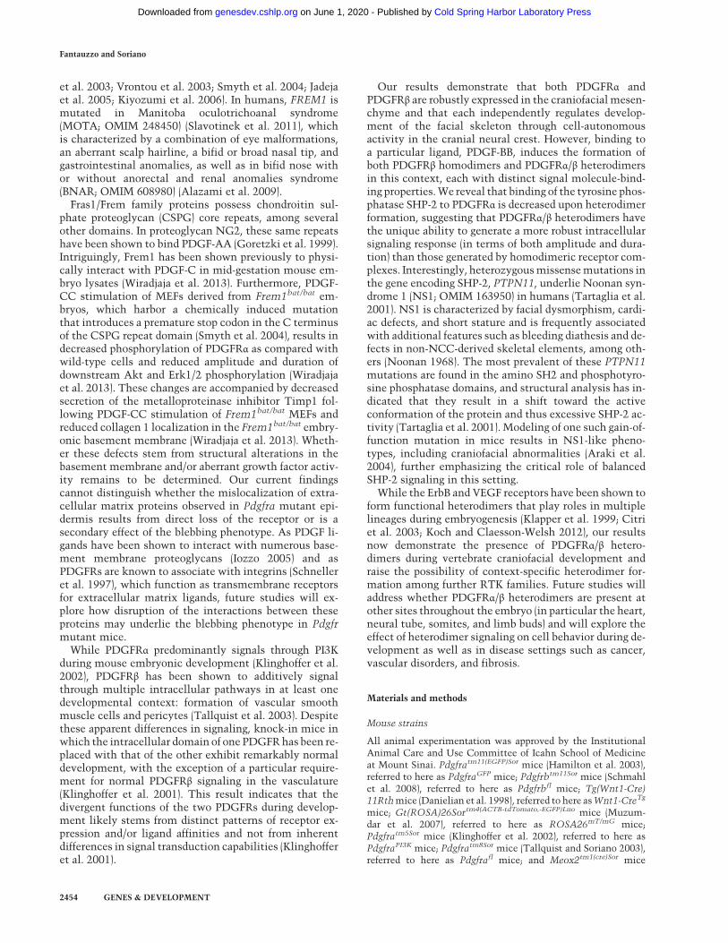
Consistent with its critical role in craniofacial develop-ment, Pdgfra is expressed in themesenchyme of the facialprocesses of embryonic day 10.5 (E10.5) heterozygous re-porter embryos in which an H2B-GFP construct hadbeen knocked into the Pdgfra locus (Fig. 1A,A′; Hamiltonet al. 2003; He and Soriano 2013). To determine the ex-pression of PDGFRβ in these same structures, we per-formed whole-mount immunohistochemistry on wild-type E10.5 embryos using an anti-PDGFRβ antibody, re-vealing expression in the medial and lateral nasal process-es, in the maxillary processes from which the palatal
shelves will extend, and in the mandibular processes(Fig. 1B,B′). Outside of the craniofacial region, expressionof both receptors was additionally detected in the heart,neural tube, somites, and limb buds at E10.5 (Fig. 1A–B′).Primary mouse embryonic palatal mesenchyme cells
(MEPMs) and primary mouse embryonic fibroblasts(MEFs) have been shown previously to independently ex-press both PDGFRα and PDGFRβ (Klinghoffer et al.2001, 2002; He and Soriano 2013; Vasudevan et al.2015), with the former cell type demonstrating respon-siveness to stimulation with PDGF-AA (He and Soriano2013; Fantauzzo and Soriano 2014; Vasudevan and Sor-iano 2014; Vasudevan et al. 2015), and the latter demon-strating responsiveness to stimulation with PDGF-AAand PDGF-BB (Klinghoffer et al. 2001, 2002). Quantitative
Figure 1. The PDGFRs are coexpressed in the craniofacial mesenchyme. (A,A′) Expression of Pdgfra as assessed by GFP fluorescence inE10.5 Pdgfra+/GFP embryos viewed laterally (A) and frontally (A′). (B,B′) Expression of PDGFRβ as assessed by whole-mount immunohis-tochemistry with an anti-PDGFRβ antibody in E10.5 wild-type embryos viewed laterally (B) and frontally (B′). No signal was detected in asecondary antibody control embryo at E10.5 (data not shown). Expression of both receptors was detected in the facial process mesen-chyme, heart, neural tube, somites, and limb bud mesenchyme. (MxP) Maxillary process; (MdP) mandibular process; (LNP) lateral nasalprocess; (MNP) medial nasal process; (Hrt) heart; (FL) forelimb; (NT) neural tube; (Som) somite. (C ) Bar graph depicting quantitative RT–PCR (qRT–PCR) values revealing reduced expression of Pdgfra inmouse embryonic palatalmesenchyme cells (MEPMs) as comparedwithmouse embryonic fibroblasts (MEFs) and similar expression of Pdgfrb across the two cell types. Data are presented asmean ± SEM. (D–G′ ′ ′)Expression of PDGFRα (red; D′ ′,F′ ′) and PDGFRβ (red; E′ ′,G′ ′) as assessed by immunofluorescence analyses in E13.5 Pdgfra+/GFP-derivedMEPMs (D–E′ ′ ′) and MEFs (F–G′ ′ ′). Expression of both receptors was detected in virtually every cell. (D′,E′,F′,G′) Pdgfra-expressing cellsare additionally indicated by GFP fluorescence (green). (D,E,F,G) Nuclei were stained with 4′,6-diamidino-2-phenylindole (DAPI; blue).Bars, 100 µm.
PDGFRs form functional heterodimers
GENES & DEVELOPMENT 2445
Cold Spring Harbor Laboratory Press on June 1, 2020 - Published by genesdev.cshlp.orgDownloaded from
RT–PCR (qRT–PCR) analyses comparing expression ofthe transcripts encoding the two receptors in primaryMEPMs and primary MEFs revealed greater expressionof Pdgfra in MEFs (2.087%± 0.3069% and 12.00%±0.7276% of mB2m expression, respectively) and similarexpression of Pdgfrb across the two cell types (18.22%±2.623% and 18.25% ± 1.078% of mB2m expression, re-spectively) (Fig. 1C). To assess coexpression of the two re-ceptors in these cell types, low-passage primary MEPMsand primary MEFs were derived from E13.5 Pdgfra+/GFP
embryos and subjected to immunofluorescence analyseswith both anti-PDGFRα and anti-PDGFRβ antibodies.Our analyses revealed expression of both receptors in vir-tually every cell (Fig. 1D–G′′ ′).
Craniofacial skeletal defects in a subset of embryoslacking Pdgfrb in the neural crest lineage
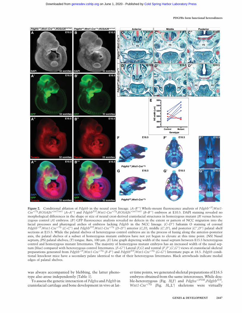
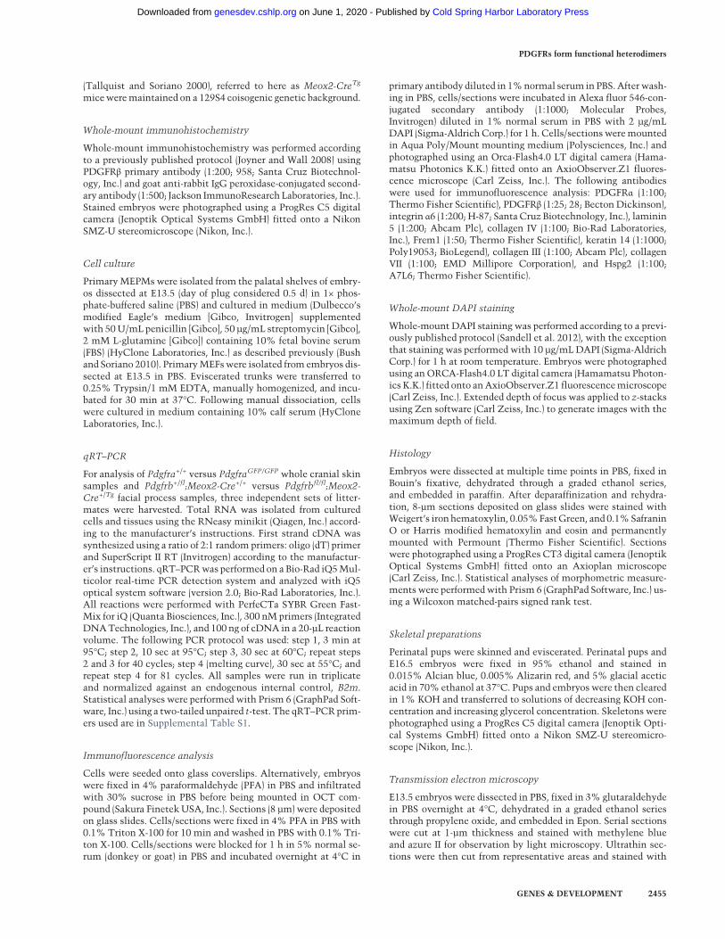
As PDGFRβ does not have an established role in murinecraniofacial development beyond a genetic interactionwith Pdgfra, we performed a series of experiments exam-ining the effect of conditionally ablating Pdgfrb in theNCC lineage using the Wnt1-Cre driver. These analysesand all further experiments in this study were performedusing mice maintained on a 129S4 coisogenic geneticbackground to prevent the effects of potential secondsite modifiers. We began by assessing the timing and ex-tent of NCC migration in this context using theROSA26mT/mG allele (Muzumdar et al. 2007), which ex-presses a double-fluorescent Cre reporter throughout theembryo, resulting in the expression ofmembrane-targetedtdTomato before Cre excision and membrane-targetedEGFP after Cre excision. This analysis revealed no obvi-ous defects in the extent or pattern of NCC migrationinto the facial processes and pharyngeal arches of embryoslacking Pdgfrb in the NCC lineage at E8.5 (n = 3) (data notshown) or E10.5 (n = 4) (Fig. 2A–B′′′). Moreover, whole-mount 4′,6-diamidino-2-phenylindole (DAPI) staining ofthese same embryos revealed no morphological differenc-es in the shape or size of neural crest-derived craniofacialstructures in experimental versus control embryos atE10.5 (Fig. 2A,B).
We next explored the effect of ablating Pdgfrb in theNCC lineage on development of the craniofacial skeleton.The majority of E15.5 homozygous mutant embryos hasan increased width of the nasal septum compared withheterozygous control littermates (521.8 µm ± 9.818 µmvs. 497.7 µm ± 4.582 µm; P = 0.0781) (Fig. 2C–E). Examina-tion of perinatal mice confirmed that this is a transientphenotype, as the nasal septum and associated structuresof Pdgfrb conditional knockout mice (Fig. 2G–G′′) are in-distinguishable from those of heterozygous littermatesat E18.5 (Fig. 2F–F′′). Furthermore, while E15.5 heterozy-gous control embryos have palatal shelves that are in theprocess of fusing along the anterior–posterior axis (Fig.2C–C′′), the palatal shelves of a subset (10%, n = 10) of ho-mozygous mutant embryos had not yet begun to elevateat this time point (Fig. 2D–D′′). This likely reflects a delayin Pdgfrb conditional knockout palatal shelf develop-ment, as these mice are ultimately born at Mendelian ra-
tios (44 pups vs. 49.5 expected pups out of 198 total; χ2
P = 0.3667) and are viable. Skeletal preparations of perina-tal mice confirmed that palatal shelf development may beonly temporarily delayed in Pdgfrbfl/fl;Wnt1-Cre+/Tg em-bryos, as all Pdgfrb conditional knockout mice examinedto date (n = 17) (Fig. 2G–G′′) have a secondary palate iden-tical to that of their heterozygous littermates (Fig. 2F–F′′).
Pdgfra and Pdgfrb genetically interact during craniofacialdevelopment
A previous skeletal analysis in which both Pdgfra andPdgfrb were conditionally ablated in the NCC lineagedid not detect additional frontonasal midline defects indouble conditional mutant embryos beyond those ob-served in Pdgfrafl/fl;Wnt1-Cre+/Tg embryos (McCarthyet al. 2016). As PdgfraPI3K/PI3K embryos have a cleft palatephenotype (Klinghoffer et al. 2002; Fantauzzo and Soriano2014) less severe than the complete facial clefting pheno-type observed in Pdgfrafl/fl;Wnt1-Cre+/Tg embryos (Tall-quist and Soriano 2003; He and Soriano 2013), we choseto use the PdgfraPI3K allele in our in vivo studies as asensitized background in the hopes of uncovering a genet-ic interaction between the two receptors in frontonasalmidline development. We thus used the constitutivePdgfraPI3K allele together with the conditional Pdgfrbfl
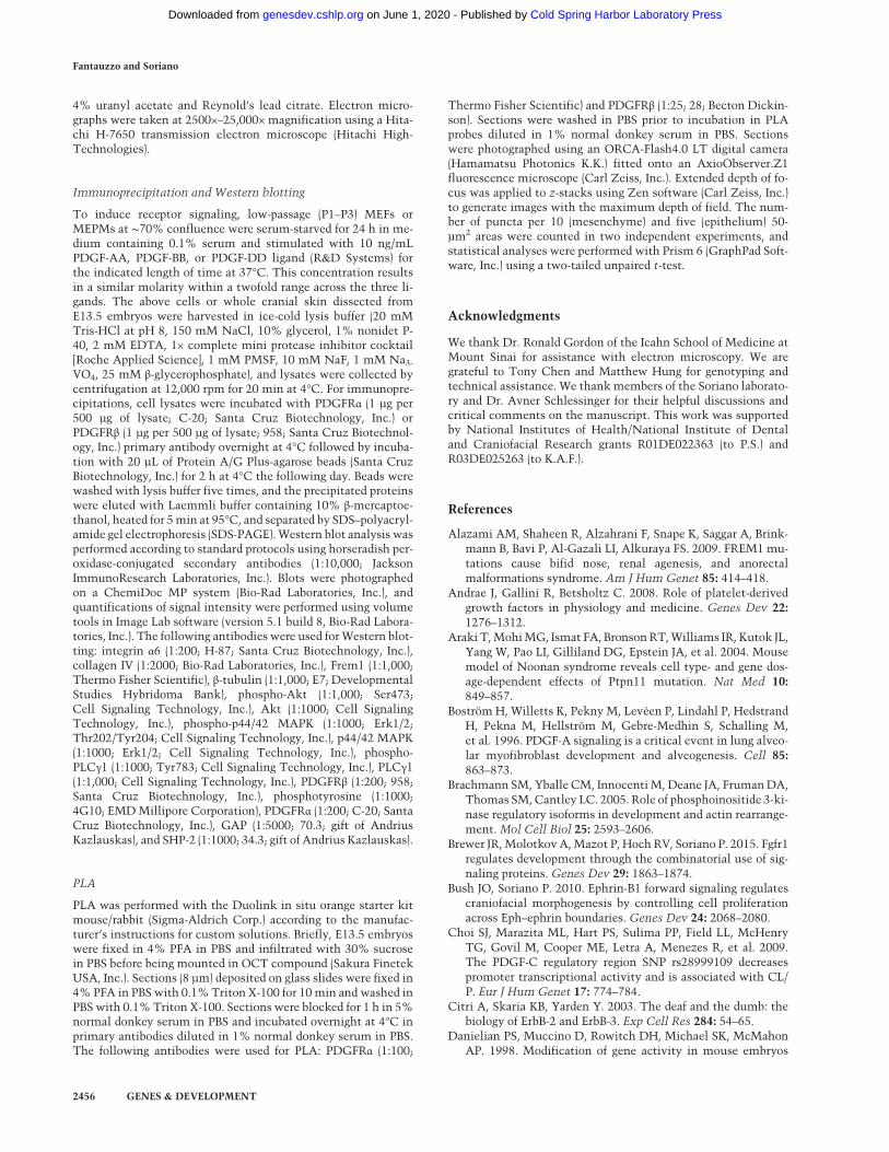
allele and the Wnt1-Cre driver to ablate various allelecombinations in the NCC lineage. We intercrossedPdgfra+/PI3K;Pdgfrbfl/fl mice with Pdgfra+/PI3K;Pdgfrb+/fl;Wnt1-Cre+/Tg mice and harvested the resulting progenyat E13.5 for gross morphological examination (Fig. 3A–
G′′). As observed previously (Fantauzzo and Soriano2014), all embryos homozygous for the PdgfraPI3K allelewere smaller than their littermates (Fig. 3B,B′,D,D′,G,G′). Double-homozygousmutant embryos were recoveredat Mendelian frequencies at E13.5 (four embryos vs. sixexpected embryos out of 97 total; χ2 P = 0.3870) and exhib-ited an overt facial clefting phenotype (100%; n = 4) (Fig.3G–G′′; Table 1) not observed among embryos possessingany of the other 11 allele combinations from the inter-crosses (n = 93) (Table 1), indicating that the two receptorsgenetically interact in the NCC lineage.
Moreover, blebbing of the surface ectoderm in the facialregion was detected in approximately half of the embryosheterozygous for the Pdgfra+/PI3K allele (53%; n = 36) (Fig.3C,C′; Table 1) and was fully penetrant among embryoshomozygous for the PdgfraPI3K allele (100%; n = 24) (Fig.3B,B′,D,D′,G,G′; Table 1) and among Pdgfra+/PI3K;Pdgfrbfl/fl;Wnt1-Cre+/Tg embryos (100%; n = 15) (Fig. 3F,F′; Table 1). Interestingly, this phenotype was alsoobserved among a subset of embryos heterozygous (14%;n = 7) or homozygous (20%; n = 5) for the Pdgfrbfl allelein combination with the Wnt1-Cre transgene in theabsence of Pdgfra mutation (Fig. 3E,E′; Table 1). Finally,facial hemorrhaging was frequently detected in embryoshomozygous for the PdgfraPI3K allele (58%; n = 24)(Fig. 3B–B′′,D–D′′,G–G′′; Table 1) and occasionally inPdgfra+/PI3K;Pdgfrbfl/fl;Wnt1-Cre+/Tg embryos (13%; n = 15)(Fig. 3F–F′′; Table 1), revealing that while hemorrhaging
Fantauzzo and Soriano
2446 GENES & DEVELOPMENT
Cold Spring Harbor Laboratory Press on June 1, 2020 - Published by genesdev.cshlp.orgDownloaded from
was always accompanied by blebbing, the latter pheno-type also arose independently (Table 1).To assess the genetic interaction of Pdgfra and Pdgfrb in
craniofacial cartilage and bone development in vivo at lat-
er time points, we generated skeletal preparations of E16.5embryos obtained from the same intercrosses. While dou-ble-heterozygous (Fig. 3J,J′) and Pdgfra+/PI3K;Pdgfrbfl/fl;Wnt1-Cre+/Tg (Fig. 3L,L′) skeletons were virtually
Figure 2. Conditional ablation of Pdgfrb in the neural crest lineage. (A–B′ ′ ′) Whole-mount fluorescence analysis of Pdgfrb+/fl;Wnt1-Cre+/Tg;ROSA26+/mT/mG (A–A′ ′ ′) and Pdgfrbfl/fl;Wnt1-Cre+/Tg;ROSA26+/mT/mG (B–B′ ′ ′) embryos at E10.5. DAPI staining revealed nomorphological differences in the shape or size of neural crest-derived craniofacial structures in homozygous mutant (B) versus hetero-zygous control (A) embryos. (B′) GFP fluorescence analysis revealed no defects in the extent or pattern of NCC migration into thefacial processes and pharyngeal arches of embryos lacking Pdgfrb in the NCC lineage. (C–D′ ′) Safranin O staining of coronalPdgfrb+/fl;Wnt1-Cre+/Tg (C–C′ ′) and Pdgfrbfl/fl;Wnt1-Cre+/Tg (D–D′ ′) anterior (C,D), middle (C′,D′), and posterior (C′ ′,D′ ′) palatal shelfsections at E15.5. While the palatal shelves of heterozygous control embryos are in the process of fusing along the anterior–posterioraxis, the palatal shelves of a subset of homozygous mutant embryos have not yet begun to elevate at this time point. (NS) Nasalseptum; (PS) palatal shelves; (T) tongue. Bars, 100 µm. (E) Line graph depicting width of the nasal septum between E15.5 heterozygouscontrol and homozygous mutant littermates. The majority of homozygous mutant embryos has an increased width of the nasal sep-tum (blue) compared with heterozygous control littermates. (F–G′ ′) Lateral (F,G) and ventral (F′,F′ ′,G′,G′ ′) views of craniofacial skeletalpreparations generated from Pdgfrb+/fl;Wnt1-Cre+/Tg (F–F′ ′) and Pdgfrbfl/fl;Wnt1-Cre+/Tg (G–G′ ′) littermate pups at 18.5. Pdgfrb condi-tional knockout mice have a secondary palate identical to that of their heterozygous littermates. Black arrowheads indicate medialedges of palatal shelves.
PDGFRs form functional heterodimers
GENES & DEVELOPMENT 2447
Cold Spring Harbor Laboratory Press on June 1, 2020 - Published by genesdev.cshlp.orgDownloaded from
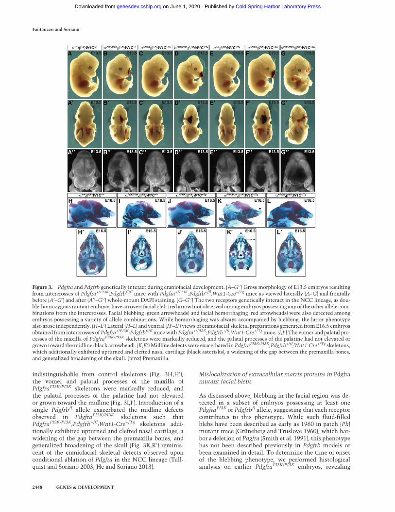
indistinguishable from control skeletons (Fig. 3H,H′),the vomer and palatal processes of the maxilla ofPdgfraPI3K/PI3K skeletons were markedly reduced, andthe palatal processes of the palatine had not elevatedor grown toward the midline (Fig. 3I,I′). Introduction of asingle Pdgfrbfl allele exacerbated the midline defectsobserved in PdgfraPI3K/PI3K skeletons such thatPdgfraPI3K/PI3K;Pdgfrb+/fl;Wnt1-Cre+/Tg skeletons addi-tionally exhibited upturned and clefted nasal cartilage, awidening of the gap between the premaxilla bones, andgeneralized broadening of the skull (Fig. 3K,K′) reminis-cent of the craniofacial skeletal defects observed uponconditional ablation of Pdgfra in the NCC lineage (Tall-quist and Soriano 2003; He and Soriano 2013).
Mislocalization of extracellular matrix proteins in Pdgframutant facial blebs
As discussed above, blebbing in the facial region was de-tected in a subset of embryos possessing at least onePdgfraPI3K or Pdgfrbfl allele, suggesting that each receptorcontributes to this phenotype. While such fluid-filledblebs have been described as early as 1960 in patch (Ph)mutant mice (Grüneberg and Truslove 1960), which har-bor a deletion of Pdgfra (Smith et al. 1991), this phenotypehas not been described previously in Pdgfrb models orbeen examined in detail. To determine the time of onsetof the blebbing phenotype, we performed histologicalanalysis on earlier PdgfraPI3K/PI3K embryos, revealing
Figure 3. Pdgfra and Pdgfrb genetically interact during craniofacial development. (A–G′ ′) Gross morphology of E13.5 embryos resultingfrom intercrosses of Pdgfra+/PI3K;Pdgfrbfl/fl mice with Pdgfra+/PI3K;Pdgfrb+/fl;Wnt1-Cre+/Tg mice as viewed laterally (A–G) and frontallybefore (A′–G′) and after (A′ ′–G′ ′) whole-mount DAPI staining. (G–G′ ′) The two receptors genetically interact in the NCC lineage, as dou-ble-homozygousmutant embryos have an overt facial cleft (red arrow) not observed among embryos possessing anyof the other allele com-binations from the intercrosses. Facial blebbing (green arrowheads) and facial hemorrhaging (red arrowheads) were also detected amongembryos possessing a variety of allele combinations. While hemorrhaging was always accompanied by blebbing, the latter phenotypealso arose independently. (H–L′) Lateral (H–L) and ventral (H′–L′) views of craniofacial skeletal preparations generated fromE16.5 embryosobtained from intercrosses of Pdgfra+/PI3K;Pdgfrbfl/flmicewith Pdgfra+/PI3K;Pdgfrb+/fl;Wnt1-Cre+/Tgmice. (I,I′) The vomer and palatal pro-cesses of the maxilla of PdgfraPI3K/PI3K skeletons were markedly reduced, and the palatal processes of the palatine had not elevated orgrown toward themidline (black arrowhead). (K,K′) Midline defectswere exacerbated in PdgfraPI3K/PI3K;Pdgfrb+/fl;Wnt1-Cre+/Tg skeletons,which additionally exhibited upturned and clefted nasal cartilage (black asterisks), a widening of the gap between the premaxilla bones,and generalized broadening of the skull. (pmx) Premaxilla.
Fantauzzo and Soriano
2448 GENES & DEVELOPMENT
Cold Spring Harbor Laboratory Press on June 1, 2020 - Published by genesdev.cshlp.orgDownloaded from
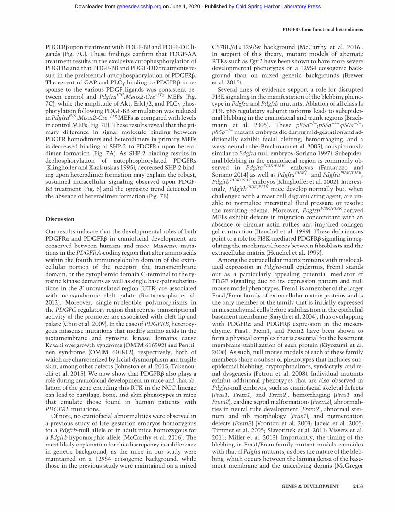
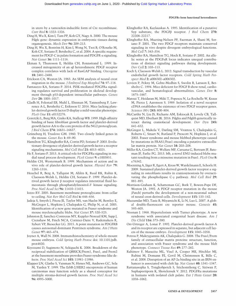
blebs as early as E11.5 (Fig. 4A,B). We next used transmis-sion electron microscopy to analyze this phenotype inmore detail in E13.5 PdgfraPI3K/PI3K embryos. Outside ofthe bleb, a two-layered epitheliumwas observed overlyingdermal fibroblasts (Fig. 4C). Moving toward the bleb, theepidermis was completely detached from the underlyingmesenchyme (Fig. 4C′). Higher-magnification images re-vealed an intact basement membrane and what appeared
to be loose collagen fibrils at the bleb interface (Fig. 4C′′,C′′′), consistent with previous findings in Pdgfc−/− (Dinget al. 2004) and Ph/Ph mutant (Erickson and Weston1983) embryos, respectively. Immunofluorescence analy-ses of E13.5 Pdgfra-null craniofacial surface tissue usingantibodies specific to the basal epidermis (keratin 14)(Supplemental Fig. S1A–B′) and the basement membrane(integrin α6) (Fig. 4D–E′) verified that these cell layers
Table 1. Pdgfra and Pdgfrb genetically interact during craniofacial development
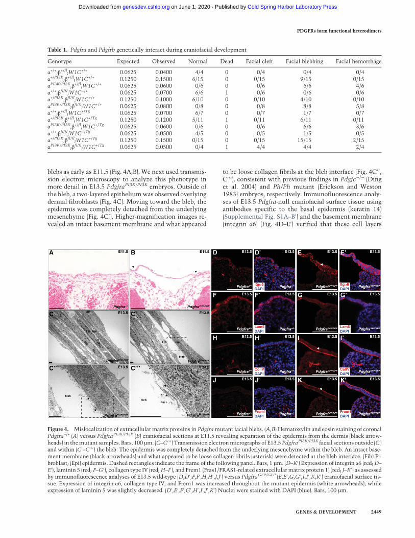
Genotype Expected Observed Normal Dead Facial cleft Facial blebbing Facial hemorrhage
α+/+;β+/fl;W1C+/+ 0.0625 0.0400 4/4 0 0/4 0/4 0/4α+/PI3K;β+/fl;W1C+/+ 0.1250 0.1500 6/15 0 0/15 9/15 0/15αPI3K/PI3K;β+/fl;W1C+/+ 0.0625 0.0600 0/6 0 0/6 6/6 4/6α+/+;βfl/fl;W1C+/+ 0.0625 0.0700 6/6 1 0/6 0/6 0/6α+/PI3K;βfl/fl;W1C+/+ 0.1250 0.1000 6/10 0 0/10 4/10 0/10αPI3K/PI3K;βfl/fl;W1C+/+ 0.0625 0.0800 0/8 0 0/8 8/8 5/8α+/+;β+/fl;W1C+/Tg 0.0625 0.0700 6/7 0 0/7 1/7 0/7α+/PI3K;β+/fl;W1C+/Tg 0.1250 0.1200 5/11 1 0/11 6/11 0/11αPI3K/PI3K;β+/fl;W1C+/Tg 0.0625 0.0600 0/6 0 0/6 6/6 3/6α+/+;βfl/fl;W1C+/Tg 0.0625 0.0500 4/5 0 0/5 1/5 0/5α+/PI3K;βfl/fl;W1C+/Tg 0.1250 0.1500 0/15 0 0/15 15/15 2/15αPI3K/PI3K;βfl/fl;W1C+/Tg 0.0625 0.0500 0/4 1 4/4 4/4 2/4
Figure 4. Mislocalization of extracellular matrix proteins in Pdgframutant facial blebs. (A,B) Hematoxylin and eosin staining of coronalPdgfra+/+ (A) versus PdgfraPI3K/PI3K (B) craniofacial sections at E11.5 revealing separation of the epidermis from the dermis (black arrow-heads) in themutant samples. Bars, 100 µm. (C–C′ ′ ′) Transmission electronmicrographs of E13.5PdgfraPI3K/PI3K facial sections outside (C )and within (C′–C′ ′ ′) the bleb. The epidermis was completely detached from the underlying mesenchyme within the bleb. An intact base-ment membrane (black arrowheads) and what appeared to be loose collagen fibrils (asterisk) were detected at the bleb interface. (Fib) Fi-broblast; (Epi) epidermis. Dashed rectangles indicate the frame of the following panel. Bars, 1 µm. (D–K′) Expression of integrin α6 (red;D–
E′), laminin 5 (red; F–G′), collagen type IV (red;H–I′), and Frem1 (Fras1/FRAS1-related extracellular matrix protein 1) (red; J–K′) as assessedby immunofluorescence analyses of E13.5 wild-type (D,D′,F,F′,H,H′,J,J′) versus PdgfraGFP/GFP (E,E′,G,G′,I,I′,K,K′) craniofacial surface tis-sue. Expression of integrin α6, collagen type IV, and Frem1 was increased throughout the mutant epidermis (white arrowheads), whileexpression of laminin 5 was slightly decreased. (D′,E′,F′,G′,H′,I′,J′,K′) Nuclei were stained with DAPI (blue). Bars, 100 µm.
PDGFRs form functional heterodimers
GENES & DEVELOPMENT 2449
Cold Spring Harbor Laboratory Press on June 1, 2020 - Published by genesdev.cshlp.orgDownloaded from

were completely detached from the underlying mesen-chyme within the mutant blebs.
Interestingly, we observed that integrin α6 expressionwas increased throughout the mutant epidermis (Fig. 4E,E′) as compared with that of wild-type littermates (Fig.4D,D′). Similar immunofluorescence analyses revealedthat expression of the extracellular matrix proteins colla-gen type IV (Fig. 4H–I′) and Frem1 (Fras1/FRAS1-relatedextracellular matrix protein 1) (Fig. 4J–K′) was also in-creased throughout the Pdgfra-null epidermis, suggestingthat these proteins may not be properly localizing to thebasementmembrane in this setting. Expression of laminin5 was slightly decreased throughout the Pdgfra-nullepidermis (Fig. 4F–G′), while localization of collagentype III (Supplemental Fig. S1C–D′), collagen type VII(Supplemental Fig. S1E–F′), and Hspg2 (also known as per-lecan) (Supplemental Fig. S1G–H′) was unchanged be-tween wild-type and mutant tissues. qRT–PCR analysescomparing expression of the transcripts encoding these ex-tracellular matrix proteins in RNA derived from E13.5wild-type versus PdgfraGFP/GFP whole cranial skin re-vealed no significant changes in transcript expression inthe mutant samples, with the exception of a modestdecrease inLamb3 expression (22.10%± 4.211%decrease;P = 0.0344) (Supplemental Fig. S2A). Moreover, Westernblotting demonstrated similar levels of integrin α6(Supplemental Fig. S2B), collagen type IV (SupplementalFig. S2C), and Frem1 (Supplemental Fig. S2D) protein inE13.5 wild-type and PdgfraGFP/GFP whole cranial skinlysates, further suggesting that the blebbing defect is asso-ciated primarily with mislocalization, and not misexpres-sion, of these extracellular matrix proteins.
PDGFRα and PDGFRβ form functional heterodimerswith properties distinct from those of homodimericreceptor complexes
Having characterized a genetic interaction betweenPdgfra and Pdgfrb during craniofacial development, we
next explored whether PDGFRα and PDGFRβ heterodi-merize in the craniofacial mesenchyme. We initiallyused the proximity ligation assay (PLA) (Söderberg et al.2006) to examine the interaction of the proteins at sin-gle-molecule resolution. We chose antibodies for each re-ceptor that generatedminimal background staining in thecraniofacial mesenchyme of embryos in which the recep-tor had been conditionally ablated in the NCC lineage(Supplemental Fig. S3B,B′,D,D′) as compared with controlembryos (Supplemental Fig. S3A,A′,C,C′). Applying thePLA technique to E13.5 craniofacial tissue, PDGFRα/β in-teractions were detected in the craniofacial mesenchyme(Fig. 5A,A′). Quantification of these puncta revealedthat these interactions were significantly increased inthe mesenchyme (8.450 ± 1.027 per 50-µm2 area) as com-pared with the epithelia (1.200 ± 0.2494 per 50-µm2 area;P < 0.0001), consistent with the expression patterns ofthe PDGFRs, as well as compared with a technical nega-tive control without the addition of primary antibodies(0.0 ± 0.0 per 50-µm2 area; P < 0.0001) (Fig. 5B,B′) and con-ditional knockout tissues in which Pdgfra (0.8000 ±0.2575 per 50-µm2 area; P < 0.0001) (Fig. 5C,C′) or Pdgfrb(2.800 ± 0.5361 per 50-µm2 area; P < 0.0001) (Fig. 5D,D′)had been ablated in the NCC lineage (Fig. 5E).
We next performed a series of in vitro biochemical ex-periments to assesswhether PDGFRα and PDGFRβ are ca-pable of forming functional heterodimers with thecapacity to activate downstream signaling pathways. Wechose to perform these experiments in primary MEFs, asthe levels of Pdgfra and Pdgfrb expression are more simi-lar in this cell type than in primary MEPMs (Fig. 1C). Inview of the demonstrated roles of Akt, Erk1/2, and PLCγsignaling downstream from PDGFR activation during cra-niofacial development (Klinghoffer et al. 2002; Moenninget al. 2009; Fantauzzo and Soriano 2014; Vasudevan et al.2015), we first assayed the ability of PDGF-AA, PDGF-BB,and PDGF-DD ligands to stimulate phosphorylation ofthese signaling proteins. To induce receptor signaling, pri-mary MEFs were starved in medium containing 0.1% calf
Figure 5. PDGFRα and PDGFRβ physically interact in the craniofacial mesenchyme. (A–D′) Interaction of PDGFRα and PDGFRβ inE13.5 craniofacial tissue as assessed by fluorescence analysis following application of the PLA. PDGFRα/β interactions were significantlyincreased in the mesenchyme (A,A′) as compared with the epithelia of experimental sections as well as compared with a technical neg-ative control without the addition of primary antibodies (B,B′) and conditional knockout tissues in which Pdgfra (C,C′) or Pdgfrb (D,D′)had been ablated in theNCC lineage. Puncta are indicated in inverse grayscale (A,B,C,D) and byCy3 fluorescence (red;A′,B′,C′,D′). Nucleiwere stained with DAPI (blue; A′,B′,C′,D′). Bars, 100 µm. (E) Scatter dot plot depicting the number of puncta per 50-µm2 area detected inthe mesenchyme (red) or epithelia (blue) for each condition. Data are presented as mean ± SEM. (∗∗∗) P = 0.001; (∗∗∗∗) P < 0.0001.
Fantauzzo and Soriano
2450 GENES & DEVELOPMENT
Cold Spring Harbor Laboratory Press on June 1, 2020 - Published by genesdev.cshlp.orgDownloaded from

serum for 24 h followed by stimulation with 10 ng/mLPDGF-AA, PDGF-BB, or PDGF-DD ligand for up to 4h. Western blot analysis of whole-cell lysates revealedthat, while PDGF-AA treatment induced a modest in-crease in phospho-Akt levels by 5 min (1.521-fold ±0.760-fold over baseline), PDGF-BB and PDGF-DD treat-ments generated sustained phospho-Akt responses thatpeaked at comparable levels by 30 min (4.451-fold ±1.691-fold and 4.302-fold ± 0.513-fold over baseline, re-spectively) (Fig. 6A). A similar examination of the timecourse of Erk1/2 phosphorylation revealed peaks at 15minutes for all three ligands, with PDGF-BB treatmentgenerating a more robust response (4.568-fold ± 0.564-fold over baseline) than PDGF-AA (2.894-fold ± 0.964-fold over baseline) and PDGF-DD (2.571-fold ± 0.174-foldover baseline) (Fig. 6B). Finally, PDGF-AA treatment re-sulted in negligible increases in phospho-PLCγ levels,while PDGF-BB and PDGF-DD treatments induced peaksof PLCγ phosphorylation at 15 min, with PDGF-BB againgenerating a stronger response (2.805-fold ± 0.246-foldover baseline) than PDGF-DD (2.159-fold ± 0.078-foldover baseline) (Fig. 6C).Having determined a general peak of intracellular sig-
naling 15 min after PDGF ligand treatment, we used thistime point to assay the ability of each PDGFR to bindthe other as well as interact with known signalingmolecules upon stimulation of primary MEFs withPDGF-AA, PDGF-BB, or PDGF-DD ligand. PDGFRα andPDGFRβ heterodimers were detected following treatmentwith PDGF-BB, a ligand that has been shown previously tobind both receptors in vitro (Andrae et al. 2008), and, to alesser extent, upon stimulation with PDGF-DD (Fig. 7A).PDGFRα/β heterodimers were similarly observed in pri-
mary MEPMs predominantly in response to PDGF-BBtreatment (data not shown). As expected, PDGF-AA stim-ulation of primary MEFs resulted in autophosphorylationof PDGFRα exclusively, while PDGF-BB and PDGF-DDtreatments induced autophosphorylation of both recep-tors (Fig. 7A), consistent with the formation of functionalheterodimers. GAP binding to PDGFRβwas noticeably in-creased upon PDGF-BB treatment and minimally in-creased upon PDGF-DD stimulation (Fig. 7A). PLCγbinding to PDGFRα, although increased over baseline lev-els, was unchanged between PDGF-AA and PDGF-BBtreatments, while binding of this signaling molecule toPDGFRβ was increased upon PDGF-BB treatment overPDGF-DD treatment (Fig. 7A). Finally, stimulation withPDGF-BB led to decreased binding of SHP-2 to PDGFRαas compared with treatment with PDGF-AA (Fig. 7A).We were never able to detect SHP-2 binding to PDGFRβunder any conditions within our system (data not shown).We next derived primary MEFs from embryos in which
Pdgfrb had been ablated throughout the embryo using theMeox2-Cre driver (Tallquist and Soriano 2000) and thuscould not engage in heterodimer formation.We confirmednear-complete knockdown of PDGFRβ in these cells (Fig.7B) and further revealed that the extent of PDGFRα auto-phosphorylation upon PDGF-BB and PDGF-DD treat-ments was reduced in this setting as compared withlevels in control cells (Fig. 7B). Consistent with the West-ern blotting results (Fig. 7B), qRT–PCR analysis revealed amodest reduction in Pdgfra transcript levels in the facialprocesses of E11.5 Pdgfrbfl/fl;Meox2-Cre+/Tg embryoscompared with control littermates (27.26%± 7.652%decrease; P = 0.0705) (Fig. 7D). To distinguish the bindingproperties of PDGFRα/β heterodimers from PDGFRβ
Figure 6. PDGF-BB treatment induces robust phosphorylation of Akt, Erk1/2, and PLCγ. (A–C) Phosphorylation time courses of Akt (A),Erk1/2 (B), and PLCγ (C ) in E13.5 primaryMEFs following treatmentwith PDGF-AA (green), PDGF-BB (red), or PDGF-DD (blue) for up to 4h. (A) Western blot analysis of whole-cell lysates revealed that, while PDGF-AA treatment induced amodest increase in phospho-Akt lev-els by 5min, PDGF-BB and PDGF-DD treatments generated sustained phospho-Akt responses that peaked at comparable levels by 30min.(B) An examination of the time course of Erk1/2 phosphorylation revealed peaks at 15 min for all three ligands, with PDGF-BB treatmentgenerating a more robust response than PDGF-AA and PDGF-DD. (C ) PDGF-AA treatment resulted in negligible increases in phospho-PLCγ levels, while PDGF-BB and PDGF-DD treatments induced peaks of PLCγ phosphorylation at 15 min, with PDGF-BB again gener-ating a stronger response than PDGF-DD. (WCL) Whole-cell lysate; (WB) Western blot. Graph data are presented as mean ± SEM.
PDGFRs form functional heterodimers
GENES & DEVELOPMENT 2451
Cold Spring Harbor Laboratory Press on June 1, 2020 - Published by genesdev.cshlp.orgDownloaded from

homodimers upon PDGF-BB stimulation, we repeated theabove experiments in primary MEFs derived from embry-os in which Pdgfra had been ablated throughout theembryo. We confirmed knockdown of PDGFRα protein
expression in these cells (Fig. 7C) and an inability ofPDGFRβ to coimmunoprecipitate with PDGFRα (datanot shown). Importantly, ablation of Pdgfra had no effecton PDGFRβ protein levels or the autophosphorylation of
Figure 7. PDGFRα/β heterodimers have properties distinct from those of homodimeric receptor complexes. (A) Biochemical analysis ofsignal molecule binding to PDGFRα and PDGFRβ in wild-type E13.5 primary MEFs following treatment with PDGF-AA, PDGF-BB, orPDGF-DD for 15 min. PDGFRα/β heterodimers were detected following treatment with PDGF-BB and, to a lesser extent, upon stimula-tion with PDGF-DD. PDGF-AA stimulation resulted in autophosphorylation of PDGFRα exclusively, while PDGF-BB and PDGF-DDtreatments induced autophosphorylation of both receptors. GAP binding to PDGFRβwas noticeably increased upon PDGF-BB treatmentandminimally increased upon PDGF-DD stimulation. PLCγ binding to PDGFRα, although increased over baseline levels, was unchangedbetween PDGF-AA and PDGF-BB treatments, while binding of this signaling molecule to PDGFRβ was increased upon PDGF-BB treat-ment over PDGF-DD treatment. Stimulation with PDGF-BB led to decreased binding of SHP-2 to PDGFRα as compared with treatmentwith PDGF-AA. (B) Biochemical analysis of control versus Pdgfrbfl/fl;Meox2-Cre+/Tg E13.5 primaryMEFs following treatmentwith PDGF-AA, PDGF-BB, or PDGF-DD for 15 min. The extent of PDGFRα autophosphorylation upon PDGF-BB and PDGF-DD treatments was re-duced in this setting as compared with levels in control cells. (C ) Biochemical analysis of signal molecule binding to PDGFRβ in controlversus Pdgfrafl/fl;Meox2-Cre+/Tg E13.5 primaryMEFs following treatment with PDGF-AA, PDGF-BB, or PDGF-DD for 15min. Ablation ofPDGFRα had no effect on PDGFRβ protein levels or the autophosphorylation of PDGFRβ upon treatment with PDGF-BB and PDGF-DDligands. The extent of GAP and PLCγ binding to PDGFRβ in response to the various PDGF ligands was consistent between control andPdgfrafl/fl;Meox2-Cre+/TgMEFs.Western blotting for a particular PDGFR served as a loading control for each immunoprecipitation of thatreceptor. (IP) Immunoprecipitation; (WB)Western blot; (WCL)whole-cell lysate. (D) Bar graph depicting qRT–PCR values revealing amod-est reduction in Pdgfra transcript levels in the facial processes of E11.5 Pdgfrbfl/fl;Meox2-Cre+/Tg embryos compared with control litter-mates. Data are presented as mean ± SEM. (E) The amplitude of Akt, Erk1/2, and PLCγ phosphorylation following PDGF-BB stimulationwas reduced in Pdgfrafl/fl;Meox2-Cre+/TgMEFs as comparedwith levels in controlMEFs.Data are presented asmean ± SEM, normalized tounstimulated samples for each genotype.
Fantauzzo and Soriano
2452 GENES & DEVELOPMENT
Cold Spring Harbor Laboratory Press on June 1, 2020 - Published by genesdev.cshlp.orgDownloaded from
PDGFRβupon treatmentwith PDGF-BB and PDGF-DD li-gands (Fig. 7C). These findings confirm that PDGF-AAtreatment results in the exclusive autophosphorylation ofPDGFRα and that PDGF-BB and PDGF-DD treatments re-sult in the preferential autophosphorylation of PDGFRβ.The extent of GAP and PLCγ binding to PDGFRβ in re-sponse to the various PDGF ligands was consistent be-tween control and Pdgfrafl/fl;Meox2-Cre+/Tg MEFs (Fig.7C), while the amplitude of Akt, Erk1/2, and PLCγ phos-phorylation following PDGF-BB stimulation was reducedin Pdgfrafl/fl;Meox2-Cre+/TgMEFs as comparedwith levelsin controlMEFs (Fig. 7E). These results reveal that the pri-mary difference in signal molecule binding betweenPDGFR homodimers and heterodimers in primary MEFsis decreased binding of SHP-2 to PDGFRα upon hetero-dimer formation (Fig. 7A). As SHP-2 binding results indephosphorylation of autophosphorylated PDGFRs(Klinghoffer and Kazlauskas 1995), decreased SHP-2 bind-ing upon heterodimer formation may explain the robust,sustained intracellular signaling observed upon PDGF-BB treatment (Fig. 6) and the opposite trend detected inthe absence of heterodimer formation (Fig. 7E).
Discussion
Our results indicate that the developmental roles of bothPDGFRα and PDGFRβ in craniofacial development areconserved between humans and mice. Missense muta-tions in the PDGFRA-coding region that alter amino acidswithin the fourth immunoglobulin domain of the extra-cellular portion of the receptor, the transmembranedomain, or the cytoplasmic domain C-terminal to the ty-rosine kinase domains as well as single base-pair substitu-tions in the 3′ untranslated region (UTR) are associatedwith nonsyndromic cleft palate (Rattanasopha et al.2012). Moreover, single-nucleotide polymorphisms inthe PDGFC regulatory region that repress transcriptionalactivity of the promoter are associated with cleft lip andpalate (Choi et al. 2009). In the case of PDGFRB, heterozy-gous missense mutations that modify amino acids in thejuxtamembrane and tyrosine kinase domains causeKosaki overgrowth syndrome (OMIM 616592) and Pentti-nen syndrome (OMIM 601812), respectively, both ofwhich are characterized by facial dysmorphism and fragileskin, among other defects (Johnston et al. 2015; Takenou-chi et al. 2015). We now show that PDGFRβ also plays arole during craniofacial development in mice and that ab-lation of the gene encoding this RTK in the NCC lineagecan lead to cartilage, bone, and skin phenotypes in micethat emulate those found in human patients withPDGFRB mutations.Of note, no craniofacial abnormalities were observed in
a previous study of late gestation embryos homozygousfor a Pdgfrb-null allele or in adult mice homozygous fora Pdgfrb hypomorphic allele (McCarthy et al. 2016). Themost likely explanation for this discrepancy is a differencein genetic background, as the mice in our study weremaintained on a 129S4 coisogenic background, whilethose in the previous study were maintained on a mixed
C57BL/6J × 129/Sv background (McCarthy et al. 2016).In support of this theory, mutant models of alternateRTKs such as Fgfr1 have been shown to have more severedevelopmental phenotypes on a 129S4 coisogenic back-ground than on mixed genetic backgrounds (Breweret al. 2015).Several lines of evidence support a role for disrupted
PI3K signaling in themanifestation of the blebbing pheno-type in Pdgfra and Pdgfrbmutants. Ablation of all class IaPI3K p85 regulatory subunit isoforms leads to subepider-mal blebbing in the craniofacial and trunk regions (Brach-mann et al. 2005). These p85a−/−;p55a−/−;p50a−/−;p85b−/−mutant embryos die duringmid-gestation and ad-ditionally exhibit facial clefting, hemorrhaging, and awavy neural tube (Brachmann et al. 2005), conspicuouslysimilar to Pdgfra-null embryos (Soriano 1997). Subepider-mal blebbing in the craniofacial region is commonly ob-served in PdgfraPI3K/PI3K embryos (Fantauzzo andSoriano 2014) as well as PdgfraPI3K/− and PdgfraPI3K/PI3K;PdgfrbPI3K/PI3K embryos (Klinghoffer et al. 2002). Interest-ingly, PdgfrbPI3K/PI3K mice develop normally but, whenchallenged with a mast cell degranulating agent, are un-able to normalize interstitial fluid pressure or resolvethe resulting edema. Moreover, PdgfrbPI3K/PI3K-derivedMEFs exhibit defects in migration concomitant with anabsence of circular actin ruffles and impaired collagengel contraction (Heuchel et al. 1999). These deficienciespoint to a role for PI3K-mediated PDGFRβ signaling in reg-ulating the mechanical forces between fibroblasts and theextracellular matrix (Heuchel et al. 1999).Among the extracellular matrix proteins withmislocal-
ized expression in Pdgfra-null epidermis, Frem1 standsout as a particularly appealing potential mediator ofPDGF signaling due to its expression pattern and nullmousemodel phenotypes. Frem1 is amember of the largerFras1/Frem family of extracellular matrix proteins and isthe only member of the family that is initially expressedinmesenchymal cells before stabilization in the epithelialbasementmembrane (Smyth et al. 2004), thus overlappingwith PDGFRα and PDGFRβ expression in the mesen-chyme. Fras1, Frem1, and Frem2 have been shown toform a physical complex that is essential for the basementmembrane stabilization of each protein (Kiyozumi et al.2006). As such, null mouse models of each of these familymembers share a subset of phenotypes that includes sub-epidermal blebbing, cryptophthalmos, syndactyly, and re-nal dysgenesis (Petrou et al. 2008). Individual mutantsexhibit additional phenotypes that are also observed inPdgfra-null embryos, such as craniofacial skeletal defects(Fras1, Frem1, and Frem2), hemorrhaging (Fras1 andFrem2), cardiac septal malformations (Frem2), abnormali-ties in neural tube development (Frem2), abnormal ster-num and rib morphology (Fras1), and pigmentationdefects (Frem2) (Vrontou et al. 2003; Jadeja et al. 2005;Timmer et al. 2005; Slavotinek et al. 2011; Vissers et al.2011; Miller et al. 2013). Importantly, the timing of theblebbing in Fras1/Frem family mutant models coincideswith that of Pdgframutants, as does the nature of the bleb-bing, which occurs between the lamina densa of the base-ment membrane and the underlying dermis (McGregor
PDGFRs form functional heterodimers
GENES & DEVELOPMENT 2453
Cold Spring Harbor Laboratory Press on June 1, 2020 - Published by genesdev.cshlp.orgDownloaded from
et al. 2003; Vrontou et al. 2003; Smyth et al. 2004; Jadejaet al. 2005; Kiyozumi et al. 2006). In humans, FREM1 ismutated in Manitoba oculotrichoanal syndrome(MOTA; OMIM 248450) (Slavotinek et al. 2011), whichis characterized by a combination of eye malformations,an aberrant scalp hairline, a bifid or broad nasal tip, andgastrointestinal anomalies, as well as in bifid nose withor without anorectal and renal anomalies syndrome(BNAR; OMIM 608980) (Alazami et al. 2009).
Fras1/Frem family proteins possess chondroitin sul-phate proteoglycan (CSPG) core repeats, among severalother domains. In proteoglycan NG2, these same repeatshave been shown to bind PDGF-AA (Goretzki et al. 1999).Intriguingly, Frem1 has been shown previously to physi-cally interact with PDGF-C in mid-gestation mouse em-bryo lysates (Wiradjaja et al. 2013). Furthermore, PDGF-CC stimulation of MEFs derived from Frem1bat/bat em-bryos, which harbor a chemically induced mutationthat introduces a premature stop codon in the C terminusof the CSPG repeat domain (Smyth et al. 2004), results indecreased phosphorylation of PDGFRα as compared withwild-type cells and reduced amplitude and duration ofdownstream Akt and Erk1/2 phosphorylation (Wiradjajaet al. 2013). These changes are accompanied by decreasedsecretion of the metalloproteinase inhibitor Timp1 fol-lowing PDGF-CC stimulation of Frem1bat/bat MEFs andreduced collagen 1 localization in the Frem1bat/bat embry-onic basement membrane (Wiradjaja et al. 2013). Wheth-er these defects stem from structural alterations in thebasement membrane and/or aberrant growth factor activ-ity remains to be determined. Our current findingscannot distinguish whether the mislocalization of extra-cellular matrix proteins observed in Pdgfra mutant epi-dermis results from direct loss of the receptor or is asecondary effect of the blebbing phenotype. As PDGF li-gands have been shown to interact with numerous base-ment membrane proteoglycans (Iozzo 2005) and asPDGFRs are known to associate with integrins (Schnelleret al. 1997), which function as transmembrane receptorsfor extracellular matrix ligands, future studies will ex-plore how disruption of the interactions between theseproteins may underlie the blebbing phenotype in Pdgfrmutant mice.
While PDGFRα predominantly signals through PI3Kduring mouse embryonic development (Klinghoffer et al.2002), PDGFRβ has been shown to additively signalthrough multiple intracellular pathways in at least onedevelopmental context: formation of vascular smoothmuscle cells and pericytes (Tallquist et al. 2003). Despitethese apparent differences in signaling, knock-in mice inwhich the intracellular domain of one PDGFRhas been re-placed with that of the other exhibit remarkably normaldevelopment, with the exception of a particular require-ment for normal PDGFRβ signaling in the vasculature(Klinghoffer et al. 2001). This result indicates that thedivergent functions of the two PDGFRs during develop-ment likely stems from distinct patterns of receptor ex-pression and/or ligand affinities and not from inherentdifferences in signal transduction capabilities (Klinghofferet al. 2001).
Our results demonstrate that both PDGFRα andPDGFRβ are robustly expressed in the craniofacial mesen-chyme and that each independently regulates develop-ment of the facial skeleton through cell-autonomousactivity in the cranial neural crest. However, binding toa particular ligand, PDGF-BB, induces the formation ofboth PDGFRβ homodimers and PDGFRα/β heterodimersin this context, each with distinct signal molecule-bind-ing properties.We reveal that binding of the tyrosine phos-phatase SHP-2 to PDGFRα is decreased upon heterodimerformation, suggesting that PDGFRα/β heterodimers havethe unique ability to generate a more robust intracellularsignaling response (in terms of both amplitude and dura-tion) than those generated by homodimeric receptor com-plexes. Interestingly, heterozygousmissensemutations inthe gene encoding SHP-2, PTPN11, underlie Noonan syn-drome 1 (NS1; OMIM 163950) in humans (Tartaglia et al.2001). NS1 is characterized by facial dysmorphism, cardi-ac defects, and short stature and is frequently associatedwith additional features such as bleeding diathesis and de-fects in non-NCC-derived skeletal elements, among oth-ers (Noonan 1968). The most prevalent of these PTPN11mutations are found in the amino SH2 and phosphotyro-sine phosphatase domains, and structural analysis has in-dicated that they result in a shift toward the activeconformation of the protein and thus excessive SHP-2 ac-tivity (Tartaglia et al. 2001). Modeling of one such gain-of-function mutation in mice results in NS1-like pheno-types, including craniofacial abnormalities (Araki et al.2004), further emphasizing the critical role of balancedSHP-2 signaling in this setting.
While the ErbB and VEGF receptors have been shown toform functional heterodimers that play roles in multiplelineages during embryogenesis (Klapper et al. 1999; Citriet al. 2003; Koch and Claesson-Welsh 2012), our resultsnow demonstrate the presence of PDGFRα/β hetero-dimers during vertebrate craniofacial development andraise the possibility of context-specific heterodimer for-mation among further RTK families. Future studies willaddress whether PDGFRα/β heterodimers are present atother sites throughout the embryo (in particular the heart,neural tube, somites, and limb buds) and will explore theeffect of heterodimer signaling on cell behavior during de-velopment as well as in disease settings such as cancer,vascular disorders, and fibrosis.
Materials and methods
Mouse strains
All animal experimentation was approved by the InstitutionalAnimal Care and Use Committee of Icahn School of Medicineat Mount Sinai. Pdgfratm11(EGFP)Sor mice (Hamilton et al. 2003),referred to here as PdgfraGFP mice; Pdgfrbtm11Sor mice (Schmahlet al. 2008), referred to here as Pdgfrbfl mice; Tg(Wnt1-Cre)11Rthmice (Danielian et al. 1998), referred to here asWnt1-CreTg
mice; Gt(ROSA)26Sortm4(ACTB-tdTomato,-EGFP)Luo mice (Muzum-dar et al. 2007), referred to here as ROSA26mT/mG mice;Pdgfratm5Sor mice (Klinghoffer et al. 2002), referred to here asPdgfraPI3K mice; Pdgfratm8Sor mice (Tallquist and Soriano 2003),referred to here as Pdgfrafl mice; and Meox2tm1(cre)Sor mice
Fantauzzo and Soriano
2454 GENES & DEVELOPMENT
Cold Spring Harbor Laboratory Press on June 1, 2020 - Published by genesdev.cshlp.orgDownloaded from
(Tallquist and Soriano 2000), referred to here as Meox2-CreTg
miceweremaintained on a 129S4 coisogenic genetic background.
Whole-mount immunohistochemistry
Whole-mount immunohistochemistry was performed accordingto a previously published protocol (Joyner and Wall 2008) usingPDGFRβ primary antibody (1:200; 958; Santa Cruz Biotechnol-ogy, Inc.) and goat anti-rabbit IgG peroxidase-conjugated second-ary antibody (1:500; Jackson ImmunoResearch Laboratories, Inc.).Stained embryos were photographed using a ProgRes C5 digitalcamera (Jenoptik Optical Systems GmbH) fitted onto a NikonSMZ-U stereomicroscope (Nikon, Inc.).
Cell culture
PrimaryMEPMswere isolated from the palatal shelves of embry-os dissected at E13.5 (day of plug considered 0.5 d) in 1× phos-phate-buffered saline (PBS) and cultured in medium (Dulbecco’smodified Eagle’s medium [Gibco, Invitrogen] supplementedwith 50U/mL penicillin [Gibco], 50 µg/mL streptomycin [Gibco],2 mM L-glutamine [Gibco]) containing 10% fetal bovine serum(FBS) (HyClone Laboratories, Inc.) as described previously (Bushand Soriano 2010). PrimaryMEFswere isolated from embryos dis-sected at E13.5 in PBS. Eviscerated trunks were transferred to0.25% Trypsin/1 mM EDTA, manually homogenized, and incu-bated for 30 min at 37°C. Following manual dissociation, cellswere cultured in medium containing 10% calf serum (HyCloneLaboratories, Inc.).
qRT–PCR
For analysis of Pdgfra+/+ versus PdgfraGFP/GFP whole cranial skinsamples and Pdgfrb+/fl;Meox2-Cre+/+ versus Pdgfrbfl/fl;Meox2-Cre+/Tg facial process samples, three independent sets of litter-mates were harvested. Total RNA was isolated from culturedcells and tissues using the RNeasy minikit (Qiagen, Inc.) accord-ing to the manufacturer’s instructions. First strand cDNA wassynthesized using a ratio of 2:1 random primers: oligo (dT) primerand SuperScript II RT (Invitrogen) according to the manufactur-er’s instructions. qRT–PCRwas performed on a Bio-Rad iQ5Mul-ticolor real-time PCR detection system and analyzed with iQ5optical system software (version 2.0; Bio-Rad Laboratories, Inc.).All reactions were performed with PerfeCTa SYBR Green Fast-Mix for iQ (Quanta Biosciences, Inc.), 300 nM primers (IntegratedDNATechnologies, Inc.), and 100 ng of cDNA in a 20-µL reactionvolume. The following PCR protocol was used: step 1, 3 min at95°C; step 2, 10 sec at 95°C; step 3, 30 sec at 60°C; repeat steps2 and 3 for 40 cycles; step 4 (melting curve), 30 sec at 55°C; andrepeat step 4 for 81 cycles. All samples were run in triplicateand normalized against an endogenous internal control, B2m.Statistical analyses were performed with Prism 6 (GraphPad Soft-ware, Inc.) using a two-tailed unpaired t-test. The qRT–PCRprim-ers used are in Supplemental Table S1.
Immunofluorescence analysis
Cells were seeded onto glass coverslips. Alternatively, embryoswere fixed in 4% paraformaldehyde (PFA) in PBS and infiltratedwith 30% sucrose in PBS before being mounted in OCT com-pound (Sakura FinetekUSA, Inc.). Sections (8 µm) were depositedon glass slides. Cells/sections were fixed in 4% PFA in PBS with0.1% Triton X-100 for 10 min and washed in PBS with 0.1% Tri-ton X-100. Cells/sections were blocked for 1 h in 5% normal se-rum (donkey or goat) in PBS and incubated overnight at 4°C in
primary antibody diluted in 1%normal serum in PBS.Afterwash-ing in PBS, cells/sections were incubated in Alexa fluor 546-con-jugated secondary antibody (1:1000; Molecular Probes,Invitrogen) diluted in 1% normal serum in PBS with 2 µg/mLDAPI (Sigma-Aldrich Corp.) for 1 h. Cells/sections weremountedin Aqua Poly/Mount mounting medium (Polysciences, Inc.) andphotographed using an Orca-Flash4.0 LT digital camera (Hama-matsu Photonics K.K.) fitted onto an AxioObserver.Z1 fluores-cence microscope (Carl Zeiss, Inc.). The following antibodieswere used for immunofluorescence analysis: PDGFRα (1:100;Thermo Fisher Scientific), PDGFRβ (1:25; 28; Becton Dickinson),integrin α6 (1:200; H-87; Santa Cruz Biotechnology, Inc.), laminin5 (1:200; Abcam Plc), collagen IV (1:100; Bio-Rad Laboratories,Inc.), Frem1 (1:50; Thermo Fisher Scientific), keratin 14 (1:1000;Poly19053; BioLegend), collagen III (1:100; Abcam Plc), collagenVII (1:100; EMD Millipore Corporation), and Hspg2 (1:100;A7L6; Thermo Fisher Scientific).
Whole-mount DAPI staining
Whole-mount DAPI staining was performed according to a previ-ously published protocol (Sandell et al. 2012), with the exceptionthat staining was performedwith 10 µg/mLDAPI (Sigma-AldrichCorp.) for 1 h at room temperature. Embryos were photographedusing an ORCA-Flash4.0 LT digital camera (Hamamatsu Photon-ics K.K.) fitted onto anAxioObserver.Z1 fluorescencemicroscope(Carl Zeiss, Inc.). Extended depth of focus was applied to z-stacksusing Zen software (Carl Zeiss, Inc.) to generate images with themaximum depth of field.
Histology
Embryos were dissected at multiple time points in PBS, fixed inBouin’s fixative, dehydrated through a graded ethanol series,and embedded in paraffin. After deparaffinization and rehydra-tion, 8-µm sections deposited on glass slides were stained withWeigert’s ironhematoxylin, 0.05%FastGreen, and 0.1%SafraninO or Harris modified hematoxylin and eosin and permanentlymounted with Permount (Thermo Fisher Scientific). Sectionswere photographed using a ProgRes CT3 digital camera (JenoptikOptical Systems GmbH) fitted onto an Axioplan microscope(Carl Zeiss, Inc.). Statistical analyses of morphometric measure-mentswere performedwith Prism 6 (GraphPad Software, Inc.) us-ing a Wilcoxon matched-pairs signed rank test.
Skeletal preparations
Perinatal pups were skinned and eviscerated. Perinatal pups andE16.5 embryos were fixed in 95% ethanol and stained in0.015% Alcian blue, 0.005% Alizarin red, and 5% glacial aceticacid in 70% ethanol at 37°C. Pups and embryos were then clearedin 1% KOH and transferred to solutions of decreasing KOH con-centration and increasing glycerol concentration. Skeletons werephotographed using a ProgRes C5 digital camera (Jenoptik Opti-cal Systems GmbH) fitted onto a Nikon SMZ-U stereomicro-scope (Nikon, Inc.).
Transmission electron microscopy
E13.5 embryos were dissected in PBS, fixed in 3% glutaraldehydein PBS overnight at 4°C, dehydrated in a graded ethanol seriesthrough propylene oxide, and embedded in Epon. Serial sectionswere cut at 1-µm thickness and stained with methylene blueand azure II for observation by light microscopy. Ultrathin sec-tions were then cut from representative areas and stained with
PDGFRs form functional heterodimers
GENES & DEVELOPMENT 2455
Cold Spring Harbor Laboratory Press on June 1, 2020 - Published by genesdev.cshlp.orgDownloaded from
4% uranyl acetate and Reynold’s lead citrate. Electron micro-graphs were taken at 2500×–25,000× magnification using a Hita-chi H-7650 transmission electron microscope (Hitachi High-Technologies).
Immunoprecipitation and Western blotting
To induce receptor signaling, low-passage (P1–P3) MEFs orMEPMs at ∼70% confluence were serum-starved for 24 h in me-dium containing 0.1% serum and stimulated with 10 ng/mLPDGF-AA, PDGF-BB, or PDGF-DD ligand (R&D Systems) forthe indicated length of time at 37°C. This concentration resultsin a similar molarity within a twofold range across the three li-gands. The above cells or whole cranial skin dissected fromE13.5 embryos were harvested in ice-cold lysis buffer (20 mMTris-HCl at pH 8, 150 mM NaCl, 10% glycerol, 1% nonidet P-40, 2 mM EDTA, 1× complete mini protease inhibitor cocktail[Roche Applied Science], 1 mM PMSF, 10 mM NaF, 1 mM Na3-VO4, 25 mM β-glycerophosphate), and lysates were collected bycentrifugation at 12,000 rpm for 20 min at 4°C. For immunopre-cipitations, cell lysates were incubated with PDGFRα (1 µg per500 µg of lysate; C-20; Santa Cruz Biotechnology, Inc.) orPDGFRβ (1 µg per 500 µg of lysate; 958; Santa Cruz Biotechnol-ogy, Inc.) primary antibody overnight at 4°C followed by incuba-tion with 20 µL of Protein A/G Plus-agarose beads (Santa CruzBiotechnology, Inc.) for 2 h at 4°C the following day. Beads werewashed with lysis buffer five times, and the precipitated proteinswere eluted with Laemmli buffer containing 10% β-mercaptoe-thanol, heated for 5min at 95°C, and separated by SDS–polyacryl-amide gel electrophoresis (SDS-PAGE).Western blot analysis wasperformed according to standard protocols using horseradish per-oxidase-conjugated secondary antibodies (1:10,000; JacksonImmunoResearch Laboratories, Inc.). Blots were photographedon a ChemiDoc MP system (Bio-Rad Laboratories, Inc.), andquantifications of signal intensity were performed using volumetools in Image Lab software (version 5.1 build 8, Bio-Rad Labora-tories, Inc.). The following antibodies were used forWestern blot-ting: integrin α6 (1:200; H-87; Santa Cruz Biotechnology, Inc.),collagen IV (1:2000; Bio-Rad Laboratories, Inc.), Frem1 (1:1,000;Thermo Fisher Scientific), β-tubulin (1:1,000; E7; DevelopmentalStudies Hybridoma Bank), phospho-Akt (1:1,000; Ser473;Cell Signaling Technology, Inc.), Akt (1:1000; Cell SignalingTechnology, Inc.), phospho-p44/42 MAPK (1:1000; Erk1/2;Thr202/Tyr204; Cell Signaling Technology, Inc.), p44/42 MAPK(1:1000; Erk1/2; Cell Signaling Technology, Inc.), phospho-PLCγ1 (1:1000; Tyr783; Cell Signaling Technology, Inc.), PLCγ1(1:1,000; Cell Signaling Technology, Inc.), PDGFRβ (1:200; 958;Santa Cruz Biotechnology, Inc.), phosphotyrosine (1:1000;4G10; EMDMillipore Corporation), PDGFRα (1:200; C-20; SantaCruz Biotechnology, Inc.), GAP (1:5000; 70.3; gift of AndriusKazlauskas), and SHP-2 (1:1000; 34.3; gift of Andrius Kazlauskas).
PLA
PLA was performed with the Duolink in situ orange starter kitmouse/rabbit (Sigma-Aldrich Corp.) according to the manufac-turer’s instructions for custom solutions. Briefly, E13.5 embryoswere fixed in 4% PFA in PBS and infiltrated with 30% sucrosein PBS before being mounted in OCT compound (Sakura FinetekUSA, Inc.). Sections (8 µm) deposited on glass slides were fixed in4% PFA in PBS with 0.1%Triton X-100 for 10min and washed inPBS with 0.1%Triton X-100. Sections were blocked for 1 h in 5%normal donkey serum in PBS and incubated overnight at 4°C inprimary antibodies diluted in 1% normal donkey serum in PBS.The following antibodies were used for PLA: PDGFRα (1:100;
Thermo Fisher Scientific) and PDGFRβ (1:25; 28; Becton Dickin-son). Sections were washed in PBS prior to incubation in PLAprobes diluted in 1% normal donkey serum in PBS. Sectionswere photographed using an ORCA-Flash4.0 LT digital camera(Hamamatsu Photonics K.K.) fitted onto an AxioObserver.Z1fluorescence microscope (Carl Zeiss, Inc.). Extended depth of fo-cus was applied to z-stacks using Zen software (Carl Zeiss, Inc.)to generate images with the maximum depth of field. The num-ber of puncta per 10 (mesenchyme) and five (epithelium) 50-µm2 areas were counted in two independent experiments, andstatistical analyses were performed with Prism 6 (GraphPad Soft-ware, Inc.) using a two-tailed unpaired t-test.
Acknowledgments
We thank Dr. Ronald Gordon of the Icahn School of Medicine atMount Sinai for assistance with electron microscopy. We aregrateful to Tony Chen and Matthew Hung for genotyping andtechnical assistance. We thank members of the Soriano laborato-ry and Dr. Avner Schlessinger for their helpful discussions andcritical comments on the manuscript. This work was supportedby National Institutes of Health/National Institute of Dentaland Craniofacial Research grants R01DE022363 (to P.S.) andR03DE025263 (to K.A.F.).
References
Alazami AM, Shaheen R, Alzahrani F, Snape K, Saggar A, Brink-mann B, Bavi P, Al-Gazali LI, Alkuraya FS. 2009. FREM1 mu-tations cause bifid nose, renal agenesis, and anorectalmalformations syndrome. Am J Hum Genet 85: 414–418.
Andrae J, Gallini R, Betsholtz C. 2008. Role of platelet-derivedgrowth factors in physiology and medicine. Genes Dev 22:1276–1312.
Araki T,MohiMG, Ismat FA, BronsonRT,Williams IR, Kutok JL,Yang W, Pao LI, Gilliland DG, Epstein JA, et al. 2004. Mousemodel of Noonan syndrome reveals cell type- and gene dos-age-dependent effects of Ptpn11 mutation. Nat Med 10:849–857.
Boström H, Willetts K, PeknyM, Levéen P, Lindahl P, HedstrandH, Pekna M, Hellström M, Gebre-Medhin S, Schalling M,et al. 1996. PDGF-A signaling is a critical event in lung alveo-lar myofibroblast development and alveogenesis. Cell 85:863–873.
Brachmann SM, Yballe CM, InnocentiM, Deane JA, FrumanDA,Thomas SM,Cantley LC. 2005. Role of phosphoinositide 3-ki-nase regulatory isoforms in development and actin rearrange-ment. Mol Cell Biol 25: 2593–2606.
Brewer JR,MolotkovA,Mazot P, Hoch RV, Soriano P. 2015. Fgfr1regulates development through the combinatorial use of sig-naling proteins. Genes Dev 29: 1863–1874.
Bush JO, Soriano P. 2010. Ephrin-B1 forward signaling regulatescraniofacial morphogenesis by controlling cell proliferationacross Eph–ephrin boundaries. Genes Dev 24: 2068–2080.
Choi SJ, Marazita ML, Hart PS, Sulima PP, Field LL, McHenryTG, Govil M, Cooper ME, Letra A, Menezes R, et al. 2009.The PDGF-C regulatory region SNP rs28999109 decreasespromoter transcriptional activity and is associated with CL/P. Eur J Hum Genet 17: 774–784.
Citri A, Skaria KB, Yarden Y. 2003. The deaf and the dumb: thebiology of ErbB-2 and ErbB-3. Exp Cell Res 284: 54–65.
Danielian PS, Muccino D, Rowitch DH, Michael SK, McMahonAP. 1998. Modification of gene activity in mouse embryos
Fantauzzo and Soriano
2456 GENES & DEVELOPMENT
Cold Spring Harbor Laboratory Press on June 1, 2020 - Published by genesdev.cshlp.orgDownloaded from
in utero by a tamoxifen-inducible form of Cre recombinase.Curr Biol 8: 1323–1326.
DingH,WuX, Kim I, TamPP, KohGY,NagyA. 2000. ThemousePdgfc gene: dynamic expression in embryonic tissues duringorganogenesis. Mech Dev 96: 209–213.
Ding H, Wu X, Boström H, Kim I, Wong N, Tsoi B, O’Rourke M,KohGY, Soriano P, Betsholtz C, et al. 2004. A specific require-ment for PDGF-C in palate formation and PDGFR-α signaling.Nat Genet 36: 1111–1116.
Ekman S, Thuresson E, Heldin CH, Ronnstrand L. 1999. In-creased mitogenicity of an αβ heterodimeric PDGF receptorcomplex correlates with lack of RasGAP binding. Oncogene18: 2481–2488.
Erickson CA, Weston JA. 1983. An SEM analysis of neural crestmigration in the mouse. J Embryol Exp Morphol 74: 97–118.
Fantauzzo KA, Soriano P. 2014. PI3K-mediated PDGFRα signal-ing regulates survival and proliferation in skeletal develop-ment through p53-dependent intracellular pathways. GenesDev 28: 1005–1017.
Gladh H, Folestad EB, Muhl L, Ehnman M, Tannenberg P, Law-rence A-L, Betsholtz C, Eriksson U. 2016. Mice lacking plate-let-derived growth factor D display amild vascular phenotype.PLoS One 11: e0152276.
Goretzki L, BurgMA,GrakoKA, StallcupWB. 1999.High-affinitybinding of basic fibroblast growth factor and platelet-derivedgrowth factor-AA to the core protein of theNG2proteoglycan.J Biol Chem 274: 16831–16837.
Grüneberg H, Truslove GM. 1960. Two closely linked genes inthe mouse. Genet Res 1: 69–90.
Hamilton TG, Klinghoffer RA,Corrin PD, Soriano P. 2003. Evolu-tionary divergence of platelet-derived growth factor α receptorsignaling mechanisms. Mol Cell Biol 23: 4013–4025.
He F, Soriano P. 2013. A critical role for PDGFRα signaling inme-dial nasal process development. PLoS Genet 9: e1003851.
Heldin CH, Westermark B. 1999. Mechanism of action and invivo role of platelet-derived growth factor. Physiol Rev 79:1283–1316.
Heuchel R, Berg A, Tallquist M, Ahlén K, Reed RK, Rubin K,Claesson-Welsh L, Heldin CH, Soriano P. 1999. Platelet-de-rived growth factor β receptor regulates interstitial fluid ho-meostasis through phosphatidylinositol-3′ kinase signaling.Proc Natl Acad Sci 96: 11410–11415.
Iozzo RV. 2005. Basement membrane proteoglycans: from cellarto ceiling. Nat Rev Mol Cell Biol 6: 646–656.
Jadeja S, Smyth I, Pitera JE, Taylor MS, van Haelst M, Bentley E,McGregor L, Hopkins J, Chalepakis G, Philip N, et al. 2005.Identification of a new gene mutated in Fraser syndrome andmouse myelencephalic blebs. Nat Genet 37: 520–525.
Johnston JJ, Sanchez-Contreras MY, Keppler-Noreuil KM, Sapp J,Crenshaw M, Finch NCA, Cormier-Daire V, Rademakers R,Sybert VP, Biesecker LG. 2015. A point mutation in PDGFRBcauses autosomal-dominant Penttinen syndrome. Am J HumGenet 97: 465–474.
Joyner A, Wall N. 2008. Immunohistochemistry of whole-mountmouse embryos. Cold Spring Harb Protoc doi: 10.1101/pdb.prot4820.
Kiyozumi D, Sugimoto N, Sekiguchi K. 2006. Breakdown of thereciprocal stabilization of QBRICK/Frem1, Fras1, and Frem2at the basementmembrane provokes Fraser syndrome-like de-fects. Proc Natl Acad Sci 103: 11981–11986.
Klapper LN, Glathe S, Vaisman N, Hynes NE, Andrews GC, SelaM, Yarden Y. 1999. The ErbB-2/HER2 oncoprotein of humancarcinomas may function solely as a shared coreceptor formultiple stroma-derived growth factors. Proc Natl Acad Sci96: 4995–5000.
Klinghoffer RA, Kazlauskas A. 1995. Identification of a putativeSyp substrate, the PDGFβ receptor. J Biol Chem 270:22208–22217.
Klinghoffer RA, Mueting-Nelsen PF, Faerman A, Shani M, Sor-iano P. 2001. The two PDGF receptors maintain conservedsignaling in vivo despite divergent embryological functions.Mol Cell 7: 343–354.
Klinghoffer RA, Hamilton TG, Hoch R, Soriano P. 2002. An alle-lic series at the PDGFαR locus indicates unequal contribu-tions of distinct signaling pathways during development.Dev Cell 2: 103–113.
Koch S, Claesson-Welsh L. 2012. Signal transduction by vascularendothelial growth factor receptors. Cold Spring Harb Per-spect Med 2: a006502–a006502.
Levéen P, Pekny M, Gebre-Medhin S, Swolin B, Larsson E, Bet-sholtz C. 1994. Mice deficient for PDGF B show renal, cardio-vascular, and hematological abnormalities. Genes Dev 8:1875–1887.
Matsui T, HeidaranM,Miki T, Popescu N, La Rochelle W, KrausM, Pierce J, Aaronson S. 1989. Isolation of a novel receptorcDNA establishes the existence of two PDGF receptor genes.Science (80-) 243: 800–804.
McCarthy N, Liu JS, Richarte AM, Eskiocak B, Lovely CB, Tall-quist MD, Eberhart JK. 2016. Pdgfra and Pdgfrb genetically in-teract during craniofacial development. Dev Dyn 245:641–652.
McGregor L, Makela V, Darling SM, Vrontou S, Chalepakis G,Roberts C, Smart N, Rutland P, Prescott N, Hopkins J, et al.2003. Fraser syndrome and mouse blebbed phenotype causedby mutations in FRAS1/Fras1 encoding a putative extracellu-lar matrix protein. Nat Genet 34: 203–208.
Miller KA, Gordon CT,WelfareMF, CaruanaG, Bertram JF, Bate-man JF, Farlie PG. 2013. bfb, a novel ENU-induced blebs mu-tant resulting from amissensemutation in Fras1. PLoSOne 8:e76342.
MoenningA, Jäger R, EgertA, KressW,WardelmannE, SchorleH.2009. Sustained platelet-derived growth factor receptor α sig-naling in osteoblasts results in craniosynostosis by overacti-vating the phospholipase C-γ pathway. Mol Cell Biol 29:881–891.
Morrison-Graham K, Schatteman GC, Bork T, Bowen-Pope DF,Weston JA. 1992. A PDGF receptor mutation in the mouse(Patch) perturbs the development of a non-neuronal subsetof neural crest-derived cells. Development 115: 133–142.
MuzumdarMD, Tasic B, Miyamichi K, Li N, Luo L. 2007. A glob-al double-fluorescent cre reporter mouse. Genesis 45:593–605.
Noonan J. 1968. Hypertelorism with Turner phenotype. A newsyndrome with associated congenital heart disease. Am JDis Child 116: 373–380.
Orr-Urtreger A, Lonai P. 1992. Platelet-derived growth factor-Aand its receptor are expressed in separate, but adjacent cell lay-ers of the mouse embryo. Development 115: 1045–1058.
Petrou P,Makrygiannis AK, Chalepakis G. 2008. The Fras1/Fremfamily of extracellular matrix proteins: structure, function,and association with Fraser syndrome and the mouse blebphenotype. Connect Tissue Res 49: 277–282.
Rahimov F, Marazita ML, Visel A, Cooper ME, Hitchler MJ,Rubini M, Domann FE, Govil M, Christensen K, Bille C,et al. 2008. Disruption of an AP-2α binding site in an IRF6 en-hancer is associated with cleft lip. Nat Genet 40: 1341–1347.
Rattanasopha S, Tongkobpetch S, Srichomthong C, Siriwan P,Suphapeetiporn K, Shotelersuk V. 2012. PDGFRa mutationsin humans with isolated cleft palate. Eur J Hum Genet 20:1058–1062.
PDGFRs form functional heterodimers
GENES & DEVELOPMENT 2457
Cold Spring Harbor Laboratory Press on June 1, 2020 - Published by genesdev.cshlp.orgDownloaded from
Richarte AM, Mead HB, Tallquist MD. 2007. Cooperation be-tween the PDGF receptors in cardiac neural crest cell migra-tion. Dev Biol 306: 785–796.
Rosenkranz S, DeMali KA, Gelderloos JA, Bazenet C, KazlauskasA. 1999. Identification of the receptor-associated signaling en-zymes that are required for platelet-derived growth factor-AA-dependent chemotaxis and DNA synthesis. J Biol Chem 274:28335–28343.
Rupp E, SiegbahnA, Ronnstrand L,Wernstedt C, Claesson-WelshL, Heldin CH. 1994. A unique autophosphorylation site in theplatelet-derived growth factor α receptor from a heterodimericreceptor complex. Eur J Biochem 225: 29–41.
Sandell LL, Kurosaka H, Trainor PA. 2012. Whole mount nuclearfluorescent imaging: convenient documentation of embryomorphology. Genesis 50: 844–850.
Schmahl J, Rizzolo K, Soriano P. 2008. The PDGF signaling path-way controls multiple steroid-producing lineages. Genes Dev22: 3255–3267.
Schneller M, Vuori K, Ruoslahti E. 1997. αvβ3 integrin associateswith activated insulin and PDGFβ receptors and potentiatesthe biological activity of PDGF. EMBO J 16: 5600–5607.
Slavotinek AM, Baranzini SE, Schanze D, Labelle-Dumais C,Short KM, Chao R, Yahyavi M, Bijlsma EK, Chu C, MusoneS, et al. 2011. Manitoba-oculo-tricho-anal (MOTA) syndromeis caused by mutations in FREM1. J Med Genet 48: 375–382.
Smith EA, Seldin MF, Martinez L, Watson ML, Choudhury GG,Lalley PA, Pierce J, Aaronson S, Barker J, Naylor SL, et al.1991. Mouse platelet-derived growth factor receptor α geneis deleted in W19H and patch mutations on chromosome 5.Proc Natl Acad Sci 88: 4811–4815.
Smyth I, Du X, Taylor MS, Justice MJ, Beutler B, Jackson IJ. 2004.The extracellular matrix gene Frem1 is essential for the nor-mal adhesion of the embryonic epidermis. Proc Natl AcadSci 101: 13560–13565.
Söderberg O, Gullberg M, Jarvius M, Ridderstråle K, LeuchowiusK-J, Jarvius J, Wester K, Hydbring P, Bahram F, Larsson L-G,et al. 2006. Direct observation of individual endogenous pro-tein complexes in situ by proximity ligation. Nat Methods3: 995–1000.
Soriano P. 1994. Abnormal kidney development and hematologi-cal disorders in PDGF β-receptor mutant mice. Genes Dev 8:1888–1896.
Soriano P. 1997. The PDGF α receptor is required for neural crestcell development and for normal patterning of the somites.Development 124: 2691–2700.
Takenouchi T, Yamaguchi Y, Tanikawa A, Kosaki R, Okano H,Kosaki K. 2015. Novel overgrowth syndrome phenotype due
to recurrent de novo PDGFRB mutation. J Pediatr 166:483–486.
TallquistMD, Soriano P. 2000. Epiblast-restricted Cre expressionin MORE mice: a tool to distinguish embryonic vs. extra-em-bryonic gene function. Genesis 26: 113–115.
Tallquist MD, Soriano P. 2003. Cell autonomous requirement forPDGFRα in populations of cranial and cardiac neural crestcells. Development 130: 507–518.
Tallquist MD, French WJ, Soriano P. 2003. Additive effects ofPDGF receptor β signaling pathways in vascular smoothmus-cle cell development. PLoS Biol 1: 288–299.
Tartaglia M, Mehler EL, Goldberg R, Zampino G, Brunner HG,Kremer H, van der Burgt I, Crosby AH, Ion A, Jeffery S, et al.2001. Mutations in PTPN11, encoding the protein tyrosinephosphatase SHP-2, cause Noonan syndrome. Nat Genet 29:465–468.
Timmer JR, Mak TW, Manova K, Anderson KV, Niswander L.2005. Tissue morphogenesis and vascular stability requirethe Frem2 protein, product of the mouse myelencephalicblebs gene. Proc Natl Acad Sci 102: 11746–11750.
VasudevanHN, Soriano P. 2014. SRF regulates craniofacial devel-opment through selective recruitment of MRTF cofactors byPDGF signaling. Dev Cell 31: 332–344.
VasudevanHN,Mazot P,He F, Soriano P. 2015. Receptor tyrosinekinases modulate distinct transcriptional programs by differ-ential usage of intracellular pathways. Elife 4: e07186.
Vissers LELM,CoxTC,MagaAM, Short KM,Wiradjaja F, JanssenIM, Jehee F, Bertola D, Liu J, Yagnik G, et al. 2011. Heterozy-gous mutations of FREM1 are associated with an increasedrisk of isolated metopic craniosynostosis in humans andmice. PLoS Genet 7: e1002278.
Vrontou S, Petrou P, Meyer BI, Galanopoulos VK, Imai K, YanagiM, Chowdhury K, Scambler PJ, Chalepakis G. 2003. Fras1deficiency results in cryptophthalmos, renal agenesis andblebbed phenotype in mice. Nat Genet 34: 209–214.
Williams LT. 1989. Signal transduction by the platelet-derivedgrowth factor receptor. Science 243: 1564–1570.
Wiradjaja F, Cottle DL, Jones L, Smyth I. 2013. Regulation ofPDGFC signalling and extracellular matrix composition byFREM1 in mice. Dis Model Mech 6: 1426–1433.
Yu JC, Heidaran MA, Pierce JH, Gutkind JS, Lombardi D, Rug-giero M, Aaronson SA. 1991. Tyrosine mutations within theα platelet-derived growth factor receptor kinase insert domainabrogate receptor-associated phosphatidylinositol-3 kinaseactivity without affecting mitogenic or chemotactic signaltransduction. Mol Cell Biol 11: 3780–3785.
Fantauzzo and Soriano
2458 GENES & DEVELOPMENT
Cold Spring Harbor Laboratory Press on June 1, 2020 - Published by genesdev.cshlp.orgDownloaded from
10.1101/gad.288746.116Access the most recent version at doi: originally published online November 17, 201630:2016, Genes Dev.
Katherine A. Fantauzzo and Philippe Soriano
αfunctional heterodimers with PDGFR regulates craniofacial development through homodimers andβPDGFR
Material
Supplemental
http://genesdev.cshlp.org/content/suppl/2016/11/17/gad.288746.116.DC1
References
http://genesdev.cshlp.org/content/30/21/2443.full.html#ref-list-1
This article cites 70 articles, 31 of which can be accessed free at:
License
Commons Creative
.http://creativecommons.org/licenses/by-nc/4.0/at Creative Commons License (Attribution-NonCommercial 4.0 International), as described
). After six months, it is available under ahttp://genesdev.cshlp.org/site/misc/terms.xhtmlsix months after the full-issue publication date (see This article is distributed exclusively by Cold Spring Harbor Laboratory Press for the first
ServiceEmail Alerting
click here.right corner of the article or
Receive free email alerts when new articles cite this article - sign up in the box at the top
© 2016 Fantauzzo and Soriano; Published by Cold Spring Harbor Laboratory Press
Cold Spring Harbor Laboratory Press on June 1, 2020 - Published by genesdev.cshlp.orgDownloaded from