Embed Size (px)
Citation preview

Biomacromol. J. Vol. 1, No. 1, 69-79, July 2015. Biomacromolecular Journal www.bmmj.org
In Vitro Cytotoxic Activity and Binding Properties of Curcumin in the Presence of β-Casein Micelle Nanoparticles
F. Mehranfar, A.-K. Bordbar* and R. Amiri
Department of Chemistry, University of Isfahan, Isfahan 81746-73441, Iran (Received 7 February 2015, Accepted 27 February 2015)
ABSTRACT Curcumin (CUR) is the active curcuminoid with many physiological, biochemical, and pharmacological properties. Solubility and stability of CUR is the limiting factors for realizing its therapeutic potential. Bovine β-casein is an abundant milk protein that is highly amphiphilic and self-assembles into stable micellar nanoparticles in aqueous solution. β-Casein nanoparticle can solubilize CUR molecules. In the present study, we introduced a drug-delivery system comprising hydrophobic anticancer drug, CUR, entrapped within β-casein-based nanoparticles. The interaction of CUR with β-casein was investigated using steady-state fluorescence spectroscopy and molecular docking calculation. Results showed that at pH 7, CUR molecules bind to β-casein micelle and formed complexes through hydrophobic interactions. Förster energy transfer measurements and molecular docking studies suggested that CUR molecules bind to the hydrophobic core of β-casein. The binding parameters including number of substantive binding sites and the binding constant were evaluated by fluorescence quenching method. Additionally, the cytotoxicity of free CUR and CUR-β-casein complex to human breast cancer cell line MCF7 was evaluated in vitro. The study revealed that the CUR-β-casein complex exhibited better cytotoxic effects on MCF7 cells compared to equal dose of free CUR. Keywords: Curcumin, β-Casein micelle, Fluorescence quenching, Cytotoxicity, Molecular docking
INTRODUCTION β-Casein (β-CN), one of the four main caseins in bovine milk, is highly amphiphilic calcium-sensitive phosphoprotein, displaying a pronounced self-association behavior under appropriate (i.e. physiological) conditions, thereby forming stable micelle like structures in aqueous solutions [1,2]. Single β-CN molecules have a radius of gyration (Rg) of 4.6 nm and isoelectric pH (pI) of 5.33. The micelles containing 15-60 β-casein molecules have Rg values ranging between 7.3 to 13.5 nm. The critical micelle concentration (CMC) ranges between 0.05 to 0.20% w/v, depending on temperature, pH, solvent composition and ionic strength [3]. Previous studies have shown that β-casein micelles may be utilized as natural nano-delivery vehicles for lipid-soluble drugs (e.g. vitamin D, vitamin A and sucrose esters). These studies suggested that β-casein
*Corresponding author. E-mail: [email protected]
OOH
OCH3H3CO
OHHO
Scheme 1. Chemical structure of curcumin (CUR) nanoparticles may entrap and deliver hydrophobic chemotherapeutics and showed that hydrophobic interactions are largely responsible for the binding of lipid-soluble molecules to β-casein [4-10]. Curcumin (CUR) is the principal curcuminoid of the popular Indian spice turmeric, which is a member of the ginger family (Zingiberaceae) [11-13]. This natural phenol is responsible for the yellow color of turmeric and its chemical structure has been shown in Scheme 1. Much attention has been given to CUR because of various biological activities. It is a lipophilic fluorescent molecule

Mehranfar et alBiomacromol. J., Vol. 1, No. 1, 69-79, July 2015.
70
with phenolic groups and conjugated double bonds. According to numerous scientific researches, CUR exhibits activities against cancer, cardiovascular diseases and diabetes. This drug also shows therapeutic effects against Alzheimer’s disease, multiple sclerosis, HIV, and drug-induced nonspecific toxicity in the heart, lung, and kidney [14-18]. The major problem with CUR is their extremely low solubility in aqueous solution and its poor bioavailability, which limits its clinical efficacy [19]. Attempts have been made through encapsulation in polymeric micelles, liposomes, polymeric nanoparticles, lipid-based nanoparticles, and hydrogels to increase its aqueous solubility and bioavailability [20-23]. In the previous studies, the potential of casein micelle (CM) to be a carrier molecule for CUR was investigated by following the interaction between CUR and CM using spectroscopic techniques [19]. These studies have suggested that CUR can produce a complex with CM in the low-polarity regions and the CM-CUR complex was efficiently internalized by HeLa cells. The interaction of αS1-casein with CUR was also investigated using spectroscopic techniques [20]. It is inferred that binding of CUR to αS1-casein is predominantly hydrophobic in nature. Also, conformation of αS1-casein did not change due to interaction. Study on incorporation of CUR into camel β-casein revealed the enhancement of solubility, cytotoxicity and antioxidant activity of CUR in the presence of camel β-Casein [24]. Generally, previous studies have shown that binding ability of casein to CUR results in its essential role in pharmacokinetic properties of CUR. Despite of these studies, there is not any report on the interaction of β-casein micelle with CUR. Our previous study on interaction of two derivatives of curcumin, Bisdemethoxycurcumin (BDMC) and Diacethylcurcumin (DAC) with β-casein micelle revealed the important role of the phenolic OH group in the binding process [25,26]. It has also been reported that this OH group is important for scavenging oxidants, and connected, with reduced cytotoxicity potential. In the present work, a comprehensive study has been done on the interaction of CUR with bovine β-casein micelle as nano-carrier. This study reveals the possible application of these nanoparticles as efficient and biocompatible drug carrier of CUR and the role of OH phenolic group in this interaction. By using the steady-state fluorescence spectroscopy, the
binding constant of the ligand to β-casein micelle was estimated and the probable binding region in β-casein was characterized. The results represent that β-casein can bind and transport such important drugs. In order to evaluate the biocompatibility and availability of encapsulated CUR in β-casein micelles, their cytotoxicity on MCF7 cells has also been investigated. The importance of this study will be revealed more by considering the possible oral administration of CUR-β-casein complex. A molecular docking study has also been done on this system for clearer visualizing of binding sites. MATERIALS AND METHOD Materials Bovine β-casein (>99%; Sigma-Aldrich, Germany) was dissolved in pH 7.0 phosphate buffer containing 80 mM NaCl, 5.65 mM Na2HPO4, and 3.05 mM NaH2PO4 with an ionic strength of 0.1 (PBS) [27]. CUR (>99%; Sigma-Aldrich) was purchased. Ethanol (>99%) was obtained from Merck Company. 3,4,5-Dimethylthiazol-2-yl-2-5-diphenyl-tetrazolium bromide (MTT,>99%) was purchased from Sigma Company. All of the solutions were prepared using double distilled water and were used freshly after preparation. For preparation of β-casein micelle solution, each protein solution was filtered through a porous membrane of 0.45 µm, to avoid large protein aggregates. Then, it was dialyzed against buffer for 24 h at 4 °C, using 1 mM ethylene diamine tetraacetic acid (EDTA) (Fluka, Switzerland) in the first part of the dialysis, to avoid a Ca2+ bridging effect. The protein concentration after dialysis was determined from the absorbance at 280 nm by an UV-Vis Spectrophotometer and adjusted to required concentration using an extinction coefficient of 4.6 (1%). CUR solutions were made up by dissolving in ethanol to give 1 × 10⁻³ M concentration. The stock solution were protected from light during the experiments and incubated. The exact concentrations of CUR solution were measured by determining light absorption at the λmax = 420 nm with ε420 nm = 55000 M-1 cm-1. For preparation of CUR-β-casein complex, a 5mM stock solution of CUR in ethanol was prepared. β-Casein (42 µM) was dissolved in sodium phosphate buffer saline (PBS), pH

In Vitro Cytotoxic Activity and Binding Properties of Curcumin/Biomacromol. J., Vol. 1, No. 1, 69-79, July 2015.
71
7. The entrapment of each CUR in β-casein nanoparticles at different CUR:β-casein molar ratios was performed by adding different volumes of the CUR solution in ethanol to a β-casein solution in PBS while continuously stirring. Fluorescence Spectroscopy Fluorescence measurements were performed using RF-5000 Shimadzu spectrofluorimeter with a cell compartment thermostated at 298 K. Protein intrinsic fluorescence was measured at a constant β-casein concentration (10 µΜ) in the presence of various concentrations of CUR. About 1800 µl of β-casein solution was placed in the cuvette. The CUR solution was added in µl volumes to the cell placed in the sample chamber. Emission spectra were individually recorded from 315-450 nm at an excitation wavelength of 295 nm. Both the excitation and emission slit widths were set at 5 nm. During the fluorescence measurements, ethanol concentration did not exceed 2% v/v. Control experiment was done on β-casein solution without CUR and proved the negligible effect of ethanol on the fluorescence intensity of β-casein protein. We measured the fluorescence of CUR by keeping its concentration constant at 5 μM and by varying the β-casein solution concentration from 0 to 20 μM. About 1800 µl of CUR solution (5 µΜ) was placed in the cuvette. β-Casein solution (1 mg ml-1, (42 µM)) was prepared in PBS, pH 7, and was consecutively added in µl volumes to the cell placed in the sample chamber. Here, the emission spectra were recorded from 450-700 nm with an excitation wavelength of 420 nm for CUR. Both the excitation and emission slit widths were set as 5 nm. In this case, free CUR solution without β-casein solution was used as control, and fluorescence was similarly recorded. The measurements have been repeated three times and the mean values were reported. Nanoparticle Size Distribution Nanoparticle size distribution of β-CN in PBS containing 1 mg ml-1 β-CN was measured by DLS analyzer (Malvern MAL 1016049), at 25 °C. Cell Culture and Cytotoxicity Assay MCF7 cells were procured from National Center for
Cell Sciences, Tehran, Iran. Cells were seeded in a 96-well plate (1 × 10⁴ cell/well). Then, were cultured at 37 °C in a humidified atmosphere that was supplied with 5% CO2 to the logarithmic phase in RPMI 1640 (Gibco, Invitrogen) supplemented with 100 ml l-1 fetal bovine serum (FBS), 100 unit/ml penicillin and 100 mg ml-1 streptomycin (Biosera). The cytotoxicity of free and β-casein micelle-entrapped CUR was assessed by MTT assay, which is based on the reduction of MTT by the mitochondrial dehydrogenase of live cells to a purple formazan product. Thereby, CUR stock solution (5 mM) was prepared in ethanol. From the stock solution, aliquots of CUR were rapidly added to the culture medium to give the final concentrations of free CUR (10-40 µM). In the case of the CUR-β-casein complex, CUR solution was diluted with β-casein solution with a final concentration for β-casein (10-40 µM) in each well. The stability of complexation system was investigated by following the absorbance of CUR-β-casein solution at various times. It has been found that no aggregation was occurred during 48 h; hence, this time has been selected as suitable incubation time for MTT assay. After the solution preparation with required concentration and after 48 h of treatment, media were removed and cells were washed with phosphate buffer saline. For cytotoxicity assay with MTT assay, briefly, 20 μl of MTT (5 mg ml-1) in the culture medium was added to each well and incubated for 4 h at 37 °C. After incubation, the medium was removed, and 100 μl of DMSO was added to each well to solubilize the formazan crystals. The amount of formazan that formed in each well was determined by measuring the absorbance at 570 nm by using a multi-well plate reader (Bioradmicroplate reader, model 680, CA). The cell viability was calculated by following equation: 100(%)
control
treated
AA
itycellviabil (1)
Where Atreated and Acontrol are the absorbance of the treated and untreated cells, respectively. The IC50 was measured as the concentration of drug at which 50% cells were viable compared with that of the control. Molecular Docking β-Casein structure was modeled using I-TASSER server,

Mehranfar et alBiomacromol. J., Vol. 1, No. 1, 69-79, July 2015.
72
which is a protein structure modeling approach based on the secondary-structure enhanced profile-profile threading alignment (PPA) and the iterative implementation of the Threading ASSEmbly Refinement (TASSER) program [24]. On the basis of experimental results, docking experiments were carried out to visualize the binding site of CUR to β-casein. All the docking calculations were performed by using Autodock4.2 Tools [29]. Protein models were first modified by adding the all hydrogens and removing of water molecules using the builder module of Autodock. The macromolecule was kept rigid, while all the torsional bonds of ligands were set free to rotate. Geometry optimization was carried out with grid resolution of 0.375 Å and grid spacing of 60 Å _ 60 Å _ 60 Å. For each ligand, 150 separate docking calculations were performed using the Lamarckian genetic algorithm (LGA) method. The docking results from each of the 150 calculation were clustered on the basis of RMSD between the Cartesian coordinates of the ligand atoms and were ranked according to the free energy of binding. The structure with lowest free energy of binding in a highly populated cluster was chosen as the optimal docking pose.
The ligand structure-optimizing calculation was carried out at the 6-31G** level by employing the Becke three-parameters Lee-Yang-Parr (B3LYP) hybrid density functional theory using the quantum chemistry software Gaussian 03. RESULTS AND DISCUSSION Determination of Aggregation Point of CUR in Aqueous Solution Due of the photosensitivity of many fluorophores to changes in the polarity of their close environment; fluorescence is a useful method to study intermolecular interactions [30]. Figure1 shows the effect of CUR concentration on its fluorescence emission spectrum in Phosphate Buffer Saline (PBS), pH 7. The fluorescence intensity increases with increasing of CUR concentration, till a value beyond which fluorescence intensity begins to increase with smaller slope, again that self-association of CUR occurs at higher concentration. The results for CUR has been shown in inset Fig. 1. This plot consists of two straight lines with different slopes. The first slope
Wavelength (nm)
450 500 550 600 650 700
Fluo
resc
ence
Inte
nsity
0
50
100
150
200
[CUR] M
0 10 20 30 40 50 60
S*10
-3
0
1
2
3
4
5
6
7
Fig. 1. Fluorescence emission spectrum of CUR at different concentrations in Phosphate Buffer Saline (PBS), pH 7.

In Vitro Cytotoxic Activity and Binding Properties of Curcumin/Biomacromol. J., Vol. 1, No. 1, 69-79, July 2015.
73
corresponds to the concentration range below the aggregation of CUR, when only monomers of CUR exist in solution. At higher concentrations, aggregation start to form and a change of slope is observed. The intersection of these two straight lines is showing aggregation point that its value is 25.9 µM for CUR. The concentration range of CUR that will be used in ligand binding study should be lower than this aggregation point. Ligand Binding Parameters for Interaction of CUR to β-Casein The changes in the fluorescence intensity of CUR upon their binding to β-casein micelles have been used for determination of their binding parameters. Fluorescence spectra of CUR in the presence of various β-casein concentrations are shown in Fig. 2. The addition of β-casein to CUR solution resulted in an increasing in the relative fluorescence intensity accompanying with a blue shift. These results represent the hydrophobic nature of binding interactions and the transfer of CUR from a hydrophilic to a more hydrophobic environment upon their interaction with β-casein [20,31,32]. The data correspond to the total area under the emission spectrum of CUR at various concentrations of β-casein were
used to analyze the binding parameters from the following equation:
(2)
Where ΔS is the change in total area under the emission spectrum of CUR in the presence of β-casein, ΔSmax is the maximum change in the total area of emission spectrum, Kb is the binding constant, and [β-Casein] is the total concentration of added protein. The double reciprocal plot of 1/ΔS as a function of 1/[β-casein] according to the above equation is given in inset Fig. 2. From the intercept and the slope of the straight line in this figure, ΔSmax and Kb can be estimated. The linearity of the plot suggests that curcuminoids interact with the β-casein to form 1:1 complexes [19]. The estimated Kb value for interaction CUR with β-Casein is (1.10 ± 0.02) × 105 M-1 that indicates the moderate affinity of CUR to β-casein micelle. This observation can be related to the significant role of the phenolic OH groups in the binding interaction. For better clarification of binding interactions of CUR to β-casein, the intrinsic fluorescence of protein was measured
Wavelength (nm)
450 500 550 600 650 700
Fluo
resc
ence
Inte
nsity
0
50
100
150
200
250
0.0 0.2 0.4 0.6 0.8 1.0 1.2 1.4 1.6 1.8 2.0
1/ S
0
1
2
3
4
5
6
1/[-Casein] (M)-1
Fig. 2. Fluorescence spectra of CUR in the presence of various β-casein concentrations in Phosphate Buffer Saline
(PBS), pH 7.

Mehranfar et alBiomacromol. J., Vol. 1, No. 1, 69-79, July 2015.
74
in the presence of various amounts of CUR. β-Casein has one Trp residue that the intrinsic fluorescence of its indol chromophores is particularly sensitive to their polarity of environments. Actually, this Trp residue is located in a primarily hydrophobic domain of the β-casein micelle, and can provide important information about the interaction and formation of CUR-β-casein complex. Figure 3 shows fluorescence emission spectra of β- casein micelle in the presence of various concentrations of CUR. The results represent the quenching effect of CUR on fluorescence spectrum of β-casein without any shift in wavelength of maximum emission. This can be related to significant change in dielectric of local environment around Trp residue [19]. Both static and dynamic processes are described by the Stern-Volmer equation as follow: ][1][1 0
0 QKQKSS
SVQ (3)
Where S0 and S are the total area under the emission spectrum in the absence and presence of quencher, respectively, [Q] is the quencher concentration and KSV is the Stern-Volmer quenching constant, which can be written
as KSV = kQτ0; where kQ is the bimolecular Quenching rate constant and τ0 is the average lifetime of the bimolecular without quencher (τ0 = 10-8 s) [11]. The values of the average Stern-Volmer quenching constant, KSV, the Gibbs energy change at 25 °C, ΔG0, bimolecular quenching rate constant, KQ, and number of binding sites, n, for the interaction of CUR with β-casein micelles are presented in Table 1. As shown in Table 1, the quenching rate constant of CUR is greater than the maximum scatter quenching constant of various quenchers with the biopolymers (kQ = 2.9 × 10¹⁰ M-1 s-1) [11]. Hence, the main mechanism of quenching should not be dynamic. The noticeable value of quenching constant of CUR suggests that phenolic OH group plays an important role in the drug–protein interaction. Fluorescence Energy Transfer (FRET) from β-Casein to CUR The addition of CUR to β-casein resulted in the quenching of the fluorescence intensity, indicating occurrence of energy transfer between ligand and protein. FRET through Förster mechanism occurs when the emission
[CUR] M
0 1 2 3 4 5 6 7
S 0/S
1.10
1.15
1.20
1.25
1.30
1.35
1.40
1.45
Wavelength (nm)
300 320 340 360 380 400 420 440
Fluo
resc
ence
Inte
nsity
0
50
100
150
200
Fig. 3. Fluorescence emission spectra of β-casein micelle in the presence of various concentrations of CUR in Phosphate Buffer Saline (PBS), pH 7.
In Vitro Cytotoxic Activity and Binding Properties of Curcumin/Biomacromol. J., Vol. 1, No. 1, 69-79, July 2015.
75
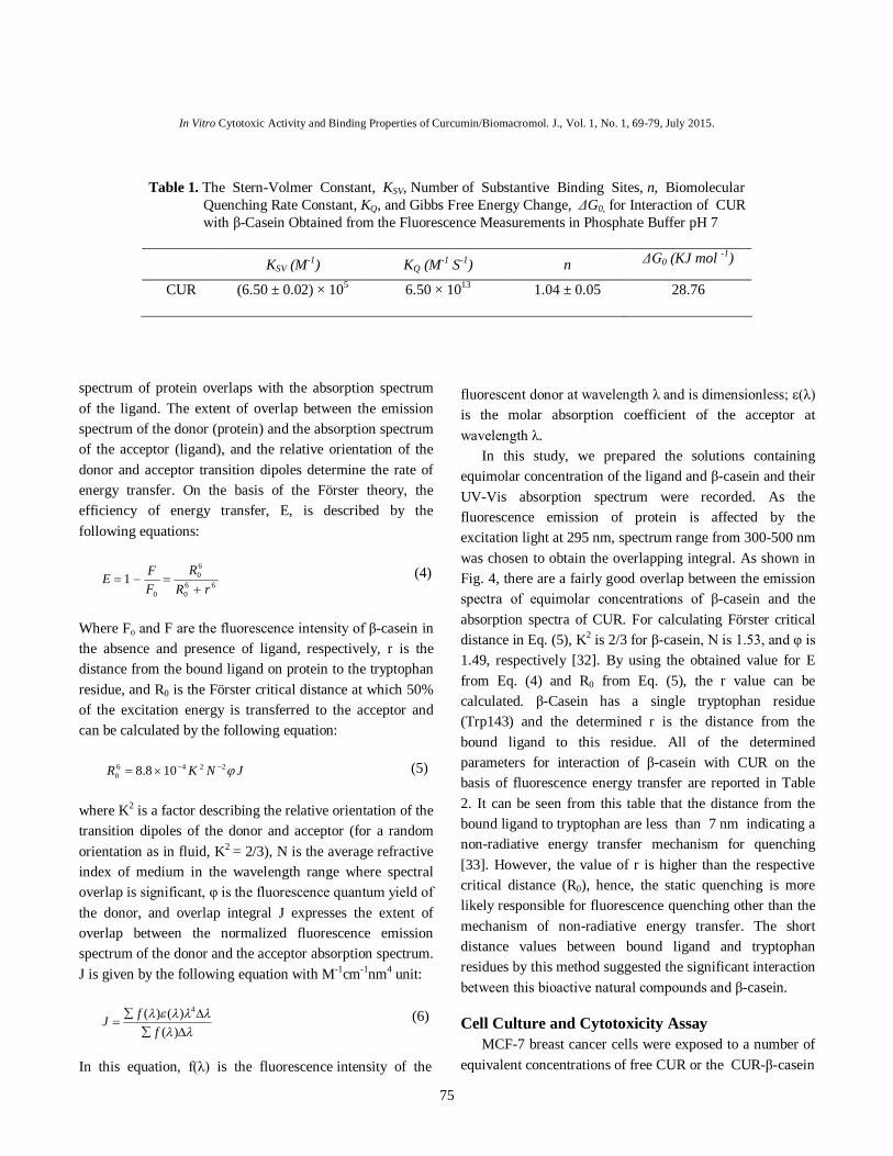
spectrum of protein overlaps with the absorption spectrum of the ligand. The extent of overlap between the emission spectrum of the donor (protein) and the absorption spectrum of the acceptor (ligand), and the relative orientation of the donor and acceptor transition dipoles determine the rate of energy transfer. On the basis of the Förster theory, the efficiency of energy transfer, E, is described by the following equations:
660
60
0
1rR
RFFE
(4)
Where Fo and F are the fluorescence intensity of β-casein in the absence and presence of ligand, respectively, r is the distance from the bound ligand on protein to the tryptophan residue, and R0 is the Förster critical distance at which 50% of the excitation energy is transferred to the acceptor and can be calculated by the following equation: JNKR 2246
0 108.8 (5) where K2 is a factor describing the relative orientation of the transition dipoles of the donor and acceptor (for a random orientation as in fluid, K2 = 2/3), N is the average refractive index of medium in the wavelength range where spectral overlap is significant, φ is the fluorescence quantum yield of the donor, and overlap integral J expresses the extent of overlap between the normalized fluorescence emission spectrum of the donor and the acceptor absorption spectrum. J is given by the following equation with M-1cm-1nm4 unit:
)()()( 4
ffJ (6)
In this equation, f(λ) is the fluorescence intensity of the
fluorescent donor at wavelength λ and is dimensionless; ε(λ) is the molar absorption coefficient of the acceptor at wavelength λ. In this study, we prepared the solutions containing equimolar concentration of the ligand and β-casein and their UV-Vis absorption spectrum were recorded. As the fluorescence emission of protein is affected by the excitation light at 295 nm, spectrum range from 300-500 nm was chosen to obtain the overlapping integral. As shown in Fig. 4, there are a fairly good overlap between the emission spectra of equimolar concentrations of β-casein and the absorption spectra of CUR. For calculating Förster critical distance in Eq. (5), K2 is 2/3 for β-casein, N is 1.53, and φ is 1.49, respectively [32]. By using the obtained value for E from Eq. (4) and R0 from Eq. (5), the r value can be calculated. β-Casein has a single tryptophan residue (Trp143) and the determined r is the distance from the bound ligand to this residue. All of the determined parameters for interaction of β-casein with CUR on the basis of fluorescence energy transfer are reported in Table 2. It can be seen from this table that the distance from the bound ligand to tryptophan are less than 7 nm indicating a non-radiative energy transfer mechanism for quenching [33]. However, the value of r is higher than the respective critical distance (R0), hence, the static quenching is more likely responsible for fluorescence quenching other than the mechanism of non-radiative energy transfer. The short distance values between bound ligand and tryptophan residues by this method suggested the significant interaction between this bioactive natural compounds and β-casein. Cell Culture and Cytotoxicity Assay MCF-7 breast cancer cells were exposed to a number of equivalent concentrations of free CUR or the CUR-β-casein
Table 1. The Stern-Volmer Constant, KSV, Number of Substantive Binding Sites, n, Biomolecular Quenching Rate Constant, KQ, and Gibbs Free Energy Change, ΔG0, for Interaction of CUR with β-Casein Obtained from the Fluorescence Measurements in Phosphate Buffer pH 7
KSV (M-1) KQ (M-1 S-1) n ΔG0 (KJ mol -1)
CUR (6.50 ± 0.02) × 105 6.50 × 10¹³ 1.04 ± 0.05 28.76

Mehranfar et alBiomacromol. J., Vol. 1, No. 1, 69-79, July 2015.
76
complex (the mole ratios of curcumin to β-casein were 0.238, 0.476, 0.714 and 0.952) and their viability was quantified using MTT assay. As shown in Fig. 5, there is dose-dependent cytotoxicity (10-40 µM) of in CUR and CUR-β-casein complex. Previous studies have been shown that CUR increase the cytotoxicity but this property was tested on different cell lines HepG2, HeLa, K-562 and human pancreatic cell lines, respectively [19,34]. The results of this study on MCF7 cell line was shown that CUR effect on the cell survival and increase Cytotoxicity, of course, enhanced cytotoxicity in cells in the presence of CUR plus β-casein combinations, as compared to individual CUR doses, that this effect is evident at higher concentration of complex. Free β-casein was found to be
non-toxic (data not shown) suggesting that the decline could not be a reflection of β-casein action alone. Also, free ethanol at different concentration was also used as control and has no harmful effect on the cells as compared with untreated cells. IC50 values for the CUR and CUR-β-casein complex were found to be 33.5 and 24 µM, respectively. Previous studies show similar results about of IC50 of CUR [35]. Results of cytotoxicity indicate that the CUR remains active after complexation with β-casein. Respect to our pervious study on DAC and BDMC, all of the effects of CUR and DAC were found to be better than those of BDMC at the same concentration. It shows that the presence of the methoxy groups in the CUR and DAC molecules is essential to cytotoxicity effect; in a similar manner the methoxy
Wavelength (nm)
250 300 350 400 450 500 550
Fluo
resc
ence
Inte
nsity
0
20
40
60
80
100
120
140
160
Abs
orba
nce
0.02
0.04
0.06
0.08
0.10
0.12
0.14
0.16
0.18
0.20
FluorescenceAbsorbance
Fig. 4. Overlay of the fluorescence emission spectra and UV-Vis of β-casein and CUR at equimolar concentrations.
Table 2. Förster Critical Distance, R0, the Binding Distance to Tryptophan Residue of Protein, r, Overlap Iintegral, J, and the Energy Transfer Efficiency, E, upon Interaction of CUR with β-Casein Measured in Phosphate Buffer pH 7
R0 (nm) r (nm) J (M-1cm-1nm4) E
CUR-β-casein 1.28 2.31 2.1×10⁻⁷ 0.028

In Vitro Cytotoxic Activity and Binding Properties of Curcumin/Biomacromol. J., Vol. 1, No. 1, 69-79, July 2015.
77
groups enhance cytotoxicity in MCF7 cell. Although further studies are warranted to explore the molecular mechanism of this enhanced cytotoxicity, combination of CUR with β-casein can be propagated as an easily accepted, economically viable and safe drug regimen. Nanoparticle Size Distribution The histogram particle size distribution of 1 mg ml-1
solution of β-CN in PBS that was measured by DLS analyzer is shown in Fig. 6. The result represents the narrow
size distribution of micelle nanoparticle with mean value of 70 nm. Docking Studies Docking studies show that CUR probably binds to β-casein at the hydrophobic core. On the basis of the experimental data, computational docking studies were performed to understand the binding site location and the best conformation of binding of CUR to β-casein. There presented build derived from the best pose with the minimal
[CUR] M
0 10 20 30 40
Via
bilit
y (%
)
0
20
40
60
80
100
120
CURCUR--Casein
Fig. 5. Viability of free CUR in the comparison of CUR-β-casein complex (the mole ratios of curcumin to β-
Casein were 0.238, 0.476, 0.714 and 0.952).
Fig. 6. The histogram of particle size distribution β-CN with 1 mg ml-1 concentration in PBS.

Mehranfar et alBiomacromol. J., Vol. 1, No. 1, 69-79, July 2015.
78
Fig. 7. Perspective of the best pose of CUR-β-casein complex with the minimal binding energy.
binding energy -7.87 kJ mol-1 for CUR is shown in Fig. 7. A careful inspection of the binding site suggested the closer contact of hydroxyl group of CUR with the tryptophan amino acid residues. This result in agreement with fluorescence quenching of β-casein due to the addition of CUR (please see Fig. 3). Also, from FRET results, r (the distance from the bound ligand on protein to the tryptophan residue) has been obtained 2.31 Å (see Table 2) that adequate for interaction between Trp and casein. The total number of hydrogen binding made by CUR is 3. These hydrogen bonds forms between hydroxyl groups of CUR and His160 (Nitrogen atom), Trp158 (Oxygen atom) and Leu142 (Nitrogen atom). Within the van der Waals contact, the CUR molecule is lined by hydrophobic residues such as Trp158, Met159, Gln181, Val198, Pro100, Ser139, Leu142, His163, His160, Pro162 and Thr141. Our results suggested the binding of CUR to β-casein predominantly by hydrophobic contacts within the hydrophobic core of protein. The docking calculation on β-casein micelle is not possible due to high complexicity. However, as we know, the structure of β-casein does not significantly changed due to formation of micelle, hence it can be easily correlated the results of β-casein monomer to β-casein micelle. CONCLUSIONS Fluorescence quenching, non-radiative energy transfer and molecular docking provided clear experimental
evidences on the binding of CUR as a one of the main active component of turmeric to the β-casein micelle. So, β-casein micelle can serve as potential carrier, especially for hydrophobic non-water soluble molecules such as the studied ligand in this article. Fluorescence spectroscopy study of the interaction between CUR and β-casein solution was observed that CUR molecules interact with β-casein by binding to the hydrophobic regions of β-casein and there are strong interaction of CUR with β-casein micelle that suggested the important role of the hydroxyl phenolic groups in the binding process (three hydrogen bonds form via these groups in the binding site). The Förster’s critical distance and the average distance between bound ligand and tryptophan residues have been determined based on the fluorescence resonance energy transfer. Results of computational docking studies indicated that CUR probably binds to β-casein at the hydrophobic core and a combination of vander Waals interactions and hydrogen bonds are encountered in the binding process. Under physiological buffer conditions, the CUR-β-casein complex yielded a nanoformulation, which exhibited similar cytotoxic effects on MCF7 cells compared to an equal dose of free CUR. Because β-casein is an edible protein, the complex has the potential to be an oral dose of CUR, however, further studies are required to confirm this hypothesis. ACKNOWLEDGEMENTS Financial supports of Research Council of Isfahan University is kindly appreciated. REFERENCES [1] G. Spada, E. Gavini, P. Giunchedi, Nanoparticles.
Engineering and Technology 76 (2011) 245. [2] Y. Liu and R. Guo. Biomacromolecules 8 (2007)
2902. [3] A. Shapira, Y.G. Assaraf, D.L. Epstein, Y.D. Livney,
Pharm. Res. 27 (2010) 2175-2186. [4] S.A. Forrest, R.Y. Yada, D. Rousseau. J. Agric. Food
Chem. 53 (2005) 8003. [5] A. Shapira, Y.G. Assaraf, Y.D. Livney,
Nanomedicine: Nanotechnology. Biology. Medicine 6 (2010) 119.

In Vitro Cytotoxic Activity and Binding Properties of Curcumin/Biomacromol. J., Vol. 1, No. 1, 69-79, July 2015.
79
[6] E. Semo, E. Kesselman, D. Danino, Y.D. Livney,
Food Hydrocolloids 21 (2007) 936. [7] A. Divsalar, M. Razmi, A.A. Saboury, A. Seyedarabi,
Medicin. Chem. 14(2014) 892. [8] M. Razmi, A. Divsalar, A.A. Saboury, Z. Izadi, T.
Haertlé, H. Mansuri-Torshizi, Colloid. Surface. B: Biointerfaces 112 (2013) 362.
[9] K. Pan, Q. Zhong, S.J. Baek, J. Agric. Food Chem. 61 (2013) 6036.
[10] Y. Luo, K. Pan, Q. Zhong, Food Chem. 155 (2014) 146.
[11] F. Mohammadi, A.K. Bordbar, K. Mohammadi, A. Divsalar, A.K. Saboury, Protein J. 28 (2009) 117.
[12] F. Mohammadi, A.K. Bordbar, K. Mohammadi, A. Divsalar, A.K. Saboury, Protein J. 28 (2009) 189.
[13] F. Mohammadi, A.K. Bordbar, K. Mohammadi, A. Divsalar, A.K. Saboury, Can. J. Cham. 88 (2010) 155.
[14] A. Graham, Biochem. Pharmacol. 17 (2007) 787. [15] B.B. Aggarwal, A. Kumar, M.S. Aggarwal, S.
Shishodia, CRC Press Boca Raton (2005) 349. [16] V. Basile, E. Ferrari, S. Lazzari, S. Belluti, F.
Pignedoli, C. Imbriano, Biochem. Pharmacol. 78 (2009) 1305.
[17] C.K. Kim, P. Ghosh, C. Pagliuca, Z.J. Zhu, S. Menichetti, V.M. Rotello, J. Am. Chem. Soc. 131 (2009) 1360.
[18] F. Yang, G.P. Lim, A.N. Begum, O.J. Ubeda, M.R. Simmons, S.S. Ambegaokar, P. Chen, R. Kayed, C.G. Glabe, S.A. Frautschy, G.M. Cole, J. Biol. Chem. 280 (2005) 5892.
[19] A. Sahu, N. Kasoju, U. Bora. Biomacromolecules 9 (2008) 2905.
[20] A.H. Sneharani, S.A. Singh, A.G. Appu Rao, J. Agric. Food Chem. 57 (2009) 10386.
[21] A.H. Sneharani, J.V. Karakka, S.A. Singh, A.G.
Appu Rao, J. Agric. Food Chem. 58 (2010) 11130. [22] C. Syng-ai, A.L. Kumariand, A. Khar, Mol. Cancer
Ther. 3 (2004) 1101. [23] A. Altunbas, S.J. Lee, S.A. Rajasekaran, J.P.
Schneider, D.J. Pochan, J. Biomaterials 32 (25) (2011) 5906.
[24] M. Esmaili, S.M. Ghaffari, Z. Moosavi-Movahedi, M.S. Atri, A. Sharifizadeh, M. Farhadi, R. Yousefi, J. M. Chobert, T. Haertlé, A.A. Moosavi-Movahedi, Food Sci. Technol. 44 (2011) 2166.
[25] F. Mehranfar, A.K. Bordbar, M. Keyhanfar, M. Behbahani, J. Lumin. 143 (2013) 687.
[26] F. Mehranfar, A.K. Bordbar, N. Fani, M. Keyhanfar, Spectrochim. Acta A: Molecular and Biomolecular Spectroscopy 115 (2013) 629.
[27] I. Portnaya, U. Cogan, Y.D. Livney, O. Ramon, K. Shimoni, M. Rosenberg, D. Danino. J. Agric. Food Chem. 54 (2006) 5555.
[28] P. Sokkar, S. Mohandass, M. Ramachandran, J. Mol. Model 17 (2011) 1565.
[29] M.F. Sanner, J. Mol. Graphics Model 17 (1999) 57. [30] A. Taheri-Kafrani, Y. Choiset, D.A. Faizullin, Y.R.
Zuev, V.V. Bezuglov, J.M. Chobert, A.K. Bordbarand, T. Haertle, Biopolymers 95(12) (2011) 871.
[31] A. Barik, K.I. Priyadarsini, H. Mohan, Photochem. Photobiol. 77 (2003) 597.
[32] J. Vörös, Biophys. J. 87 (2004) 553. [33] J. Lakowicz, Kluwer Academic, New York, 1999 (2nd
ed.). [34] H.L. Yu, Q.R. Huang, Food Chem. 119 (2010) 669. [35] D. Zaidi, N. Singh, I.Z. Ahmad, R. Sharma, A.K.
Balapure, Int. J. Pharm. Sci. 2 (2011) 212.