Embed Size (px)
Citation preview
E
PDMFHG
SmTos(
rbetp(tp
†BRFu
2
Journal of the American College of Cardiology Vol. 48, No. 1, 2006© 2006 by the American College of Cardiology Foundation ISSN 0735-1097/06/$32.00P
XPEDITED REVIEWS
athology of Drug-Eluting Stents in Humanselayed Healing and Late Thrombotic Riskichael Joner, MD,* Aloke V. Finn, MD,† Andrew Farb, MD,§ Erik K. Mont, MD,‡
rank D. Kolodgie, PHD,* Elena Ladich, MD,* Robert Kutys, MS,* Kristi Skorija, BS,*erman K. Gold, MD,† Renu Virmani, MD*aithersburg and Rockville, Maryland; Boston, Massachusetts; and Miami, Florida
OBJECTIVES This study examined human drug-eluting stents (DES) to determine the long-term effects ofthese stents on coronary arterial healing and identified mechanisms underlying late stentthrombosis (LST).
BACKGROUND Although DES reduce the need for repeat revascularization compared with bare-metal stents(BMS), data suggest the window of thrombotic risk for Cypher (Cordis Corp., Miami Lakes,Florida) and Taxus (Boston Scientific Corp., Natick, Massachusetts) DES extends far beyondthat for BMS.
METHODS From a registry of 40 autopsies of DES (68 stents), 23 DES cases of �30 days duration werecompared with 25 matched autopsies of BMS implantation. Late stent thrombosis wasdefined as an acute thrombus within a stent �30 days old.
RESULTS Of 23 patients with DES �30 days old, 14 had evidence of LST. Cypher and Taxus DESshowed greater delayed healing characterized by persistent fibrin deposition (fibrin score 2.3� 1.1 vs. 0.9 � 0.8, p � 0.0001) and poorer endothelialization (55.8 � 26.5%) comparedwith BMS (89.8 � 20.9, p � 0.0001). Moreover, DES with LST showed more delayedhealing compared with patent DES. In 5 of 14 patients suffering LST, antiplatelet therapyhad been withdrawn. Additional procedural and pathologic risk factors for LST were: 1) localhypersensitivity reaction; 2) ostial and/or bifurcation stenting; 3) malapposition/incompleteapposition; 4) restenosis; and 5) strut penetration into a necrotic core.
CONCLUSIONS The Cypher and Taxus DES result in delayed arterial healing when compared with BMS ofsimilar implant duration. The cause of DES LST is multifactorial with delayed healing incombination with other clinical and procedural risk factors playing a role. (J Am Coll
ublished by Elsevier Inc. doi:10.1016/j.jacc.2006.03.042
Cardiol 2006;48:193–202) © 2006 by the American College of Cardiology Foundation
d(sDlihsDcDDp
M
FcD1dhw
tent thrombosis remains a major cause of death andorbidity after percutaneous coronary interventions (1–4).he pivotal clinical trials that formed the basis for approvalf drug-eluting stents (DES) have not shown an increase intent thrombosis compared with bare-metal stents (BMS)5,6). However, recent publication of “real-world” patients
See page 203
eceiving DES have suggested that the window of throm-otic risk at sites where these stents have been deployedxtends far beyond that for BMS (7,8). Understanding theime course of healing in DES compared with BMS and theathologic mechanisms underlying late stent thrombosisLST) might more clearly delineate the time course ofhrombotic risk and redefine the optimal duration of anti-latelet therapy.
From *CVPath, International Registry of Pathology, Gaithersburg, Maryland;Cardiac Unit, Department of Internal Medicine, Massachusetts General Hospital,oston, Massachusetts; ‡Miami Dade County Medical Examiner Department, Heartadiology, Miami, Florida; and the §Interventional Cardiology Devices Branch, U.S.ood and Drug Administration, Rockville, Maryland. Drs. Joner and Finn contrib-ted equally to this work.
sManuscript received January 26, 2006; revised manuscript received March 10,
006, accepted March 16, 2006.
Current polymer-based sirolimus-eluting (Cypher, Cor-is Corp., Miami Lakes, Florida) and paclitaxel-elutingTaxus, Boston Scientific Corp., Natick, Massachusetts)tents are the only DES approved by the U.S. Food andrug Administration (FDA) for human use. Some pub-
ished studies of animal models with similar DES implantedn normal arteries show a substantial impairment of arterialealing relative to BMS (9–13). To date, there has been noystematic published analysis of the long-term effects ofES on arterial healing in humans. We examined 40
onsecutive autopsies of patients who died subsequent toES implantation to determine the long-term effects ofES placement on coronary arterial healing and to identify
athologic mechanisms underlying LST.
ATERIALS AND METHODS
rom a registry of 484 human coronary stents submitted foronsultation, 40 consecutive cases with evidence of one or moreES were examined (3 have been previously reported) (14–
6). Of these cases, 23 (32 stents) had DES implanted for �30ays and were included in the DES study group. Clinicalistories and cardiac catheterization reports were reviewedhen available. A total of 25 cases of BMS implantation (36
tents) of patients of similar age, gender, case duration, and
aap
dstMws�nnraMgIgDmbmnlocpss1s
wwpepStScgnnogcetd
R
Pffs((fd�s13PampDmf
FmS
194 Joner et al. JACC Vol. 48, No. 1, 2006Pathology of Drug-Eluting Stents July 4, 2006:193–202
rtery of implantation were blindly selected from our registrynd formed the control BMS group. Cases with a history ofrior brachytherapy were excluded.Stented arteries had been fixed in 10% buffered formalin,
issected off the heart, examined via radiography, andubmitted for plastic embedding. Arteries were sectioned 2o 3 mm apart and stained with hematoxylin and eosin and
ovat pentachrome as previously described (17). Casesith histologic evidence of an acute occlusive or nonocclu-
ive mural thrombus within a coronary artery stent in place30 days were defined as LST. Stented arteries with severe
arrowing, defined as in-stent luminal cross-sectional areaarrowing of �75% by neointimal growth, were defined asestenosis; and those �75% area narrowing were designateds patent stents.
orphologic and morphometric measurements. Computer-uided morphometric measurements were performed usingPLab Spectrum software (Scanalytics Inc., Vienna, Vir-inia) on sections from stents implanted for �30 days.igital images were captured (4� magnification), and areaeasurements included the internal elastic lamina, plaque
urden, stent area, and lumen area; stent lengths wereeasured from radiographs. For calculation of in-stent
eointimal growth, fibrin deposition, and surface endothe-ialization, stents with occlusive thrombi were excluded tovercome biased measurements. Ordinal data for fibrin wereollected on each stent section using a scale of 0 to 3� asreviously reported (18). Inflammation was scored at eachtent strut using a scale from 0 to 5 (with 0 for 0 to 25urrounding inflammatory cells, 1 for 25 to 50, 2 for 50 to00, 3 for 100 to 150, 4 for 150 to 200, and 5 for �200urrounding inflammatory cells). The percentage of struts
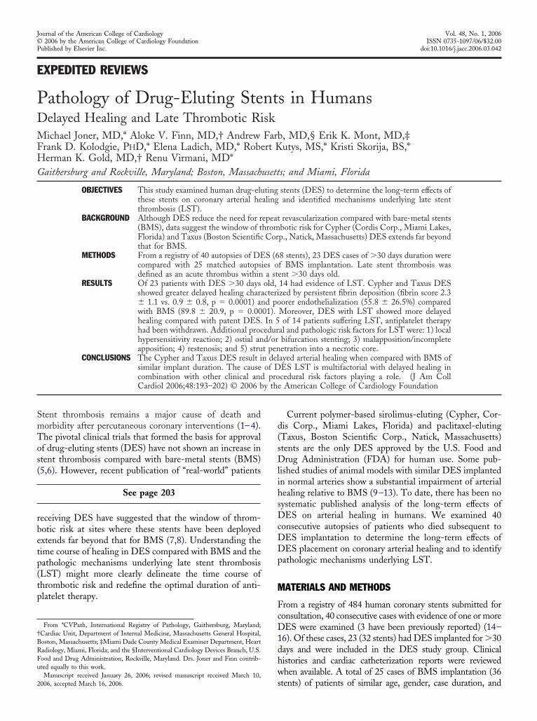
igure 1. Flow diagram illustrating cause of death in drug-eluting stents (D
Abbreviations and AcronymsBMS� bare-metal stentsDES � drug-eluting stentsFDA � Food and Drug AdministrationLST � late stent thrombosis
ajor categories depending on whether stents showed evidence of late thromboCD � sudden cardiac death.
ith surrounding fibrin and luminal surface endotheliumas also recorded. Delayed arterial healing was defined asersistence of fibrin beyond 30 days. Total eosinophils werevaluated by counting the four most severely inflamed strutser section and reported as eosinophils per strut.tatistical analysis. Continuous variables are presented as
he mean � SD and categorical variables either as mean �D or frequency (%). Continuous variables were firsthecked for normal distribution using Shapiro-Wilkoodness-of-fit test and compared by Student t test forormally distributed or a Wilcoxon rank-sum test foron-normally distributed variables. For comparison of ratesf LST between the DES group and the control BMSroup, a Fisher exact test was used. A p value �0.05 wasonsidered significant. The percentage of struts covered byndothelial cells was plotted against the duration of implanto derive a slope, intercept, and correlation coefficient toetermine relationships.
ESULTS
atient characteristics. Thirty-two DES were examinedrom 23 individuals and compared with 36 matched BMSrom 25 individuals. In patients with DES, indications fortenting were unstable angina or acute myocardial infarctionn � 5 patients for Cypher, n � 7 for Taxus), stable anginan � 5 for Cypher, n � 5 for Taxus), or restenosis (n � 1or Cypher). The location of the stents was: left anteriorescending/left diagonal (Cypher, n � 10 stents; Taxus, n
6; BMS, n � 13); right coronary artery/posterior de-cending artery (Cypher, n � 4; Taxus, n � 5; BMS, n �2); and left circumflex/left obtuse marginal (Cypher, n �; Taxus, n � 4; BMS, n � 11).rocedural outcome. At the time of coronary intervention,ll patients had angiographically successful stent deploy-ent with one DES placed in 15 patients, two DES in 7
atients, and three DES in 1 patient. Mean stent length forES was 32.1 � 17.3 mm (range 8 to 76 mm; median 25m, 75% �35 mm). Ostial/bifurcation stenting was per-
ormed in three cases (left circumflex/obtuse marginal bi-
patients with stents in place �30 days. These cases are divided into three
ES) sis, were patent, or were restenotic. AMI � acute myocardial infarction;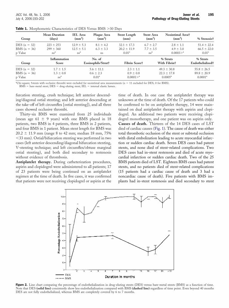
fitc
(pa2�cVowAaort
tubtddCdtwtsDcBs(np
FND
T
DBp
DBp
* uremenen.
195JACC Vol. 48, No. 1, 2006 Joner et al.July 4, 2006:193–202 Pathology of Drug-Eluting Stents
urcation stenting, crush technique; left anterior descend-ng/diagonal ostial stenting; and left anterior descending athe take-off of left circumflex [ostial stenting]), and all threeases showed occlusive thrombi.
Thirty-six BMS were examined from 25 individualsmean age 61 � 9 years) with one BMS placed in 18atients, two BMS in 4 patients, three BMS in 2 patients,nd four BMS in 1 patient. Mean stent length for BMS was0.2 � 11.9 mm (range 8 to 42 mm; median 18 mm, 75%33 mm). Ostial/bifurcation stenting was performed in two
ases (left anterior descending/diagonal bifurcation stenting,-stenting technique; and left circumflex/obtuse marginalstial stenting), and both died secondary to restenosisithout evidence of thrombosis.ntiplatelet therapy. During catheterization procedures,
spirin and clopidogrel were administered to all patients; 17f 23 patients were being continued on an antiplateletegimen at the time of death. In five cases, it was confirmedhat patients were not receiving clopidogrel or aspirin at the
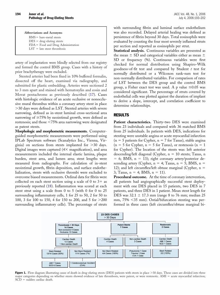
igure 2. Line chart comparing the percentage of endothelialization in dr
able 1. Morphometric Characteristics of DES Versus BMS �3
GroupMean Duration
(days)IEL Area
(mm2)Plaque Area
(mm2)
ES (n � 32) 223 � 253 12.9 � 5.3 8.1 � 4.2MS (n � 36) 299 � 360 12.5 � 5.1 6.3 � 3.3Value ns* ns* ns
GroupInflammation
ScoreNo. of
Eosinophils/Strut†
ES (n � 32) 1.7 � 1.5 5.6 � 11.1MS (n � 36) 1.3 � 0.8 0.6 � 2.3Value ns* 0.01*
Chi-square; †stents with occlusive thrombi were excluded for neointimal area measBMS � bare-metal stent; DES � drug-eluting stent; IEL � internal elastic lum
ote that DES (solid line) consistently show less endothelialization compared wES are not fully endothelialized, whereas BMS are completely covered by 6 t
ime of death. In one case the antiplatelet therapy wasnknown at the time of death. Of the 17 patients who coulde confirmed to be on antiplatelet therapy, 14 were main-ained on dual antiplatelet therapy with aspirin and clopi-ogrel. An additional two patients were receiving clopi-ogrel monotherapy, and one patient was on aspirin only.auses of death. Thirteen of the 14 DES cases of LSTied of cardiac causes (Fig. 1). The cause of death was eitherotal thrombotic occlusion of the stent or subtotal occlusionith distal embolization leading to acute myocardial infarc-
ion or sudden cardiac death. Seven DES cases had patenttents, and none died of stent-related complications. TwoES cases had in-stent restenosis and died of acute myo-
ardial infarction or sudden cardiac death. Two of the 25MS patients died of LST. Eighteen BMS cases had patent
tents, and no patients died of stent-related complications15 patients had a cardiac cause of death and 3 had aoncardiac cause of death). Five patients with BMS im-lants had in-stent restenosis and died secondary to stent
ting stents (DES) versus bare-metal stents (BMS) as a function of time.
ys
tent Length(mm)
Stent Area(mm2)
Neointimal Area†(mm2) % Stenosis†
32.1 � 17.3 6.7 � 2.7 2.8 � 1.1 51.4 � 22.420.2 � 11.9 7.7 � 3.5 4.9 � 3.0 66.5 � 22.0
0.01* ns* 0.0003�* 0.01*
Fibrin Score†% Struts
With Fibrin†% Struts
Endothelialized†
2.3 � 1.1 49.3 � 30.8 55.8 � 26.50.9 � 0.8 22.3 � 17.8 89.8 � 20.90.0001�* 0.0005* 0.0001*
ts (n � 11 excluded for DES, 0 for BMS).
ug-elu
0 Da
S
ith BMS (dashed line) regardless of time point. Even beyond 40 monthso 7 months.
rcLtcLLrCD1imsAni(s�
fiph�BtMpddlwLmhss0g(
T
TNp
*
T
A
456
4
764
63
77
*d
R
196 Joner et al. JACC Vol. 48, No. 1, 2006Pathology of Drug-Eluting Stents July 4, 2006:193–202
elated acute myocardial infarction (three cases) or suddenardiac death (two cases).ST (>30 days). Of 23 DES cases, 14 (61%) had stent
hrombosis, and of the 25 cases of BMS included asontrols, 2 (8%) had LST (p � 0.0001). Both cases of BMSST also had evidence of in-stent restenosis. The rate ofST in the control BMS group is similar to that previously
eported from our registry of BMS (19).omparison of DES and BMS implants. Stent length inES was greater than in BMS (32.1 � 17.3 vs. 20.2 �
1.9, p � 0.01) (Table 1). The DES had significantly lessn-stent neointimal growth compared with BMS (neointi-
al area 2.9 � 1.1 mm2 vs. 4.9 � 3.0 mm2, p � 0.005; %tenosis 54.4 � 23.6% vs. 66.5 � 22.0%, p � 0.05).lthough the extent of overall inflammation was not sig-ificantly different in the two groups, eosinophils surround-
ng struts were more frequent in DES compared with BMS5.6 � 11.1 vs. 0.6 � 2.3 per strut, p � 0.01). The DES alsohowed significantly higher fibrin scores (2.3 � 1.1 vs. 0.9
0.8, p � 0.0001) and percentage of struts surrounded by
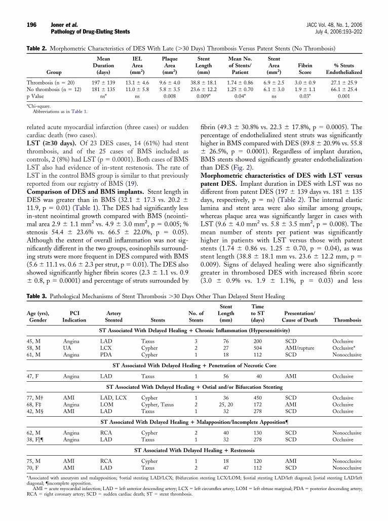
able 2. Morphometric Characteristics of DES With Late (�30
Group
MeanDuration
(days)
IELArea
(mm2)
PlaqueArea
(mm2)
hrombosis (n � 20) 197 � 139 13.1 � 4.6 9.6 � 4.0o thrombosis (n � 12) 181 � 135 11.0 � 5.8 5.8 � 3.5Value ns* ns 0.008
Chi-square.Abbreviations as in Table 1.
able 3. Pathological Mechanisms of Stent Thrombosis �30 Da
ge (yrs),Gender
PCIIndication
ArteryStented Stents
N
ST Associated With Delayed Healing
5, M Angina LAD Taxus8, M UA LCX Cypher1, M Angina PDA Cypher
ST Associated With Delayed He
7, F Angina LAD Taxus
ST Associated With Delayed Healin
7, M† AMI LAD, LCX Cypher8, F‡ Angina LOM Cypher, Taxus2, M§ AMI LAD Taxus
ST Associated With Delayed Healing
2, M Angina RCA Cypher8, F�¶ Angina LAD Taxus
ST Associated With De
5, M AMI RCA Cypher0, F AMI LAD Taxus
Associated with aneurysm and malapposition; †ostial stenting LAD/LCX; ‡bifurcaiagonal; ¶incomplete apposition.
AMI � acute myocardial infarction; LAD � left anterior descending artery; LCX � leftCA � right coronary artery; SCD � sudden cardiac death; ST � stent thrombosis.
brin (49.3 � 30.8% vs. 22.3 � 17.8%, p � 0.0005). Theercentage of endothelialized stent struts was significantlyigher in BMS compared with DES (89.8 � 20.9% vs. 55.8
26.5%, p � 0.0001). Regardless of implant duration,MS stents showed significantly greater endothelialization
han DES (Fig. 2).orphometric characteristics of DES with LST versus
atent DES. Implant duration in DES with LST was noifferent from patent DES (197 � 139 days vs. 181 � 135ays, respectively, p � ns) (Table 2). The internal elastic
amina and stent area were also similar among groups,hereas plaque area was significantly larger in cases withST (9.6 � 4.0 mm2 vs. 5.8 � 3.5 mm2, p � 0.008). Theean number of stents per patient was significantly
igher in patients with LST versus those with patenttents (1.74 � 0.86 vs. 1.25 � 0.70, p � 0.04), as wastent length (38.8 � 18.1 mm vs. 23.6 � 12.2 mm, p �.009). Signs of delayed healing were also significantlyreater in thrombosed DES with increased fibrin score3.0 � 0.9% vs. 1.9 � 1.1%, p � 0.03) and less
s) Thrombosis Versus Patent Stents (No Thrombosis)
entngth
m)
Mean No.of Stents/
Patient
StentArea
(mm2)FibrinScore
% StrutsEndothelialized
� 18.1 1.74 � 0.86 6.9 � 2.5 3.0 � 0.9 27.1 � 25.9� 12.2 1.25 � 0.70 6.1 � 3.0 1.9 � 1.1 66.1 � 25.409* 0.04* ns 0.03* 0.001
ther Than Delayed Stent Healing
fs
StentLength(mm)
Timeto ST(days)
Presentation/Cause of Death Thrombosis
ronic Inflammation (Hypersensitivity)
76 200 SCD Occlusive27 504 AMI/rupture Occlusive*18 112 SCD Nonocclusive
� Penetration of Necrotic Core
56 40 AMI Occlusive
Ostial and/or Bifurcation Stenting
36 450 SCD Occlusive25, 20 172 AMI Occlusive
32 278 SCD Occlusive
alapposition/Incomplete Apposition¶
40 130 SCD Nonocclusive32 278 SCD Occlusive
Healing � Restenosis
18 120 AMI Nonocclusive47 112 SCD Nonocclusive
tenting LCX/LOM; §ostial stenting LAD/left diagonal; �ostial stenting LAD/left
Day
StLe(m
38.823.6
0.0
ys O
o. oStent
� Ch
321
aling
1
g �
121
� M
21
layed
12
tion s
circumflex artery; LOM � left obtuse marginal; PDA � posterior descending artery;
e6Att7cotiotPwrpfitoic
�e(m(p2rAtaosanmfscta
Fadiwn((ctsi owns
197JACC Vol. 48, No. 1, 2006 Joner et al.July 4, 2006:193–202 Pathology of Drug-Eluting Stents
ndothelial coverage of stent struts (27.1 � 25.9% vs.6.1 � 25.4%, p � 0.001).ntiplatelet therapy in cases of late DES thrombosis. Of
he 14 patients with LST, maintenance on dual antiplateletherapy (i.e., aspirin and clopidogrel) could be confirmed inpatients. An additional 2 patients were either on aspirin or
lopidogrel monotherapy. Five of 14 patients with evidencef LST were not receiving any antiplatelet therapy at theime of death. In the two cases of LST after BMSmplantation, dual antiplatelet therapy was administered fornly 1 month and patients were on aspirin monotherapy athe time point of death.athologic mechanisms of DES LST. In all 14 patientsith LST delayed arterial healing was found as a cardinal
isk factor and was the only pathologic risk factor in 3 (21%)atients. Additional pathologic risk factors for LST wereound in 11 of the 14 patients (Table 3). They were groupednto five major categories: 1) chronic inflammation charac-erized by lymphocytes, macrophages, and extensive eosin-philic infiltration of the intima and media (hypersensitiv-ty) (n � 3); 2) stenting along major side branches using the
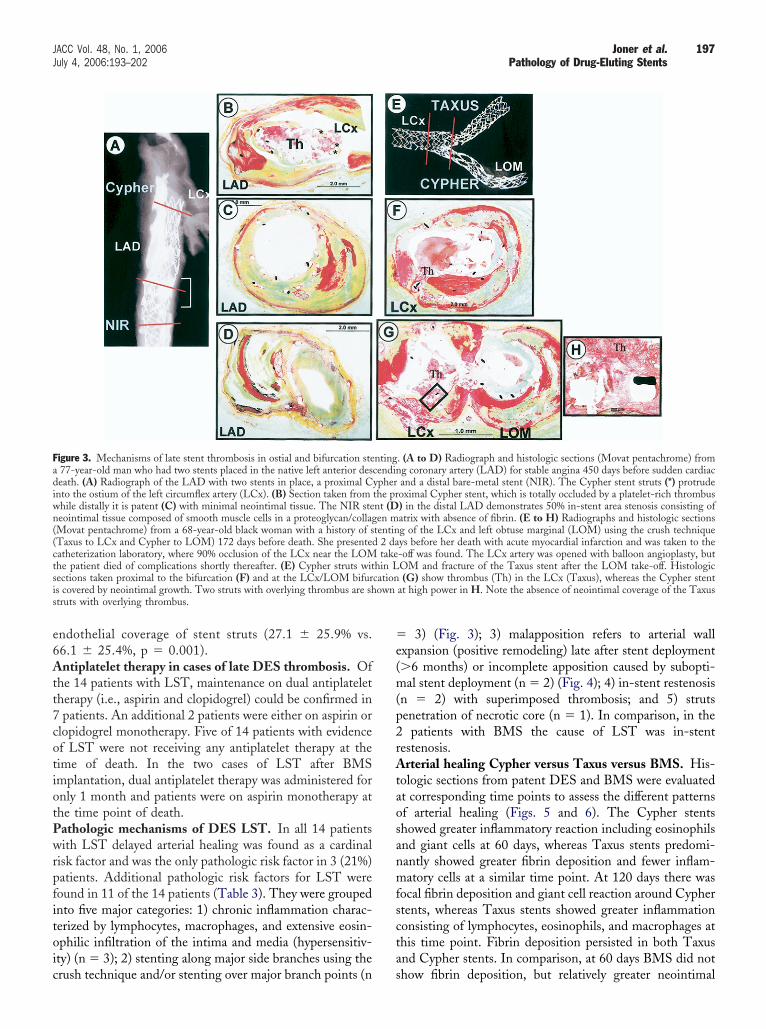
igure 3. Mechanisms of late stent thrombosis in ostial and bifurcation ste77-year-old man who had two stents placed in the native left anterior deseath. (A) Radiograph of the LAD with two stents in place, a proximal Cnto the ostium of the left circumflex artery (LCx). (B) Section taken fromhile distally it is patent (C) with minimal neointimal tissue. The NIR steeointimal tissue composed of smooth muscle cells in a proteoglycan/collagMovat pentachrome) from a 68-year-old black woman with a history of sTaxus to LCx and Cypher to LOM) 172 days before death. She presentedatheterization laboratory, where 90% occlusion of the LCx near the LOMhe patient died of complications shortly thereafter. (E) Cypher struts witections taken proximal to the bifurcation (F) and at the LCx/LOM bifurs covered by neointimal growth. Two struts with overlying thrombus are shtruts with overlying thrombus.
rush technique and/or stenting over major branch points (n s
3) (Fig. 3); 3) malapposition refers to arterial wallxpansion (positive remodeling) late after stent deployment�6 months) or incomplete apposition caused by subopti-al stent deployment (n � 2) (Fig. 4); 4) in-stent restenosis
n � 2) with superimposed thrombosis; and 5) strutsenetration of necrotic core (n � 1). In comparison, in the
patients with BMS the cause of LST was in-stentestenosis.rterial healing Cypher versus Taxus versus BMS. His-
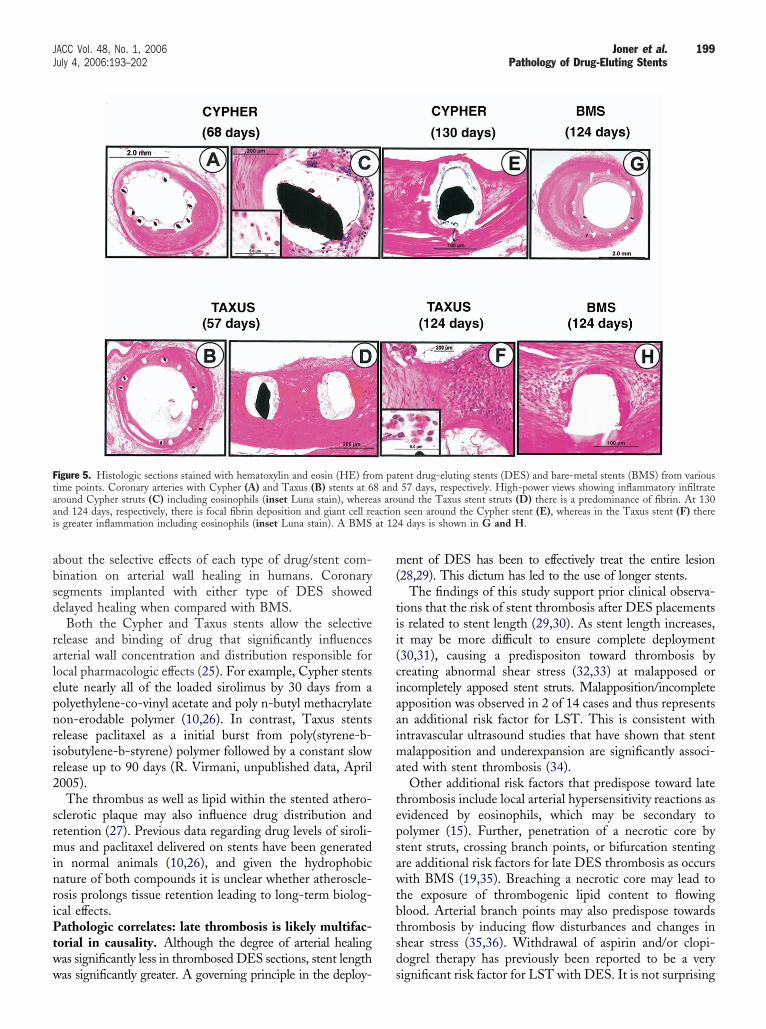
ologic sections from patent DES and BMS were evaluatedt corresponding time points to assess the different patternsf arterial healing (Figs. 5 and 6). The Cypher stentshowed greater inflammatory reaction including eosinophilsnd giant cells at 60 days, whereas Taxus stents predomi-antly showed greater fibrin deposition and fewer inflam-atory cells at a similar time point. At 120 days there was
ocal fibrin deposition and giant cell reaction around Cyphertents, whereas Taxus stents showed greater inflammationonsisting of lymphocytes, eosinophils, and macrophages athis time point. Fibrin deposition persisted in both Taxusnd Cypher stents. In comparison, at 60 days BMS did not
. (A to D) Radiograph and histologic sections (Movat pentachrome) fromg coronary artery (LAD) for stable angina 450 days before sudden cardiacand a distal bare-metal stent (NIR). The Cypher stent struts (*) protrudeoximal Cypher stent, which is totally occluded by a platelet-rich thrombus) in the distal LAD demonstrates 50% in-stent area stenosis consisting ofatrix with absence of fibrin. (E to H) Radiographs and histologic sectionsg of the LCx and left obtuse marginal (LOM) using the crush techniqueys before her death with acute myocardial infarction and was taken to the-off was found. The LCx artery was opened with balloon angioplasty, butOM and fracture of the Taxus stent after the LOM take-off. Histologic(G) show thrombus (Th) in the LCx (Taxus), whereas the Cypher stent
at high power in H. Note the absence of neointimal coverage of the Taxus
ntingcendinypherthe prnt (Den m
tentin2 datake
hin Lcation
how fibrin deposition, but relatively greater neointimal
ciTgigb
D
TrDatutsstdHLdobls
fienTcpMbhpmiwpwkBtcaaisip
FaePot D (Me
198 Joner et al. JACC Vol. 48, No. 1, 2006Pathology of Drug-Eluting Stents July 4, 2006:193–202
overage of stent struts was seen. Giant cell reaction andnflammation was substantially less in the BMS examined.he 120-day BMS showed circumferential neointimalrowth with complete coverage of stent struts. Chronicnflammatory cells (mostly lymphocytes, macrophages, andiant cells) are commonly seen in BMS at this time pointut without evidence of eosinophilic infiltrate.
ISCUSSION
he pathologic findings of our study underscore the causalelationship between the two currently FDA-approvedES and delayed arterial healing. The persistence of fibrin
nd incomplete endothelialization far beyond 30 days fromhe time of stenting form the critical pathologic substratenderlying the phenomenon of LST. These partially endo-helialized, fibrin-rich sites remain a potent thrombogenictimulus. In high-risk clinical situations such as bifurcationtenting, excessive stent length, or cessation of antiplateletherapy, these sites may develop thrombosis, leading toeath or other serious consequences.istologic findings >30 days. In patients suffering fromST after DES placement, the major pathologic findingistinguishing thrombosed from patent DES was evidencef significantly greater delay in arterial healing as manifestedy persistent peristrut fibrin deposition and poor endothe-ialization. In contrast, the control group of BMS implants
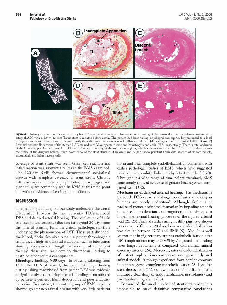
igure 4. Histologic sections of the stented artery from a 38-year-old womartery (LAD) with a 3.0 � 12-mm Taxus stent 6 months before death.mergency room with severe chest pain and shortly thereafter went into veroximal and middle sections of the stented LAD stained with Movat pentf the lumen by platelet-rich thrombus (Th) with absence of healing of thhe orifice of the diagonal branch. High-power view of the stent struts inndothelial, and inflammatory cells.
howed greater neointimal healing with very little peristrut i
brin and near complete endothelialization consistent witharlier pathologic studies of BMS, which have suggestedear-complete endothelialization by 3 to 4 months (19,20).hroughout a wide range of time points examined, BMS
onsistently showed evidence of greater healing when com-ared with DES.
echanisms of delayed arterial healing. The mechanismsy which DES cause a prolongation of arterial healing inumans are poorly understood. Although sirolimus oraclitaxel reduce neointimal formation by impeding smoothuscle cell proliferation and migration, these drugs also
mpair the normal healing processes of the injured arterialall (21–23). Animal studies using healthy pigs have shownersistence of fibrin at 28 days, however, endothelializationas similar between DES and BMS (9). Also, it is wellnown that in pig coronary arteries endothelialization afterMS implantation may be �80% by 7 days and that healing
akes longer in humans as compared with normal animaloronary arteries (24). Moreover, rates of endothelializationfter stent implantation seem to vary among currently usednimal models. Although experience from porcine coronarymplants suggests complete endothelialization 28 days aftertent deployment (11), our own data of rabbit iliac implantsndicate a clear delay of endothelialization in sirolimus- andaclitaxel-eluting stents (13).Because of the small number of stents examined, it is
o had undergone stenting of the proximal left anterior descending coronaryatient had been taking clopidogrel and aspirin, but presented to a locallar fibrillation and died. (A) Radiograph of the stented LAD. (B and C)me and hematoxylin and eosin (HE), respectively. There is total occlusiont strut regions, which are surrounded by fibrin. The stent is placed acrossovat) and E (HE) show peristrut fibrin with absence of smooth muscle,
n whThe pntricuachroe sten
mpossible to make definitive comparative conclusions
absd
ralepnrir2
srminriPtww
m(
tii(ciaaima
tepsawtbtsd
Ftaai at 12
199JACC Vol. 48, No. 1, 2006 Joner et al.July 4, 2006:193–202 Pathology of Drug-Eluting Stents
bout the selective effects of each type of drug/stent com-ination on arterial wall healing in humans. Coronaryegments implanted with either type of DES showedelayed healing when compared with BMS.Both the Cypher and Taxus stents allow the selective
elease and binding of drug that significantly influencesrterial wall concentration and distribution responsible forocal pharmacologic effects (25). For example, Cypher stentslute nearly all of the loaded sirolimus by 30 days from aolyethylene-co-vinyl acetate and poly n-butyl methacrylateon-erodable polymer (10,26). In contrast, Taxus stentselease paclitaxel as a initial burst from poly(styrene-b-sobutylene-b-styrene) polymer followed by a constant slowelease up to 90 days (R. Virmani, unpublished data, April005).The thrombus as well as lipid within the stented athero-
clerotic plaque may also influence drug distribution andetention (27). Previous data regarding drug levels of siroli-us and paclitaxel delivered on stents have been generated
n normal animals (10,26), and given the hydrophobicature of both compounds it is unclear whether atheroscle-osis prolongs tissue retention leading to long-term biolog-cal effects.athologic correlates: late thrombosis is likely multifac-
orial in causality. Although the degree of arterial healingas significantly less in thrombosed DES sections, stent length
igure 5. Histologic sections stained with hematoxylin and eosin (HE) froime points. Coronary arteries with Cypher (A) and Taxus (B) stents at 6round Cypher struts (C) including eosinophils (inset Luna stain), wherend 124 days, respectively, there is focal fibrin deposition and giant cell res greater inflammation including eosinophils (inset Luna stain). A BMS
as significantly greater. A governing principle in the deploy- s
ent of DES has been to effectively treat the entire lesion28,29). This dictum has led to the use of longer stents.
The findings of this study support prior clinical observa-ions that the risk of stent thrombosis after DES placementss related to stent length (29,30). As stent length increases,t may be more difficult to ensure complete deployment30,31), causing a predispositon toward thrombosis byreating abnormal shear stress (32,33) at malapposed orncompletely apposed stent struts. Malapposition/incompletepposition was observed in 2 of 14 cases and thus representsn additional risk factor for LST. This is consistent withntravascular ultrasound studies that have shown that stent
alapposition and underexpansion are significantly associ-ted with stent thrombosis (34).
Other additional risk factors that predispose toward latehrombosis include local arterial hypersensitivity reactions asvidenced by eosinophils, which may be secondary toolymer (15). Further, penetration of a necrotic core bytent struts, crossing branch points, or bifurcation stentingre additional risk factors for late DES thrombosis as occursith BMS (19,35). Breaching a necrotic core may lead to
he exposure of thrombogenic lipid content to flowinglood. Arterial branch points may also predispose towardshrombosis by inducing flow disturbances and changes inhear stress (35,36). Withdrawal of aspirin and/or clopi-ogrel therapy has previously been reported to be a very
ent drug-eluting stents (DES) and bare-metal stents (BMS) from various57 days, respectively. High-power views showing inflammatory infiltrate
und the Taxus stent struts (D) there is a predominance of fibrin. At 130seen around the Cypher stent (E), whereas in the Taxus stent (F) there
4 days is shown in G and H.
m pat8 andas aroaction
ignificant risk factor for LST with DES. It is not surprising
traLDft(pfcotst
tralf
Bh
DdoiftleDptbSrrti
FcDtcatb
200 Joner et al. JACC Vol. 48, No. 1, 2006Pathology of Drug-Eluting Stents July 4, 2006:193–202
hat poorly healed sites of DES placement pose a significantisk for complete thrombosis when antiplatelet therapy isbruptly discontinued.ate thrombosis: clinical correlates. The true incidence ofES LST is unknown. The reported rates of LST vary
rom 0.23% to 0.7% (7,29,37). Complicating the interpre-ation of these data are the differing definitions of LSTclinical vs. angiographic), the differing duration of anti-latelet therapy, the types of lesions stented, and duration ofollow-up. The increasing use of DES for a wide variety oflinical and anatomic situations such as bifurcation stenting,verlapping stent deployment, or acute myocardial infarc-ion, most of which have not been evaluated in randomizedtudies, means that the reported incidence of DES LST inhese trials may not reflect the real-world incidence.
It is difficult to compare the incidence of DES LST withhat from the BMS era because of the complicating issues ofestenosis with superimposed thrombosis, prior brachyther-py treatment, and differing duration and type of antiplate-et therapy used. The reported incidence of late thrombosis
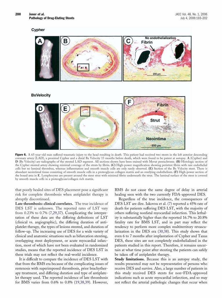
igure 6. A 65-year-old man suffered traumatic injury to the head resultinoronary artery (LAD), a proximal Cypher and a distal Bx Velocity 15 mo
(Bx Velocity) are radiographs of the stented LAD segment. All sectionhe Cypher stented artery showing minimal coverage of the struts by fibrinells but no luminal thrombus, whereas inflammation and smooth musclebundant neointimal tissue consisting of smooth muscle cells in a proteoglhe boxed area in E. Lymphocytes are present around the stent strut withy smooth muscle cells in a proteoglycan/collagen rich matrix.
or BMS varies from 0.6% to 0.8% (19,38,39). However, n
MS do not cause the same degree of delay in arterialealing seen with the two currently FDA-approved DES.Regardless of the true incidence, the consequences ofES LST are dire. Iakovou et al. (7) reported a 45% rate of
eath for patients suffering DES LST, with the majority ofthers suffering nonfatal myocardial infarction. This lethal-ty is substantially higher than the reported 16.7% to 20.8%atality rate for BMS LST, and in part may reflect theendency to perform more complex multiterritory revascu-arization in the DES era (30,38). This study shows thatven 6 to 7 months after implantation of Cypher and TaxusES, these sites are not completely endothelialized in the
atients studied in this report. Therefore, it remains uncer-ain at what time point after stenting the patients can safelye taken off of antiplatelet therapy.tudy limitations. Because this is an autopsy study, theesults presented may not be representative of persons whoeceive DES and survive. Also, a large number of patients inhis study received DES stents for non–FDA-approvedndications such as acute myocardial infarction, and so may
eath. This patient had received two stents in the left anterior descendingbefore death, which were found to be patent at autopsy. A (Cypher) andn have been stained with Movat pentachrome. (B) Histologic section ofHigh-power magnification showing peristrut fibrin with rare endothelialare only rarely observed. (E) Section of the Bx Velocity stent. There is
collagen matrix and an overlying endothelium. (F) High-power section ofal fibrin underneath the strut. The luminal surface of the stent is covered
g in dnths
s show. (C)cells
ycanminim
ot reflect the arterial pathologic changes that occur when
tsimaCsDsficDcfsihfta
ATAl
RPG
R
1
1
1
1
1
1
1
1
1
1
2
2
2
2
2
2
2
2
2
2
3
3
3
3
201JACC Vol. 48, No. 1, 2006 Joner et al.July 4, 2006:193–202 Pathology of Drug-Eluting Stents
hese stents are placed only for approved uses. In the presenttudy, however, a large number of stents were analyzed inndividuals who did and did not suffer stent-associated
orbidity, and it is likely that the results reported here arepplicable to patients receiving DES.onclusions. This is the first published study to examine
ystematically the effects of the two currently FDA-approvedES on human coronary pathology. Both DES caused a
ignificant delay in arterial healing characterized by persistentbrin deposition and delayed re-endothelialization whenompared with sites of BMS implantation. The cause ofES LST is likely multifactorial, with delayed healing in
ombination with other clinical and/or procedural riskactors such as withdrawal of antiplatelet therapy, malappo-ition/incomplete apposition, and bifurcation stenting play-ng an important role. Because the time course of arterialealing after Cypher or Taxus DES placement may varyrom patient to patient, all patients at high risk for latehrombosis should receive dual antiplatelet therapy withspirin and clopidogrel for prolonged periods of time.
cknowledgmenthe authors thank Rosalind Mathew, Lila Adams, Hedwigvallone, and Hazel M. Jenkins (CVPath) for their excel-
ent technical assistance.
eprint requests and correspondence: Dr. Renu Virmani, CV-ath, International Registry of Pathology, 19 Firstfield Road,aithersburg, Maryland 20878. E-mail: [email protected].
EFERENCES
1. Moussa I, Di Mario C, Reimers B, Akiyama T, Tobis J, Colombo A.Subacute stent thrombosis in the era of intravascular ultrasound-guided coronary stenting without anticoagulation: frequency, predic-tors and clinical outcome. J Am Coll Cardiol 1997;29:6–12.
2. Schomig A, Neumann FJ, Kastrati A, et al. A randomized comparisonof antiplatelet and anticoagulant therapy after the placement ofcoronary-artery stents. N Engl J Med 1996;334:1084–9.
3. Ong AT, Hoye A, Aoki J, et al. Thirty-day incidence and six-monthclinical outcome of thrombotic stent occlusion after bare-metal,sirolimus, or paclitaxel stent implantation. J Am Coll Cardiol 2005;45:947–53.
4. Orford JL, Lennon R, Melby S, et al. Frequency and correlates ofcoronary stent thrombosis in the modern era: analysis of a single centerregistry. J Am Coll Cardiol 2002;40:1567–72.
5. Stone GW, Ellis SG, Cox DA, et al. A polymer-based, paclitaxel-eluting stent in patients with coronary artery disease. N Engl J Med2004;350:221–31.
6. Moses JW, Leon MB, Popma JJ, et al. Sirolimus-eluting stents versusstandard stents in patients with stenosis in a native coronary artery.N Engl J Med 2003;349:1315–23.
7. Iakovou I, Schmidt T, Bonizzoni E, et al. Incidence, predictors, andoutcome of thrombosis after successful implantation of drug-elutingstents. JAMA 2005;293:2126–30.
8. McFadden EP, Stabile E, Regar E, et al. Late thrombosis indrug-eluting coronary stents after discontinuation of antiplatelet ther-apy. Lancet 2004;364:1519–21.
9. Suzuki T, Kopia G, Hayashi S, et al. Stent-based delivery of sirolimusreduces neointimal formation in a porcine coronary model. Circulation2001;104:1188–93.
0. Klugherz BD, Llanos G, Lieuallen W, et al. Twenty-eight-day efficacy
and phamacokinetics of the sirolimus-eluting stent. Coron Artery Dis2002;13:183–8.1. Drachman DE, Edelman ER, Seifert P, et al. Neointimal thickeningafter stent delivery of paclitaxel: change in composition and arrest ofgrowth over six months. J Am Coll Cardiol 2000;36:2325–32.
2. Farb A, Heller PF, Shroff S, et al. Pathological analysis of localdelivery of paclitaxel via a polymer-coated stent. Circulation 2001;104:473–9.
3. Finn AV, Kolodgie FD, Harnek J, et al. Differential response ofdelayed healing and persistent inflammation at sites of overlappingsirolimus- or paclitaxel-eluting stents. Circulation 2005;112:270–8.
4. Guagliumi G, Farb A, Musumeci G, et al. Images in cardiovascularmedicine. Sirolimus-eluting stent implanted in human coronary arteryfor 16 months: pathological findings. Circulation 2003;107:1340–1.
5. Virmani R, Guagliumi G, Farb A, et al. Localized hypersensitivity andlate coronary thrombosis secondary to a sirolimus-eluting stent: shouldwe be cautious? Circulation 2004;109:701–5.
6. Sousa JE, Costa MA, Farb A, et al. Images in cardiovascular medicine.Vascular healing 4 years after the implantation of sirolimus-elutingstent in humans: a histopathological examination. Circulation 2004;110:e5–6.
7. Farb A, Weber DK, Kolodgie FD, Burke AP, Virmani R. Morpho-logical predictors of restenosis after coronary stenting in humans.Circulation 2002;105:2974–80.
8. Kolodgie FD, John M, Khurana C, et al. Sustained reduction ofin-stent neointimal growth with the use of a novel systemic nanopar-ticle paclitaxel. Circulation 2002;106:1195–8.
9. Farb A, Burke AP, Kolodgie FD, Virmani R. Pathological mecha-nisms of fatal late coronary stent thrombosis in humans. Circulation2003;108:1701–6.
0. Grewe PH, Deneke T, Machraoui A, Barmeyer J, Muller KM. Acuteand chronic tissue response to coronary stent implantation: pathologicfindings in human specimen. J Am Coll Cardiol 2000;35:157–63.
1. Marx SO, Jayaraman T, Go LO, Marks AR. Rapamycin-FKBPinhibits cell cycle regulators of proliferation in vascular smooth musclecells. Circ Res 1995;76:412–7.
2. Poon M, Marx SO, Gallo R, Badimon JJ, Taubman MB, Marks AR.Rapamycin inhibits vascular smooth muscle cell migration. J ClinInvest 1996;98:2277–83.
3. Wiskirchen J, Schober W, Schart N, et al. The effects of paclitaxel onthe three phases of restenosis: smooth muscle cell proliferation,migration, and matrix formation: an in vitro study. Invest Radiol2004;39:565–71.
4. Virmani R, Kolodgie FD, Farb A, Lafont A. Drug eluting stents: arehuman and animal studies comparable? Heart 2003;89:133–8.
5. Levin AD, Vukmirovic N, Hwang CW, Edelman ER. Specificbinding to intracellular proteins determines arterial transport proper-ties for rapamycin and paclitaxel. Proc Natl Acad Sci U S A2004;101:9463–7.
6. Ranade SV, Miller KM, Richard RE, Chan AK, Allen MJ, HelmusMN. Physical characterization of controlled release of paclitaxel fromthe TAXUS Express2 drug-eluting stent. J Biomed Mater Res A2004;71:625–34.
7. Hwang CW, Levin AD, Jonas M, Li PH, Edelman ER. Thrombosismodulates arterial drug distribution for drug-eluting stents. Circula-tion 2005;111:1619–26.
8. Schampaert E, Cohen EA, Schluter M, et al. The Canadian study ofthe sirolimus-eluting stent in the treatment of patients with long denovo lesions in small native coronary arteries (C-SIRIUS). J Am CollCardiol 2004;43:1110–5.
9. Moreno R, Fernandez C, Hernandez R, et al. Drug-eluting stentthrombosis: results from a pooled analysis including 10 randomizedstudies. J Am Coll Cardiol 2005;45:954–9.
0. Cutlip DE, Baim DS, Ho KK, et al. Stent thrombosis in the modernera: a pooled analysis of multicenter coronary stent clinical trials.Circulation 2001;103:1967–71.
1. Cheneau E, Leborgne L, Mintz GS, et al. Predictors of subacute stentthrombosis: results of a systematic intravascular ultrasound study.Circulation 2003;108:43–7.
2. LaDisa JF, Jr., Olson LE, Guler I, et al. Stent design properties anddeployment ratio influence indexes of wall shear stress: a three-dimensional computational fluid dynamics investigation within anormal artery. J Appl Physiol 2004;97:424–30; discussion, 16.
3. Fujii K, Carlier SG, Mintz GS, et al. Stent underexpansion and
residual reference segment stenosis are related to stent thrombosis after
3
3
3
3
3
3
202 Joner et al. JACC Vol. 48, No. 1, 2006Pathology of Drug-Eluting Stents July 4, 2006:193–202
sirolimus-eluting stent implantation: an intravascular ultrasound study.J Am Coll Cardiol 2005;45:995–8.
4. Alfonso F, Suarez A, Angiolillo DJ, et al. Findings of intravascularultrasound during acute stent thrombosis. Heart 2004;90:1455–9.
5. Ge L, Airoldi F, Iakovou I, et al. Clinical and angiographic outcomeafter implantation of drug-eluting stents in bifurcation lesions with thecrush stent technique: importance of final kissing balloon post-dilation. J Am Coll Cardiol 2005;46:613–20.
6. Goodall AH. Role of shear stress and turbulence on platelets in blood
conduits and on endothelial cells in arterial conduits. In: Suigwart U,editor. Endoluminal Stenting. Philadelphia, PA: WB Saunders Co.,1996:52–9.
7. Ong AT, McFadden EP, Regar E, de Jaegere PP, van Domburg RT,Serruys PW. Late angiographic stent thrombosis (LAST) events withdrug-eluting stents. J Am Coll Cardiol 2005;45:2088–92.
8. Heller LI, Shemwell KC, Hug K. Late stent thrombosis in the absenceof prior intracoronary brachytherapy. Catheter Cardiovasc Interv2001;53:23–8.
9. Wang F, Stouffer GA, Waxman S, Uretsky BF. Late coronary stentthrombosis: early vs. late stent thrombosis in the stent era. Catheter
Cardiovasc Interv 2002;55:142–7.![Journal Papers [1-44] - biosensors.com · Polymer-Based Biolimus-Eluting Stents Versus Durable Polymer-Based Sirolimus-Eluting Stents in Patients With Coronary Artery Disease: Final](https://img.dokumen.tips/doc/110x75/5fae34968d5e227c587bb762/journal-papers-1-44-polymer-based-biolimus-eluting-stents-versus-durable-polymer-based.jpg)