Embed Size (px)
Citation preview

1
The Central Nervous System Brain structure and function
Spinal cord and reflex Danil hammoudi.MD
Central Nervous System (CNS) § CNS – composed of the brain and spinal cord § Cephalization
§ Elaboration of the anterior portion of the CNS § Increase in number of neurons in the head § Highest level is reached in the human brain
Part 1: The Brain § Composed of wrinkled, pinkish gray tissue § Surface anatomy includes cerebral hemispheres, cerebellum, and brain stem

2
Here is the vertex of a normal brain. Note the regular pattern of gyri and sulci. A sulcus is the depth between one gyrus and the next. The thin meninges cover the surface, but have been stripped away over part of the central frontoparietal region at the right to reveal more clearly the sulci. The central fissure is where the extension of the dura between the hemispheres, called the falx cerebri, would be.
The meninges have been removed at the vertex of a normal brain to reveal the A Rolandic fissure with the precentral gyrus (motor cortex) and the postcentral gyrus (somesthetic cortex).
Embryonic Development § During the first 26 days of development:
§ Ectoderm thickens forming the neural plate § The neural plate invaginates, forming the neural groove § The neural groove fuses dorsally and forms the neural tube
Primary Brain Vesicles § The anterior end of the neural tube expands and constricts to form the three primary brain vesicles
§ Prosencephalon – the forebrain § Mesencephalon – the midbrain § Rhombencephalon – hindbrain
Neural Tube and Primary Brain Vesicles
Secondary Brain Vesicles § In week 5 of embryonic development, secondary brain vesicles form
§ Telencephalon and diencephalon arise from the forebrain § Mesencephalon remains undivided § Metencephalon and myelencephalon arise from the hindbrain

3
Adult Neural Canal Regions
Space Restriction and Brain Development
Adult Brain Structures § Fates of the secondary brain vesicles:
§ Telencephalon – cerebrum: cortex, white matter, and basal nuclei § Diencephalon – thalamus, hypothalamus, and epithalamus § Mesencephalon – brain stem: midbrain § Metencephalon – brain stem: pons § Myelencephalon – brain stem: medulla oblongata

4
Divisions of the Human Brain: 1 Myelencephalon, which includes the medulla 2 Metencephalon, which includes the pons and cerebellum 3 Mesencephalon, which includes the midbrain (tectum and tegmentum) 4 Diencephalon, which includes the thalamus and hypothalamus 5 Telencephalon, which includes the cerebrum (cerebral cortex, basal ganglia, & medullary body)
§ Adult structures derived from the neural canal § Telencephalon – lateral ventricles § Diencephalon – third ventricle § Mesencephalon – cerebral aqueduct § Metencephalon and myelencephalon – fourth ventricle New Terms:
Brain Division
Telencephalon Diencephalon Mesencephalon Metencephalon Myelencephalon
Medulla
Telencephalon
–Cerebral Cortex
–Limbic system
–Basal Ganglia
Pons:
Cerebellum:
Adult Neural Canal Regions § Spinal Cord
§ Central cavity surrounded by a gray matter core § External to which is white matter composed of myelinated fiber tracts
§ Brain § Similar to spinal cord but with additional areas of gray matter § Cerebellum has gray matter in nuclei § Cerebrum has nuclei and additional gray matter in the cortex

5
Lobes and fissures of the cerebral hemispheres,
(c)
Parietal lobe
Frontal lobe
Right Cerebral hemisphere Occipital lobe
Left cerebral hemisphere
Cerebral veins and arteries covered by arachnoid
Longitudinal fissure
Posterior
Anterior
Protection of the Brain
§ The brain is protected by bone, meninges, and cerebrospinal fluid § Harmful substances are shielded from the brain by the bloodbrain barrier
Meninges § Three connective tissue membranes lie external to the CNS – dura mater, arachnoid mater, and pia mater § Functions of the meninges
§ Cover and protect the CNS § Protect blood vessels and enclose venous sinuses § Contain cerebrospinal fluid (CSF) § Form partitions within the skull

6
Dura Mater § Leathery, strong meninx composed of two fibrous connective tissue layers § The two layers separate in certain areas and form dural sinuses § Three dural septa extend inward and limit excessive movement of the brain
§ Falx cerebri – fold that dips into the longitudinal fissure § Falx cerebelli – runs along the vermis of the cerebellum § Tentorium cerebelli – horizontal dural fold extends into the transverse fissure

7
Arachnoid Mater § The middle meninx, which forms a loose brain covering § It is separated from the dura mater by the subdural space § Beneath the arachnoid is a wide subarachnoid space filled with CSF and large blood vessels § Arachnoid villi protrude superiorly and permit CSF to be absorbed into venous blood
Pia Mater § Deep meninx composed of delicate connective tissue that clings tightly to the brain
Meninges
Dura Mater
8
(b)
Occipital lobe
Superior sagittal sinus
Dura mater
Tranverse sinus
Temporal bone
Scalp
Skull
Tentorium cerebelli
Cerebellum
Arachnoid mater over medulla oblongata
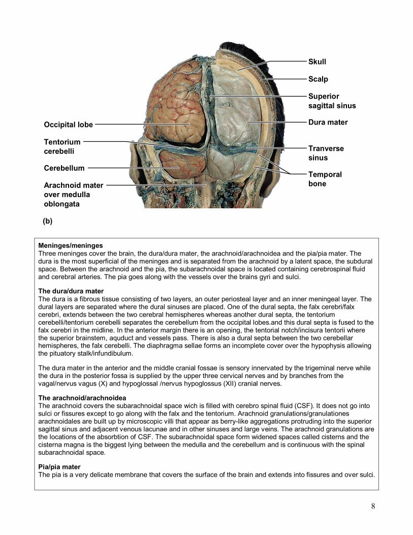
Meninges/meninges Three meninges cover the brain, the dura/dura mater, the arachnoid/arachnoidea and the pia/pia mater. The dura is the most superficial of the meninges and is separated from the arachnoid by a latent space, the subdural space. Between the arachnoid and the pia, the subarachnoidal space is located containing cerebrospinal fluid and cerebral arteries. The pia goes along with the vessels over the brains gyri and sulci.
The dura/dura mater The dura is a fibrous tissue consisting of two layers, an outer periosteal layer and an inner meningeal layer. The dural layers are separated where the dural sinuses are placed. One of the dural septa, the falx cerebri/falx cerebri, extends between the two cerebral hemispheres whereas another dural septa, the tentorium cerebelli/tentorium cerebelli separates the cerebellum from the occipital lobes.and this dural septa is fused to the falx cerebri in the midline. In the anterior margin there is an opening, the tentorial notch/incisura tentorii where the superior brainstem, aquduct and vessels pass. There is also a dural septa between the two cerebellar hemispheres, the falx cerebelli. The diaphragma sellae forms an incomplete cover over the hypophysis allowing the pituatory stalk/infundibulum.
The dura mater in the anterior and the middle cranial fossae is sensory innervated by the trigeminal nerve while the dura in the posterior fossa is supplied by the upper three cervical nerves and by branches from the vagal/nervus vagus (X) and hypoglossal /nervus hypoglossus (XII) cranial nerves.
The arachnoid/arachnoidea The arachnoid covers the subarachnoidal space wich is filled with cerebro spinal fluid (CSF). It does not go into sulci or fissures except to go along with the falx and the tentorium. Arachnoid granulations/granulationes arachnoidales are built up by microscopic villi that appear as berrylike aggregations protruding into the superior sagittal sinus and adjacent venous lacunae and in other sinuses and large veins. The arachnoid granulations are the locations of the absorbtion of CSF. The subarachnoidal space form widened spaces called cisterns and the cisterna magna is the biggest lying between the medulla and the cerebellum and is continuous with the spinal subarachnoidal space.
Pia/pia mater The pia is a very delicate membrane that covers the surface of the brain and extends into fissures and over sulci.

9
Cerebrospinal Fluid (CSF) § Watery solution similar in composition to blood plasma § Contains less protein and different ion concentrations than plasma § Forms a liquid cushion that gives buoyancy to the CNS organs § Prevents the brain from crushing under its own weight § Protects the CNS from blows and other trauma § Nourishes the brain and carries chemical signals throughout it
Circulation of CSF
Choroid Plexuses § Clusters of capillaries that form
tissue fluid filters, which hang from the roof of each ventricle § Have ion pumps that allow them to alter ion concentrations of the CSF § Help cleanse CSF by removing wastes

10
Choroid Plexuses

11
Circulation of CSF
Cerebrospinal fluid (CSF), Liquor cerebrospinalis, is a clear bodily fluid that occupies the subarachnoid space in the brain (the space between the skull and the cerebral cortex—more specifically, between the arachnoid and pia layers of the meninges). It is a very pure saline solution with microglia and acts as a "cushion" or buffer for the cortex.
Cerebrospinal fluid also occupies the ventricular system of the brain and the spinal cord. It is a prime example of the separation of brain function from the rest of the body, as all CSF is generated locally in the brain. It is produced by the choroid plexus which is formed by specialized ependymal cells. The choroid plexus enter the lateral ventricles through the choroid fissure, along the line of the fimbria/fornix, and the third and fourth ventricle through their roofs. The CSF formed by the choroid plexuses in the ventricles, circulates through the interventricular foramina (foramen of Monro) into the third ventricle and then via the mesencephalic duct (cerebral aqueduct) into the fourth ventricle, whence it exits through two lateral apertures (foramina of Luschka) and one median aperture (foramen of Magendie). It then flows through the cerebromedullary cistern down the spinal cord and over the cerebral hemispheres. It is then allowed to flow into the venous system by the arachnoid granulations.
The cerebrospinal fluid is produced by the ventricles (mostly the lateral ventricles) at a rate of 500 ml/day. Since the volume that may be contained by the brain is of 150 ml, it is frequently replaced (34 times per day turnover), exceeding amounts getting into the blood. This continuous flow through the ventricular system into the subarachnoid space and finally exiting into the venous system provides somewhat of a "sink" that reduces the concentration of larger, lipoinsoluble molecules penetrating into the brain and CSF.
The CSF contains approximately 0.3% plasma proteins, also being 15 to 40 mg/dL, depending on sampling site.

12
The cerebrospinal fluid has many putative roles including mechanical protection of the brain, distribution of neuroendocrine factors, and facilitation of pulsatile cerebral blood flow. Understanding cardiovascular dynamics is valuable as the flow pattern of arterial blood must be tightly regulated within the brain in order to ensure consistent brain oxygenation. CSF movement allows arterial expansion and contraction by acting like a spring, which prevents wide changes in intracranial blood flow. When disorders of CSF flow occur, they may therefore impact not only CSF movement, but also the intracranial blood flow, with subsequent neuronal and glial vulnerabilities. The venous system is also important in this equation. Infants and patients shunted as small children may have particularly unexpected relationships between pressure and ventricular size, possibly due in part to venous pressure dynamics. This may have significant treatment implications but the underlying pathophysiology needs to be further explored.
CSF connections with the lymphatic system have been demonstrated in several mammalian systems. Preliminary data suggest that these CSFlymph connections form around the time that the CSF secretory capacity of the choroid plexus is developing (in utero). There may be some relationship between CSF disorders, including hydrocephalus and impaired CSF lymphatic transport.
Cerebrospinal fluid can be tested for the diagnosis of a variety of neurological diseases. It is usually obtained by a procedure called lumbar puncture in an attempt to count the cells in the fluid and to detect the levels of protein and glucose. These parameters alone may be extremely beneficial in the diagnosis of subarachnoid hemorrhage and central nervous system infections (such as meningitis). Moreover, a cerebrospinal fluid culture examination may yield the microorganism that has caused the infection. By using more sophisticated methods, such as the detection of the oligoclonal bands, an ongoing inflammatory condition (for example, multiple sclerosis) can be recognized. A beta2 transferrin assay is highly specific and sensitive for the detection for e.g. cerebrospinal fluid leakage.
Lumbar puncture can also be performed to measure the intracranial pressure, which might be increased in certain types of hydrocephalus.
This fluid has an importance in anethesology. Baricity refers to the density of a substance compared to the density of human cerebral spinal fluid. Baricity is used in anesthesia to determine the manner in which a particular drug will spread in the intrathecal space.
BloodBrain Barrier § Protective mechanism that helps maintain a stable environment for the brain § Bloodborne substances are separated from neurons by:
§ Continuous endothelium of capillary walls § Relatively thick basal lamina § Bulbous feet of astrocytes
BloodBrain Barrier: Functions § Selective barrier that allows nutrients to pass freely § Is ineffective against substances that can diffuse through plasma membranes § Absent in some areas (vomiting center and the hypothalamus), allowing these areas to monitor the chemical composition of the blood
§ Stress increases the ability of chemicals to pass through the bloodbrain barrier Brain and spinal cord histology:

13
The cortical surface is composed of six layers of fibers and cell bodies, alternating between layers that receive information from other neural areas and send information to other neural areas and the spinal cord. You can also see at least one gyrus and sulcus. Notice how the axons from the cell bodies congregate at the bottom of the layers to form the white matter.

14
The standard areas of cortex (isocortex) is characterized as having six distinct layers. From outside inward:
1.Molecular layer 2.External granular layer 3.External pyramidal layer 4.Internal granular layer 5.Internal pyramidal layer 6.Multiform layer.
15
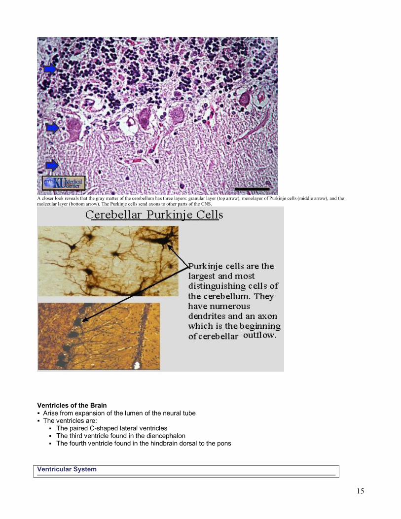
A closer look reveals that the gray matter of the cerebellum has three layers: granular layer (top arrow), monolayer of Purkinje cells (middle arrow), and the molecular layer (bottom arrow). The Purkinje cells send axons to other parts of the CNS.
Ventricles of the Brain § Arise from expansion of the lumen of the neural tube § The ventricles are:
§ The paired Cshaped lateral ventricles § The third ventricle found in the diencephalon § The fourth ventricle found in the hindbrain dorsal to the pons
Ventricular System
16
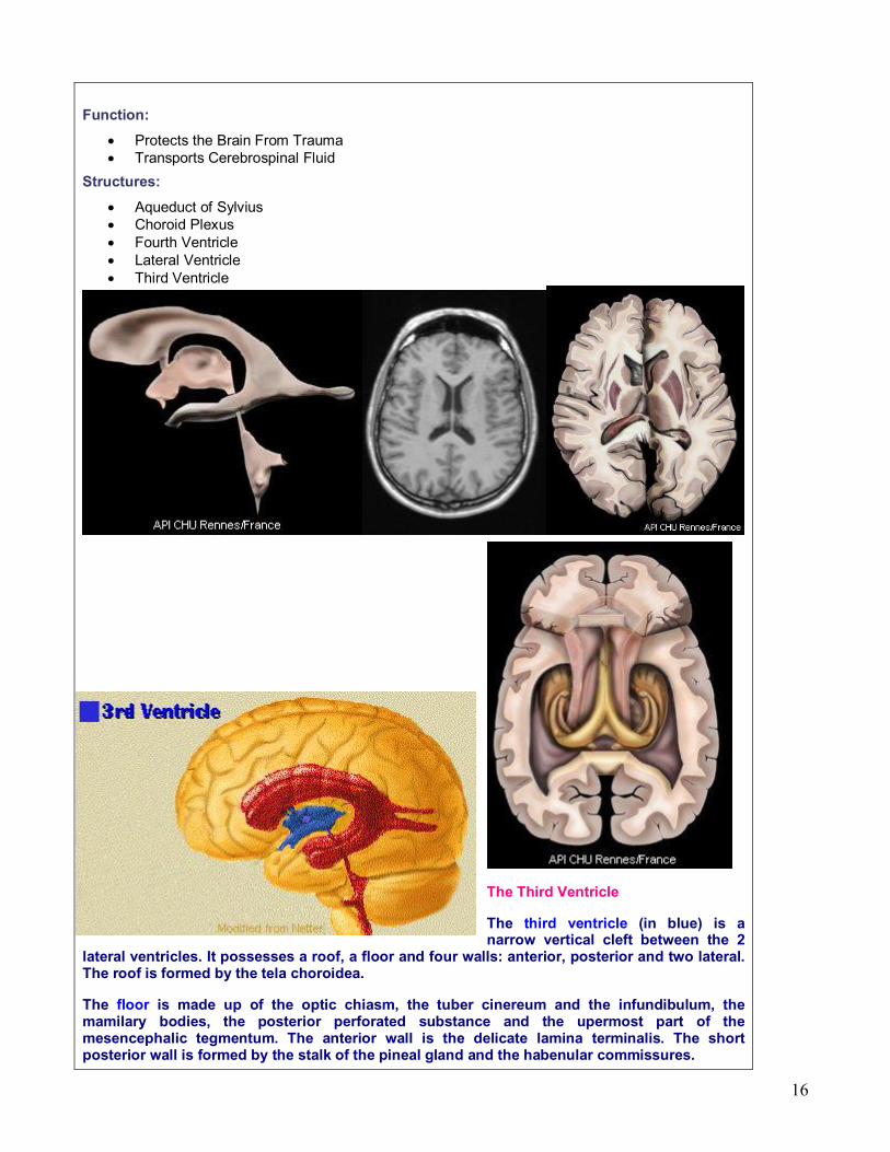
Function:
• Protects the Brain From Trauma • Transports Cerebrospinal Fluid
Structures:
• Aqueduct of Sylvius • Choroid Plexus • Fourth Ventricle • Lateral Ventricle • Third Ventricle
The Third Ventricle
The third ventricle (in blue) is a narrow vertical cleft between the 2
lateral ventricles. It possesses a roof, a floor and four walls: anterior, posterior and two lateral. The roof is formed by the tela choroidea.
The floor is made up of the optic chiasm, the tuber cinereum and the infundibulum, the mamilary bodies, the posterior perforated substance and the upermost part of the mesencephalic tegmentum. The anterior wall is the delicate lamina terminalis. The short posterior wall is formed by the stalk of the pineal gland and the habenular commissures.
17
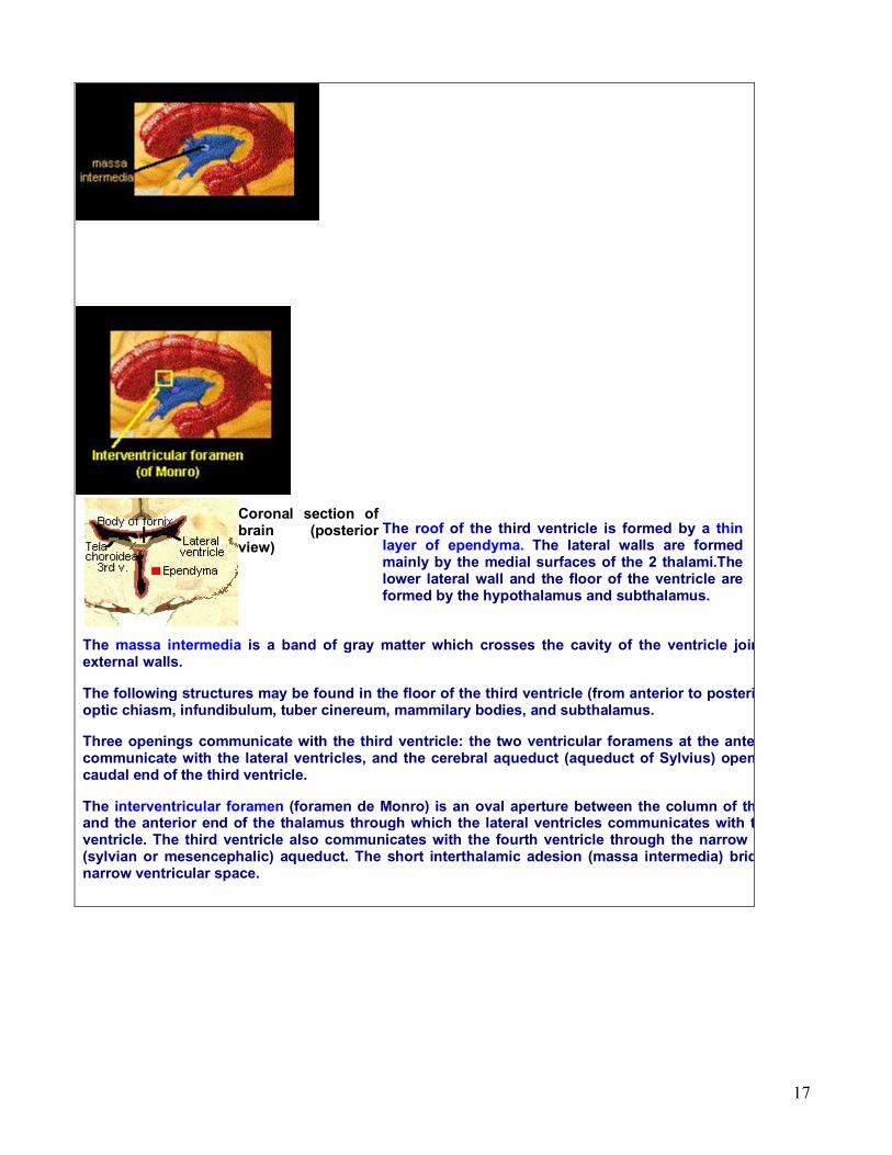
Coronal section of brain (posterior view)
The roof of the third ventricle is formed by a thin layer of ependyma. The lateral walls are formed mainly by the medial surfaces of the 2 thalami.The lower lateral wall and the floor of the ventricle are formed by the hypothalamus and subthalamus.
The massa intermedia is a band of gray matter which crosses the cavity of the ventricle joining the external walls.
The following structures may be found in the floor of the third ventricle (from anterior to posterior end): optic chiasm, infundibulum, tuber cinereum, mammilary bodies, and subthalamus.
Three openings communicate with the third ventricle: the two ventricular foramens at the anterior end communicate with the lateral ventricles, and the cerebral aqueduct (aqueduct of Sylvius) opens in the caudal end of the third ventricle.
The interventricular foramen (foramen de Monro) is an oval aperture between the column of the fornix and the anterior end of the thalamus through which the lateral ventricles communicates with the third ventricle. The third ventricle also communicates with the fourth ventricle through the narrow cerebral (sylvian or mesencephalic) aqueduct. The short interthalamic adesion (massa intermedia) bridges the narrow ventricular space.

18
The Fourth Ventricle
The fourth venticle is a cavitie which lies posterior to the pons and upper half of the medulla oblongata and anterior to the cerebellum. It is continuous with the cerebral aqueduct (mesencephalic or Sylvius) above and the central canal of the spinal cord in the lower half of the medulla. On each size, a narrow prolongation, the lateral recess, projects around the brainstem; its lateral aperture
(forame of Luschka) lies below the cerebellar flocculus.
The fourth ventricle has lateral boundaries, a roof and a floor. The lateral boundaries are formed on each side by the superior cerebellar peduncle, the inferior cerebellar peduncle and the cuneate and gracile tubercles.
Roof of the fourth ventricle Formed by thin laminae of white matter. The lower has a median aperture (foramen of Magendie); cerebrospinal fluid escapes through this opening and lateral apertures into the subarachnoid space. Because these are the only communications between the ventricular and subarachnoid spaces, their
blockage can produce one type of hydrocephalus.
The floor of the fourth ventricle, also known as rhomboid fossa, is formed by the dorsal surfaces of the pons and medulla oblongata.
The cerebral aqueduct is a narrow canal in the midline connecting the third and fourth ventricle. It is 1.5 cm long and 12 mm, in diameter. Its floor is formed by the tegmentum of the midbrain. Its roof consists of the quadrigeminal body of the midbrain and posterior comissure.
The tela choroidea is a layer of pia mater of great vascularity which invaginates close to the median plane into the cavity of the fourth ventricle to form the choroid plexus of the fourth ventricle. Anotomic findings indicate that the average normal ventricular system has a capacity of less than 16 ml.
19
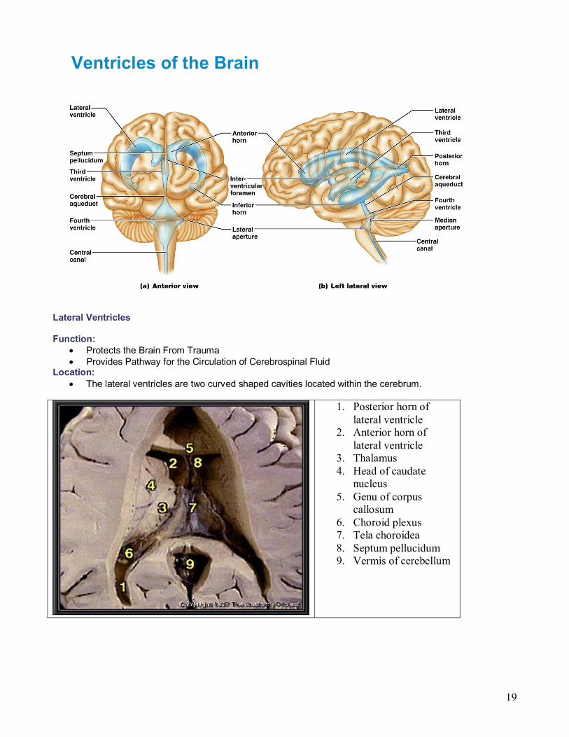
Ventricles of the Brain
Lateral Ventricles
Function: • Protects the Brain From Trauma • Provides Pathway for the Circulation of Cerebrospinal Fluid
Location: • The lateral ventricles are two curved shaped cavities located within the cerebrum.
1. Posterior horn of lateral ventricle
2. Anterior horn of lateral ventricle
3. Thalamus 4. Head of caudate
nucleus 5. Genu of corpus
callosum 6. Choroid plexus 7. Tela choroidea 8. Septum pellucidum 9. Vermis of cerebellum

20
1. Anterior horn of lateral ventricle
2. Posterior horn of lateral ventricle
3. Inferior horn of lateral ventricle
4. Choroid plexus 5. Septum pellucidum 6. Genu of corpus
callosum 7. Temporal lobe 8. Vermis of cerebellum 9. Splenium of corpus
callosum
PARTS OF THE BRAIN AND THEIR FUNCTIONS
Hindbrain Medulla Sensory and motor nerves crossover
Pons Regulation of sleepwake cycle
Cerebellum Reflexes (e.g., balance) Coordinates movement
Midbrain Hearing, vision relay point Pain registered
Forebrain Thalamus Major message relay center Regulates higher brain centers and peripheral nervous system
Hypothalamus Motivation Emotion Stress reactions
Cerebral Hemispheres
Occipital lobe Receives and processes visual information
Temporal lobe Complex vision Smell Hearing Balance and equilibrium Emotions and motivations Some language comprehension
21
Parietal lobe Sensory projection and association areas Visual/spatial abilities
Frontal lobe Goaldirected behavior, concentration Emotional control and temperament Motor projection and association areas Coordinate messages from other lobes
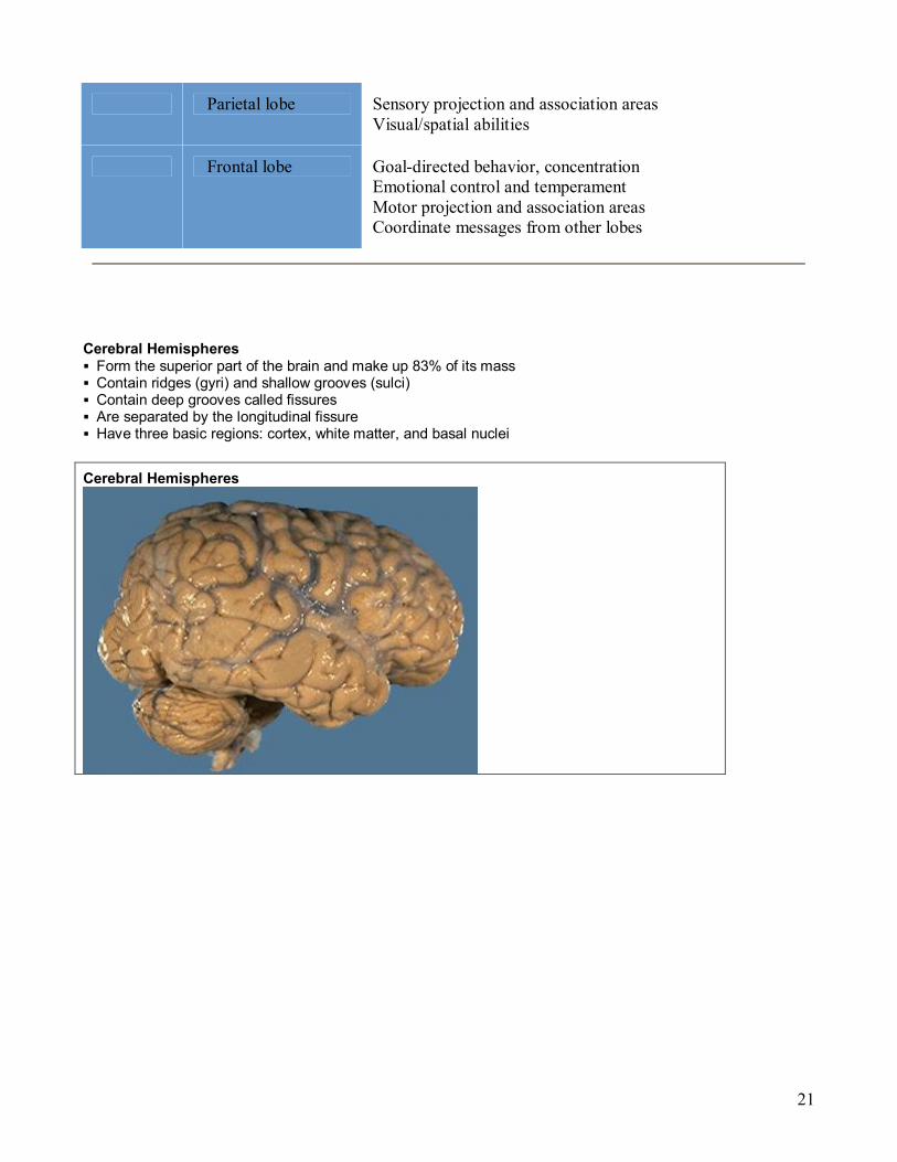
Cerebral Hemispheres § Form the superior part of the brain and make up 83% of its mass § Contain ridges (gyri) and shallow grooves (sulci) § Contain deep grooves called fissures § Are separated by the longitudinal fissure § Have three basic regions: cortex, white matter, and basal nuclei
Cerebral Hemispheres

22
• The folds and convolutions of the surface of the cerebral hemispheres vastly increase the effective surface area of the brain. Each ridge is called a gyrus and each groove between ridges is called a sulcus. A deep sulcus is referred to as a fissure.
Gyri:
• Precentral gyrus/gyrus precentralis: the convolution of the frontal lobe that borders posteriorly to the central sulcus and contains the primary motor area.
• Postcentral gyrus/gyrus postcentralis: the convolution of the parietal lobe that borders anteriorly to the central sulcus and contains the primary sensory area.
• Superior temporal gyrus/gyrus temporalis superior: the convolution of the superior temporal lobe containing the auditory cortex and language association cortex.
• Angular gyrus/gyrus angularis: the convolution of the inferior parietal lobe and participates in language reception, spatial orientation and semantic representation.
• Supramarginal gyrus/gyrus supramarginalis: the convolution of the inferior parietal lobe that surrounds the posterior end of the lateral fissure. This region is involved in spatial orientation and semantic representation.
Sulci:
• Central sulcus/sulcus centralis: a prominent sulcus on the dorsolateral part of the cerebral hemispheres formed by the precentral and postcentral gyri. The central sulcus defines the boundary between the frontal and parietal lobes.
• Precentral sulcus/sulcus precentralis: vertically oriented sulcus at the anterior margin of the precentral gyrus in the posteriuor part of the frontal lobe.
• Lateral fissure/fissura lateralis: fissure on the lateral surface of the cerebral hemisphere. The lateral fissure divides the posterior frontal and anterior parietal lobes from the superior temporal lobe. Can also be called the fissure of Sylvius.
23
• Parietooccipital sulcus/sulcus parietooccipitalis: vertically oriented sulcus on the medial aspect of the hemisphere. This sulcus divides the parietal and occipital lobes.

24

25
Major Lobes, Gyri, and Sulci of the Cerebral Hemisphere § Deep sulci divide the hemispheres into five lobes:
§ Frontal, parietal, temporal, occipital, and insula § Central sulcus – separates the frontal and parietal lobes
Brain Lobes
Major Lobes, Gyri, and Sulci of the Cerebral Hemisphere § Parietooccipital sulcus – separates the parietal and occipital lobes

26
§ Lateral sulcus – separates the parietal and temporal lobes § The precentral and postcentral gyri border the central sulcus
•4 Lobes –Frontal Lobe –Parietal Lobe –Occipital Lobe –Temporal Lobe
•Major Fissures –Central Sulcus –Longitudinal Fissure –Sylvian Fissure
Major Structures of the Cortex
•The lobes are distinguished both structurally and functionally
Cerebral Cortex § The cortex – superficial gray matter; accounts for 40% of the mass of the brain § It enables sensation, communication, memory, understanding, and voluntary movements § Each hemisphere acts contralaterally (controls the opposite side of the body) § Hemispheres are not equal in function § No functional area acts alone; conscious behavior involves the entire cortex
Functional Areas of the Cerebral Cortex § The three types of functional areas are:
27
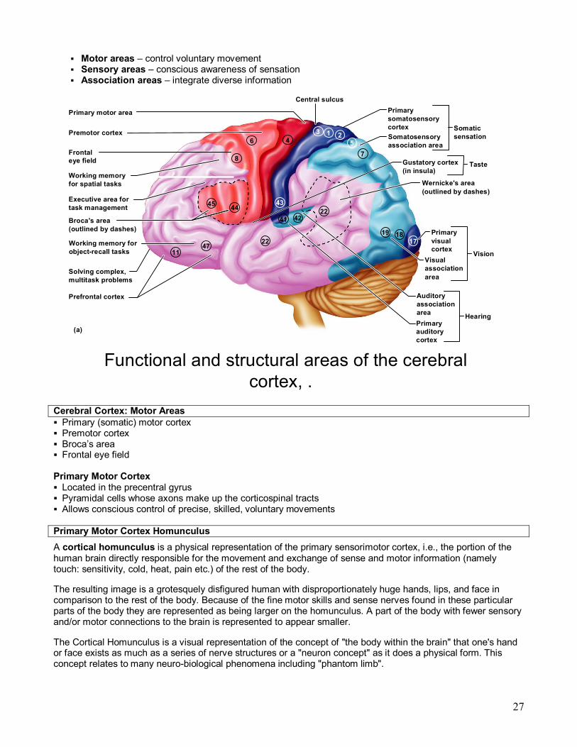
§ Motor areas – control voluntary movement § Sensory areas – conscious awareness of sensation § Association areas – integrate diverse information
Functional and structural areas of the cerebral cortex, .
(a)
Primary motor area
Premotor cortex
Frontal eye field
Working memory for spatial tasks
Executive area for task management
Working memory for objectrecall tasks
Broca's area (outlined by dashes)
Solving complex, multitask problems
Prefrontal cortex
Central sulcus Primary somatosensory cortex Somatosensory association area
Somatic sensation
Gustatory cortex (in insula)
Taste
Wernicke's area (outlined by dashes)
Primary visual cortex
Visual association area
Vision
Auditory association area Primary auditory cortex
Hearing
11 47
45 44
8
6 4 3 1 2
5
43
7
19 18 17
22 42 41
22
Cerebral Cortex: Motor Areas § Primary (somatic) motor cortex § Premotor cortex § Broca’s area § Frontal eye field
Primary Motor Cortex § Located in the precentral gyrus § Pyramidal cells whose axons make up the corticospinal tracts § Allows conscious control of precise, skilled, voluntary movements
Primary Motor Cortex Homunculus A cortical homunculus is a physical representation of the primary sensorimotor cortex, i.e., the portion of the human brain directly responsible for the movement and exchange of sense and motor information (namely touch: sensitivity, cold, heat, pain etc.) of the rest of the body.
The resulting image is a grotesquely disfigured human with disproportionately huge hands, lips, and face in comparison to the rest of the body. Because of the fine motor skills and sense nerves found in these particular parts of the body they are represented as being larger on the homunculus. A part of the body with fewer sensory and/or motor connections to the brain is represented to appear smaller.
The Cortical Homunculus is a visual representation of the concept of "the body within the brain" that one's hand or face exists as much as a series of nerve structures or a "neuron concept" as it does a physical form. This concept relates to many neurobiological phenomena including "phantom limb".

28
Premotor Cortex
§ Located anterior to the precentral gyrus § Controls learned, repetitious, or patterned motor skills § Coordinates simultaneous or sequential actions

29
§ Involved in the planning of movements
Broca’s Area § Broca’s area
§ Located anterior to the inferior region of the premotor area § Present in one hemisphere (usually the left) § A motor speech area that directs muscles of the tongue § Is active as one prepares to speak
Frontal Eye Field § Frontal eye field
§ Located anterior to the premotor cortex and superior to Broca’s area § Controls voluntary eye movement
Sensory Areas § Primary somatosensory cortex § Somatosensory association cortex § Visual and auditory areas § Olfactory, gustatory, and vestibular cortices
•4 Lobes –Frontal Lobe –Parietal Lobe –Occipital Lobe –Temporal Lobe
•Major Fissures –Central Sulcus –Longitudinal Fissure –Sylvian Fissure
Major Structures of the Cortex
•The lobes are distinguished both structurally and functionally
PrImary Somatosensory Cortex § Located in the postcentral gyrus, this area:
§ Receives information from the skin and skeletal muscles § Exhibits spatial discrimination
Primary Somatosensory Cortex Homunculus
Somatosensory Association Cortex § Located posterior to the primary somatosensory cortex § Integrates sensory information § Forms comprehensive understanding of the stimulus § Determines size, texture, and relationship of parts
Visual Areas § Primary visual (striate) cortex
§ Seen on the extreme posterior tip of the occipital lobe
30
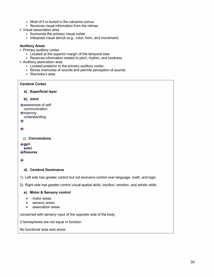
§ Most of it is buried in the calcarine sulcus § Receives visual information from the retinas
§ Visual association area § Surrounds the primary visual cortex § Interprets visual stimuli (e.g., color, form, and movement)
Auditory Areas § Primary auditory cortex
§ Located at the superior margin of the temporal lobe § Receives information related to pitch, rhythm, and loudness
§ Auditory association area § Located posterior to the primary auditory cortex § Stores memories of sounds and permits perception of sounds § Wernicke’s area
Cerebral Cortex
a). Superficial layer
b). mind awareness of self communication memory understanding
c). Convolutions gyri sulci fissures
d). Cerebral Dominance
1). Left side has greater control but not exclusive control over language, math, and logic.
2). Right side has greater control visualspatial skills, intuition, emotion, and artistic skills.
e). Motor & Sensory control • motor areas • sensory areas • association areas
concerned with sensory input of the opposite side of the body
2 hemispheres are not equal in function
No functional area acts alone.

31
i). Functional Area: Motor areas:
Found: posterior part of the Frontal Lobe

32
Include voluntary movements, repetitious movements, motor speech and voluntary eye movements. Operates left to right: .
ii). Sensory Areas:
Found in the parietal, temporal, & occipital lobes.
Conscious awareness of sensation.
Sensory areas:
1). Spatial discrimination: Parietal lobe—
skeletal muscles and skin and identifies region being stimulated.
2). Somatosensory association: Parietal lobe object being felt to produce an understanding.
3). Visuals Areas: Occipital Lobe—
Information from the eyes. Information from the right eye is mapped in the left visual cortex and vice versa. Also interprets visual stimuli using past visual experience
4). Auditory Areas: Temporal Lobe Information on pitch, rhythm, and loudness in interpreted and perceived as sound. Memories of sound are stored for reference. 5). Olfactory Cortex: Frontal Lobe & Temporal olfactory signals and perceives smells.
33
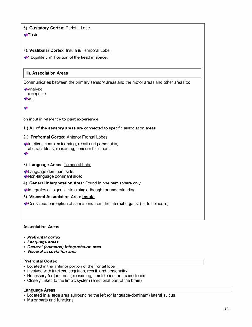
6). Gustatory Cortex: Parietal Lobe Taste
7). Vestibular Cortex: Insula & Temporal Lobe " Equilibrium" Position of the head in space.
iii). Association Areas
Communicates between the primary sensory areas and the motor areas and other areas to: analyze recognize act
on input in reference to past experience.
1.) All of the sensory areas are connected to specific association areas
2.). Prefrontal Cortex: Anterior Frontal Lobes Intellect, complex learning, recall and personality, abstract ideas, reasoning, concern for others
3). Language Areas: Temporal Lobe Language dominant side: Nonlanguage dominant side:
4). General Interpretation Area: Found in one hemisphere only integrates all signals into a single thought or understanding.
5). Visceral Association Area: Insula Conscious perception of sensations from the internal organs. (ie. full bladder)
Association Areas
§ Prefrontal cortex § Language areas § General (common) interpretation area § Visceral association area
Prefrontal Cortex § Located in the anterior portion of the frontal lobe § Involved with intellect, cognition, recall, and personality § Necessary for judgment, reasoning, persistence, and conscience § Closely linked to the limbic system (emotional part of the brain)
Language Areas § Located in a large area surrounding the left (or languagedominant) lateral sulcus § Major parts and functions:
34
§ Wernicke’s area –sounding out unfamiliar words § Broca’s area – speech preparation and production § Lateral prefrontal cortex – language comprehension and word analysis § Lateral and ventral temporal lobe – coordinate auditory and visual aspects of language

35
General (Common) Interpretation Area § Illdefined region including parts of the temporal, parietal, and occipital lobes § Found in one hemisphere, usually the left § Integrates incoming signals into a single thought § Involved in processing spatial relationships
Visceral Association Area § Located in the cortex of the insula § Involved in conscious perception of visceral sensations
Lateralization of Cortical Function § Lateralization – each hemisphere has abilities not shared with its partner § Cerebral dominance – designates the hemisphere dominant for language § Left hemisphere – controls language, math, and logic § Right hemisphere – controls visualspatial skills, emotion, and artistic skills
Cerebral White Matter § Consists of deep myelinated fibers and their tracts § It is responsible for communication between:
§ The cerebral cortex and lower CNS center, and areas of the cerebrum Types include:
§ Commissures – connect corresponding gray areas of the two hemispheres § Association fibers – connect different parts of the same hemisphere § Projection fibers – enter the hemispheres from lower brain or cord centers
Basal Nuclei
36
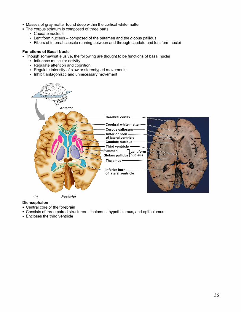
§ Masses of gray matter found deep within the cortical white matter § The corpus striatum is composed of three parts
§ Caudate nucleus § Lentiform nucleus – composed of the putamen and the globus pallidus § Fibers of internal capsule running between and through caudate and lentiform nuclei
Functions of Basal Nuclei § Though somewhat elusive, the following are thought to be functions of basal nuclei
§ Influence muscular activity § Regulate attention and cognition § Regulate intensity of slow or stereotyped movements § Inhibit antagonistic and unnecessary movement
(b)
Corpus callosum Anterior horn of lateral ventricle Caudate nucleus Third ventricle
Putamen Lentiform nucleus Globus pallidus
Thalamus
Cerebral cortex
Cerebral white matter
Anterior
Posterior
Inferior horn of lateral ventricle
Diencephalon § Central core of the forebrain § Consists of three paired structures – thalamus, hypothalamus, and epithalamus § Encloses the third ventricle

37
The diencephalons is centrally located and is nearly surrounded by the cerebral hemispheres
Thalamus § Paired, eggshaped masses that form the superolateral walls of the third ventricle § Connected at the midline by the intermediate mass § Contains four groups of nuclei – anterior, ventral, dorsal, and posterior § Nuclei project and receive fibers from the cerebral cortex
Thalamic Function § Sensual afferent impulses converge and synapse in the thalamus § Impulses of similar function are sorted out, edited, and relayed as a group § All inputs ascending to the cerebral cortex pass through the thalamus § Mediates sensation, motor activities, cortical arousal, learning, and memory

38
Selected structures of the diencephalon, p. 445.
(a) (b)
Dorsal nuclei
Ventral nuclei
Medial
Anterior nuclear group
Reticular nucleus
Ventral anterior
Ventral lateral
Ventral posterior lateral
Lateral geniculate body
Medial geniculate body
Pulvinar
Lateral dorsal
Lateral posterior
Preoptic nucleus
Supraoptic nucleus Suprachiasmatic nucleus
Anterior hypothalamic nucleus
Dorsomedial nucleus
Paraventricular nucleus
Fornix Anterior commissure
Posterior hypothalamic nucleus Lateral hypothalamic area Ventromedial nucleus
Optic chiasma Infundibulum (stalk of the pituitary gland)
Pituitary gland
Arcuate nucleus
Mammillary body
Thalamus Two lobes that relay sensory projection fiber info to the cerebral cortex
Hypothalamus
Lies at the base of the brain
Controls and regulates the endocrine system (hormones), autonomic system, species survival (the four Fs) and sleeping.
Contains many nuclei and fiber tracts
• The thalamus comprises a system of lamellae (made up of myelinated fibers) separating different thalamic subparts.
• Other areas are defined by distinct clusters of neurons, such as the periventricular gray, the intralaminar elements, the "nucleus limitans", and others.
• These latter structures, different in structure from the major part of the thalamus, have been grouped together into the allothalamus as opposed to the isothalamus
• This distinction simplifies the global description of the thalamus.
• The thalamus is known to have multiple functions. Deduced from the design of the isothalamus, it is generally believed to act as a translator for which various "prethalamic" inputs are processed into a form readable by the cortex. The thalamus is believed to relay information selectively to various parts of the cortex, as one thalamic point may reach one or several regions in the cortex.
• The thalamus also plays an important role in regulating states of sleep and wakefulness.
• Thalamic nuclei have strong reciprocal connections with the cerebral cortex, forming thalamocortico thalamic circuits that are believed to be involved with consciousness.
• The thalamus plays a major role in regulating arousal, the level of awareness and activity.
• An animal with a severely damaged or severed thalamus suffers permanent coma.
• Many different functions are linked to the system to which thalamic parts belong.

39
• This is at first the case for sensory systems (which excepts the olfactory function) auditory, somatic, visceral, gustatory and visual systems where localised lesions provoke particular sensory deficits.
• A major role of the thalamus is devoted to "motor" systems.
• This has been and continues to be a subject of interest for investigators. VIm, the relay of cerebellar afferences, is the target of stereotactians particularly for the improvement of tremor.
• The role of the thalamus in the more anterior pallidal and nigral territories in the basal ganglia system disturbances is recognized but still poorly known.
• The contribution of the thalamus to vestibular or to tectal functions is almost ignored.
• The thalamus has been thought of as a "relay" that simply forwards signals to the cerebral cortex. Newer research suggests that thalamic function is more complicated
Cerebrovascular accidents (strokes) can cause thalamic syndrome which results in a contralateral hemianaesthesia, burning or aching sensation on one half of a body (painful anaesthesia), often accompanied by mood swings. Ischaemia of the territory of the paramedian artery, if bilateral, causes serious troubles including akinetic mutism accompanied or not by oculomotor troubles.
Korsakoff's Syndrome, stems from mammillary bodies, mammilothalamic, or thalamic lesions.
• All sensory modalities relay through the thalamus
Thalamus

40
Hypothalamus § Located below the thalamus, it caps the brainstem and forms the inferolateral walls of the third ventricle § Mammillary bodies
§ Small, paired nuclei bulging anteriorly from the hypothalamus § Relay station for olfactory pathways
§ Infundibulum – stalk of the hypothalamus; connects to the pituitary gland § Main visceral control center of the body
Hypothalamic Nuclei
Hypothalamic Nuclei Hypothalamic Nuclei

41
Hypothalamus
• A group of nuclei critical for regulating homeostasis, the four Fs, and hormones
Hypothalamic Function § Regulates blood pressure, rate and force of heartbeat, digestive tract motility, rate and depth of breathing, and many other visceral activities
§ Perception of pleasure, fear, and rage § Maintains normal body temperature § Regulates feelings of hunger and satiety § Regulates sleep and the sleep cycle
Endocrine Functions of the Hypothalamus § Releasing hormones control secretion of hormones by the anterior pituitary § The supraoptic and paraventricular nuclei produce ADH and oxytocin
Epithalamus § Most dorsal portion of the diencephalon; forms roof of the third ventricle § Pineal gland – extends from the posterior border and secretes melatonin
§ Melatonin – a hormone involved with sleep regulation, sleepwake cycles, and mood § Choroid plexus – a structure that secretes cerebral spinal fluid (CSF)
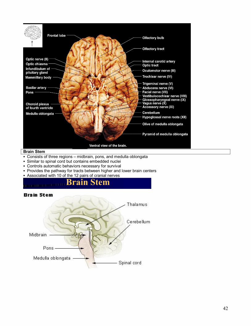
Human Brain: Ventral Aspect
42
Brain Stem § Consists of three regions – midbrain, pons, and medulla oblongata § Similar to spinal cord but contains embedded nuclei § Controls automatic behaviors necessary for survival § Provides the pathway for tracts between higher and lower brain centers § Associated with 10 of the 12 pairs of cranial nerves
Brain Stem

43
Figure 12.12: Midsagittal section of the brain illustrating the diencephalon and brain stem, p. 445.
Parietal lobe of cerebral hemisphere Corpus callosum
Choroid plexus Occipital lobe of cerebral hemisphere Thalamus (encloses third ventricle)
Pineal body/gland (part of epithalamus)
Posterior commissure
Corpora quadrigemina Cerebral aqueduct
Arbor vitae Fourth ventricle Choroid plexus Cerebellum
Septum pellucidum
Interthalamic adhesion (intermediate mass of thalamus) Frontal lobe of cerebral hemisphere Interventricular foramen Anterior commissure Hypothalamus Optic chiasma Pituitary gland
Temporal lobe of cerebral hemisphere Mammillary body
Pons Medulla oblongata Spinal cord
Midbrain
Fornix
Midbrain § Located between the diencephalon and the pons § Midbrain structures include:
§ Cerebral peduncles – two bulging structures that contain descending pyramidal motor tracts § Cerebral aqueduct – hollow tube that connects the third and fourth ventricles § Various nuclei
Midbrain Nuclei § Nuclei that control cranial nerves III (oculomotor) and IV (trochlear) § Corpora quadrigemina – four domelike protrusions of the dorsal midbrain § Superior colliculi – visual reflex centers § Inferior colliculi – auditory relay centers § Substantia nigra – functionally linked to basal nuclei § Red nucleus – largest nucleus of the reticular formation; red nuclei are relay nuclei for some descending motor pathways
Pons
§ Bulging brainstem region between the midbrain and the medulla oblongata § Forms part of the anterior wall of the fourth ventricle § Fibers of the pons:
§ Connect higher brain centers and the spinal cord § Relay impulses between the motor cortex and the cerebellum
§ Origin of cranial nerves V (trigeminal), VI (abducens), and VII (facial) § Contains nuclei of the reticular formation

44
Medulla Oblongata § Most inferior part of the brain stem § Along with the pons, forms the ventral wall of the fourth ventricle § Contains a choroid plexus of the fourth ventricle § Pyramids – two longitudinal ridges formed by corticospinal tracts § Decussation of the pyramids – crossover points of the corticospinal tracts

45
Medulla Nuclei § Inferior olivary nuclei – gray matter that relays sensory information § Cranial nerves X, XI, and XII are associated with the medulla § Vestibular nuclear complex – synapses that mediate and maintain equilibrium § Ascending sensory tract nuclei, including nucleus cuneatus and nucleus gracilis § Cardiovascular control center – adjusts force and rate of heart contraction § Respiratory centers – control rate and depth of breathing § Additional centers – regulate vomiting, hiccuping, swallowing, coughing, and sneezing
The Cerebellum § Located dorsal to the pons and medulla § Protrudes under the occipital lobes of the cerebrum § Makes up 11% of the brain’s mass § Provides precise timing and appropriate patterns of skeletal muscle contraction § Cerebellar activity occurs subconsciously Figure 12.17: Cerebellum, p. 452.
(a) (b)
(c) (d)
Anterior lobe Primary fissure
Posterior lobe
Vermis
Horizontal fissure
Vermis
White matter of cerebellum
Deep cerebellar nuclei Granule cells
in granular layer
Purkinje cells
Site of basket cells and stellate cells in outer cortex (molecular layer)
Caudal (inferior)
Brain stem (midbrain) Cerebellar cortex
Vermis (cut)

46

47
48
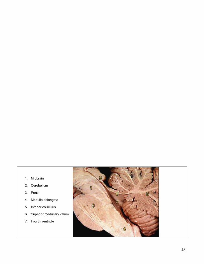
1. Midbrain
2. Cerebellum
3. Pons
4. Medulla oblongata
5. Inferior colliculus
6. Superior medullary velum
7. Fourth ventricle

49
1. Vermis 2. Central lobule 3. Anterior lobe 4. Superior cerebellar
peduncle 5. Middle cerebellar
peduncle 6. Nodule of vermis 7. Inferior cerebellar
peduncle 8. Flocculus 9. Posterior lobe
Anatomy of the Cerebellum § Two bilaterally symmetrical hemispheres connected medially by the vermis § Folia – transversely oriented gyri § Each hemisphere has three lobes – anterior, posterior, and flocculonodular § Neural arrangement – gray matter cortex, internal white matter, scattered nuclei § Arbor vitae – distinctive treelike pattern of the cerebellar white matter
Cerebellar Peduncles § Three paired fiber tracts that connect the cerebellum to the brain stem § All fibers in the cerebellum are ipsilateral § Superior peduncles connect the cerebellum to the midbrain § Middle peduncles connect the pons to the cerebellum § Inferior peduncles connect the medulla to the cerebellum
50
Cerebellar Processing § Cerebellum receives impulses of the intent to initiate voluntary muscle contraction § Proprioceptors and visual signals “inform” the cerebellum of the body’s condition § Cerebellar cortex calculates the best way to perform a movement § A “blueprint” of coordinated movement is sent to the cerebral motor cortex
Cerebellar Cognitive Function § Plays a role in language and problem solving § Recognizes and predicts sequences of events
Functional Brain System § Networks of neurons working together and spanning wide areas of the brain § The two systems are:
§ Limbic system § Reticular formation
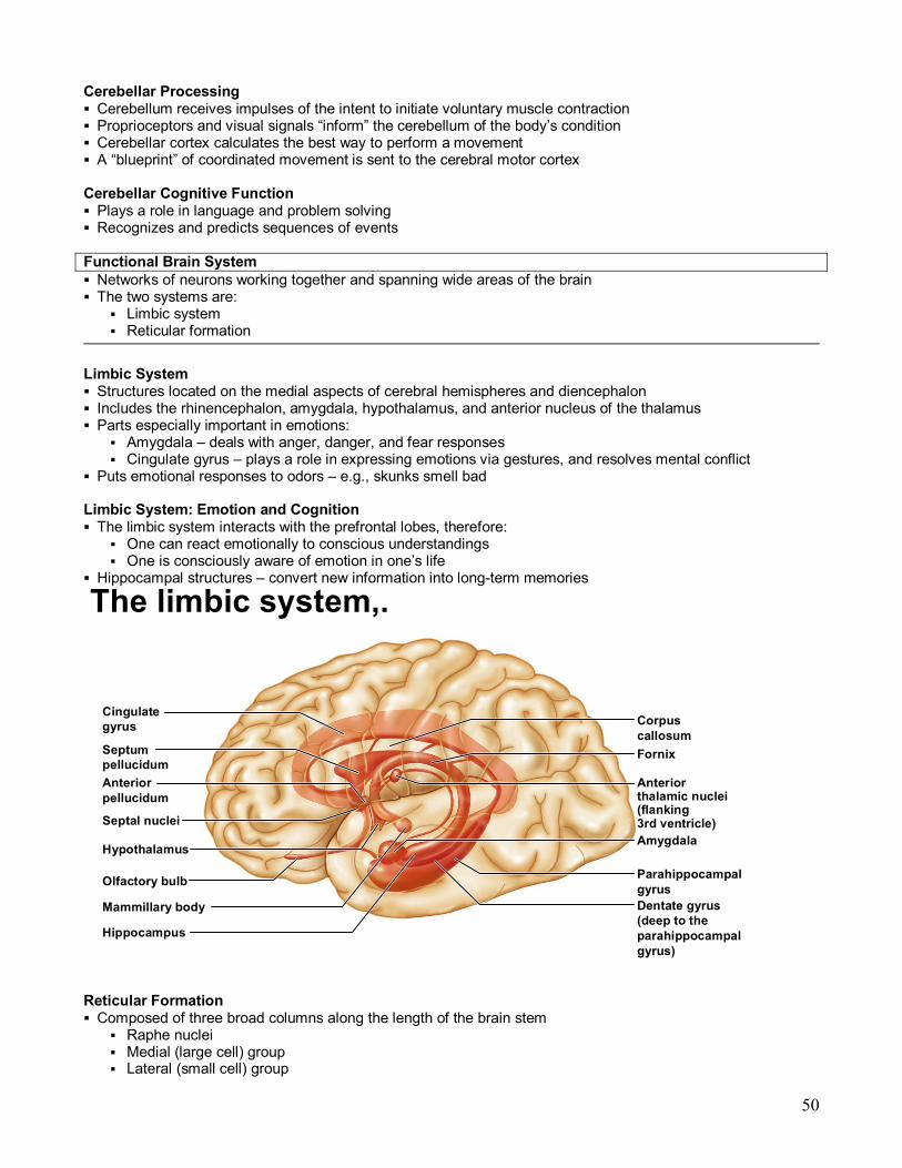
Limbic System § Structures located on the medial aspects of cerebral hemispheres and diencephalon § Includes the rhinencephalon, amygdala, hypothalamus, and anterior nucleus of the thalamus § Parts especially important in emotions:
§ Amygdala – deals with anger, danger, and fear responses § Cingulate gyrus – plays a role in expressing emotions via gestures, and resolves mental conflict
§ Puts emotional responses to odors – e.g., skunks smell bad
Limbic System: Emotion and Cognition § The limbic system interacts with the prefrontal lobes, therefore:
§ One can react emotionally to conscious understandings § One is consciously aware of emotion in one’s life
§ Hippocampal structures – convert new information into longterm memories
The limbic system,.
Cingulate gyrus Corpus
callosum Fornix
Anterior thalamic nuclei (flanking 3rd ventricle) Amygdala
Parahippocampal gyrus
Septum pellucidum
Septal nuclei
Hypothalamus
Anterior pellucidum
Olfactory bulb
Mammillary body
Hippocampus
Dentate gyrus (deep to the parahippocampal gyrus)
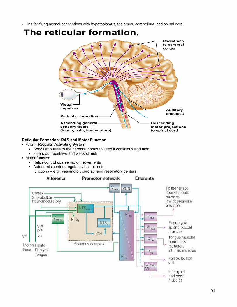
Reticular Formation § Composed of three broad columns along the length of the brain stem
§ Raphe nuclei § Medial (large cell) group § Lateral (small cell) group
51
§ Has farflung axonal connections with hypothalamus, thalamus, cerebellum, and spinal cord
The reticular formation,
Visual impulses
Reticular formation
Ascending general sensory tracts (touch, pain, temperature)
Descending motor projections to spinal cord
Auditory impulses
Radiations to cerebral cortex
Reticular Formation: RAS and Motor Function § RAS – Reticular Activating System
§ Sends impulses to the cerebral cortex to keep it conscious and alert § Filters out repetitive and weak stimuli
§ Motor function § Helps control coarse motor movements § Autonomic centers regulate visceral motor functions – e.g., vasomotor, cardiac, and respiratory centers

52
Brain Waves § Normal brain function involves continuous electrical activity § An electroencephalogram (EEG) records this activity § Patterns of neuronal electrical activity recorded are called brain waves § Each person’s brain waves are unique § Continuous train of peaks and troughs § Wave frequency is expressed in Hertz (Hz)
Types of Brain Waves § Alpha waves – regular and rhythmic, lowamplitude, slow, synchronous waves indicating an “idling” brain § Beta waves – rhythmic, more irregular waves occurring during the awake and mentally alert state § Theta waves – more irregular than alpha waves; common in children but abnormal in adults § Delta waves – highamplitude waves seen in deep sleep and when reticular activating system is damped
Brain Waves: State of the Brain § Change with age, sensory stimuli, brain disease, and the chemical state of the body § EEGs used to diagnose and localize brain lesions, tumors, infarcts, infections, abscesses, and epileptic lesions
§ A flat EEG (no electrical activity) is clinical evidence of death
Epilepsy § A victim of epilepsy may lose consciousness, fall stiffly, and have uncontrollable jerking, characteristic of epileptic seizure
§ Epilepsy is not associated with, nor does it cause, intellectual impairments § Epilepsy occurs in 1% of the population
Epileptic Seizures § Absence seizures, or petit mal – mild seizures seen in young children where the expression goes blank § Grand mal seizures – victim loses consciousness, bones are often broken due to intense convulsions, loss of bowel and bladder control, and severe biting of the tongue
Control of Epilepsy § Epilepsy can usually be controlled with anticonvulsive drugs § Valproic acid, a nonsedating drug, enhances GABA and is a drug of choice § Vagus nerve stimulators can be implanted under the skin of the chest and can keep electrical activity of the brain from becoming chaotic
Consciousness § Encompasses perception of sensation, voluntary initiation and control of movement, and capabilities associated with higher mental processing
§ Involves simultaneous activity of large areas of the cerebral cortex § Is superimposed on other types of neural activity § Is holistic and totally interconnected § Clinical consciousness is defined on a continuum that grades levels of behavior – alertness, drowsiness, stupor, coma
Types of Sleep § There are two major types of sleep:
§ Nonrapid eye movement (NREM) § Rapid eye movement (REM)
§ One passes through four stages of NREM during the first 3045 minutes of sleep § REM sleep occurs after the fourth NREM stage has been achieved

53
Types and Stages of Sleep: NREM § NREM stages include:
§ Stage 1 – eyes are closed and relaxation begins; the EEG shows alpha waves; one can be easily aroused § Stage 2 – EEG pattern is irregular with sleep spindles (highvoltage wave bursts); arousal is more difficult § Stage 3 – sleep deepens; theta and delta waves appear; vital signs decline; dreaming is common § Stage 4 – EEG pattern is dominated by delta waves; skeletal muscles are relaxed; arousal is difficult
Types and Stages of Sleep: REM § Characteristics of REM sleep
§ EEG pattern reverts through the NREM stages to the stage 1 pattern § Vital signs increase § Skeletal muscles (except ocular muscles) are inhibited § Most dreaming takes place
Sleep Patterns § Alternating cycles of sleep and wakefulness reflect a natural circadian rhythm § Although RAS activity declines in sleep, sleep is more than turning off RAS § The brain is actively guided into sleep § The suprachiasmatic and preoptic nuclei of the hypothalamus regulate the sleep cycle § A typical sleep pattern alternates between REM and NREM sleep
Importance of Sleep § Slowwave sleep is presumed to be the restorative stage § Those deprived of REM sleep become moody and depressed § REM sleep may be a reverse learning process where superfluous information is purged from the brain § Daily sleep requirements decline with age
Sleep Disorders § Narcolepsy – lapsing abruptly into sleep from the awake state § Insomnia – chronic inability to obtain the amount or quality of sleep needed § Sleep apnea – temporary cessation of breathing during sleep
Memory § Memory is the storage and retrieval of information § The three principles of memory are:
§ Storage – occurs in stages and is continually changing § Processing – accomplished by the hippocampus and surrounding structures § Memory traces – chemical or structural changes that encode memory
Memory Processing
Stages of Memory § The two stages of memory are shortterm memory and longterm memory § Shortterm memory (STM, or working memory) – a fleeting memory of the events that continually happen § STM lasts seconds to hours and is limited to 7 or 8 pieces of information § Longterm memory (LTM) has limitless capacity
Transfer from STM to LTM § Factors that effect transfer of memory from STM to LTM include:
§ Emotional state – we learn best when we are alert, motivated, and aroused § Rehearsal – repeating or rehearsing material enhances memory § Association – associating new information with old memories in LTM enhances memory

54
§ Automatic memory – subconscious information stored in LTM Categories of Memory § The two categories of memory are fact memory and skill memory § Fact (declarative) memory:
§ Entails learning explicit information § Is related to our conscious thoughts and our language ability § Is stored with the context in which it was learned
Skill Memory § Skill memory is less conscious than fact memory and involves motor activity § It is acquired through practice § Skill memories do not retain the context in which they were learned
Structures Involved in Fact Memory § Fact memory involves the following brain areas:
§ Hippocampus and the amygdala, both limbic system structures § Specific areas of the thalamus and hypothalamus of the diencephalon § Ventromedial prefrontal cortex and the basal forebrain
Structures Involved in Skill Memory § Skill memory involves:
§ Corpus striatum – mediates the automatic connections between a stimulus and a motor response § Portion of the brain receiving the stimulus § Premotor and motor cortex
Mechanisms of Memory § Neuronal RNA content is altered § Dendritic spines change shape § Extracellular proteins are deposited at synapses involved in LTM § Number and size of presynaptic terminals may increase § More neurotransmitter is released by presynaptic neurons § New hippocampal neurons appear
Mechanisms of Memory § Longterm potentiation (LTP) is involved and is mediated by NMDA receptors § Synaptic events involve the binding of brainderived neurotropic factor (BDNF) § BDNF is involved with Na + , Ca 2+ , and Mg 2+ influence at synapses
Cerebrovascular Accidents (Strokes) § Caused when blood circulation to the brain is blocked and brain tissue dies § Most commonly caused by blockage of a cerebral artery § Other causes include compression of the brain by hemorrhage or edema, and atherosclerosis § Transient ischemic attacks (TIAs) – temporary episodes of reversible cerebral ischemia § Tissue plasminogen activator (TPA) is the only approved treatment for stroke
Degenerative Brain Disorders § Alzheimer’s disease – a progressive degenerative disease of the brain that results in dementia § Parkinson’s disease – degeneration of the dopaminereleasing neurons of the substantia nigra § Huntington’s disease – a fatal hereditary disorder caused by accumulation of the protein huntingtin that leads to degeneration of the basal nuclei

55