Embed Size (px)
Citation preview
Chapter XX
PAROTITIS AND MASTOIDITIS
ACUTE POSTOPERATIVE PAROTITIS
Before radiation therapy was employed for acute postoperative parotitis, this condition was frequently a fatal surgicalcomplication. Although it probably does not occur more oftenthan once in every 2,000 surgical cases, the morbidity and mortality should entitle the disease to more consideration than ithas received in the literature on radiation therapy in the past,especially in view of the fact that x-ray therapy is so effective.
The statistics cited by Blair and Olch21 place the mortalityof surgical cases in which parotitis is a complication at 42.8 percent unless operative treatment is used, in which case the mortality is 26 per cent. Bowing and Fricke22 quoted Green as saying that the mortality in American statistics varies from 25 to60 per cent. Using radium, Bowing and Fricke's mortality ratewas 22.8 per cent in 184 cases. With x-ray therapy as used atpresent, the mortality should never exceed 10 per cent.
The etiology of acute postoperative parotitis is still obscure.Many theories have been mentioned, among them oral sepsis,seasonal influences, dehydration, reflex disturbance from peritoneal irritation and anesthetic trauma. None seems to fit verywell with Bowing and Fricke's observation that this complication is 13 times as frequent following colon surgery as it isfollowing all other surgery.
No light is shed on the possible cause in our small series fromCreighton Memorial St. Joseph's Hospital, in which 19 patientsrecovered uneventfully, one had suppuration necessitating surgicaJr drainage, and two died. Of these 22 cases,colon surgeryhad been performed in only one, unless appendectomy can beconsidered surgery involving the colon. One case occurred postpartum, one after abortion, and one followed implantation ofradium needles in a breast cancer. The others followed laparotomy for appendectomy, synechtenterotomy, cholecystectomyand various pelvic surgical procedures. One case, not includedin this group because it was not a postoperative complication,
332

Parotitis and Mastoiditis 333
was in a 10 year old child with empyema thoracis. The parotitisresponded promptly, and rib resection was done two weeks laterfor the empyema. Although 14 of the 22 cases were in females,the total number is probably not large enough for conclusiveevidence of a variation in frequency in sexes. All but two ofthe patients were over 20.
The therapy recommended in most surgical texts consists ofpacks, careful oral hygiene, probing Stensen's duct and surgicalincision in the presence of suppuration. Rankin and Palmer23
reported a definite decrease in the mortality when radium therapy was used. Bowing and Fricke's results have been quoted.Robinson and Spencer24 reported excellent results with a singledose of 300 r units of high voltage, heavily filtered x-ray. Hodgesand Berger16 mentioned this condition as one in which roentgentherapy is valuable. Repeated small doses of low voltage x-rayshave been highly effective in our hands.
In our opinion, x-ray therapy has been as valuable in surgicalmumps as in gas bacillus infection. A review of many casestreated during the past few years discloses that a small grouptreated with x-rays stands out from the rest and suggestsstrongly that singular therapeutic action may be claimed forx-rays in surgical mumps. For many years, the mortality ratefor surgical mumps was high. When a patient who was seriously ill contracted the disease, he died.
In our experience, and in the literature as far as we eanascertain, there are no reeords prior to the use of radiationtherapy of any patients reeovering from surgical mumps onlyto die a week or two later of their original disease. Anyone whohad a disease serious enough to be the cause of death in a shorttime did not have the resistance to survive also the acute toxemiaof surgical mumps. When surgical mumps developed, it wasconsidered a terminal manifestation and the patient soon died.
In t}-le last few years, however, several patients in whomsurgical mumps developed responded promptly to x-ray therapy.But after they were free from all evidence of surgical mumps,they continued to fail because of the original illness and diedin a short time.
These patients obviously did not recover from surgical mumpsbecause of any improvement in their original disease, since the~y
died of their original disease two to six weeks after they had
334 Roentgen Treat11tent of Infections
made a complete recovery from surgical mumps. The use of x-raytherapy is the only reasonable explanation, then, for their recovery from the mumps.
The ability of x-rays to overcome the toxemia and cause complete regression of surgical mumps in seriously ill patients is astriking example of their therapeutic value and ranks, it seems,with the effect secured in treating gas bacillus infection. Theantitoxic effect of x-rays in acute infections deserves greaterrecognition, wider usage and further study.
In cases in which response was most prompt, treatment wasstarted in the painful or early swelling stage of the disease;and when treatment is started at this time no complicationsoccur. But if treatment is started late, after pus is present,nothing but surgical intervention and a long convalescence canbe expected, or even death.
Fur practical clinical purposes, and to emphasize the value ofearly x-ray treatment, the disease may be divided into threestages:
Stage l.-This, the painful stage, may be absent occasionally,but usually the patient first notices the disease because of painin the jaw when eating. As the nurse records this in the bedsidenotes on the chart, the "stage of suspicion" at least is established.
Stage 2.-This, the stage of swelling, is characterized by enlargement of the parotid gland or tissues adjacent to it. Generalsigns of an acute infection appear. These may be the first findings noted. This period of swelling may last a few days. It isassociated with considerable toxemia and other manifestationsof a severe infection. If a favorable turn does not follow andregression occurs, the gland may suppurate.
Stage B.-After this, the stage of suppuration, is establishedsurgical intervention becomes inevitable and the outcome of thecase Jess certain. The x-rays, as far as it was possible to determine, do not cause suppuration; in fact, if treatment is startedearly and small doses are given at frequent intervals, the chanceof suppuration seems to be diminished. If treatment is notstarted until the third stage, little benefit will come from x-raytherapy, but reduction of the toxemia even at this stage may bea great help.
X-rays localize the disease, relieve the pain, control the tox·
Parotitis· and Mastoiditis 335
emia, in general shorten the course and lessen the chance ofcomplications. These effects on the patient's general comfortmay also be an important factor in recovery from the originaldisease as well as from the mumps.
Surgical mumps is one of the conditions in which radiationtherapy is imperative. The mobile therapy unit makes the treatment simple, regardless of how sick the patient may be. \¥e feel
.that small repeated doses at short intervals are less likely to becomplicated by suppuration than one or two heavy doses atgreater intervals.
X-RAY TECHNIC
\Vith the mobile therapy unit, repeated treatments do notdisturb the patient in any way. This is an important advantagein treating the extremely toxic patient.
The dose varies from 60 to 100 I' units twice daily for at leasttwo to four days, each involved gland being treated. \Ve use100 kv. and 1 or 2 mm. Al filter, depending on the amount ofswelling. Good results have been obtained with low voltageswhen an ordinary mobile diagnostic unit has been used, andmany patients have been treated with this type of equipment.Hesponse within 24 hours is the rule, and seldom do more than48 hours pass without definite improvement. \Vhen it is necessary to continue treatment for several days, additional filter isused and skin tolerance never exceeded.
In one case, there was definite evidence of suppuration beforex-ray therapy was started. The patient was a young womanwith pelvic cellulitis and peritonitis following an abortion; thecomplication of parotitis made the prognosis grave. Bilateralincision of the neck and drainage were resorted to in additionto x-ray therapy, and the patient recovered.
As in gas bacillus infection and peritonitis, reports on surgicalmumpjl .should include the morbidity as well as the mortalityrate. In the morbidity report, the number of days in the hospital must be stated as well as whether or not suppuration occurred. If facial paralysis, parotid fistula or some other seriousflequela remains as a result of the mumps, it should be mentioned.
In surgical mumps, as in other acute infections, the earlierx-ray treatment is started, the better the results, for an early
336 Roentgen Treatntent of Infections
start of the treatments quite definitely lessens the complications.
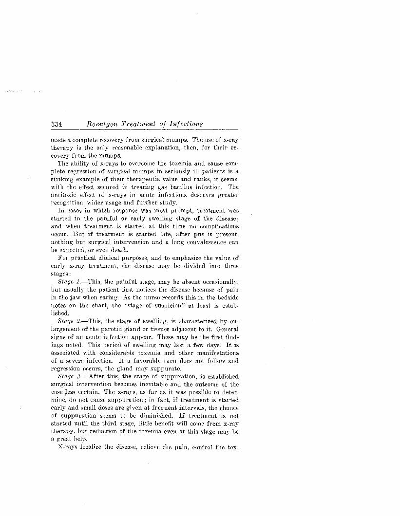
CASE 71.-A. M., a woman aged 47, was admitted to the hospitalSeptember 13, 1937, for a cholecystectomy. The third postoperativeday, parotitis developed on the left side. The patient was treatedwith x-rays twice daily for two days and once daily for two additionaltreatments. She improved promptly, convalescence was uneventful
tI
~y I
IIII ',"I"I.I"bl.1
I'
.' +C'~i-t , .ic:IT-1=I._~[ i--jJ --: - _.- ~ ~,~ --~g;: Et:::f£{d-~ f"cf
, . ,t:-i':c~ . ct· ''1=!::,c-f$'=;;=:''.. '2~ 'c]. L ~~ "I: ..t·Jif"i ''-j='~
.~..:EE. :;tt::-::. '-+" .
HI cL~'¥cT:'-f$gL:jo--r" '1:-T;:~~.- ::p:' -; ..;,. .3.::.Lft"ff~1=tEJ=1=fc @ .
-.- .E.':'=' . Ci--+ct~·'-;cg·: ;!--.--ft._. ~~::I-.-i: :.,;' ::!";---H+ ~i;:+:'-f';
I?
'" .
I.
.,'
HOUFl I'll 13./
,,.,10.' ~
III lOJ' :~
~ 1~' i-~ Ill!':-
I~:!=T'
FIG. !lO.-Case 71. Typical response of patient with acute postoperativeparotitis. Technical factors were: 100 kv.; 5 ma.; 40 em. distance; 1 rnm. Al
filter; left parotid gland as port. Result was excellent.
and she was dismissed the thirty-first postoperative day. The clinicalchart (Fig. 90) shows an immediate drop in temperature at the beginning of the x-ray treatment.
X-Fay therapy for this condition is truly the surgeon's friend.
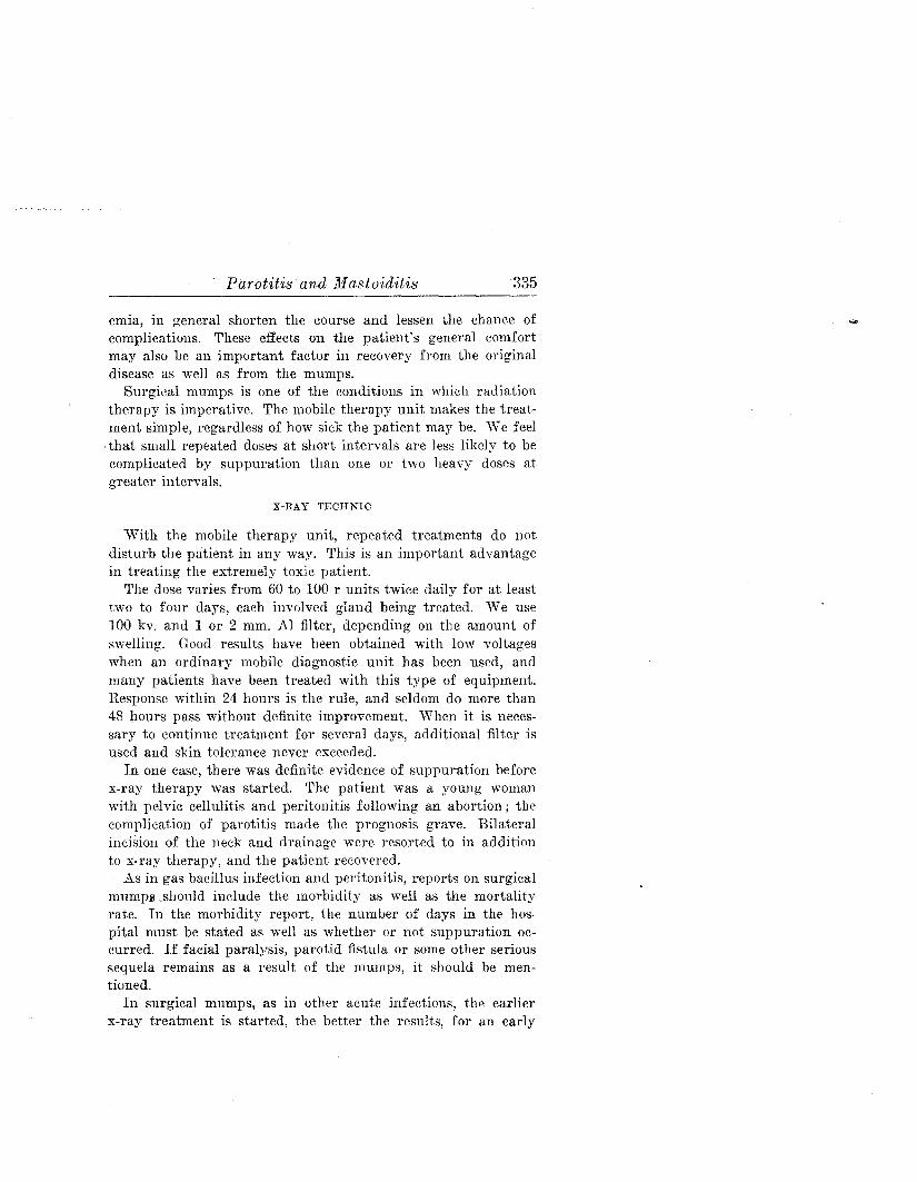
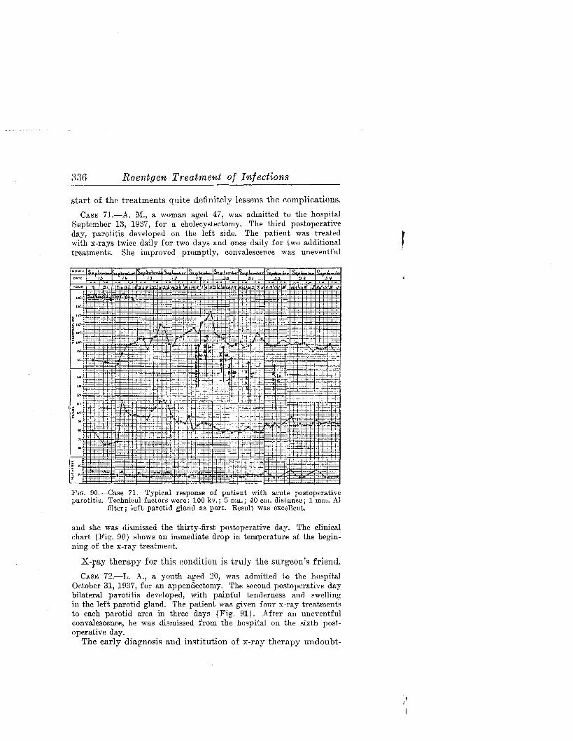
CASE 72.-L. A., a youth aged 20, was admitted to the hospitalOctober 31, 1937, for an appendectomy. The second postoperative daybilateral parotitis developed, with painful tenderness and swellingin the left parotid gland. The patient was given four x-ray treatmentsto each parotid area in three days (Fig. 91). After an uneventfulconvalescence, he was dismissed from the hospital on the sixth postoperative day.
The early diagnosis and institution of x-ray therapy undoubt-
Parotitis and Mastoiditis 337
edly permitted the early dismissal of this patient. X-ray therapyis a safe treatment for this serious complication.
CASE 73.-E. W., a woman aged 68, was admitted to the hospitalJune 3, 1935, with a diagnosis of strangulated inguinal hernia on the
DATE I
HOUR . I
lOS'
un' I"' .a: •~ 102' :a: •~ 101' •:!: •"' :~ 100' :
!<o
130;
120:110 :
"'In •5 100 I
<l.
90
80
"';..6IJ
i~
J
JI~I 131 I'll IJ I" '1P. fol. A.~.
1 3 1'1 13 I 111 I
------- +_._. -----=-r::'- ~..~, -L-.....-... ~:
FIG. 91.-Case 72. This behavior of temperature and pulse is typical forpatients '\vllO receive treatment early when first complaining of pain in thejaw. Technical factors were: 90 kv.; 5 ma.; 40 em. distance; 1 mm. Al
filter; both parotid glands as ports. Result was excellent.
right side with intestinal obstruction. She was treated expectantlyuntil June 11. She was then operated on, and a portion of gangrenousileum was removed, a Murphy button anastomosis was made, and anenterostomy performed above the resection. The parotid gland wasswollen the day following the operation. She received one x-ray
338 Roentgen Treatment of Infections
treatment, but died the following day before further x-ray treatmentcould be given.
In this case the pathologic process, shock and toxemia fromthe abdominal condition were sufficient to cause death. The rOleplayed by. parotitis in this case was questionable, but the case islisted among the group of deaths caused by surgical mumps.The patient received no x-ray therapy for peritonitis.
CASE 74.-8. G., a woman aged 22, was admitted to the hospitalMarch 11, 1936, complaining of severe abdominal pain and vomitingof three days' duration. There was boardlike rigidity with abdominaltenderness, more severe in the pelvic region. The white blood cellcount was 41,000, temperature 100.2 F., pulse rate 122 and respiratoryrate 22.
Diagnosis was pelvic abscess, general peritonitis, paralytic ileus andsepticemia. At operation on March 13, the pelvic abscess was drainedand about 16 oz. of pus evacuated, and a rubber tube and two tamponswere inserted for drainage. Transfusions were given March 18 and19. X-ray treatments included one on March 19 and two on March 20.The patient died at 7: 00 P. M. March 20.
This patient received no x-ray treatment for the pelvic condition and received one treatment for the mumps late on March19, when the swelling in the parotid region was first noted, andtwo treatments the next day, which was the day she died. It isdoubtful that surgical parotitis was the major cause of death, asit had hardly developed when the patient died. She was exceedingly toxic from the peritonitis at the time of admission andshowed no improvement at any time despite surgical drainage,blood transfusions and other measures. ''Ve were unable to secure permission to treat the peritonitis, and we believe that ifthe best results were to be obtained we should have treated bothconditions.
CASE 75.-T. McM., a woman aged 62, was hospitalized December7, 1937. Diagnosis on admission was chronic cholecystitis with stonesand aI'lcites. She was operated on December 13, when she was foundto have empyema of the gallbladder with multiple stones, and about agallon of free fluid in the peritoneal cavity due to acute hepatitis.Twelve gall stones were removed. The suction apparatus was insertedshortly after the operation and left in about 20 days. On January 4,1938, the right parotid gland was swollen and sore. She received twox-ray treatments on the fourth and two on the fifth. The mumpspromptly subsided, but the stormy course continued. The suction apparatus was re-inserted January 6 and left for several days. On
Parotitis and Mastoiditis 339
January 9, bedsores added to the distress. The patient died February8, without recurrence of the surgical mumps.
This case is one in which the effectiveness of x-rays in surgicalmumps is evident. Despite the serious illness, the patient hadfully recovered from the mumps long before death. This waspractically unheard of before the use of x-rays. Patients as illas this patient were never able to live through the toxic phase ofthe mumps when it was added to another serious disease.
CASE 76.-N. L., a woman aged 72, was hospitalized September 10,1937. The diagnosis was complete uterine prolapse. The day afteradmission vaginal hysterectomy and plastic repair were done. Thetemperature had reached 101 F. daily until the diagnosis of pelvicabscess was made. This abscess was drained by way of a posteriorcolpotomy September 25. The course continued to be somewhat stormy.Pain and swelling of the left parotid gland were noted October 19.Two treatments were given on this and the following day, one onOctober 21 and on each of the following three days. The mumps responded, but the patient's condition continued to grow worse despitea transfusion on October 30. Death occurred November 5.
Death occurred after the surgical mumps had apparentlysubsided. Again, surgical mumps which developed during along, toxic process seemed to be entirely controlled, but the patient's general condition continued to be poor and death finallyintervened.
CASE 77.-J. H., a man aged 23, a medical student, was admitted tothe hospital March 18, 1938, with a diagnosis of acute appendicitis.He was operated on the same day. Convalescence was stormy. Anabdominal abscess developed which was drained on the twenty-seventhhospital· day. Two days later severe bilateral parotitis was present.The temperature reached 103.8 F. and the patient was decidedly toxic.X-ray therapy was started, the temperature dropped promptly, andtoxic manifestations quickly responded (Fig. 92). He received sixtreatments in five days.
The attending surgeon was skeptieal at first, but the results inthis c£se served to make him more friendly to x-ray therapy forsurgieal parotitis.
SUPPURATIVE PAROTITIS, NONSURGICAL
CASE 78.-E. C., a woman aged 77, was hospitalized April 22, 1936.The provisional diagnosis was senility with a complication of suppurative parotitis. Her chief complaint was swelling and pain over
342 Roentgen Treatment of Infections
a definite problem when x-ray therapy is considered, becausesome organisms respond much more readily than others, at leastin our present system of treatment with x-rays. As time goeson and accurate records accumulate on established types ofinfections, the data will probably lead to a more successful technic for infections which are stubborn or entirely resistant tox-ray therapy. Some organisms appear adequately controlledwith the present-day technic, and 110 change or little changeseems indicated or desirable. Others are still not responsive,and more study is indicated before the correct technic will befound.
1. Acute Simple Mastoiditis with and without Suppuration
This localized infection generally responds well to irradiation.If the causative organism is sensitive to irradiation and x-raytreatments are given in the early stage or even in the mostactive stage, the results are good. As in other infections, theearlier treatment is started, the more prompt the response andthe better the result. Irradiation during the early stage willoften abort the infection and render surgical intervention unnecessary.
Obviously, x-ray treatment started in the late stage of thedisease after destruction of cell walls has taken place and theabscess cavity is formed will be less effective and surgery willbe necessary. In this type of case, a treatment or two preoperatively, then more treatment after operation and after drainageis established have been of definite help in shortening thecourse of the infection even at this late stage. The point toremember, however, is that when the mastoid cells have beendestroyed, surgical intervention is indicated. In any event,irradiation preoperatively is decidedly beneficial and should notbe neglected.
2. Postoperative Suppurative Mastoiditis
When x-ray therapy is started in the late stage, after operation and after drainage is established, it is still of some valueif started within a few hours of operation. It aids in localizingthe disease, which otherwise takes on renewed activity in tissuesto which it has just gained access as a result of the operation.Those who object to irradiation before operation certainly have

Parotitis and Mastoiditis 343
no legitimate excuse for refusing x-ray treatment immediatelyafter operation.
If x-ray treatment is delayed until two weeks or longer afterthe operation, the infection is much less responsive. It is doubtful that any good will come from such delayed therapy, or thatit is even indicated. At this late stage, however, it is worth trying in the desperate case; in the occasional case, it may endthe seropurulent discharge and greatly hasten recovery. Thisresult is the exception and not the rule. In cases treated late,the course generally continues, and unless a secondary operationis necessary, x-rays do little good.
If another operation is indicated, preoperative irradiation andtherapy continued immediately after the operation for a fewdays will do a great amount of good.
3 and 4. Osteomyelitis of Bones Adjacent to Mastoid;Mastoid with Reactivation of Old Infection
These conditions, as well as old mastoiditis which suddenlybecomes actively infected, call for judicious pre- and postoperative x-ray treatment. Except for localizing the infection. aftereach new surgical procedure, it is not as clearly indicated as insome of the other mastoid conditions. A longer preoperativecourse of radiation than is usually given may be necessary tolocalize the infection thoroughly. Operative procedure shouldnot be undertaken, if it can possibly be avoided, until the infection is localized. X-rays are one agent which may be appliedwell beyond the known limits of infection and repeatedly applied to the tissues with the assurance that they will lessen theextent of the infection, make it more accessible to surgical intervention and do no harm to the normal structures.
5. Specifie Mastoiditis
Mastoiditis which develops as a complication of pneumoniaor some other specific disease may call for specific therapy. Butx-ray therapy may be all that is necessary in other cases. X-rayand specific therapy may be used in combination. The most important thing is early diagnosis and treatment.
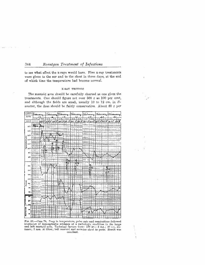
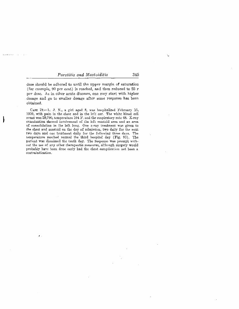
In Case 79, the diagnosis of pneumonia and mastoiditis wasmade on admission to the hospital. Because of the lung involvement, x-ray therapy was decided on, and operation was deferred
344 Roentgen Treatment of Infections
to see what effect the x-rays would have. Five x-ray treatmentswere given to the ear and to the chest in three days, at the endof which time the temperature had become normal.
X-RAY TECHNIC
The mastoid area should be carefully charted as one gives thetreatments. One should figure not over 500 r as 100 per cent,and although the fields are small, usually 10 to 12 em. in diameter, the dose should be fairly conservative. About 80 r per
....J;r~ -: ',. =E .. \:Ef-~!!~: s;c::tc:H~'., .: ::t=t~. __
MONTH ~\..YUlro."
DATE I:" "
HOUR
·,·-r i""'='t:-+
... ---r----- -.- :!~1-
---.:::-,J U
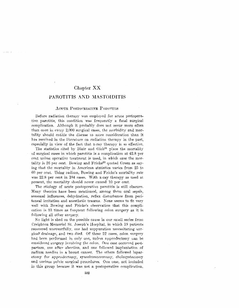
FIG. 93.-Case 79. Drop in temperature, pulse rate and respirations followedtreatment of demonstrable evidence of a pathologic condition in the lungsand left mastoid cells. Technical factors were: 100 ky.; 5 rna.; 40 em. dis·tance; 3 mm. Al filter; left mastoid and anterior chest as ports. Result was
excellent.
Parotitis and Mastoiditis 345
dose should be adhered to until the upper margin of saturation(for example, 90 per cent) is reached, and then reduced to 25 rper dose. As in other acute diseases, one may start with higherdosage and go to smaller dosage after some response has beenobtained.
CASE 79.-L. J. N., a girl aged 8, was hospitalized February 15,1939, with pain in the chest and in the left ear. The white blood cellcount was 29,700, temperature 104 F. and the respiratory rate 48. X-rayexamination showed involvement of the left mastoid area and an areaof consolidation in the left lung. One x-ray treatment was given tothe chest and mastoid on the day of admission, two daily for the nexttwo days and one treatment daily for the following three days. Thetemperature reached normal the third hospital day (Fig. 93). Thepatient was dismissed the tenth day. The response was prompt without the use of any other therapeutic measures, although surgery wouldprobably have been done early had the chest complication not been acontraindication.
¥ ..