Embed Size (px)
Citation preview
SKIN ACCESSORIES AND APPENDAGES
Pages 119-124
© 2015 Pearson Education, Inc.
APPENDAGES OF THE SKIN
all exocrine glands (secretions via ducts)
Sebaceous glands Sweat glands
Hair/hair follicles Nails
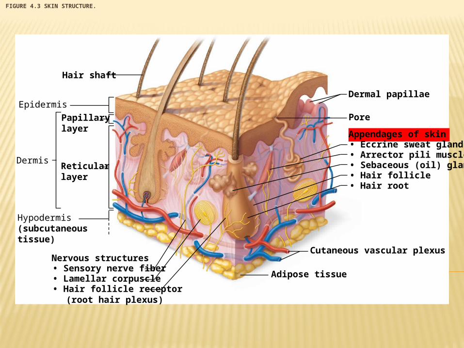
FIGURE 4.3 SKIN STRUCTURE.
Dermal papillae
Hair shaft
Pore
Appendages of skin• Eccrine sweat gland• Arrector pili muscle• Sebaceous (oil) gland• Hair follicle• Hair root
Cutaneous vascular plexus
Adipose tissue
Epidermis
Dermis
Papillarylayer
Reticularlayer
Hypodermis(subcutaneoustissue)
Nervous structures• Sensory nerve fiber• Lamellar corpuscle• Hair follicle receptor (root hair plexus)
© 2015 Pearson Education, Inc.
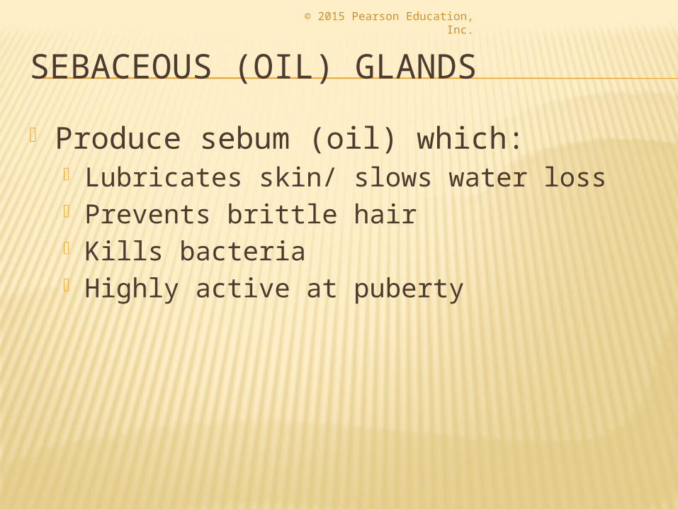
SEBACEOUS (OIL) GLANDS
Produce sebum (oil) which: Lubricates skin/ slows water loss Prevents brittle hair Kills bacteria Highly active at puberty
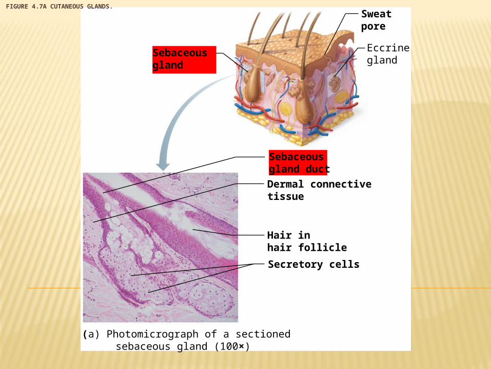
FIGURE 4.7A CUTANEOUS GLANDS.
Eccrinegland
Sebaceousgland
Sweatpore
Sebaceousgland duct
Dermal connectivetissue
Hair inhair follicle
Secretory cells
(a) Photomicrograph of a sectioned sebaceous gland (100×)
© 2015 Pearson Education, Inc.
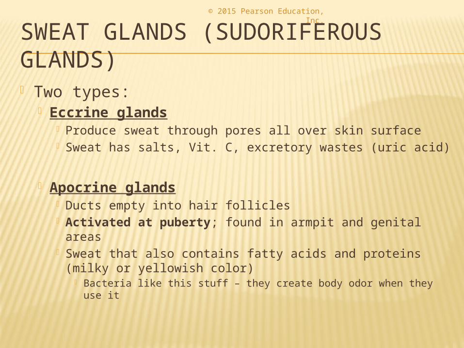
SWEAT GLANDS (SUDORIFEROUS GLANDS) Two types:
Eccrine glands Produce sweat through pores all over skin surface Sweat has salts, Vit. C, excretory wastes (uric acid)
Apocrine glands Ducts empty into hair follicles Activated at puberty; found in armpit and genital
areas Sweat that also contains fatty acids and proteins (milky
or yellowish color) Bacteria like this stuff – they create body odor when they use it
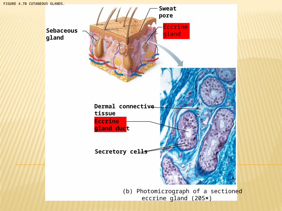
FIGURE 4.7B CUTANEOUS GLANDS.
EccrineglandSebaceous
gland
Sweatpore
Dermal connectivetissueEccrinegland duct
Secretory cells
(b) Photomicrograph of a sectioned eccrine gland (205×)
© 2015 Pearson Education, Inc.
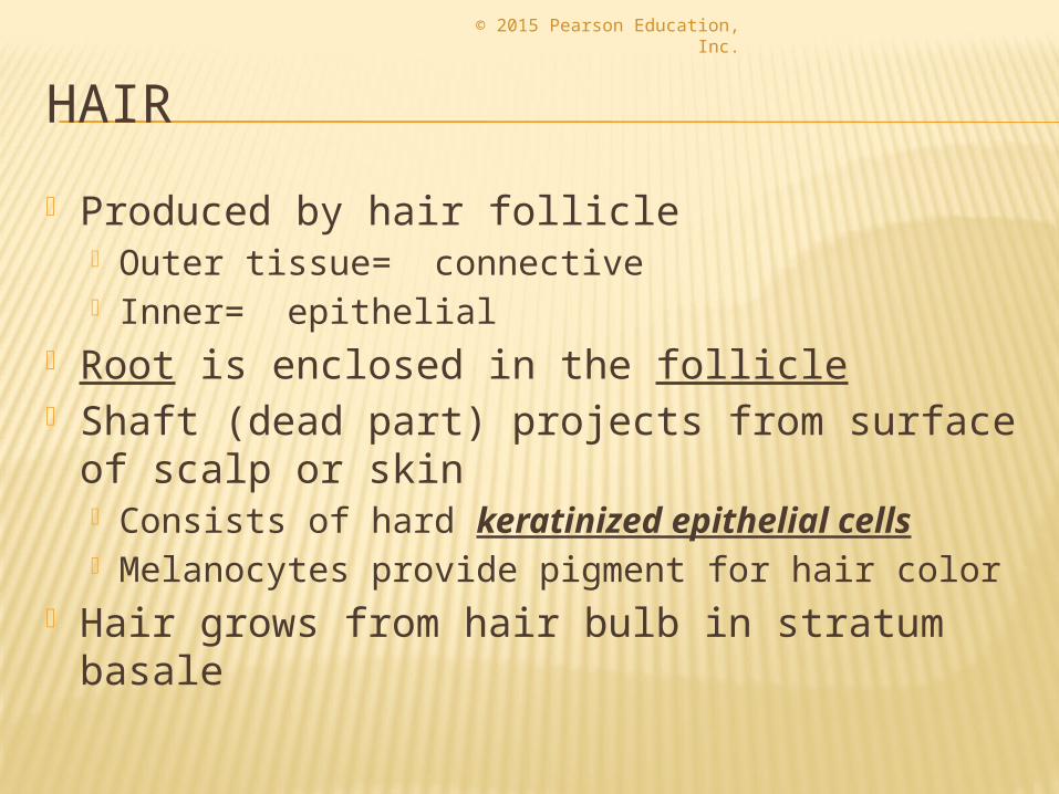
HAIR
Produced by hair follicle Outer tissue= connective Inner= epithelial
Root is enclosed in the follicle Shaft (dead part) projects from surface of
scalp or skin Consists of hard keratinized epithelial cells Melanocytes provide pigment for hair color
Hair grows from hair bulb in stratum basale
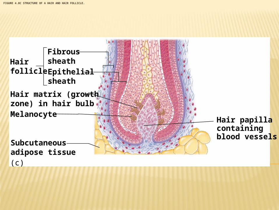
FIGURE 4.8C STRUCTURE OF A HAIR AND HAIR FOLLICLE.
Hairfollicle
(c)
FibroussheathEpithelialsheath
Hair matrix (growthzone) in hair bulbMelanocyte
Subcutaneousadipose tissue
Hair papillacontainingblood vessels
© 2015 Pearson Education, Inc.
APPENDAGES OF THE SKIN
Arrector pili muscle Smooth muscle tissue Pulls hairs upright when person is cold or
frightened (gives us goosebumps) You could consider this a vestigial structure- it
has lost all of its function/use for humans
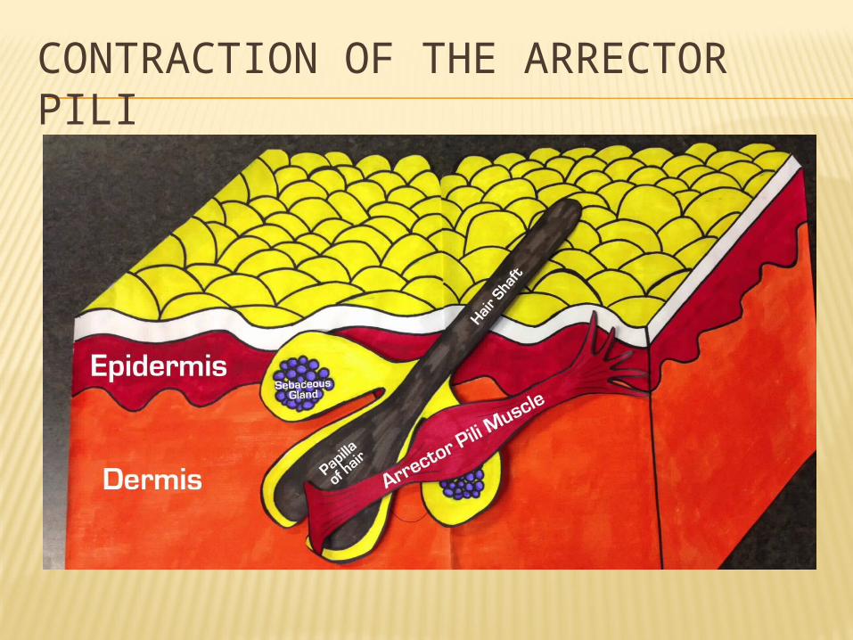
CONTRACTION OF THE ARRECTOR PILI
© 2015 Pearson Education, Inc.
NAILS
Heavily keratinized = very hard Stratum basale is responsible for growth Lack of pigment makes them colorless Growth is similar to that of hair
Functions: Protection Tools Scratch an itch!