Embed Size (px)
Citation preview
ORIGINAL RESEARCH ARTICLEpublished: 02 September 2014doi: 10.3389/fmicb.2014.00459
Oxygenic photosynthesis as a protection mechanism forcyanobacteria against iron-encrustation in environmentswith high Fe2+ concentrationsDanny Ionescu1,2*†, Bettina Buchmann1†, Christine Heim3, Stefan Häusler1, Dirk de Beer1 and
Lubos Polerecky1,4*
1 Microsensor Group, Max-Planck Institute for Marine Microbiology, Bremen, Germany2 Department of Stratified Lakes, Leibniz Institute for Freshwater Ecology and Inland Fisheries, Stechlin, Germany3 Department of Geobiology, Geoscience Center, Georg-August University of Göttingen, Göttingen, Germany4 Department of Earth Sciences – Geochemistry, Faculty of Geosciences, Utrecht University, Utrecht, Netherlands
Edited by:
Hongyue Dang, Xiamen University,China
Reviewed by:
Kathleen Scott, University of SouthFlorida, USAMarie-Ève Garneau, University ofZurich, Switzerland
*Correspondence:
Danny Ionescu, Department ofStratified Lakes, Leibniz Institute forFreshwater Ecology and InlandFisheries, IGB, Dep 3, ExperimentalLimnology, Alte Fischerhütte 2, OTNeuglobsow, 16775 Stechlin,Germanye-mail: [email protected];Lubos Polerecky, Department ofEarth Sciences – Geochemistry,Faculty of Geosciences, UtrechtUniversity, Budapestlaan 4, 3584CD, Utrecht, Netherlandse-mail: [email protected]
†These authors have contributedequally to this work.
If O2 is available at circumneutral pH, Fe2+ is rapidly oxidized to Fe3+, which precipitatesas FeO(OH). Neutrophilic iron oxidizing bacteria have evolved mechanisms to preventself-encrustation in iron. Hitherto, no mechanism has been proposed for cyanobacteriafrom Fe2+-rich environments; these produce O2 but are seldom found encrusted in iron.We used two sets of illuminated reactors connected to two groundwater aquifers withdifferent Fe2+ concentrations (0.9 μM vs. 26 μM) in the Äspö Hard Rock Laboratory (HRL),Sweden. Cyanobacterial biofilms developed in all reactors and were phylogeneticallydifferent between the reactors. Unexpectedly, cyanobacteria growing in the Fe2+-poorreactors were encrusted in iron, whereas those in the Fe2+-rich reactors were not.In-situ microsensor measurements showed that O2 concentrations and pH near thesurface of the cyanobacterial biofilms from the Fe2+-rich reactors were much higherthan in the overlying water. This was not the case for the biofilms growing at lowFe2+ concentrations. Measurements with enrichment cultures showed that cyanobacteriafrom the Fe2+-rich environment increased their photosynthesis with increasing Fe2+concentrations, whereas those from the low Fe2+ environment were inhibited atFe2+ > 5 μM. Modeling based on in-situ O2 and pH profiles showed that cyanobacteriafrom the Fe2+-rich reactor were not exposed to significant Fe2+ concentrations. Wepropose that, due to limited mass transfer, high photosynthetic activity in Fe2+-richenvironments forms a protective zone where Fe2+ precipitates abiotically at a non-lethaldistance from the cyanobacteria. This mechanism sheds new light on the possible role ofcyanobacteria in precipitation of banded iron formations.
Keywords: oxygenic phototrophs, Cyanobacteria, Fe(II), iron-encrustation, banded iron formations, oxygen
microgradients, pH microgradients
INTRODUCTIONAbiotic oxidation of Fe2+ to Fe3+ and subsequent precipitationof FeO(OH) is a function of pH and O2 concentration, and isrelatively rapid when O2 is available at circumneutral pH (Khalilet al., 2011). To prevent iron self-encrustation, iron oxidizing bac-teria developed a variety of protection mechanisms, includingformation of organic matter stalks (Chan et al., 2009; Suzuki et al.,2011) or sheaths (Van Veen et al., 1978) that provide a templatefor FeO(OH) nucleation. For some phototrophic iron oxidiz-ers, a low-pH microenvironment generated by the cell’s protonpumps has been suggested as a mechanism preventing iron self-encrustation (Hegler et al., 2010). Additional adaptations includea hydrophilic cell membrane with a near neutral surface charge toprevent the adhesion of Fe2+/Fe3+ (Saini and Chan, 2013).
Oxygenic phototrophs are often found in microbial matsin Fe2+-rich environments (Pierson et al., 1999; Brown et al.,2005, 2010; Wieland et al., 2005). It is known that when the
photosynthetically active cells are densely packed in a volumewhere transport is limited by diffusion (e.g., in photosyntheticbiofilms or mats), their activity leads to a locally increased pHand O2 concentration (Pierson et al., 1999; Wieland et al., 2005),which should favor locally higher rates of Fe2+ oxidation andprecipitation. Yet, oxygenic phototrophs in Fe2+-rich environ-ments have seldom been found encrusted in precipitated iron(Pierson and Parenteau, 2000). A defense mechanism againstiron self-encrustation that would enable oxygenic phototrophsto thrive in Fe2+-rich environments has hitherto not been sug-gested. Although cyanobacteria that accumulate iron precipitatesintracellularly were described from Yellowstone National Park(Brown et al., 2010), this phenomenon has not been foundelsewhere.
In the Fe2+-rich environment of the Äspö Hard RockLaboratory (HRL), Sweden, which is a man-made research tun-nel, a series of flow-through reactors were set up in 2006 as
www.frontiersin.org September 2014 | Volume 5 | Article 459 | 1
Ionescu et al. Oxygenic phototrophs in Fe(II)-rich environments
part of a study on iron oxidizing bacteria (Ionescu et al., 2014a).Part of the reactors were connected to two aquifers that emergefrom the tunnel wall at the top and bottom of the tunneland differ markedly with respect to concentrations of dissolvedFe2+ (top: ∼ 26 μM, bottom: ∼ 0.9 μM). Half of the reactorswere illuminated and the other half were not. After four yearsof undisturbed incubation, we found that inside the illumi-nated reactors cyanobacterial biofilms developed. Interestingly,cyanobacteria in the Fe2+-poor reactors were mostly encrustedin iron precipitates, whereas those from the Fe+2-rich reactorswere not.
In this study we aimed to understand this counter-intuitiveobservation. Past studies showed that only some morphotypesof cyanobacteria are found encrusted in iron (Pierson andParenteau, 2000; Parenteau and Cady, 2010) while others arenot; however a mechanism to account for these differences wasnot suggested. Pierson et al. (1999) showed that cyanobacterialmats from iron rich environments were characterized by highphotosynthetic O2 production, which was similar to our obser-vation in the cyanobacterial biofilms growing in the Fe2+-richreactors. Hence, we hypothesized that it is the high rate of oxy-genic photosynthesis that allows cyanobacterial proliferation inthe Fe2+-rich environment of the Äspö HRL. Specifically, dueto mass transfer limitations, cyanobacterial photosynthesis cre-ates a microenvironment with elevated O2 concentrations andpH that causes high rates of Fe2+ oxidation and precipitation ata non-lethal distance from the cells and thus prevents their ironself-encrustation.
MATERIALS AND METHODSREACTORSThe Äspö HRL is a man-made research tunnel (length 3.6 km)operated since 1995 by the Swedish Nuclear Waste ManagementFacility (SKB) in the south-east of Sweden. The tunnel inter-sects several groundwater aquifers, each characterized by spe-cific water chemistry (Laaksoharju et al., 1999; Ionescu et al.,2014a). In 2006 two sets of two illuminated reactors were con-nected directly to the aquifers at sites TASF (depth 460 m)and TASA-1327B (depth 185 m) using valves KF0069A01 andHA1327B, respectively. Only chemically inert materials such aspolytetrafluoroethylene (PTFE, Teflon®), PTFE-fiber glass, fluori-natedethylene propylene (FEP) and special PTFE-foam were usedas construction materials to avoid biological contamination fromthe surrounding environment and chemical contamination fromglass- and plastic-ware. The reactors and connection tubing weresterilized with ethanol (70%, overnight) before undergroundinstallation. In each set, reactor 1 (R1) had an air headspaceand gas exchange was allowed through a 0.2 μm membrane fil-ter. Reactor 2 (R2) did not have an air headspace, which wasachieved by elevating the outflow pipe above the height of thereactor. Both reactors were illuminated using two fluorescentlamps (WL11, Brennenstuhl, Tübingen, Germany; Figure S1).Light penetrated inside the reactors through an FEP-window inthe center of the lid. Irradiance behind the window was 60 μmolphotons m−2 s−1, as determined by a quantum irradiance sensor(LI-190 Quantum) connected to a light meter (LI-250, both fromLI-COR Biosciences).
DNA EXTRACTION AND SEQUENCINGTo characterize the composition of the cyanobacterialcommunities developed in the reactors, DNA was extractedas previously described (Ionescu et al., 2012). Pyrosequencingwas done by MrDNA laboratories (Shallowater, TX) using generalbacterial primers 27F and 519R (Lane, 1991). Sequences wereanalyzed using the SILVA NGS pipeline (Ionescu et al., 2012) andthe SILVA 111 database (Quast et al., 2012).
CULTURINGGreen cyanobacterial biomass that developed in the aeratedFe2+-poor (TASF-R1) and aerated/non-aerated Fe2+-rich (1327-R1/R2) reactors was physically separated from the rusty-orangebiomass of iron oxidizing bacteria and transferred on site to sterileBG-11 medium (Rippka et al., 1979). Upon arrival to the labora-tory the samples were transferred to both solid and liquid BG-11medium and grown in constant light (irradiance 60–70 μmolphotons m−2 s−1) at 15◦C to provide light and temperature con-ditions close to those in their natural habitat. To allow for latermicrosensor measurements (see below) without the loss of addediron, cultures were allowed to grow on GF/F filters (Whatmann,diameter 47 mm) that were soaked in Fe2+-free media andplaced on top of pre-grown BG-11 (Fe2+-free) agar plates.Filters were used once the green cyanobacterial biomass wasvisible.
MICROSENSOR MEASUREMENTSDissolved O2 concentration was measured with a fast-respondingClark-type microelectrode (tip diameter ∼ 30 μm). pH was mea-sured using shielded liquid ion exchange glass microelectrodes.Both microsensors were constructed and calibrated as previouslydescribed (Revsbech, 1989; de Beer and Stambler, 2000). In-situmeasurements were conducted either directly in the reactors or, ifnot possible, directly next to them, placing the biofilms in waterfrom the respective reactor. Illumination during all experimentswas provided by a Schott KL1500 lamp (Carl Zeiss AG, Göttingen,Germany), with the incident irradiance adjusted to match thevalue inside the reactors (60 μmol photons m−2 s−1).
For laboratory measurements 0.5 cm filter stripes with a well-developed cyanobacterial biomass were placed in an equally wideflow-through chamber (volume ∼5 mL) connected to a mediareservoir using a peristaltic pump (Figure S2). The medium wascontinuously purged with N2 gas to maintain anoxic conditions.During measurements, Fe2+ was added to the media reservoirwhile subsamples for iron determination were periodically takenfrom the chamber to assure the required concentration (1, 5,10, 30, 50 μM) is maintained. Iron was added from an acidicFe(II)Ammonium Sulfate stock solution resulting in a slight acid-ification of the medium (<0.2 pH units). Used medium was notrecycled.
A similar iron addition experiment was performed also in-situusing a biofilm collected from the aerated Fe2+-poor reactor(TASF-R1). The sample was placed in a 1 L beaker and coveredwith water from the same reactor. The water was purged with N2
and iron was added to a final concentration of 25 μM.In all experiments Fe2+ was measured colorimetrically using
ferrozine as previously described (Riemer et al., 2004).
Frontiers in Microbiology | Aquatic Microbiology September 2014 | Volume 5 | Article 459 | 2
Ionescu et al. Oxygenic phototrophs in Fe(II)-rich environments
NanoSIMS MEASUREMENTSTo test whether iron oxides are being precipitated insidecyanobacterial filaments in a similar way as described by Brownet al. (2010), 57Fe2+ was added to a culture from the non-aerated Fe2+-rich reactor at a concentration of 50 μM. Sampleswere transferred to polycarbonate filters and cyanobacterialfilaments were identified using auto-fluorescence. Areas thatcontained the filaments were marked by a laser dissection micro-scope (LMD7000, Leica), which allowed their later localiza-tion and identification in the nanoSIMS instrument using thebuilt-in CCD camera. NanoSIMS analysis was performed withthe nanoSIMS 50 L instrument (Cameca) available at the MPIBremen. Before each analysis the sample was pre-sputtered witha Cs+ primary ion beam (16 keV, 1.1–3.5 pA) focused on a spotof ∼120 nm diameter for 60–90 s. Subsequently, the same beamwas scanned over the sample in a 256 × 256 pixel raster witha counting time of 1 ms per pixel, and the following ions weredetected: 12C−, 12C14N−, 32S−, 56Fe16O−, and 57Fe16O−. Themass resolution power during all measurements was >8000. Foreach region of interest 30–100 planes were acquired at a rastersize of 15 × 15 or 30 × 30 μm. Data analysis was done with theLook@NanoSIMS software (Polerecky et al., 2012). As a con-trol of the method, the same 57Fe2+-addition experiment andnanoSIMS analysis were performed using samples from mats ofiron oxidizing bacteria.
Fe2+ PROFILE MODELINGDepth profiles of Fe2+ concentrations around the biofilm surfacecould not be measured and were therefore modeled numeri-cally. First, a depth profile of Fe2+ oxidation rates was calculatedaccording to Morgan and Lahav (2007) based on the profile ofO2 concentrations and pH measured in-situ. Subsequently, theserates were used to calculate the evolution of the Fe2+ concentra-tion profile, starting from a flat profile (i.e., Fe2+ concentrationsin the entire modeled domain equal to that measured in the reac-tor water). Specifically, during each time-step, the net decreasein the Fe2+ concentration at a given depth was the result ofthe local Fe2+ oxidation rate combined with the transport bydiffusion determined from the Fe2+ concentrations above andbelow. Fe2+ concentrations at distances greater than the thick-ness of the diffusive boundary layer (DBL) were kept constantand equal to the initial concentrations during the entire cal-culation. DBL thickness was derived from the measured O2
profile. Calculation proceeded in time-steps of 1 × 10⁻³ s until
a steady state was reached. Steady state was defined when thechanges in Fe2+ concentration across the entire profile fell below10−6 μM. Equilibrium constants required for the iron specia-tion were corrected for the ionic strength and temperature ofthe specific feeding aquifer using the Davies (Davies, 1962) andVan’t Hoff (Atkins, 1978) equations, respectively. Ionic strengthof the different waters was calculated using Visual MINTEQ (Ver.3.0). Iron diffusion coefficient was set to 5.82 × 10⁻⁶ cm2 s−1
(Yuan-Hui and Gregory, 1974). As no pH profile was availablefrom the aerated Fe2+-rich reactor, modeling was done only forbiofilms from the non-aerated Fe2+-rich and aerated Fe2+-poorreactors.
RESULTSWATER CHEMISTRYWater in the top aquifer is mainly influenced by the Baltic Seaand recent meteoric water and has a retention time in the bedrockof several weeks (Laaksoharju et al., 1999). It is rich in dissolvedFe2+ (around 26 μM) and has pH of 7.4 and O2 concentrations ofabout 2 μM. In contrast, water in the bottom aquifer is a mixtureof glacial, ancient marine and brine water and was dated to theBoreal age (SICADA, SKB Database). It has markedly lower Fe2+concentrations (0.9 μM), higher pH (8), and O2 concentrationsof about 4 μM. Other notable differences include dissolved inor-ganic carbon (DIC), total alkalinity (Alk), and total dissolved salt(TDS) concentrations, which are about 10- and 100-fold lowerand 5-fold higher in the bottom aquifer, respectively (Table 1).
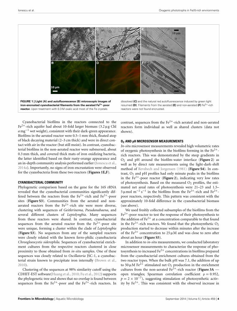
IN-SITU BIOMASSFilamentous cyanobacteria in the aerated reactors connected tothe Fe2+-poor aquifer formed veil-like biofilms (2–3 cm long,2–3 mm thick) that slowly but continuously moved due to theslow water flow in the reactor. Their pale-green color coin-cided with a low chlorophyll a (Chl a) concentration (0.2 μgChl a mg−1 wet weight) and thus presumably low cyanobac-terial biomass. Microscopic observations of biofilm subsamplesrevealed clear iron encrustation and largely diminished auto-fluorescence of the filaments (Figures 1A,B). Upon addition of0.3 M oxalic acid, most of the Fe-oxide crystals dissolved andthe red auto-fluorescence induced by green light, which is typ-ical for cyanobacteria due to their Chl a and phycocyanin con-tent, significantly increased (Figures 1C,D). Due to an extremelylow biomass, biofilms from the non-aerated reactor from theFe2+-poor site were not studied.
Table 1 | Physico-chemical characteristics of the different aquifers connected to the reactors.
Site Sampled pH T Fe2+ O2 Alk DIC DOC TDS H2S NH4 NO3
water ◦C µM µM µM µM C µM C g/l µM µM µM
TASF (Fe2+-poor) Aquifer 7.98 12 0.89 3.7 110 133 183 23.6 0.53 0.61 1.05
R1# 7.94 12 0.89 32.5 110 133 167 23.5 0.53 0.67 1.03
TASA (1327B Fe2+-rich) Aquifer 7.41 15 25.8 2.2 3277 1691 492 5.1 1.88 215 0.12
R1# 7.40 15 27.1 7.2 3277 1933 617 5.1 1.17 217 1.15
R2# 7.39 15 26.4 3.9 3245 1825 558 4.9 0.88 212 1.48
#R1 and R2 are aerated and non-aerated flow reactors, respectively.
www.frontiersin.org September 2014 | Volume 5 | Article 459 | 3
Ionescu et al. Oxygenic phototrophs in Fe(II)-rich environments
FIGURE 1 | Light (A) and autofluorescence (B) microscopic images of
iron-encrusted cyanobacterial filaments from the aerated Fe2+-poor
reactor. Upon treatment with 0.3 M oxalic acid most of the Fe crystals
dissolved (C) and the natural red autofluorescence induced by green lightresumed (D). Filaments from the aerated (E) and non-aerated (F) Fe2+-richreactors were not found encrusted.
Cyanobacterial biofilms in the reactors connected to theFe2+-rich aquifer had about 10-fold larger biomass (3.2 μg Chla mg−1 wet weight), consistent with their dark-green appearance.Biofilms in the aerated reactor were 0.5–1 mm thick, floated atopof black decaying material (2–3 cm thick) and were in direct con-tact with air in the reactor (but still moist). In contrast, cyanobac-terial biofilms in the non-aerated reactor were submersed, about0.3 mm thick, and covered thick mats of iron oxidizing bacteria,the latter identified based on their rusty-orange appearance andan in-depth community analysis performed earlier (Ionescu et al.,2014a). Importantly, no signs of iron encrustation were observedfor the cyanobacteria from these two reactors (Figures 1E,F).
CYANOBACTERIAL COMMUNITYPhylogenetic comparison based on the gene for the 16S rRNArevealed that the cyanobacterial communities significantly dif-fered between the reactors from the Fe2+-rich and Fe2+-poorsites (Figure S3). Communities from the aerated and non-aerated reactors from the Fe2+-rich site were more diverse,clustering with sequences of Geitlerinema, Pseudanabaena, andseveral different clusters of Leptolyngbia. Many sequencesfrom these reactors were shared. In contrast, cyanobacterialsequences from the aerated reactor from the Fe2+-poor sitewere unique, forming a cluster within the clade of Leptolyngbia(Figure S3). No sequences from any of the sampled reactorswere closely related with the known ferro-philic cyanobacteriaChroogloeocystis siderophila. Sequences of cyanobacterial enrich-ment cultures from the respective reactors clustered in closeproximity to those obtained from in-situ samples. One of thesesequences was closely related to Oscillatoria JSC-1, a cyanobac-terial strain known to precipitate iron internally (Brown et al.,2010).
Clustering of the sequences at 98% similarity cutoff using theCDHIT-EST software(Huang et al., 2010; Fu et al., 2012) supportsthe phylogenetic tree and shows that no overlap is found betweensequences from the Fe2+-poor and the Fe2+-rich reactors. In
contrast, sequences from the Fe2+-rich aerated and non-aeratedreactors form individual as well as shared clusters (data notshown).
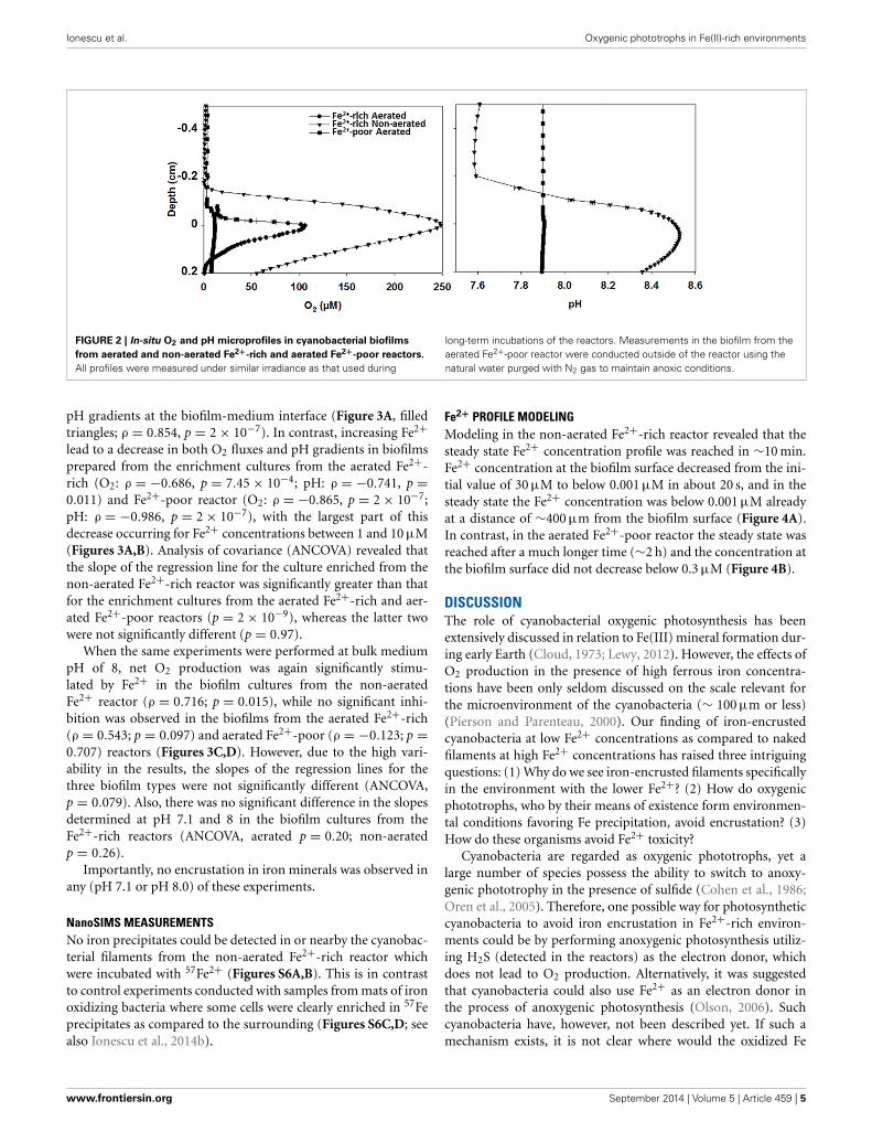
O2 AND pH MICROSENSOR MEASUREMENTSIn-situ microsensor measurements revealed high volumetric ratesof oxygenic photosynthesis in the biofilms forming in the Fe2+-rich reactors. This was demonstrated by the steep gradients inO2 and pH around the biofilm-water interface (Figure 2) aswell as by direct rate measurements using the light-dark-shiftmethod of Revsbech and Jorgensen (1981) (Figure S4). In con-trast, O2 and pH profiles had only minute peaks in the biofilmsin the Fe2+-poor reactor (Figure 2), indicating very low ratesof photosynthesis. Based on the measured O2 profiles, the esti-mated net areal rates of photosynthesis were 21–23 and 1.5–5 μmol m−2 s−1 in the biofilms from the Fe2+-rich and Fe2+-poor reactors, respectively. This pattern was consistent with theapproximately 10-fold difference in the cyanobacterial biomass(see above).
We used freshly collected subsamples of the biofilms from theFe2+-poor reactor to test the response of their photosynthesis tothe addition of Fe2+ at a concentration comparable to that foundin the Fe2+-rich reactors. We found that the photosynthetic O2
production started to decrease within minutes after the increaseof the Fe2+ concentration to 25 μM and was close to zero afterabout an hour (Figure S5).
In addition to in-situ measurements, we conducted laboratorymicrosensor measurements to characterize the response of pho-tosynthesis to increased Fe2+ concentrations in biofilms preparedfrom the cyanobacterial enrichment cultures obtained from thetwo reactor types. When the bulk pH was 7.1, the addition of upto 50 μM Fe2+ stimulated net O2 production in the enrichmentcultures from the non-aerated Fe2+-rich reactor (Figure 3A —open triangles; Spearman correlation coefficient ρ = 0.952,p = 2 × 10⁻⁷), suggesting stimulation of photosynthetic activ-ity by Fe2+. This was consistent with the observed increase in
Frontiers in Microbiology | Aquatic Microbiology September 2014 | Volume 5 | Article 459 | 4
Ionescu et al. Oxygenic phototrophs in Fe(II)-rich environments
FIGURE 2 | In-situ O2 and pH microprofiles in cyanobacterial biofilms
from aerated and non-aerated Fe2+-rich and aerated Fe2+-poor reactors.
All profiles were measured under similar irradiance as that used during
long-term incubations of the reactors. Measurements in the biofilm from theaerated Fe2+-poor reactor were conducted outside of the reactor using thenatural water purged with N2 gas to maintain anoxic conditions.
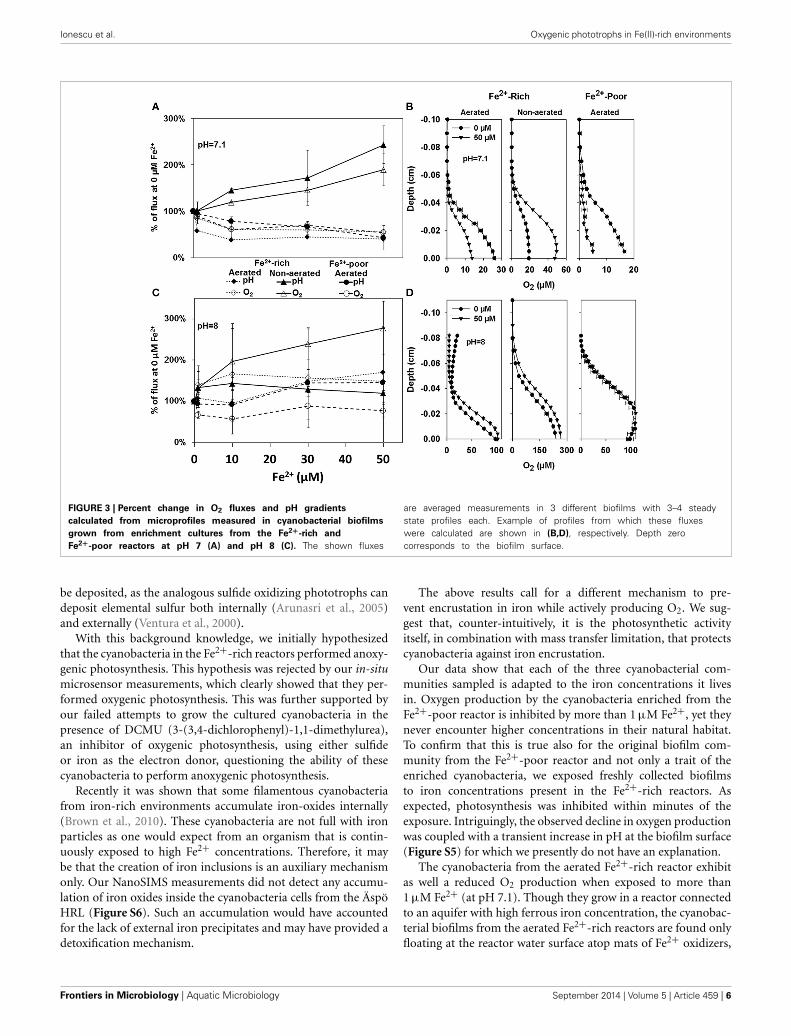
pH gradients at the biofilm-medium interface (Figure 3A, filledtriangles; ρ = 0.854, p = 2 × 10⁻⁷). In contrast, increasing Fe2+lead to a decrease in both O2 fluxes and pH gradients in biofilmsprepared from the enrichment cultures from the aerated Fe2+-rich (O2: ρ = −0.686, p = 7.45 × 10⁻⁴; pH: ρ = −0.741, p =0.011) and Fe2+-poor reactor (O2: ρ = −0.865, p = 2 × 10⁻⁷;pH: ρ = −0.986, p = 2 × 10⁻⁷), with the largest part of thisdecrease occurring for Fe2+ concentrations between 1 and 10 μM(Figures 3A,B). Analysis of covariance (ANCOVA) revealed thatthe slope of the regression line for the culture enriched from thenon-aerated Fe2+-rich reactor was significantly greater than thatfor the enrichment cultures from the aerated Fe2+-rich and aer-ated Fe2+-poor reactors (p = 2 × 10⁻⁹), whereas the latter twowere not significantly different (p = 0.97).
When the same experiments were performed at bulk mediumpH of 8, net O2 production was again significantly stimu-lated by Fe2+ in the biofilm cultures from the non-aeratedFe2+ reactor (ρ = 0.716; p = 0.015), while no significant inhi-bition was observed in the biofilms from the aerated Fe2+-rich(ρ = 0.543; p = 0.097) and aerated Fe2+-poor (ρ = −0.123; p =0.707) reactors (Figures 3C,D). However, due to the high vari-ability in the results, the slopes of the regression lines for thethree biofilm types were not significantly different (ANCOVA,p = 0.079). Also, there was no significant difference in the slopesdetermined at pH 7.1 and 8 in the biofilm cultures from theFe2+-rich reactors (ANCOVA, aerated p = 0.20; non-aeratedp = 0.26).
Importantly, no encrustation in iron minerals was observed inany (pH 7.1 or pH 8.0) of these experiments.
NanoSIMS MEASUREMENTSNo iron precipitates could be detected in or nearby the cyanobac-terial filaments from the non-aerated Fe2+-rich reactor whichwere incubated with 57Fe2+ (Figures S6A,B). This is in contrastto control experiments conducted with samples from mats of ironoxidizing bacteria where some cells were clearly enriched in 57Feprecipitates as compared to the surrounding (Figures S6C,D; seealso Ionescu et al., 2014b).
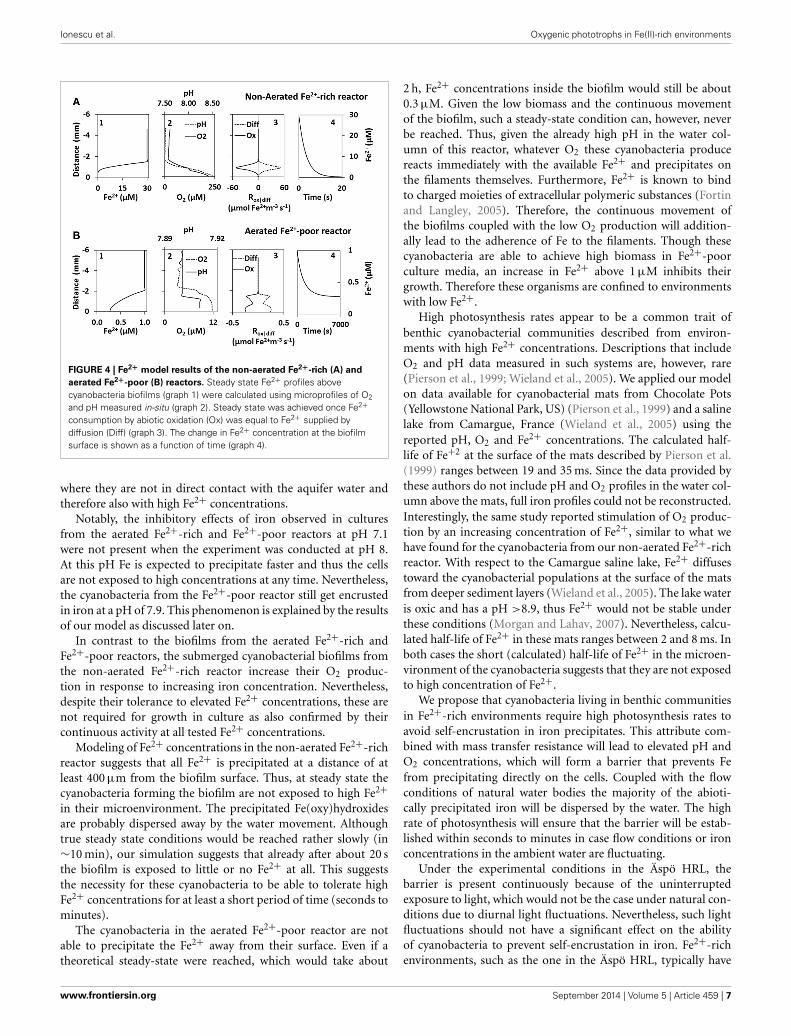
Fe2+ PROFILE MODELINGModeling in the non-aerated Fe2+-rich reactor revealed that thesteady state Fe2+ concentration profile was reached in ∼10 min.Fe2+ concentration at the biofilm surface decreased from the ini-tial value of 30 μM to below 0.001 μM in about 20 s, and in thesteady state the Fe2+ concentration was below 0.001 μM alreadyat a distance of ∼400 μm from the biofilm surface (Figure 4A).In contrast, in the aerated Fe2+-poor reactor the steady state wasreached after a much longer time (∼2 h) and the concentration atthe biofilm surface did not decrease below 0.3 μM (Figure 4B).
DISCUSSIONThe role of cyanobacterial oxygenic photosynthesis has beenextensively discussed in relation to Fe(III) mineral formation dur-ing early Earth (Cloud, 1973; Lewy, 2012). However, the effects ofO2 production in the presence of high ferrous iron concentra-tions have been only seldom discussed on the scale relevant forthe microenvironment of the cyanobacteria (∼ 100 μm or less)(Pierson and Parenteau, 2000). Our finding of iron-encrustedcyanobacteria at low Fe2+ concentrations as compared to nakedfilaments at high Fe2+ concentrations has raised three intriguingquestions: (1) Why do we see iron-encrusted filaments specificallyin the environment with the lower Fe2+? (2) How do oxygenicphototrophs, who by their means of existence form environmen-tal conditions favoring Fe precipitation, avoid encrustation? (3)How do these organisms avoid Fe2+ toxicity?
Cyanobacteria are regarded as oxygenic phototrophs, yet alarge number of species possess the ability to switch to anoxy-genic phototrophy in the presence of sulfide (Cohen et al., 1986;Oren et al., 2005). Therefore, one possible way for photosyntheticcyanobacteria to avoid iron encrustation in Fe2+-rich environ-ments could be by performing anoxygenic photosynthesis utiliz-ing H2S (detected in the reactors) as the electron donor, whichdoes not lead to O2 production. Alternatively, it was suggestedthat cyanobacteria could also use Fe2+ as an electron donor inthe process of anoxygenic photosynthesis (Olson, 2006). Suchcyanobacteria have, however, not been described yet. If such amechanism exists, it is not clear where would the oxidized Fe
www.frontiersin.org September 2014 | Volume 5 | Article 459 | 5
Ionescu et al. Oxygenic phototrophs in Fe(II)-rich environments
FIGURE 3 | Percent change in O2 fluxes and pH gradients
calculated from microprofiles measured in cyanobacterial biofilms
grown from enrichment cultures from the Fe2+-rich and
Fe2+-poor reactors at pH 7 (A) and pH 8 (C). The shown fluxes
are averaged measurements in 3 different biofilms with 3–4 steadystate profiles each. Example of profiles from which these fluxeswere calculated are shown in (B,D), respectively. Depth zerocorresponds to the biofilm surface.
be deposited, as the analogous sulfide oxidizing phototrophs candeposit elemental sulfur both internally (Arunasri et al., 2005)and externally (Ventura et al., 2000).
With this background knowledge, we initially hypothesizedthat the cyanobacteria in the Fe2+-rich reactors performed anoxy-genic photosynthesis. This hypothesis was rejected by our in-situmicrosensor measurements, which clearly showed that they per-formed oxygenic photosynthesis. This was further supported byour failed attempts to grow the cultured cyanobacteria in thepresence of DCMU (3-(3,4-dichlorophenyl)-1,1-dimethylurea),an inhibitor of oxygenic photosynthesis, using either sulfideor iron as the electron donor, questioning the ability of thesecyanobacteria to perform anoxygenic photosynthesis.
Recently it was shown that some filamentous cyanobacteriafrom iron-rich environments accumulate iron-oxides internally(Brown et al., 2010). These cyanobacteria are not full with ironparticles as one would expect from an organism that is contin-uously exposed to high Fe2+ concentrations. Therefore, it maybe that the creation of iron inclusions is an auxiliary mechanismonly. Our NanoSIMS measurements did not detect any accumu-lation of iron oxides inside the cyanobacteria cells from the ÄspöHRL (Figure S6). Such an accumulation would have accountedfor the lack of external iron precipitates and may have provided adetoxification mechanism.
The above results call for a different mechanism to pre-vent encrustation in iron while actively producing O2. We sug-gest that, counter-intuitively, it is the photosynthetic activityitself, in combination with mass transfer limitation, that protectscyanobacteria against iron encrustation.
Our data show that each of the three cyanobacterial com-munities sampled is adapted to the iron concentrations it livesin. Oxygen production by the cyanobacteria enriched from theFe2+-poor reactor is inhibited by more than 1 μM Fe2+, yet theynever encounter higher concentrations in their natural habitat.To confirm that this is true also for the original biofilm com-munity from the Fe2+-poor reactor and not only a trait of theenriched cyanobacteria, we exposed freshly collected biofilmsto iron concentrations present in the Fe2+-rich reactors. Asexpected, photosynthesis was inhibited within minutes of theexposure. Intriguingly, the observed decline in oxygen productionwas coupled with a transient increase in pH at the biofilm surface(Figure S5) for which we presently do not have an explanation.
The cyanobacteria from the aerated Fe2+-rich reactor exhibitas well a reduced O2 production when exposed to more than1 μM Fe2+ (at pH 7.1). Though they grow in a reactor connectedto an aquifer with high ferrous iron concentration, the cyanobac-terial biofilms from the aerated Fe2+-rich reactors are found onlyfloating at the reactor water surface atop mats of Fe2+ oxidizers,
Frontiers in Microbiology | Aquatic Microbiology September 2014 | Volume 5 | Article 459 | 6
Ionescu et al. Oxygenic phototrophs in Fe(II)-rich environments
FIGURE 4 | Fe2+ model results of the non-aerated Fe2+-rich (A) and
aerated Fe2+-poor (B) reactors. Steady state Fe2+ profiles abovecyanobacteria biofilms (graph 1) were calculated using microprofiles of O2
and pH measured in-situ (graph 2). Steady state was achieved once Fe2+consumption by abiotic oxidation (Ox) was equal to Fe2+ supplied bydiffusion (Diff) (graph 3). The change in Fe2+ concentration at the biofilmsurface is shown as a function of time (graph 4).
where they are not in direct contact with the aquifer water andtherefore also with high Fe2+ concentrations.
Notably, the inhibitory effects of iron observed in culturesfrom the aerated Fe2+-rich and Fe2+-poor reactors at pH 7.1were not present when the experiment was conducted at pH 8.At this pH Fe is expected to precipitate faster and thus the cellsare not exposed to high concentrations at any time. Nevertheless,the cyanobacteria from the Fe2+-poor reactor still get encrustedin iron at a pH of 7.9. This phenomenon is explained by the resultsof our model as discussed later on.
In contrast to the biofilms from the aerated Fe2+-rich andFe2+-poor reactors, the submerged cyanobacterial biofilms fromthe non-aerated Fe2+-rich reactor increase their O2 produc-tion in response to increasing iron concentration. Nevertheless,despite their tolerance to elevated Fe2+ concentrations, these arenot required for growth in culture as also confirmed by theircontinuous activity at all tested Fe2+ concentrations.
Modeling of Fe2+ concentrations in the non-aerated Fe2+-richreactor suggests that all Fe2+ is precipitated at a distance of atleast 400 μm from the biofilm surface. Thus, at steady state thecyanobacteria forming the biofilm are not exposed to high Fe2+in their microenvironment. The precipitated Fe(oxy)hydroxidesare probably dispersed away by the water movement. Althoughtrue steady state conditions would be reached rather slowly (in∼10 min), our simulation suggests that already after about 20 sthe biofilm is exposed to little or no Fe2+ at all. This suggeststhe necessity for these cyanobacteria to be able to tolerate highFe2+ concentrations for at least a short period of time (seconds tominutes).
The cyanobacteria in the aerated Fe2+-poor reactor are notable to precipitate the Fe2+ away from their surface. Even if atheoretical steady-state were reached, which would take about
2 h, Fe2+ concentrations inside the biofilm would still be about0.3 μM. Given the low biomass and the continuous movementof the biofilm, such a steady-state condition can, however, neverbe reached. Thus, given the already high pH in the water col-umn of this reactor, whatever O2 these cyanobacteria producereacts immediately with the available Fe2+ and precipitates onthe filaments themselves. Furthermore, Fe2+ is known to bindto charged moieties of extracellular polymeric substances (Fortinand Langley, 2005). Therefore, the continuous movement ofthe biofilms coupled with the low O2 production will addition-ally lead to the adherence of Fe to the filaments. Though thesecyanobacteria are able to achieve high biomass in Fe2+-poorculture media, an increase in Fe2+ above 1 μM inhibits theirgrowth. Therefore these organisms are confined to environmentswith low Fe2+.
High photosynthesis rates appear to be a common trait ofbenthic cyanobacterial communities described from environ-ments with high Fe2+ concentrations. Descriptions that includeO2 and pH data measured in such systems are, however, rare(Pierson et al., 1999; Wieland et al., 2005). We applied our modelon data available for cyanobacterial mats from Chocolate Pots(Yellowstone National Park, US) (Pierson et al., 1999) and a salinelake from Camargue, France (Wieland et al., 2005) using thereported pH, O2 and Fe2+ concentrations. The calculated half-life of Fe+2 at the surface of the mats described by Pierson et al.(1999) ranges between 19 and 35 ms. Since the data provided bythese authors do not include pH and O2 profiles in the water col-umn above the mats, full iron profiles could not be reconstructed.Interestingly, the same study reported stimulation of O2 produc-tion by an increasing concentration of Fe2+, similar to what wehave found for the cyanobacteria from our non-aerated Fe2+-richreactor. With respect to the Camargue saline lake, Fe2+ diffusestoward the cyanobacterial populations at the surface of the matsfrom deeper sediment layers (Wieland et al., 2005). The lake wateris oxic and has a pH >8.9, thus Fe2+ would not be stable underthese conditions (Morgan and Lahav, 2007). Nevertheless, calcu-lated half-life of Fe2+ in these mats ranges between 2 and 8 ms. Inboth cases the short (calculated) half-life of Fe2+ in the microen-vironment of the cyanobacteria suggests that they are not exposedto high concentration of Fe2+.
We propose that cyanobacteria living in benthic communitiesin Fe2+-rich environments require high photosynthesis rates toavoid self-encrustation in iron precipitates. This attribute com-bined with mass transfer resistance will lead to elevated pH andO2 concentrations, which will form a barrier that prevents Fefrom precipitating directly on the cells. Coupled with the flowconditions of natural water bodies the majority of the abioti-cally precipitated iron will be dispersed by the water. The highrate of photosynthesis will ensure that the barrier will be estab-lished within seconds to minutes in case flow conditions or ironconcentrations in the ambient water are fluctuating.
Under the experimental conditions in the Äspö HRL, thebarrier is present continuously because of the uninterruptedexposure to light, which would not be the case under natural con-ditions due to diurnal light fluctuations. Nevertheless, such lightfluctuations should not have a significant effect on the abilityof cyanobacteria to prevent self-encrustation in iron. Fe2+-richenvironments, such as the one in the Äspö HRL, typically have
www.frontiersin.org September 2014 | Volume 5 | Article 459 | 7
Ionescu et al. Oxygenic phototrophs in Fe(II)-rich environments
very low O2 concentrations (Pierson et al., 1999; Ionescu et al.,2014a). Thus, in the dark, when there is no photosynthetic activ-ity, the microenvironment in the cyanobacterial biofilms willrapidly turn anoxic due to the combined effects of respirationand mass transfer resistance, and the cells will not face an encrus-tation problem. The need for a protection mechanism againstiron encrustation returns as soon as the photosynthetic activityresumes upon onset of illumination. Here, a high rate of pho-tosynthesis to minimize the exposure time to high Fe2+ whileproducing O2 will be essential.
In addition to iron self-encrustation, cyanobacteria fromFe2+-rich environments need to deal also with Fe2+ toxicity(Brown et al., 2005; Shcolnick and Keren, 2006). While the iron-barrier provides a protection against iron toxicity during daytime, it will have no effect in the dark. Therefore, we suggest thatthese cyanobacteria require a second mechanism to deal with thehigh Fe2+ concentrations at night, such as elaborate metal exportsystems (Shcolnick et al., 2009).
Single cells or biofilms below a critical biomass appear to getencrusted in iron (Pierson et al., 1999). The amount of biomassnecessary to create the suggested “iron barrier” probably can-not be reached under the conditions in which the cells wouldneed such a barrier. Hence, we suggest that new biofilms of therequired critical biomass are formed in a “Fe2+ neutral” envi-ronment (e.g., floating on the surface or on the shores of a waterbody). When such biofilms become permanently exposed to highFe2+, their survival is determined by the rapid achievement ofa steady state in the distribution of Fe2+ in the microenviron-ment around the biofilms. This in turn depends on biomass, theoverall photosynthesis rate of the biofilms and the ability of thebiofilms to respond to the change in Fe2+ concentrations. Thisalso suggests that a biofilm that contains a critical biomass of“iron tolerant cyanobacteria” may shelter less tolerant species,which could explain the observed partial overlap between thecyanobacterial communities from the aerated and non-aeratedFe2+-rich reactors.
The ability of cyanobacteria to survive in Fe2+-rich environ-ments without getting encrusted while at the same time facili-tating precipitation of iron oxides through their O2 producingphotosynthetic activity may have implications for our under-standing of early Earth geology. Cyanobacteria have been oftensuggested to be involved in the creation of the banded iron for-mations (BIF), the largest Fe depositions on Earth. Whether theirimpact was directly in the water body (Cloud, 1965, 1973) orindirectly by oxygenation of the atmosphere remains controver-sial (Cloud, 1968). However, despite cyanobacterial microfossilsbeing found in BIFs (Cloud, 1965), the overall abundance oforganic matter is scarce (Bontognali et al., 2013). If cyanobacte-ria were directly involved in the formation of BIFs, the existenceof a cyanobacterial protective “iron barrier” may explain the lackof biomass in the Fe precipitates. Such a barrier mechanismwould prevent the cyanobacteria from becoming entrapped inthe precipitated iron. The Fe2+ supplied from the deep primor-dial ocean (Wang et al., 2009) would have been oxidized in thewater column below the chlorophyll maximum and sink down asparticulate Fe(III) without the carryover of biomass. Intuitively,marine cyanobacteria appear to be suitable model organisms to
study the existence of such a mechanism. Nevertheless, modernoceans have been poor in iron for about 2 Ga, a substantial periodfor evolutionary adaptation, which makes it unlikely for marinecyanobacteria today to be adapted to such elevated Fe2+ concen-trations. Hence, in this case, cyanobacteria from brackish-saline,iron-rich environments such as the Äspö HRL still provide thebest model organisms for studying ancient Earth marine analogs.
ACKNOWLEDGMENTSWe are grateful to the SKB staff for technical, logistical, andanalytical support at the Äspö HRL. Joachim Reitner is acknowl-edged for the initiation of the project “Biomineralisation,Biogeochemistry and Biodiversity of chemolithotrophicMicroorganisms in the Tunnel of Äspö (Sweden),” which pro-vided funding for this study through the DFG (German ResearchFoundation) research unit FOR 571. We thank the reviewers forvaluable comments that helped improve the manuscript.
SUPPLEMENTARY MATERIALThe Supplementary Material for this article can be found onlineat: http://www.frontiersin.org/journal/10.3389/fmicb.2014.
00459/abstractFigure S1 | Pictures of an aerated (A) and a non-aerated (B) reactor set up
in the ÄSPÖ Hard Rock Laboratory. The draining tube of the non-aerated
reactor was bended such that flow-through was obtained only when the
reactor was full to the top.
Figure S2 | (A) Experimental setup for laboratory microsensor
measurements. N2-purged medium was run through a small flow
chamber (V∼5 ml) (B) in which filters overgrown with cyanobacterial
biofilms were placed and measured using microsensors (C).
Figure S3 | Maximum Likelihood tree of cyanobacterial sequences from
the different reactors together with reference sequences. The sequences
are labeled according to their origin: 1327 R1 and R2 for the Fe2+-rich
aerated and non-aerated reactors, respectively; TASF R1 for the Fe2+-poor
aerated reactor. Bold labels were used in the case of larger clusters of
sequences of measured cultures.
Figure S4 | O2 microprofiles measured in the cyanobacterial biofilms in the
aerated and non-aerated Fe2+-rich reactors in the dark (filled symbols) and
at incident irradiance of 60 µmol photons m−2 s−1 (open symbols). The
bars represent gross photosynthesis rates (in μM O2 s−1) as measured by
the light-dark-shift method in 100 μm steps in the biofilm. All
measurements were done in-situ.
Figure S5 | In-situ O2 and pH microprofiles measured in the cyanobacterial
biofilm from the aerated Fe2+-poor reactor before and after the addition of
25 µM of Fe2+ at time points indicated in the legend. All profiles were
measured using similar light intensity as used in the reactors. The
measurements were conducted outside of the reactor using the natural
water. N2 gas was bubbled continuously to maintain anoxic conditions.
Figure S6 | Nanoscale Secondary Ion Mass Spectrometry (nanoSIMS)
analysis of a cyanobacterial enrichment culture and an iron oxidizing mat
obtained from the non-aerated Fe2+-rich reactor and incubated with
57Fe2+. The Secondary electron panels (A,C) show the surface topography
with color bar representing signal intensity. No enrichment with 57Fe2+
can be seen near or on the cyanobacterial filaments (B), while an overall
high concentration of 57Fe2+ was detected in the iron oxidizing mat
including a highly enriched single filament (D).
Frontiers in Microbiology | Aquatic Microbiology September 2014 | Volume 5 | Article 459 | 8
Ionescu et al. Oxygenic phototrophs in Fe(II)-rich environments
REFERENCESArunasri, K., Sasikala, C., Ramana, C. V., Süling, J., and Imhoff, J. F. (2005).
Marichromatium indicum sp. nov., a novel purple sulfur gammaproteobac-terium from mangrove soil of Goa, India. Int. J. Syst. Evol. Microbiol. 55,673–679. doi: 10.1099/ijs.0.02892-0
Atkins, P. W. (1978). Physical Chemistry, 1st Edn. Oxford: Oxford University Press.de Beer, D., and Stambler, N. (2000). A microsensor study of light enhanced Ca2+
uptake and photosynthesis in the reef-building hermatypic coral Favia sp. Mar.Ecol. Prog. Ser. 194, 75–85. doi: 10.3354/meps194075
Bontognali, T. R. R., Fischer, W. W., and Föllmi, K. B. (2013). Siliciclasticassociated banded iron formation from the 3.2Ga Moodies Group,Barberton Greenstone Belt, South Africa. Precambrian Res. 226, 116–124.doi: 10.1016/j.precamres.2012.12.003
Brown, I. I., Bryant, D. A., Casamatta, D., Thomas-Keprta, K. L., Sarkisova,S. A., Shen, G., et al. (2010). Polyphasic characterization of a thermo-tolerant siderophilic filamentous cyanobacterium that produces intracellulariron deposits. Appl. Environ. Microbiol. 76, 6664–6672. doi: 10.1128/AEM.00662-10
Brown, I. I., Mummey, D., and Cooksey, K. E. (2005). A novel cyanobacteriumexhibiting an elevated tolerance for iron. FEMS Microbiol. Ecol. 52, 307–314.doi: 10.1016/j.femsec.2004.11.020
Chan, C. S., Fakra, S. C., Edwards, D. C., Emerson, D., and Banfield, J. F.(2009). Iron oxyhydroxide mineralization on microbial extracellular polysac-charides. Geochim. Cosmochim. Acta 73, 3807–3818. doi: 10.1016/j.gca.2009.02.036
Cloud, P. (1973). Paleoecological significance of the banded iron formation. Econ.Geol. 68, 1135–1143. doi: 10.2113/gsecongeo.68.7.1135
Cloud, P. E. (1965). Significance of the gunflint (Precambrian) microflora:photosynthetic oxygen may have had important local effects before becom-ing a major atmospheric gas. Science 148, 27–35. doi: 10.1126/science.148.3666.27
Cloud, P. E. (1968). Atmospheric and hydrospheric evolution on the primi-tive earth: both secular accretion and biological and geochemical processeshave affected earth’s volatile envelope. Science 160, 729–736. doi: 10.1126/sci-ence.160.3829.729
Cohen, Y., Jørgensen, B. B., Revsbech, N. P., and Poplawski, R. (1986). Adaptationto hydrogen sulfide of oxygenic and anoxygenic photosynthesis amongCyanobacteria. Appl. Environ. Microbiol. 51, 398–407.
Davies, C. W. (1962). Ion Association. Washington, DC: Butterworth.Fortin, D., and Langley, S. (2005). Formation and occurrence of biogenic
iron-rich minerals. Earth-Sci. Rev. 72, 1–19. doi: 10.1016/j.earscirev.2005.03.002
Fu, L., Niu, B., Zhu, Z., Wu, S., and Li, W. (2012). CD-HIT: accelerated for clus-tering the next-generation sequencing data. Bioinformatics 28, 3150–3152. doi:10.1093/bioinformatics/bts565
Hegler, F., Schmidt, C., Schwarz, H., and Kappler, A. (2010). Does a low-pHmicroenvironment around phototrophic Fe(II) -oxidizing bacteria prevent cellencrustation by Fe(III) minerals? FEMS Microbiol. Ecol. 74, 592–600. doi:10.1111/j.1574-6941.2010.00975.x
Huang, Y., Niu, B., Gao, Y., Fu, L., and Li, W. (2010). CD-HIT suite: a web serverfor clustering and comparing biological sequences. Bioinformatics 26, 680–682.doi: 10.1093/bioinformatics/btq003
Ionescu, D., Heim, C., Polerecky, L., Ramette, A., Häusler, S., Bizic-Ionescu, M.,et al. (2014a). Diversity of iron oxidizing and reducing bacteria in flow reactorsin the Äspö Hard Rock Laboratory. Geomicrobiol. J. (in press).
Ionescu, D., Heim, C., Polerecky, L., Thiel, V., and de Beer, D. (2014b). Biotic andabiotic oxidation and reduction of iron at circumneutral pH are inseparableprocesses under natural conditions. Geomicrobiol. J. (in press).
Ionescu, D., Siebert, C., Polerecky, L., Munwes, Y. Y., Lott, C., Häusler, S.,et al. (2012). Microbial and chemical characterization of underwater freshwater springs in the Dead Sea. PLoS ONE 7:e38319. doi: 10.1371/jour-nal.pone.0038319
Khalil, M., Teunissen, C., and Langkammer, C. (2011). Iron and neurodegener-ation in multiple sclerosis. Mult. Scler. Int. 2011:606807. doi: 10.1155/2011/606807
Laaksoharju, M., Tullborg, E., Wikberg, P., Wallin, B., and Smellie, J.(1999). Hydrogeochemical conditions and evolution at the Äspö HRL,Sweden. Appl. Geochemistry 14, 835–859. doi: 10.1016/S0883-2927(99)00023-2
Lane, D. (1991). “16S/23S rRNA sequencing,” in Nucleic Acid Techniques in BacterialSystematics, eds E. Stackebrandt and M. Goodfellow (New York; Chichester:John Wiley and Sons), 115–175.
Lewy, Z. (2012). Banded iron formations (BIFs) and associated sedediments do notreflect the physical and chemical properties of early precambrian seas. Int. J.Geosci. 03, 226–236. doi: 10.4236/ijg.2012.31026
Morgan, B., and Lahav, O. (2007). The effect of pH on the kineticsof spontaneous Fe(II) oxidation by O2in aqueous solution–basic princi-ples and a simple heuristic description. Chemosphere 68, 2080–2084. doi:10.1016/j.chemosphere.2007.02.015
Olson, J. M. (2006). Photosynthesis in the Archean era. Photosynth. Res. 88,109–117. doi: 10.1007/s11120-006-9040-5
Oren, A., Ionescu, D., Lipski, A., and Altendorf, K. (2005). Fatty acid analysisof a layered community of cyanobacteria developing in a hypersaline gypsumcrust. Arch. Hydrobiol. Suppl. Algol. Stud. 117, 339–347. doi: 10.1127/1864-1318/2005/0117-0339
Parenteau, M. N., and Cady, S. L. (2010). Microbial piosignatures iniron-mineralized phototrophic mats at Chocolate Pots hot springs,Yellowstone National Park, United States. Palaios 25, 97–111. doi:10.2110/palo.2008.p08-133r
Pierson, B. K., and Parenteau, M. N. (2000). Phototrophs in high iron microbialmats: microstructure of mats in iron-depositing hot springs. FEMS Microbiol.Ecol. 32, 181–196. doi: 10.1111/j.1574-6941.2000.tb00711.x
Pierson, B. K., Parenteau, M. N., and Griffin, B. M. (1999). Phototrophs in high-iron-concentration microbial mats?: physiological ecology of phototrophs in aniron-depositing hot spring. Appl. Environ. Microbiol. 65, 5474–5483.
Polerecky, L., Adam, B., Milucka, J., Musat, N., Vagner, T., and Kuypers, M. M. M.(2012). Look@NanoSIMS–a tool for the analysis of nanoSIMS data in environ-mental microbiology. Environ. Microbiol. 14, 1009–1023. doi: 10.1111/j.1462-2920.2011.02681.x
Quast, C., Pruesse, E., Yilmaz, P., Gerken, J., Schweer, T., Yarza, P., et al.(2012). The SILVA ribosomal RNA gene database project: improved data pro-cessing and web-based tools. Nucleic Acids Res. 41, 1–7. doi: 10.1093/nar/gks1219
Revsbech, N., and Jorgensen, B. B. (1981). Primary production of microal-gae in sediments measured by oxygen microprofile, H14CO3 2 fixa-tion and oxygen exchange methods. Limnol. Oceangr. 26, 717–730. doi:10.4319/lo.1981.26.4.0717
Revsbech, N. P. (1989). An oxygen microsensor with a guard cathode. Limnol.Oceanogr. 34, 474–478. doi: 10.4319/lo.1989.34.2.0474
Riemer, J., Hoepken, H. H., Czerwinska, H., Robinson, S. R., and Dringen, R.(2004). Colorimetric ferrozine-based assay for the quantitation of iron incultured cells. Anal. Biochem. 331, 370–375. doi: 10.1016/j.ab.2004.03.049
Rippka, R., Deruelles, J., Waterbury, J. B., Herdman, M., and Stanier, R. Y.(1979). Generic assignments, strain histories and properties of pure cultures ofcyanobacteria. J. Gen. Microbiol. 111, 1–61. doi: 10.1099/00221287-111-1-1
Saini, G., and Chan, C. S. (2013). Near-neutral surface charge and hydrophilicityprevent mineral encrustation of Fe-oxidizing micro-organisms. Geobiology 11,191–200. doi: 10.1111/gbi.12021
Shcolnick, S., and Keren, N. (2006). Metal homeostasis in cyanobacteria andchloroplasts. Balancing benefits and risks to the photosynthetic apparatus. PlantPhysiol. 141, 805–810. doi: 10.1104/pp.106.079251
Shcolnick, S., Summerfield, T. C., Reytman, L., Sherman, L. A., and Keren, N.(2009). The mechanism of iron homeostasis in the unicellular cyanobacteriumsynechocystis sp. PCC 6803 and its relationship to oxidative stress. Plant Physiol.150, 2045–56. doi: 10.1104/pp.109.141853
Suzuki, T., Hashimoto, H., Matsumoto, N., Furutani, M., Kunoh, H., andTakada, J. (2011). Nanometer-scale visualization and structural analy-sis of the inorganic/organic hybrid structure of Gallionella ferrugineatwisted stalks. Appl. Environ. Microbiol. 77, 2877–2881. doi: 10.1128/AEM.02867-10
Van Veen, W. L., Mulder, E. G., and Deinema, M. H. (1978). The Sphaerotilus-Leptothrix group of bacteria. Microbiol. Rev. 42, 329–356.
Ventura, S., Viti, C., Pastorelli, R., and Giovannetti, L. (2000). Revision of speciesdelineation in the genus Ectothiorhodospira. Int. J. Syst. Evol. Microbiol. 50,583–591. doi: 10.1099/00207713-50-2-583
Wang, Y., Xu, H., Merino, E., and Konishi, H. (2009). Generation of banded ironformations by internal dynamics and leaching of oceanic crust. Nat. Geosci. 2,781–784. doi: 10.1038/ngeo652
www.frontiersin.org September 2014 | Volume 5 | Article 459 | 9

Ionescu et al. Oxygenic phototrophs in Fe(II)-rich environments
Wieland, A., Zopfi, J., Benthien, M., and Kühl, M. (2005). Biogeochemistry ofan iron-rich hypersaline microbial mat (Camargue, France). Microb. Ecol. 49,34–49. doi: 10.1007/s00248-003-2033-4
Yuan-Hui, L., and Gregory, S. (1974). Diffusion of ions in sea water and indeep-sea sediments. Geochim. Cosmochim. Acta 38, 703–714. doi: 10.1016/0016-7037(74)90145-8
Conflict of Interest Statement: The authors declare that the research was con-ducted in the absence of any commercial or financial relationships that could beconstrued as a potential conflict of interest.
Received: 03 June 2014; accepted: 13 August 2014; published online: 02 September2014.
Citation: Ionescu D, Buchmann B, Heim C, Häusler S, de Beer D and Polerecky L(2014) Oxygenic photosynthesis as a protection mechanism for cyanobacteria againstiron-encrustation in environments with high Fe2+ concentrations. Front. Microbiol.5:459. doi: 10.3389/fmicb.2014.00459This article was submitted to Aquatic Microbiology, a section of the journal Frontiersin Microbiology.Copyright © 2014 Ionescu, Buchmann, Heim, Häusler, de Beer and Polerecky.This is an open-access article distributed under the terms of the Creative CommonsAttribution License (CC BY). The use, distribution or reproduction in other forumsis permitted, provided the original author(s) or licensor are credited and that theoriginal publication in this journal is cited, in accordance with accepted academicpractice. No use, distribution or reproduction is permitted which does not comply withthese terms.
Frontiers in Microbiology | Aquatic Microbiology September 2014 | Volume 5 | Article 459 | 10