Embed Size (px)
Citation preview


OXFORD MEDICAL PUBLICATIONS
Cardiothoracic Critical Care

Oxford Specialist Handbooks published and forthcoming General Oxford Specialist HandbooksA Resuscitation Room Guide Addiction Medicine Day Case SurgeryPerioperative Medicine, 2ePharmaceutical MedicinePostoperative Complications, 2e Renal Transplantation
Oxford Specialist Handbooks in AnaesthesiaAnaesthesia for Medical and Surgical
EmergenciesCardiac Anaesthesia Neuroanaesthesia Obstetric Anaesthesia Ophthalmic AnaesthesiaPaediatric Anaesthesia Regional Anaesthesia, Stimulation and
Ultrasound TechniquesThoracic Anaesthesia
Oxford Specialist Handbooks in CardiologyAdult Congenital Heart Disease Cardiac Catheterization and Coronary
Intervention Cardiac Electrophysiology and
Catheter AblationCardiovascular Computed
TomographyCardiovascular Magnetic Resonance Echocardiography, 2eFetal Cardiology Heart Failure, 2eHypertension Inherited Cardiac DiseaseNuclear Cardiology Pacemakers and ICDs Pulmonary HypertensionValvular Heart Disease
Oxford Specialist Handbooks in Critical CareAdvanced Respiratory Critical CareCardiothoracic Critical Care
Oxford Specialist Handbooks in End of Life CareEnd of Life Care in CardiologyEnd of Life Care in DementiaEnd of Life Care in Nephrology End of Life Care in Respiratory
DiseaseEnd of Life in the Intensive
Care Unit
Oxford Specialist Handbooks in NeurologyEpilepsy Parkinson’s Disease and Other Movement
Disorders Stroke Medicine
Oxford Specialist Handbooks in OncologyPractical Management of Complex
Cancer Pain
Oxford Specialist Handbooks in PaediatricsPaediatric Dermatology Paediatric Endocrinology and Diabetes Paediatric Gastroenterology,
Hepatology, and Nutrition Paediatric Haematology and Oncology Paediatric Intensive CarePaediatric Nephrology, 2ePaediatric Neurology, 2ePaediatric Radiology Paediatric Respiratory Medicine Paediatric Rheumatology
Oxford Specialist Handbooks in Pain MedicineSpinal Interventions in Pain Management
Oxford Specialist Handbooks in PsychiatryChild and Adolescent Psychiatry Forensic PsychiatryOld Age Psychiatry
Oxford Specialist Handbooks in RadiologyInterventional Radiology Musculoskeletal Imaging Pulmonary ImagingThoracic Imaging
Oxford Specialist Handbooks in SurgeryCardiothoracic Surgery, 2e Colorectal SurgeryGastric and Oesophageal SurgeryHand Surgery Hepatopancreatobiliary Surgery NeurosurgeryOperative Surgery, 2e Oral and Maxillofacial Surgery Otolaryngology and Head and Neck
Surgery Paediatric SurgeryPlastic and Reconstructive Surgery Surgical Oncology Urological Surgery Vascular Surgery

1
Oxford Specialist Handbooks in Critical Care
Cardiothoracic Critical CareEdited by
Robyn SmithDepartment of AnaesthesiaGolden Jubilee National HospitalClydebank, UK
Mike HigginsGolden Jubilee National HospitalClydebank, UK
Alistair MacfieDepartment of AnaesthesiaGolden Jubilee National HospitalClydebank, UK

3Great Clarendon Street, Oxford, OX2 6DP, United Kingdom
Oxford University Press is a department of the University of Oxford. It furthers the University’s objective of excellence in research, scholarship, and education by publishing worldwide. Oxford is a registered trade mark of Oxford University Press in the UK and in certain other countries
© Oxford University Press 204
The moral rights of the authors have been asserted
First Edition published in 204
Impression:
All rights reserved. No part of this publication may be reproduced, stored in a retrieval system, or transmitted, in any form or by any means, without the prior permission in writing of Oxford University Press, or as expressly permitted by law, or under terms agreed with the appropriate reprographics rights organization. Enquiries concerning reproduction outside the scope of the above should be sent to the Rights Department, Oxford University Press, at the address above
You must not circulate this work in any other form and you must impose this same condition on any acquirer
Published in the United States of America by Oxford University Press 98 Madison Avenue, New York, NY 006, United States of America
British Library Cataloguing in Publication Data
Data available
Library of Congress Control Number: 203954327
ISBN 978–0–9–969295–8
Printed in Great Britain by Ashford Colour Press Ltd, Gosport, Hampshire
Oxford University Press makes no representation, express or implied, that the drug dosages in this book are correct. Readers must therefore always check the product information and clinical procedures with the most up-to-date published product information and data sheets provided by the manufacturers and the most recent codes of conduct and safety regulations. The authors and the publishers do not accept responsibility or legal liability for any errors in the text or for the misuse or misapplication of material in this work. Except where otherwise stated, drug dosages and recommendations are for the non-pregnant adult who is not breast-feeding.
Links to third party websites are provided by Oxford in good faith and for information only. Oxford disclaims any responsibility for the materialscontained in any third party website referenced in this work.

v
The specialty of intensive care is relatively new, starting in the 950s with the polio epidemics, and maturing over the years to the point where it is now recognized as a specialty in its own right in the UK and several other countries worldwide.
Patients undergoing cardiac surgery have always been admitted directly to ICU post-operatively. Cardiothoracic intensive care, usually delivered by cardiothoracic anaesthetists, has resulted in excellent outcomes from major cardiac surgery. This system of patient care and its results are the envy of many other branches of surgery.
With developments in surgery, newer extracorporeal systems for sup-port of cardiac and respiratory function, the survival of patients with con-genital cardiac anomalies into adulthood and greater expectations from clinicians and patients, cardiothoracic intensive care is becoming a super-specialty of both intensive care medicine and anaesthesia. Indeed we are seeing the development of the cardiothoracic intensivist who is not also an anaesthetist.
The authors are established consultants in cardiothoracic anaesthesia and intensive care who bring many years of practical experience in the specialty to this handbook. Trainee attachments to cardiac critical care are often shorter than in days gone by, whilst the technology moves on apace. A gap had arisen for a practical guide to the specialty written by experts suit-able for use by trainees and more experienced clinicians in intensive care medicine, anaesthesia, and surgery alike. A strength of this volume is that it will not only be of use in specialist critical care, patients present to general intensive care units with significant cardiac and respiratory problems and this handbook contains many chapters relevant to all intensivists includ-ing up to date management of heart failure and cardiac pacing. Echo both transthoracic and transoesophageal, skills spilling out into wider critical care practice, are well covered.
A particular example of the utility of this book to the general intensiv-ist and anaesthetist is the comprehensive chapter on the anatomical and physiological consequences of congenital heart disease, its treatment and long term sequelae. Clear descriptions and illustrations will help the non- specialist formulate care plans for the management of these complex patients when presenting for none cardiac surgery, obstetric care or as an emergency.
Oxford specialist handbooks are recognized as high quality, concise, and authoritarian, this volume is no exception. Writing and editing such a wide ranging and comprehensive textbook is an epic, indeed herculean task and the authors are to be commended for their efforts.
Anna BatchelorConsultant in Anaesthesia and Intensive Care Medicine
Royal Victoria InfirmaryNewcastle, UK
Foreword


vii
This is an exciting time in intensive care medicine. The specialty has grown and matured rapidly from its early roots in anaesthesia. In the UK the for-mation of the new Faculty of Intensive Care Medicine marks an unequivocal coming of age. And yet, almost unnoticed, on the margins of the main story, another immensely significant development has been gathering pace; the crystallisation of cardiothoracic intensive care as a clinical entity in its own right. Just as general intensive care grew from the emergence of effective technologies to support the failing lung, so cardiothoracic intensive care has been catalysed by new and effective techniques of circulatory support.
Cardiothoracic intensive care has seen recent major developments in mechanical cardiovascular support (intra arterial balloon pump, ventricular assist devices and extracorporeal membrane oxygenation) and drug thera-pies such as nitric oxide and new inotropic agents. Clinical management has seen a dramatic shift from pragmatic, experience-based algorithms to care based on early diagnosis, proactive intervention and pathophysiologi-cal understanding. A major factor in this shift has been the development of sophisticated bed-side echocardiography.
The new clinical infant is still undeveloped. The shape of its future growth politically and clinically is not yet clear. Traditionally much of the workload has been based around care of patients after cardiac surgery, but new workstreams are developing rapidly, particularly in advanced heart failure and around cardiological interventions. One thing is clear, cardiothoracic intensive care is now at the cutting edge of intensive care medicine.
This book is primarily aimed at cardiothoracic intensive care residents, fellows and nursing staff. It is intended as a practical tool for those who find themselves in positions of responsibility, often at the start of a steep learning curve. It is a bedside handbook, not a textbook. It can be used as a quick clinical reference source, as a guide to deeper study and as an aide memoire. It provides an overview of the subspecialty focussing on the pro-cedures, skills, guidelines, and technologies which are particular to cardiotho-racic intensive care. It forms a complimentary text to companion volumes on cardiothoracic anaesthesia and general intensive care.
The material is grouped into four intuitive headings. These sections are broken up into brief chapters each focusing on a key topic and designed to allow quick access to relevant information. The style is deliberately didactic with boxes and bullet points, summary diagrams, key learning points and references for further reading. Where possible the material is based on evidence-based critical appraisal of recent trial studies and international guidelines and it follows British and European accreditation programmes.The authors are all experienced practitioners in cardiothoracic critical care, but above all they are enthusiasts for their subject and the specialty. If this text supports the clinical development of a new generation of enthusiasts, it will have done its work well.
Preface


ix
Symbols and Abbreviations xi
Contributors xvii
Part Early postoperative management
The normal postoperative cardiac patient 3 2 Risk prediction and outcome 3 3 Fast-tracking the low-risk patient 2 4 Resuscitation in the cardiac intensive care setting 25 5 Myocardial ischaemia and infarction 3 6 Bleeding management 39 7 Postoperative hypotension 49 8 Glucose, lactate, and acid–base 57 9 Postoperative medications 67 0 Information handover and care planning 75
Part 2 Organ dysfunction
Heart 83 2 Lung 07 3 Kidney 5 4 Gastrointestinal 27 5 Nervous system 39 6 Immune system 49 7 Haematology 57
Part 3 Specific patient groups
8 The thoracic patient 7 9 The adult patient with congenital heart disease 87
Contents

contentsx
20 The obstetric patient with cardiac disease 20 2 Heart failure resistant to standard
medical therapy 209 22 The transplant patient 227 23 Acute cardiology 239
Part 4 Treatments and procedures
24 Central venous cannulation, pulmonary
artery catheter, and minimally invasive cardiac
output monitoring 253 25 Airway management 263 26 Respiratory management 275 27 Circulatory support 287 28 Epicardial pacing 303 29 Sedation and pain relief 33 30 Wound management 32 3 Infection control and prevention 329 32 Bedside echocardiography 339
Index 37

xi
b cross-referencei increasedd decreased> greater than< less thanM website7 approximately2D two-dimensionalACE angiotensin-converting enzymeACHD adult congenital heart diseaseACR albumin:creatinine ratioACS acute coronary syndromeACT activated clotting timeACV assist control ventilationAF atrial fibrillationAHA American Heart AssociationAKI acute kidney injuryALI acute lung injuryALS Advanced Life SupportAPTT activated partial thromboplastin timeAR aortic regurgitationARDS acute respiratory distress syndromeAS aortic stenosisAUC area under the curveAV atrioventricularAVR aortic valve replacementBBB bundle branch blockBIPAP bi-level positive airway pressureBMI body mass indexBNP brain natriuretic peptideBP blood pressureBPF bronchopleural fistulaCABG coronary artery bypass graftCALS Cardiac Surgery Advanced Life SupportcAMP cyclic adenosine monophosphateCDI Clostridium difficile infection
Symbols and Abbreviations

SymbolS and abbreviationSxii
CF cystic fibrosisCFM colour flow mappingcGMP cyclic guanosine monophosphateCICU cardiac intensive care unitCKD chronic kidney diseaseCMV cytomegalovirusCNI calcineurin inhibitorCNS central nervous systemCO cardiac outputCO2 carbon dioxideCOPD chronic obstructive pulmonary diseaseCPAP continuous positive airway pressureCPB cardiopulmonary bypassCPP cerebral perfusion pressureCPR cardiopulmonary resuscitationCRP C-reactive proteinCRRT continuous renal replacement therapyCRT cardiac resynchronization therapyCT computed tomographyCTx cardiac transplantationCVC central venous cannulationCVP central venous pressureCVS cardiovascular systemCVVH veno-venous haemofiltrationCWD continuous wave DopplerCXR chest X-rayDHCA deep hypothermic circulatory arrestDHF decompensated heart failureDLCO carbon monoxide diffusing capacityDLT double-lumen tubeDVT deep vein thrombosisEACTS European Association for Cardio-Thoracic SurgeryECF extracellular fluidECG electrocardiogramECLS extracorporeal lung supportECMO extracorporeal membrane oxygenationEDD end-diastolic dimensionEF ejection fractioneGFR estimated glomerular filtration rateER enhanced recovery

SymbolS and abbreviationS xiii
ERC European Resuscitation CouncilESA erythropoiesis stimulating agentESC European Society of CardiologyESPEN European Society for Parenteral and Enteral NutritionETT endotracheal tubeFATE focus-assessed transthoracic echocardiographyFEV forced expiratory volume in secondFFP fresh frozen plasmaGFR glomerular filtration rateGI gastrointestinalGTN glyceryl trinitrateHAI hospital-acquired infectionhb haemoglobinHDU high dependency unitHIT heparin induced thrombocytopeniaHOCM hypertrophic obstructive cardiomyopathyHR heart rateIABP intra-aortic balloon pumpICU intensive care unitIE infective endocarditisIJV internal jugular veinINR international normalized ratioIPAP inspiratory positive airway pressureIPF idiopathic pulmonary fibrosisIPPV intermittent positive pressure ventilationIRRT intermittent renal replacement therapyITU intensive therapy unitIV intravenousJVP jugular venous pressureJVP jugular venous pressureK+ potassiumKDIGO Kidney Diseases: Improving Global OutcomesL litreLAX long axisLBBB left bundle branch blockLIMA left internal mammary arteryLMWH low-molecular-weight heparinLTx lung transplantationLV left ventricle/ventricularLVAD left ventricular assist device

SymbolS and abbreviationSxiv
LVEF left ventricular ejection fractionLVH left ventricular hypertrophyLVOT left ventricular outflow tractLVRS lung volume reduction surgeryMAP mean arterial pressuremcg microgramMCS mechanical circulatory supportmg milligramMI myocardial infarctionMMF mycophenolate mofetilMR mitral regurgitationMRI magnetic resonance imagingMRSA meticillin-resistant Staphylococcus aureusMS mitral stenosisMV mitral valveMVO
2 myocardial oxygen consumptionMVP mitral valve prolapseNG nasogastricNIBP non-invasive blood pressureNICE National Institute for Health and Clinical ExcellenceNIV non-invasive ventilationNMDA N-methyl-D-aspartateNOMI non-occlusive mesenteric ischaemiaNSAID non-steroidal anti-inflammatory drugNSR normal sinus rhythmNT-proBNP N-terminal-pro brain natriuretic peptideNYHA New York Heart AssociationO2 oxygenOOHCA out-of-hospital cardiac arrestPA pulmonary arteryPAC pulmonary artery catheterPAFC pulmonary artery flotation catheterPAP pulmonary artery pressurePAWP pulmonary artery wedge pressurePCR protein:creatinine ratioPCWP pulmonary capillary wedge pressurePDA posterior descending coronary arteryPE pulmonary embolismPEA pulseless electrical activityPEEP positive end-expiratory pressure

SymbolS and abbreviationS xv
PF platelet factorPFT pulmonary function testPISA proximal isovelocity surface areaPPE personal protective equipmentPPI proton pump inhibitorp-SIMV pressure synchronized intermittent mandatory ventilationPSV pressure support ventilationPT prothrombin timePVR pulmonary vascular resistancePW pressure wavePWD pulsed wave DopplerRCA right coronary arteryROSC return of spontaneous circulationRRT renal replacement therapyRV right ventricle/ventricularRVAD right ventricular assist deviceSAM systolic anterior motionSAX short axisSBE standard base excessSCV subclavian veinSIMV synchronized intermittent mandatory ventilationSIRS systemic inflammatory response syndromeSMR standardized mortality ratioSpO
2 saturation of peripheral oxygenSV stroke volumeSVC superior vena cavaSvO2 mixed venous oxygen saturationSVR systemic vascular resistanceTCI target-controlled infusionTOE transoesophageal echocardiographyTPN total parenteral nutritionTR tricuspid regurgitationTS tricuspid stenosisTTE transthoracic echocardiographyTV tricuspid valveV/Q ventilation/perfusionVAC vacuum-assisted closureVAD ventricular assist deviceVAP ventilator-associated pneumoniaVAS Visual Analogue Scale

SymbolS and abbreviationSxvi
VATS video-assisted thoracoscopic surgeryVCV volume controlled ventilationVF ventricular fibrillationVILI ventilator-induced lung injuryVSD ventricular septal defectVT ventricular tachycardiaVTE venous thromboembolismVTI velocity-time integral

xvii
Lynne AndersonConsultant AnaesthetitstDepartment of AnaesthesiaGolden Jubilee National HospitalClydebank, UK
Michael BrettAnaesthetics and Intensive Care MedicineRoyal Alexandra HospitalPaisley, UK
Anton ButerConsultant General SurgeonGeneral SurgeryRoyal Alexandra HospitalPaisley, UK
Coralie CarleConsultant in Anaesthesia and Critical Care MedicinePeterborough City HospitalPeterborough,UK
Ian ColquhounConsultant Cardiothoracic SurgeonWest of Scotland Heart and Lung UnitGolden Jubilee National HospitalClydebank, UK
Mark DavidsonPaediatric IntensivistPaediatric Intensive Care UnitRoyal Hospital for Sick ChildrenGlasgow, UK
Joel DunningCardiothoracic UnitJames Cook University HospitalMiddlesbrough, UK
Clare GardnerConsultant AnaesthetistSt John’s HospitalLivingston, UK
Roy GardnerScottish National Advanced Heart Failure ServiceGolden Jubilee National HospitalClydebank, UK
Donna GreenhalghConsultant in Cardiothoracic AnaesthesiaUniversity Hospital of South ManchesterStockport UK
Doshi HarikrishnaDepartment of Congenital Cardiac SurgeryAlder Hey Children’s HospitalLiverpool, UK
Chris HawthorneClinical LecturerAcademic Unit of Anaesthesia, Pain and Critical Care MedicineUniversity of GlasgowGlasgow, UK
Stephen HickeyDepartment of AnaesthesiaGolden Jubilee National HospitalClydebank, UK
Mike HigginsMedical DirectorGolden Jubilee National HospitalClydebank, UK
Contributors

xviii CONTRIBUTORS
Martin HughesConsultant in Anaesthesia and Intensive Care MedicineIntensive Care UnitGlasgow Royal InfirmaryGlasgow, UK
Theresa InksterConsultant MicrobiologistDepartment of MicrobiologyGlasgow Royal InfirmaryGlasgow, UK
Nitish KhannaConsultant MicrobiologistDepartment of MicrobiologySouthern General HospitalGlasgow, UK
Adarsh LalDepartment of AnaesthesiaGolden Jubilee National HospitalClydebank, UK
Ninian LangClinical Lecturer in CardiologyUniversity of Edinburgh Edinburgh, UK
Alistair MacfieDepartment of AnaesthesiaGolden Jubilee National HospitalClydebank, UK
Ken McKinlayConsultant in Anaesthesia and Intensive CareDepartment of AnaesthesiaGolden Jubilee National HospitalClydebank, UK
Naomi MaySpecialist Registrar in AnaestheticsDepartment of AnaestheticsWest of ScotlandGlasgow, UK
Neal PadmanabhanConsultant NephrologistRenal UnitWestern InfirmaryGlasgow, UK
John PayneScottish National Advanced Heart Failure ServiceGolden Jubilee National HospitalClydebank, UK
Jorg PrinzlinDepartment of AnaesthesiaGolden Jubilee National HospitalClydebank, UK
Isma QuasimDepartment of AnaesthesiaGolden Jubilee National HospitalClydebank, UK
Stefan SchraagDepartment of AnaesthesiaGolden Jubilee National HospitalClydebank, UK
Jason ShahinDepartment of Critical CareDivision of pulmonary medicineMcGill UniversityMontreal, Quebec, Canada
Ben ShelleyClinical Research FellowUniversity of GlasgowGlasgow, UK
Andrew SinclairDepartment of AnaesthesiaGolden Jubilee National HospitalClydebank, UK
Robyn SmithDepartment of AnaesthesiaGolden Jubilee National HospitalClydebank, UK

xixCONTRIBUTORS
Tim StrangDepartment of AnaesthesiaUniversity of South ManchesterManchester, UK
Alison TaylorRegistrar in NephrologyGlasgow Renal and Transplant UnitWestern InfirmaryGlasgow, UK
Niki WalkerConsultant CardiologistGolden Jubilee National HospitalClydebank, UK
Stephen WebbDepartment of Anaesthesia and Intensive CarePapworth Hospital NHS Foundation TrustCambridge, UK
Alice YoungDietitianGolden Jubilee National HospitalClydebank, UK


Early postoperative management
The normal postoperative cardiac patient 3 2 Risk prediction and outcome 3 3 Fast-tracking the low-risk patient 2 4 Resuscitation in the cardiac intensive care setting 25 5 Myocardial ischaemia and infarction 3 6 Bleeding management 39 7 Postoperative hypotension 49 8 Glucose, lactate, and acid–base 57 9 Postoperative medications 67 0 Information handover and care planning 75
Part


3Chapter
The normal postoperative cardiac patient
Introduction 4Effects of surgery and cardiopulmonary bypass 5Typical progression 7Routine management 9Warning signs

4 ChaPTER The normal postoperative cardiac patient
IntroductionCardiac surgery involves considerable physiological trespass. Nevertheless, most cardiac patients recover uneventfully with comfortable margins of safety.
however, a small proportion of routine patients develop an important treatable complication. a second small group of patients are critically ill and are dependant on exact optimization of their physiology, either because of their preoperative condition or their operative course or both. The latter group are usually obvious and are often already on substantial support and invasive monitoring when they leave theatre. Recognizing the first group is crucial and is a key skill of cardiac intensive care. The key to success with these patients is early proactive management. In turn, this requires attention to detail, an understanding of normal postoperative course, close serial observation, and excellent diagnosis employing early use of additional monitoring and investigation.

EFFECTs oF suRGERy aNd CaRdIoPulMoNaRy ByPass 5
Effects of surgery and cardiopulmonary bypassInflammatory responseMajor surgery and cardiopulmonary bypass (CPB) both cause a systemic inflammatory response. all patients who have undergone cardiac sur-gery will have some degree of inflammatory activation, even those who have undergone off-pump surgery. In a substantial proportion of patients this response will be clinically apparent and in a small fraction it will be problematic.
Coolingduring CPB patients are often cooled to 32°C or lower. despite initial re-warming, after separation from bypass there may be a re-distribution of heat to poorly perfused tissues leading to an ‘after drop’ in core tem-perature. once anaesthesia wears off, the low core temperature will trigger reflex shivering. This in turn leads to i cardiac output (Co) and i respira-tory workload to service the muscle activity. Re-warming normally occurs at 0.5–2°C/hour and is dependent on a normal Co and blood volume.
Pulmonary effectsGas exchange is often measurably impaired after cardiac surgery. This is due to a combination of causes including atelectasis as a result of anaesthesia and lung collapse during CPB, and i lung water secondary to the inflamma-tory response. Relatively large doses of opioid drugs may be used during cardiac anaesthesia which may result in postoperative respiratory depres-sion. Conversely, pain contributes to atelectasis and impaired gas exchange.
Urine output and fluid balancePatients have significant fluid derangements as a result of CPB. There is a fluid load as a result of the pump-prime which leads to an increase in total body fluid. however, the intravascular space may be depleted as a result of capillary leak. The kidneys are at risk of insult from CPB. There is a tradition of giving the osmotic diuretic mannitol during bypass to promote clearance of excess fluid. This leads to high postoperative urine volumes in the first few hours after surgery. however, at the same time patients commonly require a positive fluid balance to maintain pre-load because of inflamma-tory activation.
PainPain immediately after median sternotomy is generally less problematic than after major abdominal surgery and can be managed by a range of appropri-ate techniques.
Cardiac functionCardiac contractility may be adversely affected by ischaemia during bypass or intraoperative handling and by reperfusion injury (stunning). This process may worsen in the first few hours after surgery.

6 ChaPTER The normal postoperative cardiac patient
BleedingCPB is associated with abnormal haemostasis through a series of mecha-nisms involving coagulopathy, i fibrinolysis, haemodilution, and qualitative and quantitative platelet dysfunction. only a minority of patients will have a clinically significant lesion.

TyPICal PRoGREssIoN 7
Typical progressionThe following represents patients following a traditional progression. This may be modified by modern enhanced recovery techniques.
On admission• Cold core (34–36°C)• large core–peripheral gradient (0°C)• Polyuric• deeply sedated• Fully ventilated• Good gas exchange• Variable mediastinal drainage• Normal coagulation• Variable inotrope and pressor support• anaemic (haemoglobin (hb) 70–0g/l)• supine position• high/normal blood glucose• May be relatively bradycardic and pacing dependent.
First hour• Increasing core temp (0.5–2°C over the hour)• Core–peripheral gradient remains high• May shiver• high urine output (00–300ml/h)• Remains sedated• Gas exchange stable• drain loss up to 200ml• Variable pressor support but often tendency to hypotension requiring
moderate volume transfusion 500–000ml (± low-dose pressors)• May have increasing or decreasing inotrope requirements• May require insulin to control blood glucose• Falling K+ requires supplementation.
–4 hours• Rewarmed to 36.8°• Core–peripheral gradient normalized (<3°C)• urine output tapering to normal (0.5–ml/kg/hour)• sat up >30°: gas exchange stable or improving with posture change• stabilized blood pressure (BP) with• stabilized pressor requirements• stabilized low inotrope requirement• decreasing volume requirements• stable and normal acid–base levels• Commonly in positive fluid balance (up to l)• decreasing mediastinal drainage tapering to <50ml/hour• usually stable/increasing haematocrit (capillary leak); hb >70g/l• Gas exchange good or improving• Normal or slightly raised (>7.0) Co2 (mild narcotization)• Minimal sedation and interacting purposefully

8 ChaPTER The normal postoperative cardiac patient
• low/no pain• Breathing spontaneously• Interacting purposefully• May be extubated• May require insulin to control blood glucose• Falling K+ requires supplementation• stabilizing rhythm (decreasing pacing dependence).
4–6 hours• as for –4 hours• often d pressor requirements as sedation stopped• Most are extubated.

RouTINE MaNaGEMENT 9
Routine managementThe majority of patients require minimal intervention and are best served by following routine protocols.
VentilationPatients are usually ventilated for several hours after cardiac surgery. Typical ventilator settings employ relatively low tidal volumes (0.7ml/kg) with a small amount of positive end-expiratory pressure (PEEP) (5cmh2o) to counteract atelectasis. Patients are weaned onto a spontaneous breathing mode as soon as possible. Patients should be extubated once they reach the required criteria and do not need progressive weaning of mechanical support. a relatively high Co2 is likely to indicate narcosis rather than res-piratory muscle insufficiency and may be tolerated if otherwise clinically appropriate.
Extubation criteria• awake and responding to requests• Comfortable• sustained spontaneous respirations• Fio2 <0.5 with sao2 >95%• Co2 <7.0kPa• Core temperature ≥36.8°C• No excessive blood loss (see b Fluid and electrolyte management,
p. 9)• Cardiovascularly stable.
Re-warming• Re-warming may be passive or aided by warm-air re-warmers.• shivering should be suppressed by increasing sedation, treating with
pethidine 25–50mg IV, or, if severe, by non-depolarizing muscle relaxants (in this case patient must be unconscious).
Inotropes and vasopressorsRoutine patients may require low doses (e.g. up to 0.mcg/kg/min of adren-aline or noradrenaline or 8mcg/kg/min of dopamine) of inotropes or vaso-pressors in the first few hours after operation. In general, noradrenalin as a sole agent should be reserved to use in patients with good ventricular func-tion. Inotropes and pressors can often be weaned over the first few hours after operation. Physiological targets vary from patient to patient (see b Postoperative hypotension, p. 49). Relative hypotension can be tolerated if there is good evidence of adequate perfusion (e.g. normal urine output, normal cerebration, good peripheral perfusion).
Fluid and electrolyte management• Maintain K+ 4.5–6.0mmol/l• Insulin to keep glucose management is 5.0–0.0mmol/l (see b Glucose, p. 65)• Colloids as required to maintain pre-load• oral fluids once extubated.• Transfuse packed red cells to keep hb >70g/dl.

10 ChaPTER The normal postoperative cardiac patient
Analgesia and antiemesisThere is no single agreed optimum postoperative analgesic regimen. an acceptable approach includes the following elements:• opioids titrated to effect• antiemetics (e.g. ondansetron 0mg IV, buccal prochlorperazine 3mg)
as required• Regular paracetamol g 6-hourly.

WaRNING sIGNs 11
Warning signsany of the following should prompt clinical review and careful ongoing attention:• Failure to warm• Increasing base excess (worse than −5)• high or increasing lactate (>3.0mmol/l)• hypotension unresponsive to fluid challenge or low-dose vasopressor
(0.mcg/kg noradrenaline)• oliguria (<0.5ml/kg/min or <ml/kg/min in the first 3 hours if the
patient has had mannitol)• Poor peripheral perfusion• low cardiac index (<2.2l/m2)• Excessive chest drainage:
• >300mLinfirst hour• >200mLeachhourforany2consecutive hours• >50mLeachhourforany4consecutive hours
• high central venous pressure (CVP) (>5mmhg)• Fio2 >0.5 to maintain normal saturation• Frequent or sustained dysrhythmia• >2l positive fluid balance over first 2 hours.


13
Risk prediction and outcome
Prognostic models 4Organ failure scores 7Cardiac surgery prognostic models 8
Chapter 2

14 ChaPter 2 Risk prediction and outcome
Prognostic modelsPrognostic models allow the prediction of patient outcomes in intensive care units (ICUs) based on multiple risk factors. they are often referred to as prognostic models, risk prediction models, or severity of illness scores.
What are prognostic models?• Statistical models using commonly measured risk factors to predict
patient outcome.• risk factors usually consist of:
• Physiologicalvariables• Chronichealthconditions• ICUadmissiondiagnosis.
• Usually measured within first 24 hours of ICU admission.• hospital mortality is the most common and reliable outcome.
Commonly used prognostic models for adult general ICUs include:• aPaChe (acute Physiology and Chronic health evaluation—table 2.)• SaPS (Simplified acute Physiology Score)• MPM (Mortality Probability Model)• ICNarC (Intensive Care National audit & research Centre).
How are prognostic models developed?• accurate data on risk factors and outcomes are collected prospectively.• risk factors are chosen based on literature review, existing prognostic
models, and expert opinion.• Model is developed using regression techniques.• relative weights are assigned to risk factors based on regression model.• Model performance is assessed by testing for discrimination (ability to
discriminate between individuals with and without the outcome) and calibration (how well the model mirrors the true outcome rate).
What are prognostic models used for?By controlling for differences in risk, prognostic models allow clinicians, managers, policymakers, patients, and relatives to make informed decisions. the most common examples are:• Benchmarking: to assess quality of care delivered by ICUs using
standardized mortality ratios (SMrs):• SMRistheratioof observeddeathstoexpecteddeaths(estimated
by the model) and is used to compare an ICU to a reference group (e.g. the national average).
• Performance assessment: to allow ICUs to compare current performance with past performance.• Resource utilization: to allow appropriate resource allocation to ICUs.• Clinical trials: to select patients for inclusion into clinical trials based
on risk.• Clinical decision-making: as a bedside clinical decision-making tool:
• Hasnotbeenvalidatedforthisuseandmaynotbeaccurate.

PrO
gN
OSt
IC M
Od
elS15
Table 2. aPaChe II score
Score
Acute Physiology score 4 3 2 0 2 3 4
temperature (°C) ≤29.9 30–3.9 32–33.9 34–35.9 36–38.4 38.5–38.9 39–40.9 ≥4
Mean arterial pressure (mmhg) ≤49 50–69 70–09 0–29 30–59 ≥60
heart rate ≤39 40–54 55–69 70–09 0–39 40–79 ≥80
respiratory rate (non-ventilated or ventilated)
≤5 6–9 0– 2–24 25–34 35–49 ≥50
Oxygenation status
a–a gradient mmhg (if FiO2≥0.5)
PaO2 mmhg (if FiO2 <0.5) <55 55–60 6–70
<200
>70
200–349 350–499 ≥500
Acid–base status
arterial ph (if aBg present)
Serum hCO3 (meQq/l) (if no aBg)
≥7.7
≥52
7.6–7.69
4–5.9
7.5–7.59
32–40.9
7.33–7.49
22–3.9
7.25–7.32
8–2.9
7.5–7.24
5–7.9
<7.5
<5
Serum Na (mmol/l) ≥80 60–79 55–59 50–54 30–49 20–29 –9 ≤0
Serum K (mmol/l) ≥7 6–6.9 5.5–5.9 3.5–5.4 3–3.4 2.5–2.9 <2.5
Serum creatinine (µmol/l) no acute renal failurea
≥305 70–304 30–69 54–29 <54
(Continued)

16C
ha
Pter
2 Risk prediction and outcome
Score
Acute Physiology score 4 3 2 0 2 3 4
hematocrit (%) ≥60 50–59.9 46–49.9 30–45.9 20–29.9 <20
White blood cell count (× 03/mm3)
≥40 20–39.9 5–9.9 3–4.9 –2.9 <
GCS: score is 5 − actual GCS
total acute Physiology score= sum of above points.
total aPaChe score = acute physiology score + age points + chronic health points.
diagnostic category weight: final score is modified depending on diagnosis.adouble points if acute renal failure.
aBg: arterial blood gas; gCS: glasgow Coma Score.data from Knaus et al., ‘aPaChe II: a severity of disease classification system’, Critical Care Medicine, 3, 0, pp. 88–829, 985, the Williams & Wilkins Co.
Table 2. (Continued)

OrgaN FaIlUre SCOreS 17
Organ failure scoresUnlike prognostic models, organ failure models assess the degree of organ dysfunction in major organ systems (cardiovascular, respiratory, renal, hepatic, haematological, neurological):• Can be measured daily (not restricted to first 24 hours of admission)• Points allocated based on degree of organ dysfunction• easy to calculate• allows ICU monitoring of ICU trajectory• Not initially designed for risk prediction• gastrointestinal (gI) and endocrine systems are not included because of
difficulty in assessment of degree of organ dysfunction.
Commonly used organ failure models for adult general ICUs include:• SOFa (Sequential Organ Failure assessment)• MOdS (Multiple Organ dysfunction Score)• lOdS (logistic Organ dysfunction System).

18 ChaPter 2 Risk prediction and outcome
Cardiac surgery prognostic modelsdeath after cardiac surgery is related to the patient’s preoperative risk pro-file. hospital mortality is used as a marker of quality of care after cardiac surgery. In order to compare hospital mortality rates for different hospitals and different surgeons, patients’ preoperative risk profiles must be adjusted using the patient preoperative risk profile.
Commonly used preoperative cardiac surgery prognostic models include:• euroSCOre• Parsonnet Score.
EuroSCORE• developed using a cohort of 4,799 patients from 28 hospitals in eight
european countries.• Widely used across europe.• the model consists of three groups of risk factors:
• Patient-relatedfactors• Cardiac-relatedfactors• Operation-relatedfactors.
• two models exist (table 2.2).• additive model:
• Simple to use• Canbecalculatedatbedside• Simpleadditionof points• Underestimatesriskinhigh-riskgroups.
• logistic model:• Useslogisticequation• Sameriskfactorsasadditive model• Bettersuitedforhigh-riskpatients.
• a revised score is being developed—EuroSCORE 200:• Beingdevelopedin>300centresindifferentcountries• Expectedtoprovidemoreaccurateriskpredictionforadiverse
patient population• EuroSCOREcalculatorcanbefoundatM <http://euroscore.org/
index.htm>.
Parsonnet score• redeveloped in 2000 using a cohort of 0,703 patients in 0 US centres.• Calculation is based on simple addition of scores with graphical
interpretation (Fig. 2.).• helpful as a guide to patients and family in estimating cardiac
surgical risk.

CardIaC SUrgery PrOgNOStIC MOdelS 19
Table 2.2 euroSCOre additive and logistic regression models
Risk factor Description Points β coefficient
Patient-related factors
age Per 5 years or part thereof >60 years
0.0666354
Sex Female 0.3304052
Serum creatinine >200 µmol/l 2 0.652653
extracardiac arteriopathy
any or more of the following: claudication, carotid occlusion or >50% stenosis, previous or planned intervention on the abdominal aorta, limb arteries, or carotids
2 0.655897
Pulmonary disease long-term use of bronchodilators or steroids for lung disease
0.49334
Neurological dysfunction
disease severely affecting ambulation or day-to-day functioning
2 0.84626
Critical preoperative status
any or more of the following: ventricular tachycardia or fibrillation or aborted sudden death, preoperative cardiac massage, preoperative ventilation before arrival in the anaesthetic room, preoperative inotropic support, intra-aortic balloon counterpulsation or preoperative acute renal failure (anuria or oliguria <0ml/h)
3 0.905832
active endocarditis Patient still under antibiotic treatment for endocarditis at the time of surgery
3 .0265
Previous cardiac surgery
requiring opening of the pericardium
3 .002625
Cardiac-related factors
Unstable angina rest angina requiring IV nitrates until arrival in the anaesthetic room
2 0.5677075
recent MI <90 days 2 0.546028
lV dysfunction lVeF 30–50% 0.49643
lVeF <30% 3 .094443
Pulmonary hypertension
Systolic PaP >60mmhg 2 0.7676924
(Continued)

20 ChaPter 2 Risk prediction and outcome
Further readingafessa B, gajic O, Keegan Mt. Severity of illness and organ failure assessment in adult intensive care
units. Crit Care Clinics 2007;23:639–58.altman dg, Vergouwe y, royston P, Moons KgM. Prognosis and prognostic research: validating a
prognostic model. BMJ 2009;338:605.Bernstein ad, Parsonnet V. Bedside estimation of risk as an aid for decision making in cardiac surgery.
Ann Thorac Surg 2000;69:823–8.Nashef SaM, roques F, Michel P, gauducheau e, lemeshow S, Salamon r. european system for
cardiac operative risk evaluation (euroSCOre). Eur J Cardiothorac Surg 999;6:9–3.roques F, Michel P, goldstone ar, Nashef SaM. the logistic euroSCOre. Eur Heart J 2003;24:–2.
Risk factor Description Points β coefficient
Operation-related factors
emergency operation
Carried out on referral before the beginning of the next working day
2 0.727953
Ventricular septal rupture
4 .462009
Other than isolated coronary surgery
Major cardiac procedure other than or in addition to CaBg
2 0.5420364
thoracic aortic surgery
For disorder of ascending arch or descending aorta
3 .59787
CaBg: coronary artery bypass graft; IV: intravenous; lV: left ventricular; lVeF: left ventricular ejection fraction; MI: myocardial infarction; PaP: pulmonary artery pressure.data from roques F, Michel P, goldstone ar, Nashef SaM, ‘the logistic euroSCOre’, European
Heart Journal, 2003, 24, 9, pp. –2, Oxford University Press and european Society of Cardiology.
0
90
80
70
Estim
ated
risk
, per
cent
60
50
40
30
20
10
0
5 10 15 20 25
Upper 95% con�dence limit
Lower 95% con�dence limit
Total score
30 35 40 45 50
Figure 2. graphic tool used to calculate risk score. reprinted from The Annals of Thoracic Society, 69, 3, ad Bernstein and V Parsonnet, ‘Bedside estimation of risk as an aid for decision-making in cardiac surgery’, pp. 823–828, Copyright 2000, with permission from elsevier and the american thoracic Society.
Table 2.2 (Continued)

21
Fast-tracking the low-risk patient
Fast-track concepts 22Enhanced recovery 24
Chapter 3

22 ChaptEr 3 Fast-tracking the low-risk patient
Fast-track conceptsachieving a rapid and sustained recovery is one of the primary goals in the modern management of cardiac surgical patients.
advancements in surgery, anaesthesia, and improved patient selection and preparation have led to the development of fast-track programmes.
the defining characteristic of these programmes is the reduction in the use of critical care resources in the postoperative period. Instead, the patient is managed in a specialized recovery of postsurgical care unit and stepped down to the cardiac surgical ward directly from there.
Principles of fast-tracking the low-risk patientthe key elements are:• Patient: low risk profile, preserved ventricular function, minimal
co-morbidity.• Surgery: attention to good haemostasis, minimal blood loss, and
meticulous wound management• Anaesthesia: achieving haemodynamic stability and using short-acting
anaesthetic drugs.• Recovery unit: aggressive normothermia, no postoperative sedation,
clear bleeding protocol, tight extubation protocol.
applying these principles has been shown to be equally safe for the patient when compared to the traditional critical care pathway.
When looking at the overall in-hospital mortality in a series of almost 8000 patients, a retrospective analysis could not demonstrate an increased risk of adverse outcome when compared to historical controls.
however, this publication also showed that in spite achieving faster initial recovery and extubation, this not necessarily translated in shorter hospi-tal length of stay and stresses the need that working fast-track concepts should be part of a wider enhanced recovery (Er) programme to utilize its potential.
Examples of fast-track protocolstwo major versions of fast-track protocols after cardiac surgery have been described. they share similarities, but depend on the hospital’s resources and infrastructure.• The recovery room concept (Fig. 3.): this means that the patient is
either extubated in theatre and then transferred to the theatre’s main recovery room or extubation will take place there shortly after arrival. the patient is looked after by specially trained recovery nurses and a dedicated anaesthetist and surgeon overlook the patient’s recovery, able to troubleshoot if required. When achieving predefined criteria, the patient is discharged straight to the surgical high dependency unit (hDU).• The cardiac recovery unit concept (Fig. 3.2): in this version the hospital
provides a special unit of recovery beds close to theatres. all suitable patients will be admitted either extubated or they will be recovered according to strict protocols that are designed to avoid unnecessary delays. Once a patient has achieved a certain level of recovery they will be discharged to hDU. Dependent on the capacity, there is the

Fast-traCk COnCEpts 23
option to recover the patient further without moving them for another 24 hours, followed by a discharge straight to the ward.
Both models include the option that any patient who deteriorates or for other reasons is not suitable for either concept will be admitted in the ICU.
Further readingEnder J, Borger Ma, scholz M, Funkat ak, anwar n, sommer M, et al. Cardiac surgery fast-track
treatment in a postanesthetic care unit. Anesthesiology 2008;09:6–6.häntschel D, Fassl J, scholz M, sommer M, Funkat ak, Wittmann M, et al. Leipzig “Fast-track” proto-
col in cardiac anaesthesia. Effective, safe and economical. Anaesthesist 2009;58:379–86.svircevic V, nierich ap, Moons kG, Brandon Bravo Bruinsma GJ, kalkman CJ, van Dijk D. Fast-track
anesthesia and cardiac surgery: a retrospective cohort study of 7989 patients. Anesth Analg 2009;08:727–33.
Theatre PACUHDU
Figure 3. the recovery room concept. reproduced from Dunning J et al., ‘Guideline for resuscitation in cardiac arrest after cardiac surgery', European Journal of Cardio-Thoracic Surgery, 2009, 36, pp. 3–28, by permission of the European association for Cardio-thoracic surgery, the European society of thoracic surgeons, and Oxford University press.
Theatre CRUHDU
WARD
Figure 3.2 the cardiac recovery unit concept. reproduced from Dunning J et al., ‘Guideline for resuscitation in cardiac arrest after cardiac surgery', European Journal of Cardio-Thoracic Surgery, 2009, 36, pp. 3–28, by permission of the European association for Cardio-thoracic surgery, the European society of thoracic surgeons, and Oxford University press.

24 ChaptEr 3 Fast-tracking the low-risk patient
Enhanced recoveryWhat is enhanced recovery?Er is a high-quality surgical care pathway and a concept to get patients bet-ter, sooner. the major goals behind Er are:• to reduce morbidity and mortality• to allow stable mental recovery• to reduce hospital length of stay• to improve functional capacity• to reduce inflammatory response• to reduce development of chronic pain.
this development was influenced and supported by improved and less inva-sive surgical techniques, better understanding of controlling surgical pain, newer and cleaner general and local anaesthetics, and more evidence-based perioperative supportive care.
the potential improvements with Er for cardiac surgical patients include:• savings (ICU, staff, improved capacity)• patient experience (earlier discharge)• Economic (rehabilitation into work force)• Clinical (reduced hospital complications).
Components of ERas a multidisciplinary programme, Er focuses on the attitude and work ethic of combining various components and elements that individually are evidence-based and applicable for the surgical pathway.
the following components have been identified as positive contributors to Er:• pre-assessment: comprehensive consent, motivated patient• scheduling: suitable surgical teams and resources• patient preparation: reduce fasting times, allow carbohydrate drinks• surgical technique: minimal invasive procedures, haemostasis,
normothermia, local anaesthesia• anaesthetic technique: short-acting drugs, regional anaesthesia,
cardiovascular stability• Early extubation• Drains: timely removal of chest drains, central venous lines and urinary
catheter• physiotherapy: intense chest physiotherapy and mobilization• nutrition: early oral nutrition and balanced diet.
Further readingholte k, kehlet h. Fluid therapy and surgical outcomes in elective surgery: a need for reassessment
in fast-track surgery. J Am Coll Surg 2006;202:97–89.kehlet h. Fast-track surgery. an update on physiological care principles to enhance recovery.
Langenbecks Arch Surg 20;396:585–90.Wilmore DW, kehlet h. Management of patients in fast-track surgery. BMJ 200;322:473–6.

25
Resuscitation in the cardiac intensive care setting
Cardiac Surgery Advanced Life Support 26
Chapter 4

26 ChApter 4 Resuscitation in the cardiac intensive care
Cardiac Surgery Advanced Life Supportthe Cardiac Surgery Advanced Life Support (CALS) course was developed in response to recognized differences which are required for management of arrested patients post sternotomy in contrast to standard european resuscitation Council (erC) Advanced Life Support (ALS) guidelines. CALS forms the european Association for Cardio-thoracic Surgery (eACtS) guidelines.
these modifications to standard ALS have now been taken on by the erC and incorporated into the ‘Special Situation – post cardiac surgery’ section. the following text therefore relates only to patients up to 0 days post sternotomy who arrest on a cardiac ICU (CICU).
Differences from ‘standard’ ward arrest• Many more available skilled staff members.• Arrest is identified immediately.• Much wider range of possible therapies.• patient is often highly monitored and intubated.• Chest re-opening is a standard part of resuscitation—indeed when
considering the 4hs and 4ts in the post-sternotomy setting all are resolved by chest opening as the cause of arrest on the CICU is often mechanical, e.g. tamponade, blown graft, loss of pacing.
Key components of arrest management• early identification of patient pre-arrest.• Key specific roles for team members on arrival at scene.• Adoption of modified protocol regarding defibrillation/drugs (see Fig. 4.).• Use of simplified chest opening kit.• Use of ‘all in one’ sterile drape (central adhesive window)—no prep
to wound.• Successful outcome in 48% if chest opened rapidly.
Key specific roles and modifications from ‘standard’ ALSSee Fig. 4.2.
Identification of arrest• Do not be sidetracked by monitor traces!• Defibrillate or pace before massage if equipment at bedside.• efficacy of massage can be judged from A line trace.• Switch intra-aortic balloon pump (IABp) to pressure mode.
Airway role• Identification of tension pneumothorax may be difficult in a noisy ICU
environment.
Defibrillation• In ventricular fibrillation (VF) three shocks are administered before chest
opening—anticipate the need for internal paddles.• Asystole—attach pacer, DDD, rate 90, maximum output, anticipate the
need for internal wires.• pulseless electrical activity (peA)—turn off pacer to unmask covert VF.

CArDIAC SUrgery ADVAnCeD LIFe SUpport 27
Team leader• Make early decision to open chest.• gown and glove, rapidly minimal (if any) hand wash.• Do not apply cleaning agents to wound—these work by drying and
delay opening.• Apply ‘all-in-one drape’ to patient after massager has removed wound
dressing.• Massage over sterile dressing until chest opening kit delivered.• open chest, deliver internal massage/shocks, identify cause of arrest.
Drugs• Do NOT give adrenaline (will cause catastrophic hypertension once
tamponade relieved). Stop all infusions especially if drug error a possibility.
Coordinator role• Coordinate sending for help.• Assist others to rapidly gown and deliver opening kit.
Pitfalls of chest opening• note scalpel cannot be packaged in a sterile kit—use disposable scalpel.• Chest opening itself is harmless—even without sternal retraction
tamponade may be resolved.• Internal massage can carry hazards—avoid dislodging left internal
mammary artery (LIMA), grafts, and pacing wires.• only use two-handed technique—one placed gently posterior to LV.• Single-handed technique may result in thumb through right ventricle
(rV) or annular disruption!
Contents of emergency reopening pack (only 5 items)• Sternal retractors• Wire cutters• Large forceps• Scissors• heavy needle holder.
Further readingDunning J, Fabbri A, Kolh ph, Levine A, Lockowandt U, Mackay J, et al. guideline for resuscitation
in cardiac arrest after cardiac surgery. Eur J Cardiothorac Surg 2009;36:3–28. M <http://ejcts.oxfordjournals.org/content/36//3.long>.
Dunning J, nandi J, Ariffin S, Jerstice J, Danitsch D, Levine A. the Cardiac Surgery Advanced Life Support Course (CALS): delivering significant improvements in emergency cardiothoracic care, Ann Thorac Surg 2006;8(5):767–72. M <http://dx.doi.org/0.06/j.athoracsur.2005.2.02>.
Mackay Jh, powell SJ, osgathorp J, rozario CJ. Six-year prospective audit of chest reopening after cardiac arrest. Eur J Cardiothorac Surg 2002;22(3):42–5.
the Cardiac Surgery Advanced Life Support course: M <http://www.csu-als.com>.A video of this protocol in practice can be found at: M http://www.youtube.com/
watch?v=phgyZDgQJgcA handbook of this protocol can be found at: M <http://www.lulu.com/content/4428266>.CALS manual: M <http://webapp.doctors.org.uk/redirect/www.lulu.com/content/442826>.Demonstration of chest opening: M <http://webapp.doctors.org.uk/redirect/www.youtube.
com/watch?v=phgyZDgQJgc>.

28 ChApter 4 Resuscitation in the cardiac intensive care
CARDIAC ARREST
assess rhythm
start basic life support
prepare for emergency resternotomy
ventricular�brillation ortachycardia
DC shock(3 attempts)
amiodarone300mg
via centralvenous line
continue CPR withsingle DC shock
every 2 minutes untillresternotomy
continue CPRuntil
resternotomy
airway and ventilation• If ventilated turn FiO2 to 100% and switch off PEEP.
• Change to bag/valve with 100% O2, verify ET tube position and cuff inflationand listen for breath sounds bilaterally to exclude a pneumothorax or haemothorax.• If tension pneumothorax suspected, immediately place large bore cannula in the
2nd rib space anterior mid-clavicular line.
DO NOT GIVE ADRENALINE unless a senior doctor advises this.If an IABP is in place change to pressure trigger.
Do not delay basic life support for defibrillation or pacing for more than one minute.
continue CPRuntil
resternotomy
atropine 3mgconsiderexternalpacing
if paced, turno� pacing to
excludeunderlying VF
asystole orsevere
bradycardia
pace(if wiresavailable)
pulselesselectricalactivity
Figure 4. Arrest protocol. reproduced from Dunning J. et al., ‘guideline for resuscitation in cardiac arrest after cardiac surgery’, European Journal of Cardio-thoracic Surgery, 2009, 36, pp. 3–28, by permission of the european Association for Cardio-thoracic Surgery, the european Society of thoracic Surgeons, and oxford University press.

CArDIAC SUrgery ADVAnCeD LIFe SUpport 29
5
2
1
46
3
Ventilator
De�brillator
SternotomyTrolley
Patient
SyringeDrivers
Six key roles in the cardiac arrest:1. External cardiac massage
2. Airway and breathing3. De�brillation4. Team leader
5. Drugs and syringe drivers6. ICU co-ordinator
Figure 4.2 Six key roles in a cardiac arrest. reproduced from Dunning J. et al., ‘guideline for resuscitation in cardiac arrest after cardiac surgery’, European Journal of Cardio-thoracic Surgery, 2009, 36, pp. 3–28, by permission of the european Association for Cardio-thoracic Surgery, the european Society of thoracic Surgeons, and oxford University press.


31
Myocardial ischaemia and infarction
Introduction 32How to diagnose myocardial ischaemia 33The diagnosis of myocardial infarction 36Management: general principles 37
Chapter 5

32 CHapTer 5 Myocardial ischaemia and infarction
IntroductionMyocardial ischaemia is an important cause of myocardial dysfunction in cardiac surgical patients in the postoperative period.
It is essential that the signs of myocardial ischaemia are recognized and treated early before a vicious deteriorating spiral culminating in MI occurs. The possible causes of myocardial ischaemia are:• an imbalance of supply/demand balance resulting in subendocardial
ischaemia• Spasm of coronary artery or arterial conduit• Thrombosis and/or occlusion of coronary graft or native
coronary artery• Mechanical issues such as kinking of a coronary artery graft related to
CaBG surgery.

How To dIaGnoSe MyoCardIal ISCHaeMIa 33
How to diagnose myocardial ischaemiaIn the CICU, clinical situations to consider myocardial ischaemia include:• Unanticipated myocardial failure• ST changes in pattern consistent with a distribution of coronary artery
of graft• ST elevation or depression which is episodic which may suggest
coronary spasm• echocardiogram may show a new wall-motion abnormality• angina type pain may be present in the awake patient.
The right coronary artery (rCa) supplies the inferior and inferoseptal walls of the lV and much of the rV (Fig. 5.). electrocardiogram (eCG) changes will be seen in the inferior standard leads II, III, and aVF.
The left anterior descending branch of the left coronary artery supplies the anterior and anteroseptal walls. eCG changes will be seen in standard lead and anterior chest leads V–3.
The circumflex artery branch of the left coronary artery supplies the lat-eral and posterior wall of the lV. eCG changes will be seen in standard lead and lateral ventricular lead aVl.
In 85% of patients the rCa is said to be ‘dominant’ because it supplies circulation to the inferior portion of the interventricular septum via the right posterior descending coronary artery (or pda) branch.
non-specific ST and T wave changes are common after cardiac surgery and may be due to pericardial reaction which makes recognition of myocar-dial ischaemic difficult. Intraventricular conduction defects may compound these difficulties.
ECG definitions of myocardial ischaemiaIn the absence of left ventricular hypertrophy (lVH) and left bundle branch block (lBBB):
ST elevationST elevation from J point in two contiguous leads with the cut off points:≥0.2mV in men and 0.5mV in women in leads V2–3 and/or ≥0.5mV in any other leads.
ST depression and T wave changesnew horizontal or downsloping depression ≥0.05Mv in two contiguous leads; and/or T wave inversion in ≥0.mV in two contiguous leads with prominent r wave or r/S >.
Contiguous leads mean lead groups such as• anterior leads V–6• Inferior leads II, III, aVF• lateral/apical leads I and aVl.
Determinants of myocardial oxygen supply• anaemia (Hb <8.0g/dl) may predispose to myocardial ischaemia.• Tachycardia: myocardial oxygen supply is determined by diastolic
filling time.

34 CHapTer 5 Myocardial ischaemia and infarction
• Hypotension: the diastolic Bp and end-diastolic pressure gradient. The perfusion of the lV occurs in diastole while the rV can be perfused in both systole and diastole.• Increased demand due to the use of inotropes such as adrenalin and
dobutamine.
Myocardial oxygen demand is determined by heart rate (Hr) and systolic Bp which can be quantified as the rate pressure product. Therefore hyper-tension and tachycardias should be avoided. Myocardial ischaemia results from an imbalance between myocardial oxygen supply and demand.
Echocardiographic findings in myocardial ischaemia or infarctiona new regional wall-motion abnormality may indicate myocardial ischae-mia or infarction. This may reflect the distribution of a particular coronary artery or be more global in character. There may also be global lV or rV systolic and/or diastolic dysfunction. In some cases, new mitral regurgita-tion or ventricular septal defect (VSd) may be identified.
Myocardial ischaemia can be induced even in the presence of normal coronary arteries.
Investigations in suspected myocardial ischaemia• 2-lead eCG• echocardiogram• review of operative intervention.

How To dIaGnoSe MyoCardIal ISCHaeMIa 35
Four chamber view Two chamber view
Long axis view Mid short axis view
CxLAD RCA
Figure 5. Typical regions of myocardium perfused by each of the major coronary arteries to the left ventricle. other patterns occur as a result of normal anatomical variations or coronary disease with collateral flow. lad: left anterior descending; Cx: circumflex; rCa: right coronary artery. reprinted from the Journal of the American Society of Echocardiography, 2, 0, Shanewise, J. et al., ‘aSe/SCa guidelines for performing a comprehensive intraoperative multiplane transesophageal echocardiography examination: recommendations of the american Society of echocardiography Council for Intraoperative echocardiography and the Society of Cardiovascular anesthesiologists Task Force for Certification in perioperative Transesophageal echocardiography', pp. 884–900, Copyright 999, with permission from american Society of echocardiography and elsevier.

36 CHapTer 5 Myocardial ischaemia and infarction
The diagnosis of myocardial infarctionThe diagnosis of MI is difficult after cardiac surgery. elevation of cardiac biomarkers such as troponin occurs to various degrees after cardiac surgery due to myocardial cell necrosis. This may occur as a result of:• Surgical trauma• Cardiac handling• Coronary dissection• Global or regional ischaemia due to inadequate myocardial protection
and reperfusion injury• damage from oxygen free radicals• Inadequate revascularization.
However, major elevations of biomarkers are associated with worsened prognosis.
The european Society of Cardiology (eSC)/american Heart association (aHa) consensus guidelines redefined the types of MI in 2007.
Type 5 myocardial infarction (post cardiac surgery)In CaBG with normal baseline troponin values, elevation of cardiac bio-markers above the 99th percentile upper reference limit (Url) is indica-tive of perioperative myocardial necrosis. By convention:• Increases in biomarkers >5 × 99th percentile during the first 72 hours
after CaBG:• pluseithernewpathologicalQwavesornew LBBB• orangiographicallynewgraftocclusionornativecoronaryocclusion• orimagingevidenceof newlossof myocardiumhavebeen
designated as defining CaBG-related MI.• elevated biomarkers levels are associated with a worse prognosis.

ManaGeMenT: General prInCIpleS 37
Management: general principlesInitial interventions are targeted at reducing ischaemia• Control tachycardia Hr <00bpm.• normalize Bp: Map 70–00mmHg.• reduce end-diastolic pressures with vasodilators such as glyceryl
trinitrate (GTn).• If hypertensive reduce inotropes.• Correct hypotension.• This may be due to hypovolaemia or ischaemia-induced myocardial
dysfunction or vasodilation.• Beware: inotropes may increase myocardial oxygen demand.• administer aspirin if bleeding is controlled.
If simple measures are ineffective:• Consider the insertion of an IaBp early as it is beneficial to the supply/
demand balance and may allow weaning of inotropes.• Consider surgical intervention with re-exploration if malfunctioning graft
is suspected.• emergency coronary angiography with possible percutaneous coronary
intervention (pCI).
If the diagnosis of MI is established in the postoperative period, therapy is similar to the management of patients in a non-cardiac setting with the exception that thrombolysis is contraindicated in the postoperative period due to the risks of bleeding.
MI can be complicated with:• arrhythmias• Cardiogenic shock• organ dysfunction• VSd• papillary muscle rupture and severe mitral regurgitation (Mr).
Supportive therapy is targeted on the following aims:• re-establish coronary perfusion if possible• Maintain systemic perfusion• avoid organ dysfunction• Treat arrhythmias• Identify and treat any complications• Secondary prevention with aspirin and statins.
Cardiogenic shock may require treatment with:• IaBp• Inotropes• Vasodilators such as milrinone• Ventricular assist device (Vad).
Key pointIn myocardial ischaemia, every effort should be made to identify remedi-able causes early as technical issues with coronary grafts may be amenable to intervention by reoperation or pCI.


39
Bleeding management
Introduction 40Excessive bleeding 42Overt bleeding 44Concealed bleeding 45Cardiac tamponade 46
Chapter 6
40 ChaptEr 6 Bleeding management
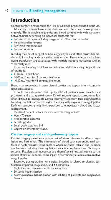
IntroductionCardiac surgery is responsible for 5% of all blood products used in the UK.
all cardiac patients have some drainage from the chest drains postop-eratively. this is variable in quantity and blood content with wide variations between units depending on individual protocols for:• Bypass circuit types including oxygenators, filters, and cannulae• heparin and its reversal• perfusion temperatures• Bypass duration.
Bleeding may be of surgical or non-surgical types and often causes haemo-dynamic instability and/or cardiac tamponade. these effects and subse-quent transfusion are associated with multiple negative outcomes and an i mortality rate.
Excessive bleeding is difficult to define and definitions vary. a good rule of thumb is:• >300mL in first hour• >200mL/hour for 2 consecutive hours• >50mL/hour for 4 consecutive hours.
Blood may accumulate in open pleural cavities and appear intermittently in significant aliquots.
It could be anticipated that up to 20% of patients may breach local protocols and that approximately 5% will require repeat sternotomy. It is often difficult to distinguish surgical haemorrhage from true coagulopathic bleeding, but left untreated surgical bleeding will progress to coagulopathy. Early re-sternotomy may limit exposure to unnecessary blood and factor replacement.
Identified patient factors for excessive bleeding include:• age >70 years• preoperative anaemia• Female gender• Small body size/low BMI• Urgent or emergency status.
Cardiac surgery and cardiopulmonary bypassCardiac surgery provides a unique set of circumstances to affect coagu-lation and fibrinolysis. the interaction of blood with non-endothelial sur-faces in CpB releases tissue factors which activates cellular and humoral mechanisms; including the coagulation cascade, complement and fibrinolytic systems. platelets and leucocytes are thereafter stimulated leading to the clinical effects of oedema, tissue injury, hyperfibrinolysis and a consumptive coagulopathy.
Excessive postoperative non-surgical bleeding is related to platelet dys-function, impaired coagulation, and i fibrinolysis.
technique and disease-specific issues include:• Systemic heparinization• Normovolaemic haemodilution with dilution of platelets and coagulation
factors

INtrOdUCtION 41
• Substantive transfusion of salvaged blood may increase dilutional effects• hypothermia which continues into immediate perioperative period and
increases with bypass duration• antithrombotic drug therapy• antiplatelet therapy• Warfarin therapy—inhibits vitamin K-dependent synthesis of factors II,
VII, IX, X.
42 ChaptEr 6 Bleeding management
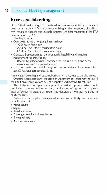
Excessive bleedingUp to 5% of cardiac surgical patients will require re-sternotomy in the early postoperative period. Stable patients with higher than expected blood loss may return to theatre but unstable patients are best managed in the ItU environment (Fig. 6.).
Bleeding may be:• Overt with rapid or ongoing haemorrhage:
• >300mLinfirst hour• >200mL/hourfor2consecutive hours• >50mL/hourfor4consecutive hours
• Concealed presenting as haemodynamic instability and ongoing requirement for transfusion:• Beware pleural collections, consider chest X-ray (CXr) and echo
examination of the pleural spaces• Localized to the pericardial cavity and present with cardiac tamponade.
See b Cardiac tamponade, p. 46.
If untreated, bleeding and its complications will progress to cardiac arrest.Ongoing assessment and proactive management are important to avoid
the additional complications of coagulopathy and massive transfusion.the decision to re-open is complex. the patient’s preoperative condi-
tion including recent anticoagulation, the duration of bypass, and any sur-gical difficulties in theatre all inform the decision of whether to perform re-sternotomy.
patients who require re-exploration are more likely to have the complications of:• renal failure• Sepsis• atrial fibrillation• prolonged mechanical ventilation• i hospital stay• i overall mortality.

EXC
ESSIVE B
LEEdIN
g43
YES
YES
NO
NO
Continue
For re-sternotomyand transfusion
according toconsultant advice
Does the patient requireurgent re-sternotomy?
If platelets <100 000 orthe MA <50 mm
If the haparinaseTEG ‘r’ time
>9 min
If theLY30 >7.5%
If the �brinogenlevel <1 g litre–1
Give 10 units ofcryoprecipitate
Give 2 unitsof FFP
If INR >1.5with a normal
TEG result
If the standardTEG ‘r’ time is
greater than twicethe heparinase ‘r’
Give 50 mg ofprotamine
Give one adult bag ofplatelets
Give 2 units ofFFP
Give aprotinin 0.5million unit bolus,then 0.5 million
units h–1
Is there excessive bleeding?
>4 mLkg–1 h–1 in any one h>2 mLkg–1 h–1 for two consecutive h>5 mLkg–1 in the �rst four h post-op
Figure 6. Management of coagulopathy. reproduced from p diprose, ‘reducing allogeneic transfusion in cardiac surgery: a randomized double-blind placebo-controlled trial of antifibrinolytic therapies used in addition to intra-operative cell salvage', British Journal of Anaesthesia, 2005, 94, 3, pp. 27–278, by permission of the Board of Management and trustees of the British Journal of anaesthesia and Oxford University press.

44 ChaptEr 6 Bleeding management
Overt bleedingthe pathophysiology of bleeding in cardiac surgery is complex and requires multiple approaches to minimize blood loss and transfusions.
Transfusion• transfuse to keep the hb >7g/dL postoperatively.• there is no evidence supporting transfusion where hb >0g/dL.• given that a significant volume of colloid has been added during bypass,
it is normal to tolerate lower hb concentrations in the immediate postoperative period in the expectation that the patient will increase their urine output and the haematocrit will rise over the first few hours post bypass.

CONCEaLEd BLEEdINg 45
Concealed bleedingpatients may have concealed haemorrhage if they require more volume replacement than anticipated in the first 2–4 hours after surgery. they are more likely to be tachycardic and Bp will improve with transfusion of vol-ume, but then fall again. red cells will be required to maintain the haemo-globin >7g/dL:• It is important to know if the pleurae are open following surgery, as
blood may collect in the pleural spaces. a CXr will show i opacification if there is a pleural collection.• the peritoneum may be breached at sternotomy, and may become a
reservoir for concealed haemorrhage.• Both the upper and lower gI tract may conceal haemorrhage, and
should be considered if the source of bleeding cannot be identified.• Consider retroperitoneal haemorrhage if an IaBp has been inserted.• persisting bleeding from the legs may occur if vein has been harvested
during surgery. Inspection of the legs should be included in the search for hidden haemorrhage.

46 ChaptEr 6 Bleeding management
Cardiac tamponadeCardiac tamponade is the clinical syndrome when blood collects in the peri-cardial space reducing ventricular filling and causing haemodynamic com-promise. this can be produced with rapid accumulations of <50mL.
PhysiologyNormal cardiac filling is bimodal:• Initially venous return increases with the onset of ventricular contraction
and subsequent decrease in intrapericardial pressure (x descent of CVp).• Later rV filling in diastole with opening of tricuspid valve (y descent).
this is diminished or absent in tamponade.
diastolic filling is diminished when the transmural distending pressures are insuf-ficient to overcome the intrapericardial pressures. Venous return is impeded with right atrial and ventricular collapse. Output is initially maintained by tachy-cardia. the compliance of the pulmonary vascular bed causes blood to pool in the venous circulation at the expense of LV filling and CO.
Whilst the pericardium may be reconstituted after valve surgery, this is rarely carried out in coronary revascularization. despite the large potential space if the pleura is opened, localized tamponade on the right atrium can have significant haemodynamic effects.
Mediastinal chest drains are inserted as standard postoperative practice. however, they may occlude despite measures to maintain patency.
Early detection and treatment of cardiac tamponade is crucial in postop-erative management. It often coexists in complex patients with low output states.
Classical features include:• i hr• pulsus paradoxus (exaggerated decrease in systolic Bp in inspiration).
May be 20–40mmhg in tamponade but difficult to diagnose in hypotensive or ventilated patients• i jugular venous pressure ( JVp)/CVp• ECg—low voltage complexes or electrical alternans.• d Bp• d CO• d urinary output• Cardiomegaly on CXr.
atypical features:• Failure to rewarm• Unexpected dysrhythmias• Lower than expected CO• Metabolic/lactic acidosis.
diagnosis may be difficult especially in patients with low output states. Echocardiography is a useful adjunct to diagnosis but a high index of clinical suspicion should always be present in:• high-risk patients• When higher than average mediastinal losses suddenly diminish• Failure of patients to progress as expected.

CardIaC taMpONadE 47
EchocardiographyCardiac tamponade cannot be excluded with echocardiography. Classical features include:• Echo-free space ant/post/global• diastolic collapse of rV free wall (parasternal short axis at aortic valve)• Late diastolic compression/collapse of ra.
Difficulties• poor transthoracic access due to sternal wound/drains/pacing wires• Organized clot may be difficult to distinguish from myocardium and
mediastinal structures• Ubiquitous left pleural effusion.
Differential diagnosis• tension pneumothorax• hypovolaemic shock• acute right heart failure• MI• Congestive cardiac failure.
Further readingdiprose p, herbertson MJ, O'Shaughnessy d, deakin Cd, gill rS. reducing allogenic transfusion in
cardiac surgery: a randomized double blind placebo controlled trial of anti-fibrinolytic therapy used in addition to intra-operative cell salvage. BJA 2005;94(3):27–8.
Society of thoracic Surgeons Blood Conservation guideline task Force, Ferraris Va, Ferraris Sp, Saha Sp, hessel Ea 2nd, haan CK, et al. peri-operative Blood transfusion and Blood Conservation in Cardiac Surgery: the Society of thoracic Surgeons and Society of Cardiovascular anaesthesiologists Clinical practice guideline. Ann Thor Surg 2007;83:S27–86.


49
Postoperative hypotension
Introduction 50Management of hypotension: general principles 5Reversible causes of hypotension 53
Chapter 7

50 ChapteR 7 Postoperative hypotension
Introductionhypotension is a common occurrence in up to 75% of patients in the early postoperative period after cardiac surgery. as the Bp is easy to measure it is used as a surrogate for impaired tissue perfusion. In fact the Bp and the flow are equally important to regional perfusion.
there are no global accepted definitions for hypotension, however, for the purposes of this chapter we shall define hypotension as a fall in the systolic Bp to <90mmhg or the Map to <65mmhg.
an understanding of the relationship between Bp and flow is essential. In many ways the Map is a more important measure than the systolic pressure which is more influenced by damping and vascular impedance and is less directly linked to flow.
When assessing a critical care patient, the perfusion pressure should be calculated by subtracting CVp from the Map. patients with high CVps require higher Map to achieve an adequate perfusion pressure.
the perfusion pressure is directly proportional to the product of the cardiac index (CI) and the systemic vascular resistance (SVR) and may be manipulated by influencing these indices by pharmacological or mechanical interventions.
autoregulation across a vascular bed results in stable flow across a range of Maps. In the cerebral circulation this is typically from a cerebral perfusion pressure (Cpp) of 50–50mmhg. the autoregulation curve may be shifted to the right in various diseased states such as long-standing hypertension or when autoregulation is impaired.
In the assessment of hypotensive patients in the postoperative period after cardiac surgery, it is important to know what the Bp was before sur-gery as this acts as a baseline.
also other indices of perfusion must be considered alongside the Bp such as:• Urine output• arterial h+
• plasma lactate• Mixed venous saturation.
Goals for Map should be set on an individual patient basis taking into account preoperative factors such as a history of hypertension and baseline ventricular function.

ManaGeMent of hypotenSIon: GeneRal pRInCIpleS 51
Management of hypotension: general principlesDiagnosishypotension is commonly a result of the following (see table 7.):• preload reduction (hypovolaemia)• Impaired cardiac function• Reduced afterload (vasoplegia).
Preload• Maintain preload• assess response to fluid challenge (see algorithm in fig. 7.)• Beware concealed blood loss.
preload may be difficult to assess. the absolute CVp is a poor measure of preload and the dynamic response of the Map, hR, CVp, and other indirect indices of perfusion to a fluid challenge may be better.
an i pulse pressure variation may indicate hypovolaemia and suggest volume responsiveness.
In patients who are unresponsive to fluid challenge, more information may be necessary to guide treatments.
a full assessment of the past medical history, ventricular function, con-duct of the operation including any clinical difficulties in theatre, and clinical examination often raises suspicion of a possible likely cause.
a bedside echocardiogram can help differentiate between hypovolaemia, impaired cardiac function and vasoplegia. the insertion of a pulmonary artery flotation catheter (pafC) will help differentiate between low Co syndrome and vasoplegia.
Table 7. early postoperative hypotension: the common causes, helpful investigations, and typical interventions
Diagnosis MAP CVP HR Treatment Investigation
hypovolaemia −− −− ++ fluid challenge echo
Myocardial failure −− ++ ++/0 Inotropes, IaBp, VaD
echo, pafC, 2-lead eCG
tamponade −− ++ ++/0 Surgical reopening echo, CXR
tension pneumothorax
−− ++ +/− pleural drain CXR
Vasoplegia
−−
+/−
+/−
noradrenaline, vasopressin, methylene blue
echo, pafC

52 ChapteR 7 Postoperative hypotension
Unresponsive to�uid challenge
Clinical assessment
Poor function Vasodilatation
Still unstable
More informationPAFC, TTE, TOE, CXR
Low dose inotrope Low dose vasopressor
Hypovolaemia
Optimize rhythm
Vasodilatation
Poor LV
Poor RVReversible causesTamponadeTension pneumothoraxSurgery related:Valve repair/replacementDysfunctionLVOT obstructionMyocardial ischaemia
Hypotension
Figure 7. Management algorithm for hypotension post cardiac surgery.

ReVeRSIBle CaUSeS of hypotenSIon 53
Reversible causes of hypotensionDysrhythmiaChanges in heart rhythm can precipitate hypotension. the heart rate and rhythm is assessed and corrected if possible. When tachydysrhythmias are associated with profound hypotension and compromised perfusion, DC cardioversion should be considered. Bradycardias may be treated by pacing if epicardial pacing wires have been placed during surgery. loss of atrioven-tricular (aV) synchrony may have a disproportionate effect in patients with impaired ventricular function and/or diastolic dysfunction and lV hypertro-phy. DDD pacing may be beneficial.
Hypovolaemia and tamponadeexcessive bleeding will cause hypotension and can be diagnosed by i surgi-cal drainage (>200ml/hour).
Beware of concealed haemorrhage as this may occur particularly when the pleural spaces have been opened. a clinical examination and a CXR will identify a significant haemothorax.
hypotension may be a result of cardiac tamponade which is a clinical diagnosis. the hallmarks of cardiac tamponade are:• hypotension• tachycardia• Raised CVp• poor perfusion (delayed capillary filling and adverse trending of
perfusion indices).
When in doubt an echocardiogram can confirm the diagnosis (see b Management of hypotension: general principles, p. 5).
Specific surgery-related causes of hypotension• Regional myocardial ischaemia• Valve replacement or repair dysfunction• lV outflow tract (lVot) obstruction after replacement or repair.
an index of suspicion must be maintained for the recognition of reversible causes of hypotension which are specific to the cardiac surgical procedure.
poor myocardial performance may be due to regional myocardial ischae-mia. this may be suspected if there are eCG changes in the territory of a single coronary artery and/or matching new wall-motion abnormality on echocardiography. Suspected myocardial ischaemia may indicate a techni-cal issue with a CaBG and be an indication for reoperation or coronary angiography and pCI.
If, after valve repair or replacement, resistant hypotension is problematic this may indicate a failure of the operative procedure. Valvular annuloplasty rings can become dehisced and leaflets of mechanical valves may be stuck. these can be diagnosed by transoesophageal echocardiography (toe) and reoperation may be indicated.

54 ChapteR 7 Postoperative hypotension
after mitral valve repair or aortic valve replacement, lVot obstruction may cause early hypotension. these should be excluded by echocardiogra-phy; however, generally an index of suspicion will be present from intraop-erative echocardiographic findings and this should have been handed over to the cardiac intensive care staff on admission.
Features of low cardiac output syndrome (CI <2.2L/min)• low Co• Cold peripheries• Impaired capillary refilling• hypotension• Raised h+ and lactate• low urine output• low mixed venous saturation (<60%).
Treatment• optimize hR/usually 80–90bpm• Rhythm (aim for normal sinus rhythm (nSR) and aV synchrony)• optimize preload• address any reversible causes• pharmacological support: inotropes: catecholamines, phosphodiesterase
inhibitors, calcium sensitization• optimize and normalize biochemical milieu (K+, Ca2+, h+)• Improve oxygen carrying by correcting anaemia• Mechanical support: IaBp or VaD• Reduced afterload.
Vasoplegic syndromeVasoplegic syndrome occurs in 8.8% of cardiac surgical patients in the early postoperative period. It can be defined by the presence of the following five criteria:• hypotension Map <50mmhg• low filling pressures: CVp <5mmhg; pulmonary capillary wedge
pressure (pCWp) <0mmhg• high or normal CI >2.5l/min/m2
• low peripheral resistance, SVR <800dyne/sec/cm5
• a vasopressor requirement.
At-risk groups• transplants• poor ejection fraction (ef)%• Use of angiotensin-converting enzyme (aCe) inhibitors.
Typically associated with:• CpB• long bypass runs• Systemic inflammatory response syndrome (SIRS)• i use of constrictors on CpB.

ReVeRSIBle CaUSeS of hypotenSIon 55
Treatment• Confirm diagnosis with echo and/or pafC• Minimize use of vasodilators (Gtn, milrinone, and sedation)• Pharmacological vasoconstriction:
• Noradrenaline: infusionstartingat0.05mcg/kg/min• Vasopressin: infusionstartingat2IU/hour(workingrange0–4IU/
hour)• Methyleneblue: methylenebluecomesas%(0mg/mL).Bolus
dose .5mg/kg. Make up to 50ml with sterile water and infuse over 20–30 minutes.
Tension pneumothoraxClinical signs include:• hypotension• high airway pressures• arterial desaturation• air leak into drains• Unequal air entry on auscultation• evidence of tracheal shift away from pneumothorax.
If the patient is relatively stable then time can be taken to perform a CXR which will be diagnostic. In extremis, if clinical suspicion exists, a drainage cannula should be inserted into the 2nd intercostal space to relieve the ten-sion. once stabilized, an intercostal drain should be inserted under sterile conditions.
Further readinglevin Rl, Degrange Ma, Bruno Gf, Del Mazo CD, taborda DJ, Griotti JJ, et al. Methylene blue
reduces mortality and morbidity in vasoplegic patients after cardiac surgery. Ann Thorac Surg 2004;77(2):496–9.


57
Glucose, lactate, and acid–base
Introduction 58Acid–base biochemistry 60Lactate 64Glucose 65
Chapter 8

58 ChApter 8 Glucose, lactate, and acid–base
IntroductionAcid–base biochemistry, lactate metabolism, and even glucose metabo-lism are complex. In an individual patient it may be difficult to interpret with certainty the precise mechanisms underlying the measured trends. Nevertheless, these measurements are useful as indicators of potential physiological derangement, as guides to treatment, and as indicators of prognosis.
Despite the underlying complexity, a pragmatic approach extracts most of the immediate clinical value from the data with remarkably little mental effort.

INtroDuCtIoN 59

60 ChApter 8 Glucose, lactate, and acid–base
Acid–base biochemistryOverviewhydrogen ion concentration in living tissue is tightly controlled because small changes in hydrogen ion concentration can have large qualitative and quantitative changes on a myriad of metabolic reactions. plasma hydro-gen ion concentration, which equates to extracellular fluid hydrogen ion concentration, is the only hydrogen ion concentration amenable to easy measurement. however this reflects only one physiological compartment and in particular, does not give reliable information about what is happening intracellularly.
ph is a traditional notation for hydrogen ion concentration that derives from the physical sciences and has become embedded in biological culture largely through historical custom. It is the negative logarithm of the actual hydrogen ion concentration and is useful for describing changes across large ranges. It is a non-linear scale. hydrogen ion concentration [h+] is better for describing changes in the relevant clinical range.
the hydrogen ion concentration in an aqueous solution reflects the degree of dissociation of water. It is influenced by dissolved ions, by tem-perature and by buffers.
there are two common ways of conceptualizing the control of hydro-gen ions in biological solutions. these are the traditional henderson–hasselbalch or bicarbonate-centred framework, and the Stewart (strong ion) hypothesis. In practice and mathematically these descriptions are equivalent. Strong ion theory is useful because it provides an immediate conceptual explanation for the clinical importance of chloride ions and other phenomena such as dilution acidosis. It does not, however, lend itself to easy calculation and the verity of its claims to provide insights into the causal mechanisms of hydrogen ion concentration remains debatable and experimentally unproven.
Stewart hypothesisthe hydrogen ion concentration of an aqueous biological solution is determined only by three variables from which it may be derived by a fourth-order polynomial equation. these are: [SID], [Atot], and pCo2, where [SID], the strong ion difference is the difference in the net charge concentration (mmol/L) of the fixed cations and anions fully dissociated in solution, [Atot] is the concentration of partially dissociated weak acids (albumin, other serum proteins and inorganic phosphate), and pCo2 is the partial pressure of dissolved Co2.
Since [SID] is given by
[SID] = [Na ] + [K ] + [Ca ] + Mg ] [Cl ] [other strong anions]+ + 2+ 2+ − −−

ACID–bASe bIoChemIStry 61
It is easy to see (at least qualitatively) how hyperchloraemia (relative to [Na+]) is associated with acidosis and hypochloraemia is associated with alkalosis. Similarly it can be seen that changes in plasma volume due to dehy-dration or dilution will change the absolute size of the strong ion difference with resultant effects on plasma [h+].
Blood gas analysisClinical blood gas analysis is based on the bicarbonate-centred approach. It assumes plasma acidity is determined by the concentration of volatile acid (Co2) and the concentration of non-volatile acids (all other acids apart from Co2).
An acid tendency caused by excess Co2 is termed a respiratory acidosis. An alkaline tendency caused by lack of Co2 is termed a respiratory alkalosis. Similarly the effects of excess or deficiency of non-volatile acids are termed metabolic acidosis and alkalosis respectively. these effects may compensate for each other and a combination of metabolic and respiratory effects is often present. resultant changes in the actual [h+] are termed acidaemia (high [h+]) and alkalaemia (low [h+]).
An effective approach to blood gas analysis may be based on three key variables: [h+], Sbe (the standard base excess), and pCo2
Respiratory acidosis is caused by accumulation of Co2 in the plasma. Co2 is acidic because it forms carbonic acid:
CO + H O = H CO = H + HCO Eqn 82 2 2 3+
3− .1( )
the pCo2 defines the size of the respiratory acidosis or alkalosis. high pCo2 indicates respiratory acidosis, low pCo2 indicates respiratory alkalosis.
Co2 is the end product of metabolism and is excreted through the lungs. excess Co2 thus occurs as a result of i production or d clearance. the commonest cause of d clearance in the early postoperative period is opi-oid–mediated respiratory depression (this contrasts with the sick patient who has been ventilated for a longer period where mechanical respiratory insufficiency and poor gas exchange are likely to be important factors.) the treatment is adjustment of analgesia ± temporary prolongation of mechani-cal support. the commonest cause of i production in the early postopera-tive period is shivering.
Practice pointConsider shivering as a cause of respiratory acidosis in the re-warming patient. treat with pethidine (25mg IV repeated once if necessary) esca-lating to i sedation in the intubated patient and, if still problematic, neu-romuscular blockade (only in anaesthetized patients).

62 ChApter 8 Glucose, lactate, and acid–base
Metabolic acidosisSbe, the standard base excess, defines the size of the metabolic acidosis or alkalosis.
Sbe is the amount of acid or alkali required to titrate the extracellular fluid [h+] back to normal in the presence of a normal pCo2. the term ‘standard’ refers to the fact that the value is corrected for the buffering effect of haemoglobin so as to estimate the base excess for whole body extracellular fluid (eCF) rather than the base excess of the plasma in the sample. by convention, base excess is described as meq/L of alkali so a negative base excess is an acidosis.
the key importance of a metabolic acidosis is in the diagnosis and treat-ment of the underlying cause. Important causes of metabolic acidosis in the early postoperative period include low Co (often accompanied by high lactate) and renal insufficiency (failure of h+ excretion: lactate is usually nor-mal). A transient lactic acidosis may develop in the first few postoperative hours in a patient who is otherwise well, particularly in patients receiving exogenous adrenaline.
Treatment of metabolic acidosisDirect treatment of metabolic acidosis by alkali administration in the form of bicarbonate remains controversial. Clinical evidence is lacking. theoretical arguments are contradictory. A pragmatic approach may be taken:• make a careful search for and manage the underlying cause.• Leave moderate degrees of abnormality (up to about base excess −0)
to self-correct.• In the acidotic patient with acute cardiac decompensation, administering
bicarbonate to partially correct the acidosis may increase cardiac contractile force and the efficacy of exogenous inotropes and vasopressors, which might contribute to a virtuous circle of recovery. Give bicarbonate in aliquots of 50–00mmol titrated against blood gas measurements, while monitoring for volume overload. In the ventilated patient, simultaneously increase ventilation to rigorously maintain normocapnia, or mild hypocapnia. Co2 diffuses freely across cell membranes and the Co2 load associated with the neutralization of the carbonic acid (see eqn 8.) may otherwise worsen intracellular acidosis. the total amount of bicarbonate required to correct an existing acidosis can be calculated from:
Dose (mEq) = 0.3 weight (Kg) SBE(mEq/L) Eqn 8 2× × .( )most clinicians would regard total correction as too aggressive.

ACID–bASe bIoChemIStry 63
Mixed effectsmetabolic and respiratory acidosis may coexist, e.g. in the unconscious or under-ventilated patient with cardiogenic shock. In this case their effects on [h+] will be additive and there will be an acidaemia. the metabolic and respiratory effects may be in opposite directions as the body attempts to normalize the [h+], e.g. in the case of prolonged type 2 respiratory failure. In this case the compensation is almost always incomplete and the [h+] indi-cates the direction of the primary abnormality, e.g. if the [h+] is high there is a primary acidosis with a compensatory respiratory alkalosis. however the main clue is usually provided by the clinical history.
Practice pointtemperature correction. blood gas and acid base results should be inter-preted at 37°C rather than corrected for the patient’s actual temperature (analogous to alpha stat ph management during bypass).
Practice pointthe patient’s acid–base status in the immediate postoperative period will reflect fluid management during theatre. An acidic pump prime will leave a small residual metabolic acidosis.
Practice pointblood gas results and acid–base chemistry should always be interpreted in the light of the clinical history.

64 ChApter 8 Glucose, lactate, and acid–base
Lactatemetabolite lactate is formed from pyruvate which is a normal product of metabolism.
Lactate metabolism and its control is complex both at a whole-body level and a cellular level. Changes in concentration may reflect changes in pro-duction or changes in removal. A raised plasma lactate is not thought to be harmful in itself. Lactate is formed during anaerobic metabolism in muscles and thus i lactate concentrations may reflect inadequate tissue perfusion, for instance, caused by low Co. however, other causes of raised lactate in the immediate postoperative period include i production due to β 2 receptor stimulation (adrenaline) and the systemic inflammatory response (cytokine-mediated stimulation).
A raised or rising lactate should thus precipitate a careful diagnostic review for signs of circulatory insufficiency but, by itself, is not diagnostic of an adverse situation. It is usefully reassuring if the lactate concentration is normal.
A high lactate (>3mmol/L) at entry to intensive care is associated with a raised mortality in observational studies.
Practice pointSwitching a patient away from an adrenaline-based inotrope regimen may normalize a raised lactate

GLuCoSe 65
Glucoseblood glucose concentration rises during and after cardiac surgery due to stress response and exogenous beta stimulation.
there is no strong evidence for treating blood glucose after cardiac surgery differently than in other critical care situations. Glucose should be kept in the range 3.5–0.0mmol/L using continuous infusion of insu-lin adjusted according to a protocol. the main danger of tighter control is hypoglycaemia.
the causal relationship between high blood glucose and lactataemia is unclear.
An important feature of glucose control is keeping input constant. enteral or parenteral feeding should thus be administered continuously over 24 hours. If feeding is interrupted for any reason insulin infusions should be stopped or reduced and glucose concentration monitored closely.
TerminologyWhole blood glucose concentrations differ from plasma values, because the concentration in whole red cells is lower due to their high lipid content. Whole blood concentrations can be converted to plasma concentrations by multiplying by . at normal haematocrit. Since 200, all laboratory and point-of-care resting values should be reported as plasma concentrations regardless of the actual method of measurement.
Further readingGrogono AW. Acid-Base Tutorial. Available at: M <http://www.acid-base.com>.Kurtz I, Kraut J, ornekian V, Nguyen mK. Acid-base analysis: a critique of the Stewart and
bicarbonate-centered approaches. Am J Physiol Renal Physiol 2008;294:F009–F03.


67
Postoperative medications
Postoperative medications 68Postoperative medication following coronary bypass/valve
surgery 70Preoperative medications 72
Chapter 9

68 ChaPter 9 Postoperative medications
Postoperative medicationsPostoperative critical care involves getting the basics right, attention to detail, and avoidance of omissions. Most postoperative medications are prescribed according to a standard protocol (see example protocol in table 9.). Cardiac surgical patients are commonly on numerous medica-tions some of which must be continued into the postoperative period and others which should be stopped. this chapter covers the common drugs prescribed in the postoperative period after cardiac surgery.
Antibiotic prophylaxis• Prophylaxis against surgical site infection is routinely continued into the
postoperative period. Local policies will vary. these are generally not continued beyond 24 hours after surgery. See b antimicrobial therapy, p. 34.
Stress ulcer prophylaxis• Prophylaxis is essential in patients mechanically ventilated for >48 hours.• the combination of major surgery, hypoperfusion, systemic
anticoagulation, and potential coagulopathy makes patients high risk for development of stress ulcers—prophylaxis is started preoperatively.
Electrolytes• hypokalaemia and hypomagnesaemia are common in the postoperative
period and put patients at i risk of arrhythmia.• Plasma potassium should be maintained >4.0mmol/L, or >4.5mmol/L
if arrhythmias are present.• IV magnesium is a safe and effective antiarrhythmic agent and is
commonly used to prevent postoperative arrhythmias:• Caution: rapidinfusionof magnesiumcancausehypotensionand
bradycardia.
Antiemetics• Should be available as required to treat PONV. Postoperative ileus and
opiates may also cause PONV.
Shivering• Shivering increases oxygen demand and should be avoided.• Can be due to hypothermia requiring resedation and further warming.• Postoperative shivering often responds to pethidine.
Glucose control• hyperglycaemia is common after cardiac surgery and associated with i
mortality and complications in patients both with and without diabetes mellitus.• Blood glucose should be controlled by using a continuous, IV insulin
infusion, e.g. by sliding scale, to maintain median blood glucose control between 3.5mmol/L and 0mmol/L. Perioperative blood glucose control after cardiac surgery reduces mortality:• Caution: avoidhypoglycaemia.

POStOPeratIVe MedICatIONS 69
Deep venous thrombosis prophylaxis• Patients are at risk for dVt due to the procoagulant effect of major
surgery, immobility, and the loss of saphenous veins/bandaging of limbs.• Subcutaneous low-molecular-weight heparin (LMWh) is delayed until
postoperative day because of the risks of bleeding associated with cardiopulmonary bypass and cardiac surgery:• e.g.dalteparin5000IUSConce daily.
Arrhythmia (atrial fibrillation) prophylaxis• early postoperative administration of β-blockers in patients without
contraindications is standard therapy to reduce the incidence and/or clinicalsequelaeof atrialfibrillation.(Usuallystartedonpostoperativeday in patients not receiving inotropes—see b Secondary prevention, p. 70.)• amiodarone is effective prophylaxis for high-risk patients with
contraindications to β-blockers.
Table 9. example protocol for postoperative prescribing on the day of surgery
Sedation and analgesia
Propofol 0–5mcg/mL IV by target controlled infusion
Paracetamol g IV 6-hourly
Morphine Nurse-controlled IV boluses until extubated then5–0mg SC PrN up to hourly
Antibiotic prophylaxis
Flucloxacillin
Gentamicin
g IV 6-hourly (for 3 doses)Single dose only at induction
Stress ulcer prophylaxis
ranitidine 50mg IV 8-hourly until extubated
Electrolytes
Potassium chloride 2.5–25mmol IV PrN by infusion
Magnesium sulphate 0–20mmol IV PrN by infusion
Antiemetics
Ondansetron 4mg IV PrN up to 8-hourly
Control of shivering
Pethidine 25–50mg IV PrN
Glucose control
actrapid® insulin actrapid®50IUin50mLbyslidingscale
Note: this table represents normal practice in the author's institution but should not be considered absolute. refer to local guidelines.

70 ChaPter 9 Postoperative medications
Postoperative medication following coronary bypass/valve surgeryMedication following coronary bypass surgery
Prevention of coronary graft thrombosis• aspirin is the drug of choice for prophylaxis against early saphenous vein
graft (SVG) thrombosis and should be given 6 hours post-operatively or as soon as bleeding has ceased:• e.g.aspirin300mgPR(continuedat75mgPOdailylong term).• Thebenefitof postoperativeaspirinonSVGpatencyislostwhen
started >48 hours after surgery.• Clopidogrel is an acceptable alternative to aspirin for the maintenance
of graft patency in patients intolerant of aspirin:• Superiorityoveraspirinhasnotbeenestablished.• Patientsonlong-termdualtherapy(aspirinandclopidogrel),e.g.
stents, should have the dual therapy recommenced.
Secondary prevention (postoperative day onwards)• It is indicated to start and continue β-blocker therapy in all patients after
MI, aCS, or LV dysfunction, unless contraindicated (see erC/eaCtS guidelines, b Further reading, p. 7).• β-blockers should be recommenced or considered for all patients
without contraindications:• Contraindications: asthmaorbronchospasm,HR<60bpmor
pacemaker-dependent,BP<00mmHgsystolic,oroninotropes.• Dosestartedlowfollowedbyupwarddosetitration.• e.g.bisoprolol.25mgPO daily.
• aCe inhibitors should be started and continued indefinitely in all patients withLVEF<40%andforthosewithhypertension,diabetes,orchronickidney disease, unless contraindicated.• aCe inhibitors should be gradually recommenced/considered in all
patients (particularly patients with evidence of LV dysfunction):• Contraindications: acuteorunstablerenalimpairment,BP
<00mmHgsystolic,oroninotropes.• e.g.ramipril2.5mgPO daily.
• Statins should be recommenced or introduced in all patients:• Contraindications: acuteliverdisease,persistentlyabnormalliver
function tests.• e.g.Simvastatin40mgPO nocte.
Prevention of coronary graft spasm• radial artery grafts are particularly prone to spasm therefore
vasodilators are commonly used:• e.g.glyceryltrinitrate–5mg/hourIVstartingintraoperatively.• e.g.amlodipine5mgPOdailycontinuedfor3–6 months.

71POStOPeratIVe MedICatION FOLLOWING COrONary ByPaSS
Medication following valve surgeryPrevention of valve thrombosis• all patients with mechanical valves require anticoagulation.• Bioprosthetic valves have an i risk of thromboemboli during the first
3 months.• Oral anticoagulation is recommended for the following situations:
• Lifelongforallpatientswithmechanicalvalvesirrespectiveof valvetype or date of introduction.
• Lifelongforpatientswithbioprosthesesormitralrepairwhohaveother indications for anticoagulation, e.g. atrial fibrillation and impaired LV function.
• Forthefirst3 months,inallpatientswithbioprosthesesormitral valve repair involving the use of a prosthetic annuloplasty ring. (there is widespread use of aspirin as an alternative to anticoagulation for the first 3 months in patients with no other indications for anticoagulation.)
• target INr depends on valve position, risk of thrombosis, and type of valve (see table 9.2).• Warfarin should be commenced on postoperative day unless chest
drains remain in situ for bleeding.
Further readingtask Force on Myocardial revascularization of the european Society of Cardiology (eSC) and the
european association for Cardio-thoracic Surgery (eaCtS), et al. Guidelines on myocardial revascularisation. Eur Heart J 200;3:250–55.
Table 9.2 target INr following valve surgery
Mechanical aortic: no risk factorsa
—low/medium/high-risk valve*
2.5/3.0/3.5
Mechanical aortic: with risk factorsa
—low/medium/high-risk valveb
3.0/3.5/4.0
Mechanical mitral valve
—low/medium/high-risk valveb
3.0 /3.5/4.0
Bioprosthesis: aortic—no risk factorsa aspirin only
Bioprosthesis: aortic—risk factorsa 2.5 (3 months)
Bioprosthesis: mitral 2.5 (3 monthsaRiskfactors: atrialfibrillation,leftatrium>50mm,ejectionfraction<35%,additionalvalvereplacements, hypercoagulability, history of thromboembolism.bValve risk: low risk: Medtronic hall, St Jude, Carbomedics. Medium risk: bileaflet valves with insufficient data, Bjork–Shiley. high risk: Lillehei–Kaster, Omniscience, Starr–edwards.
european Society of Cardiology. recommendations for the management of patients after heart valve surgery. Eur Heart J 2005;26:2463–7

72 ChaPter 9 Postoperative medications
Preoperative medicationsContinuing essential medications
Drugs for neurological disordersEpilepsy• Generalized seizures occurring in the perioperative period can increase
morbidity and mortality, thus patients with pre-existing seizure disorders require anticonvulsant medications to be continued.• Many antiepileptic drugs are available as a suspension that can be given
via NG tube. Phenytoin, valproate, and levetiracetam are available parenterally. Carbamazepine can be given Pr. there are no parenteral forms of gabapentin, topiramate, or lamotrigine—patients taking these medications in which the oral route is contraindicated require conversion to an alternative agent in consultation with a neurologist.
Parkinson's disease• Liaison with specialist services is advised preoperatively.• adequate antiparkinsonian therapy is essential for satisfactory
postoperative respiratory and bulbar muscle function.• dopaminergic drugs should be restarted as soon as possible
postoperatively to reduce the risk of neuroleptic malignant syndrome (secondary to acute withdrawal).• Many dopamine receptor agonists and levodopa/decarboxylase
combinations can be administered via NG tube. Subcutaneous apomorphine or topical preparations of rotigotine (Neupro®) are available for patients in whom the oral route is contraindicated.• Caution is required when prescribing other drugs—phenothiazines,
butyrophenones, metoclopramide, and clonidine should be avoided.
Myasthenia gravis• Careful management of anticholinesterase medication is essential to
avoid myasthenic crisis and respiratory failure postoperatively.• anticholinesterases can be administered via NG tube and are usually
restarted when the patient is haemodynamically stable, prior to weaning from mechanical ventilation and at the patient's usual dose.• If the oral route is contraindicated, IV neostigmine is substituted for
pyridostigmine at /30th the usual oral dose.• Many drugs can worsen muscular weakness in myasthenia gravis.
aminoglycoside antibiotics should be avoided.• Caution: patients with myasthenia gravis are often on corticosteroid
therapy and will require additional supplementation.
Drugs for respiratory diseasesInhaled bronchodilators and steroids should be continued in the intubated patient via nebulizer or metered dose inhaler attachment to the ventilator circuit. Beware tachycardia from β2 agonists.
CorticosteroidsPatients taking chronic glucocorticoid therapy require perioperative supple-mentation due to suppression of the hypothalamic–pituitary–adrenal axis.

PreOPeratIVe MedICatIONS 73
• Patients taking >0mg prednisolone per day or who have stopped therapy<3 monthsagoareat risk.• For major surgery, in addition to a bolus at induction of anaesthesia,
patients require IV hydrocortisone 25–50mg 3 times a day for 48–72 hours postoperatively. Patients with high vasopressor requirement may require a prolonged course.
Restarting preoperative medicationsCardiovascular drugsβ-blockers, aCe inhibitors, and statins should be recommenced in patients with ischaemic heart disease (see b Secondary prevention, p. 70). antihypertensive, antianginal, and diuretic requirements may be different post-operatively and may not be required or can be titrated to effect.
Drugs for diabetes mellitusGenerally speaking, IV insulin infusions are continued until patients are eat-ing and drinking when preoperative oral hypoglycaemics/SC insulins are recommenced:• Type (and type 2 patients on insulin): long-acting insulins are withheld
and total daily insulin requirement is administered as short-acting actrapid® insulin (in 3–4 divided doses) until blood glucose is stable. Long-acting agents are then reintroduced.• Type 2: caution should be exercised with certain drugs:
• Sulfonylureasstimulateinsulinsecretionandmaycausehypoglycaemia. Patients on high doses should start low and titrate upwards.
• Metforminshouldnotberestartedinpatientswithrenalinsufficiency,significant hepatic impairment, or congestive heart failure.
• Thiazolidinedionesshouldnotberestartedinpatientswithcongestive cardiac failure, problematic fluid retention, or liver function abnormalities.
Drugs for psychiatric disordersCan generally be restarted on postoperative day . Caution is required with the following:• drugs prolonging Qt-interval (particularly in association with class III
antiarrhythmics).• Selective serotonin reuptake inhibitors in combination with other drugs
affecting serotonin levels (e.g. tramadol and ondansetron).
Sedation and analgesia• See b Pharmacology, p. 37.


75
Information handover and care planning
Introduction 76ICU admission 77Shift handover and ICU discharge 78Conclusion 79
Chapter 0

76 Chapter 0 Information handover and care planning
Introductioneffective communication is an essential part of good team work which in turn helps set the scene for optimizing patient safety. Most human errors are not the result of poor technical knowledge or ability but are due instead to ‘non-technical’ aspects of performance such as communication.
Within a multidisciplinary team and between different tiers of a clinical team effective communication can be problematic, with differing commu-nication styles complicated by hierarchical, ethnic, and gender influences.
effective handover communication is vital at initial admission, staff changeovers, and on discharge from the cardiothoracic ICU. Within an intensive care environment a single patient’s care may be ‘handed over’ within the multidisciplinary team over 5 times within any single 24-hour period.
Missing or incomplete information during admission or subsequent hand-over is a common cause of error and patient harm.
During the handover of information distractions or interruptions increase cognitive demands leading to inefficiency and an i risk of error. this cogni-tive stress may be exacerbated when the patient is acutely unwell.
Good situation awareness and effective role and task allocation start with effective communication.
handovers require the concise transfer of accurate and relevant patient information. this process can be aided by a standardized written handover sheet which details the objective clinical data. the verbal handover should include a short summary and highlight areas of clinical concern, outstanding tasks or plans, and take place between all health professionals of the same discipline or between different disciplines. there is no need to verbally repeat all the clinical data on the handover sheet as this will only increase the cognitive demands on the recipient.
One method of enabling effective handover is using a modified SBAR tool, developed by Kaiser permanente, which is an easy focused way of setting expectations of a response that is appropriate and timely. SBar encom-passes the following:• Situation:
• Identifyself,patient,bed space• Brieflystatecurrentissuesandduration
• Background:• Diagnosis• Numberof days in ICU• Currentrespiratorysupport• Currentdrugtherapies• Relevantrecentvital signs• Relevantlabresults
• assessment:• Whatdoyouthinkisgoing on?• Whatisyouropinion?
• response:• Whatactionsarerequired?• Overwhattimescale?

ICUaDmIssION 77
ICU admissionPreparation for the patient’s arrival• ICU multidisciplinary team brief/planning and role allocation• equipment ready and checked• Infusions prepared• handover sheet from theatre with relevant patient information including
endotracheal tube (ett) sizing, lines, inotropes, etc.
Patient arrival in ICU• patient monitoring transfer• patient support transfer (ventilator, inotropes, drain suction)• Verbal handover:
• Onevoice,all team watching and listening• Noothertasksundertakenduringhandover• Clearandconcisehandover• sBaRtechnique• Handoversheet allowshandovertobemoresubjective
• encourage questions from all staff groups at end• Summarize plans at end.
Post handover• ICU team task allocation• appropriate investigations• Multidisciplinary review of care plan.

78 Chapter 0 Information handover and care planning
Shift handover and ICU dischargeWritten handover information can be used to provide the objective clini-cal data with the more subjective clinical impression and outstanding tasks being delineated by the following SBar format.• Situation:
• Concisesummary• Patient name• Primarydiagnosis• Bed space• age• Weight
• Background:• Diagnoses• Lengthof stay• systems-basedconcise review• Currenttherapies
• assessment:• Currentissuesinasystem•basedapproach
• response:• Outstandingtasksandtimescale• Clinicalteamsinvolved.

CONCLUsION 79
Conclusioneffective and efficient communication plays a central role if excellent multi-disciplinary care is to be delivered in any ICU.
Written and verbal communication can work hand in hand to allow such communication and care planning. the written data should provide the hard facts and the verbal handover, using an SBar format, allows participants to focus on the pertinent facts, tasks, and areas of clinical concerns.
all disciplines in the ICU should continually be seeking to improve the care delivered. By taking a short period of time to think about how we all handover information at each stage of the patient’s journey we have the potential to ensure that the patient’s safety is paramount and optimal and timely care is delivered.
Further readingInstitute for healthcare Improvement website: M <http://www.ihi.org/ihi>NHsInstituteforInnovationandImprovementwebsite: M <http://www.institute.nhs.uk/>.Scottish patient Safety programme website: M <http://www.patientsafetyalliance.scot.nhs.uk>.


Organ dysfunction
Heart 83 2 Lung 07 3 Kidney 5 4 Gastrointestinal 27 5 Nervous system 39 6 Immune system 49 7 Haematology 57
Part 2


83Chapter
Heart
Left ventricular failure 84Right ventricular failure 86Valve heart disease 95Common arrhythmias and their management 04

84 CHapteR Heart
Left ventricular failurethe assessment and management of the LV is covered in other chapters of this book. the anatomy and physiology are summarized here, followed by reference to the relevant chapters covering functional assessment and management.
Anatomy and physiology of the LVAnatomythe LV receives oxygenated blood from the pulmonary veins and pumps it to the systemic circulation. It consists of:• the mitral valve, forming the inlet to the ventricle• the conical apical portion, containing fine trabeculations• the outflow tract leading to the aortic valve.
the muscle fibres of the LV are formed in multiple overlapping sheets. Myocardial contractility is spiral, producing radial contractility and longitudi-nal shortening. the LV appears circular in short-axis view.
Physiology• the systemic LV is a high-pressure system.• Coronary perfusion occurs in diastole over four phases; isovolumic
relaxation, rapid filling, diastasis, and atrial systole.• Volume preloading increases stroke volume within the limits of the
Starling effect.• the ventricle responds to i afterload through i contractility and over
time through muscle hypertrophy.
LV functional assessmentCardiac outputChapter 24 on central venous cannulation (CVC), pulmonary artery cath-eter (paC) and minimally invasive CO monitoring describes the indications for and advantages of the invasive paC and minimally invasive CO moni-toring in the assessment and ongoing management of cardiogenic shock. the paFC provides direct measurement of pulmonary pressures, SvO2, and CO, while pulse contour analysis, thoracic bioimpedance measurement, and oesophageal Doppler provide minimally invasive derived measures of CO.
Anatomical and functional assessmentChapter 32 on bedside echocardiography describes the indications of both transthoracic echocardiography (tte) and transoesophageal echocardiog-raphy (tOe) in the CICU. the size, degree of filling, and contractility of the LV can be assessed. In addition, regional wall-motion abnormalities can be defined and culprit vessels identified.
Management of the failing LVManagement: general principles and the management of reversible causesChapter 7 on hypotension and low CO describes the general principles of managing patients with a poorly functioning LV following a standard aBC

LeFt VeNtRICULaR FaILURe 85
approach. In addition, identification and management of reversible causes including myocardial ischaemia are described.
Contractility: pharmacological supportChapter 27 on circulatory support details the pharmacological actions of different classes of inotropes together with their indications. the selec-tion and safe use of a particular vasoactive agent is based on knowledge of its cardiovascular effects, clinical indications, and an understanding of its clinical use.
Contractility: mechanical supportChapter 27 on circulatory support goes on to describe the indications and use of mechanical and extracorporeal life support. this includes the use of IaBp counterpulsation, short-term VaDs, long-term VaDs, and extracor-poreal membrane oxygenation (eCMO).

86 CHapteR Heart
Right ventricular failureAnatomy and physiology of the RVthe RV differs from the LV in structure and function. the right side of the heart can be more difficult to assess and treat. Its interdependence with the LV highlights the importance of its careful assessment and optimization in the compromised patient.
Anatomythe RV receives blood from systemic venous return and pumps it to the pulmonary circulation. It consists of:• the sinus (inflow) below the tricuspid valve• the free wall (providing contractility) which is thin walled• the infundibulum (outflow) leading to the pulmonary valve.
the RV contracts in a peristaltic pattern from the sinus to the infundibulum. It relies on longitudinal shortening more than the LV.
the RV appears triangular when viewed from the side and crescentic when viewed in cross section.
Ventricular interdependencethe RV wraps around the LV: up to 30% of the contractile energy of the RV is generated by the LV.
the LV and RV are enclosed in a non-expanding pericardium. Overdistention of the RV within this closed sac compromises LV contractile performance primarily by reducing LV preload.
Physiology• the RV is a low-pressure system (Fig. .).• RV wall stress is low. Coronary perfusion occurs in systole and diastole.
an acute increase in afterload reduces coronary perfusion, so reducing contractile performance.• Loss of forward flow from the RV increases Ra pressure, reducing
systemic venous return.
LV
RV
Volume
Pres
sure
Figure . pressure–volume loop. RV: ejection starts earlier in systole, and is against a low-resistance bed. Reproduced from Heart, F Sheehan and a Redington, ‘Non-invasive imaging: the right ventricle: anatomy, physiology and clinical imaging', 94, , copyright 2008, with permission from BMJ publishing Group Ltd.

RIGHt VeNtRICULaR FaILURe 87
RV functional assessmentthe complex morphology of the RV makes volume and functional assess-ment difficult. the current clinical standard is echocardiographic visual assessment of the RV with surrogate markers of contractility. assessment should be corroborated in multiple views.
RV dilatationtOe: mid-oesophageal four-chamber view or tte: apical four-chamber view:• RV should be approximately half LV size• LV should form the apex.
Paradoxical septal motiontOe: transgastric short-axis view of LV or tte: parasternal short axis:• paradoxical septal motion in systole, reflecting the presence of RV
systolic overload.
the patient in Fig. .2a exhibited normal septal motion with a pure spheri-cal LV. the patient in Fig. .2b exhibited paradoxical septal motion at the end of systole and beginning of diastole reflecting systolic overload of the RV.
The right ventricle• Is a thin-walled low-pressure system.• It is exquisitely volume sensitive.• there is functional interdependence with the LV.• the RVs response to i preload or afterload is overdistention.
(a) (b)
Figure .2 Short-axis view of the left ventricle by a transgastric approach. Reproduced from a. Vieillard-Baron, ‘assessment of right ventricular function', Current Opinion in Critical Care, 5, 3, pp. 254–260, copyright 2009, with permission from Wolters Kluwer.

88 CHapteR Heart
Surrogate markers of contractilityTAPSE (normal > 6mm)tricuspid annular plane systolic excursion can be assessed in the tte api-cal four-chamber view with M-mode, measuring the distance of tricuspid annular movement between end-diastole and end-systole.
Fractional area change (normal > 35%)Other indirect measures of RV function include:
ECG• RV strain• New atrial fibrillation (aF).
Pulmonary artery floatation catheter• the only way to directly measure right heart pressures. the gold
standard for CO monitoring.• Direct measurement of pap allows continuous monitoring of therapy to
reduce pap.• paWp provides an indirect measure of left heart filling pressure and
thus preload.
Management of the failing RVRV function has often been overlooked in critically ill patients. the last decade has seen a significant advance in the understanding of RV haemodynamics and appreciation of their importance in global cardiac performance.
the RV can fail because of:• Volume overload (preload)• Impaired contractility, or• pressure overload (afterload).
When to suspect RV failureIn the CICU, clinical situations to consider RV failure include:• Disproportionate rise in CVp (suggesting i preload, particularly in
association with tricuspid valve incompetence)• Myocardial stunning following reperfusion• RV ischaemia and infarction, either due to sustained hypotension or
coronary occlusion• When the LV is underfilled but the CVp is high and the RV is distended
there may be RV failure• Septic shock• pulmonary hypertension (primary or acquired)• acute respiratory distress syndrome (5% develop right heart failure)• Secondary to LV failure (ventricular interdependence).
Management: general principlesOptimize ventilation• FiO2 to maintain normoxia• avoid high intrathoracic pressures:
• Ventilateat6–8mL/kg,avoidinspiratorypressure>30mmHg• Avoidauto-PEEP
• Avoidhypoxia,acidosis,and/orhypercarbiaasthesecontributetopulmonary vasoconstriction.

RIGHt VeNtRICULaR FaILURe 89
Maintain sinus rhythm (aim for HR 90–0bpm)• Consider electrical cardioversion for atrial tachyarrhythmias• Or pacing for symptomatic bradyarrhythmias.
Optimize preload• If uncertain, 00mL crystalloid fluid challenge and assess response.• If volume overloaded use IV diuretics in the first instance. If there is no
response to diuretics consider ultrafiltration.• If underfilled, decide on appropriate replacement fluid and continue to
bolus in 00mL aliquots until euvolaemic.• Support contractility:
• Pharmacological(seeb pharmacological support, p. 288)• Surgical(seeb Mechanical circulatory support: extracorporeal life
support, p. 294).
Reduce afterload• pulmonary vasodilators such as inhaled nitric oxide, or nebulized
iloprost.• Once stable consider IV or oral vasodilators such as sildenafil or
epoprostenol.• Minimize blood production transfusion.
the general principles are described in Fig. .3.
Further readingHaddad F, Couture p, tousignant C, Denault aY. Right ventricular function in cardiovascular dis-
ease, part II: pathophysiology, clinical importance, and management of right ventricular failure. Circulation 2008;7:77–3.
Contractility: pharmacological supportthere is no evidence for the best inotrope regimen to use in right heart fail-ure. Useful drugs are outlined here. Use should be tailored to the patient’s haemodynamics and local guidelines. One logical sequence is suggested in the management algorithm (see Fig. .3).
Adrenaline• Infusion starting at 0.05mcg/kg/min (working range 0.02–0.2mcg/kg/min)• Useful resuscitation drug and first-line inotrope• Direct acting on α, β, α2 and β2 receptors• Undesirable effects:
• Proarrhythmic• Hypertension• Hyperglycaemia• i myocardial oxygen demand.
Key pointIn acute RV failure, every effort should be made to avoid systemic hypo-tension, which may lead to a vicious cycle of RV ischaemia and further hypotension.
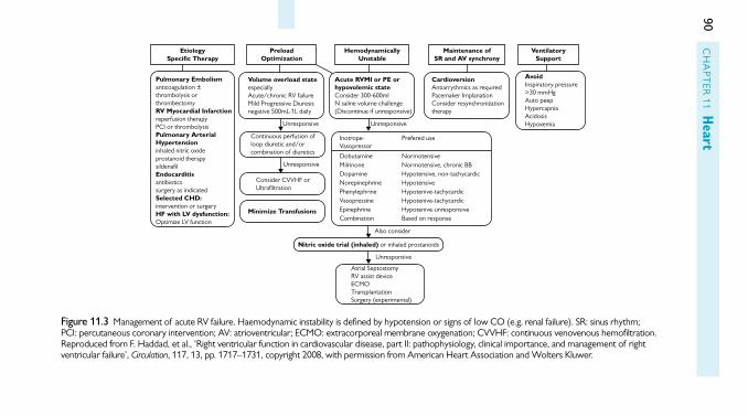
90C
Ha
pteR
Heart
EtiologySpeci�c Therapy
Pulmonary Embolismanticoagulation ±thrombolysis orthrombectomyRV Myocardial Infarctionreperfusion therapyPCI or thrombolysisPulmonary ArterialHypertensioninhaled nitric oxideprostanoid therapysildena�lEndocarditisantibioticssurgery as indicatedSelected CHD:intervention or surgeryHF with LV dysfunction:Optimize LV function
PreloadOptimization
HemodynamicallyUnstable
Maintenance ofSR and AV synchrony
VentilatorySupport
Volume overload stateespeciallyAcute/chronic RV failureMild Progressive Diuresisnegative 500mL-1L daily
Continuous perfusion ofloop diuretic and/orcombination of diuretics
Unresponsive
Also consider
Unresponsive
Minimize Transfusions
Consider CVVHF orUltra�ltration
Unresponsive
Unresponsive
Inotrope-Vasopressor
Prefered use
Dobutamine NormotensiveMilrinone Normotensive, chronic BB Dopamine Hypotensive, non-tachycardicNorepinephrine HypotensivePhenylephrine Hypotenive-tachycardicVasopressine Hypotenive-tachycardicEpinephrine Hypotenive-unresponsiveCombination Based on response
Atrial SeptostomyRV assist deviceECMOTransplantationSurgery (experimental)
Acute RVMI or PE orhypovolemic stateConsider 300-600mlN saline volume challenge(Discontinue if unresponsive)
CardioversionAntiarrythmics as requiredPacemaker ImplanationConsider resynchronizationtherapy
AvoidInspiratory pressure>30 mmHgAuto peepHypercapniaAcidosisHypoxemia
Nitric oxide trial (inhaled) or inhaled prostanoids
Figure .3 Management of acute RV failure. Haemodynamic instability is defined by hypotension or signs of low CO (e.g. renal failure). SR: sinus rhythm; pCI: percutaneous coronary intervention; aV: atrioventricular; eCMO: extracorporeal membrane oxygenation; CVVHF: continuous venovenous hemofiltration. Reproduced from F. Haddad, et al., ‘Right ventricular function in cardiovascular disease, part II: pathophysiology, clinical importance, and management of right ventricular failure’, Circulation, 7, 3, pp. 77–73, copyright 2008, with permission from american Heart association and Wolters Kluwer.

RIGHt VeNtRICULaR FaILURe 91
Dopamine• Infusion starting at 5mcg/kg/min (working range 2.5–25mcg/kg/min)• Inotrope with vasoconstriction at higher concentrations• endogenous precursor of noradrenaline with predominantly
β-receptor activity. at higher doses, inotropic effects mediated through β receptors. Vasoconstrictor effects via α and 5Ht receptors.• Undesirable effects:
• Extravasationmaycauselocalnecrosis• Proarrhythmicsecondarytoabnormalcalciumloading.
Milrinone• Infusion starting at 0.375mcg/kg/min (working range 0.–0.75mcg/kg/
min)• Inotrope and vasodilation• phosphodiesterase inhibitor inhibiting caMp breakdown• Undesirable effects:
• Pro-arrhythmic• Oftenrequiresconcomitantvasopressor.
Dobutamine• Infusion starting at 2.0mcg/kg/min (working range 0–20mcg/kg/min)• Inotrope, chronotrope, and vasodilator at higher doses• Synthetic catecholamine acting on β, β2, and α receptors• Undesirable effects:
• Proarrhythmic• Hypotensionathigher doses• Tachyphylaxisprolongedinfusionover96hourshasbeenassociated
with a decrease in the haemodynamic effect by as much as 50%.
Noradrenaline• Infusion starting at 0.05mcg/kg/min (working range 0.02–0.2mcg/kg/min)• Vasoconstriction• endogenous agonist at α and α2 adrenoreceptors (modest effect on β
adrenoreceptors)• Undesirable effects:
• Reducedrenalandsplanchnicblood flow• i pulmonary vascular resistance (pVR)• Proarrhythmic.
Vasopressin• Infusion starting at 2IU/hour (working range 0–4IU/hour)• Direct vasoconstriction• peptide hormone agonist at aVpRa receptor• Undesirable effects:
• Localtissuenecrosis• Reducedsplanchnicblood flow.
Phenylephrine• Infusion starting at 0.5mcg/kg/min (working range 0.4–9mcg/kg/min)• Vasopressor• α adrenoreceptors agonist

92 CHapteR Heart
• Undesirable effects:• Arrhythmiaincludingreflexbradycardia• Severeperipheralandvisceralvasoconstriction.
Mechanical supportMechanical cardiac support for short-term use covers a spectrum of tech-niques that can be classified as follows:• Intra-aortic counter pulsation with an IaBp• extracorporeal ventricular support—short-term:
• Short-term VADs• ECMO.
In patients with severe RV dysfunction, a right ventricular assist device (RVaD) may be considered. RVaDs may be used alone or in combination with LVaDs.
Short-term VaDs have a potential duration of support of days to weeks. an exit strategy must be identified before device implantation. this may be ventricular recovery, or as bridge to destination therapy or transplantation. If there is associated hypoxia there may be benefit in combining short-term ventricular support with oxygenation in an eCMO circuit.
Commonly used short-term devices in the UK include the Levitronix CentriMag™ VaD. this device can act as a RVaD, LVaD, or BiVaD. the pump is outside the body. Flow is non-pulsatile, but residual ventricular ejection may add pulsatility.
See b Mechanical circulatory support: extracorporeal life support, p. 294 for further discussion of mechanical circulatory support.
Further readingShuhaiber JH, Jenkins D, Berman M, parameshwar J, Dhital K, tsui S, et al. the papworth experience
with the Levitronix CentriMag ventricular assist device. J Heart Lung Transplant 2008;27:58–64.
Afterload reductionPulmonary hypertension• Meanpulmonaryarterypressure≥25mmHgatrestor>30mmHgon
exercise.
acute management options in severe pulmonary hypertension:• Seek expert help.• High-flow oxygen is a potent pulmonary vasodilator.• Specific pulmonary vasodilator therapy—first line:
• If intubated,inhalednitric oxide• If notintubated,nebulizediloprost.
• IV and oral agents may be used to wean inhaled nitric oxide, consider sildenafil, epoprostenol.• Consider eCMO ± RVaD if the RV is failing despite medical therapy.
Key pointan exit strategy should always be identified before embarking on any form of mechanical support.

RIGHt VeNtRICULaR FaILURe 93
• atrial septostomy is used in chronic pulmonary hypertension, but may be considered in the acute setting. this creates a right-to-left shunt at atrial level with afterload reduction of the right heart. this is at the expense of resultant cyanosis.
Nitric oxide• Delivered via a dedicated ventilator circuit• Start at 20ppm (range 0–20ppm)• Smooth muscle relaxation mediated through cGMp• Undesirable effects:
• Humidificationshouldbeavoidedtopreventformationof nitric acid• Withdrawalshouldbecautioustoavoidreboundpulmonary
hypertension.
Iloprost• First dose: 2.5mcg nebulized• Thereafter: 5mcg 6–9 times/day (>2 hours apart)• Inhaled prostacyclin pGI2• Undesirable effects:
• Systemichypotension.
Sildenafil• IV 0mg three times a day (named patient prescription)• Oral 20mg three times a day• phosphodiesterase-5 inhibitor, blocks metabolism of nitric oxide• Undesirable effects:
• Systemichypotensionandheadache.
Epoprostenol (Flolan®)• Infusion starting at 2ng/kg/min• Increment by –2ng/kg/min every 5 minutes• prostacyclin• Short half-life, potential to titrate in the unstable patient• Undesirable effects:
• Avoid in severe LVSD• Hypotension• Flushing,headache.
Reversible causesReversible causes should be identified and treated once initial management is underway.
RV myocardial stunning• transient (usually up to 24 hours) systolic and diastolic dysfunction
persisting after ischaemia and subsequent reperfusion.• Intraoperative volatile anaesthesia offers pre-conditioning benefit.• Inotropic support improves performance of stunned myocardium.
RV myocardial infarction• Classical triad of hypotension, elevated CVp, and clear lung fields.• Diagnosis:

94 CHapteR Heart
• ECG: STelevationininferior(II,III,andaVF)and/orrightventricularleads (right-sided chest leads). Caution not to overlook posterior changes (St depression in V–3 with dominant R-wave pattern).
• Echo: thisisthekeydiagnostictooldemonstratingthattheRVisdilated with systolic dysfunction.
• If this occurs in the postoperative period after cardiac surgery immediate revascularization should be considered. this may be surgical but is increasingly achieved with pCI. time is myocardium!
Pulmonary hypertension—acquired, mitral valve disease• Severe mitral stenosis (valve area <.0cm2) is associated with reversible
pulmonary hypertension. Supportive measures may be required in the immediate postoperative phase after mitral valve replacement.• Untreated severe mitral stenosis can be diagnosed with
echocardiography. tOe will demonstrate the mechanism of the valve lesion and can identify whether transcatheter balloon valvuloplasty is feasible. If not then urgent valve surgery may be required.
Pulmonary hypertension—acquired, thromboembolic disease• although rare in the immediate postoperative period in cardiac surgery,
pulmonary embolism (pe) may be seen in long-term patients and those admitted for non-surgical care.• pe presenting with cardiogenic shock has a mortality of 20–50%.• Management of acute pe depends on the severity of haemodynamic
consequence. If there are no features of shock (tachycardia or hypotension) then anticoagulation with heparin is sufficient. If the patient is haemodynamically compromised by pe then the clot should be dispersed. the options include:• Thrombolysis• Catheterfragmentation• Surgicalembolectomy.

VaLVe HeaRt DISeaSe 95
Valve heart diseaseAortic stenosis
Aetiologythe majority of patients with symptomatic aortic stenosis (aS) are elderly. the predominant aetiology is degenerative calcific valve disease. Some may present because of rheumatic heart disease and may have other valve involvement.
In a young person there should be a high index of suspicion of a con-genitally abnormal valve, e.g. bicuspid. this has an association with aortic coarctation.
the differential diagnosis for aS is LVOt obstruction.
Definition• transvalvular velocity and hence gradient is dependent on LV systolic
function, and should be interpreted in this context. a patient with poor LV function will generate a lower gradient across a valve with reduced area than a patient with a good ventricle. See table ..• timing of corrective intervention is determined by the presence or
absence of symptoms.
Pathophysiologythe LV undergoes concentric hypertrophy to overcome LV outflow obstruc-tion. the i wall tension together with thickened myocardium renders the endocardium vulnerable to ischaemia. the ventricle may then progress to fail. the failing ventricle will dilate and the walls will progressively thin.
Natural historythere is a long latent period in the development of aS where patients remain asymptomatic and the risk of sudden death is low. the presenta-tion of severe aS is with dyspnoea, exercise limitation, and syncope. the average survival after the onset of symptoms is 2–3 years with a high risk of sudden death.
Management optionsSurgical aortic valve replacement (aVR) is indicated in patients with:• Symptomatic severe aS• Severe or moderate aS when undergoing surgery to the coronaries, the
aorta or other heart valves.
Table . Quantification of aortic stenosis
Valve area Mean gradient Jet velocity
Mild >.5cm2 <25mmHg <3.0m/s
Moderate .0–.5cm2 25–40mmHg 3.0–4.0m/s
Severe <.0cm2 >40mmHg >4.0m/s

96 CHapteR Heart
Surgical aVR may be considered in patients with asymptomatic aS when:• there is an abnormal response to exercise or a likelihood of rapid
progression of disease. In addition if valve area is <0.6cm2 and predicted surgical mortality is low• LV systolic dysfunction.
percutaneous aortic balloon valvuloplasty may be considered:• In a haemodynamically unstable adult as a bridge to surgery• as palliation in a patient with severe aS who is unfit for surgery.
transcatheter aortic valve implantation is an alternative to aVR in patients:• at high risk because of comorbidities• With a hostile chest—previous sternotomy, thoracic radiotherapy,
porcelain aorta precluding aortic cannulation.
Postoperative pitfallsWhile the postoperative course of the patient undergoing aVR follows the typical pattern outlined in part of this book, there are unique pitfalls to be aware of:• the hypertrophied LV is at risk of poor myocardial protection, and so
may have a prolonged period of stunning after CpB.• the stunned heart is volume sensitive and requires careful attention to
maintaining preload. there is a dependence on atrial filling, and DDD pacing is preferred. In addition, the hypertrophied myocardium requires a high perfusion pressure to maintain endocardial perfusion and may require vasopressors.• typically, once contractile function has returned, the ventricle is
contracting against a reduced afterload, and so there is a tendency to systemic hypertension.• aV dissociation is a recognized postoperative complication and a
proportion of patients will require permanent pacing.• all patients will require attention to postoperative anticoagulation.
Subaortic obstructionSubaortic stenosis may present as a fixed or dynamic obstruction below the level of the aortic valve. It may present de novo or consequent to mitral valve (MV) repair or aV surgery.
Aetiology• Hypertrophic obstructive cardiomyopathy (HOCM) is morphologically
characterized by LV hypertrophy, and may have associated MV anomaly. Subaortic obstruction occurs in the form with high septal hypertrophy or a sigmoid septum.• Systolic anterior motion (SaM) of the anterior leaflet of the MV may
occur after MV repair when the anterior leaflet of the MV is sucked into the LVOt by the Venturi effect.• Mid-ventricular obstruction and SaM after aortic surgery.• Fixed discrete subaortic stenosis, which may occur in association with
other congenital anomalies (VSD, patent ductus arteriosus, coarctation of aorta), or may be an acquired lesion.

VaLVe HeaRt DISeaSe 97
Presentationpresentation may range from mild exertional symptoms to syncope and in the surgical patient as failure to wean from CpB.
ManagementMedical management focuses on:• Maintenance of sinus rhythm with a slow normal HR• avoid hypovolaemia• avoid the use of inotropes.
Surgical correction of the underlying lesion provides definitive treatment.
Aortic regurgitation—chronicAetiologyChronic aortic regurgitation (aR) most commonly presents in the elderly as a degenerative disease.
DefinitionChronic aR is always associated with i LV dimensions. the timing of correc-tive intervention is determined by LV function and dimensions. See table .2.
Pathophysiologythe LV is volume overloaded. there is an increase in end-diastolic volume. i compliance prevents an increase in end-diastolic pressure and the ventricle hypertrophies to maintain eF. Over time the end-diastolic pressure increases, the LV dilates, and the thickened myocardium renders the endocardium vulner-able to ischaemia. the ventricle may then progress to fail.
Natural historythere is a long latent period in the progression of disease. prognosis is associated with LV size rather than onset of symptoms. Regular follow-up and imaging is thus essential.
Management options• Surgical aVR is indicated in patients with:
• SevereARwhoaresymptomaticorhaveLVsystolicdysfunction• SevereARwhoareundergoing CABG
• Surgical aVR is reasonable in patients with:• SevereAR,preservedLVsystolicfunction,butseverelydilatedLV
(>75mm)• Surgical aVR may be considered in patients with:
• ModerateARwhoareundergoingsurgeryontheascendingaortaorcoronary arteries.
Table .2 Quantification of chronic aortic regurgitation
Colour Doppler jet width of LVOT
Doppler vena contracta width
Regurgitant fraction
Mild <25% LVOt <0.3cm <30%
Moderate 0.3–0.6cm 30–50%
Severe >65%LVOT >0.6cm >50%

98 CHapteR Heart
Postoperative pitfallsthe LV is dilated, may have reduced systolic function, and is working against an i afterload following valve replacement:• the ventricle will require careful attention to maintain pre-load.• Consider early institution of inotropic support.• aV dissociation is a recognized postoperative complication and a
proportion of patients will require permanent pacing.• all patients will require attention to postoperative anticoagulation.
Aortic regurgitation—acuteAetiologyacute aR is most commonly associated with bacterial endocarditis and aor-tic dissection. typically the LV is not dilated.
DefinitionClassification of severity is the same as in chronic aR but the LV dimensions are unchanged. See table .3.
Pathophysiologythe acute volume loading of the non-adapted LV results in i LV end-diastolic and left atrial pressures. patients typically present in pulmonary oedema and cardiogenic shock. this deterioration is exacerbated in patients with a poorly compliant ventricle, for example, the hypertensive patient who presents with aR secondary to aortic dissection.
Natural historyDeath due to pulmonary oedema, ventricular arrhythmias, electromechanical dissociation, or circulatory collapse is common in acute severe aR, even with intensive medical management. the timing of surgical intervention in patients with bacterial endocarditis depends on the degree of compromise. Ideally the infection is eradicated at the time of surgery by 6 weeks of antibiotics. Sometimes the patient’s deteriorating clinical condition precludes this.
Management optionsthe timing of surgical intervention in patients with bacterial endocarditis depends on the degree of cardiorespiratory compromise. Ideally a full course of antibiotics will be completed preoperatively.
Medical management may temporize clinical deterioration:• Dobutamine or dopamine to augment forward flow and GtN to reduce
after load• aVR provides definitive treatment.
Table .3 Quantification of acute aortic regurgitation
Colour Doppler jet width of LVOT
Doppler vena contracta width
Regurgitant fraction
Mild <25% LVOt <0.3cm <30%
Moderate 0.3–0.6cm 30–50%
Severe >65%LVOT >0.6cm >50%

VaLVe HeaRt DISeaSe 99
Postoperative pitfallsas for patients after aVR for aR, with the compounding variable of a septic picture.
Aortic regurgitation—functionalAetiologyFunctional aR is caused by aortic root dilatation. the valve is usually mor-phologically normal with a dilated annulus. typically the LV is not dilated.
DefinitionClassification of severity is the same as in chronic aR. LV dimensions are unchanged.
Pathophysiologythe pathophysiology of functional aR mirrors chronic aR. Volume over-loading of the LV leads to progressive dilation and eventual decompensa-tion of the ventricle.
Management optionsDecision-making considers both the sequele of the aR and the disease of the ascending aorta.
the aortic root may be abnormal because of Marfan’s syndrome, dis-section, or chronic dilatation secondary to hypertension or a bicuspid aortic valve.
In general, aVR and aortic root reconstruction are indicated in patients with disease of the aortic root or proximal aorta and aR of any severity when:• Dilatation of the aorta or aortic root reaches or exceeds 5.0cm by
echocardiography (some consider surgery at 4.5cm)• Based on a rate of increase of 0.5cm per year or greater.
Postoperative pitfallsthe postoperative management is as for patients after aVR for aR, with the additional caveat that the systolic pressure should be controlled to protect the ascending aorta.
Mitral stenosisAetiologyMitral stenosis (MS) is most commonly secondary to rheumatic carditis.
Congenital MS is rare as are other acquired causes of MV obstruction. Other causes include left atrial myxoma, ball valve thrombus, mucopolysac-charidosis, and severe annular calcification.
DefinitionMitral stenosis is an obstruction to LV inflow at the level of the mitral valve. Normal MV area is 4–5cm2. See table .4.

100 CHapteR Heart
Pathophysiologythere is progressive dilation of the La, with subsequent development of aF, pulmonary hypertension and the risk of atrial clot formation which may embolize systemically.
Natural historyPatientsremainasymptomaticwhiletheMVareais>.5cm2. Symptoms of dyspnoea are associated with reduced cardiac output and i flow across the MV, initially with exercise and subsequently at rest. Symptoms accelerate with the development of atrial arrhythmias and pulmonary hypertension.
Management optionsMV surgery is indicated for patients with severe MS and moderate MS associated with severe symptoms, pulmonary hypertension, or coexisting moderate to severe MR.
Postoperative pitfallsWhile the postoperative course of the patient undergoing MVR follows the typical pattern outlined in part of this book, there are unique pitfalls to be aware of:• pulmonary hypertension may take months to resolve after MVR
and patients are prone to right heart failure in the immediate postoperative phase.• aF is common postoperatively.
Mitral regurgitation—organicAetiologyOrganic MR incudes all aetiologies where leaflet abnormalities are the cause of the regurgitation. there is a declining incidence of rheumatic fever and degen-erative changes are now the most common cause of organic MR.
DefinitionSee table .5.
Table .4 Quantification of mitral stenosis
Valve area Pressure half time Mean pressure drop
Mild .6–2.0cm2 7–39ms <5mmHg
Moderate .0–.5cm2 40–29ms 5–0mmHg
Severe <.0cm2 >29ms >0mmHg
Table .5 Quantification of organic mitral regurgitation
Vena contracta Jet area/LA PISA radius
Mild <0.3cm <20% <0.4cm
Moderate 0.3–0.7cm 2–39% 0.4–.0cm
Severe >0.7cm >40% >.0cm

VaLVe HeaRt DISeaSe 101
Pathophysiologythere is progressive dilation of the LV with deterioration of LV systolic function as the end-diastolic dimension (eDD) increases. Caution in inter-preting LV systolic function must be exercised, as the MR can flatter the appearance of the ventricular function.
at the same time the La is volume overloaded and aF and pulmonary hypertension secondary to pulmonary congestion may develop.
Natural historyMR may develop acutely, and patients present with rapidly developing symp-toms or it may develop chronically with symptoms of more insidious onset.
patients develop symptoms of LV failure as the function of the LV dete-riorates, or when they decompensate with aF. the development of pulmo-nary hypertension is associated with breathlessness.
Management optionsthe mitral valve may be surgically repaired or replaced. When replaced the mitral valve apparatus may be preserved or removed.
While there are no randomized trials it is accepted that where feasible and the expertise exists, mitral valve repair is the optimal surgical treat-ment. preservation of the MV apparatus leads to better postoperative LV function.
Surgery is indicated in acute severe MR. patients with chronic MR are considered for surgery when they become symptomatic, and develop aF or pulmonary hypertension.
Postoperative pitfallsthe management of the postoperative patient follows the pattern outlined in part of this book:• the LV tends to have a high eDD and requires careful attention to
preload.• pulmonary hypertension may take months to resolve and patients may
be prone to right heart failure in the immediate postoperative period.
Mitral valve prolapseDefinitionMitral valve prolapse (MVp) is the systolic billowing of one or both mitral leaflets into the La. It may or may not be associated with MR.
By definition, the valve prolapse is of 2mm or more above the mitral annulus in the long-axis parasternal view and other views.
AetiologyMV prolapse can be familial or non-familial.
Natural historythe natural history of MVp is heterogeneous with most following a benign course.
When associated with MR, MVp will progress to dilatation of the La and subsequent development of aF and pulmonary hypertension secondary to venous congestion. MR may also result in LV dysfunction and congestive heart failure.
If a chordae ruptures, the mitral valve leaflet may become flail.

102 CHapteR Heart
Management options and postoperative pitfallsas for mitral regurgitation—organic.
Mitral regurgitation—functionalAetiologythe mitral valve is structurally normal and the MR results from changes in LV geometry secondary to ischaemic changes. Functional MR is seen in LV dysfunction of ischaemic origin and cardiomyopathy.
Natural historyFunctional MR is associated with a poorer prognosis, as by definition it is associated with LV dysfunction.
Management optionsthe evidence base for the management of functional MR is weak. the pre-dominant surgical technique is restrictive annuloplasty ring at the time of coronary revascularization when the MR is severe. Historically the opera-tive risk is perceived as high; however, case series from experienced centres give good results. the management of mild or moderate MR is less clear.
Postoperative pitfallsas for mitral regurgitation—organic.
Tricuspid valve regurgitationDefinitiontrivial tricuspid regurgitation (tR) is frequently seen on echocardiography. pathological tR is usually associated with RV dysfunction secondary to vol-ume or pressure overload. See table .6.
Aetiology and pathophysiologySevere tR is nearly always consequent to annular dilatation and RV dysfunc-tion, whether it be because of pressure or volume overload.• RV pressure overload:
• Left-sidedheartdisease• Corpulmonale• Idiopathicpulmonaryhypertension
• RV volume overload:• Atrialseptaldefects• IntrinsicRVdisease
• primary tR may result from trauma or endocarditis.
Table .6 Quantification of tricuspid valve regurgitation
Hepatic vein flow
Vena contracta Jet area/LA PISA radius
Mild Sys dominance Undefined <5cm2 <0.5cm
Mod Sys blunting <0.7cm 5–0cm2 0.6–0.9cm
Severe Sys reversal >0.7cm >0cm2 >0.9cm
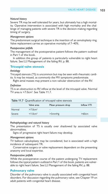
VaLVe HeaRt DISeaSe 103
Natural historySevere tR may be well tolerated for years, but ultimately has a high mortal-ity. Operative intervention is associated with high mortality and the chal-lenge of managing patients with severe tR is the decision-making regarding timing of surgery.
Management optionsthe predominant surgical technique is the insertion of an annuloplasty ring. Valve replacement carries an operative mortality of 7–40%.
Postoperative pitfallsthe management of the postoperative patient follows the pattern outlined in part of this book.
In addition this group of patients is particularly vulnerable to right heart failure. See b Management of the failing RV, p. 88.
Tricuspid valve stenosisAetiologytricuspid stenosis (tS) is uncommon but may be seen with rheumatic cardi-tis. It may be missed, as commonly the MV symptoms predominate.
Right atrial masses may present a non-valvular obstruction of the RV.
DefinitiontS is an obstruction to RV inflow at the level of the tricuspid valve. Normal TVareais>7.0cm2. See table .7.
Pathophysiology and natural historythe presentation of tS is usually over shadowed by associated valve abnormalities.
Signs of progressive right heart failure may develop.
Management optionstV balloon valvuloplasty may be considered, but is associated with a high incidence of subsequent tR.
Conservative surgery or valve replacement dependent on the presenting anatomy and local expertise.
Postoperative pitfallsWhile the postoperative course of the patient undergoing tV replacement follows the typical pattern outlined in part of this book, patients are vulner-able to right heart failure. See b Management of the failing RV, p. 88.
Pulmonary valveDisorder of the pulmonary valve is usually associated with congenital heart disorders. For discussion regarding the pulmonary valve, see Chapter 9 on adult patients with congenital heart disease.
Table .7 Quantification of tricuspid valve stenosis
Valve area Mean pressure drop Inflow VTI
Normal >7.0cm2 –
Severe <.0cm2 >5mmHg >60cm

104 CHapteR Heart
Common arrhythmias and their managementBradyarrhythmiaspostoperative bradyarrhythmias requiring permanent pacemaker implan-tation occur in up to 4% of patients, most commonly after aortic valve surgery. the most common abnormalities are a nodal rhythm, sinus node dysfunction, and complete aV block. a further group of patients require heart rate support in the immediate postoperative period to achieve a tar-get heart rate.
epicardial pacing is the mainstay of heart rate in the postoperative phase. transvenous pacing provides the backstop. transcutaneous pacing and pharmacological therapies are useful to temporize.
PacingMost postoperative patients have epicardial pacing leads and their heart rates can be controlled (see b Introduction, p. 304).
transcutaneous pacing may be administered through an external defibril-lator as a temporizing measure when the bradyarrhythmia is associated with a low Bp and no other therapy is immediately available. Heart rate is selected and gain i until capture is achieved. It is uncomfortable for the patient, who may require sedation.
transvenous pacing requires the placement of a transvenous temporary pacing wire in the RV under X-ray guidance.
Pharmacological managementAtropine• Bolus 0.–0.6 mg• Max. 3mg• anticholinergic, increases firing at Sa node increasing heart rate• Competitive antagonist for muscarinic acetyl-choline receptor, reducing
parasympathetic activity• Undesirable effects:
• Short-actingandineffectiveinAV block• Vasodilation• Reducedsweatingandlacrimation• Centralnervoussystem(CNS)effects—confusion,excitation• Dilatedpupils.
Isoprenaline• Bolus 20mcg• Then infusion –4mcg/min titrated to response• Sympathomimetic βand β2 adrenergic agonist• Chronotrope, ionotrope, and peripheral vasodilation effects• Undesirable effects:
• Proarrhythmic• Rarelyparadoxicalairwaysresistancerequiringcessationof the drug.

COMMON aRRHYtHMIaS aND tHeIR MaNaGeMeNt 105
Consider both directly and indirectly acting chronotropes, e.g. adrenaline (direct) and dobutamine (indirect) if both inotropy and chronotropy is appropriate to the patient’s condition.
Tachyarrhythmias—atrial fibrillationtachyarrhythmias, particularly aF occur in up to 40% of patients following CaBG and in up to 60% of patients following CaBG and valve replacement.
Riskthe risk of postoperative aF is highest in those with preoperative aF, i age, impaired LVeF, and hypokalaemia and hypomagnesaemia. peak incidence is on day 3.
Prophylaxisthere is some evidence for pharmacological prophylaxis of aF. the mainte-nance of potassium, magnesium, and calcium within the high normal range is effective first-line prophylaxis.
amiodarone and β-blockers may be considered for prophylaxis of aF following cardiac surgery.
TreatmentPatients in AF with haemodynamic compromise should have synchronized DC cardioversion.
Rate controlaF without haemodynamic compromise following CaBG can be treated with rate limiting therapy as 80% revert to sinus rhythm spontaneously within 24 hours and 90% revert spontaneously within 6 weeks.
treatment options for rate limitation include the following:• electrolytes: the maintenance of potassium, magnesium, and calcium
within the high normal range.• amiodarone provides rate control with the additional benefit that the
patient may chemically cardiovert.• β-blockers provide good rate control, but are not always appropriate to
start in the very early postoperative period.• Digoxin may be considered for rate control in aF.
Persisting AFaF is more likely to persist following valve surgery and may persist in a small number of patients following CaBG.
anticoagulation for patients with aF persisting for 48 hours should be considered on a case-by-case basis. the benefit of preventing thrombotic emboli and stroke needs to be weighed against the risk of bleeding in the postoperative period.
patients who tolerate rate-controlled aF may not require cardioversion. those who tolerate it poorly should be considered for chemical cardiover-sion or for elective synchronized DC cardioversion.
Where pharmacological therapy is used to treat AF, the need for persisting therapy should be reviewed within 6 weeks of hospital discharge.

106 CHapteR Heart
Tachyarrhythmias—ventricularVentricular tachycardias associated with CaBG have a variable effect on prognosis. Non-sustained Vt and VF is commonly seen when coming off bypass, it is thought to be re-perfusion induced, and is typically benign. Ventricular tachycardias occurring later in the clinical course may be associ-ated with a reversible underlying pathology such as cardiac tamponade or myocardial ischaemia which should be identified and treated.
Prophylaxis• amiodarone is indicated for prophylaxis against ventricular arrhythmias
in cardiac surgery.• Maintenance of electrolytes (potassium, magnesium, and calcium) in
the high normal range is indicated for prophylaxis against ventricular arrhythmias in cardiac surgery.
Pulseless VF and VT; VT with a pulse and haemodynamic compromisepatients with VF or pulseless Vt should be defibrillated immediately.
those with Vt and a pulse who have haemodynamic compromise may require sedation before cardioversion.
patients with refractory VF or pulseless Vt should be managed by the CaLS algorithm with early sternal re-opening, internal cardiac massage, and internal defibrillation. they may require re-institution of cardio-pulmonary bypass (see b Cardiac Surgery advanced Life Support, p. 26).
VT with a pulse and no haemodynamic compromiseeven in the absence of haemodynamic compromise, patients with persis-tent Vt require cardioversion. this may be pharmacological with amiodar-one, or electrical with synchronized DC cardioversion. patients who are conscious will require sedation for this.
Special circumstancetorsade de point (polymorphic Vt characterized by a change in amplitude of the QRS complexes with twisting around an isoelectric line) in an other-wise stable patient is managed medically with magnesium.
Further readingScottish Intercollegiate Guidelines Network (SIGN). Cardiac Arrhythmias in Coronary Heart Disease.
SIGN Guideline 94. edinburgh: SIGN; 2007.

107
Lung
Acute lung injury/acute respiratory distress syndrome 08Ventilator-associated pneumonia 3
Chapter 2

108 ChApter 2 Lung
Acute lung injury/acute respiratory distress syndromeIntroductionAcute respiratory distress syndrome (ArDS) is a clinical condition compris-ing respiratory distress, hypoxaemia refractory to oxygen, d lung compli-ance, and the appearance of diffuse infiltrates on a CXr.
DefinitionArDS is the severe form of a disease spectrum that occurs in the lungs of critically ill patients. Acute lung injury (ALI) is the ‘milder’ form of the spectrum and although it has a similar pathogenesis, it has a better outcome.
the diagnostic criteria for ALI/ArDS are as follows:• the onset is acute• the presence of bilateral pulmonary infiltrates on a CXr• the pulmonary artery wedge pressure (pAWp) is ≤8mmhg or no
clinical evidence of left atrial hypertension• In ALI the paO2/FiO2 ratio is <40 (300mmhg)• In ArDS the paO2/FiO2 ratio is <27 (200mmhg).
the Murray Lung Injury Score can stratify severe acute respiratory failure (table 2.).
Table 2. the Murray Lung Injury Score
Score 0 2 3 4
paO2/FiO2 (mmhg)
≥300 225–299 75–224 00–74 <00
Chest radiographic alveolar consolidation
None quadrant 2 quadrants 3 quadrants 4 quadrants
peep (cmh2O) ≤5 6–8 9– 2–4 ≥5
Compliance ≥80 60–79 40–59 20–39 ≤9
the final value is obtained by dividing the aggregate sum by the number of components that were used.
No lung injury 0
Mild–moderate lung injury 0.–2.5
Severe lung injury >2.5
reprinted with permission of the American thoracic Society. Copyright © 204 American thoracic Society. Murray JF Matthay MA et al. An expanded definition of the adult respiratory distress syndrome. Am Rev Respir Dis. 988; 38: 720–723. Official Journal of the American thoracic Society.
Incidencethe reported incidence of ALI varies widely; incidence ranging from 6–34 per 00,000 person-years in europe and Australia whereas in the US the incidence is 78 per 00,000 person-years.

ACUte LUNg INJUry/ACUte reSpIrAtOry DIStreSS SyNDrOMe 109
two decades ago the mortality ranged from 50–70% but has since declined and is now in the region of 30–40%. this is similar in patients who present with ALI or ArDS. the cause for the decline is not entirely clear but advances in general supportive care and the use of ventilatory strategies may account for the change.
Risk factorsthere is a probable genetic predisposition with higher death rates in African Americans and males:• pulmonary risk factors:
• Pneumonia• Aspiration• Lungcontusion• Inhalational injury• PE• Drowning
• extrapulmonary risk factors:• Sepsis• Shock• Trauma• Pancreatitis• Bloodtransfusion• CPB.
In recent years transfusion-related ALI (trALI) and novel viral pathogens (severe acute respiratory syndrome or SArS) have emerged as important risk factors for the development of ALI.
PathophysiologyThe acute phase• Disruption of the normal alveolar–capillary barrier allowing protein-rich
fluid to leak into alveoli; neutrophils, red blood cells, and fibroblasts all enter alveoli.• pro-inflammatory cytokines are secreted by alveolar macrophages.• Anti-inflammatory cytokines are also found in the alveoli, but in ALI/
ArDS the balance is disrupted and the pro-inflammatory effect predominates.• Other mechanisms in the acute phase involve coagulation abnormalities
leading to microvascular occlusion in the lungs due to platelet and fibrin-rich thrombi and abnormal fibrinolysis.
The resolution phase• Depends on repair of the alveolar epithelium and removal of the
protein-rich fluid from the alveolar space.• proliferation of type II cells occurs which then differentiate into
type I cells.• removal of alveolar fluid dependent on active transport of sodium
via type II cells, and then water follows by osmosis via channels on type I cells.• Insoluble proteins removed by macrophages and neutrophil clearance
may be by apoptosis.

110 ChApter 2 Lung
Fibrosing alveolitis• Associated with a higher mortality.• May begin as early as 5–7 days after onset of ALI/ArDS.• Alveolar spaces get filled with inflammatory cells, fibrin, collagen, and
blood vessels leading to fibrosis.
Managementthe management of ALI/ArDS is aimed at:• Identifying and treating the underlying cause:
• Considerantimicrobialtherapy• Considersurgicaldrainageof anintra-abdominalcollection• Removalof invasivelinesinthecaseof acatheter-relatedblood
stream infection• providing supportive care:
• Adequatenutrition• Stressulcerprophylaxis• DVTprophylaxis• Haemodynamicmanagement• Optimalfluidmanagementremainsunclear.Conservativestrategy
associated with improved lung function and d duration of mechanical ventilation and intensive care
• Maintaining oxygenation using a protective ventilatory strategy. Ventilating at high tidal volumes (volutrauma) and pressures (barotrauma) can cause disruption of the alveolar–capillary interface; biotrauma (the release of inflammatory cytokines from neutrophils) is due to shearing forces as collapsed alveoli are repeatedly opened and closed and can lead to distant organ damage:• TheARDSnetgrouphavepublishedatablelookingatthe
combinations of FiO2 and peep for maintaining arterial oxygenation in ArDS (table 2.2)
• Achieving paO2 >8kpa or SpO2 88–95%.
Lung-protective strategies• FiO2: accept a paO2 >8kpa. prolonged high concentrations of oxygen
can contribute to lung injury due to oxygen toxicity• PEEP (positive end-expiratory pressure) improves oxygenation by
recruiting collapsed alveoli, improving ventilation/perfusion (V/Q) matching, and reducing intrapulmonary shunt.• Low tidal volume ventilation:
• Maintainatidalvolumeof 6mL/kgusingpredictedbody weight• Keepthepeakairwaypressure<30cmH2O• Permissivehypercapnia(pH >7.).

ACUte LUNg INJUry/ACUte reSpIrAtOry DIStreSS SyNDrOMe 111
Alternative strategies for improving profound hypoxaemia include:• Recruitment manoeuvres (RM) and high PEEP:
• Theaimistoopenuppreviouslycollapsedandfloodedalveoliwhich should lead to improved oxygenation and a reduction in ventilator-induced lung injury from the shear stresses from repetitive opening and closing of alveoli.
• However,‘normal’alveoliwillbesubjectedtotheriskofoverdistensionand damage and there is a possibility of haemodynamic compromise.
• RMwithhighPEEPorhighPEEPaloneisbestconsideredearlyinArDS with severe hypoxaemia when plateau airway pressures are <30cmh2O.
• Donotconsiderthesestrategiesinpatientswithpneumothoracesorwhen the disease is focal.
• Prone positioning:• V/Qmismatchisimprovedbyrecruitingdependentatalectatic
lung areas.• Oxygenationdoesimprove,butthereisnoclearevidencethat
mortality is reduced.• Therearetechnicaldifficultiesassociatedwithturningpatients,such
as accidental dislodgement of invasive catheters and endotracheal/ tracheostomy tubes as well as the risk of pressure ulcers and facial oedema.
• High-frequency oscillatory ventilation (HFOV):• Useshighairwaypressuretoachievelungrecruitmentandthus
improve oxygenation.• Anoscillatingpistoncreatescyclesof pressureaboveandbelowthe
mean airway pressure at a high frequency with small tidal volumes.• Shouldnotbeusedinpatientswithshock,airwayobstruction,
intracranial haemorrhage, or refractory barotrauma.• Sedationisneededandsevereacidosismayoccurduetolimited
CO2 excretion.
Table 2.2 the ArDSnet group combinations of FiO2 and peep for maintaining arterial oxygenation in ArDS
FiO2 PEEP (cm H2O)
0.3 5
0.4 5–8
0.5 8–0
0.6 0
0.7 0–4
0.8 4
0.9 6–8
.0 8–24
Data from the Acute respiratory Distress Syndrome Network, ‘Ventilation with lower tidal volumes as compared with traditional tidal volumes for acute lung injury and the acute respiratory distress syndrome’, The New England Journal of Medicine, 342, 8, pp. 30–308.

112 ChApter 2 Lung
• Inhaled nitric oxide:• Apotentvasodilatorwhichpromotesthedistributionof bloodflow
towards ventilated portions of the lung minimizing V/Q mismatch and improving oxygenation.
• Littlesystemicabsorptionandrapidlyinactivated.• Steroids:
• SteroidscouldstoptheprogressiontosevereALI/ARDSbyinhibiting neutrophil activation, collagen deposition, and the proliferation of fibroblasts.
• Clinicaltrialshavefailedtoshowabenefittopatientsgivensteroidsin early ALI and an i mortality was noted in patients given steroids after 4 days of diagnosis.
• Routineuseisnottoberecommendedbutcouldbeconsideredinlife-threatening hypoxaemia when other measures have failed.
• Lowdoseof methylprednisolone(mg/kg/day)shouldbeused,andstopped if no improvement occurs after 3 days.
• Extracorporeal lung support (ECLS):• Veno-venouslifesupportcircuitthatremovesbloodfromthe
patient and circulates it through a membrane oxygenator providing gaseous exchange and allowing the lungs to recover. there are two main types of eCLS that have been used: extracorporeal membrane oxygenation (eCMO) which is a modified form of cardiopulmonary bypass and extracorporeal CO2 removal.
• Themainrisksareduetotheneedforanticoagulationandlarge-bore central venous cannulae. the CeSAr trial (Conventional ventilation or eCMO for severe adult respiratory failure) showed an improvement in the survival and absence of severe disability at 6 months in the intervention group.
• ECMOorextracorporealCO2 removal should only be considered when all other therapies have failed and at a centre with experience in extracorporeal lung support. It should not be used in patients with contraindications to anticoagulation and in those patients ventilated with high airway pressures for >7 days.
Further readingArDS Clinical Network website: M <http://www.ardsnet.org>.

VeNtILAtOr-ASSOCIAteD pNeUMONIA 113
Ventilator-associated pneumoniaDefinitionNICe defines ventilator-associated pneumonia (VAp) as ‘pneumonia which develops 48 hours or more after intubation with an endotracheal or trache-ostomy tube, and which was not present before’:• In the US up to 30% of ventilated ItU patients have a VAp.• patients who develop VAp have significantly longer duration of
mechanical ventilation and ItU stay.• Mortality is i in patients by 30% if they have a VAp and can reach more
than 70% if multiresistant pathogens are involved.
DiagnosisVAp is a difficult diagnosis to make. Ideally clinical and radiological signs and positive microbiology will all coincide. Clinical signs may be non-specific, radiological signs may also be non-specific and lag behind clinical changes. Microbiological confirmation will be affected by:• Sampling technique:
• Sputum sample• Trachealaspirate• Bronchoalveolar lavage• Protectedspecimen brush
• recent antimicrobial therapy• pathogenicity of organisms• Culture technique; quantitative vs non-quantitative.
A pragmatic approach used in Scottish ICUs is:• the presence of new or persistent infiltrates on CXr and any 2 of:
• Febrile >38°C• Leucocytosisorleukopenia(>or >3.5)• Purulenttracheobronchialsecretions.
Aetiologythe micro-organisms responsible for VAp vary according to the popula-tion of patients in the ItU, the duration of hospital and ItU stays, and the diagnostic method used:• AerobicGram-negativebacilli(GNB)areresponsibleforaround60%
of VAP.ThepredominantGNBwerePseudomonas aeruginosa and Acinetobacter spp. followed by Proteus, Escherichia coli, Klebsiella, and Haemophilus influenzae.• gram-positive pneumonias are increasingly seen with Staphylococcus
aureus being the most common.• polymicrobial infection in occurs in 20–50% of VAp.
Pre-disposing factors• prolonged antibiotic treatment increases the risk of superinfection
with multiresistant organisms and delays the occurrence of nosocomial infection.• ett, reintubation, and tracheostomy: the presence of an ett not only
bypasses normal host defences but causes local trauma.

114 ChApter 2 Lung
• Ng tube, enteral feeding, and supine position.• respiratory equipment: ventilator circuits may be a source of bacteria
responsible for VAp. the use of heat and moisture exchangers (hMes) instead of heat humidifiers in suitable patients has been associated with a decrease in the risk of developing VAp
Prevention—VAP bundle• Elevation of the head of the bed to 35°.• Daily ‘sedation breaks’ reduce duration of mechanical ventilation.• Daily oral care with chlorhexidine: dental plaque can be a reservoir for
potential pathogens that cause VAp.• Peptic ulcer prophylaxis: stress ulceration is the most common cause of
gI bleeding in the critically ill and is associated with a 5-fold increase in mortality compared with those ItU patients without bleeding. Although there is a trend towards a lower pneumonia rate in patients treated with sucralfate rather than ph altering drugs, h2 blockers were more effective in preventing peptic ulcer disease and so are the preferred agents.• DVT prophylaxis: this is an appropriate intervention in all sedentary
patients.
Further readingMeade MO, Cook DJ, guyatt gh, Slutsky AS, Arabi yM, Cooper DJ, et al.; Lung Open Ventilation
Study Investigators. Ventilation strategy using low tidal volumes, recruitment maneuvers, and high positive end-expiratory pressure for acute lung injury and acute respiratory distress syndrome: a randomized controlled trial. JAMA 2008;299(6):637–45.
MercatA,RichardJ-CMVielleB,JaberS,OsmanD,DiehlJ-L,et al.; expiratory pressure (express) Study group. positive end-expiratory pressure setting in adults with acute lung injury and acute respiratory distress syndrome: a randomized controlled trial. JAMA 2008;299(6):646–55.

115
Kidney
Acute kidney injury 6Chronic kidney disease 24
Chapter 3

116 ChApter 3 Kidney
Acute kidney injuryIntroductionthe principal functions of the kidney are:• regulation of body fluid volume and osmolality• excretion of metabolic end products and foreign substances• regulation of acid–base• production and secretion of enzymes and hormones (erythropoietin,
renin, and ,25 dihydroxyvitamin D3).
In healthy subjects any change in body fluid volume, Bp, or acid–base bal-ance is corrected in a matter of hours, however in disease states these regulatory processes are disturbed.
the true incidence of acute kidney injury (AKI) in cardiac surgery is not clear, as there is significant variability in studies over how abrupt or severe the kidney dysfunction must be before it is defined as AKI. It is, however, a common complication of critical illness and an independent risk factor for postoperative death. the risk for death is proportional to the severity of kidney injury with mortality being as high as 50% in the general intensive care setting. Mortality is higher in patients with a sustained elevation of creatinine than in those who recover function.
All patients undergoing cardiac surgery are at risk of AKI either through their presenting illness or subsequent iatrogenic injury. Specific risk factors are listed in table 3. and these should be considered prior to elective surgery. the most common pathogenesis of AKI following cardiac surgery is disrupted renal blood flow and the accepted prevention strategies are therefore:• Appropriate IV volume expansion• Optimization of CO• Maintenance of renal blood flow and renal perfusion pressure.
‘Renal dose dopamine’there is no clinical benefit for its use in the prevention or treatment of AKI. recent studies have shown it can worsen renal perfusion in patients with established AKI.
Furosemide infusionFurosemide does not prevent the development of, or hasten the recovery from, AKI. Its inappropriate use can lead to intravascular hypovolaemia and worsening renal function. Loop diuretics can be used to correct volume overload in patients who are still responsive to treatment.
Key point: contrast-induced nephropathyIdentify patients at risk and carefully assess their fluid status. If indicated use pre- and postprocedure volume expansion with 0.9% sodium chlo-ride or isotonic sodium bicarbonate. the use of N-acetyl cysteine is not recommended. Diuretics, NSAIDs, ACe inhibitors, and metformin should be withheld near the time of the procedure. As metformin is exclusively excreted by the kidneys, patients who develop AKI are at i risk of lactic acidosis. If creatinine clearance is <60mL/min stop for 24 hours prior to contrast being given.

ACute KIDNey INjury 117
Further readingBellomo r, Chapman M, Finfer S, hickling K, Myburgh j. Low-dose dopamine in patients with early
renal dysfunction: a placebo-controlled randomised trial. ANZICS Clinical trials Group. Lancet 2000;356:239–43.
Friedrich jO, Adhikari N, herridge MS, Beyene j. Meta-analysis: low dose dopamine increases urine output but does not prevent renal dysfunction or death. Ann Intern Med 2005;42:50–24.
ho KM, Sheridan Dj. Meta-analysis of furosemide to prevent or treat AKI. BMJ 2006;333(7565):420–5.ho KM, power BM. Benefits and risks of furosemide in AKI. Anaesthesia 200;65:283–93.Lauschke A, teichgräber uK, Frei u, eckardt Ku. Low-dose dopamine worsens renal perfusion in
patients with AKI. Kidney Int 2006;69:669–74.
Definition and stagingthe term acute renal failure implies that the function of the kidney is sig-nificantly deranged. renal failure is a spectrum of disease from less severe forms of injury to more advanced injury where renal replacement therapy (rrt) may be required. A small increase in serum creatinine level is an inde-pendent predictor of morbidity and mortality and for this reason the term acute kidney injury (AKI) is now used to describe acute changes in renal function.
Table 3. risk factors for the development of AKI following cardiac surgery
Preoperative Intraoperative Postoperative
Advanced ageFemale sexChronic renal diseaseDiabetes mellitusChronic cardiac failureAortic diseaseperipheral vascular diseaseChronic liver diseaseGenetic predispositionIABpDrug nephrotoxicity
prolonged CpB timeCombined surgical proceduresemergency surgeryprevious cardiac surgeryAortic clamp placement
Low CO syndromeAcute cardiac dysfunctionMediastinal haemorrhagehypovolaemiarhabdomyolysisIntra-abdominal hypertensionMultiple organ dysfunctionDrug nephrotoxicity
Key point: biomarkers in renal diseaseCreatinine is currently the only biomarker of renal disease used in clinical practice. It is easily measured and specific for renal function; however, an acute decline in kidney function is not reflected by a rise in serum creati-nine for several hours. For this reason urine output is used as a clinical monitor of renal function.

118 ChApter 3 Kidney
AKI is generally defined as ‘an abrupt and sustained decrease in kidney function’. the uK renal Association recommends that the Acute Kidney Injury Network (AKIN) definition as adopted and refined by the Kidney Diseases: Improving Global Outcomes (KDIGO) International guideline group be used (table 3.2).
AKI can be diagnosed when one of the following criteria is met:• Serum creatinine rises by ≥26µmol/L from baseline within 48 hours or• Serum creatinine rises ≥.5× from baseline value which is known or
presumed to have occurred within one week or• urine output documented <0.5mL/kg/hour for >6 consecutive hours.
General managementthe aims of non-rrt management should be prevention of further kidney injury and supportive care to allow functional recovery. Management should begin with prompt diagnosis and correction of the underlying cause of renal injury. Although the most common pathologies in AKI post-cardiac surgery involve disruption of the renal blood flow all causes should be considered and excluded where appropriate. Sepsis is known to be a leading contrib-uting factor to AKI in critical illness and should be diagnosed and treated promptly.
Clinical assessment general management measures• Assess fluid balance and optimize haemodynamic status:
• Clinicalassessment—pulse,meanarterialbloodpressure,capillaryrefill time, jugular or central venous pressure, Glasgow Coma Scale (GCS), acid–base balance, lactate, and serum albumin measurements
• Ensureadequatefluidreplacement• Considertheuseof vasopressorsorinotropes.
Table 3.2 KDIGO staging of AKI
Stage Serum creatinine Urine output
.5–.9 × baselineOr≥0.3mg/dL (≥26.5µmol/L) increase
<0.5mL/kg/h for 6–2 hours
2 2.0–2.9 × baseline <0.5mL/kg/h for ≥2 hours
3 3.0 × baselineOrIncrease in serum creatinine to ≥4.0mg/dL (≥353.6µmol/L)OrInitiation of rrtOrIn patients <8 years, decrease in eGFr to <35mL/min per .73m2
<0.3mL/kg/h for ≥24 hoursOrAnuria for ≥2 hours
Kidney Disease: Improving Global Outcomes (KDIGO) Acute Kidney Injury Work Group. KDIGO 202 Clinical practice Guideline for Acute Kidney Injury. Kidney Inter., Suppl. 203; 3: –50, reproduced with permission.

ACute KIDNey INjury 119
• Assess for complications of AKI:• Correcthypoxaemiaif pulmonaryoedema—continuouspositive
airway pressure (CpAp) or intubation and ventilation may be necessary until fluid can be removed with rrt
• Recognizeandtreathyperkalaemia• Assessforsignsof uraemia—pericarditis,encephalopathy,
drowsiness, seizures.• Consider and correct reversible causes of AKI.• establish preoperative renal function.• exclude renal tract obstruction using ultrasonography.• Identify and treat sepsis appropriately.• Stop and avoid nephrotoxic medication and dose adjust those drugs with
renal clearance.• treat metabolic acidosis.
Interpret a low urine output in the context of the patient’s cardiovascular parameters:• the oliguric patient may not be hypovolaemic.• urine output is influenced by the patient’s cardiovascular parameters,
hydration state, the use of diuretics, the presence of renal tract obstruction, and the stress response to injury.
Key pointhyperkalaemia when associated with eCG change is a medical emergency.
Causes of AKI to consider• pre-renal (reduced renal perfusion):
• Volumedepletion,d Bp, d CO, intrarenal vasoconstriction (drugs, radiographic contrast), hepatorenal syndrome.
• renal or intrinsic:• Vascular(renalarterialdisease,aorticatheroma/cholesterol
emboli), nephrotoxins, infections, interstitial nephritis, accelerated phase hypertension, vasculitis, glomerulonephritis.
• post-renal (obstruction):• Prostatehypertrophy,neurogenicbladder,intraureteral
obstruction (stones, tumour, thrombus, crystals), extra ureteral obstruction (tumour or retroperitoneal fibrosis).
Treatment of hyperkalaemiaensure stable cardiac rhythm:• IVcalciumgluconateorcalciumcarbonateif ECGchanges—follow
ALS guidelines.
Stop exogenous sources of potassium:• Nutrition (oral/enteral/parenteral), potassium supplementation, IV
fluid (e.g. hartmann’s solution).
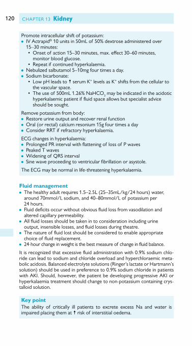
120 ChApter 3 Kidney
Fluid management• the healthy adult requires .5–2.5L (25–35mL/kg/24 hours) water,
around 70mmol/L sodium, and 40–80mmol/L of potassium per 24 hours.• Fluid deficits occur without obvious fluid loss from vasodilation and
altered capillary permeability.• All fluid losses should be taken in to consideration including urine
output, insensible losses, and fluid losses during theatre.• the nature of fluid lost should be considered to enable appropriate
choice of fluid replacement.• 24-hour change in weight is the best measure of change in fluid balance.
It is recognized that excessive fluid administration with 0.9% sodium chlo-ride can lead to sodium and chloride overload and hyperchloraemic meta-bolic acidosis. Balanced electrolyte solutions (ringer’s lactate or hartmann’s solution) should be used in preference to 0.9% sodium chloride in patients with AKI. Should, however, the patient be developing progressive AKI or hyperkalaemia treatment should change to non-potassium containing crys-talloid solution.
promote intracellular shift of potassium:• IV Actrapid® 0 units in 50mL of 50% dextrose administered over
5–30 minutes:• Onsetof action5–30minutes,max.effect30–60minutes,
monitor blood glucose.• Repeatif continuedhyperkalaemia.
• Nebulized salbutamol 5–0mg four times a day.• Sodium bicarbonate:
• LowpHleadstoi serum K+ levels as K+ shifts from the cellular to the vascular space.
• Theuseof 500mL.26%NaHCO3 may be indicated in the acidotic hyperkalaemic patient if fluid space allows but specialist advice should be sought.
remove potassium from body:• restore urine output and recover renal function• Oral (or rectal) calcium resonium 5g four times a day• Consider rrt if refractory hyperkalaemia.
eCG changes in hyperkalaemia:• prolonged pr interval with flattening of loss of p waves• peaked t waves• Widening of QrS interval• Sine wave proceeding to ventricular fibrillation or asystole.
the eCG may be normal in life-threatening hyperkalaemia.
Key pointthe ability of critically ill patients to excrete excess Na and water is impaired placing them at i risk of interstitial oedema.

ACute KIDNey INjury 121
Renal replacement therapyrrt is an extracorporeal method of removing fluid and solutes from blood outside the body. During treatment blood comes in to contact with a semipermeable membrane where solute transfer occurs prior to it being returned to the patient. this can be carried out using intermittent (Irrt) or continuous (Crrt) techniques. In the acute setting Irrt is usually in the form of haemodialysis and Crrt is usually in the form of continuous veno-venous haemofiltration (CVVh) although hybrids of both techniques do exist.
IRRT—haemodialysis• Solutesareremovedbyprocessof diffusion.• Solute removal by this method is rapid and efficient.• Fluid can be removed (ultrafiltration) by adjusting the pressures on the
blood side of the semipermeable membrane.
CRRT—CVVH• Solutes are removed by process of convection across a highly
permeable filter.• Convectivesoluteremovalislessefficientthandiffusivebutthe
continuous nature of the treatment compensates for this.• the solute removal in CVVh is completely reliant on fluid removal
therefore ‘solute-free’ fluid is replaced during the process either before (predilution) or after (postdilution) the blood comes in to contact with the semipermeable membrane
Consideration should be given to the type and dose of rrt chosen, the timing of initiation and discontinuation, access for rrt, and anticoagulation during rrt.
Type of RRT chosen• CRRToffersnosurvivaladvantageoverIRRTinpatientswith AKI.• the type of rrt chosen does not influence the rate of renal recovery
following AKI.• Crrt is associated with a significantly i risk of recurrent filter clotting
compared to Irrt.• Due to the continuous nature of Crrt there may be greater
haemodynamic stability in patients with multiorgan failure and this is used in preference to Irrt in most ICus.
Possible indications for urgent RRT• refractory hyperkalaemia >6.5mmol/L• refractory volume overload• refractory severe metabolic acidosis (ph <7.)• Signs of uraemia (e.g. pericarditis, neuropathy, seizures, or an
otherwise unexplained decline in metal status)• Certain drug and alcohol intoxications.

122 ChApter 3 Kidney
Initiation and discontinuation of RRTthere is lack of evidence to guide the optimal time to initiate rrt; however, for patients with multiorgan failure in the intensive care setting early initia-tion of rrt should be considered. When the urine output is ≥400mL/24 hours and the clinical condition of the patient is improving, discontinuation of rrt can be considered. Accurate assessment of fluid, electrolyte, and metabolic status is essential and rrt should be reconsidered if clinically indicated.
Dose of RRTthe dose of rrt can be altered by adjusting the blood flow, the area of the semipermeable membrane, or the duration of treatment. the prescribed dose should be assessed daily for continuous techniques and prior to each session for intermittent techniques.• IRRT: the best evidence suggests patients should receive at least 3
sessions/week with delivered Kt/V value of .2. Most patients require daily or alternate day treatments to achieve this value.• CRRT: the best evidence suggests patients should receive treatment
doses equivalent to post dilution ultrafiltration rates ≥25mL/kg/hour.
AnticoagulationDue to the requirement of extracorporeal blood flow in both Irrt and Crrt some form of anticoagulation is usually required to prevent throm-bosis in the blood circuit. type of rrt and risk of bleeding should be taken in to consideration when choosing type of anticoagulation.• heparin: bolus 000–2000u at the start of rrt session followed by
continuous infusion of 300–400u/hour. Aim for Appt in the venous limb .5–2 × control.• regional citrate anticoagulation: continuous infusion of isosmotic trisodium
citrate solution (02mmol/L) in to the arterial side of the dialyser.• regional anticoagulation with protamine reversal: technically difficult
with i risk of rebound bleeding 2–4 hours post rrt.
Access• In the acute setting access is veno-venous and the access site depends
on the clinical condition of the patient.• ultrasound guidance should be used to aid placement of venous
catheters and post-procedure CXr should confirm position.• Internal jugular catheters should be 5–20cm in length and femoral
catheters should be 20–25cm in length.
Indications for IRRT in preference to CRRT• Drug poisoning: removal of specific toxins such as Li+,
theophyllines, etc.• Bleeding risk: during Crrt blood is in contact with the extracorporeal
circuit for longer and circuit clotting is more common, necessitating anticoagulation.• Mobilization: rehabilitation and mobilization is difficult in patients
receiving Crrt and in patients no longer confined to bed Irrt should be considered.

ACute KIDNey INjury 123
• tunnelling of the venous catheter is desirable if rrt is likely to be prolonged (>3 weeks).
Complications of vascular access• Catheter-related bacteraemia: the catheter should be reserved for
extracorporeal treatment only to minimize the risk of catheter-related infection. the exit site should be carefully monitored and temporary access should be changed if there is clinical suspicion of catheter-related infection.• Arrhythmia: insertion of internal jugular or subclavian catheters in the
hyperkalaemic patient risks the guidewire touching the endocardium resulting in life-threatening and refractory arrhythmias.• Clotting of dialysis catheter lumens: between periods of Irrt
catheters should be locked with heparin (000u/mL to the lumen volumes). this lock must be removed prior to recommencing rrt to avoid introducing heparin into the systemic circulation.
Complications of RRT• hypotension: can occur with both Irrt and Crrt. i likelihood with
high ultrafiltration rate, reduced LV function, and concurrent use of antihypertensive medications.• recirculation: caused when blood leaving the dialyser outlet returns
to the inlet without having first passed through the peripheral bloodstream. this is more common with the use of femoral catheters and if the venous catheter used is an inadequate length.• Infection: immune system dysregulation is multifactorial and patients
with AKI have an i susceptibility to infection.• Cardiac arrhythmias: can occur with rapid change in serum electrolyte
concentration. Carefully monitor and replace potassium, phosphate, and magnesium if necessary.• haemolysis.• Air embolism.• rupture of the dialysis membrane: can lead to blood loss and entry
of microorganisms to the bloodstream. usually avoided by ensuring ultrafiltration rate (transmembrane pressure) is not too high.• reaction to dialysis membrane: rare complication. Clinical
manifestations can vary from anaphylaxis to abdominal pain, fevers, or headaches.• Disequilibrium syndrome: neurological signs and symptoms caused
by cerebral oedema due to a rapid reduction in blood urea nitrogen. Onset of symptoms is usually during or shortly after Irrt, however the syndrome is now rare as rrt is initiated at an earlier stage.• transmission of blood-borne viruses.
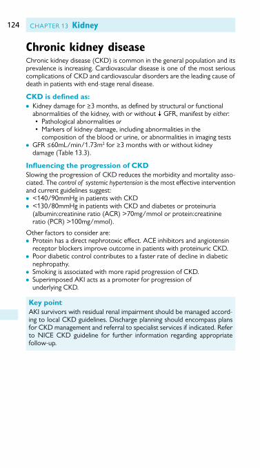
124 ChApter 3 Kidney
Chronic kidney diseaseChronic kidney disease (CKD) is common in the general population and its prevalence is increasing. Cardiovascular disease is one of the most serious complications of CKD and cardiovascular disorders are the leading cause of death in patients with end-stage renal disease.
CKD is defined as:• Kidney damage for ≥3 months, as defined by structural or functional
abnormalities of the kidney, with or without d GFr, manifest by either:• Pathologicalabnormalities or• Markersof kidneydamage,includingabnormalitiesinthe
composition of the blood or urine, or abnormalities in imaging tests• GFr ≤60mL/min/.73m2 for ≥3 months with or without kidney
damage (table 3.3).
Influencing the progression of CKDSlowing the progression of CKD reduces the morbidity and mortality asso-ciated. the control of systemic hypertensionisthemosteffectiveinterventionand current guidelines suggest:• <40/90mmhg in patients with CKD• <30/80mmhg in patients with CKD and diabetes or proteinuria
(albumin:creatinine ratio (ACr) >70mg/mmol or protein:creatinine ratio (pCr) >00mg/mmol).
Other factors to consider are:• Proteinhasadirectnephrotoxiceffect.ACEinhibitorsandangiotensin
receptor blockers improve outcome in patients with proteinuric CKD.• poor diabetic control contributes to a faster rate of decline in diabetic
nephropathy.• Smoking is associated with more rapid progression of CKD.• Superimposed AKI acts as a promoter for progression of
underlying CKD.
Key pointAKI survivors with residual renal impairment should be managed accord-ing to local CKD guidelines. Discharge planning should encompass plans for CKD management and referral to specialist services if indicated. refer to NICe CKD guideline for further information regarding appropriate follow-up.

ChrONIC KIDNey DISeASe 125
Complications of CKD• Cardiovascular disease: the traditional Framingham risk factors apply
in CKD –3 but the Framingham tables significantly underestimate the risk of cardiovascular disease in patients with more advanced CKD. Statins reduce cardiovascular mortality in CKD stages –4. the role of statins in CKD5D patients is unclear and still under review.• hypertension: Bp is a major risk factor for both cardiovascular and
renal mortality. Dietary salt restriction should be suggested to all hypertensive patients with CKD. target values are outlined as follows:• Anaemia: consider the use of erythropoiesis stimulating agent (eSA)
in anaemic patients with eGFr <25mL/min (<40mL/min in diabetic patients). exclude causes other than renal impairment and ensure patients have adequate stores of iron, vitamin B2 and folate prior to commencing eSA treatment. Aim for a target hb of –2g/dL.• hyperparathyroidism and renal osteodystrophy: bone metabolism is
disrupted in patients with moderate to severe CKD and accelerates vascular disease. In patients with CKD stage 3+ guidelines currently suggest serum calcium levels should be kept within normal limits, serum phosphate at or below .8mmol/L and pth below 2–3 × the upper limit of normal. Specialist advice should be sought in patients who have evidence of renal bone disease.• Metabolic acidosis: CKD stage 3+.• Infection: alterations in both cellular and humoral immunity occur
leading to a significant i susceptibility of infection. this worsens with progression of uraemia and stage of CKD.• Malnutrition: anorexia and nausea are commonly found in patients
with advancing CKD. Vitamin supplementation may be required.
Table 3.3 KDIGO staging of CKD
Stage Description GFR (mL/min/.73m2)
Kidney damage with normal or i GFr ≥90
2 Kidney damage with mild d GFr 60–89
3A
3B
Moderate d GFr 45–59
30–44
4 Severe d GFr 5–29
5 Kidney failure <5 (or dialysis)
P suffix: the addition of p to a stage is used to denote the presence of proteinuria (this is defined as ACr ≥30mg/mmol or pCr ≥50mg/mmol).
T suffix: the addition of t to a stage indicates the patient has a renal transplant.
D suffix: the addition of d to a stage indicates the patient is on dialysis.
Kidney Disease: Improving Global Outcomes (KDIGO) CKD Work Group. KDIGO 202 Clinical practice Guideline for the evaluation and Management of Chronic Kidney Disease. Kidney Inter., Suppl. 203; 3: –50, reproduced with permission.


127
Gastrointestinal
Gastrointestinal complications after cardiac surgery 28Investigating the acute abdomen 32Gastrointestinal bleeding 34Nutrition in CICU 36
Chapter 4

128 Chapter 4 Gastrointestinal
Gastrointestinal complications after cardiac surgeryGastrointestinal (GI) complications after cardiac operations are infrequent (0.3–3%) but serious. the overall mortality from GI complications ranges from –72% and has not d significantly during the last two decades. GI conditions range from mild, but troublesome, complications like ileus to devastating complications such as bowel ischaemia and liver failure.
Common complications• GI bleeding (33%), mortality 20%• Mesenteric ischaemia (4%), mortality 50–00%• pancreatitis (%), mortality 20%• acute cholecystitis (9%), mortality 26%• perforated peptic ulcer (5%), mortality 36%• Liver failure (2%), mortality 56%.
Risk factorsNumerous risk factors for GI complications have been reported and can be classified as pre-, intra-, and postoperative. the factors that have been identified on multivariate analysis are:• preoperative: age >70 years, low CO (22-fold i risk), peripheral
vascular disease, reoperative surgery and chronic renal failure• Intra- and postoperative: i need for blood transfusions, prolonged CpB,
dysrhythmias (7-fold i risk), IaBp.
these factors are general markers of how unwell these patients are as well as proxy markers for the complexity of their surgery. Mesenteric ischaemia is often the final part of a tragic tale of organ dysfunction in patients who likely would have died from circulatory failure anyway, with or without GI complications.
the cause of GI complications is still unknown but most theories impli-cate low CO resulting in visceral hypoperfusion and ischaemia. this process triggers a hormonal stress response and leads to a mediator-driven sys-temic inflammatory response.
the role of CpB in the aetiology of GI complications is controversial. One randomized trial showed a significant decrease of GI complications in coronary bypass off-pump vs on-pump. these results have not been dupli-cated and most studies found no difference with regard to GI complica-tions, gastric mucosal oxygenation, or superior mesenteric arterial blood flow when comparing on-pump with off-pump CaBG.
Other perioperative interventions have been shown to reduce the inci-dence of GI complications like stress-ulcer prophylaxis and goal-directed haemodynamic therapy and even intraoperative manoeuvres like epi-aortic scanning prior to clamping shows promise.
Ischaemic bowelOccurs after 0.6% of all cardiac operations and has a mortality of 50% on average but may be as high as 00% in some reports. the patients are com-monly on mechanical ventilation and sedated, thus making the usual signs of

GastrOINtestINaL COMpLICatIONs after CardIaC sUrGery 129
abdominal pain and tenderness unreliable. furthermore, many patients with multiple organ dysfunction in intensive care develop distended and tender abdomens with ileus making it a diagnostic challenge to identify the patient with ischaemic bowel.
the onset is usually gradual and non-specific with Ileus, abdominal disten-sion, i NG output, i need for organ support, raised inflammatory markers, hyperkalaemia, unexplained lactic acidosis, and lower GI bleeding. all of these are non-specific and contribute to a delay in diagnosis with imorbid-ity and mortality.
Mesenteric ischaemia can be occlusive, non-occlusive and rarely due to mesenteric venous thrombosis.• Non-occlusive mesenteric ischaemia (NOMI) is more common and
thought to be secondary to hypoperfusion and the use of vasopressor drugs. the onset is slower and more gradual than occlusive ischaemia. If suspected and the patient does not need urgent laparotomy for bowel necrosis (as indicated by severe acidosis, peritonitis, Ct findings) the diagnosis can be made with mesenteric angiography showing the characteristic ‘string of sausages’ signs due to constrictions in the mesenteric arteries.• Management in the early stages include: broad-spectrum antibiotics,
aggressive resuscitation to maximize mesenteric blood flow, NG tube decompression, and intra-arterial papaverine infusion (bolus of 60mg followed by continuous infusion of 0.7mg/kg per hour) through the intra-arterial catheter placed into the sMa during arteriography.• If bowel necrosis is already established urgent laparotomy is required.• Occlusive mesenteric ischaemia may be caused by emboli or
thrombus formation and has sudden onset with acute clinical deterioration. diagnosis is commonly made on Ct angiography. Management is usually operative with laparotomy, revascularization, and resection of necrosed bowel. alternatives to mesenteric bypass like embolectomy, angioplasty, stenting, and thrombolytics have been reported but their use in post-cardiac surgery (especially thrombolytics) needs to be established.• Mesenteric venous thrombosis leads to swelling of the bowel and
eventual arterial occlusion. the diagnosis is made on contrast-enhanced Ct scan and management includes urgent anticoagulation.
the outcome of mesenteric ischaemia is dependent on early diagnosis which requires a high index of suspicion and a willingness to aggressively investigate and explore patients early. the principles of operative surgery are as follows:• revascularize before resection to minimize bowel loss• Liberal use of second-look laparotomy.
Where there is agreement that bowel ischaemia is part of progressive and irreversible organ dysfunction syndrome due to poor perfusion, a multidis-ciplinary decision (along with relatives) should be made to limit intervention in a patient who is deemed to be in an end of life situation.

130 Chapter 4 Gastrointestinal
Acute pancreatitisany patient with abdominal symptoms and signs post-cardiac surgery should have a s-amylase test. hyperamylasemia occurs in 30–40% of patients after cardiac surgery, but only –3% of these have clinical pancreatitis. amylase may also be raised in numerous other conditions including perforated viscus and ischaemic bowel. Contrast-enhanced Ct should always be performed to confirm the diagnosis and rule out other conditions that may require urgent operative intervention.
hyperamylasemia on its own does not warrant any specific treatment whereas the presence of clinical and radiological evidence of pancreatitis needs supportive management:• adjust IV fluid therapy to meet the i demands of massive
retroperitoneal fluid sequestration.• No longer should ‘bowel rest’ be employed. enteral feeding (normal
oral route, NG, or nasojejunal is acceptable) should always be encouraged above total parenteral nutrition (tpN). the route should be adjusted to compensate for vomiting, gastric stasis, and ileus which commonly occur.• routine antibiotics are not recommended in the absence of necrosis.• repeat Ct if condition deteriorates to diagnose and manage pancreatic
necrosis and infected necrosis, increasingly by radiological and percutaneous drainage rather than open necrosectomy.
Acute cholecystitisCholecystitis typically occurs 5–5 days postoperatively and rarely presents with classical symptoms of right upper quadrant pain. Instead, patients develop sIrs and/or i unexplained haemodynamic instability. diabetics are at i risk and do not present typical clinical signs. as opposed to cholecystitis in the general population, at least half of cases post cardiac surgery have acalculous disease and approximately half of patients will have gangrenous cholecystitis. It is therefore important to intervene before gangrene and perforation occurs (within 24 hours of onset of symptoms) as the mortality rises sharply thereafter.
Management has to be individualized:• percutaneous cholecystostomy (at the bedside under ultrasound
guidance) is an option for unstable patients in the absence of perforation.• Cholecystectomy, partial or total is the treatment of choice and can be
done open or laparoscopically. Large drains are recommended as cystic duct/gallbladder remnant bile leaks are not uncommon. this can be managed with endoscopic retrograde cholangiopancreatography (erCp) and temporary biliary stenting.
Liver failure and hyperbilirubinaemiaafter cardiac surgery up to 25% of patients may experience a rise in biliru-bin, usually peaking on the st or 2nd postoperative day with a mortality rate of around 4%. Liver failure, as defined by the loss of synthetic and metabolic function of the liver (d production of clotting factors and albu-min) and the development of hepatic encephalopathy, occurs in <0.% of

GastrOINtestINaL COMpLICatIONs after CardIaC sUrGery 131
cardiac patients but carries a mortality rate of >50%. It is often a manifesta-tion of multiple organ dysfunction syndrome, but may be drug induced.
Management includes:• Improving haemodynamics and liver perfusion• stopping hepatotoxic drugs• Liver screen for underlying hepatic and haematological disease• Ultrasound and Ct to rule out biliary obstruction, portal venous
obstruction, and hepatic vein thrombosis.
GI complications after cardiac surgery are fortunately rare as they carry a significant mortality rate. It remains one of the greatest clinical and diag-nostic challenges in cardiac ItU. a general surgeon should be involved early as a delay in diagnosis and treatment has a high mortality. a team approach to management is ideal to ensure that patients receive timely intervention when needed but also, and just as important, that patients for whom death is inevitable do not suffer pointless and frankly cruel interventions in the final stages of their lives.

132 Chapter 4 Gastrointestinal
Investigating the acute abdomenpost-cardiac surgery patients in ICU can present with abdominal symptoms in a variety of ways. the presentation may be obvious with acute onset of symptoms and signs in a patient who is not sedated and on minimal support-ive therapy. It is easier to diagnose these patients more accurately with clini-cal examination, laboratory, and imaging investigations. On the other end of the spectrum, patients may be profoundly unwell with multiple organ dysfunction on an array of life support therapies with subtle and insidious signs of GI disturbance. these patients remain one of the greatest diagnos-tic challenges and often the correct diagnosis is only made at laparotomy or postmortem.
Clinical history and examinationpost-cardiac surgery patients are often sedated and ventilated. symptoms are therefore not reported and physical signs can be unreliable and may only become obvious at a late and irreversible stage. that said, there is no reliable laboratory or imaging indicator (especially for mesenteric ischae-mia). the only objective ‘test’ is laparotomy. therefore, clinical examination by an experienced acute GI surgeon is still the only, although imperfect, modality to essentially decide on the need for surgery.
a careful history should include:• Nature of cardiac surgery, intra- and postoperative course, time elapsed
since operation• previous history of GI disease, abdominal surgery• ‘subtle’ signs of GI complications such as abdominal distension,
non-absorption of feed, and i NG output• deterioration of organ dysfunction for no other apparent reason.
Physical examination• focal tenderness is a more indicative sign than generalized tenderness.• Unless peritonitis is present, abdominal examination may remain
unremarkable.• In the presence of equivocal findings, repeated examination by the
same surgeon is invaluable as it is often subtle changes that are not communicated that are the most helpful.
Laboratory testNo laboratory test can reliably establish or eliminate the diagnosis of mes-enteric ischaemia. the following blood tests are used as an adjunct:• arterial blood gas—ph• s-lactate• s-phosphate• s-amylase• Liver function tests (Lfts)• full blood count (fBC)—white cell count.
as expected, most of these will be abnormal in patients who are often haemodynamically unstable and have a high incidence of organ dysfunction. acid–base status, s-lactate, and s-phosphate are altered by renal replace-ment therapy.

INVestIGatING the aCUte aBdOMeN 133
Imaging• abdominal X-ray: findings are usually non-specific, e.g. dilated loops of
bowel, thickened bowel wall, and ascites which are late findings. It can be normal in up to 25%. Its role is more to rule out perforation and bowel obstruction.• Ultrasound and mesenteric duplex doppler is not very useful in the
acute setting as distended gas-filled loops of bowel degrade and obscure the images.• Mesenteric angiogram is still regarded as the gold standard but its use
in critically ill patients is limited because of its invasive, time-consuming nature and the need for high doses of contrast. In selected, stable patients without established infarction it may be useful and even therapeutic allowing interventions such as stents, angioplasty, and in the case of NOMI, intra-arterial infusion of papaverine.• Ct: multidetector row Ct has overcome most of the limitations
of earlier scanners with faster scanner and thinner collimation. sophisticated post processing can render images of the mesenteric vasculature comparable to angiography. In the critically ill patient, often with poor CO and renal impairment, the constraints in accurate timing and dose of contrast reduces the sensitivity and specificity (correlates with laparotomy findings in <50% of cases where mesenteric ischaemia is suspected). It is, however, useful to diagnose pancreatitis, bowel obstruction, cholecystitis, and perforation.
early aggressive investigation of patients who show clinical and radio-logical signs of paralytic ileus with one or more of the following signs should be promptly investigated with high-quality Ct and/or mesenteric arteriography:• No bowel movement 3 days after cardiac surgery despite laxatives• Bloated, distended abdomen• high output from NG tube• Non-absorption of feed.
Ultimately, the only objectively reliable ‘investigation’ is laparotomy and this should be offered early in patients with suspicious findings. In the absence of frank dead bowel it may be difficult, even at laparotomy, to distinguish mesenteric ischaemia. Limited reports of diagnostic laparoscopy found it safe and accurate in this setting.

134 Chapter 4 Gastrointestinal
Gastrointestinal bleedingGI bleeding occurs in 0.3–% of post-cardiac surgery patients and accounts for about 33% of GI complications.
Upper GI bleedingthe mortality rate of bleeding from the upper GI tract after cardiac sur-gery is high (5–35%) and not dissimilar to the mortality seen in other hospitalized patients who bleed. acute stress ulceration due to ischaemia/reperfusion injury is the most common cause, further complicated by the ubiquitous use of anticoagulants and/or antiplatelet drugs. another risk fac-tor is prolonged ventilation. there is some evidence that a prior history of GI bleeding/ulceration increases risk. Helicobacter pylori does not appear to be associated with stress ulceration in post-cardiac surgery patients.
the bleeding lesion is acid related in 75% of cases: gastritis, gastric ulcer, duodenal ulcer, and erosive oesophagitis. Less common lesions include varices (rare in cardiac surgery patients), Mallory–Weiss syndrome and vas-cular lesions, e.g. dieulafoy lesions.
the majority of upper GI bleeding is self-limiting. significant, ongoing bleeding needs urgent management.
Principles of management• resuscitation. adequate haemodynamic stability and transfusion of
blood products is obviously important.• prophylactic antibiotics. therapeutic endoscopic procedures cause
transient bacteraemia putting patients with prosthetic valves etc. at risk for endocarditis.• anticoagulant, antiplatelet therapy should be stopped and coagulopathy
should be corrected.• endoscopy. the timing depends on the situation. stable patients need
an urgent endoscopy to establish and treat the source of bleeding. Unstable patients despite adequate resuscitation need intubating (if not already intubated) and endoscopy should take place as part of the resuscitation.• endoscopic haemostasis is the treatment of choice and >90% successful
in stopping the bleeding. It may be necessary to lavage the stomach to remove blood clots to identify the source of bleeding. the prokinetic effect of IV erythromycin is a proven way of reducing blood clots in the stomach. Combination therapy (adrenaline injection with either heater probe or haemoclips) is superior to monotherapy. When faced with an adherent clot to the ulcer base this should be removed to treat the underlying lesion.• IV ppI infusion post endostasis reduces the risk of rebleeding.
Omeprazole 80mg IV followed by an infusion of 8mg/hour for 72 hours. this is followed by 20mg of oral omeprazole for at least 8 weeks or indefinite maintenance for patients on non-steroidal anti-inflammatories.• enteral nutrition should be resumed as soon as bleeding has stopped
and the patient is stable and able to tolerate feeding.

GastrOINtestINaL BLeedING 135
• Where endostasis fails, laparotomy and suturing of the bleeding point is the only option. this is a self-selecting group with a higher mortality approaching 45%.
Lower GI bleedingLower GI bleeding in the CICU setting is associated with a 7–20% mortal-ity. the most common causes remain diverticular bleeding and angiodys-plastic lesions. Bleeding from these lesions is self-limiting in >90% of cases, provided anticoagulant and antiplatelet therapy is stopped. Other causes are colitis (mostly ischaemic), neoplasia, and anorectal bleeding. It must be kept in mind that up to 0% of lower GI bleeding is actually bleeding from an upper GI source.
Management• resuscitation and transfusion.• Correct coagulopathy.• rule out upper GI source especially if massive bleed causing
haemodynamic instability.• all patients should have colonoscopy after proper bowel prep in the
weeks to come once discharged from ICU and able to tolerate the procedure. the main purpose is to rule out underlying colorectal malignancy.• In the minority who do not stop bleeding the options are:
• Colonoscopyinunpreparedbowelwhichisdifficultyieldingadiagnosis in 70–80%.
• Itmaybepossibletouseendostasistechniques.• AngiographyorCTangiographytoestablishthebleedingpoint
followed by segmental resection.• Indesperatecasesasubtotalcolectomymaybetheonlyviable
option.

136 Chapter 4 Gastrointestinal
Nutrition in CICUIntroductionIt is estimated that 0–40% of all patients in hospital are undernourished. In addition, critically ill patients lose approximately 5–0% of skeletal muscle mass per week during their ICU stay. It is therefore recommended that criti-cal care patients are fed to counteract catabolic state.
there is a lot of literature on nutrition in critically ill patients, but few high-quality trials. these recommendations are based on guidelines pro-duced by the Canadian Critical Care Network, by espeN (the european society for parenteral and enteral Nutrition) and NICe.
Requirements• 20–30kcal/kg body weight increasing to 25–30kcal/kg• Nitrogen 0.7–0.2g/kg body weight (–.25g protein/kg).
this does not allow for age, sex, obesity and metabolic state and so an individual patient’s requirements should be assessed by a dietitian.
Feedingthe provision of nutrition should be started within 24–48 hours of admis-sion to CICU if the patient is not expected to become established on oral diet within 3 days (espeN). enteral tube feeding uses the physiological route, is cheap, and is generally safe. In the presence of a functioning GI tract it is the preferred route of nutrition support.
Enteral nutritionthe simplest technique is via NG tube:• reducing the risk of aspiration:
• NGtubepositionshouldbeconfirmedonplacementandcheckedregularly
• 45°head-upposition• promoting enteral feed:
• CheckgastricemptyingaspiratefromNGtube4-to6-hourly.acceptable volume of aspirates vary as per local policy (up to 400mL), if able return some/all aspirate to maintain gut function.
high aspirate is a common reason why patients do not receive full pre-scribed volume of feed therefore consider the following:• reduced use of opiates.• the use of prokinetic agents—metoclopramide 0mg four times a day
and/or erythromycin. there is not a standard regimen for erythromycin when used as a prokinetic agent. With IV erythromycin, satisfactory outcomes have been achieved with doses of 300mg daily in divided doses, with 250mg four times a day, and with single doses of 70mg and 200mg.• ensure that feed rate is not reduced based on one aspirate, monitor
trend and review rate in line with clinical assessment—abdomen, regurgitation, nausea, and vomiting.

NUtrItION IN CICU 137
If aspirates prevent adequate feeding consider post-pyloric feeding with a naso-jejunostomy tube. to ensure correct positioning this may require endoscopic or radiological placement.
Administrationstart feed at low rate (25–30mL/hour); if tolerated increase in 25–30mL increments until target rate met.
fluid requirements should be decided by medical staff. depending on volume available for feeding, standard (.0kcal/mL) or high-energy and/or protein feeds (.2–2.0kcal/mL) can be used.
Parenteral nutritionIf enteral feeding is not achieving nutrition targets, consider early conver-sion to parenteral feeding. Where available the multidisciplinary nutrition team will assess and advise.
Consider:• When the GI tract is not functional or cannot be accessed• When the patient’s nutrient needs are greater than those which can be
met through the GI tract.
parenteral nutrition (pN) may be delivered through peripheral venous access in the short term. Ideally it is delivered into a central vein through a peripherally inserted central catheter (pICC), a dedicated central venous feeding line, or through a dedicated lumen of a central line. pharmacy will advise on pN bags available. asepsis must be strictly adhered to.
AdministrationOptions are ‘all-in-one’ mixtures with added electrolytes and micro-nutrients or individually tailored pN bags:• some of the energy should come from lipid sources but there is paucity
of evidence to suggest any particular lipid formulations. Caution should be applied when other lipid containing infusions are also in use (e.g. propofol). Lipid infusion is safe at rates up to .5g/kg/day.• Glucose can be given at 4–5g/kg/day. In all critically ill patients avoid
hyperglycaemia (blood glucose >0mmol/L) by minimizing IV dextrose and using insulin administration when necessary.• Nitrogen requirements should be met in standard solutions.• estimate electrolyte requirements based on clinical condition and
discuss with pharmacy regarding additions to pN bag.• Micronutrient solutions providing vitamins and trace elements should be
added by pharmacy prior to dispensing.
assess volume requirements and infusion rate. pN should be infused using a volumetric pump normally over 24 hours.
MonitoringNutritional support needs to be monitored to assess efficacy and detect any complications. the measurements and their frequency depend on the severity of the patient’s illness and the stage of feeding.• Biochemistry—urea & electrolytes (U&es) daily; glucose at least once
daily; bone profile daily, once stable twice weekly; Lfts twice weekly.• fluid balance—fluid balance daily, patient weight daily.
138 Chapter 4 Gastrointestinal
• Nutritional status—weight and nitrogen balance weekly. Interpret both with care re: fluid balance. there is wide individual response.• trace elements and vitamin status—inflammatory status of patient
(C-reactive protein (Crp)) should be assessed before requesting nutrition screen as results are difficult to interpret. discuss with biochemistry department.
Further readingKreymann KG, Berger MM, deutz Ne, hiesmayr M, Jolliet p, Kazandjiev G, et al. espeN guidelines on
enteral nutrition: intensive care. Clin Nutr 2006;25;20–23.National Institute for health and Clinical excellence (NICe). Nutritional Support for Adults
Oral Nutritional Support, Enteral Tube Feeding and Parenteral Nutrition. Clinical Guideline 32. London: NICe; 2006.
todorovic V, Mickelwright a (eds) on behalf of the parenteral and Nutrition Group of the British dietetic association. A Pocket Guide to Clinical Nutrition, 4th ed. London: parenteral and enteral Nutrition Group; 20.

139
Nervous system
Delirium 40Neurological complications of cardiac surgery 44
Chapter 5

140 Chapter 5 Nervous system
Risk factors• Multiple factors have been associated with i risk of delirium post
cardiac surgery (table 5.2).
Pathophysiologyaetiology of delirium is complex and involves the interaction of the risk factors in table 5.2 with:• pharmacological effects (including anaesthesia, analgesia and drug
withdrawal)• Ischaemia (secondary to embolization and hypoperfusion)• Inflammation and infection• environmental factors (overstimulation and sleep deprivation)• pathophysiological effects of CpB are implicated in the aetiology of
delirium. however, delirium remains a problem following off-pump surgery• Underlying neuropathological mechanisms are poorly understood
but there is evidence that an imbalance of central neurotransmission including a reduction in cholinergic and an increase in dopaminergic neurotransmission is likely to be important.
Delirium• Delirium is an acute and fluctuating change in cognition characterized
by inattention with either a fluctuating level of consciousness or disorganized thinking.• part of the spectrum of neuropsychiatric changes commonly seen after
cardiac surgery and often referred to as ‘pump-head’ or post-perfusion syndrome.• Significant crossover with stroke in terms of risk factors,
pathophysiology, and clinical presentation.
Incidence• Incidence varies depending on the technique used for measurement, the
age group assessed and the type of surgery (table 5.).
Table 5. Incidence of delirium varies depending on the measurement technique, age group, and type of surgery
Measurement technique
Patient age group Surgery Incidence
Daily clinical assessment
adult patients CaBG on CpBCaBG off CpBValve or combined
7.9%2.3%.2%
Interviews with nurse <65 years
>65 years
CaBG
CaBG
3%
9%
Validated diagnostic tool
>60 years CaBG, valve, combined
52%
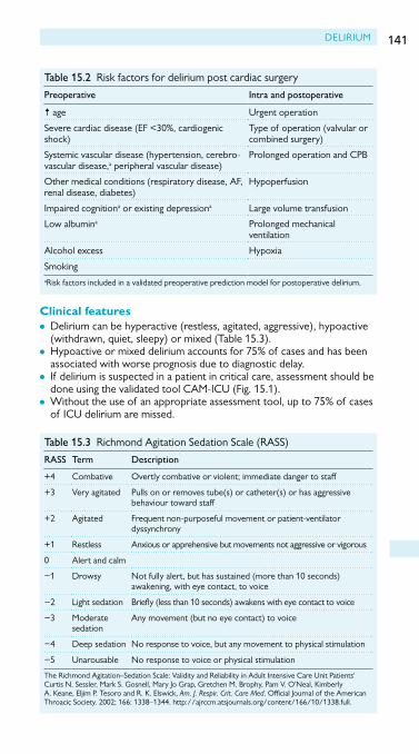
DelIrIUM 141
Clinical features• Delirium can be hyperactive (restless, agitated, aggressive), hypoactive
(withdrawn, quiet, sleepy) or mixed (table 5.3).• hypoactive or mixed delirium accounts for 75% of cases and has been
associated with worse prognosis due to diagnostic delay.• If delirium is suspected in a patient in critical care, assessment should be
done using the validated tool CaM-ICU (Fig. 5.).• Without the use of an appropriate assessment tool, up to 75% of cases
of ICU delirium are missed.
Table 5.2 risk factors for delirium post cardiac surgery
Preoperative Intra and postoperative
i age Urgent operation
Severe cardiac disease (eF <30%, cardiogenic shock)
type of operation (valvular or combined surgery)
Systemic vascular disease (hypertension, cerebro-vascular disease,a peripheral vascular disease)
prolonged operation and CpB
Other medical conditions (respiratory disease, aF, renal disease, diabetes)
hypoperfusion
Impaired cognitiona or existing depressiona large volume transfusion
low albumina prolonged mechanical ventilation
alcohol excess hypoxia
Smokingarisk factors included in a validated preoperative prediction model for postoperative delirium.
Table 5.3 richmond agitation Sedation Scale (raSS)
RASS Term Description
+4 Combative Overtly combative or violent; immediate danger to staff
+3 Very agitated pulls on or removes tube(s) or catheter(s) or has aggressive behaviour toward staff
+2 agitated Frequent non-purposeful movement or patient-ventilator dyssynchrony
+ restless anxious or apprehensive but movements not aggressive or vigorous
0 alert and calm
− Drowsy Not fully alert, but has sustained (more than 0 seconds) awakening, with eye contact, to voice
−2 light sedation Briefly (less than 0 seconds) awakens with eye contact to voice
−3 Moderate sedation
any movement (but no eye contact) to voice
−4 Deep sedation No response to voice, but any movement to physical stimulation
−5 Unarousable No response to voice or physical stimulation
the richmond agitation–Sedation Scale: Validity and reliability in adult Intensive Care Unit patients' Curtis N. Sessler, Mark S. Gosnell, Mary Jo Grap, Gretchen M. Brophy, pam V. O'Neal, Kimberly a. Keane, eljim p. tesoro and r. K. elswick, Am. J. Respir. Crit. Care Med. Official Journal of the american throacic Society. 2002; 66: 338–344. http://ajrccm.atsjournals.org/content/66/0/338.full.

142 Chapter 5 Nervous system
Investigation• perform thorough physical examination and review medications.• Further investigation will be guided by clinical exam but should include
bloods (FBC, U&e, lFt, glucose, Ca), eCG, and sepsis screen as a minimum.• Cerebral imaging is unlikely to affect clinical management in the delirious
patient in the absence of focal neurological signs. In addition, sedation may be required to facilitate imaging and can prolong the course of delirium.
Prevention• a number of strategies to prevent neurological complications in cardiac
surgery have been proposed (see b Neurological complications of cardiac surgery, p. 44).• there is a randomized trial that found a reduced incidence of delirium
comparing the α-2 agonist dexmedetomidine with benzodiazepines for ICU sedation.• the ‘aBCDe’ bundle has been proposed as an evidence-based
structured approach to minimizing brain dysfunction in mechanically ventilated ICU patients.• Awakening and Breathing Coordination involving spontaneous
awakening trials combined with spontaneous breathing trials• Delirium monitoring using a validated assessment tool• Exercise and early mobility.
Confusion Assessment Method for the ICU (CAM-ICU) Flowsheet
CAM-ICU negativeNO DELIRIUM
CAM-ICU negativeNO DELIRIUM
CAM-ICU negativeNO DELIRIUM
CAM-ICU positiveDELIRIUM Present
• “Squeeze my hand when I say the letter ‘A’.” Read the following sequence of letters: S A V E A H A A R T ERRORS: No squeeze with ‘A’ & Squeeze on letter other than ‘A’• If unable to complete Letters → Pictures
2. Inattention:
• Is there an acute change from mental status baseline? OR• Has the patient’s mental status �uctuated during the past 24 hours?
1. Acute Change or Fluctuating Course of Mental Status:
1. Will a stone float on water?2. Are there fish in the sea?3. Does one pound weigh more than two?4. Can you use a hammer to pound a nail?
4. Disorganized Thinking:
Command: “Hold up this many �ngers” (Hold up 2 �ngers) “Now do the same thing with the other hand” (Do not demonstrate) OR “Add one more �nger” (If patient unable to move both arms)
Current RASS level
3. Altered Level of Consciousness
NO
YES
RASS otherthan zero
0–2Errors
0–1Error
> 1 Errors
> 2 Errors
RASS = zero
Figure 5. Confusion assessment method for the ICU. Copyright © 2002, e. Wesley ely, MD, Mph and Vanderbilt University, all rights reserved.

DelIrIUM 143
• NICe guideline on delirium advocates a multi-component intervention package in patients identified as at risk of delirium. Care should be taken to:• Provideanappropriatelystimulatingenvironmentandreorientateas
required• Avoidhypoxia• Ensureadequatehydrationandavoidconstipation• Identifyandtreatinfection• Ensureadequatenutrition• Identifyandtreat pain• Avoidsleepdisturbance• Avoidsensorydeprivationbyensuringvisualandhearingaidsare
working• Encourageearlymobilization• Reviewmedicationsandconsidersideeffects.
Treatment• primary strategy is to identify and treat any underlying causes.• If the patient is distressed then use verbal and non-verbal de-escalation
techniques.• If these are ineffective then consider a short course of haloperidol or
second-generation olanzapine, quetiapine, risperidone starting at the lowest appropriate dose.• alpha-2 agonists are commonly used as second-line treatment.
Dexmedetomidine is not licensed in the UK so clonidine is the drug of choice.
Outcomes• Following cardiac surgery, cerebral complications including delirium have
been associated with i ICU and hospital stay and iin-hospital mortality.• there is evidence that delirium post coronary artery bypass surgery
increases the risk of late mortality up to 0 years.• however, a recent study has shown no i rates of postoperative
cognitive decline at 3 months post CaBG compared to total hip joint replacement or coronary angiography done without general anaesthesia.
Further readingCurtis N, Sessler MS, Gosnell MJ, Grap GM, Brophy pV, O'Neal Ka, et al. the richmond agitation–
Sedation Scale: validity and reliability in adult intensive care unit patients. Am J Respir Crit Care Med 2002;66:338–44.
ely eW, Inouye SK, Bernard Gr, Gordon S, Francis J, May l, et al. Delirium in mechanically ventilated patients: validity and reliability of the confusion assessment method for the intensive care unit (CaM-ICU). JAMA 200;286:2703–0.
rudolph Jl, Jones rN, levkoff Se, rockett C, Inouye SK, Sellke FW, et al. Derivation and validation of a preoperative prediction rule for delirium after cardiac surgery. Circulation 2009;9:229–36.
Young J, Murthy l, Westby M, akunne a, O'Mahony r; Guideline Development Group. Diagnosis, prevention, and management of delirium: summary of NICe guidance. BMJ 200;34:c3704.

144 Chapter 5 Nervous system
Risk factors• With the exception of female sex, largely represent risk factors for
systemic vascular disease:i age, hypertension, diabetes, chronic kidney disease, peripheral vascular disease, previous history of cerebrovascular disease, known atherosclerosis of the ascending aorta or carotids, aF.• patients presenting with a recent MI, unstable angina, or moderate to
severe lV systolic dysfunction are at i risk.• Several prediction models exist that account for the additive effect of
risk factors in an individual patient.
Neurological complications of cardiac surgeryIntroductionNeurological complications are a major source of morbidity and mortality following cardiac surgery. present as a spectrum of overlapping disorders, which can be classified as: stroke, encephalopathy (often presenting as neu-rocognitive deficits including delirium), and peripheral neuropathy.
Stroke• Stroke is the rapid onset of neurological deficit secondary to infarction
or haemorrhage and lasting >24 hours.• Majority of strokes following cardiac surgery are secondary to
infarction.
Incidence• Significant variation in the reported incidence of perioperative stroke in
cardiac surgery.• table 5.4 presents data from a large cohort from a single institution
and from the Society of thoracic Surgeons National adult Cardiac Surgery Database (StS NCD).• Surgery involving combined procedures, the mitral valve, or the aorta
has a particularly high risk of perioperative stroke.
Table 5.4 high variation in the incidence of stroke demonstrated in data derived from a single institution and from the StS NCD
Cardiac surgical procedure STS NCD Single institution
CaBG .4% 4.%
Valve .6% 3.%
CaBG/Valve 2.9% 7.9%

NeUrOlOGICal COMplICatIONS OF CarDIaC SUrGerY 145
Pathophysiology• Stroke can be classified as intraoperative or postoperative depending on
whether the deficit is present immediately on waking from anaesthesia.• aetiology is likely multifactorial but a primary mechanism is thought to
be atheromatous embolism arising from the ascending aorta.• Cerebral emboli of particulate matter also arise from cardiac thrombi,
severely calcified valves, and carotid artery atherosclerosis.• Gaseous emboli arise from open cardiac chambers, vascular cannulation
sites, or arterial anastomoses.• Cerebral hypoperfusion may contribute to intraoperative stroke by
reducing the washout out of embolic material from watershed areas of brain at the borders of vascular territories.• the inflammatory process and hypercoagulability may contribute to
postoperative stroke.
Clinical features• Depend on the location of brain injury and whether the ischemia is
regional or global.• any combination of hemiparesis, ataxia, hemisensory loss, visual
disturbance, dysarthria, dysphasia, or effects on consciousness or cognition.
Investigation• Neuroimaging will confirm the diagnosis of ischaemic or haemorrhagic
stroke. perform routine investigations to identify aetiological factors and exclude alternative causes of neurological impairment.• eCG—aF, other arrhythmia.• echocardiogram—intracardiac thrombus, new valve pathology.• Bloods—include FBC, coagulation screen, U&es, calcium, glucose, lFts,
and thyroid function tests as first line.• Imaging should be performed as soon as possible to exclude
haemorrhage.• Ct will identify about 50% of infarcts.• Magnetic resonance imaging (MrI) is the technique of choice.
Diffusion-weighted imaging can detect acute ischemic events related to microemboli. It is more likely to demonstrate multiple lesions in a watershed pattern of distribution than t2 or FlaIr imaging.
Prevention• a number of strategies to prevent neurological complications
including stroke have been proposed. evidence supporting individual neuroprotective techniques is sparse.• preoperative—combine thorough preoperative assessment with the
use of scoring systems to identify high-risk patients and thus focus resources.• Off-pump surgery—no consensus regarding the avoidance of CpB
to reduce the incidence of neurological complications but off-pump surgery can be considered.

146 Chapter 5 Nervous system
• Surgical technique—epiaortic ultrasound guidance and ‘no touch technique’ may help to reduce aortic atheroma embolization. Use of aortic cannulae with improved flow characteristics and with filters may be beneficial.• CpB—membrane oxygenators reduce emboli compared to bubble
oxygenators. No evidence to support ph over α-stat management or pulsatile over non-pulsatile flow. targeting of higher haematocrit (>27%) and higher Map (minimum >50mmhg) may be beneficial. hypothermia used for neuroprotection despite conflicting evidence.• Multimodal neuromonitoring—optimization of intraoperative care
using a combination of cerebral monitors such as transcranial Doppler, electroencephalogram, and cerebral oximetry using near-infrared spectroscopy has been suggested. Cerebral desaturation has been shown to correlate with neurological impairment but protocolized treatment of desaturations did not improve outcome.• pharmacotherapy—no evidence to support the use of any agent for
pharmacological neuroprotection. thiopentone is often used during deep hypothermic circulatory arrest.• Combined carotid endarterectomy and cardiac surgery—there is no
evidence that this approach reduces the risk of stroke in patents with carotid stenosis and outcomes may be worse.
Treatment• antiplatelet therapy—shown to be safe and effective at reducing
ischaemic complications including stroke following CaBG. aspirin 300mg first-line treatment of acute ischaemic stroke.• anticoagulation—should not be used routinely for treatment of acute
stroke. If surgically indicated because of prosthetic valve, risk:benefit assessment with surgeon and neurologist.• IV thrombolysis increasingly used in ischaemic stroke but
contraindicated in context of recent surgery.• Intra-arterial thrombolysis and mechanical clot disruption are under
investigation for reperfusion in specific patients.• Neurosurgical referral if:
• Largemiddlecerebralarteryinfarctionforconsiderationof decompressive craniectomy.
• Primaryintracranialhaemorrhagewithhydrocephalus.• Supportive therapy:
• Supplementaloxygenonlyif SaO2 <95%.• BP—if hypertensiveemergency(target<85/0mmHg).• Glycaemiccontrol—hyperglycaemiaassociatedwithpoor
neurological outcome following both cardiac surgery and stroke. Blood glucose should be maintained 4–mmol/l although no evidence that this improves outcome.
• Temperature—hyperthermiashouldbeavoidedduetoassociationwith poor outcome following cardiac surgery and stroke.
• Non-pharmacologicalDVTprophylaxis.• Ongoing management of stroke should involve a specialist
multi-disciplinary team who can address issues such as nutrition, physiotherapy, and secondary prevention.

NeUrOlOGICal COMplICatIONS OF CarDIaC SUrGerY 147
Outcomes• Several studies have shown an association with poor outcome and
perioperative stroke in cardiac surgery. For example, following CaBG stroke was associated with i ICU stay,i hospital stay, and i hospital mortality (4.4% vs 2.7%).
Further readinghogue CW Jr, palin Ca, arrowsmith Je. Cardiopulmonary bypass management and neurologic out-
comes: an evidence-based appraisal of current practices. Anesth Analg 2006;03:2–37.McKhann GM, Grega Ma, Borowicz lM Jr, Baumgartner Wa, Selnes Oa. Stroke and encephalopathy
after cardiac surgery: an update. Stroke 2006;37:562–7.Stamou SC, hill pC, Dangas G, pfister aJ, Boyce SW, Dullum MK, et al. Stroke after coronary artery
bypass: incidence, predictors, and clinical outcome. Stroke 200;32:508–3.


149
Immune system
Inflammatory response 50Heparin-induced thrombocytopenia 52
Chapter 6

150 CHapter 6 Immune system
Inflammatory responseMajor surgery triggers a systemic inflammatory response. this is particularly so in cases involving CpB due to the exposure of the blood to the artificial surface. Biochemical and physiological signs of this response may be found in all post-cardiac surgery patients if one investigates carefully enough; how-ever, it is clinically apparent in only a proportion and problematic in a minor-ity. the systemic inflammatory response syndrome (SIrS) associated with major surgery is clinically indistinguishable from that triggered by infection (SIrS associated with infection is called sepsis).
Despite the avoidance of bypass a marked SIrS can still occur in off-pump patients as a result of the surgical trespass, cardiac manipulation, and circu-latory disturbance.
potential clinical features include:• Vasodilation and subsequent hypotension• raised lactate• Capillary leak (relative systemic hypovolaemia, impaired gas exchange, d
pulmonary compliance)• renal impairment• i susceptibility to infection• Delirium.
ARDSrarely, full blown arDS (‘pump lung’) may occur as part of the inflamma-tory response after cardiac surgery. treatment is supportive, as for arDS from other causes.
Vasoplegiararely, vasodilation associated with the SIrS triggered by cardiac surgery may be pronounced and intractable to normal therapy with alpha adren-ergic agonists and vasopressin (severe vasoplegia). Nitric oxide is a pow-erful mediator of inflammatory vasodilation. Methylene blue inhibits nitric oxide-mediated vasodilation by a number of mechanisms (scavenges nitric oxide, inhibits nitric oxide synthetase and inhibits guanylate cyclase) and may be effective where other pressor agents are failing:
Dose regimen for methylene blueIn the presence of a good cardiac index, give a bolus of .5mg/kg (make up to 50mL with sterile water and infuse over 30 minutes). response is rapid if it is going to occur. No major side effects are expected at this dose range. Urine and skin may become discoloured with the skin discolouration
Practice pointFever, changes in white cell count, and rise in Crp are unreliable signs of infection immediately after cardiac surgery. Clinical history, the presence of other clinical signs (e.g. X-ray changes, sputum production), and culture results should be used in deciding whether to commence antimicrobial therapy in the first 48 hours postoperatively.

INFLaMMatory reSpoNSe 151
potentially interfering with pulse oximetry (but not oxygenation). the bolus may be followed by an infusion of 0.25–.0mg/kg/hour for 6 hours starting 2 hours after the initial bolus.
Further readingFaber p, ronald a, Millar BW. Methythioninium chloride: pharmacology and clinical applications with
special emphasis on nitric oxide mediated vasodilatory shock during cardiopulmonary bypass. Anaesthesia 2005;60: 575–87.

152 CHapter 6 Immune system
Heparin-induced thrombocytopeniaHeparin induced thrombocytopenia (HIt type 2) is an immunologi-cally mediated condition where IGG antibodies are formed against heparin-platelet factor 4 (pF4) complexes. Heparin has a high affinity for pF4 and although neither is immunogenic by themselves antibodies are formed to the complexes. the antibodies cause pathological platelet activa-tion and release of tissue factor (promotes thrombosis) from monocytes. HIt may be more likely when heparin exposure occurs in association with conditions that themselves increase the amount of circulating pF4, such as surgery and burns.
a non-immune HIt or HIt type is also described. this is a benign self-limiting condition requiring no treatment. It is a result of the binding of heparin to pF4 per se. It is not associated with antibodies. It causes transient mild thrombocytopenia immediately within the first few days of heparin exposure due to platelet aggregation and sequestration.
the term HIt in common clinical usage refers to HIt type 2. HIt as discussed in this chapter refers to HIt type 2.
Incidence of HIt after exposure to unfractionated heparin has been esti-mated at between % and 5%, incidence is 5–0 times less than that after exposure to LMWH.
platelet counts should be monitored regularly (usually daily) in all patients in cardiothoracic intensive care.
Clinical featuresThrombocytopenia• Usually occurs 5–0 days after start of heparin therapy.• Count usually increases within 4–4 days of stopping heparin.• Count falls to 40,000–80,000 (<20,000 suggests alternate diagnosis).
Thrombosis• Varies with clinical setting.• Usually more common venous than arterial (4:).• Usually occurs after but may occur before thrombocytopenia.• Includes DVt, pe, CVa, limb ischaemia, myocardial infarction and skin
necrosis.• May manifest as repeated circuit clotting in patient on haemofiltration.• May occur after cessation of heparin therapy due to persistence of
antibodies (antibodies usually disappear within 4 months).
HIt may be triggered by small amounts of heparin, e.g. in heparin flush used in central venous catheters or arterial lines.
Bleeding is uncommon in the absence of other causes of platelet dysfunction.
DiagnosisDiagnosis is based on a combination of clinical signs and antibody detection.
HIt is associated with thrombocytopenia but is a pro-thrombotic condi-tion. It has a high mortality (30%) in its full clinical manifestation.

HeparIN-INDUCeD tHroMBoCytopeNIa 153
Laboratory tests
antibody assays are divided into those that test for platelet activation (functional assays) and other methods. Functional assays have a very high specificity (though still variable sensitivity) but are resource intensive and technically demanding. Most laboratories use other methods such as enzyme-linked immunoassays (eLISa) or particle gel immunoassay. these tests (particularly eLISa) are highly sensitive (close to 00%) and thus have a high negative predictive value, but are variably specific. Furthermore only a subset of antigens detected by these tests is functionally active in terms of platelet activation. they thus have a high false-positive rate (possibly in the order of 50% vs clinically significant HIt). Ideally, characterization requires detection with a high sensitivity test and characterization with a functional assay but this is will only be possible in laboratories with a special interest.
Practice pointUnderstand the type and significance of the HIt antibody tests used by your local laboratory.
Because of these difficulties a probabilistic approach must be taken. a formal scoring system such as the 4 ts score can be incorporated into decision-making (table 6.).
Table 6. the 4 ts scoring system
Score
Category 2 0
Degree of thrombocytopenia
>50% decrease or nadir 20–00 x 09/L
30–50% decrease or nadir 0–9 x 09/L
<30% decrease or nadir <0 x 09/L
timing of thrombocytopenia
5–0 days after exposure or < day if previous exposure within month
>0 days after exposure or < day if previous exposure between month and year or unclear
No heparin in last year
other causes of thrombocytopenia
None possible Definite
thrombotic phenomena/other clinical
existing thrombosis, after heparin bolus, new skin necrosis or acute systemic reaction
Silent or recurrent thrombosis or erythematous skin lesions
None
reproduced from Springer and Current Science, Current Haematology Reports, 2, 2, 2003, pp. 48–57, ‘Laboratory diagnosis of immune heparin-induced thrombocytopenia’, Warkentin te, Heddle NM, with kind permission from Springer Science+Business Media B.V.

154 CHapter 6 Immune system
Score of 3 points or fewer gives a low risk of HIt and no further action is required. No laboratory tests are undertaken. Heparin does not need to be discontinued. Score of 6 points or more indicates a high risk of HIt. Score of 4 or 5 indicates an intermediate risk. If the score is 4 or over (intermediate- or high-risk patients) heparin should be discontinued, and antibody assay information sought. an alternative anticoagulation regimen should be started immediately as prophylaxis against thrombosis without waiting for laboratory results.
However, diagnosis is difficult as only a proportion of patients (5–30%) with antibodies develop thrombocytopenia. only a proportion (30–70%) of patients with antibody-related thrombocytopenia develop thrombotic complications. Furthermore there are many causes of thrombocytopenia in critically ill patients. In a minority of patients with HIt (5%) thrombosis may occur with a normal platelet count. If there are pre-existing antibodies from a previous exposure the usual delay in onset may not occur (‘rapid onset’ HIt). Finally antibody detection varies depending on the method used and between laboratories. See Box 6..
Box 6. Differential diagnosis of thrombocytopeniaMany, but include:• Sepsis• Drug-induced• Haemofiltration• eCMo/VaDS• CpB• Dilutional• Disseminated intravascular coagulation• Idiopathic thrombocytopenic purpura• pe• Liver disease• Hypersplenism• Malignancy• Immune disease (e.g. antiphospholipid syndrome, systemic lupus
erythematosus)• (Very rarely) thrombotic thrombocytopenic purpura.
Managementonce HIt is diagnosed the principles of management are:• Stop all heparin administration (including LMWHs and low-dose
heparin flush)• Start anticoagulation with an alternative parenteral anticoagulant.
See table 6.2 for recommendations for cardiac surgery for patients with HIt.
Warfarin and other vitamin K antagonists in patients with HITWarfarin (and other vitamin K antagonists) are contraindicated in patients in the acute phase of HIt as initiation of warfarin therapy is associated with a procoagulant phase that lasts several days. Lower limb venous gangrene and severe skin necrosis have been described. In patients who are already on

HeparIN-INDUCeD tHroMBoCytopeNIa 155
warfarin when they develop HIt, warfarin should be stopped and vitamin K should be administered.
Warfarin can be started once platelet count is >50 × 09/L under cover of an alternative anticoagulant. Dosing should be cautious (5mg/day initially).
Platelet transfusionIt is unclear whether platelet transfusion actively precipitates thrombosis in HIt. prophylactic platelet transfusion is unnecessary as bleeding is rare.
Alternative anticoagulantsthis is an area which is currently evolving. all alternative strategies have balancing advantages and disadvantages. In patients without thrombosis the alternative anticoagulant should be continued until the platelet count has recovered to a stable plateau. options include:• Factor Xa antagonists:
• Danaparoid—half-lifeof 24hourswithnospecificantidote.Partiallyexcreted in urine so accumulates in renal dysfunction. Monitor anti Xa level. occasional (<0% cases) cross-reactivity with HIt IgG. Not available in USa.
• Fondaparinux—syntheticheparinanalogue.FormsantiPF4/heparinantibodies but these tend not to trigger HIt. Half-life of 8 hours. renal excretion. No specific antidote.
• Direct thrombin inhibitors:• Shorthalf-life.GenerallygivenbyIVinfusion.Nospecificantidotes.
partly or largely excreted in urine so accumulate in renal failure.• Lepirudin.• argatroban.
• Hepatobiliaryexcretion.Contraindicatedinlivercellfailure.Monitorby appt.
• Bivalirudin:• 80%clearedbyenzymicproteolysisthereforeadvantageousin
significant liver and kidney dysfunction. requires dose adjustment in renal failure.
Table 6.2 Cardiac surgery for patients with HIt
Clinical picture Immunological assay
Functional assay
Recommendation for surgery
remote HIt Negative Negative Use unfractionated heparin
Sub-acute HIt positive Negative Delay surgery until immune assay negative Or: use bivalirudin
acute HIt
positive
positive
Delay surgery until both assays negative Or: use bivalirudin

156 CHapter 6 Immune system
Duration of treatmentBilateral lower limb compression ultrasound should be performed in all patients with HIt whether or not there is clinical evidence of DVt. the presence of a DVt influences duration of anticoagulation.• patients with HIt and proven venous thrombosis (HItt) should be
anticoagulated for 3–6 months.• patients with HIt without proven venous thrombosis (isolated HIt)
have an elevated risk of thrombosis for at least 30 days. they should be anticoagulated for at least month.
Further readingCuker a, Crowther Ma. Clinical Practice Guideline on the Evaluation and Management of
Heparin-Induced Thrombocytopenia (HIT). 2009. available at: M <http://www.hematology.org/practice/Guidelines/2934.aspx>.
Linkins La, Dans aL, Moores LK, Bona r, Davidson BL, Schulman S, et al. american College of Chest physicians. treatment and prevention of heparin-induced thrombocytopenia: antithrombotic therapy and prevention of thrombosis, 9th ed: american College of Chest physicians evidence-Based Clinical practice Guidelines. Chest 202;4(2 suppl):e4955–e5303.

157
Haematology
Coagulopathies 58Blood conservation 60Point-of-care testing 64
Chapter 7

158 ChaPter 7 Haematology
CoagulopathiesCardiac surgery as a specialty is almost uniquely placed to provoke the development of coagulation abnormalities. there are numerous reasons why cardiac surgical patients may become coagulopathic.
Haemodilution• Dilution of red cells occurs from addition of –.5L of colloid/
crystalloid pump prime, the effect being exaggerated in patients with smaller blood volumes, i.e. low body weight.• haemodilution may also occur from volume replacement subsequent to
haemorrhage or as treatment for postoperative hypotension.• In addition, coagulation factors are also diluted—it is estimated that
reductions to <30% of control levels are necessary before significant bleeding will occur although there is some individual variation. Factors V and VIII are most commonly reduced.
Haemorrhage• Subsequent volume and red cell replacement may lead to the
development of coagulopathy due to dilution, loss of factors, and loss of platelets.
Platelet damage• aside from dilution of platelets on bypass, mechanical damage from
roller pumps or adsorption onto the membrane of the oxygenator may occur.• hypothermia-induced splenic sequestration of platelets has also been
reported.
Antiplatelet therapy• a significant number of cardiac patients are on antiplatelet medication
prior to surgery.• Most will be on aspirin alone, some will be on clopidogrel alone (or
an alternative drug acting on the P2Y2 receptor), and some will be on both.• Dual antiplatelet therapy is a significant concern with regard to
perioperative bleeding and where safe to do so, most surgeons will stop clopidogrel 5 days in advance of surgery.• Some emergency patients may also have been given glycoprotein IIb/
IIIa antagonists, e.g. tirofiban, which are very potent antiplatelet agents, acting as they do on the final common pathway of platelet activation.
Heparin–protamine mismatch• there are numerous dosing regimens for administration of heparin for
CPB and its subsequent reversal.• It is important that heparin is adequately reversed at the end of the
procedure.• When the residual pump blood is administered to the patient towards
the end of the case, some clinicians administer a further dose of protamine to cover any heparin present in that blood.
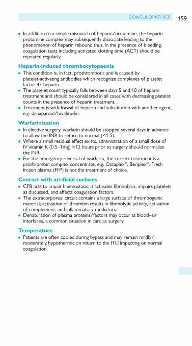
CoaguLoPathIeS 159
• In addition to a simple mismatch of heparin/protamine, the heparin–protamine complex may subsequently dissociate leading to the phenomenon of heparin rebound thus, in the presence of bleeding, coagulation tests including activated clotting time (aCt) should be repeated regularly.
Heparin-induced thrombocytopaenia• this condition is, in fact, prothrombotic and is caused by
platelet-activating antibodies which recognize complexes of platelet factor 4/ heparin.• the platelet count typically falls between days 5 and 0 of heparin
treatment and should be considered in all cases with decreasing platelet counts in the presence of heparin treatment.• treatment is withdrawal of heparin and substitution with another agent,
e.g. danaparoid/bivalirudin.
Warfarinization• In elective surgery, warfarin should be stopped several days in advance
to allow the INr to return to normal (<.5).• Where a small residual effect exists, administration of a small dose of
IV vitamin K (0.5–mg) >2 hours prior to surgery should normalize the INr.• For the emergency reversal of warfarin, the correct treatment is a
prothrombin complex concentrate, e.g. octaplex®, Beriplex®. Fresh frozen plasma (FFP) is not the treatment of choice.
Contact with artificial surfaces• CPB acts to impair haemostasis; it activates fibrinolysis, impairs platelets
as discussed, and affects coagulation factors.• the extracorporeal circuit contains a large surface of thrombogenic
material; activation of thrombin results in fibrinolytic activity, activation of complement, and inflammatory mediators.• Denaturation of plasma proteins/factors may occur at blood–air
interfaces, a common situation in cardiac surgery.
Temperature• Patients are often cooled during bypass and may remain mildly/
moderately hypothermic on return to the Itu impacting on normal coagulation.

160 ChaPter 7 Haematology
Blood conservationFrom what has already been discussed, it can be no surprise that cardiac surgery is a high blood-using specialty, estimated to use 5% of the uK blood supply. While the blood supply could be considered to be the safest it has ever been there are still recognizable risks associated with transfusion of red cells and blood products.
Shot (Serious hazards of transfusion) reports annually on adverse events associated with transfusion:• Incorrect component transfusion accounts for 25% of reports• acute and haemolytic transfusion reactions account for 3%• transfusion-related acute lung injury accounts for 2%• transfusion-transmitted infections 0.6%• the risk of variant Creutzfeldt–Jakob disease is, as yet, unknown.
In addition, several large retrospective studies have demonstrated an asso-ciation between transfusion and long- and short-term increases in mortality even if only unit of red cells has been transfused.
Blood is also an expensive and finite resource. these facts considered, it is appropriate that attempts are made to decrease our use of red cells and blood products and to use the resource appropriately.
Blood conservation could be considered to address three specific areas:• Increasing or optimizing patients’ haemoglobin preoperatively• Decreasing blood loss intra- and postoperatively• optimizing transfusion practice using the best available evidence.
Increasing preoperative haemoglobinAnaemia• anaemia is an independent predictor of mortality in the cardiac surgical
population.• It has been demonstrated that up to a quarter of patients presenting
for cardiac surgery have d iron stores. these patients have lower admission hb, receive more red cells during their hospital stay, and have lower discharge hb than their normal counterparts. however screening patients far enough in advance for treatment to be useful presents logistical problems for most units.• oral iron salts should increase hb by g/dL every 0 days and, although
usually well tolerated, treatment should last for at least 4–6 weeks to allow for an increase in hb.• IV iron has been associated with allergic reactions so is not without risk
compared to oral formulations.• erythropoietin is not currently approved in the uK for preoperatively
increasing hb (except in Jehovah’s Witnesses).
Autologous transfusionMost health Boards do offer autologous transfusion, however cardiac dis-ease is a contraindication.
Decreasing blood loss intra- and postoperativelythere are several possible points of intervention:

BLooD CoNSerVatIoN 161
Acute normovolaemic haemodilutionthe principle behind this technique is that:• after induction in suitable patients (e.g. hb >2g/dL), a volume of
blood (e.g. 5mL/kg) can be removed from the patient and stored for re-infusion at the end of surgery.• the volume removed is replaced with colloid to maintain
normovolaemia.• the advantages of this technique are that it is fairly simple, relatively
inexpensive, and means that blood lost intraoperatively is of a lower haematocrit, therefore fewer red cells are lost.• the blood transfused at the end of the procedure will raise the
haematocrit and hb and also contains platelets which function as well as they did preoperatively.
Pharmaceutical interventionsAntifibrinolytic agents• tranexamic acid:
• Alysineanaloguewhichbindstothelysinebindingsiteof theplasminogen molecule displacing it from fibrin.
• Ithasbeenproventodecreasebloodloss,decreasetransfusion,andto decrease the frequency of surgical re-exploration.
• Havinglargelyreplacedaprotininincardiacpractice,severalrecentstudies have suggested that its use may be associated with similar complications.
• aprotinin:• Aprotinin’slicencehasbeenwithdrawn,however,some
centres continue to use it for high-risk patients off-licence on a named-patient only basis.
DDAVP®
• a synthetic vasopressin derivative which acts by recruitment of von Willebrand factor thus increasing platelet adhesion.• It also increases concentrations of factor VIII and tissue plasminogen
activator.• It is not commonly used in the uK and good evidence of benefit is slim.
NovoSeven®
recombinant factor VIIa has been used in an attempt to arrest catastrophic haemorrhage in extremis:• It has been reported that there is a significant increase in thrombotic
events in those treated outside its licensed indications for haemophilia and its use has accordingly become controversial.• Its use therefore is at the discretion of the attending medical staff in
discussion with haematologists.
Cell salvage• evidence supports the use of intraoperative cell salvage in cardiac surgery.• however, there is a requirement for significant financial investment
in terms of machine purchase, machine maintenance, purchase of disposables, and training of staff.

162 ChaPter 7 Haematology
• given the cost of unit of red cells may be quoted as £50, cell salvage offers potentially significant savings.• Blood lost from the surgical field is washed to remove contaminants,
debris, heparin, cytokines, etc. and is resuspended in normal saline for reinfusion.• Most systems concentrate the blood to a haematocrit of at least 60%.• this blood, however, will contain no coagulation factors or platelets.• a postoperative system which acts as a mediastinal drain is also offered
by haemonetics (cardioPat®) and has proved useful in our practice.• It is important to note that some Jehovah’s Witness patients may accept
cell salvage as part of their treatment.
Decreasing haemodilution by pump primeMoving the machine as close as possible to the patient and cutting arte-rial and venous lines to an appropriate length can decrease the amount of volume added at the time of bypass and hence decrease haemodilution.
Mini bypassthis is a relatively new technique which is used in few units and is somewhat more than simple miniaturization of a traditional bypass circuit requiring specific training for the perfusionists.
however, its aim is to minimize the inflammatory response to bypass which is done in several ways:• haemodilution is d (may be able to reduce prime to as little as 50mL).• Minimizes the surface area of the circuit and oxygenator.• Lower suction is applied to the venous side of the circulation than in
conventional bypass, decreasing damage to blood cells.• the centrifugal pump is also less damaging to red cells and platelets.• there is no blood–air interface as there is no venous reservoir.
accordingly:• Bloodloss is d.• Transfusion is d.
• May decrease myocardial injury and postoperative dysrhythmias.• May minimize renal and intestinal injury.
there have, however, been significant concerns regarding the risk of air embolism.
Use of topical haemostatic agentsuse of these agents has also been suggested:• employing localized compression as a scaffold for clot formation, e.g.
surgicel or• as anastomotic sealants as a spray.• there seems to be some evidence of d bleeding but limited evidence of d transfusion requirements.
Topical antifibrinolyticsthere is some evidence of a benefit of administration of antifibrinolytic agents such as tranexamic acid directly to the wound at closure.

BLooD CoNSerVatIoN 163
Off-pump proceduresthese may confer advantages (oPCaB) by avoiding some of the potential provoking factors of bypass (haemodilution, contact with artificial surfaces, platelet damage).
Avoid excesses of blood pressurehypertension may lead to i blood loss from suture lines after aortotomy.
Good surgical haemostasisNever to be underestimated in reducing blood loss.
Optimizing transfusion practiceMSBOS (maximum surgical blood ordering schedule)unit-specific protocols for ordering a predetermined number of units of blood for each surgical procedure based on likelihood of use have been proven to decrease blood wastage. good practice states that the cross-match to transfusion ratio should be no greater than 2:.
Guidelines/transfusion triggersthere are several different guidelines available to optimize transfusion prac-tice, some looking specifically at cardiac surgery.
recent StS/ SCa guidelines suggests:• adoption of a transfusion trigger of 7g/dL hb postoperatively although
each patient should be assessed individually for evidence of impaired oxygen delivery.• there is no evidence supporting transfusion where hb >0g/dL.• a trigger of 6g/dL is suggested during bypass.• given that a significant volume of colloid has been added during bypass,
it is normal to tolerate lower hb concentrations in the expectation that the patient will increase their urine output and the haematocrit will rise over the first few hours post bypass.
AlgorithmsIntroduction of a transfusion algorithm alone has been proven to decrease transfusion requirements. these should, of course, be evidence-based.
Near-patient testingthis will be discussed further (see b Viscoelastic tests, p. 65) however there are several cardiac surgical publications demonstrating d red cell and blood product use with roteM®/teg® and publications which also dem-onstrate significant cost savings.
Individually the effect of many of these interventions is small. It is only through the adoption of a multimodal and multidisciplinary approach to blood conservation that the best results can be obtained.

164 ChaPter 7 Haematology
Point-of-care testingPoint-of-care testing may be defined as any analytical test performed by a healthcare professional outside the laboratory setting which should generate results comparable to the local laboratory with matching refer-ence ranges. unfortunately laboratory testing can be slow and results will reflect the patient’s status at the time the samples were taken; in cardiac surgery the situation may have altered significantly by the time the results are reported. the aim with all point-of-care tests is to obtain results more quickly than could be obtained by regular laboratory testing, that they may more appropriately guide therapy.
Blood gas analysisWill give rapid and reliable information on:• hb concentrations and haematocrits to guide transfusion decisions• electrolyte disturbances (with particular reference to K and Ca)• acid–base abnormalities and lactate concentrations• glucose.
Haemoglobin analysersMost units would use blood gas analysis for hb measurement nowadays; however, analysers such as hemoCue® are available for assessment of hb.
Coagulation monitorshand-held monitors of coagulation are available giving prothrombin time (Pt) and INr results.
ACTalthough the aCt is a fairly crude test (using a tube containing an activa-tor such as diatomaceous earth and a magnet rotating in a well) it is com-monly used in cardiac units as a means of monitoring the effect of heparin administration.• there are several devices available some offering heparinase-adjusted
results, some offering accelerated results.• unit protocols dictate the acceptable levels for commencement of CPB
but 400 or 480 seconds are common.• It is important to note that the aCt is not solely dependent on
the effects of heparin; it also depends on platelet and fibrinogen interactions.• After protamine administration, a further test is performed to assess a return
towards baseline.
Hepcon®
an automated device which measures:• the heparin dose response• heparin–protamine titration, predicting the required dose of protamine• Some authors have suggested such direct measurements of heparin
concentration as a more appropriate measurement and as a better method to monitor anticoagulation on bypass and heparin reversal.

PoINt-oF-Care teStINg 165
Viscoelastic teststhese tests were first described in the 940s and have regained favour in the last 5 years as concerted attempts have been made to decrease blood transfusion.• there are two commercially available systems—roteM® and teg®.• Both are viscoelastic tests, i.e. they test the ‘stickiness’ of clot and
provide an overview of the coagulation system including assessment of platelet function and of the plasma-based factors.• they test not only how rapidly clot forms but if the clot becomes and
remains stable.• the systems both rely on placing blood in a cuvette into which a pin is
inserted. In roteM® this pin rotates back and forth by approximately 4° (Fig. 7.) and in teg the cup rotates by a similar amount. as clot forms between the pin and the wall of the cup, this change in tension is detected and is transformed into the roteM®/teg® trace (Fig. 7.2).• the traces produced are somewhat similar and each parameter can be
related to certain aspects of the coagulation process allowing a rapid and accurate diagnosis of any coagulopathy and therefore the institution of early appropriate treatment.• Ct (clotting time)/r time = the time from the start of the test until the
first clot forms—prolonged by factor deficiency and heparin excess. Performance of a hePteM/heparinase test would help differentiate.• CFt (clot formation time)/k time = the time from clot first forming
until the amplitude reaches 20mm—prolonged by factor deficiency or platelet abnormality.• alpha angle reflects rate of clot formation.• MCF (maximum clot firmness) reflects deficiencies of fibrin or platelet
function abnormality. Performance of a FIBteM test would help differentiate.• Ma (maximum amplitude) is the equivalent of MCF for teg® and is said
to reflect platelet abnormality although functional fibrinogen testing is available for teg®.• ML (maximum lysis) reflects fibrinolysis.• It has been suggested that these tests are the gold standard in diagnosing
hyperfibrinolysis.
there are various tests available for both machines. roteM® specifically uses the addition of different activating agents to further localize any abnor-mality as part of the diagnostic algorithm:• It is important to recognize that neither system will identify the effects
of drugs (antiplatelet agents, warfarin) on routine testing.• While it is important to rapidly identify the presence of coagulopathy,
it is equally important to identify the absence of a coagulopathy which would imply a surgical cause for any haemorrhage allowing early re-exploration.
use of thromboelastometry or thromboelastography has been rec-ommended by QIS (Quality Improvement Scotland) as a clinical and cost-effective measure in cardiac surgery.

166 ChaPter 7 Haematology
oscillating axis (+–4,75°)
mirror
counterforce spring
ball bearingdetector
data processor
sensor pin
cuvette + sample
cuvette holder clot formation
temperature
controlled
LED light source
Figure 7. the roteM® mechanism of action. With kind permission from tem International gmbh <http://www.rotem.de/site/index.php?option=com_content&view=article&id=&Itemid=7&lang=en>.
100
Am
plitu
de in
mm
(Firm
ness
) 80604020
0
CT
CFT
0 10 20 30 40 50 60
Time in min
CTCFTalphaA10MCFLI30ML
MCF
LI 30A10
alpha
Clotting timeClot formation timeAlpha-angleAmplitude 10 min after CTMaximum clot �rmnessLysis index 30 min after CTMaximum lysis
ML
Figure 7.2 a roteM® trace. With kind permission from tem International gmbh <http://www.rotem.de/site/index.php?option=com_content&view=article&id=&Itemid=7&lang=en>.

PoINt-oF-Care teStINg 167
Platelet function testsgiven the increasing use of antiplatelet agents either alone or in combi-nation, platelet function testing may be an increasingly useful/valuable diagnostic tool:• there are several devices commercially available, e.g. Multiplate®,
VerifyNow®, PFa-00®.• While each use different technologies to obtain the results, they will all
give information on platelet function and most give information on the effects of antiplatelet agents.• While resistance to aspirin is rare, responsiveness to clopidogrel varies
between studies but resistance may be as high as 40%. those who are not responsive are clearly no more likely to bleed than those not on the drug.• these tests may allow patients at genuinely i risk of bleeding
perioperatively to be targeted by some of the more expensive blood conservation strategies.


Specific patient groups
8 The thoracic patient 7 9 The adult patient with congenital heart disease 87 20 The obstetric patient with cardiac disease 20 2 Heart failure resistant to standard medical therapy 209 22 The transplant patient 227 23 Acute cardiology 239
Part 3


171Chapter 8
The thoracic patient
Routine care of the thoracic patient 72Postoperative respiratory failure 75Post-thoracotomy acute lung injury 77Chest drainage 79Bronchopleural fistula 83Thoracic airway obstruction 84Lung volume reduction surgery 85

172 CHAPTeR 8 The thoracic patient
Routine care of the thoracic patientThoracic surgery in adults includes surgery to the lungs (including lung trans-plantation), pleura, thymus, oesophagus, and other thoracic structures as well as the chest wall. Thoracic procedures include lobar resection, pneu-monectomy for malignant and non-malignant conditions, mediastinoscopy and mediastinotomy, and bronchoscopy for diagnostic and interventional indications. Video-assisted thoracoscopic surgery (VATS) is performed for drainage and investigation of effusions, lung resection, sympathectomy, and removal of mediastinal tumours. Other procedures include the sur-gical management of air-leaks, management of empyema, operations on the chest wall, endobronchial laser surgery, and tracheal stenting and oesophagectomy. All of these patient groups may require admission to cardiothoracic ICU.
The routine care of thoracic surgical patients is aimed at promot-ing rapid recovery and reducing complications. Thoracotomy is a major operation and is one of the most painful. The nature of the surgery and the common comorbidities of this patient group such as respiratory disease result in an i incidence of respiratory complications. Patients may be admitted to critical care for routine postoperative care in level 2 beds or for level 3 care as a result of a complication of surgery, whether immediately postoperatively such as bleeding or as a later complication such as respiratory failure.
Routine postoperative careAnalgesiaPain is the enemy of recovery and will prevent adequate coughing and deep breathing. After lung resection, the patient will generally have a regional technique in situ such as paravertebral blockade or thoracic epi-dural analgesia. This is usually part of a balanced analgesic technique with the administration of regular paracetamol and non-steroidal analgesics. Patient-controlled analgesia is often used to supplement analgesic efficacy of paravertebral block.
Oxygen therapyOxygen therapy should be given to overcome the V/Q mismatch associ-ated with thoracic surgery. Generally humidified oxygen is administered to avoid the drying effects on secretions of the pipeline gases. Oxygen should be prescribed to achieve a SpO2 of 94–98% or 88–92% in patients with chronic obstructive pulmonary disease (COPD) at risk of hypercapnic respiratory failure. High oxygen concentrations may promote absorption atelectasis and lung injury and may also be dangerous in COPD patients with hypoxia-dependant respiratory drive.
Chest drainageIntercostal chest drains are inserted at the end of thoracic surgery to allow drainage of blood (basal drain) and air (apical drain) from the damaged lung. Typically these are each connected to an underwater drainage bottle which operates as a one-way valve which is set at 2cmH2O. An additional bottle may be added to allow low pressure suction to be applied to encourage

ROUTIne CARe Of THe THORACIC PATIenT 173
lung expansion. Low-level suction at 20cmH2O can be applied. Continued air leak is evidenced by bubbling of the drains.
The pleural drain should swing with respiration if it is patent and bubbling indicates air leak from damage lung and if severe may suggest bronchop-leural fistula.
Automated portable drain systems are now available which are battery driven and generate a constant negative pressure and display a digital display of the air leak in mL/minute.
Physiotherapy and mobilizationPhysiotherapy and early mobilization are essential for the thoracic patient. The thoracic patient should be managed sitting up at all times. On day they should be mobilized to a chair and begin weight bearing as soon as possible.
Some patients who are at risk of atelectasis and hypoxemia in the postop-erative period should have CPAP or BIPAP as preventive measures.
MinitracheostomyWhen the there is concern that a patient may be unable to cough adequately due to weakness, laryngeal nerve palsy, or excessive secretions, some units will consider the use of a minitracheostomy to assist secretion clearance.
Venous thromboembolism prophylaxisAll thoracic patients should have venous thromboembolism (VTe) prophy-laxis usually in the form of LMWH commenced preoperatively. epidural catheters should not be inserted or removed within 2 hours of the admin-istration of LMWH. The timing of LMWH should allow preoperative epi-dural catheter insertion. We give LMWH at 6pm the night before surgery and every 24 hours thereafter.
Fluid administrationIt is generally accepted that thoracic patient should be relatively fluid restricted and fluids should be prescribed with caution. A typical post-op prescription would be mL/kg/hour of Hartmann’s solution up to a maxi-mum of 70mL/hour. There is an association between post thoracotomy ALI and excessive fluid administration; however, the causal relationship has never been conclusively demonstrated. Units will have local policies for fluid administration.
Antibiotic prophylaxisAntibiotics are commonly administered after thoracotomy for 24 hours. Local protocols should be followed.
BronchodilatorsIn asthmatics and patients with reversibility of their airways disease, bron-chodilators are indicated. Generally the routine inhalers are continued through the perioperative period although inhalers may be converted to nebulizers. Salbutamol may contribute to the risk of postoperative atrial fibrillation and as such should not be routinely prescribed. Ipratropium is better tolerated and can be used where the benefits of β2 agonists are borderline.

174 CHAPTeR 8 The thoracic patient
Nutritionnutritional deficiencies are common in thoracic patients as a consequence of chronic infection (empyema), cancer cachexia, and dysphagia (oesoph-ageal disorders). early feeding is encouraged and where appetite is poor and/or dysphagia prevents consumption of adequate calories enteral feed-ing should be established.

POSTOPeRATIVe ReSPIRATORy fAILURe 175
Postoperative respiratory failureRespiratory complications occur in 25% of patients after lung resection. Of these, the more severe will require admission to ICU and up to 5% may require positive pressure ventilation. The reasons for postoperative respiratory failure are multifold. Patients may have significant underlying respiratory disease and then may fail to tolerate the loss of lung function associated with the lung resection. Recovery from surgery is complicated by postoperative pain and its consequences, such as the restriction in cough-ing and deep breathing. Opiate analgesia may also suppress cough reflexes, and cause sedation and hypoventilation. These variables conspire to cause atelectasis and sputum retention which may develop into postoperative pneumonia. ALI after lung resection is a major contributing factor in many patients and may be underdiagnosed.
Aetiology of postoperative respiratory failure• Atelectasis• ALI/ARDS• Pneumonia• Severe underlying respiratory disease• excessive resection• Pe• Aspiration.
Thoracic conditions which may result in respiratory failure• Blunt chest trauma and lung contusion• Chest stabbing• Bronchopleural fistula• empyema.
Principles of managementWhen respiratory failure occurs after thoracic surgery, the causes are often multifactorial and treatment is both supportive and aimed at the underlying causes. nosocomial respiratory infection may accompany atelectasis and ALI. non-invasive ventilation reduces the requirement for intubation and intermittent positive pressure ventilation (IPPV) and improves outcomes in type 2 respiratory failure. The following interventions may be indicated:• non-invasive ventilation:
• Nasal CPAP• BiPAP• CPAPandpressuresupport• Hood CPAP
• Minitracheostomy• Intubation and IPPV• early tracheostomy• Physiotherapy and mobilization• Sputum culture and antibiotics:
• Hospitalacquiredvscommunityacquired• fluid restriction

176 CHAPTeR 8 The thoracic patient
• Bronchodilators:• Betaagonists: salbutamol• Cholinergic: ipratropium• Steroids: beclometasone
• Systemic steroids• enteral feeding• DVT prophylaxis.

POST-THORACOTOMy ACUTe LUnG InjURy 177
Post-thoracotomy acute lung injuryformerly known as post-pneumonectomy pulmonary oedema but also occurs after lesser resections. Often rapidly progressive syndrome of non-cardiogenic pulmonary infiltration and hypoxia developing after thora-cotomy. Clinical, radiological, and histopathological characteristics are simi-lar to ARDS. A bimodal distribution of onset has been described.
DefinitionALI/ARDS diagnosed as per the American-european consensus definition:• Acute onset of hypoxaemia: PaO2/fiO2 ratio <300mmHg for
ALI/<200mmHg for ARDS (regardless of level of PeeP).• Bilateral infiltrates on CXR.• PAWP <8mmHg (but rarely measured in this group) or no evidence of
left atrial hypertension.
Incidence• Reports vary: 2–5% after lobectomy, 2.5–5% after pneumonectomy.
Outcome• Mortality 40–60%. Some series report 00% for ARDS requiring
intubation.
Early or ‘primary’ ALI• Postoperative days 0–3.
PathophysiologyBoth ipsilateral and contralateral lungs are exposed to inflammatory and oxidative insults secondary to:• Ipsilateral: atelectasis, surgical manipulation, ischaemia-reperfusion• Contralateral: hyperoxia, high airway pressures (barotrauma),
hyperinflation (volutrauma), hyperperfusion.
Risk factors• High intraoperative ventilatory pressures (both duration of one lung
ventilation and peak airway pressure appear to be important)• Large tidal volumes on one-lung ventilation• excessive IV volume replacement• Pneumonectomy > lobectomy > lesser resections• Right sided resection > left• Preoperative alcohol abuse• Preoperative respiratory function (forced expiratory volume in second
(feV)).
Late or ‘secondary’ ALI• Post-operative days 3–0• Secondary to obvious causes:
• Pneumonia• Aspiration• Bronchopulmonaryfistula.

178 CHAPTeR 8 The thoracic patient
PreventionPrevention is key. early use of non-invasive ventilation is favoured to avoid intubation. Mortality increases dramatically if positive pressure ventilation is required.• Lung protective ventilatory strategies during one-lung ventilation are
widely believed to be of benefit (limited evidence base):• ReducedFiO2: 30–50% in absence of hypoxia• Lowphysiologicaltidalvolume∼6mL/kg or less• Pressure-limitedventilation—minimizepeakpressure• Preventionof atelectasisthroughuseof PEEP∼5cmH2O• Useof recruitmentmanoeuvres
• fluid restriction:• MinimizeIVI—restrictive/goal-directedapproachintraoperatively
and for following 24–48 hours• Useof vasopressorstotreat(non-haemorrhagic)hypotension(e.g.
noradrenaline 0.02–0.2mcg/kg/min)• Inhaled β2 agonists (e.g. nebulized salbutamol 2.5–5mg 4-hourly)• Chest physiotherapy, early mobilization• early use of non-invasive ventilation in at-risk patient.
ManagementIt is essential to identify and treat any underlying primary cause for respira-tory failure, e.g. aspiration, pneumonia, pneumothorax, Pe:• Oxygen• Chest physiotherapy• Close attention to fluid balance:
• Considerinduceddiuresis• Useof vasopressorstotreat(non-haemorrhagic)hypotension(e.g.
noradrenaline 0.02–0.2mcg/kg/min)• non-invasive ventilation• Intubation and positive pressure ventilation:
• Lungprotectiveventilationwithlimitedtidalvolumesandminimizingairway pressures (see Chapter 26)
• experimental/proposed therapies described in the literature (predominantly observational data in small numbers of patients):• Corticosteroids• Inhalednitric oxide• ExtracorporealCO2 removal.
Further readingLicker M, de Perrot M, Spiliopoulos A, Robert j, Diaper j, Chevalley C, et al. Risk factors for acute
lung injury after thoracic surgery. Anesth Analg 2003;97:558–65.

CHeST DRAInAGe 179
Chest drainageThe intrapleural drain is essential for treatment and prevention of pneu-mothorax and drainage of pleural collections of blood, pleural effusion, or empyema. They are routinely sited after thoracotomy and lung resection. Classically two drains are inserted: an apical drain for air drainage and a basal drain for drainage of blood. Single drains placed apically with basal sideholes are now popular and cause less pain.
After cardiac surgery, mediastinal and pleural drains are inserted for drainage of blood.
Insertion of intercostal drains can be dangerous particularly when there are pleural adhesions. Special care should be taken when there has been previous intrathoracic surgery.
full aseptic technique should be used and the drain position should be confirmed on CXR afterwards.
Drains in situ should never be advanced further into chest as this can introduce infection.
Indications for chest drain insertion• Pneumothorax in any ventilated patient• Tension pneumothorax after initial needle decompression• Persistent or recurrent pneumothorax after simple aspiration• Large secondary spontaneous pneumothorax in patients >50 years• Malignant pleural effusion• empyema and complicated parapneumonic pleural effusion• Traumatic haemopneumothorax• Postoperative, e.g. thoracotomy, oesophagectomy, cardiac surgery.
Pre-insertion check• Consent.• Bleeding risk: check coagulation screen and platelets if risk of
abnormality.• Diagnostic challenges: it is important to differentiate between the
presence of collapse and a pleural effusion when the chest radiograph shows a unilateral ‘whiteout’. Ultrasound can be useful.• Lung densely adherent to the chest wall throughout the hemithorax is
an absolute contraindication to chest drain insertion.• The drainage of a post-pneumonectomy space should only be carried
out by or after consultation with a thoracic surgeon.
Potential complications• Bleeding• Damage to underlying lung• Cardiac damage with trochar• Hepatic or splenic injury• Injury to intercostal neurovascular bundle• Infection of intrapleural space and sepsis.
Drainage arrangementsSee figs 8., 8.2, and 8.3.

180 CHAPTeR 8 The thoracic patient
Tube from patient
2cm
Tube to atmosphere
Figure 8. An underwater seal for pneumothorax. Courtesy of Atrium, Maquet Getinge Group.
20cm
Suction Control Water Seal
–20cmH2O
Collection
Figure 8.2 An underwater seal combined with drainage bottle and suction bottle. Courtesy of Atrium, Maquet Getinge Group.
Tube To Suction Tube From Patient
Collection Chamber
2 cm
Water SealSuction Control
Figure 8.3 An example of a modern drainage system which allows underwater seal, drainage of blood or fluids, and the application of controlled suction in one sealed unit. Courtesy of Atrium, Maquet Getinge Group.

CHeST DRAInAGe 181
Removal of drains• Drains for pneumothorax are generally not removed until bubbling has
stopped and CXR shows lung re-expansion.• When bleeding has become insignificant.• There is no benefit in clamping the chest drain before removal.• CXR should be repeated after drain removal.
Thoracic surgical drainage system• Suction of 0–20cmH2O can be applied if the is lung not fully inflated
by attaching an additional underwater drain bottle and attaching to wall with low pressure suction.
Electronic drainage systemselectronic systems driven by battery and/or mains have potential benefits for thoracic surgery. The pump applies continuous suction and is small and has no fluid level which allows easier patient mobility. The pump applies a constant level of suction and gives a digital display of air leak in mL/minute. This allows better quantification of air leak and the drains can generally be removed when the air leak falls below 50mL/hour.
Chest drainage techniques• Chest drainage is a dangerous procedure and should be only be
performed by trained individuals.• Bedside ultrasound reduces the risk of intercostal chest drain (ICD)
insertion.• A Seldinger technique for introduction of small-bore ICD (8–4fG) may
reduce risks of damage to the underlying lung and is used for pleural fluid aspiration.• A large-bore (>24fG) size of ICD tube must be inserted when blood
drainage is required.• The insertion site should be in the ‘triangle of safety’ (see fig. 8.4).
This is the triangle bordered by the anterior border of the latissimus dorsi, the lateral border of the pectoralis major muscle, a line superior to the horizontal level of the nipple, and an apex below the axilla.
Figure 8.4 Diagram to illustrate the ‘safe triangle’. Reproduced from Thorax, D. Laws, et al., BTS guidelines for the insertion of a chest drain, 58, Suppl 2, pp. 53–59, copyright 2003, with permission from BMj Publishing Group Ltd and British Thoracic Society.

182 CHAPTeR 8 The thoracic patient
Mobile pleural drainageWhen patients with intrapleural drains are transferred a flutter valve (or Heimlich valve) can be used if there is air leak without significant fluid or blood drainage.
Further readingLaws D, neville e, Duffy j; Pleural Diseases Group, Standards of Care Committee, British Thoracic
Society. BTS guidelines for the insertion of a chest drain. Thorax 2003;58(suppl 2):ii53–ii59.

BROnCHOPLeURAL fISTULA 183
Bronchopleural fistulaA bronchopleural fistula (BPf) is a persistent communication between the bronchial tree and the pleural space. Patients with bronchopleural fistula present particular management challenges. The BPf results in an air leak from the lung and consequently there is acquired infection of the pleural space and empyema. Initial treatment often involves drainage of the pleural space which can be difficult as there is often loculation of the empyema. The chronic infection in the pleural space progressively results in debility and weight loss. Spillage of infected pleural fluid into the airway contaminates the good lung and results in recurrent chest infection.
Causes of BPF• Post-pneumonectomy stump dehiscence• Carcinoma of lung• Tuberculosis• Traumatic.
ManagementIn the event of a patient with BPf requiring intubation and ventilation, spe-cial consideration should be given to the risk of inadequate ventilation due to massive air leak and airway contamination. If the BPf is post pneumonec-tomy, lung isolation may be required and the placement of a double-lumen tube may be necessary. A rapid sequence induction with pre-oxygenation and accurate placement of a double-lumen tube with the help of a fibre-scope is the technique of choice. Alternative ventilation strategies such as the use of jet ventilation or high-frequency oscillation may have a place when the air leak is persistent and severe. These techniques are not uni-versally available.
The surgical management of the BPf will be guided by the thoracic surgical team.

184 CHAPTeR 8 The thoracic patient
Thoracic airway obstructionThoracic patients with life-threatening obstruction to their trachea and lower airways may be admitted to cardiothoracic ICU for emergency man-agement. After initial stabilization, investigations and diagnosis lead to defini-tive treatment.
Aetiology of airway obstruction• foreign body• Trauma (post intubation, burns, and other forms of trauma)• neoplasm of the trachea or major bronchus• Mediastinal lymphoma• Thymic tumours.
Diagnostic investigations often include CT scan, rigid bronchoscopy, and mediastinoscopy with biopsy for histological diagnosis. Relief of airway obstruction may require initial intubation and treatment with steroids and/or radiological placement of airway stents prior to definitive treatment with further surgery or chemotherapy/radiotherapy.

LUnG VOLUMe ReDUCTIOn SURGeRy 185
Lung volume reduction surgeryConventional lung volume reduction surgery (LVRS) is a palliative opera-tion in patients with severe emphysema. The surgical removal of emphy-sematous lung areas reduces the hyperexpanded lung volume and gives mechanical advantage. This may result in improvement in life expectancy and symptoms. Lesser forms of surgery such as simple bullectomy may also be performed in emphysematous patients. A major study of LVRS showed that overall, LVRS increases the chance of improved exercise capacity but does not confer a survival advantage over medical therapy. It does yield a survival advantage for patients with both predominantly upper-lobe emphy-sema and low baseline exercise capacity. Patients previously reported to be at high risk and those with non-upper-lobe emphysema and high baseline exercise capacity were poor candidates for LVRS, because of i mortality and negligible functional gain.
LVRS can be performed by various surgical approaches such as VATs, minithoracotomy, thoracotomy, or sternotomy. These may be unilateral or bilateral, or staged bilateral.
The major complications seen are respiratory failure often complicated by persistent air leaks from staple lines in the poor quality lungs. The requirement for positive pressure ventilation carries a very high morbidity and mortality.
Further readingfishman A, Martinez f, naunheim K, Piantadosi S, Wise R, Ries A, et al. A randomized trial com-
paring lung-volume-reduction surgery with medical therapy for severe emphysema. NEJM 2003;348(2):2059–73.


187
The adult patient with congenital heart disease
Introduction 88Predictors of ICU outcome in ACHD 96Lesion-specific considerations 99
Chapter 9

188 CHAPter 9 Patient with congenital heart disease
Introductionthe success of paediatric cardiac services has resulted in a significant growth in the population of adults with congenital heart disease (ACHD):• In Westernized societies the number of adults with congenital heart
disease is now greater than the number of children.• Survival to adulthood is now estimated to be 790%.• the majority of adults with ACHD have a good quality of life with
families to support and jobs to maintain.
the spectrum of anatomies comprising ACHD is wide, ranging from simple lesions such as patent ductus arteriosus to complex single ventricle mor-phologies. the anatomy, however, is only part of the condition. the more challenging aspect in the care of these patients is appreciation of the result-ant physiology. A ‘simple’ patent ductus arteriosus may manifest in the adult as eisenmenger syndrome with pulmonary hypertension.
Patients with ACHD may present to CICU in three circumstances:Postoperatively following cardiac interventionVery few congenital cardiac procedures are curative. Adults may require cardiac intervention (surgical or transcatheter) in several circumstances:• Primary repair of anatomy• repair of sequelae of primary anatomical repair• Intervention for unrelated problem, e.g. CABG• Cardiac and/or pulmonary transplantation.
Postoperatively following non-cardiac surgeryPatients with complex ACHD benefit from perioperative CICU care for non-cardiac surgical procedures, including obstetric interventions. the special-ist’s appreciation of the physiological balance optimizes the ICU outcome.
Management of medical emergencyMedical emergencies may critically destabilize a patient with ACHD; for exam-ple, acute severe pneumonia in a single ventricular physiology. these patients also benefit from CICU care although the limited evidence base suggests that despite this specialist care the outcome for these patients is often poor.
the additional consideration for patients with ACHD is that their car-diac history may be only part of their condition. there are a wide range of syndromes that include cardiac defects. these syndromes need to be understood to fully appreciate a patient’s needs. Some patients with ACHD have learning difficulties that significantly impacts communication.
Key pointFor all patients with ACHD contact their base clinical team (cardiology/GP) to ensure that the CICU team have a full understanding of the anat-omy, physiology, and comorbidities.
ACHD has many eponymous syndromes and operations. this confounds the understanding of specific lesions and interventions. Some of the more common terms are described in table 9..

IntroDUCtIon 189
Table 9. List of more common terms and descriptions of lesions and interventions
Term Description
ALCAPA Anomalous left coronary artery from pulmonary artery: usually presents and repaired in childhood, but may cause left ventricular systolic dysfunction
ASD Atrial septal defect: most commonly secundum and potentially repairable with transcatheter device, primum defects also called partial atrioventricular defect, superior sinus venosus defects are associated with partial anomalous pulmonary venous drainage, uncommon defects include inferior sinus venosus defects and coronary sinus defects
AVSD Atrioventricular septal defect: these may be partial affecting either the atrial septum or ventricular septum, or complete with a resultant common atrioventricular valve
Balanced circulation Pulmonary blood supply arises from the systemic arterial circulation. the balance of systemic and pulmonary flow is critical. Systemic vasodilation (e.g. anaesthetic agents) reduces pulmonary blood flow. Pulmonary vasoconstriction reduces pulmonary blood flow increasing cyanosis. Systemic vaodilatation can be countered with systemic vasoconstriction, e.g. with phenylephrine
Blalock–taussig shunt
Palliative shunt connecting the subclavian artery to the ipsilateral PA, to increase pulmonary blood flow. Classical shunt transected the subclavian artery to fashion an end-to-side anastomosis with the PA. Modified shunts use a synthetic interposition tube graft. operation achieved through thoracotomy. Blood pressure may be falsely low in the ipsilateral arm
Concordance Connection of two structures on the same side morphologically—right atrium to right ventricle
Congenitally corrected transposition of the great arteries (cctGA; Fig. 9.3)
Atrioventricular and ventriculo-arterial discordance (double discordance): pulmonary venous return to left atrium to right ventricle to aorta. Long-term problems with conduction abnormalities and systemic right ventricular failure
Damus–Kaye–Stansel procedure
Main PA is directly anastomosed to the aorta. the branch PAs are supplied by a conduit or a shunt. Augments aortic or sub-aortic hypoplasia/stenosis and provides adequate systemic outflow. Usually in univentricular repair but can be in biventricular repair if rV–PA conduit (Fig. 9.2)
Discordance A term used in the sequential segmental description of cardiac morphology. Atrioventricular (AV) discordance, e.g. right atrium connects to the left ventricle (nB the AV valve relates to the ventricle therefore this would be a mitral valve). Ventriculo-arterial discordance, e.g. left ventricle connects to the PA
Double inlet ventricle
>50% of each AV valve connects to one ventricle (usually left ventricle)
(Continued)

190 CHAPter 9 Patient with congenital heart disease
Term Description
Double outlet ventricle
>50% of each great vessel arises from one ventricle (usually right ventricle)
eisenmenger syndrome
Congenital heart defect that initially causes a major left-to-right shunt, induces severe pulmonary vascular disease and pulmonary arterial hypertension, and then finally causes reversal of the shunt with consequent cyanosis
Fenestration Iatrogenic connection in interatrial, interventricular, or baffled septum allowing right-to-left shunt in context of raised pulmonary vascular resistance, improving cardiac output but at the expense of cyanosis
Fontan procedure (Fig. 9.7)
Systemic venous return is connected to the pulmonary arteries without ventricular pump. original operation described connection of the right atrial appendage to the PA. Contemporary operations have extracardiac and intracardiac (e.g. lateral tunnel) conduit variations. Most have a small fenestration that allows for a small right-to-left shunt
Glenn connection
Superior vena cava (SVC) connected to PA. Bidirectional Glenn describes the SVC connecting to confluent (connected) right and left pulmonary arteries
Hemitruncus right or left branch PA originating from the aorta
Konno procedure
operation to enlarge aortic annulus and left ventricular outflow tract (LVot). LVot enlarged with patch through right ventriculotomy. Complications include heart block and ventricular septal defect
Left SVC Persistence of connection from left subclavian vein and coronary sinus. Coronary sinus usually dilated. If coronary sinus unroofed or fenestrated may act as potential source of right-to-left shunt
Mustard procedure (Fig. 9.5)
Atrial switch procedure for transposition of the great arteries. the atrial septum is resected and then an intra-atrial baffle of pericardial tissue constructed to divert systemic venous return to the mitral annulus and then the sub-pulmonary left ventricle. Long-term complications include atrial arrhythmias and systemic right ventricular failure
norwood procedure
Palliative series of operations for hypoplastic left heart syndromes achieving a single ventricle repair with systemic rVStage I: highly complex procedure that achieves connection of PA to aorta to enlarge outflow, shunt from aorta to PAs, and atrial septectomyStage II: shunt taken down and bidirectional Glenn procedureStage III: completion of Fontan procedure
Pott shunt Palliative shunt connecting the descending aorta to the left PA
Table 9. (Continued)
(Continued)

IntroDUCtIon 191
Table 9. (Continued)
Term Description
rastelli operation Specific procedure for transposition of the great arteries, with ventricular septal defect and pulmonary stenosis. Left ventricular outflow is baffled through a ventricular septal defect to the malpositioned aorta. A tube graft connects the right ventricle to the PA
ross procedure replacement of aortic valve with native pulmonary valve (autograft). A homograft replaces the pulmonary valve. Utilized particularly in children to facilitate graft growth. Long-term potential problems with autograft and homograft failure. May require LVot enlargement, i.e. ross–Konno procedure
Senning procedure
Intra-atrial baffling of venous return in transposition of the great arteries. Similar to the Mustard procedure but using atrial tissue and no prosthetic material
Single ventricle Also known as univentricular. Functionally one pumping chamber, although there is often a vestigial remnant of a second ventricular chamber
Straddling Valve with chordal attachments crossing a ventricular septal defect, thus limiting definitive repair
Subclavian aortoplasty
Surgical repair of coarctation of the aorta using the left subclavian artery to patch augment the coarctation segment. resultant reduction or absence of palpable pulses or measureable BP in the left arm
Switch operation (Fig. 9.6)
Surgical repair for transposition of the great arteries restoring ventriculo-arterial concordance by an arterial switch repair of the great vessels with transfer of coronary arteries to the aorta. Long-term issues include stenoses at pulmonary, aortic, or coronary anastomoses or branch PA stenoses
tetralogy of Fallot (Fig. 9.)
Key features are ventricular septal deviation with aortic override and right ventricular outflow tract obstruction with consequent right ventricular hypertrophy. Primary repair now more common, but if unstable and cyanotic maybe palliated with shunts (Blalock–taussig, Waterston or Pott) to allow growth. Long-term issues include pulmonary valve regurgitation or stenosis, right ventricular dilatation, and ventricular tachyarrhythmias
total cavopulmonary connection (Fig. 9.8)
Also known as a tCPC. Contemporary variation of the Fontan procedure with SVC connected to PA (Glenn connection) and IVC connected to the PA usually via an extra-cardiac connection
transposition of the great arteries (Fig. 9.4)
Ventriculo-arterial discordance: aorta arises from right ventricle
truncus arteriosus Single vessel outlet from the heart from which aortic and pulmonary arterial flows then develop
Waterston shunt Palliative shunt connecting the ascending aorta and right PA
reproduced from Matthew Barnard and Bruce Martin, Oxford Specialist Handbook: Cardiac Anaesthesia, 200, table 30., with permission from oxford University Press.

192 CHAPter 9 Patient with congenital heart disease
Arti�cial conduitfrom RV to PA
VSD closed witheccentric path to restoreLV to Aortic connection
Figure 9.2 rV–PA conduit. reproduced with kind permission from Yorkshire and Humber Adult Congenital Heart Disease network.
The morphologicright ventricle isin the systemicposition
RV
LV
Figure 9.3 Congenitally corrected transposition of the great arteries (CCtGA). reproduced with kind permission from Yorkshire and Humber Adult Congenital Heart Disease network.
Ventricularseptal defect
Thickenedright ventricle
Pulmonarystenosis
Figure 9. tetralogy of Fallot. reproduced with kind permission from Yorkshire and Humber Adult Congenital Heart Disease network.

IntroDUCtIon 193
The great arteries arise fromthe incorrect ventricle
Figure 9.4 transposition of the great arteries (tGA). reproduced with kind permission from Yorkshire and Humber Adult Congenital Heart Disease network.
The morphologic right ventricleacts as the systemic venticle
from body
from lungs
to body
to lungsto lungs
Figure 9.5 the Mustard repair. reproduced with kind permission from Yorkshire and Humber Adult Congenital Heart Disease network.

194 CHAPter 9 Patient with congenital heart disease
Pulmonary artery stretchedinto correct position
Aorta sewn intocorrect position
Coronary arteriesre-implanted
Figure 9.6 the Switch procedure. reproduced with kind permission from Yorkshire and Humber Adult Congenital Heart Disease network.
Blue blood directedstraight into lungs
Tricuspid atresia
Singleventricle
Figure 9.7 the Fontan procedure. reproduced with kind permission from Yorkshire and Humber Adult Congenital Heart Disease network.

IntroDUCtIon 195
PA
RA
IVC
Atriopulmonary Fontan
RA
PA
Total cavopulmonary connection(TCPC, lateral tunnel)
TCPC (Extracardiac conduit*)
SVC
IVC
RA
PA
SVC
IVC
LPA
RA
RPA
SVC
Classical Glenn
IVC
PA
RA
SVC
Bidirectional Glenn
IVC
PA
CVSR CVSL
Hepatic veins
RA
Bilateral bidirectional Glenn
Figure 9.8 total cavopulmonary connection (tCPC). reproduced from SG Myerson et al., Emergencies in Cardiology, 2006, p. 243, with permission from oxford University Press.

196 CHAPter 9 Patient with congenital heart disease
Predictors of ICU outcome in ACHDLesion complexity in ACHD is described according to the Canadian Cardio-vascular Society’s Consensus Classification (see b Further reading, p. 98).
Increasing lesion complexity is associated with higher ICU morbidity and mortality:
In elective postoperative admissions to ICU morbidity increases with:• Preoperative renal, liver, or thyroid dysfunction• Haemodynamically significant residual lesions.
ICU admissions prompted by acute deterioration secondary to arrhythmias can be managed successfully with a good outcome.
In emergency medical admissions (except arrhythmias) ICU mortality is high (36%) and accurately predicted by the APACHe II score.
ACHD patients admitted to ICU with an acute medical problem should be managed using standard principles of care. However, advice is often sought from CICU specialists particularly with regard to ventilatory and haemodynamic support.
General considerationsthe key factor to CICU care for patients with ACHD is an appreciation of their anatomy, physiology, and function. this includes knowledge of their usual haemoglobin, oxygen saturation, and BP. even this can reveal the com-plexities of care as identifying where to measure the ‘true’ BP can be chal-lenging in a patient with previous shunts, or the correct oxygen saturation in an eisenmenger patient with an unrepaired ductus arteriosus.
Complications of ACHD include rV dysfunction, arrhythmia, endocardi-tis, and pulmonary hypertension. these aspects are covered in detail in their relevant chapters. Chapter 20 also highlights the specific aspects of care of pregnancy in ACHD.
ArrhythmiasArrhythmias may be difficult to detect. Knowledge of the baseline eCG is helpful.
Atrial tachyarrhythmias are life-threatening to patients with single ventricular physiologies. If an atrial tachyarrhythmia is diagnosed then prompt action is required to manage the patient:• Electrical cardioversion is treatment of choice.• Amiodarone can potentially slow the rate of atrial conduction and risk
: AV conduction with consequent haemodynamic collapse.
CyanosisCyanosis is an important feature of unrepaired or some palliated ACHD syndromes. Hypoxaemia is caused by right-to-left shunts or mixing of pul-monary venous and systemic venous returns in a common chamber. A sec-ondary erythrocytosis results with an elevated haematocrit and subsequent hyperviscosity.
Any arrhythmia with haemodynamic compromise should be managed in the standard way, with prompt electrical cardioversion.

PreDICtorS oF ICU oUtCoMe In ACHD 197
• Preoperatively efforts should be made to optimize iron stores to ensure that the patient is iron-replete with optimal haemoglobin for their physiology.• Venesection is very infrequently performed; essentially only if
symptomatic hyperviscosity syndrome.• Cyanotic patients are at risk of systemic emboli: new neurological
findings should prompt urgent brain imaging for potential cerebral emboli.• Chronic cyanosis has many sequelae including; haemostatic
abnormalities, renal impairment, and development of aorto-pulmonary collateral arteries.• Cyanotic patients often have a coagulopathy associated with
thrombocytopenia consequent to their secondary erythrocytosis. there are also some reports of d levels of coagulation factors V, VII, VIII, and IX and reduced von Willebrand multimers.• the patient may develop critically compromised oxygen delivery
perioperatively due to hypovolaemia or relative anaemia. transfusion may be required, not to a specific target haemoglobin, but rather to achieve adequate oxygen delivery.
Haemoptysis may be life threatening. If eisenmenger physiology, remember that systemic and pulmonary pressures are similar; if hypertensive, avoid vasodilators, and treat with β-blockers ± sedation.
HaemoptysisHaemoptysis is a worrying symptom in ACHD. A small-volume bleed may be the herald of a life-threatening haemoptysis and so should be investi-gated thoroughly. the source of bleed may be related to bronchitis, Pe, and in pulmonary hypertensive patients, pulmonary arterial rupture or rupture of an aorto-pulmonary collateral vessel. In patients with tracheostomy or prolonged ventilation a tracheo-arterial fistula may form. In patients with aortic aneurysm including coarctation repairs and Marfan syndrome there may be erosion into the airway (or oesophagus).
If major bleeding, consider selective intubation of non-bleeding bron-chus or bronchial blocker.
Prompt investigation includes CXr, bronchoscopy, Ct angiography, and invasive catheterization with the potential of embolizing the bleeding vessel.
Heart failurePatients with ACHD may develop heart failure. this should be managed according to standard management strategies although there is less evi-dence of benefit, particularly in systemic right ventricles.
the evidence for cardiac resynchronization therapy (Crt) in ACHD is minimal but it can be considered in specific cases, and may be appropriate in the CICU setting where a patient may have become pacing dependent postoperatively and subsequently develops systemic ventricular failure. Access to achieve Crt may be challenging and may require an epicardial lead to be positioned.

198 CHAPter 9 Patient with congenital heart disease
Advanced heart failure strategies may be required, including mechanical circulatory support and transplantation. In the postoperative period ACHD patients may require eCMo support or a VAD. VAD technology has been a significant advance in the postoperative management of right ventricular dysfunction in the ACHD population. Cardiac transplantation in ACHD patients is appropriate for consideration, but requires significant technical consideration and planning.
General principles in care of patients with ACHD• the aim of care is to achieve adequate end-organ oxygen delivery.• oxygen therapy is not harmful to patients with ACHD. therefore
oxygen can be used as in general patients. However, oxygen saturations should be monitored with consideration of the patient’s usual saturation.• Meticulous care of IV access should be maintained to avoid bubbles. Air
bubbles may enter the systemic circulation. Similarly, cerebral abscesses may form due to paradoxical emboli into the systemic circulation. new neurological signs should be investigated with advanced brain imaging.• Invasive monitoring requires consideration. Arterial access should be
sited to avoid pressure damping from previous shunts or coarctation repairs. Central venous lines should avoid cavopulmonary connections as these connect directly with the pulmonary arteries. Pulmonary artery flotation catheters are fraught with problems in the ACHD patient and often cause more problems than they solve: in general they should be avoided.
Further readingPrice S, Jaggar SI, Jordan S, trenfield S, Khan M, Sethia B, et al. Adult congenital heart disease: inten-
sive care management and outcome prediction. Intensive Care Med 2007;33:652–9.Warnes CA, Liberthson r, Danielson GK, Dore A, Harris L, Hoffman JI, et al. task force : the chang-
ing profile of congenital heart disease in adult life. JACC. 200;37:70–5.

LeSIon-SPeCIFIC ConSIDerAtIonS 199
Lesion-specific considerationsTetralogy of Fallotthe most common cause of planned ICU admission in adults with tetralogy of Fallot is re-do surgery for pulmonary valve replacement. In planning these patients monitoring in ICU attention should be paid to previous surgeries:• If a patient has had previous Blalock–taussig shunts then they are likely
to have a falsely low arterial BP in the arm on the same side as the thoracotomy scar. • Many patients will recover promptly from their surgery, however there
is potential for ICU complications.• RV dysfunction frequently causes morbidity and this should be promptly
managed using the strategies described in b Management of the failing rV, pp. 88: optimization of ventilation, ensure heart rate and rhythm are optimal, preload optimization, modification of afterload including early use of nitric oxide, and consideration of advanced mechanical support.• the role of inter-ventricular dependence in LV dysfunction is unclear, but
additional factors in LV dysfunction include coronary artery disease. LV dysfunction should be managed with standard heart failure strategies.• Arrhythmias are potentially problematic and need prompt attention.
Sustained ventricular arrhythmias may require electrical cardioversion. optimization of electrolytes and consideration of anti-arrhythmic agents including amiodarone may be required in the acute setting, and a proportion of the tetralogy patients require implantable defibrillators pre-discharge.
Transposition of the great arteries—Mustard or Senning repairSurgery in adults with previous Mustard or Senning repair may be for obstructed or leaking baffles. they are likely to have postoperative renal and hepatic dysfunction secondary to chronic venous congestion that may be slow to resolve.• Systemic RV dysfunction is often challenging to manage. Standard heart
failure strategies should be instigated although the response is not predictable.
Transposition of the great arteries—switch repairoperative indications in patients with previous arterial switch include repair of the pulmonary, aortic, or coronary artery anastomoses, or valve repair or replacement of the aortic or pulmonary valves.• the major concern in these patients is systemic left ventricular failure
often due to coronary ischaemia.
Congenitally corrected transposition of the great arteriesthere are two frequent presentations of adults with CCtGA: bradyar-rhythmia requiring pacemaker, and systemic atrioventricular (tricuspid) valve regurgitation. tricuspid valve regurgitation may be a manifestation of

200 CHAPter 9 Patient with congenital heart disease
systemic rV failure, and this may be challenging to manage in the postopera-tive period. these patients may require support from a VAD.
Fontan and TCPC circulationsthe Fontan circulation is a univentricular physiology which depends on pas-sive pulmonary blood flow. IPPV will drop the cardiac output compared with spontaneous breathing, by preventing the negative intrathoracic pres-sure generated during inspiration which increases pulmonary blood flow. Cardiac output in the Fontan physiology requires an adequate preload and avoidance of elevated pulmonary vascular resistance.• Avoidance of IPPV is preferred, however IPPV can be tolerated if the
change in cardiac output is anticipated and managed. the lowest airway pressures should be utilized with minimal PeeP. early extubation is advocated but not at the risk of airway compromise.• Fontan physiology is associated with systemic venous hypertension with
chronic changes in renal, liver, and haematological function all of which are challenging in the postoperative setting.
In the CICU setting Fontan patients require careful management. In addi-tion to the systemic problems already described, potential cardiovascular problems include:• Systemic ventricular dysfunction: manage with standard strategies.
Preliminary evidence suggests that calcium sensitizers such as levosimendan may be beneficial. Maximizing cardiac output may require aggressive management of pleural effusions and ascites (which may splint diaphragmatic function), optimization of ventilator parameters in combination with bronchodilatation and reduction of pulmonary vascular resistance with pulmonary vasodilators (potentially in combination for maximal effect).• Arrhythmias: Fontan heart rate is usually between 40–90bpm. Atrial
arrhythmias may be difficult to discern from usual rhythm. Atrial eCG may be possible postoperatively. Arrhythmias are poorly tolerated and prompt electrical cardioversion should be considered.
Conclusion• the management of ACHD patients in CICU is complex and
challenging, but highly rewarding in terms of the impact on a young population.• Information about the past history and contemporary investigations is
key to appreciating the current anatomy and physiology.• Support and advice should be sought from ACHD specialists—
cardiologists, cardiac surgeons, anaesthetists, and intensivists.
Further readingGriffiths M, Cordingley J, Price S (eds). Cardiovascular Critical Care. oxford: Wiley-Blackwell; 200.

201
The obstetric patient with cardiac disease
Introduction 202The physiology of pregnancy 204Anaesthetic techniques and the delivery 206Special consideration for cardiac intensive care 207
Chapter 20

202 ChApTer 20 The obstetric patient with cardiac disease
IntroductionCardiac disease can complicate pregnancy and can result in admission to a critical care unit. The most recent CMACe report 2006–2008 reported 2.3 deaths per 00,000 maternities (Fig. 20.). Cardiac causes remain first in the list of causes of maternal death (Fig. 20.2). Increasingly women with severe cardiac disease are opting for pregnancy with the expectation of good maternal and neonatal outcomes. This presents huge challenges for obstetric services and good outcomes are dependent on cardiac obstetric networks including excellent cardiac critical care.
2.50
Rat
e pe
r 10
0 00
0 m
ater
nitie
s
2.00
1.50
1.00
0.50
0.00
Car
diac
dise
ase
Oth
er in
dire
ctca
uses
Indi
rect
neur
olog
ical
cond
ition
sSe
psis
Pre-
ecla
mps
ia a
ndec
lam
psia
Thr
ombo
sis a
ndth
rom
boem
bolis
m
Am
niot
ic �
uid
embo
lism
Psyc
hiat
ric c
ause
s
Earl
y pr
egna
ncy
deat
hs
Hae
mor
rhag
e
Ana
esth
esia
Oth
er d
irect
Indi
rect
mal
igna
ncie
s
Figure 20. Leading causes of maternal death per 00,000 maternities, UK: 2006–2008. Other indirect causes are separated into neurological and other, and other direct included fatty liver and direct cancer. reproduced from Lewis, G. et al., Saving mothers’ lives: reviewing maternal deaths to make motherhood safer: 2006–2008, BJOG: An International Journal of Obstetrics and Gynaecology, 8, s, pp. –203, Wiley, © 20 Centre for Maternal and Child enquiries (CMACe), BJOG.

InTrOdUCTIOn 203
Other congenital*
Unknown heart failure
Endocarditis†
Myocardial infarction
* Miltral valve prolapse, repaired secundum atrial septal defect† Repaired coarctation, bicuspid aortic valve, normal heart‡ 2 Marfan, 2 previously normal, 1 hypertension§ 3 Primary, 2 Eisenmenger, 1 repaired atrial septal defect and pulmonary hypertension, 1 talc granuloma, 1 unknown
Aortic dissection‡
Peripartumcardiomyopathy
Pulmonaryhypertension§
Cardiomyopathy/myocarditis
0 2 4 6 8 10
Figure 20.2 Cardiac causes of maternal deaths in the UK: confidential enquiry into maternal deaths 997–999 (total maternal deaths = 409, cardiac deaths = 4). reproduced from Heart and Education in Heart, SA Thorne, pregnancy in heart disease, 90, 4, pp. 450–456, copyright 2004, with permission from BMJ publishing Group Ltd.

204 ChApTer 20 The obstetric patient with cardiac disease
The physiology of pregnancypregnancy makes great demands on the cardiac function during pregnancy (Fig. 20.3). patients with limited cardiac reserve can decompensate during pregnancy risking both maternal and fetal well-being. Complications can occur for up to several days after delivery. In the immediate aftermath of delivery there is an autotransfusion of placental blood which may precipi-tate cardiac volume overload.
The risk of maternal deathAntenatal advice should be given to patients with significant cardiac disease. exact prediction of maternal and fetal outcomes can be difficult with the progressive advances in medical care and the limited experience of preg-nancy in rare cardiac diseases.
Mothers with complex cardiac disease should attend a joint clinic with a specialist cardiologist and the high-risk pregnancy obstetric team. The role of the clinic is to assess and advise the mother on the risk of pregnancy and to develop a plan to minimize these risks through close follow-up and medical intervention.
patients at high risk of cardiac mortality should be delivered in a hospital which can provide specialist cardiac obstetric services (Table 20.). After delivery, close supervision and treatment in a cardiac critical care unit is beneficial.
90 90 8
Plas
ma
volu
me
(mL)
3750
3500
3250
3000
2750
2500
2250
Hea
rt r
ate
(bpm
)St
roke
vol
ume
(mL)
Car
diac
out
put (
1/m
in)
80
80 6
70
70 60Non-
pregnant4 8 12 16 20 24 28 32 36 Post-
delivery
Plasma volume
Heart rate
Cardiac outputand stroke volume
Gestation (weeks)
4
Figure 20.3 physiological changes in pregnancy. Systemic and pulmonary vascular resistance (pVr) fall during pregnancy. Bp may fall in the 2nd trimester, rising slightly in late pregnancy. note that cardiac output and stroke volume peak by 6 weeks of gestation. reproduced from Heart and Education in Heart, SA Thorne, pregnancy in heart disease, 90, 4, pp. 450–456, copyright 2004, with permission from BMJ publishing Group Ltd.

The phySIOLOGy OF preGnAnCy 205
Independent predictors of cardiac risk are:• Cyanotic heart disease• pulmonary hypertension• poor functional status (new york heart Association (nyhA) III or IV)• poor systemic ventricular function• Severe left sided obstruction.
More patients with congenital heart disease are surviving to adulthood and are wishing to risk pregnancy. eisenmenger’s syndrome is known to carry a major risk with published maternal mortality rates of 40–50%. These types of patients require preoperative counselling of the risks involved. Complex cardiac disease in pregnancy is a classic example of a clinical condition where careful planning and excellent teamwork between multiple disci-plines is required. These include obstetricians, cardiologists, paediatricians, midwifes, anaesthetists, and intensivists. Obstetric patients with complex cardiac disease should be managed in specialist units with an experienced cardiac obstetric team and access to cardiac critical care facility.
Medical interventions are sometimes indicated to improve maternal and neonatal outcome during pregnancy, such as mitral balloon valvulo-plasty, coarctation repair, and treatment with anticoagulants or pulmonary vasodilators.
Table 20. risk of maternal death in AChd
Condition Risk of death
normal healthy woman in 20,000
Average for the population in 0,000
Corrected Fallot’s or similar in 000
Severe aortic stenosis in 00
Severe pulmonary hypertension in 3
eisenmenger’s complex in 2
reproduced from MA Gatzoulis et al., Adult congenital heart disease: A practical guide, Copyright © 2005 by Blackwell publishing Ltd, with permission.

206 ChApTer 20 The obstetric patient with cardiac disease
Anaesthetic techniques and the deliveryThe gestation and method of delivery will be determined by the high-risk obstetric team depending on fetal growth and well-being and the cardiac assessment of the mother. In complex cardiac disease, the mother and fetus are assessed serially. The cardiac demands increase as pregnancy pro-gresses; however, this has to be balanced against the fetal risk of premature delivery and the growth and maturity of the baby.
In moderate-risk pregnancies a spontaneous delivery is preferred; how-ever, in high-risk patients a caesarean section may be the delivery method of choice.
The anaesthetic technique will be decided by the responsible anaesthetist depending on a host of factors. Awake regional anaesthesia is the preferred option adopted by many centres.
Basic principles• Lateral tilt at all times to minimize caval compression from gravid uterus.• Careful administration of Syntocinon® (5 units IV slowly) post delivery.• Very cautious fluid administration.• Vasopressor infusion for spinal anaesthesia. phenylephrine is our
preference.• Full cardiac backup should be available for the high-risk cardiac obstetric
caesarean section. This should include the option of cardiac surgical intervention and/or mechanical support.
Benefits of regional technique• Mother is awake• Lower risk of aspiration• Avoid general anaesthetic side effects• Good pain relief.
Commonly accepted exceptions to the use of regional technique• Severe left-sided obstruction (aortic or mitral stenosis)• Severe cardiomyopathy• Spinal/combined spinal epidural contraindicated by an absolute
contraindication such as dual antiplatelet therapy or anticoagulation.
Benefits of general anaesthesia• Avoid side effects of regional anaesthesia, i.e. vasodilation• Control of ventilation• easier to escalate support and insert lines.• TOe monitoring possible.
Risks of general anaesthesia• CVS depression and decompensation• raised intrathoracic pressure• i pVr• i uterine atony• neonatal depression• pulmonary aspiration.

SpeCIAL COnSIderATIOn FOr CArdIAC InTenSIVe CAre 207
Special consideration for cardiac intensive careCareful planning of postoperative care is essential. While specialist cardiac intensive care may be lifesaving, all the facilities of an obstetric unit should be available in the peripartum period. This plan should include considera-tion of the following:• Appropriate level of CVS monitoring:
• Arterialpressure• Centralvenouspressure• Urinary output• Cardiac output• SvO2• PApressures• Echoif required
• Analgesia:• Paracetamol• NSAIDs• Epiduralorspinalopiates• Rescueoralopiateanalgesia
• Bleeding complications:• ReduceduterinetonetreatedwithroutineSyntocinon® infusion• Ergometrine• Carboprostforuncontrolledpostpartumhaemorrhage (PPH)• Theavailabilityof obstetricsurgical team• Interventionalradiologycapacityforuncontrollable PPH• Hysterectomymayberequiredif allelse fails.• Antibiotic prophylaxis according to local protocol• VTe prophylaxis should be given• emotional support:
• Separationfromnewbornbabyisdifficultfor mother• Webcamcanhelpviewbabyinneonatal ICU• Moralsupportthroughoutfrompartner
• Cardiac arrest in the pregnant patient:• ResuscitationshouldfollowERCguidelines• Emergencycaesareansectionwithin5minutesof cardiac arrest• Lateral tilt• Cricoidpressureforintubationasi risk of aspiration.


209
Heart failure resistant to standard medical therapy
Introduction 20Initial management 28Escalation 22End of life 223Step down/weaning 225
Chapter 2

210 ChaptEr 2 Heart failure resistant
IntroductionDecompensated heart failure (DhF) is common. the majority of patients with DhF respond well to standard medical therapy, however some do not, experiencing worsening signs and symptoms. this chapter will focus on the assessment and general management of patients where standard medical therapy is failing and more advanced options are required. these are intravenous inotropic therapy, intra-aortic balloon pump (IaBp) counter-pulsation, ultrafiltration, formal mechanical circulatory support (MCS), and/or cardiac transplantation. Further information on these spe-cific strategies will be found in respective individual chapters.
Presentationheart failure may be new or an exacerbation of chronic disease. the clinical syndrome is characterized by breathlessness associated with accumulation of fluid within the lung's interstitial and alveolar spaces resulting from ele-vated cardiac filling pressures. Other organ dysfunction may be present and the patient may be hypotensive (cardiogenic shock). hF is most commonly due to LV systolic or diastolic dysfunction; other cardiac abnormalities may also exist such as coronary artery or valvular disease.
Specialist versus non-specialist carethe recognition and management of patients refractory to standard medical care is challenging and ideally such patients are best managed in specialized CICUs which can provide a full range of options. however, most patients will present to more generalist units and be managed outside intensive care. the identification of the at-risk subgroup, the decision to refer, and the tim-ing of transfer are crucial. Early consultation with specialist units should be encouraged, if possible as part of a formal clinical network and underpinned by formal protocols and good clinical relationships.
Assessment
HistoryCan be challenging in presence of of respiratory distress or depressed con-scious level. Use accompanying relative or carer if necessary.
priorities:• Clarify diagnosis:
• Isthisheartfailureoramimic?(seeTable 2.)• Hasheartfailurebeendiagnosedpreviously?• Istheredualpathology?e.g.heartfailureandpneumonia
Factors which may precipitate decompensation• patient compliance• acute medical condition, e.g. lower respiratory tract infection• Delay in diagnosing DhF• arrhythmia, for example, aF or ventricular tachycardia• Fluid resuscitation at point of admission• Use of a β-blocker.

IntrODUCtIOn 211
• assess symptoms:• Breathlessness,orthopnoea,paroxysmalnocturnaldyspnoea• GradeefforttoleranceusingtheNew YorkHeartAssociation
(NYHA)classification
• Peripheraloedema: oftennotpresent,especiallyinyoungerpatients despite considerable relative fluid overload due to undetected ascites and bowel oedema
Table 2. Differentialdiagnosisof cardiacfailure
respiratory COpDasthmapulmonary thromboembolism (acute or chronic)arDSpneumoniapulmonary haemorrhageInterstitial (fibrotic) lung diseasere-expansion pulmonary oedema (pneumothorax)pulmonary veno-occlusive disease
renal dysfunction acute or chronic kidney diseasenephrotic syndromeBilateral renal artery stenosis
Drugs (fluid retention/oedema)
nSaIDSDihydropyridine calcium channel antagonists
‘high output cardiac failure’
Severe anaemiathyrotoxicosisthiamine deficiency (beriberi)Sepsisarterio-venous shuntingpaget’s disease
hepatic dysfunction
hypoalbuminaemia
Chronic venous insufficiency
neurogenic pulmonary oedema
Intracerebral haemorrhageElectroconvulsive therapyhead injury
Drug overdose Opiate toxicitySalicylate toxicity
• Abdominalswelling(ascites),tightness,andbloating• Weightchanges:
• —Gain: fluidretention• —Loss: cardiaccachexia

212 ChaptEr 2 Heart failure resistant
Examinationairway, breathing, circulation, disability, exposure (aBCDE).
General• Doesthepatientlookunwell?• Skeletal muscle loss may indicate cardiac cachexia.
Cardiovascular• pulse and peripheries:
• Lowvolumewithcool,cyanosedperipheries(peripheralvasoconstriction)
• Capillary refill• Tachycardia: foundinmostpatientswithDHFoftendespiterate
limiting medication• AF: causeorconsequenceof decompensation
• try to define cause for decompensation or acute presentation:• Symptomsof ischaemia,arrhythmia,hypotension• Recentmedicationchangesorcompliance issues• Cardiotoxicdrugsordrugsthatmayexacerbatefluidretention(see
Box 2.)• Non-adherencetofluid,salt,oralcoholrestriction• Bewareof the‘nilbymouth’surgicalpatient.Hascardiacmedication
beenwithheld?HaspatientreceivedexcessiveIV fluid?• Define comorbidities:
• Associatedwithworseprognosis(seeBox 2.2).• Canimpactontransplantcandidacy(seeChapter 22)
• Establish usual quality of life and whether an advanced directive is in place.Wishesof patientshouldbeof centralimportance.
Box 2. Potentially reversible cardiac depressants• acidosis• Sepsis• hypoxia• Drugswithnegativeinotropiceffects: e.g.calciumchannelantagonists,
most anti-arrhythmic agents, overdose.
Box 2.2 Common non-cardiac comorbidities in patients with heart failure• anaemia• renal dysfunction• Diabetes mellitus• COpD• arthritis• Cognitive dysfunction• Depression.

IntrODUCtIOn 213
• Bp:• Usuallylowbutmaybenormalorhigh,especiallyif hypertensionis
the underlying cause of heart failure• Pulsusparadoxusmayindicatecardiactamponade
• Jugular venous pressure ( JVp):• ElevatedJVPreflectinghighcardiacfillingpressures• Bewareof othercausesof elevatedJVP: tricuspidregurgitation,
pE, cardiac tamponade, constrictive pericarditis, right ventricular infarction, obstruction of the SVC
• praecordium:• Palpation: diffuse,laterallydisplacedapexbeat(LVdilatation);RV
heave (elevated pulmonary artery pressures or right heart failure)• auscultation:
• 3rdheartsound(‘galloprhythm’)• Pansystolicmurmurof mitralregurgitation• Thepresenceof anymurmurmayindicatevalvularpathology,ora
septal defect, in the aetiology.
Respiratory• tachypnoea at rest• Bibasal crepitations although absence does not exclude
pulmonary oedema• Pleuraleffusions.
Abdominal• tender hepatomegaly caused by liver congestion and right heart failure
is common.
Oedema• a cardinal feature of cardiac failure but has many other causes and does
not correlate well with systemic venous pressure. Often absent in acute de novo heart failure and in young heart failure patients.
InvestigationClinical haematology and biochemistry• Full blood count: anaemia can exacerbate cardiac failure and,
occasionally, is the primary cause of it.• renal dysfunction is common and often improves (paradoxically) with
aggressive diuretic therapy.• Electrolytes: frequently disturbed either as a consequence of the disease
or, more commonly, as a result of treatment.• Glucose,lipids,andthyroidfunction.• Liver function tests: often abnormal as a result of hepatic congestion
caused by cardiac failure.• Clotting screen: hepatic congestion in heart failure often associated with
deranged coagulation parameters.• Cardiomyopathy ‘screen’: should be checked when the underlying
diagnosis has not been ascertained. Should include:• Viralserology(viralmyocarditis/cardiomyopathy),ferritin
(haemochromatosis), thyroid function tests, calcium, serum aCE (cardiac sarcoidosis), 24-hour urine collection for metanephrines

214 ChaptEr 2 Heart failure resistant
(phaeochromocytoma), creatine kinase (neuromuscular disease), auto-antibodies (connective tissue disease).
• troponin: may be marked elevated in myocardial ischaemia and infarction. however, modest elevations also occur in DhF (whatever the cause), myocarditis, and severe non-cardiac illness, e.g. pneumonia.• natriuretic peptides such as brain natriuretic peptide (Bnp) and its
precursor n-terminal-pro Bnp (nt-proBnp). released in response to myocardial wall stress. May help guide therapy in DhF. Bnp should fall as condition improves.
Electrocardiogram
• Can provide important clues to the underlying aetiology of the cardiac failure:• PathologicalQwavesandSTsegmentorTwavechanges: suggest
the presence of ischaemic heart disease.• LargevoltageQRScomplexessuggestLVhypertrophy.• LowvoltageQRScomplexes: considerhypothyroidism,amyloidosis
orpericardialeffusion.• Electricalalternans: alternatingamplitudeof QRScomplex
sometimesseenwithpericardialeffusion.• provides evidence of conduction problems or arrhythmia:
• Bradycardiaor2nd-or3rd-degreeAVblock: causemaybeorganicconductionsystemdiseaseordrugsideeffects.
• Atrialarrhythmias: particularlycommonandmayeitherbeacauseor a consequence of heart failure.
Chest X-ray• CXr can often confirm or refute the diagnosis, may provide clues to
aetiology, provide alternative diagnoses or highlight complications or comorbidity such as superimposed pneumonia (see table 2.2).• Beware: normal cardiothoracic ratio has low sensitivity and specificity
for the detection of heart failure (normal in 740–50% of patients with LV dysfunction).• Be careful: the relationship between haemodynamic and pulmonary
vascular abnormalities are variable. Some patients with severe heart failure may not have X-ray features of pulmonary venous congestion or oedema despite very high pulmonary capillary pressures.
Echocardiography (transthoracic and/or transoesophageal)• the echocardiogram remains one of the most important investigations
in the patient with heart failure. It can detect the presence, the aetiology,andtheseverityof heartfailure.SeeChapter 32.
Right heart catheterization• the utility and appropriateness of right heart catheterization in patients
with heart failure is controversial.• TheESCAPErandomizedcontrolledtrialrecruited433patientswith
DhF and examined outcomes in those assigned to invasive monitoring
• Rarelynormalinheartfailure.NormalECGcarriesanegativepredictive value of 798%.

IntrODUCtIOn 215
in the form of right heart catheterization versus those managed by clinical assessment alone. after a 6-month follow-up period there was nodifferenceinthenumberof dayspatientswerealiveandoutof hospitalandnodifferenceinoverallmortality.• there is no similar data for patients with DhF who require intensive care;
many specialist heart failure units consider right heart catheterization an important investigation in this patient group. For example, a normal or low pulmonary artery capillary wedge pressure should question the diagnosis of heart failure and/or may significantly change a frequent model of treatment, aggressive diuresis. thus, selected patients with severe DhF may benefit from right heart catheterization.• right heart catheterization is a routine means of assessment of patients
potentially suitable for cardiac transplantation.
Endomyocardial biopsy• an aha/aCC/ESC consensus document providing guidance on the
appropriateness of endomyocardial biopsy has been published (see Table 2.3).ThosescenariosinwhichbiopsyisconsideredtohaveaClass I indicationprimarilyfocusonthedetectionordifferentiationof giant cell myocarditis from other myocarditides for which the prognosis andtherapeuticstrategymaydiffersubstantially.• Endomyocardial biopsy might also be considered in heart failure
with suspected infiltrative processes such as amyloid, sarcoid, and haemochromatosis, as well as in eosinophilic myocarditis and restrictive cardiomyopathy of unknown origin.• Cardiac biopsy should only be carried out by operators and centres
with experience in performing the procedure.
Table 2.2 the CXr in heart failure
Classic CXR features of cardiac failure
CXR features providing clues to the aetiology
Cardiomegaly (cardiothoracic ratio >0.5 (postero-anterior radiograph))
prominent hilar vessels (pulmonary hypertension)
Upper lobe venous diversion peribronchial cuffing
Bulging left heart border with retrosternal ‘double density’ (left ventricular aneurysm)
‘Kerley B lines’ Left atrial enlargement (mitral valve disease)
Fluid in horizontal fissure pericardial calcification (constrictive pericarditis)
Bilateral alveolar oedema incl. ‘bat’s wing’ perihilar shadowing
Valvular calcification (valvular heart disease)
Pleuraleffusion(bilateralorR>L) rib notching (coarctation of the aorta)
adapted from Lang nn, newby DE. assessment of patients with Suspected Chronic heart Failure. In: Oxford Medical Library: Chronic Heart Failure. Oxford University press, UK. 2008.

216 ChaptEr 2 Heart failure resistant
Table 2.3 Clinical indications for endomyocardial biopsy
Scenario Number
Clinical Scenario
Class of Recommendation (I, IIa, IIb, III)
Level of Evidence (A,B,C)
new-onset heart failure of <2 weeks' duration associated with a normal-sized or dilated left ventricle and hemodynamic compromise
I B
2 new-onset heart failure of 2 weeks’ to3 months'durationassociatedw/a dilated left ventricle and new ventricular arrhythmias, second- or third-degree heart block, or failure to respond to usual care within to 2 wks
I B
3 Heartfailureof >3 months'duration associated with a dilated left ventricle and new ventricular arrhythmias, second- or third-degree heart block, or failure to respond to usual care within to 2 weeks
IIa C
4 heart failure associated with a DCM of any duration associated with suspected allergic reaction and/or eosinophilia
IIa C
5 heart failure associated with suspected anthracycline cardiomyopathy
IIa C
6 heart failure associated with unexplained restrictive cardiomyopathy
IIa C
7 Suspected cardiac tumors IIa C
8 Unexplained cardiomyopathy in children
IIa C
9 new-onset heart failure of 2 weeks’ to3 months'durationassociatedwith a dilated left ventricle, without new ventricular arrhythmias or second- or third-degree heart block, that responds to usual care within to 2 weeks
IIb B
0 Heartfailureof>3 months'durationassociated with a dilated left ventricle, w/o new ventricular arrhythmias or second- or third-degree heart block, that responds to usual care within o 2 weeks
IIb C
(Continued)

IntrODUCtIOn 217
Scenario Number
Clinical Scenario
Class of Recommendation (I, IIa, IIb, III)
Level of Evidence (A,B,C)
heart failure associated with unexplained hCM
IIb C
2 Suspected arVD/C IIb C
3 Unexplained ventricular arrhythmias IIb C
4 Unexplained atrial fibrillation III C
reproduced from Lt Cooper et al., ‘the role of endomyocardial biopsy in the management of cardiovascular disease: a scientific statement from the american heart association, the american College of Cardiology, and the European Society of Cardiology’, Circulation, 6, 9, pp. 226–2233,copyright2007,withpermissionfromtheAmericanHeartAssociation.
Table 2.3 (Continued)

218 ChaptEr 2 Heart failure resistant
Initial management• airway assessment• Ensure adequate oxygenation (continuous pulse oximetry). Oxygen
supplementation can be titrated in order to keep the patient comfortable and arterial oxygen saturation >90%. this can be provided by:• Non-rebreatherfacemaskdeliveringhigh-flowdefinedpercent
oxygen.• Non-invasivepositivepressureventilation(NIV).If respiratory
acidosis, respiratory distress, and/or hypoxia, consider nIV as the preferred initial modality of assisted ventilation as long as the patient does not have a contraindication (see Chapter 26).
• Conventionalmechanicalventilationmaybenecessaryinpatientswithrespiratory failure who do not tolerate or have contraindications to nIV, or in patients who fail nIV (see Chapter 26).
• repeated assessment of Bp.• Continuous ECG.• IV access.• Upright posture.• Urine output monitoring, preferably with the placement of a urethral
catheter.• PharmacologicalVTEprophylaxis(e.g.LMWH)isindicatedinpatients
admitted with decompensated hF who are not already anticoagulated and have no contraindication to anticoagulation.• Diuretic therapy:
• Loopdiuretics,e.g.furosemideintravenouslyasacontinuousinfusion(more controllable than boluses). Start at a dose of 5mg/hour increasing to 0mg/hour and if necessary 20mg/hour until diuresis is achieved.
• Aimfornegativefluidbalancebetween500mLand000mLper24 hours.
• Monitorureaandelectrolytescloselyforhyponatraemiaandhypokalaemia. potassium supplementation may be necessary especially if cardiac rhythm is labile.
• Ultrafiltration:• Understandably,therehasbeensomeinterestintheuseof this
method in patients with DhF. however, studies are small and although they show improvements in morbidity, such as duration of hospital readmission, no mortality benefits have been shown.
• Theroleof ultrafiltrationinDHFhasyettobedefinedandshouldbeused with caution.
• Vasodilator therapy (if Bp allows):• GTNinfusion;startat5mcg/minute,increaseby5mcg/minuteto
20mcg/minute. If no response at 20mcg/minute, may increase by 0–20mcg/minuteevery3–5minutes(generallyacceptedmaximumdose 400mcg/minute).
• Morphine or other mu-opioid.• Treatsdyspnoeaandanxietyandagitation.Avoidovernarcotization.
2–5mg as required. target-controlled remifentanil is good alternative with excellent controllability.

INITIALMANAGEMENT 219
• avoid or stop:• β-blockers(seeBox2.3).If concernregardingtachyarrhythmia
consider amiodarone as a substitute.• Calcium-channelblockerssuchasnifedipine,diltiazem,andverapamil
have negative inotropic activity and for this reason are generally avoided in patients with heart failure due to systolic dysfunction. Other calcium channel blockers such as felodipine and amlodipine appear to be safe in heart failure and can be used to treat hypertension although this is rarely a problem in patients with heart failure resistant to standard medical therapy.
• Consider CVp monitoring• Consider right heart catheterization (See ‘right heart catheterization’
section in b Introduction, p. 29).
• IV inotropes and vasopressors:• Inotropicagentssuchasdobutamineand/ormilrinonemaybe
used in selected patients with severe LV systolic dysfunction and low cardiac output syndrome (reduced peripheral perfusion and end-organ dysfunction) for whom treatment may be restricted by borderline systemic Bp or inadequate response to vasodilator and diuretic therapy. Such measures are often temporary and act as a bridge to stabilization and/or more definitive treatment.
Consider intravenous inotropes such as dobutamine or milrinone:• In DhF patients with severe LV systolic dysfunction with low systemic
Bp (<90mmhg) despite adequate filling pressures or are unresponsive to or intolerant of IV vasodilators.• In patients with evidence of fluid overload unresponsive to IV diuretics.
the use of IV inotropes in the context of severe DhF should be accompa-nied by continuous or frequent Bp monitoring and continuous monitoring of cardiac rhythm. If tachyarrhythmias or hypotension occur in response to the use of inotropes, consider withdrawal or dose reduction.
Arrhythmia managementBoth supraventricular and ventricular arrhythmias can occur in DhF.
Atrial fibrillationaF is a common in patients with DhF. the relationship between decom-pensation and aF include:• DhF can precipitate aF secondary to i cardiac filling pressures, i left
atrial pressure, and subsequent wall stretch.• In this scenario successful treatment of components of the
decompensation such as pulmonary oedema may slow the ventricular rate or precipitate cardioversion to sinus rhythm.• aF can precipitate decompensation, particularly if the ventricular rate
is high.
nB prolonged supine or head-down positioning for central line insertion may precipitate further decompensation or even cardiac arrest

220 ChaptEr 2 Heart failure resistant
• a rate control strategy is adequate in the first instance using drugs such as amiodarone or digoxin note: medium- to long-acting β-blockers and non-dihydropyridine calcium channel blockers should be avoided; short-acting formulations of β-blockers such as IV esmolol are sometimes used.• aF may be chronic and not have a direct association with
decompensation.• rate control is often the preferred initial strategy for the following
reasons:• Cardioversionpriortotheresolutionof acuteHFwilloftenbe
followed by early recurrence of aF because DhF can precipitate aF.• AFisoftenachronicconditionandmoreoftenthannotpoorrate
control is a response to decompensation rather than causative.
heparin should be started prior to cardioversion, if possible.
Ventricular arrhythmiasVentricular tachycardia in the context of DhF may be life threatening and so prompt electrical cardioversion or defibrillation is often required.
If Vt recurs after reversion, antiarrhythmic therapy, particularly with ami-odaronemaybeeffective.
Box 2.3 β-blockersβ-blockers reduce mortality when used in the long-term management of patients with stable heart failure. thus most patients with known heart failure because of LV systolic dysfunction will be taking a β-blocker prior to hospital admission.
the continuation of β-blockers following admission is reasonable in a patient responding to standard medical therapy. however in unstable patients with DhF, the continuation of β-blockers can result in a poor response to usual medical therapy such as intravenous loop diuretic. Moreover, the initiation of a β-blocker in such an unstable patient can result in cardiogenic shock (negatively inotropic) and is a common cause for the deterioration which results in emergency referral to a tertiary cen-tre for advanced therapies.
Many patients with DhF will have a resting tachycardia in response to the decompensation (as a response to maintain cardiac output in the face of a fixed stroke volume). the temptation to attribute the heart failure to the tachycardia is usually misguided but is often used to justify the use of β-blockers in the decompensated patient.
As a general principle, β-blockers should be avoided in DHF. If rate or rhythm control is necessary for AF or broad complex tachycardia, the use of amiodarone is recommended since it is usually well tolerated in the decom-pensated patient.

ESCaLatIOn 221
EscalationIntra-aortic balloon pumpIABPrepresentsareliableandeffectivedeviceforstabilizingpatientswithDhF. nonetheless it is invasive, is not complication free, and renders the patient immobile. Identification of patients who may benefit for IaBp can be challenging:• patients with cardiogenic pulmonary oedema and cardiogenic shock
should be considered candidates for IaBp and if necessary formal MCS. they usually have a systolic arterial pressure <90mmhg, a cardiac index <2L/min per m2,andaPCWP>8mmHg,despiteadequatepharmacological therapy.• Some patients present so acutely that such invasive measurements
are not available and/or their trajectory is towards severe cardiogenic shock and death. Emergency IaBp insertion should be considered in such patients.• IaBp may be inserted as a bridge to formal MCS and/or transplantation.• IaBp may be inserted to stabilize DhF, improve renal perfusion, and
precipitate a diuresis; following a period of stability they can often be removed safely without the need for reinsertion or further escalation of therapy.
Short-term/long-term VADIABPsarefrequentlyeffectiveatstabilizingapatientwithcardiogenicshock.however, some will decline despite the presence of an IaBp. the use of a short-termVADorECMOoffersmoredefinitivecardiovascular supportand can be used as:• Bridge to transplantation (Btt).• Bridge to long-term LVaD implantation if suitable donor organ does not
become available whilst Btt.• Bridge to decision, if neurological status is unknown and might be
expected to improve with improved ‘cardiac output’. Decision may be to move to Btt or palliation if neurological status poor.• Bridge to recovery if myocardial recovery is plausible, for example, in
myocarditis.• Bridge to retransplantation in acute allograft rejection post first cardiac
transplant.
Femoral vein to femoral artery ECMOthe advantages of ECMO:• Is that it can provide oxygenation (relevant in a patient with severe
pulmonary oedema)• It leaves the chest untouched making cardiac transplantation and/or
long-term LVaD implantation attractive for the future.
Disadvantages include:• requirement for powerful anticoagulation increasing bleeding risk• the period on ECMO is limited to –2 weeks• Finally the cardiac chambers are not decompressed.

222 ChaptEr 2 Heart failure resistant
an alternative to ECMO is a central short-term VaD. Both left and right hearts can be supported if necessary (BiVaD) for up to 2 months with decompression of the cardiac chambers being possible. Unfortunately central short-term VaDs require sternotomy and cardiotomy rendering the patient higher risk for future cardiac surgery if upgrade to a long-term LVaD or transplant is required.
Long-term LVADthese devices are not usually considered in patients in cardiogenic shock because of the relatively poor outcomes seen in such patients married with the cost of the device in comparison to ECMO or a short-term VaD. additionally, they only support the left heart and so are dependent on the presence of reasonable rV function.
Transplantationthe most common indication for heart transplantation is severe heart fail-ure refractory to medical therapy. Some patients are ambulatory at home prior to admission for transplantation. however, increasingly transplan-tation is occurring in patients with decompensated heart failure who are inotrope and/or IaBp and/or VaD/ECMO dependent (see b heart transplantation, pp. 228–9).

EnD OF LIFE 223
End of liferecognizing when management should focus on palliation rather than prog-nosis is important.
average life expectancy following diagnosis of heart failure is <6 years.Unpredictability in the course of heart failure makes identifying the end of
life in patients with heart failure challenging, whether that be an out-patient or intensive care setting. Identification of patients requiring end of life care and palliation is not easy:• Seek palliative care team advice (if available) early• take into consideration functional status of the patient pre-admission• Seek advice from cardiologist regarding prognosis:
• Usemultidisciplinaryteamdiscussionif available• Discusstheissuewiththepatient• Discussissuewithfamilyif patientconsents.
Communicating with the patient and family• the life-limiting nature of heart failure should be acknowledged to
patient and family early in care.• poor prognosis should be conveyed using a structured and planned
‘breaking bad news’ conversation. Suggested structure includes:• Askpatientwhatthey know• Answeranyqueriesthey have• Conveyanypiecesof informationcrucialtodecision-making• Askif theyhaveanyfurtherquestions• Confirmthattheyhaveunderstoodthekey issues• Summarizetheplanof carefromhereon.• the uncertainty of prognosis estimates should be emphasized.• regular daily (and if necessary more frequent) updates to patient and
family should be planned.• Issues that should not be overlooked are cardiopulmonary resuscitation
status and the deactivation of implantable cardioverter-defibrillators (discussed in ‘treatment’).• If palliation is feasible outside the ICU, and there is time, consider
transfer home, to a hospice, or a favoured ward.• Remembertoofferconnectiontospiritualservices.
TreatmentKey aim is palliation of symptoms.
thoroughly assess symptoms. ask whether the patient is experiencing dyspnoea, pain, nausea, sleep disturbance, depression/anxiety, constipa-tion, and anorexia.
Do not wait for patients to volunteer symptoms, consider using a stand-ardized questionnaire.
to avoid repeated shocks, discuss deactivation of implantable cardioverter-defibrillatorwith patient if relevant. Present turning off thedefibrillator function as a simple step to improving the quality of the last few days of life. Emergency deactivation can usually be accomplished by placing a magnet over the defibrillator box; the magnet must remain in place for continued deactivation.

224 ChaptEr 2 Heart failure resistant
Manystandardheartfailuretreatmentsareeffectiveatalleviatingsymp-tom burden and should be used in palliation/end of life setting. they include:• Oxygen. ask the patient what mode of administration they prefer.• Morphine or other mu-opioid. treats dyspnoea and anxiety and
agitation. avoid over-narcotization. 2–5 mg as required. alternatively give as continuous intravenous or subcutaneous infusion.• Diuretics. Fluid overload should be treated with diuretics such as loop,
thiazide, and aldosterone antagonists. Consider oral or intravenous administration. remember that frusemide can be given subcutaneously if necessary.• aCE inhibitors, angiotensin II receptor antagonists, and β-blockers.
their role in a dying patient with decompensation is unknown but unlikely to improve symptoms acutely in a patient with end-stage DhF. Indeed,B-blockersarelikelytomakesymptomsworse(seeBox2.3,p. 220). have a low threshold to withdraw these drugs in palliated patients who are in-patients.

STEPDOWN/WEANING 225
Step down/weaning• Weaningof inotropes,artificialventilation,andIABPsupportiscommon
if a patient’s condition improves.• Commonly, weaning from a ventilator is the first goal, followed by
weaning from inotropes, and finally weaning from balloon pump.• Ventilator weaning (see bWeaningstrategies,p. 283).• Weaningfrominotropes(seeb Management of hypotension, p. 5).
Weaning from IABPIt is usual practice to lower the frequency of balloon pump inflation prior to removal. For example, 4 hours at one inflation in every two cardiac cycles (‘ in 2’) would be the relatively rapid weaning in a patient that might be expected to tolerate removal. alternatively for patients who have been bal-loon pump-dependent for several weeks, 24 hours at ‘ in 2’ then a further 24hoursat‘in3’mightbeamorejudiciousplanpriortoIABPremoval.Care should be taken to monitor patients closely in the first 24 hours post IaBp removal looking for evidence of decompensation.


227
The transplant patient
Cardiac transplantation 228Lung transplantation 233Heart–lung transplantation 236Immunosuppression 237
Chapter 22

228 CHapter 22 The transplant patient
Cardiac transplantationCardiac transplantation (Ctx) is the final option for patients with heart fail-ure who remain symptomatic despite optimal medical and device therapy. patients need to be free of significant comorbidities so that they can toler-ate major cardiac surgery and subsequent immunosuppression.
Indications for CTx• advanced chronic heart failure refractory to maximum tolerated
medical, device, or surgical therapy. patients who benefit have an expected annual mortality rate in excess of 25%.• Life-threatening acute heart failure unresponsive to initial therapy (e.g.
post-MI cardiogenic shock)• refractory life-threatening arrhythmias• Intractable angina not amenable to revascularization or other methods
of pain control (uncommon indication).
Prognosisthe -year survival following Ctx is 85%, with a conditional median survival of 3 years. patients bridged to Ctx with mechanical circulatory support have a higher risk of mortality.
Patient selection for heart transplantationthe selection of patients for Ctx is difficult. It is important to identify those at the highest risk of mortality prior to listing, as Ctx has a -year mortality of 75%.
Conventional criteria for heart transplantation• Severe LV systolic function with NYHa class III or IV symptoms• Optimal medical therapy (maximum doses of β-blockers, aCe inhibitor,
aldosterone antagonists) and Crt-p/D or implantable cardioverter defibrillator device implanted (if indicated).• evidence of a poor prognosis, e.g.:
• VO2 max. <2mL/kg/min (on β-blocker) or <4mL/kg/min (if not on β-blocker), ensuring respiratory exchange ratio ≥.05
• ElevatedBNP(orNT-proBNP)serumconcentration• Establishedcompositeprognosticscoringsystem,suchastheHFSS
orSeattleHeartFailure Model.
Risk factors and contraindicationsWhenever possible, intrinsic organ damage should be differentiated from reversible abnormalities secondary to heart failure. Contraindications include:• active malignancy (absolute)—collaborate with oncologists• active infection (absolute)• Irreversible pulmonary hypertension (pVr >5 Wood units,
transpulmonary gradient >5mmHg)• Advancedirreversiblerenaldisease(eGFR<40mL/min)• advanced irreversible hepatic disease• Irreversible pulmonary parenchymal disease• peripheral or cerebrovascular disease

CarDIaC traNSpLaNtatION 229
• Life expectancy markedly compromised by other systemic disease• Inability to comply with immunosuppressive regimen• Continuing smoking, alcohol, or substance misuse• advanced age (>65 in practice, due to comorbidity)• Diabetes mellitus with microvascular complications• HepatitisB,C,orHIVpositive• Severeobesity(BMI >30)• recent pe (i.e. within 3 months)• Severe osteoporosis.
Immediate post-transplant managementthe perioperative management of heart transplant recipients offers sev-eral unique challenges to the cardiothoracic intensivist. as well as the usual postoperative problems of bleeding and haemodynamic instability, there is the added burden of:• arrhythmia• Fluidstatusmanagement• Bleeding• pericardial effusion• Myocardial stunning due to a long ischaemic time• rV dysfunction• early graft failure• Immunosuppressive therapy• Opportunistic infection.
ArrhythmiaSinus bradycardia and sinus arrest are common. transient aV block is com-mon but usually resolves within the first few hours.
Management• epicardial pacing wires are placed on the atrium and ventricle at the time
of surgery. patients are paced DDD at a rate >0bpm. Daily checks of threshold and sensitivity should be made. See b Mandatory checks, p. 30.• Implantation of a permanent pacemaker is required in <5% of
recipients.
Fluid status management• Maintain CVp 5–2mmHg to provide adequate filling without rV
overload. Consider colloid replacement in first 24 hours.• IV loop diuretics by infusion may be required to maintain fluid balance,
± sequential nephronal blockade using thiazide diuretics or aldosterone antagonists.• CVVH may be required for fluid management.
BleedingOf particular concern in patients undergoing redo surgery or following bridging with an LVaD. early reversal of anticoagulation and the monitoring of haemostatic function with a thromboelastogram can be of assistance in the management of such patients. platelet transfusion may be required, especially in those who have been receiving dual antiplatelet drug therapy,

230 CHapter 22 The transplant patient
but the use of blood products may increase pVr and so be associated with a higher risk of right heart failure.
Bloodproductsshouldbeleucocytedepleted.If thedonorandrecipientare CMV negative, all blood products should also be CMV negative.
Myocardial stunningthe total ischaemic time of the donor heart (cross-clamp on donor heart to cross-clamp off after implantation) is directly correlated with postoperative myocardial performance. While the shorter the time the better, transplant teams aim for an ischaemic time of <4 hours with an i mortality observed for total ischaemic time >5 hours.
Acute RV failureFrequently seen in the immediate post-bypass period and is of criticalimportance. rV failure is precipitated by both the transplanted heart being exposed to an i afterload with high pVr and having reduced contractil-ity secondary to myocardial stunning. the rV tends to overdistend, and so loses forward flow down the rCa. anastomotic complications, such as kinking or torsion at the pulmonary anastomosis, can contribute to postop-erative rV dysfunction.
MonitoringAPAFCshouldbeinsertedtoallowcontinuousmonitoringof cardiacout-put and pulmonary artery pressures (paps). Some surgeons also use a left atrial line to allow direct monitoring of left-sided filling pressures. tOe is useful in monitoring rV distension.• High pa pressures with rap >20mmHg, Lap <0mmHg, and
decreasing cardiac output (CO) and mean arterial pressure (Map).
Management• Maintain sinus rhythm or pace DDD at a heart rate of over 0bpm.
reducing diastolic time helps to reduce distension of the rV.• preload—the rV is very preload sensitive and fluid boluses should be
restricted to 50–00mL then re-assessed.• Inotropic support of rV. agents are chosen for combined actions:
• Milrinoneisaninotropewhichvasodilatesthepulmonaryvascularbed and reduces systemic vascular tone. Infuse at 0.375–0.75mcg/kg/min.
• Dobutamineiscommonlyinfusedwithalowdoseof dopaminefortheircombinedinotropyandvasodilatation.Bothat2.5–5mcg/kg/min.
• reduce afterload. pVr should be managed with inhaled and systemic vasodilator therapy:• Inhalednitricoxidemaybeusedprophylacticallyinpatientsknown
to have i pVr and should be initiated early where there is any sign of right ventricular distension or dysfunction. Start at 20ppm and titrate down.
• Onceitisestablishedthei pVr is reversible, inhaled nitric oxide therapy may be converted to IV or oral therapy. Sildenafil IV 0mg three times daily or oral 20mg three times daily.

CarDIaC traNSpLaNtatION 231
Early graft failureFailure to achieve satisfactory haemodynamics with adequate CO andacceptable filling pressures, without the use of excessive inotropic support, should prompt a complete diagnostic reassessment to exclude technical problems and rejection. Occasionally, short-term support with a VaD (e.g. Levitronix system) or veno-arterial eCMO is necessary to avoid a slide into multisystem organ failure. retransplantation for acute graft failure early after the original Ctx has a 40–60% risk of death at year.
Immunosuppressive therapySee b Immunosuppression, p. 237.
Opportunistic infectionprophylactic antibiotics are used in the early postoperative phase.
Cardiac allograft rejectionrejection can be either cellular or antibody mediated, although in the non-sensitized patient, cellular rejection is the most common form of acute rejection. ISHLt registry data suggests that ∼30% of Ctx recipients have a rejection episode in the first postoperative year. risk factors include:• Female sex• Younger patients• patients receiving immunosuppressive induction therapy.
patients who do not have an acute rejection episode in the first year have a better 3-year survival (94% vs 88%, p=0.00), and are less likely to develop cardiac allograft vasculopathy.
early rejection is often asymptomatic but clinical signs suspicious of car-diac allograft rejection include:• pyrexia• atrial arrhythmias• Changes in eCG voltage• 3rd heart sound• Heart failure.
Endomyocardial biopsyIn 973, philip Caves first described transvenous endomyocardial biopsy to diagnose cardiac allograft rejection. Most centres continue to use this tech-nique in the early phase after Ctx, when the risk of rejection is highest, and while immunosuppressive therapy is slowly weaned to maintenance doses. this is generally performed from a right internal jugular approach, although the femoral route can be employed with the use of a long venous sheath. an example of a biopsy protocol is:• Weekly for the first 6 weeks• Fortnightlyfrom6weeksto3 months• 6-weekly up to year• thereafter only when rejection is clinically suspected.
Potential complications(total procedural risk of ∼3%.)• arrhythmias

232 CHapter 22 The transplant patient
• tamponade• tricuspid valve trauma• pneumothorax• Conduction disturbance• air embolism• Nerve palsy.
Treatment of cardiac allograft rejection(See table 22..)• Maintain oral immunosuppressive therapy at optimum levels and ensure
compliance.• Grade r aCr without evidence of allograft dysfunction does not
require further therapy.• ForGrades2Rand3RACR,oneof thefollowingadditionalmeasuresis
prescribed:• MethylprednisolonegIVdailyfor 3 days• Antithymocyteglobulin(ATG)0.5mL/kg/dayfor 3 days• Monomurab-CD3(OKT3).
Table 22. The2005revisionof theWorkingFormulationforclassification of acute cellular rejection of the heart. aCr, acute cellular rejection; r, revised (avoiding confusion with the grades used in the 990WorkingFormulation)
Grade Category Description
0 No rejection –
r Mild aCr Multifocal interstitial and/or perivascular mononuclear infiltrates of lymphocytes, some macrophages and occasional eosinophils ± one focus of myocytolysis
2r Moderate aCr two or more foci of mononuclear cell infiltrates expanding interstitium and with two or more foci of myocyte damage
3r
Severe aCr
Diffuse mononuclear cell infiltrates expanding interstitium ± oedema ± haemorrhage ± neutrophils ± widespread myocyte necrosis ± vasculiti
reprinted from The Journal of Heart and Lung Transplantation, 24, , Stewart, S., et al, ‘revision of the990WorkingFormulationfortheStandardizationof NomenclatureintheDiagnosisof Heart rejection’, pp.70–720, Copyright 2005, with permission from International Society for Heart and Lung transplantation, and elsevier.

LuNG traNSpLaNtatION 233
Lung transplantationLung transplantation (Ltx) is an option for patients with end-stage lung dis-ease, although the number of available donor organs remains far fewer than the number of patients who would stand to benefit from this procedure. Ltx was first performed in 963 by James Hardy, although the patient only survived for 8 days. Multiple other attempts were thwarted by rejection until Joel Cooper performed the first successful single Ltx in 983, followed by double Ltx in 986.
Indicationsthree conditions account for 70% of Ltx: emphysema/COpD, often sin-gle lung, idiopathicpulmonaryfibrosis (IPF), andcysticfibrosis (CF).Theprimary goal of Ltx is to improve prognosis, and this has been shown with CFandIPF,buttheoutcome inpatientswithCOPDare lesscompelling.Guidelines for these common indications are:• COPD: BODE(BMI,airwaysobstruction,dyspnoea,andexercise
capacity), index of 7–0, or hospitalization with acute hypercapnia (pCO2 >50mmHg (6.7kpa)), or pulmonary hypertension despite O2 therapy,orFEV <20% and DLCO (diffusing capacity of the lung for carbon monoxide) <20%, or homogenous distribution of emphysema.• CF: FEV<30%predicted,orarapiddeclineinFEV; exacerbation
requiring Itu, or frequent use of antibiotics; refractory/recurrent pneumothoraces; pulmonary hypertension, O2-dependent respiratory failure; hypercapnia.• IPF: DLCO<39%predicted;>0%fallinforcedvitalcapacity(FVC)
in 6 months; SpO2 <88% in a 6-minute walk test; honeycombing on high-resolution Ct (fibrosis score >2).
Contraindications• Malignancy within 5 years• advanced major organ dysfunction (heart, kidney, liver)• poor compliance• poor social support• Substance abuse• age—the prognosis is best in patients <50 years old• OvertsepsisorhepatitisB/C,HIVinfection• Obesity(BMI >30)• Other significant comorbidity.
Prognosisthe prognosis following Ltx is poorer than following Ctx (5.3 vs 0 years). a better outcome is seen in:• Bilaterallungtransplantation(comparedwithsingle lung)• Younger recipients.
Postoperative management of lung transplant recipientsFollowingLTx,patientsaremanaged inthe ICUwithclosemonitoringof graft function, immunosuppression, and for signs of infection (including CMV). Initial priorities include:• reverse isolation• early extubation (8–24 hours)

234 CHapter 22 The transplant patient
• early mobilization• Negative fluid balance.
Respiratory managementMaintain paO2 >80mmHg (0.7kpa) with the lowest inspired O2 and peep possible.
Targets• tidal volume 6–8mL/kg• peep 4–8cm/H2O (note: a high peep may cause a deterioration in
bronchial anastomotic healing, barotrauma, or over-inflation of the native lung in single Ltx)• peak inspiratory pressure <30cmH2O• pulmonary toilet 2-hourly• Chest drain <5cm/H2O suction• Fibreopticbronchoscopyif atelectasis/infiltrationseen,orpriorto
extubation (to assess bronchial anastomosis and colour of distal bronchus).
Haemodynamic targets• Central venous pressure (CVp) <0cmH2O• Map >80mmHg• Mean pap <50mmHg• urine output >0.5mL/kg/hour• Haematocrit >0.3.
Graft dysfunctionearly graft dysfunction occurs in ∼20% of Ltx. If oxygenation deteriorates, prompt investigation (including CXr, perfusion scintigraphy, echocardiog-raphy, and Ct chest) should be carried out looking for the precipitating cause. Causes include:• pulmonary thrombosis/stenosis• pulmonary hypertension• Mediastinal shift in single Ltx/COpD• re-implant response (reperfusion injury caused by endothelial
dysfunction, leading to pulmonary hypertension, pulmonary oedema, and respiratory failure).
LTx rejectionacute rejection is characterized by perivascular mononuclear cell infil-trates, whereas chronic rejection is manifest by fibrous scarring, involving the bronchioles and sometimes associated with accelerated fibrointimal changes affecting pulmonary arteries and veins (table 22.2). Infection and LTxrejectionoftenoccurtogetherandcanbeconfusedhistologically.Forthat reason, infection needs to be rigorously excluded for the accurate and reproducible interpretation of pulmonary allograft biopsies.
Investigations performed to identify Ltx rejection include:• CXr: performed twice daily for the first week, daily for the second
week, then twice weekly. Ill-defined peri-hilar and lower zone nodules and septal lines may raise the suspicion of rejection, although a normal appearance does not exclude an episode of rejection.• Spirometry: dailypulmonaryfunctiontests(PFTs)usingahand-held
spirometer,andformalPFTsatdischargeandat3,6,and2 months.A >5%reductioninFEV or VC may be suggestive of rejection.
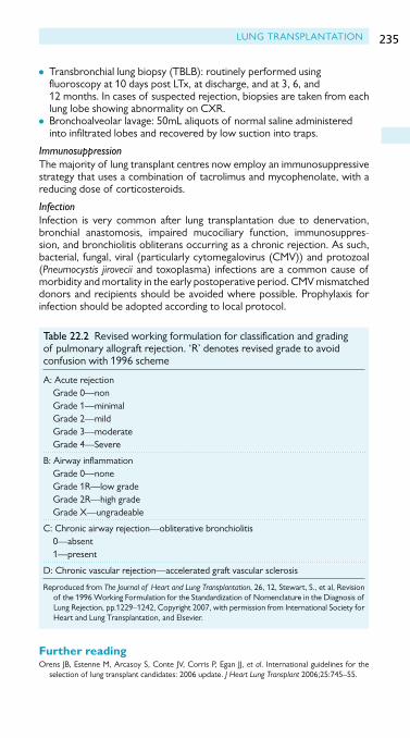
LuNG traNSpLaNtatION 235
• Transbronchiallungbiopsy(TBLB): routinelyperformedusingfluoroscopy at 0 days post Ltx, at discharge, and at 3, 6, and 2 months. In cases of suspected rejection, biopsies are taken from each lung lobe showing abnormality on CXr.• Bronchoalveolarlavage: 50mLaliquotsof normalsalineadministered
into infiltrated lobes and recovered by low suction into traps.
Immunosuppressionthe majority of lung transplant centres now employ an immunosuppressive strategy that uses a combination of tacrolimus and mycophenolate, with a reducing dose of corticosteroids.
InfectionInfection is very common after lung transplantation due to denervation, bronchial anastomosis, impaired mucociliary function, immunosuppres-sion, and bronchiolitis obliterans occurring as a chronic rejection. as such, bacterial, fungal, viral (particularly cytomegalovirus (CMV)) and protozoal (Pneumocystis jirovecii and toxoplasma) infections are a common cause of morbidity and mortality in the early postoperative period. CMV mismatched donors and recipients should be avoided where possible. prophylaxis for infection should be adopted according to local protocol.
Table 22.2 revised working formulation for classification and grading of pulmonary allograft rejection. ‘r’ denotes revised grade to avoid confusion with 996 scheme
a: acute rejection Grade 0—non Grade —minimal Grade 2—mild Grade 3—moderate Grade 4—Severe
B: Airwayinflammation Grade 0—none Grade r—low grade Grade 2r—high grade Grade X—ungradeable
C: Chronic airway rejection—obliterative bronchiolitis 0—absent —present
D: Chronic vascular rejection—accelerated graft vascular sclerosis
reproduced from The Journal of Heart and Lung Transplantation, 26, 2, Stewart, S., et al, revision of the996WorkingFormulationfortheStandardizationof NomenclatureintheDiagnosisof Lung rejection, pp.229–242, Copyright 2007, with permission from International Society for Heart and Lung transplantation, and elsevier.
Further readingOrens JB,EstenneM,ArcasoyS,Conte JV,CorrisP,EganJJ,et al. International guidelines for the
selection of lung transplant candidates: 2006 update. J Heart Lung Transplant 2006;25:745–55.

236 CHapter 22 The transplant patient
Heart–lung transplantationthe first successful heart–lung transplant was performed in 98 by Dr BruceReitzatStanfordHospital.Thenumberof proceduresreportedtobe performed worldwide is declining, with 50% of centres performing only one such transplant per year.
the primary indications for heart–lung transplantation are:• Congenital heart disease (39%)• primary pulmonary hypertension (25%)• CF (4%).
The procedurethe donor heart and lungs are harvested with minimal handling, with the heart flushed with cold cardioplegia solution, and the lungs with modified Collins solution.
UsingCPB, the recipient’s heart and lungs are removedwhilst care istaken to preserve the phrenic nerves and to avoid postoperative bleed-ing complications from the bronchial artery circulation. the donor heart and lungs are inserted in the anastomotic order: trachea, right atrium, aorta. Care should be taken to keep the donor trachea as short as possible because of the limited vascularity of the area.
Prognosisalthough the prognosis has improved in recent years, the -year mortality is 728%, and around half of patients are dead at 3 years. However, patients surviving the first year have an average life expectancy of nearly 0 years.

IMMuNOSuppreSSION 237
Immunosuppressionthe calcineurin inhibitors (CNIs) ciclosporin and tacrolimus are the corner-stone of immunosuppressive regimens. traditionally, either is used in com-bination with corticosteroids and an antiproliferative agent (mycophenolate or azathioprine). the use of induction therapy may delay the introduction of a CNI and reduce the incidence of perioperative renal dysfunction. tacrolimus is thought to be somewhat more effective than ciclosporin, and mycophenolate has been shown to provide more effective prophylaxis against acute rejection compared with azathioprine. the combination of tacrolimus, mycophenolate, and corticosteroids may therefore be consid-ered to be the most effective form of maintenance immunosuppression at least for the first 6–2 months after transplantation.
Calcineurin inhibitorsCiclosporin (cyclosporine)• Commenced when haemodynamics are stable, without evidence of
hepatic or renal failure.• Metabolized via cytochrome p450 pathway• Starting dose: 4mg/kg/day orally or .5mg/kg/day IV in two divided
doses (the IV dose is approximately a third of the oral dose).• Subsequent dose depends on blood levels and renal function, and can
be measured in two ways: C0—trough level, or C2—2 hours post dose.• target C0 levels:
• 300–400mcg/Lforthefirst4 weeks• 200–250mcg/Lfrom4weeksto6 months• 50–200mcg/Lfrom6 monthsto year• 00–50mcg/Lafter year.
Tacrolimus• CNI• 0.5–0.3mg/kg/day orally.
Antiproliferative immunosuppressantsMycophenolate mofetil (MMF)• Assoonasthepatientisabletotakeoralmedications,MMFis
commenced at a dose of .5g twice a day.• adjusted to mitigate side effects.
Azathioprine• Day 0: 4mg/kg IV at induction• postoperatively: 2mg/kg/day as a single dose if white cell count >4• DiscontinuedwhenMMFcommenced.
Corticosteroids• Intraoperative: g methylprednisolone at release of cross-clamp• Days and 2: 25mg methylprednisolone IV 2-hourly then oral
prednisolone 60mg/day decreasing by 5mg each day and then stopped• thereafter, steroids given for acute rejection.

238 CHapter 22 The transplant patient
Target of rapamycin inhibitorsSirolimus and everolimus• Similarefficacyinprophylaxisagainstacuterejectionas MMF.• Limited use due to adverse effects:
• Impairedwoundhealing• Pericardialeffusions• Bacterialandfungalinfections• GIeffects• Pneumonitis(sirolimus)• PotentiatesCNInephrotoxicity• Druginteractions(cytochromeP450pathway).
Induction therapyMany centres use a form of induction therapy with an anti-t-cell antibody to provide additional immunosuppression during the early perioperative period, lower rates of acute rejection, possible host hyporesponsiveness to alloantigen, and renal protection by allowing the delayed introduction of the CNIs. agents that have been used for this purpose include:• Monomurab-CD3 (OKT3)• antithymocyte globulin (atG)• Basiliximabanddaclizumab(monoclonalantibodiestotheIL-2receptor)• alemtuzumab (CaMpatH-H)—humanized antibody against CD52.
profoundly depletes lymphocytes. used in kidney transplantation; however, there is limited data in heart transplantation and there has been concern about potential cardiac toxicity.

239
Acute cardiology
Out-of-hospital cardiac arrest 240Endocarditis 243Aortic dissection 246
Chapter 23

240 ChAptEr 23 Acute cardiology
Out-of-hospital cardiac arrestIntroductionCardiovascular disease is the leading cause of death in the developed world. there is a wide variation in the reported incidence and outcome of patients who have out-of-hospital cardiac arrest (OOhCA). It is unfortunately a common event with a high mortality, and survival rate to discharge home is typically <0%. the most common cause for adult cardiac arrest is acute coronary syndrome (ACS), however it is important to eliminate other potential causes and treat them appropriately.
Principles of management• Effective cardiopulmonary resuscitation (Cpr) with minimal interruption• Optimize oxygenation• Early defibrillation• Appropriate drug treatment.
During circulatory arrest, organ injury can result from hypoxia, which is fur-ther exacerbated by reperfusion injury once return of spontaneous circula-tion (rOSC) is established. this insult can result in systemic inflammatory response syndrome (SIrS) and subsequent multiorgan failure (MOF). Early, aggressive management of these patients has potential to significantly influ-ence their outcome and neurological status.
Post-cardiac care bundle (AHA guidelines). Early coronary reperfusionEarly pCI is superior to thrombolysis with lower stroke, death, and reinfarction rates.
2. Hemodynamic optimizationFollowing cardiac arrest there is often a period of transient myocardial dys-function; hypotension, reduced CO, frequent arrhythmias, and impaired contractility. treatment includes:• IABp may augment the failing myocardium by both offloading the heart
and increasing coronary perfusion• Inotropic/vasopressor support• Invasive cardiac monitoring.
3. Control of ventilationManagement should aim to avoid ALI:• Aim for normocarbia and oxygen saturation 94–98%• Lung protective ventilation strategies:
• Tidalvolumes 6mL/kg• Useof PEEP• FiO2 <60%• Permissivehypercapniawithplateaupressures<30cmH2O.
4. Blood glucose controlBloodglucoseshouldbekeptwithin6–0mmol/L.

OUT-OF-HOsPiTaLcardiac arrEsT 241
5. Treatment of seizuresSeizures are common following hypoxic neurological insult:• Insufficient evidence for prophylactic treatment.• tonic clonic seizures treated with phenytoin/sodium valproate/
benzodiazepines.• Myoclonus can be treated with clonazepam.• the use of EEG monitoring allows for identification of subclinical
seizures, or seizure activity in paralysed patients.
6. Temperature controlpyrexia is common in the 48 hours following cardiac arrest; poor neuro-logical outcome has been associated with temperatures of 38°C and above.
Therapeutic hypothermiathere is increasing interest in the use of therapeutic hypothermia follow-ing OOhCA. two landmark randomized control trials (rCts) looked at the effect of cooling patients following VF arrest. Both trials demonstrated improvement in anoxic neurological injury, relating to improved functional outcomes and reduced mortality.
the International Liaison Committee on resuscitation (ILCOr) recom-mends the use of therapeutic hypothermia in patients who have rOSC though remain comatose following OOhCA regardless of initial presenting rhythm.
Indications for therapeutic hypothermiaPatientsaccepted for icUtreatmentdirectlyorvia thecatheter labwithfollowing features:• OOhCA with rOSC regardless of presenting rhythm• Comatose at the initial point of treatment with GCS score <9• Cerebral irritation following rOSC requiring intubation• No other known cause of coma (e.g. metabolic, trauma)• No coma state prior to arrest• No terminal illness• Not pregnant (request hCG test if <50 years)• Not haemodynamically unstable, e.g. high dose of inotropic support• No severe respiratory compromise.
Due to the adverse physiological effects of cooling, it is important to assess the risk of benefit versus harm on an individual basis.
Temperature monitoring (in order of preference)• pAFC• Bladder• rectal• Oesophageal• Do not use tympanic probe for temperature monitoring—this is
inaccurate.
Protocol for therapeutic hypothermiaSee Fig. 23..

242 ChAptEr 23 Acute cardiology
Further readingintensive care society (ics), UK. Standards for the Management of Patients After Cardiac Arrest. M<http://www.ics.ac.uk/ics-homepage/guidelines-standards/>.
START COOLINGSET TARGET TEMP 33°C
Temp 32–33°C at coolingstart + 1 hour
YES NO
Maintain temp 32–34°Cfor 24 hrs
Cold IV Hartmann’sstart 10 mL/kg
Temp 32–34°C at coolingstart + 2 hours?
YES
NO
Inform Medical Sta�
Consider:Further cold IV Hartmann’s stat(up to 30 mL/kg)Cold saline washout via NGAim to achieve temp 32–34°C
Stop cooling after 24 hrs
Controlled rewarming
Increase target temp by 0.3°C/hr to 36.5°C
Temp > 36°C at rewarmingstart + 12 hrs
YES NO
Prevent pyrexia for 72 hrsafter rewarming:Frequent temp measurementIf temp > 37.5°C cool to 36.5°CInform medical sta�Consider paracetamol,cold IV �uidsBlood cultures
Inform medical sta�
Consider warm IV �uids
Figure 23. (a) and (b) An example of a protocol for therapeutic hypothermia. reproducedwithkindpermissionfromJorgPrinzlinandKennethMcKinlay,copyrightGolden Jubilee National hospital.

ENDOCArDItIS 243
EndocarditisIntroductionEndocarditis is inflammation of the endocardium, which may or may not include the heart valves. Despite advances in medicine over the last 30 years neither the incidence nor mortality of the condition has changed. however, both the predisposing factors and causative agents have altered. previously considered a disease of the young with rheumatic heart disease, now this condition is associated with older patients following invasive procedures with or without prosthetic heart valves.
Predisposing factors• Male > female• Increasing age• IV drug abuse• Valve prostheses• Degenerative valve sclerosis• Multiple invasive procedures• Chronic haemodialysis• Diabetes• Intravascular devices.
Microbiology85% of cases are blood culture positive for the following typical bacteria:• Staphylococcus aureus: commonest causative bacteria, coagulase
negative Staphylococcus is frequently associated with prosthetic valve endocarditis (pVE)• Streptococcus; S. sangius,S. mitis,S. salivarius,S. mutans, and Gemella
morbillorum. Infection with S. milleri and S. anginosus is associated with abscess formation and disseminated disease• Enterococcus; E. faecalis,E. faecium,E. durans.
Blood cultures are frequently negative with the following organisms:• Variant Streptococcus• Gram-negative bacilli• HacEKgroup(Haemophilus,Actinobacillus,Cardiobacterium,Eikenella,King
ella)• Brucella, and fungi.
Upto5%of casesaretheresultof Coxiella burnetii (Q fever), Bartonella,Legionella,Mycoplasma, and Chlamydia. these organisms will always have nega-tive blood cultures and can only be identified with serology and cell culture.
Common presenting features• New regurgitant murmur• Embolic event—30% patients present with brain, lung, or spleen
infarction• Fever,signsof sepsis,pyrexiaof unknownorigin (PUO).

244 ChAptEr 23 Acute cardiology
Additional features• Splinter haemorrhages (particularly on nail beds)• Janeway lesions (painless macular erythematous lesions)• roth spots (retinal haemorrhages)• Osler’s nodes (painful nodules on digits)• rigors, fatigue, anaemia, night sweats, microscopic haematuria.
Diagnosisthe Duke criteria for diagnosis of infective endocarditis require 2 major or major and 3 minor or 5 minors:
Major criteria• positive blood cultures: either typical organism in 2 sets, or persistently
positive blood cultures (3+ sets >2 hours apart), or single positive blood culture for Q fever/phase IgG antibody titre > :800.• Endocardium involvement: echocardiogram evidence of vegetation/
abscesses or new valvular incompetence.
Minor criteria• predisposing factors• pyrexia > 38°C• positive blood cultures• Echocardiogram that do not meet major criteria• Vascular or immunological signs.
Investigations• Blood cultures: at least 3 sets taken 30 minutes apart from separate sites.• Echocardiogram: ttE is first line in all patients; tOE has a higher
sensitivity and specificity and is indicated in patients with a high suspicion of infective endocarditis (IE) despite a negative ttE and in all patients who have a positive ttE (abscess/vegetations visualized). In patients in whom a high suspicion remains despite previously negative findings should be rescanned 7–0 days later.• Urinalysis: microscopic haematuria.• CXR: cardiomegaly.• ECG: conduction abnormalities, typically st-degree heart block.• Blood tests: normochromic, normocytic anaemia, leukopenia, raised ESr
and Crp.
ManagementEarly antibiotic therapy has been shown to reduce mortality.
Empirical therapy for native valves with acute presentation:• Flucloxacillin8–2g/dayin4–6doseswithgentamycinmg/kg/day
three times a day.• In penicillin allergic patients: vancomycin g/2 hours, and gentamycin
mg/kg/day three times a day.

ENDOCArDItIS 245
Subacute presentation:• Penicillin7.2gin6divideddoseswithgentamycinmg/kg/daythree
times a day, or• amoxicillin2g/dayin6divideddoses(moreactivityagainstHacEK
organisms).
Prosthetic valves• Vancomycin and gentamycin doses as previously with the addition of
rifampicin600–200mg/dayintwooral doses.
Once organism is isolated therapy should be directed in accordance with microbiological advice.
Early surgery prevents progressive heart failure and valvular destruc-tion; however it also carries a higher mortality risk. the indications for surgery are:• Uncontrolledinfectiondespitetreatment• prevention of emboli• heart failure.
ProphylaxisCertain patients are considered at a higher risk of developing endocarditis:• patients with prosthetic valves/material for cardiac valve repair• patients with previous infective endocarditis• Structural congenital heart disease—not including isolated atrial septal
defect, repaired ventricular septal defect, or patent ductus arteriosus• hypertrophic cardiomyopathy• Acquired valve heart disease (stenosis/incompetence).
prophylactic antibiotics:• No longer indicated for all dental or non-dental procedures as the risk
of anaphylaxis outweighs the benefit of use.• Advice is given regarding recognizing symptoms, good oral hygiene, and
avoidance of potential causative procedures.• however, if a high risk patient has an infection which requires
antibiotics,andisundergoingGU/Gisurgeryatthesiteof theinfectionthen antibiotics given should cover organisms causative for IE.
prognosis is dependent on four features:• Patient factors: elderly, pVE, coexisting disease – respiratory, renal,
cardiac, or IDDM.• Infective organism: fungi, Staphylococcus aureus, and Gram-negative bacilli
all have poorer outcomes.• Complications of IE are heart or renal failure, CVA, and septic shock.• Echocardiogram evidence of pulmonary hypertension, severe valvular
incompetence, raised diastolic pressure and reduced LV ejection fraction.
Further readinghabib G, hoen B, tornos p, thuny F, prendergast B, Vilacosta I, et al. Guidelines on the preven-
tion, diagnosis, and treatment of infective endocarditis (new version 2009): the task Force on the prevention, Diagnosis, and treatment of Infective Endocarditis of the European Society of Cardiology (ESC). Eur Heart J2009;30:2369–43.
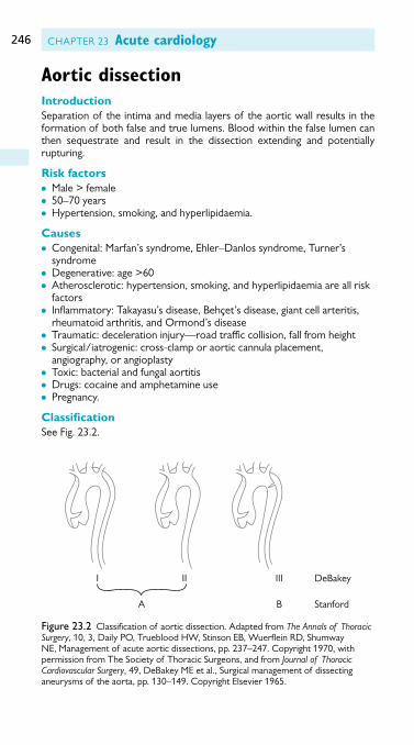
246 ChAptEr 23 Acute cardiology
Aortic dissectionIntroductionSeparation of the intima and media layers of the aortic wall results in the formation of both false and true lumens. Blood within the false lumen can then sequestrate and result in the dissection extending and potentially rupturing.
Risk factors• Male > female• 50–70 years• hypertension, smoking, and hyperlipidaemia.
Causes• Congenital: Marfan’s syndrome, Ehler–Danlos syndrome, turner’s
syndrome• degenerative: age >60• Atherosclerotic: hypertension, smoking, and hyperlipidaemia are all risk
factors• Inflammatory: takayasu’s disease, Behçet’s disease, giant cell arteritis,
rheumatoid arthritis, and Ormond’s disease• traumatic: deceleration injury—road traffic collision, fall from height• Surgical/iatrogenic: cross-clamp or aortic cannula placement,
angiography, or angioplasty• toxic: bacterial and fungal aortitis• Drugs: cocaine and amphetamine use• pregnancy.
ClassificationSee Fig. 23.2.
A
I II III DeBakey
B Stanford
Figure 23.2 Classification of aortic dissection. Adapted from The Annals of Thoracic Surgery, 0, 3, Daily pO, trueblood hW, Stinson EB, Wuerflein rD, Shumway NE, Management of acute aortic dissections, pp. 237–247. Copyright 970, with permission from the Society of thoracic Surgeons, and from Journal of Thoracic Cardiovascular Surgery, 49, DeBakey ME et al., Surgical management of dissecting aneurysmsof theaorta,pp. 30–49.copyrightElsevier 965.

AOrtIC DISSECtION 247
Stanford
• type A: the ascending aorta is involved in the dissection.• type B: no involvement of the ascending aorta.DeBakey (further subdivides the dissection)2
• type I the whole aorta is involved.• type II only the ascending aorta is involved.• type III only the descending aorta is involved in the dissection.A new classification includes the aetiology of the dissection (table 23.).
Presentation• Abrupt onset of severe chest pain, typically radiating through to back.• pain often described as sharp, tearing, ripping, or stabbing.• Associated syncope, cardiac failure, cardiac tamponade, aortic
regurgitation, hypo/hypertension.• Depending on the anatomy of the extension, aortic root occlusion can
occur, affecting arms/legs (20%), renal (5%), brain (5%), cardiac (0%), and bowel (3%).
DiagnosisEarly diagnosis requires good history taking and a high index of suspicion in patients with risk factors.• ECG: eliminate acute MI as the diagnosis. Extension of the dissection
involving the coronary ostia may present as ischaemia on the ECG.• CXR: mediastinal widening, cardiomegaly, aortic knuckles, pleural
capping, deviation of trachea to right, and blunted costophrenic angles secondary to haemothorax.• CT: high sensitivity and specificity of dissection, rapid test, and able to
establish the extent of the lesion.• MRI: highest sensitivity and specificity for diagnosis, also allows for
functional assessment of aortic valve and left ventricle. May be impractical in critically ill patient.
Table 23. European Society of Cardiology classification of aortic dissection
Class Classical aortic dissection
Class 2 Intramural haematoma/haemorrhage
Class 3 Subtle, discrete aortic dissection
Class 4 plaque rupture/ulceration
Class 5 traumatic/iatrogenic aortic dissection
reproduced from Erbel r. et al., 'Diagnosis and management of aortic dissection: task Force on Aortic Dissection, European Society of Cardiology', European Heart Journal, 200, 22, 8, pp.642–68,bypermissionof theEuropeansocietyof cardiology.
reprinted from The Annals of Thoracic Surgery, 0, 3, Daily pO, trueblood hW, Stinson EB, Wuerflein rD, Shumway NE, Management of acute aortic dissections,pp. 237–247. Copyright 970, with permission from the Society of thoracic Surgeons.
2 this classification was published in Journal of Thoracic Cardiovascular Surgery, 49, DeBakey ME et al., surgicalmanagementof dissectinganeurysmsof theaorta,pp.30–49.copyrightElsevier965.

248 ChAptEr 23 Acute cardiology
• TOE: allows for real time assessment of dissection, involvement of the aortic valve, degree of regurgitation, and left ventricular cardiac function can be assessed.• Aortography: previously considered the gold standard, though
inappropriate in unstable patient.
Differential diagnosis• ACS• Aortic regurgitation• Aortic aneurysm without dissection• pericarditis• pE• pleurisy.
ManagementType A aortic dissection• requires surgical intervention and has a higher mortality compared to
typeB(60%vs 0%).• Mortality increases –2% per hour in the untreated acute ascending
aorta dissections in the first 48 hours.
Type B aortic dissection• Medical therapy is aimed at reducing hypertension.• No evidence to suggest an improved survival in patients who have a
surgical repair of type B aortic dissections.• Surgery or interventional radiology may be considered in this group if
the dissection is causing organ ischaemia.
Initial managementStrategy should be to prevent further extension of the dissection and ves-sel rupture:• A, B, C.• tight Bp control: IV labetalol to maintain systolic Bp 80–00mmhg.
Sodium nitroprusside (SNp), GtN and hydralazine are alternatives.• reduce LV contractility without comprising coronary perfusion.• Adequate analgesia.• iVaccess,cross-match,fullbloodcount,coagulation,U&Es,lactate.• Establish invasive monitoring—arterial line ideally placed in left radial
artery as innominate artery may be involved in dissection, affecting right radial readings.
Conduct of anaesthesia• Maintain haemodynamic stability.• type B dissections—thoracotomy incisions require double-lumen tube
placement, usually left.• Depending on position of aortic clamp patients are at risk of bowel or
renal ischaemia and anterior spinal artery syndrome/paralysis.• Meticulous review of urine output, metabolic status, and coagulation is
important to anticipate any complications.• tOE can help guide surgical decision-making.• CpB—it may be necessary to establish CpB rapidly in a patient in
extremis and femoral bypass can be established prior to sternotomy.

AOrtIC DISSECtION 249
Deep hypothermic circulatory arrest (DHCA)CpB allows for the maintenance of organ perfusion following cardioplegia induced cardiac arrest. Complete circulatory arrest can be advantageous in aortic surgery, providing improved operating conditions and reducing basal metabolic rate (BMr) and organ ischaemia. By reducing the body tempera-ture to 8–20°C prolongation of the CpB and aortic cross-clamp times are possible. periods of up to 30 minutes of DhCA are tolerated without significant neurological dysfunction; longer durations are linked to a sharp increase in neurological injury and poorer outcome.
It is possible to selectively perfuse the cerebral circulation, using retro-grade cerebral perfusion or selective antegrade cerebral perfusion. By using these methods DhCA can be prolonged safely.
Postoperative care• Establish normothermia.• Correct any coagulopathy and acidosis.• Maintain MAp ∼65mmHgforatleastthefirst4hoursfollowing
the operation and if no significant bleeding noted increase the MAp ∼75mmhg.• Careful fluid balance, risk of cerebral oedema formation, may require
mannitol or frusemide therapy.• Keepthepatientventilatedandsedatedinitially,oncehaemodynamically
stable for >2 hours and no signs of bleeding, wean patients and assess neurological status.
Further readingErbel r, Alfonso F, Boileau C, Dirsch O, Eber B, haverich A, et al. Diagnosis and management of
aortic dissection. Eur Heart J200;22:642–8.hebballi r, Swanevelder J. Diagnosis and management of aortic dissection. CEACCP 2009;9:4–8.

Treatments and procedures
24 Central venous cannulation, pulmonary
artery catheter, and minimally invasive
cardiac output monitoring 253 25 Airway management 263 26 Respiratory management 275 27 Circulatory support 287 28 Epicardial pacing 303 29 Sedation and pain relief 33 30 Wound management 32 3 Infection control and prevention 329 32 Bedside echocardiography 339
Part 4


253Chapter 24
Central venous cannulation, pulmonary artery catheter, and minimally invasive cardiac output monitoring
Central venous cannulation 254Pulmonary artery catheter 256Minimally invasive cardiac output monitoring 260

254 ChAPtER 24 Central venous cannulation
Central venous cannulationIndications• Measurement of central venous pressure (CVP)• If peripheral venous access is difficult to establish• Administration of drugs which cannot be given peripherally.
Administration of tPN and other hypertonic solutions• Siting of PAFC• transvenous cardiac pacing• Renal replacement therapy.
Preventing complications5% of patients with central venous catheters have a complication.• Use standard Seldinger wire technique.• Ultrasound technique reduces risk of arterial puncture and
pneumothorax. Particularly useful in internal jugular vein (IJV) cannulation.• Choose multilumen catheters to avoid multiple cannulations.• Get experienced help if more than 3 cannulation attempts: (risk i× 6).• Prevent air embolism: occlude lumina during insertion, use head-down
position (may not be tolerated with poor cardiac function).• Arterial puncture? Check pressure trace or compare blood sample
to ABG.• Arrhythmias: check ECG whilst advancing Seldinger wire or catheter.• Perforation: usually delayed complication due to erosion of vessel wall.
Risk of damaging veins (stiff PAC introducer) and endocardium (PAC).• Central venous thrombosis: i risk in IJV, femoral vein, and large
catheters.• Pneumothorax: i risk with hyperinflation (large tV, PEEP, chronic
obstructive airways disease). If suspected do not attempt contralateral IJV/subclavian vein (SCV) cannulation.
Preventing infection• Risk of bloodstream infection is 3–8%.• Mechanism of catheter infection:
• <5 days: tipcolonizationwithskinorganismsviaext. tract.• >5 days: contaminationof catheter,organismsviaint. lumen.
• Maximal precautions: hat, mask, gown, gloves, large sterile drapes. Skin antisepsis with chlorhexidine is significantly superior.• Use teflon or polyurethane catheters.• Antimicrobial catheters d infection risk but may i antibiotic resistance.
Consider if infection rate is high despite preventative measures. Antibiotic ointments at catheter site i resistance and fungal infections.• Give lipids (tPN, propofol) via dedicated lumina.• Changing of CVC over guidewires i infection risk.• Scheduled replacement of CVC does not reduce infection risk.• Remove CVC as soon as not required.

CENtRAl VENoUS CANNUlAtIoN 255
Central vein routesIn cardiothoracic patients IJV, SCV, and femoral veins are commonly used.
Internal jugular cannulation• Easily accessible and high success rate.• low complication rate with landmark technique, best with ultrasound.• Safer than SCV approach in deranged coagulation. External pressure on
punctured carotid artery possible.• Right IJV preferred because of straight access into SVC. low left-sided
approach carries risk of thoracic duct damage (chylothorax).• May be less comfortable for awake patients than subclavian route.• Awake patient: insertion with lA infiltration or superficial cervical
plexus block.• head-down tilt to increase venous filling and prevent air embolus.• Venous thrombosis risk approximately 4× that of the SCV.• high approach in neck reduces risk of pneumo/haemothorax.
Subclavian vein cannulation• More comfortable for patient• least risk of infection• Ultrasoundlessusefulasveinismoredifficulttovisualize• Risk i of pneumothorax; arterial puncture—compression difficult.
Femoral vein cannulation• Arterial puncture: easily compressible• No risk of pneumothorax• Indicated in SVC obstruction• Difficulttomobilizepatients• iriskof infectioninpatientswithBMI >28• i risk of thrombosis (up to 2%).
Central line tip position• to avoid vessel wall erosion and pericardial tamponade catheter
tip must lie outside the RA and parallel to the SVC just above the pericardial inflection (CXR: the carina is the landmark just above this).• Innominate vein position: thrombosis risk with inotropes/irritant drugs.• Right-sided catheters should be sited above the carina.• left-sided catheters may need to be positioned distal to the carina to
avoid the tip abutting on SVC wall.• CVC tip changes 2–3cm with respiration, arm, and torso position.• If in doubt about the correct length, insert a longer catheter deep and
withdraw after CXR.

256 ChAPtER 24 Central venous cannulation
Pulmonary artery catheterthere is controversy on safety and clinical benefit of the PAC. It continues to be used in CICU as the only device that can assess intrathoracic pres-sures, Co, and Svo2 simultaneously. Standard PAC features: 0cm mark-ings, proximal lumen for CVP and IV fluids, distal lumen for PAWP, balloon lumen and thermistor for Co measurement near the tip, and fibreoptic bundles for continuous Svo2 measurement. See table 24..
Indication• Monitoring of Svo2, cardiac pressures, Co, and derived data: trends
more useful than single values• Differentiation of causes of shock states• Assessment of pulmonary hypertension• Differentiation of high- versus low-pressure pulmonary oedema• Aspiration of air emboli.
Contraindications• tricuspid or pulmonary valve mechanical prosthesis or endocarditis• Right heart mass (thrombus/tumour).
Insertion• Insert PAFC introducer. Preferred routes: right IJV, left SCV (no acute
angle to enter SVC); femoral veins can be difficult (in enlarged RV).• Full-barrier asepsis for insertion. Check balloon, pre-fill other lumina.
Inside sterile plastic sleeve catheter can be freely handled.• Attach transducer to distal lumen. observe pressure changes as
catheter is advanced. Calibrate (usually automatic), reference (as for CVP)andzeropriormeasurements.Avoiddampening(bubbles)—check with rapid flush test.• Patient supine or head-up. Curved catheter tip may help positioning
(RIJ: curve to l shoulder, in RV turn ¼ clockwise to ease tip into PA).• In RA inflate the balloon gently (ml/sec) and ‘float’ from the RV into
PA. Max. filling of balloon .5ml. As the balloon occludes a PA vessel the pressure trace changes (Fig. 24.). this is the PAWP.• Deflate the balloon and ensure PA trace becomes visible again. If
permanent wedging occurs, gently withdraw the catheter.• In-between PAWP measurement, ensure balloon stays deflated to avoid
PA damage and infarction. Monitor PAP trace continuously.• CXR: tip position below level of left atrium (only 60% of catheters).• Aim to remove catheter within 72 hours to reduce complications.
Difficulties floating the catheter• In RA or RV dilation, severe tR, very poor RV function:
• Optimizepatientposition: headupandslightlytowardstherightsideputs pulmonary valve at the highest level. When the catheter is in the RV outflow tract (PVCs), pause ventilator in inspiration and advance catheter into PA.
• Consider fluoroscopy.

PUlMoNARy ARtERy CAthEtER 257
Complications• Mechanical and infective complications as with central venous access.• Arrhythmia: usuallyPVCs,short-livedVT.SignificantVT/VFin<%.
transient right bundle branch block (BBB) in 5% (risk of 3rd-degree heart block with pre-existing left BBB).• Infection (i risk after day 3).• Coiling/knotting: suspectif >20cmadvancedintotheRVwithout
waveform changes. Withdraw slowly to avoid knotting. Get experienced help, consider fluoroscopy, interventional radiology.• Damage to valves, myocardium.• Rupture/damage to PA: rare (∼% but 50% fatal). Risk i: pulmonary
hypertension,>60 years,anticoagulation.Signs: suddenhaemoptysisafter inflation of PAC balloon. Management: position bleeding side down,lungisolationwithDLT,PEEP,embolizationorlobectomy.• Pulmonaryinfarction: (<7%)Cause: migrationof PACorthrombi.• PACnotinzone3;suspectif: meanPAWP>meanpulmonaryartery
diastolic pressure (PADP), PAP trace swings ++ with respiration.
Table 24. Cardiac pressures related to PAC length and position (RIJ approach)
Position Length (cm) Pressure (mmHg)
RA 20–25 0-8/0–8
RV 30–35 5–30/3–8
PA 40–45 5–30/4–2
PA wedge 45–55 2–5
605040
(mm
Hg)
3020100
Right atrium Right ventricle Pulmonaryartery
Pressure tracings during pulmonary artery catheterization
Wedge position(PCWP)
Figure 24. PAC waveform changes on insertion. SVC /RA: = CVP waveform; RV trace: after crossing tV, steady increase in diastolic pressure; PAP trace: after crossing PV, sudden rise in diastolic pressure with steady fall in baseline; PAWP: similar to CVP trace. Reproduced from Punit S. Ramrakha and Kevin P. Moore, Oxford Handbook of Acute Medicine 2nd Edition, 2004, figure in chapter 6, p. 879, with permission from oxford University Press.

258 ChAPtER 24 Central venous cannulation
• Cardiac tamponade: i RA pressure on inspiration (Kussmaul sign), mean RA pressure = RVDP = PADP = PAWP.• Cardiogenicpulmonaryoedema: likelyif PAWP>8–20mmHg(if
colloid pressure and permeability normal).• PAWP overestimates lVED pressure in mitral stenosis, PEEP ventilation,
left atrial myoma, pulmonary hypertension.• PAWP underestimates lVED pressure in non-compliant lV, lVED
>25mmHg,aorticregurgitation.
Difficulties determining the PAC position• large v waves may make it difficult to differentiate PA trace and PCWP.
look for subtle waveform differences (loss of the PA dicrotic notch) or compare distal blood sample with and without balloon inflation (PAWP = Svo2, PAP = Sao2).• overwedging: wedge pressure trace rises excessively, no pulsatile trace,
balloon volume less than half the maximum.
Cardiac pressures• PAP: approx. /5 of systemic pressures.• PAWP: pressure at the distal catheter tip as balloon occludes a PA
branch. Reflects delayed pressure changes in the lA (similar to CVP trace). PAWP is an approximation of the true lV preload (lVEDV), indirectly represents myocardial fibre length (Starling law). helpful when RA pressures (CVP) do not reflect lV function, e.g. in lV failure, severe BBB, pulmonary hypertension, cardiac tamponade, constrictive pericarditis, valvular disease. 7–4mmhg less than PADP. Measure PAWP at end-expiration (IPPV: trough of trace, spont. breathing: peak of trace).
Cardiac output monitoringCalculations based on Fick principle:• Intermittent: 0ml cold fluid boluses into RA, blood temperature
measured by thermistor at tip, use average from 3–4 measurements.• Continuous CO (CCO): pseudo-random cycles from RA heat element every
40–60 seconds registered by thermistor. temperature–time-curve is inversely proportional to Co. Changes may lag up to 2 minutes.• Pitfalls: large left-to-right shunt: overestimation of Co, large right-to-left
shunt: underestimation of Co, severe tR: may over or underestimate Co.
Interpretation(See table 24.2.)
Table 24. 2 haemodynamic parameters in relation to common disorders
CO PAWP CVP SVR
Cardiogenic d i i i
Vasodilatation i n/d n/d d
hypovolaemia d d d i

PUlMoNARy ARtERy CAthEtER 259
Mixed venous oximetryIndicates Vo2/Do2 balance and adequacy of global circulation. high risk of poor tissue oxygenation if Svo2<40%.SeeTable 24.3.
Table 24. 3 Common causes of abnormal Svo2
Low SvO2 High SvO2
VO2i DO
2d VO
2d DO
2i
Shivering hb d Sedation hb i
Stress/pain Fio2d hypothermia Fio2i
hyperthermia Co d Muscle relaxation Co i
Seizures Sepsis (shunting)
Wedged PAC (artefact)

260 ChAPtER 24 Central venous cannulation
Minimally invasive cardiac output monitoringSeveral alternatives to the PAC are being used to assess cardiac function:
Pulse contour analysisArea under the curve (AUC) of a central arterial pressure wave (PW) correlates with the stroke volume (SV) calculated from aortic compli-ance and pressures. these are estimated by mathematical modelling from peripheral arterial PW. Difficulties arise from the accuracy of determining the systolic part of arterial PW, estimating varying arterial compliance and quality of PW.
Pulse contour analysis with calibration• lithium indicator dilution (liDCoplus):• Calibration for SV: an arterial sensor measures the concentration of
a lithium bolus. Co calculated from this. Recalibration ≤8 hours or in haemodynamic instability.• Continuous mode: assumes changes in the arterial PW to reflect SV.• Accuracy decreases with: AF, IABP, AV disease, quaternary muscle
relaxants, severe hyponatraemia. the system should not be used in st trimesterpregnancy,concurrentlithiumtherapy,bodyweight <40kg.transpulmonary cold indicator dilution (PiCCo).• Calibration for SV: cold saline injected into central vein. temperature
changes detected by a peripheral arterial sensor. Allows estimation of extra vascular lung water (EVlW). Needs frequent recalibration in haemodynamic instability.• Continuous mode: analyses systolic AUC, shape of PW, aortic
compliance and SVR calculated Co.• Accuracy d in cardiac shunt, severe AV disease, and IABP.
Non-calibrated pulse contour analysis (Vigileo™)• No venous access or calibration required.• Algorithm uses demographics, analysis of pulse pressure (∼ to SV), SD
of 2000 data points of arterial PW to estimate arterial compliance and calculate SV and Co.• Accuracy d with AR, severe peripheral arterial vasoconstriction. Not
validated with concurrent use of IABP.
Thoracic bioimpedance measurement• thoracic electrodes emit and sense a small alternating current and relate
it to the ECG. Baseline impedance reflects total thoracic fluid volume. the change in impedance over the cardiac cycle generates a waveform similar to the aortic flow curve and allows calculation of SV, Co, and myocardial contractility.• Accuracy may be affected by pulmonary oedema and changes in PVR.
Validity in critically ill and cardiac patients is uncertain.

MINIMAlly INVASIVE CARDIAC oUtPUt MoNItoRING 261
Oesophageal Doppler• Velocity of blood flow is proportional to frequency of reflected waves
(Doppler shift). Probe is rotated for optimal velocity-time signals. Shape of waveform reflects peak velocity, lV preload, contractility, and afterload. Aortic cross-section area estimated from nomograms or measured by oesophageal probe. Calculation of SV and Co assumes flow through the ascending aorta stays constant.• Accuracy d by hypovolaemia, regional anaesthesia. Good correlation
with PAC for Co. Better reflection of preload (end-diastolic volume) than PAWP.
Further readingFunk DJ, Moretti EW, Gan tJ. Minimally invasive cardiac output monitoring in the perioperative set-
ting. Anesthes Analg 2009;08:887–97.NICE. Guidance on the use of Ultrasound locating devices for placing central venous catheters, technology
Appraisal Guidance No 49. london: National Institute for Clinical Excellence; 2002.Practice Guidelines for Pulmonary Artery Catheterization, An updated Report by the American
Societyof AnesthesiologistsTaskForceof onPulmonaryArteryCatheterization.Anaesthesiology 2003;99:988–04.
taylor RW, Paligri AV. Central venous cannulation. Crit Care Med 2007;35:390–6.


263
Airway management
Intubation 264Tracheostomy 266Extubation 272
Chapter 25

264 ChapTEr 25 Airway management
IntubationIndications for intubation• hypoxaemia• hypercapnia• Exhaustion• To reduce the work of breathing and oxygen consumption• To protect the airway (reduced GCS score/impaired laryngeal reflexes)• To treat or prevent obstructed airway• To facilitate investigations/treatment or critical care transfer.
The decision to institute mechanical ventilation can be difficult. It should be undertaken as a treatment for respiratory failure or critical illness if:• The patient had an acceptable premorbid quality of life• There is a reversible cause for the deterioration• NIV is inappropriate• The patient consents to the treatment or has not expressed any prior
wishes regarding treatment limitation.
Technique of endotracheal intubationEquipment and preparation• assess the airway—anticipate difficult intubation where possible.• allocate roles to appropriate skilled personnel.• have a plan in place for failed intubation.• Check suction and ventilator or anaesthetic machine.• Ensure there is an alternative working oxygen supply available.• have available a bag/valve/mask, laryngoscope, airway adjuncts, and
various ETTs.• attach monitoring to association of anaesthetists of Great Britain and
Ireland (aaGBI) standards: pulse oximeter, non-invasive blood pressure (NIBp), and ECG. airway gas analysis including end-tidal CO2 and airway pressure monitoring should be available immediately after intubation.• In critically ill patients, insert an arterial line if time permits.• Establish IV access with fluid attached.• Draw up and label drugs of choice and emergency drugs• Check trolley tips or head of bed able to tilt down• Optimize patient position: neck flexed about 35o and head extended
5o. a pillow is required
Technique• preoxygenate with 00% oxygen with a tight fitting face mask for 3–5
minutes. In emergencies four vital capacity breaths may be used.• administer a sleep dose of a preselected IV induction agent followed
by either a dose of depolarizing muscle relaxant in a rapid-sequence intubation (rSI) or non-depolarizing muscle relaxant.• For example: thiopentone 3–7mg/kg followed by
suxamethonium 00mg.• If cricoid pressure is being used in an rSI then this should be applied by
a skilled operator before loss of consciousness.
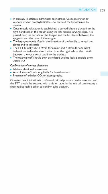
INTuBaTION 265
• In critically ill patients, administer an inotrope/vasoconstrictor or vasoconstrictor prophylactically—do not wait for hypotension to develop.• Once muscle relaxation is established, a curved blade is placed into the
right-hand side of the mouth using the left-handed laryngoscope. It is passed over the surface of the tongue and the tip placed between the epiglottis and the base of the tongue.• The laryngoscope is lifted in the direction of the handle to reveal the
glottis and vocal cords.• The ETT (usually size 8–9mm for a male and 7–8mm for a female)
is then inserted under direct vision from the right side of the mouth between the vocal cords and into the trachea.• The tracheal cuff should then be inflated until no leak is audible or to
30cmh2O.
Confirmation of correct placement• Bilateral chest wall movement• auscultation of both lung fields for breath sounds• presence of exhaled CO2 on capnography.
Once tracheal intubation is confirmed, cricoid pressure can be removed and the ETT should be secured with a tie or tape. In the critical care setting a chest radiograph is taken to confirm tube position.

266 ChapTEr 25 Airway management
TracheostomyThis section deals with elective insertion of temporary tracheostomies in critically ill patients.
Some indications are absolute:• airway protection (neurological dysfunction)• upper airway obstruction
Some indications are relative:• Bronchial toilet• patient may be cared for in lower dependency area• patient is poorly tolerant of ETT.
Most tracheostomies are placed in patients who are undergoing, or are at risk of, prolonged mechanical ventilation.
anecdotal advantages:• Improved patient comfort• reduction in sedation requirement• Improved mouth care• reduced dead space, tube resistance, and work of breathing• Improved weaning and reduced ICu length of stay.
Two recent prospective randomized trials evaluating early vs late placement of tracheostomy did not demonstrate any significant advantages of early tracheostomy, but did confirm that a policy of early tracheostomy inevitably means additional procedures and procedural complications.
TypesPercutaneous versus surgicalWhere the expertise exists, percutaneous tracheostomy is now the pre-ferred procedure in ICu (it is at least equivalent, possibly with less wound infections).
Internal/external diameterNomenclature used by different manufacturers is confusing. There is a reasonable amount of variation in the ratio of internal to external diam-eter, and different tubes all of the same ‘size’ may have significantly dif-ferent internal diameters. The smaller the internal diameter the higher the imposed work of breathing.
Inner cannula and cuffsTubes with inner cannulae are safer, especially in areas without airway expertise, but the internal diameter is necessarily reduced compared with external diameter.
In the critical care setting, cuffed tubes are customarily the initial choice, allowing airway protection and positive pressure ventilation. Cuff pressure should be 20–30cmh2O, to reduce Vap without causing mucosal ischaemia.
FenestrationsIf bulbar function is intact and the patient has been weaned from respira-tory support, fenestrated tubes allow airflow through the upper airways and

TraChEOSTOMy 267
therefore phonation and speaking. If the tube is not fenestrated, deflating the cuff serves a similar function.
Shape and sizeThere is no perfect tracheostomy tube that will have the correct intra-stomal length, intra-tracheal length, and angulation for every patient. alternatives should therefore be available in order to avoid problems with the tube abutting the anterior or posterior wall, tube displacement, or tracheal ero-sion. adjustable flange tubes may be useful, but have no inner cannula.
Performing a tracheostomyFor guidance on how to perform a tracheostomy, see the Intensive Care Society’s guidance on standards for the care of an adult patient with a tem-porary tracheostomy (see b Further reading, p. 27.
Complications of tracheostomiesImmediate• pneumothorax or pneumomediastinum• Tracheo-oesophageal fistula• Injury to great vessels or recurrent laryngeal nerves• Bleeding, e.g. from divided thyroid isthmus.
Early• Secretions and mucus plugging• Dislodged tube, respiratory arrest• post obstructive pulmonary oedema (when tracheostomy is performed
in a patient with longstanding upper airway obstruction).
Late• Bleeding from tracheoinnominate fistula (can be torrential)• Tracheal stenosis (from ischemia induced by a cuffed
tracheostomy tube)• Tracheo-oesophageal fistula• Tracheocutaneous fistula• Cosmetic deformity.
Practical tracheostomy issuesChanging a tracheostomyThere is little evidence to guide practice. Facilities to reintubate via oral route should be immediately available.• There is usually a defined stoma after 0 days.• First elective change is best undertaken between 7 and 0 days. avoid
changes <72 hours after tracheostomy unless absolutely necessary.• railroad new tube over a fibreoptic scope (direct vision) or paediatric
ETT (may be used to ventilate) or a suction catheter.
Tracheostomy emergenciesThe main life-threatening complications associated with a tracheostomy are blockage, dislodgement, and bleeding (see Fig. 25.).

268 ChapTEr 25 Airway management
Blockage and displacementa blocked or displaced tracheostomy tube presents with respiratory diffi-culty. a partly dislodged tracheostomy tube is just as dangerous, if not more dangerous, as a completely removed tracheostomy tube.• Tracheostomy tubes may become dislodged or displaced when a
ventilated patient is turned, or moved from their bed to a trolley.• restless or agitated patients may pull at their tracheostomy tubes, or
ventilator tubing attached to the tracheostomy.
Action• Don’t panic!• Call for help—senior medical and nursing staff, other ahps with
tracheostomy care skills (e.g. physiotherapist).• reassure patient.• assess patency of airway (a) and patient’s breathing (B). Is air passing
through the tracheostomy tube or stoma? Is the patient breathing via mouth or nose?• Check capnography and oxygen saturation with a pulse oximeter.• If airway is not patent, it must be cleared immediately.• If the blocked tracheostomy tube has an inner lumen, remove the inner
lumen and ventilate through the outer lumen.• If a single-lumen tracheostomy tube is blocked or displaced, obstructing
the airway, remove it to ventilate through the stoma or the mouth/nose.• Once removed, only experienced staff should attempt to re-insert a
tracheostomy.• any attempt to re-insert a tracheostomy should be quickly abandoned if
unsuccessful.• Awell-formedtrackfromskintothetrachealstomatakes>72
hourstoformfollowingsurgicaltracheostomyor>7 daysafterpercutaneous tracheostomy.
• If unable to ventilate through the stoma, re-establish the airway in the usual fashion. Tilt the head back to extend the head on the neck, perform a jaw thrust. If necessary insert an oral airway (Guedel).• Note that if ‘bag and mask’ ventilation is attempted, air will escape
through the stoma. In this situation, get a colleague to occlude the stoma by applying pressure over it with gauze swabs or a pad.• Tracheal intubation may be needed. It may be necessary to push the
tracheal tube distal to the stoma. (use an ‘uncut’ tube.)• If the patient is breathing adequately at this point, there may be no
need to artificially assist ventilation. Check the oxygen saturation with a pulse oximeter and administer oxygen as required via a facemask or resuscitation bag with an oxygen reservoir.• If artificial ventilation is needed, use a resuscitation bag and mask in the
standard fashion.• Maintain occlusion of the stoma as described earlier to prevent an
air leak. Capnography will confirm tracheal intubation. Measurement of oxygen saturation with a pulse oximeter will confirm adequacy of oxygenation.

TraChEOSTOMy 269
• at this point the patient is safe, with a patent airway and adequate respiration. Do not panic.• a decision about re-insertion of the tracheostomy tube can now be
made by the senior anaesthetist.
Emergency tracheostomy management -Patent upper airway
Tracheostomy tube partiallyobstructed or displacedContinue ABCDE assessment
The tracheostomy tube is patentPerform tracheal suctionConsider partial obstructionVentilate (via tracheostomy)if not breathingContinue ABCDE assessment
Tracheostomy STOMA ventilationPaediatric face mask applied to stomaLMA applied to stoma
Attempt intubation of STOMASmall tracheostomy tube/6.0 cu�ed ETTConsider Aintree catheter and �breoptic‘scope/Bougie/Airway exchange catheter
Can you pass a suction catheter?
Remove speaking valve or cap (if present)Remove inner tube
Some inner tubes need re-inserting to connect to breathing circuits
REMOVE THE TRACHEOSTOMY TUBELook, listen & feel at the mouth and tracheostomy. Ensure oxygen re-applied to face and stoma
Use waveform capnography or Mapleson C if available
No
No
Yes
Yes
Yes
No
Standard ORAL airway manoeuvresCover the stoma (swabs/hand). Use:
Bag-valve-maskOral or nasal airway adjunctsSupraglottic airway device e.g. LMA
Attempt ORAL intubationPrepare for di�cult intubationUncut tube, advanced beyond stoma
Yes
De�ate the cu� (if present)Look, listen & feel at the mouth and tracheostomy
Use waveform capnography or Mapleson C if available
No
Secondary emergency oxygenationPrimary emergency oxygenation
Assess tracheostomy patency
Is the patient breathing?
Call for airway expert helpLook, listen & feel at the mouth and tracheostomy
A Mapleson C system (e.g. ‘Waters circuit’) may help assessment if availableUse waveform capnographywhen available: exhaled carbon dioxide indicates a patent or partially patent airway
Call Resuscitation TeamCPR if no pulse/signs of life
Apply high �ow oxygen to BOTHthe face and the tracheostomy
Call Resuscitation teamCPR if no pulse/signs of life
Continue ABCDEassessment
Is the patient breathing?
Is the patient stable or improving?
Figure 25. Emergency tracheostomy management—patent upper airway. reproduced from McGrath Ba, Bates L, atkinson D, Moore Ja. Multidisciplinary guidelines for the management of tracheostomy and laryngectomy airway emergencies. Anaesthesia. 202 Jun 26. doi: 0./j.365-2044.202.0727, with permission from the association of anaesthetists of Great Britain & Ireland/Blackwell publishing Ltd.

270 ChapTEr 25 Airway management
Bleeding from a tracheostomyBleeding is the most common complication of tracheostomy.• Bleeding may occur early (within 48 hours of formation of the
tracheostomy).• Bleeding may be late (several days afterwards).
It may be minor (settles with simple conservative management) or major (requiring transfusion of blood and/or blood products) and surgical explo-ration may be needed to identify and deal with the source of bleeding.
Early minor bleedingOozing from the stoma site is the most common type of bleeding seen fol-lowing formation of a tracheostomy. Most commonly, this is the result of the effects of the vasoconstrictor used to infiltrate the incision site wearing off. Blood staining of the dressings may be noted, or there may be blood staining of tracheal secretions.• Whilst maintaining control of the tracheostomy tube, remove the
tracheostomy tube holder and dressing.• Clean stoma site with sterile saline.• apply manual pressure to any obvious bleeding point—this may be
sufficient to stop minor oozing. Suturing locally may also be effective.• If still bleeding, infiltrate any obvious bleeding point with dilute
adrenaline (:80,000 to :200,000). If no obvious bleeding point, infiltrate the stoma margins with dilute adrenaline.• If still oozing, apply Kaltostat® packing to stoma to promote local clot
formation.• If bleeding is not stopped by these measures, refer for surgical
exploration.
Major early bleedingLarge volumes of blood in the trachea may cause respiratory embar-rassment—the patient may need further respiratory support in a critical care area.
• Ensure that cross-matched blood is made available.• Check full blood count and a coagulation screen. Correct any
abnormalities in the standard way.
Late bleedingLate bleeding may occur because of erosion of blood vessels in and around the stoma site. This is more likely if there has been infection of the stoma site. Such bleeding may settle with conservative management, as described in the early bleed guideline.
Beware of the risk of the tracheostomy tube becoming occluded by blood clot.
In most situations of significant bleeding, secure the airway by trans-laryngeal intubation with the cuff below the stoma so the airway is pro-tected from blood entering the trachea from the stoma. Then temporary haemostasis of the stoma can be achieved by digital pressure, packing the wound with gauze or deep tension sutures.

TraChEOSTOMy 271
• Don’t panic.• Call for help—senior medical and nursing staff, other ahps with
tracheostomy care skills.• reassure the patient.• Bleeding may be temporarily reduced or stopped by applying finger
pressure to the root of the neck in the sternal notch, or by inflating the tracheostomy tube cuff (if present) with a 50mL syringe of air. This inflation should be done slowly and steadily to inflate the balloon to a maximum volume without bursting it. This may be anywhere between 0mL and 35mL.• urgent referral for surgical exploration must be made.• Ensure that cross-matched blood is made available.• Check full blood count and a coagulation screen. Correct any
abnormalities in the standard way.
Weaning and decannulationOnce on low-level respiratory support, the time spent breathing through a T piece is gradually increased. The cuff is normally deflated at this stage, allowing use of a speaking valve. use of decannulation cap is not required, and will increase work of breathing unnecessarily. When the patient has not required respiratory support for an acceptable period (at least 24 hours) decannulation may be undertaken if there is:• patent upper airway• adequate cough for amount of secretions• Intact bulbar function.
use of a minitracheostomy has been shown to reduce sputum retention and the need for re-intubation in thoracic surgical patients.
Further readingBonde p, papachristos I, McCraith a, Kelly B, Wilson C, McGuigan Ja, et al. Sputum retention after
lung operation: prospective, randomized trial shows superiority of prophylactic minitracheos-tomy in high-risk patients. Ann Thoracic Surg 2002;74()96–203.
Intensive Care Society. Standards for the Care of Adult Patients with a Temporary Tracheostomy. 2008. M<http://www.ics.ac.uk/ics-homepage/guidelines-standards/>.
Terragni pp, antonelli M, Fumagalli r, Faggiano C, Berardino M, pallavicini FB, et al. Early vs late tracheotomy for prevention of pneumonia in mechanically ventilated adult ICu patients: a rand-omized controlled trial. JAMA 200;303(5):483–9.
Late bleeding may be the result of erosion of a major artery in the root of the neck where there has been pressure from the tracheostomy tube itself or the cuff.

272 ChapTEr 25 Airway management
Extubationremoval of the ETT (extubation) is undertaken after successful weaning from mechanical ventilation. In patients with COpD or neuromuscular dis-ease extubation may precede fully completed weaning.
Pre-conditions for extubation• adequate oxygenation (paO2:FiO2ratio>27kPa).• Normal paCO2 (except in those known to be chronically hypercapnic)• adequate level of consciousness, compatible with maintaining an airway
and clearing secretions (obeying commands)• adequate cough (to deal with current secretion volume)• adequate bulbar function• adequate respiratory muscle strength (vital capacity 2–5mL/kg)• Adequatecuffleakinpatientsventilatedfor>24hours(see‘Cuffleak
test’ later in this topic)• Successful trial of spontaneous breathing (see b Weaning strategies,
p. 283).
Process of extubationPreparation• Drugs and equipment for reintubation should be immediately available
including equipment for a difficult airway.• prepare an oxygen mask and system capable of delivering humidified
oxygen.
Process• Often a two-person job.• Inform the patient and surrounding staff.• Ensure enteral feed is stopped (preferably for 4 hours).• aspirate gastric contents via NG tube.• Sit patient up.• Suction mouth and oropharynx and aspirate subglottic drain if
applicable.• undo tube ties.• In order to deal with secretions present above cuff, deflate the pilot
balloon of the ETT while suction catheter remains in trachea.• Withdraw the ETT with constant aspiration of the suction catheter.• administer humidified oxygen.
Extubation failurepost extubation failure and re-intubation is associated with a marked increase in mortality and ICu length of stay. Signs include stridor, dyspnoea, sweating, use of accessory muscles, tachypnoea, tachycardia, hypotension or hypertension, hypoxaemia, and hypercapnia.
Ideally, identify the cause of extubation failure before reintubation—it is often difficult in retrospect. perform an examination, arrange for ECG and echocardiogram, CXr, and SvO2. If stridor is present, fibreoptic laryngos-copy will delineate the underlying aetiology. Often the degree of respiratory distress will appropriately curtail these investigations.

EXTuBaTION 273
Causes of extubation failureEarly failure is usually due to upper airway swelling, LV failure, or inadequate assessment.
Stridorpost-extubation stridor can be caused by laryngospasm, laryngeal oedema, or excessive dynamic airway collapse and tracheobronchomalacia. It is often under-recognized and occurs in 0–5% of patients who are extubated after >24 hours of ventilation. Risk factors include medical admissions,female sex, traumatic or difficult intubations, cuff pressure >30cmH2O, prolonged intubation, and a history of self extubation.
Cuff leak test• During IppV a leak of <0mL or <8% tidal volume predicts post
extubation stridor.• If breathing spontaneously, the presence of an audible leak while using
pEEp 0cmh2O is a sensible alternative measure.• Steroids reduce post-extubation stridor in those with inadequate cuff
leak (dexamethasone 4mg four times a day for 24–48 hours).
ManagementLaryngospasm, excessive dynamic airway collapse or tracheobronchomalacia:• application of high-flow oxygen• Cpap• reintubation if these measures fail.
Laryngeal oedema:• Nebulized adrenaline (the levo-isomer is recommended, though with
poor supporting evidence).• Dexamethasone 4mg four times daily.• In the presence of significant respiratory distress re: intubation should
not be delayed.
Left ventricular failureExtubation places considerable strain on the left ventricle.• removal of positive intrathoracic pressure increases preload and
afterload• Extubation can be frightening—i sympathetic drive may add to
myocardial work.• i minute ventilation will increase oxygen demand.
underlying causes should be treated, full medical therapy should be insti-tuted, and aggressive diuresis carried out before further attempt at extuba-tion, even if LV function improves post reintubation.
Respiratory dysfunctionrespiratory dysfunction post extubation is often multifactorial:• Loss of positive intrathoracic pressure and alveolar hypoventilation due
to muscle weakness cause atelectasis. The result is i work of breathing and poor gas exchange.• Large volume or viscous secretions cause alveolar hypoventilation,
consolidation, i work of breathing, and poor gas exchange.

274 ChapTEr 25 Airway management
• a poor cough or associated muscle weakness may exacerbate both these problems.• Wheeze (asthma, COpD, LV failure).• LV dysfunction.
Inadequate assessmentpatients whose underlying pathology has not resolved sufficiently, or who have not met the preconditions for extubation (described earlier) are unlikely to tolerate extubation.
Non-invasive ventilation• use of NIV to manage respiratory failure post extubation has been
shown to increase mortality. These patients should be reintubated.• use of NIV electively in hypercapnic patients or patients with COpD
reduces the rate of reintubation.
Reintubation• reintubation may be complicated by laryngeal oedema following
prolonged intubation.• avoid suxamethonium in patients at risk of critical illness
polymyoneuropathy (severe sepsis or prolonged mechanical ventilation). Severe hyperkalaemia may result.

275
Respiratory management
Positive pressure ventilation 276CPAP and non-invasive ventilation 28Weaning strategies 283
Chapter 26

276 ChAPter 26 Respiratory management
Positive pressure ventilationModes of mechanical ventilation
Positive pressure ventilatorsVentilators and ventilator modes may be classified according to input power, mode of triggering, inspiratory characteristics, mode of cycling, the pattern of mandatory and spontaneous breaths, and method of synchronization.
Input powerInput is either electrical or pneumatic.
Triggering—the start of inspirationVentilators measure pressure, volume, flow, and time. Inspiration is started (triggered) when one of these variables reaches a preset value. Breaths may be triggered by the patient or ventilator. If the respiratory rate is set at 0/min, a controlled mechanical breath will be commenced every 6 seconds (time triggering). For patient-triggered breaths, it is usually a change in flow or pressure which results in the start of a supported spontaneous breath (e.g. pressure support ventilation, PSV) or a mandatory breath (e.g. syn-chronized intermittent mandatory ventilation (SIMV) in the synchronization window, or assist control ventilation (ACV)). Alternate triggers are possible (e.g. diaphragmatic contraction or chest wall motion in children).
Inspiratory phaseControl modeVentilators are either pressure controllers, or flow (or volume) control-lers. In practice flow and volume controllers behave almost identically (direct control of flow means indirect control of volume and vice versa) and both are called ‘volume controlled ventilation’ (VCV). Most VCV uses flow control.
Pressure control ventilation versus volume control ventilationthe diagrams demonstrate the difference between a pressure-controlled and a volume-controlled breath.
With pressure control, inspiratory pressure is chosen by the clinician (a). Flow in a passive patient is decelerating (b first curve). With increasing patient effort sine wave flow becomes more prominent. Airway pressure is controlled, but the volume delivered depends on respiratory system imped-ance and inspiratory time (c).
Usually with volume control, tV and inspiratory flow are chosen by the clinician (inspiratory flow is often set indirectly by choosing the respiratory rate, inspiratory:expiratory ratio, and tidal volume, e.g. rr 20, I:e :2, tV 600mL= second for inspiration, therefore inspiratory flow of 36L/min; 2b). Delivered volume is controlled, but airway pressure is dependent on resistance (for peak pressure) and compliance (for plateau pressure) (2a).
No benefit in significant outcomes has ever been demonstrated for either mode over the other. Many of the trials of ventilatory strategies use volume control ventilation. Compared with VCV, PCV has the following theoretical advantages:

PoSItIVe PreSSUre VeNtILAtIoN 277
• Alveolar pressure is limited and cannot be higher than the set inspiratory pressure.• Peak airway pressures will be lower for an equivalent tidal volume.• For a given peak airway pressure, mean airway pressure is higher.
oxygenation may therefore be improved (but will also depend on the plateau pressure and PeeP).• there may be improved distribution of ventilation. there is less
end inspiratory gradient of pressure among regional units with heterogeneous time constants. Co2 elimination is improved.
the main disadvantage of PCV is the variation in tidal volume. In addition, when there is vigorous patient inspiratory effort, pleural pressure drops significantly and the transpulmonary pressure (one of the crucial causes of ventilator-induced lung injury (VILI)) may be high.
Studies do not demonstrate a difference in the incidence of VILI and there are similar haemodynamic consequences.
Modern ventilators can deliver breaths with characteristics of both types of breath, called dual control or hybrid breaths (e.g. pressure regulated volume control, where pressure control and decelerating flow patterns are combined with volume cycling).
Limitthe term limit refers to any variable which reaches a preset value before inspiration ends. It sustains inspiration. ‘Limit’ is sometimes interchange-able with ‘control’, e.g. pressure-controlled breaths are equivalent to pressure-limited breaths which are equivalent to pressure-targeted breaths. this terminology may also lead to confusion: the phrase ‘volume limited’ usually means flow limited and volume cycled (VCV).
Inspiratory to expiratory cycling—the start of expirationexpiration starts when a preset value of flow, time, volume (or pressure) is reached. Mandatory breaths are generally time cycled (PCV) or volume cycled (VCV). Spontaneous supported breaths (PSV) are usually flow cycled (expiration usually starts at 25–33% of peak inspiratory flow. this is adjust-able on some ventilators).
Pressure cycling is now only used as a safety backup for other forms of cycling, i.e. it will terminate the breath if pressure rises to the preset limit.
Mandatory vs spontaneous and patient-ventilator synchronyMandatory breaths are machine triggered or cycled. Mandatory breaths that are patient triggered are called assisted.
Spontaneous breaths are patient triggered and cycled. Spontaneous breaths may be supported or unsupported.
Synchrony is the agreement between the patient's own (neural) and the ventilator (mechanical) inspiratory and expiratory time, and includes the matching of patient effort with delivered tidal volume (more effort should result in i volume). each mode of ventilation has specific rules governing interaction with the patient. these rules are made clear in all ventilator man-uals, but this subject is outside the scope of this chapter. As an example we will examine the differences between pressure SIMV (P-SIMV) and BIPAP:

278 ChAPter 26 Respiratory management
Complications encountered in mechanical ventilationComplications of endotracheal tube• Ventilator-associated pneumonia• tracheal stenosis• Vocal cord injury• tracheo-oesophageal fistula• Sinusitis.
Complications of mechanical ventilation• VILI• Air leaks including bronchopleural fistulae• oxygen toxicity• reduction in Co• reduction in renal or splanchnic blood supply• Fluid retention (i renin, angiotensin, aldosterone, and antidiuretic
hormone; d atrial natriuretic peptide)• Ventilator-induced diaphragmatic dysfunction.
Complications due to immobility• Vte• Pressure sores.
Complications related to critical illness• GI bleeding, GI dysmotility, endocrine disease, polymyoneuropathy.
P-SIMV and BIPAP are identical in a passive patient.In an active patient, patient ventilator interaction during expiration is the
same. there are two expiratory time windows. During the first period of expiration, the patient is allowed to breathe spontaneously or with sup-port (PSV). During the second (trigger) window, patient effort will trigger a time-cycled mandatory breath.
the modes, however, differ during inspiration. With BIPAP, the patient can breathe throughout inspiration (with a floating valve opening to prevent excessive airway pressure). Patient efforts are allowed, and superimposed on the time-cycled inspiratory pressure. Cycling to expiration is also syn-chronized with patient expiration. With P-SIMV, patient effort is unrec-ognized during inspiration, and may result in breath termination due to a breach of the high pressure safety limit.
SynchronyIt is important to recognize that no current mode of ventilation that uses pneumatic signals to trigger and cycle (including modes such as PSV/assisted spontaneous breathing) is without significant synchrony problems. Difficulties include inspiratory trigger delay, ineffective triggering, double triggering, auto triggering, inspiratory time extension, early expiratory cycling, and failure of expiratory cycling. Neurally adjusted ventilatory assist (NAVA) may reduce some of the common difficulties by using diaphrag-matic contraction to trigger breaths and to guide support levels.

PoSItIVe PreSSUre VeNtILAtIoN 279
Effects of positive intrathoracic pressurethe mean intrathroracic pressure depends on the PeeP, plateau pressure, and time spent in inspiration (the I:e ratio).
Cardiovascular side effects due to heart–lung interactions are common. Both changes in lung volume and changes in intrathoracic pressure con-tribute to these consequences. Full discussion is beyond the scope of this chapter.
respiratory side effects constitute the best documented short- and long-term causes of morbidity and mortality related to ventilation. these range from sudden onset of tension pneumothorax to the development of diffuse alveolar damage, the release of inflammatory mediators, and subse-quent other organ dysfunction (VILI).
Potential benefits of positive intrathoracic pressure• Alveolar recruitment:
• Reduced shunt• i V/Q.• Improvedcomplianceandreducedworkof breathing.• reduced LV preload and afterload in cardiogenic pulmonary oedema.• reduced PVr by increasing lung volumes towards normal and reversing
hypoxic pulmonary vasoconstriction.
Potential harms• i shunt fraction—elevating mean airway pressure will increase PVr in
compliant, healthy areas of lung and divert blood to consolidated areas.• i deadspace—d blood flow to ventilated areas, especially in apical
regions.• d compliance—overdistended lung on the flat upper portion of the P/V
(compliance) curve.• i PVr (if lung is overdistended).• d Co:
• ReducedLVpreloadori PVr in normo- or hypovolaemic states.• ReducedCOmayincreaseV/Q,butwillreduceoxygendelivery
and Svo2.
Detrimental heart lung interactions can be minimized by preventing both hyperinflation and alveolar derecruitment, reducing work of breathing, pre-venting volume overload during weaning, and avoiding negative pressure swings in intrathoracic pressure during spontaneous breathing.
Expirationexpiration is passive in all modes except high frequency oscillation. PeeP is almost always applied in critically unwell patients. It increases FrC, recruits alveoli, reduces shunt, helps prevent atelectrauma and reduces preload and afterload. A full discussion of the best way to set the level of PeeP is out with the scope of this chapter.
Ventilator Induced Lung Injurythis term encompasses barotrauma (injury due to excessive pressure) and volutrauma (injury due to excessive volume) – in combination these pro-duce alveolar strain (defined as the ratio between the amount of gas volume delivered compared with the amount of aerated lung receiving it). Crucially,

280 ChAPter 26 Respiratory management
damage is also caused by atelectrauma (injury due to repeated opening and closing of alveoli) and biotrauma (distal organ dysfunction caused by release of inflammatory mediators because of VILI). Preventing VILI is difficult and the best way to do so is not established. In ArDS, avoid plateau pressures > 30cm h2o, use tidal volumes of no more than 6ml/kg of ideal body weight, select PeeP to prevent end expiratory alveolar closure without increasing alveolar strain (see further reading), and perhaps use intermittent recruitment manoeuvres. these techniques, in combination with permissive hypercapnia and permissive hypoxia, are together termed ‘Lung Protective Ventilation’.
In concept, high Frequency oscillatory Ventilation seemed the ideal way to achieve these ends, but two recent trials have suggested that at best outcomes are not improved, or that mortality may be worse when using this technique on unselected patients with ArDS.
Acknowledgement this section is adapted from Anaesthesia & Intensive Care Medicine, 4, 0, Martin hughes et al., ‘Ventilatory support in the intensive care unit’, pp. 466–47, Copyright 203, with permission from elsevier.
Further readingFerguson N.D. et al. high Frequency oscillation in early Acute respiratory Distress Syndrome.
N Engl J Med 203;368:795–805.hughes M, Black r (eds). Advanced Respiratory Critical Care. oxford: oxford University Press; 20.Pinsky Mr. heart lung interactions. Curr Opin Crit Care 2007;3(5):528–3.Strachan L, hughes M. Ventilatory support in the intensive care unit. Anaesthes Intens Care Med
200;():469–473..Young D. et al. high frequency oscillation for acute respiratory Distress Syndrome. N Engl J Med
203;368:806–3.

CPAP AND NoN-INVASIVe VeNtILAtIoN 281
CPAP and non-invasive ventilationCPAPCPAP is the application of constant positive airway pressure throughout the respiratory cycle in the spontaneously breathing patient. CPAP mainly improves oxygenation (see b effects of positive intrathoracic pressure p. 279). effect on work of breathing is variable and depends on respiratory mechanics.
NIVNIV usually refers to the application of higher inspiratory (inspiratory positive airway pressure (IPAP)) than expiratory positive airway pressure (ePAP). It is usually patient triggered and flow cycled. It will improve oxy-genation, increase minute ventilation, and usually reduce work of breathing.
Contraindications to CPAP and NIV• respiratory arrest• Unprotected airway/reduced conscious level• Upper airway obstruction• Inability to clear secretions• haemodynamic instability• Untreated pneumothorax• Base of skull or facial fractures.
Complications of NIV• Patient discomfort• Pressure sores• Gastric insufflation• Aspiration• Barotrauma.
Use of CPAP and NIVNon-invasive ventilation modes are useful in the treatment of reversible disease processes of limited duration. It has limited application in the treat-ment of prolonged disease processes like ALI.
Cardiogenic pulmonary oedemai lung water secondary to interstitial and alveolar oedema increases the elastic workload of the lung and reduces compliance. CPAP recruits alveoli, reduces intrapulmonary shunt, and reduces preload and afterload. It often results in a rapid improvement in the patient's condition, and reduces mor-tality. the addition of an IPAP does not improve outcome.
Acute respiratory failure in COPDIn CoPD, ePAP overcomes the i threshold load due to intrinsic PeeP, and IPAP compensates for the additional inspiratory resistance from bronchoc-onstriction. Work of breathing is reduced and tidal volumes are increased.

282 ChAPter 26 Respiratory management
In hypercapnic, acidotic exacerbations of CoPD, BIPAP reduces intuba-tion rates, and reduces in-hospital mortality. the risk reduction is directly proportional to the severity of the respiratory acidosis.
Othersevidence for the use of NIV in other disease processes is limited. If the patient requires respiratory support continuously (as opposed to CoPD where breaks can be taken), or for a long period (as opposed to LV failure where improvement is often relatively rapid), a tight-fitting facemask is often eventually poorly tolerated because of facial discomfort or skin breakdown.
In addition, gastric insufflation, difficulty clearing secretions, and an absence of airway protection reduce the effectiveness of prolonged use of CPAP and NIV. Where no clinical improvement is seen, intubation should be considered.
Used prophylactically after extubation it reduces reintubation in patients with CoPD or who are hypercapnic pre extubation. It does not work well for respiratory failure post extubation.
Further readingBurns KeA, Adhikari NKJ, Keenan SP, Meade M. Use of non-invasive ventilation to wean critically ill
adults off invasive ventilation: meta-analysis and systematic review. BMJ 2009;338:b574.Gray A, Goodacre S, Newby De, Masson M, Sampson F, Nicholl J; on behalf of the 3 CPo trial-
ists. Noninvasive ventilation in acute cardiogenic pulmonary oedema. NEJM 2008;359(2):42–5.Peter JV, Moran JL. Noninvasive ventilation in exacerbations of chronic obstructive pulmonary dis-
ease: implications of different meta-analytic strategies. Ann Intern Med 2004;4(5):W78–9.Peter JV, Moran JL, Phillips-hughes J, Graham P, Bersten AD. effect of non-invasive positive pres-
sure ventilation (NIPPV) on mortality in patients with acute cardiogenic pulmonary oedema: a meta-analysis. Lancet 2006;367:5–63.
ram FSF, Picot J, Lightowler J, Wedzicha JA. Non-invasive positive pressure ventilation for treatment of respiratory failure due to exacerbations of chronic obstructive pulmonary disease. Cochrane Database Syst Rev 2004;3:CD00404.
Weng C, Zhao t, Liu Q, Fu CJ, Sun F, Ma YL, et al. Meta-analysis: non-invasive ventilation in acute cardiogenic pulmonary edema. Ann Intern Med 200;52(9):590–600.

WeANING StrAteGIeS 283
Weaning strategiesWeaning is the process of liberation from mechanical ventilation. It need not be a prolonged process, and many patients are weaned immediately postoperatively. this chapter deals with weaning from mechanical ventila-tion in the presence of acute or chronic respiratory disease.
Unnecessary prolongation of ventilation is costly and associated with an i risk of ventilator-associated pneumonia, lung injury, and delirium. risk factors for prolonged weaning include old age, severity of acute illness, duration of mechanical ventilation, chronic respiratory disease, and neuro-muscular or musculoskeletal disorders.
Titration of respiratory supportA process of active weaning is different from the titration of respiratory support while the wean screen (see later in topic) excludes the patient from a spontaneous breathing trial. this is the routine management of ventila-tory support, progressing from high Fio2 and airway pressures towards a spontaneous breathing trial.
Consider both the support required for oxygenation, and the support required for work of breathing. In general, Fio2 is reduced to ≤0.6 before there is a significant reduction in mean airway pressure. At this point, PeeP is lowered if oxygenation is acceptable. Inspiratory pressures, on the other hand, are reduced as tidal volume improves with i compliance, and if work of breathing is reasonable.
Assessment of suitability for weaningPrediction of successful weaning has been the subject of much investiga-tion. Minute ventilation, maximal inspiratory pressure, P0./Pimax and CroP score (Compliance respiratory rate oxygenation and Pressure) are all associated with an i chance of success. Unfortunately, the likelihood ratios are not large enough to make these measurements a real alternative to spontaneous breathing trials.
As well as a daily sedation vacation, a wean screen should be completed. If the wean screen is passed, a spontaneous breathing trial is undertaken and suitability for extubation (see b extubation, p. 272) considered.
Wean screen• Adequate resolution of underlying disease• Pao2/Fio2 > 25kPa, PeeP ≤8cmh2o• Low-dose inotropic or vasoconstrictor support• Capable of spontaneous breaths.
Spontaneous breathing trialsSpontaneous breathing trials do not take account of the preconditions to extubation (see b extubation, p. 272). however, they are currently the best predictor of a patient's ability to breathe independently.
A spontaneous breathing trial is 30–20 minutes long in uncomplicated patients. After a prolonged weaning process, progressively increasing the dura-tion of the trial until the trial lasts ≥24 hours may be appropriate. traditionally, spontaneous breathing was assessed using a t-piece. Many clinicians add PeeP of 5 to reproduce physiological PeeP, and pressure support of 5–8cmh2o to

284 ChAPter 26 Respiratory management
overcome the resistance of the tube. too much support will lead to an i failed extubation rate. too little support will lead to unnecessary failure of the spon-taneous breathing trial and delayed extubation.
the purpose of the trial is to reproduce the postextubation work of breathing conditions before extubation. While in most patients PS and PeeP will most accurately replicate these conditions, there are two subgroups in whom a t-piece trial may be useful:• Patients with known LV systolic dysfunction. Assessment without the
beneficial effects of PeeP is helpful.• Patients with known or potential upper airway swelling (e.g. prolonged
intubation, significant oedema) where the postextubation work of breathing is likely to be higher.
As the majority of patients in the CICU setting have an element of LV systolic dysfunction, a successful t-piece trial will reduce the rate of failed extubation.
A spontaneous breathing trial should be discontinued if there is:• respiratory compromise: Pao2/Fio2 <25kPa, respiratory rate >35
breaths/minute or increase of >50%, accessory muscle use, fatigue• Cardiovascular instability: systolic BP >80mmhg or <90mmhg, hr
>20bpm.• Neurological dysfunction: agitation, anxiety, sweating, or reduction in
GCS score.
the daily sedation hold, wean screen, and spontaneous breathing trial con-stitute a weaning protocol. Despite a reluctance to use such protocols, in trials they outperform or match more traditional methods of weaning.
Management of weaning failureIdentify factors which increase respiratory load or decrease respiratory capacity.
RespiratoryCt chest is increasingly the investigation of choice in ICU. It allows accurate assessment of lung parenchyma and pleural disease:• exclude infection. If in doubt perform bronchoalveolar lavage.• Drain pleural effusions, unless small bilateral transudates and diuresis is
underway.• treat bronchospasm.• reduce Fio2 to achieve a normal Spo2 for that patient.• Normalize PaCo2 unless chronically hypercapnic. If respiratory
acidosis has resulted in compensatory metabolic alkalosis, several days of mandatory mode of ventilation (with high MV), combined with acetazolamide (to eliminate more quickly the metabolic alkalosis) is reasonable. Continue until bicarbonate is normal.• Bronchoscopy may help to re-expand lobar or segmental collapse.
Cardiovascular• If echocardiography is normal, repeat during a spontaneous
breathing trial.• treat LV dysfunction, myocardial ischaemia and dysrhythmias.• Aggressive diuresis—clear the CXr.

WeANING StrAteGIeS 285
Central nervous system• Avoid oversedation.• reduce precipitating factors for delirium including benzodiazepines.
Metabolic• Correct hypophosphataemia.• optimize nutrition.
General management• Draw up an individual plan and keep it at the bedside. Deviation from
the plan should be clinically justified.• During the day, progressively wean pressure support and increase the
time with no respiratory support.• During a prolonged wean, night time should be a period of rest to
allow sleep, re-recruit the lung, and correct hypercapnia. PSV alone is associated with i sleep disturbance (compared with assist control ventilation) because of apnoeas and micro-arousals: a mandatory backup rate prevents this.• Demedicalize the environment: tV, reading materials, and items from
home including normal clothes.• Sitting improves functional residual capacity, and promotes normality.• encourage eating and drinking.• Speaking valves improve morale and vocal cord function.
remember the possibility of long-term (home) ventilation for some patients who are unable to wean.


287
Circulatory support
Pharmacological support 288Mechanical circulatory support: extracorporeal life support 294
Chapter 27

288 ChaPter 27 Circulatory support
Pharmacological supportthere is a lack of robust clinical evidence demonstrating a positive impact on survival concerning the use of vasoactive agents. their use is considered part of supportive care allowing time for definitive treatment of underlying disease processes. this treatment might involve antibiotics for sepsis, management of arrhythmias, or, in the context of cardiothoracic critical care, interventions such as PCI, surgical revascularization, or the institution of mechanical support (dis-cussed in b Mechanical circulatory support, p. 294).
the selection and safe use of a particular vasoactive agent is based on knowledge of its cardiovascular effects, clinical indications, and an under-standing of its clinical use.
Inotropic agentsCatecholaminesCatecholamines exert their cardiovascular effects via activity at specific adr-energic (a, β, β2) and dopaminergic (D) receptors. Sensitive β receptor effects are evident at low plasma concentrations while α effects become more prominent at higher concentrations.
Myocardial contractility is enhanced by activity at β receptors leading to i levels of caMP and intracellular Ca2+ with the inevitable consequence of i myocardial oxygen consumption (MVO2).
Dobutamine• Synthetic catecholamine.• Direct β >β2 agonist (3: ratio) with some α activity.
Dose-dependent CVS effects:• i contractility (β with α).• i hr and aV conduction (β).• Vasodilatation (β2 with mixed α agonism/antagonism) at low doses
(<5mcg/kg/min).• Variable effects on vasculature at intermediate doses.• Vasoconstriction (α) at higher doses (>5mcg/kg/min).
Predominant effects are increases in SV and CO with SVr moderately reduced or unchanged. MaP may thus fall, remain unchanged, or rise with increasing CO. i MVO2 is attenuated by a reduction in preload/afterload and offset by augmented myocardial blood flow.
Clinical indications:• First-line agent to support borderline CO post cardiac surgery.• Low CO (DhF, cardiogenic shock, sepsis-induced myocardial dysfunction).
Clinical use:• Dosing: 2.5–25mcg/kg/min; max. 40mcg/kg/min.• Onset 2 minutes; peak effect 0 minutes; plasma half-life 2 minutes.• addition of a vasopressor (e.g. noradrenaline) is often required to
maintain MaP.• Increases hr more than adrenaline for similar increases in CO and may
be used as indirect chronotrope.• tachycardia and arrhythmias limit upper dose.• tachyphylaxis occurs after 48–72 hours.

PharMaCOLOgICaL SuPPOrt 289
Dopamine• endogenous catecholamine precursor to noradrenaline.• α, β/β2, D agonist with release of neuronal noradrenaline.
Dose-dependent CVS effects:• i contractility (β)• i hr and aV conduction (β)• Vasodilatation at lower doses (0.5–3mcg/kg/min)renal, mesenteric,
coronary (D), peripheral vessels (β2)• Mild vasoconstriction (α) (3–0mcg/kg/min)• Vasoconstriction (α) at higher doses (0–20mcg/kg/min)
Predominant effects are an increase in SV and CO with mild increases in SVr and i MaP. Vasoconstriction at higher doses raises SVr and MaP but also increases PVr and MVO2.
renal effects:• i renal blood flow via i CO (β) and vasodilatation (D).• i urine output secondary to i renal blood flow and enhanced natriuesis
(D) without altering creatinine clearance
Clinical indications:• First-line agent to support borderline CO post cardiac surgery.• Shock (cardiogenic, vasodilatory).• augmentation of diuresis in DhF.
Clinical use:• Dosing: 2–20mcg/kg/min; max. 50mcg/kg/min.• Peak effect 5–0 minutes; plasma half-life 2 minutes.• Plasma concentrations unpredictable due to variable clearance in the
critically ill.• tachyarrhythmias may limit use even at lower doses.• If inadequate response at <0mcg/kg/min consider switch to
adrenaline or addition of a second agent.• Low-dose dopamine is not effective in the prevention or treatment of
acute renal failure.
Adrenaline• endogenous catecholamine.• Potent α, β/β2 agonist.
Dose-dependent CVS effects:• i contractility (β, β2, α).• i hr and aV conduction (β).• Vasodilatation (β2) at low doses (<0.03mcg/kg/min).• Vasoconstriction (α) at higher doses (>0.03mcg/kg/min).
Increases in SV, hr, CO, and SVr result in significant increases in MaP and MVO2. In septic shock, increases in MaP are achieved predominantly by increasing SV.
additional effects:• Bronchodilation (β2).• Mast cell stabilization.

290 ChaPter 27 Circulatory support
• i lactate levels (enhanced glycogenolysis and glycolysis with reduced pyruvate utilization).• i glucose levels (enhanced glycogenolysis).
Clinical indications:• First-line agent to support low CO post cardiac surgery.• Shock (cardiogenic, vasodilatory).• Bronchospasm/anaphylaxis.• Cardiac arrest.
Clinical use:• Dosing: 0.0–0.5mcg/kg/min; >0.05mcg/kg/min rarely indicated
following cardiac surgery.• greater efficacy than the other catecholamines.• Can be used in combination with a phosphodiesterase (PDe) inhibitor
to augment CO without the same increases in afterload and MVO2.• tachyarrhythmias remain relatively common.
Phosphodiesterase inhibitorsMilrinone and enoximone• Inhibit PDe breakdown of caMP in cardiac myocytes and vascular
smooth muscle.
Dose-dependent CVS effects:• i contractility.• enhanced diastolic relaxation.• Modest increase hr and aV conduction.• Vasodilatation.
Increases in SV and CO are achieved via moderate increases in contractility along with reduced SVr/PVr. MaP is commonly reduced. reductions in preload/afterload have a beneficial effect on MVO2.
Clinical indications:• Second-line agents to support low CO post cardiac surgery.• Low CO with raised SVr.• Biventricular or rV failure associated with i PVr, e.g. before and after
cardiac transplant, mitral valve surgery.
Clinical use:• Milrinone: loading dose 25–50mcg/kg; infusion 0.375–0.75mcg/kg/min;
half-life 2–4 hours.• enoximone: loading dose 0.5–mg/kg; infusion 2.5–20mcg/kg/min;
half-life 4–6 hours.• Long half-life means titration to effect is achieved over hours rather than
minutes and effects (beneficial and adverse) are similarly prolonged.• Synergistic effect on CO when used with catecholamines.• Vasodilatation, which limits clinical use, often requires the addition of a
vasopressor (e.g. noradrenaline).• useful in the setting of adrenergic receptor downregulation, e.g. chronic
hF, or after chronic β-agonist administration.• Less tachycardia and risk of arrhythmias than with dobutamine.

PharMaCOLOgICaL SuPPOrt 291
LevosimendanIn common with other members of the calcium sensitizer class levosi-mendan exerts its main inotropic effects by increasing calcium binding to troponin C. It also opens atP sensitive K+ channels and inhibits PDe.
Dose-dependent CVS effects• i contractility (Ca2+ sensitization).• Vasodilatation (K+ channel opening).• enhanced diastolic relaxation (PDe inhibitor activity).
Overall effects are similar to those of PDe inhibitors with increases in SV and CO and a reduction in SVr/PVr. MaP is reduced or unchanged. Minimal effect on MVO2 while coronary blood flow is i.
Clinical indications• Currently third-line agent to support CO post cardiac surgery.• Low CO (cardiogenic shock, DhF).
Clinical use• Loading dose 6–2mcg/kg; infusion 0.05–0.2mcg/kg/min for 24 hours.• active metabolites; effects persist for up to 9 days.• May also be useful in adrenergic receptor downregulation.• When compared with dobutamine it provides similar improvements in
CO with smaller increases in MVO2.• Some evidence to suggest improved outcomes in cardiac surgery.• No long-term survival benefit over dobutamine in treatment of
acute DhF.• Potential for Qt prolongation and ventricular arrhythmias.
VasopressorsNoradrenaline• endogenous catecholamine.• Potent α agonist; modest β and limited β2 activity.
Dose-dependent CVS effects• Vasoconstriction (α) characterizes its clinical effect.• Modest increases in contractility (β).• Minimal increase in hr (β), or reflex decrease (α).• Increases PVr.
Increases in SVr, with modest increases in SV result in elevated systolic, diastolic and pulse pressure with minimal impact on CO. i afterload and contractility result in i MVO2 though coronary flow is i secondary to ele-vated diastolic BP.
Clinical indications• Shock (vasodilatory).
Clinical use• Dosing: 0.0–0.3mcg/kg/min; titrated according to MaP or SVr.• Can be used alone if CO is satisfactory or in combination with an
inotrope (e.g. dobutamine/milrinone) if CO is compromised.• risk of arterial conduit vasospasm and a reduction in renal/splanchnic
blood flow particularly at higher doses.

292 ChaPter 27 Circulatory support
Vasopressin• endogenous peptide hormone.• activity at V, V2, V3, and oxytocin-type receptors (Otr).
Dose-dependent CVS effects• Vasoconstriction (Va) characterizes its clinical effect.• relative sparing of pulmonary, coronary, and cerebral circulations.
Splanchnic, skeletal muscle, and cutaneous vessels are particularly affected. Impact on CO is dependent on increases in SVr and reflex changes in hr.
Clinical indications• Shock (vasodilatory, e.g. vasoplegic syndrome).
Clinical use• Dosing: 0.0–0.u/min; commonly 0.04u/min.• effects relatively preserved during hypoxia and acidosis.• reduces splanchnic blood flow more than noradrenaline and may
precipitate myocardial ischaemia at higher doses.• Often introduced in the context of escalating noradrenaline requirements.
Maintains vascular tone when noradrenaline sensitivity is reduced.
VasodilatorsGlyceryl trinitrate• Vascular smooth muscle relaxation via i cgMP production.
Dose-dependent CVS effects• Vasodilatation (venous > arterial)• d preload results in a fall in CO, MVO2, and a modest reduction in MaP.
higher dosing results in a fall in SVr and MaP.
Clinical indications• hypertension.• acute DhF with pulmonary oedema.• Myocardial ischaemia.
Clinical use• Dosing: –0mg/hour; titrated upwards according to MaP.• although potentially beneficial in the treatment of pulmonary
hypertension its use is limited by falls in SVr/MaP.• Beneficial effects on myocardial ischaemia are primarily due to
reductions in MVO2 rather than coronary vasodilatation.• tachyphylaxis may occur with prolonged infusion.
Sodium nitroprusside• Smooth muscle relaxation via release of nitric oxide, increasing cgMP.
Dose-dependent CVS effects• Vasodilatation (arterial and venous).• SVr is reduced. CO is maintained or i.
Clinical indications• hypertension secondary to i SVr.• acute DhF.

PharMaCOLOgICaL SuPPOrt 293
Clinical use• Dosing: 0.–2mcg/kg/min; titrated according to MaP.• Commonly added to gtN for treatment of resistant hypertension.• Potential for rebound increases in SVr/MaP on withdrawal.• Cyanide toxicity is dose and duration dependent and results in tissue
hypoxia despite an adequate PaO2.
Inhaled nitric oxide• Vascular smooth muscle relaxation via i cgMP.
Dose-dependent effects• Pulmonary vasodilatation.• reduced PVr/rV afterload and improved V/Q matching. rapidly
inactivated by haemoglobin minimizing systemic effects.
Clinical indications• Pulmonary hypertension and/or rV dysfunction with potential
reversibility, e.g. following cardiac transplant, mitral valve surgery.• arDS with refractive hypoxaemia.
Clinical use• Initial dose: 20ppm, with subsequent reduction to achieve minimum
effective dose (usually –5ppm).• titrated according to pulmonary artery pressure measurements.• administered via inspiratory limb of a primed ventilator circuit to
minimize nitrogen dioxide levels• Potential for rebound pulmonary hypertension when weaning dose;
sildenafil can be used as an adjunct to withdrawal.• Improvements in PaO2 in arDS are not sustained.• No studies have shown that its use reduces mortality.

294 ChaPter 27 Circulatory support
Mechanical circulatory support: extracorporeal life supportMechanical support of the circulation can be lifesaving. Intra-aortic balloon pump (IaBP) counterpulsation has long been used as an effective form of circulatory support post cardiotomy; however, it has limitations in advanced circulatory failure. the IaBP's main mechanism of action is augmentation of the diastolic myocardial perfusion pressure and reduction in afterload. the improvement in cardiac function often allows weaning of inotropes and the avoidance of harm from their deleterious effects. IaBP may be ineffective if cardiac function is pro-foundly impaired and has the disadvantage that the patient is confined to bed. Short-term ventricular assist devices (VaDS) provide full circulatory support which can bridge the patient to either recovery, decision, or transplantation. Where recovery or transplantation is not anticipated in a period of 28 days but is likely to occur, a long-term VaD may be used. extracorporeal mem-brane oxygenation (eCMO) may be used for cardiorespiratory support and has a place in the management of both children and adults. the indications, con-traindications, and management of these devices are considered in this section.
Intra-aortic balloon pump counterpulsationIaBPs inflate in diastole which augments the diastolic pressure and improves myocardial oxygen supply, deflation on the r wave immediately before sys-tole decreases afterload, and myocardial oxygen demand (Figs 27. and 27.2). the improvement in this supply-demand balance improves ischaemic myocardial failure and systemic perfusion.
the commonly used indications for an IaBP are:• Myocardial ‘stunning’ after cardiac surgery• refractory unstable angina awaiting CaBg• refractory ischaemic ventricular arrhythmias• the complications of acute MI:
• Acuteseveremitralregurgitation• Ventricularseptaldefect (VSD)
• the unstable primary PCI patient• refractory ventricular failure.
Contraindications• Severe aortic regurgitation• Severe calcific aorto-iliac disease and/or peripheral vascular disease• Disease of the descending aorta (aortic coarctation or aneurysm)• Sheathless insertion in severe obesity.
Potential risks and complications• Ischaemic limb• Bleeding from the insertion site• aorta perforation or dissection• retroperitoneal bleeding• Patient confined to bed and <45° head up• Infection risk particularly with femoral cannulation

295MeChaNICaL CIrCuLatOry SuPPOrt
• Limb ischaemia• thrombocytopenia• Balloon leak• Compartment syndrome.
120C
D
INCREASED CORONARY ARTERY PERFUSION
B
EA
F
mmHg
100
80
A one Complete Cardiac cycleB Unassisted Aortic End Diastolic PressureC Unassisted Systolic PressureD Diastolic AugmentationE Assisted Aortic End Diastolic PressureF Reduced Systolic Pressure
REDUCEDMYOCARDIAL O2DEMAND
Figure 27. IaBP pressure tracing showing diastolic augmentation. Courtesy of MaQuet Cardiac assist Division.
Intra-aortic balloon
Intra-aorticballoon pump
Figure 27.2 Diagram demonstrating an IaBP in situ. Courtesy of MaQuet Cardiac assist Division.

296 ChaPter 27 Circulatory support
Short-term ventricular assist devicesShort-term VaDs are used to support a failing circulation for up to 28 days. the commonest device in use currently in the uK is the Levotronix CentriMag® (Fig. 27.3) which is licensed for 28 days. the CentriMag® is a centrifugal blood pump based on bearingless motor design, magnetic forces are generated to levitate and spin the rotors which propel the blood pro-ducing non pulsatile flow.
Short-term VaDs have increasingly been used in CICus as a means to support a failing circulation until recovery has occurred or more definitive treatment can be instituted (Fig. 27.4).
the circuit and pump is designed to minimize risks of clot formation and embolic complications. Despite this anticoagulation with unfractionated heparin is required which must be carefully monitored by frequent activated partial thromboplastin times (aPtts).
Treatment strategy• Bridge to decision• Bridge to recovery• Bridge to bridge (long-term device)• Bridge to transplantation.
Indications• Dilated cardiomyopathy:
• Ischaemic• Myocarditis• Peripartum
• Post cardiotomy• Congenital heart disease• Primary graft failure post cardiac transplantation.
Figure 27.3 the Levitronix CentriMag®. reprinted with the permission of thoratec Corporation.

297MeChaNICaL CIrCuLatOry SuPPOrt
Cannulation optionsLVAD• Inflow cannula:
• Leftatrium(viarightupperpulmonary vein)• Ventricular apex
• Outflow cannula:• Ascending aorta• Descending aorta.
RVAD• Outflow cannula from right atrium• Inflow cannula to pulmonary artery.
Characteristics of the Levotronix CentriMag®
• Flows up to 9.9L/min• No seals or bearings, one moving part• Magnetically levitated impeller• Licensed for 30 days• Cost-effective• Mobility limited.
Different treatment modalities• Left VaD• right VaD• Bi-VaD• eCMO.
Figure 27.4 a patient with a short-term ventricular assist device. reprinted with the permission of thoratec Corporation.

298 ChaPter 27 Circulatory support
Components of a long-term LVAD• Battery pack• Mains power module when not mobilizing• Drive line• Pump• Inflow cannula from LV apex to pump• Outflow cannula form pump usually to ascending aorta.
Clinical issues of a long-term LVAD• requires functioning rV• anticoagulation with warfarin to INr .5–2.5• Battery life 4 hours• Mobilization possible• home discharge is the norm• resuscitation:
• No CPR• Always defibrillate a shockable rhythm
• Complicates surgical procedure for heart transplantation.
Complications in the early postoperative period in ICU• Bleeding can be a major problem:
• Thecausesaresurgical,liverdysfunction,residualanticoagulation,and inflammatory-induced coagulopathy and platelet dysfunction.
Long-term ventricular assist devicesthe main indication for long-term VaDs in the uK is bridge to trans-plantation. Worldwide as the survival on VaDs has improved, the use of long-term VaDs for destination therapy has i.
Indications• Bridge to transplant• Bridge to recovery.
Types of long VADS (Fig. 27.5)• st generation: pulsatile displacement pumps• 2nd generation; continuous flow by rotating impellers• 3rd generation: centrifugal miniaturized to size of D battery.
Figure 27.5 the thoratec heartMate II®. reprinted with the permission of thoratec Corporation.

299MeChaNICaL CIrCuLatOry SuPPOrt
• Earlyinterventiontostophaemorrhageisimportant.• Surgicalreopeningiscommon.• Anticoagulationshouldberestartedcautiouslyoncebleedingis
controlled.• right heart failure:
• MaybeduetoRVmyocardialdysfunction,ventricularinterdependence, and i afterload.
• ExcessivebloodproductadministrationmayelevatethePVRandincrease rV overload.
• RVmaybeimpairedduetounderlyingaetiologyof heartfailure.• Short-termRVADhayberequired.• RVfailuremaycontributetothe‘suckdowneffect’wherethewalls
of the left ventricle collapse onto the outflow cannula.• hypovolaemia may result in poor pump flow due to lack of venous
return.• respiratory failure.• hepto-renal dysfunction.
Long-term complicationsthe survival at 2 years of patients who were Nyha IIIb or IV and ineligible for transplantation and treated with a continuous flow pump for destination therapy was 58% at 2 years. Complications were common and tended to be infection, bleeding or thromboembolic related.• Stroke 8%:
• Ischaemic• Haemorrhagic
• Sepsis 36%:• Drivelineinfection• Pumpandpocketinfection• Non-LVADinfection
• Pump replacement 9%• right heat failure 20%• renal and hepatic failure.
ECMOextracorporeal life support can be used for cardiorespiratory support (veno-arterial (V-a) eCMO) or respiratory support (veno-venous (V-V) eCMO). In the circuit, generally centrifugal pumps have replaced roller pumps. Compared to CPB, in eCMO a membrane oxygenator designed for long-lasting use is inserted into the circuit. the circuit is simplified to minimize the risk of clotting and inflammatory activation so there is no car-diotomy reservoir.
V-A ECMOV-a eCMO can be used as ‘crash and burn’ resuscitative option in patients with acute cardiogenic shock. the technique allows assessment of neuro-logical function and the potential for end-organ recovery prior to embarking on a long-term VaD. It is commonly used in paediatric cardiac surgery for post cardiotomy failure.• Can be introduced open or percutaneously.• Femoral arterial cannulation is popular for percutaneous option.

300 ChaPter 27 Circulatory support
• Venous cannulation is achieved directly from the right atrium or percutaneously via the right internal jugular or femoral veins.
Risks and complications• air embolism• Bleeding• thrombotic risk• Stroke• Ischaemic limbs• haemolysis• Infection• Immobility.
V-V ECMOV-V eCMO is used to allow resting of the lungs in patients with severe respiratory failure. It has been used mainly in neonatal and paediatrics. the duration of support is usually –2 weeks.
rDS in the adult population has a mortality in excess of 50%. the CeSar study has demonstrated benefits from specialist eCMO centres in the man-agement of these patients.
Cannulation is by percutaneous cannulation of a major vein (internal jugu-lar and/or femoral) with fluoroscopic (or tOe) guidance. recent bicaval two-stage cannula (avalon cannula) can be introduced via the IJV.
the indications and outcomes of eCMO are collected by the eLSO reg-istry. In adults, survival with respiratory V-V eCMO support are better than cardiac V-a eCMO at 55% vs 39% at discharge.
Table 27. eCLS registry report
Total Patients
Survived ECLS Survived to DC or Transfer
Neonatal
respiratory 24,770 20,95 85% 8,558 75%
Cardiac 4,375 2,649 6% ,723 39%
eCPr 694 438 63% 270 39%
Pediatric
respiratory 5,009 3,25 65% 2,785 56%
Cardiac 5,423 3,468 64% 2,609 48%
eCPr ,347 720 53% 539 40%
adult
respiratory 2,620 ,655 63% ,428 55%
Cardiac ,680 894 53% 660 39%
eCPr 59 225 38% 73 29%
Total 46,509 34,25 74% 28,745 62%
Data from extracorporeal Life Support Organization.

301MeChaNICaL CIrCuLatOry SuPPOrt
Respiratory aetiology of patients treated with ECMO• Meconium aspiration in neonates• Viral pneumonitis including influenza and hN• Other pneumonias• respiratory distress syndrome• trauma including postoperative.
Indications for adult ECMO in the CESAR study• age 8–65 years• Severe respiratory failure but potentially reversible• Failed conventional management• Murray score >3.0 or pH< 7.2• Early referral <7 days with peak inspiratory pressure >30cmH2O and
FiO2 >0.8.• No contraindication to heparin.
Circuit requirements• Suitable PMP oxygenators include the Maquet Quadrox D® and the
Medos hi-Lite® 7000.• Centrifugal pump, ideally of modern design such as the Levitronix or
Maquet rotaflow®.• an ultrasonic flowmeter.• Monitor pre- and post-membrane pressures.• Monitor venous drainage (inlet) pressure.• No venous reservoir.• No additional connections to reduce risk of air embolism or bleeding.• high-flow stop-cocks on the drainage and return lines to allow insertion
of a bridge.
Targets on V-V ECMO• Protective lung ventilation:
• FiO2 30%, PIP 20–25, PeeP 0–5, rate 0.• Pressure controlled ventilation or BIPaP.• target oxygenation PaO2 6–8kPa adjusted with eCMO circuit flow.• PaCO2 4–6kPa by adjusting sweep gas flow through oxygenator.• unfractionated heparin is infused to maintain an aCt at 780–200
seconds or an aPtt ratio of 2–3. these targets depend on the type of aCt machine and local policies.• Maintain hB at 20–40g/L.
Extracorporeal lung support ECLSNovalung®
the Novalung® is a new pumpless extracorporeal membrane which effi-ciently removed CO2 and also improves oxygenation but less efficiently. Indications are ventilator refractory respiratory failure with predominant hypercapnia and respiratory acidosis. there is a developing role in the management of lung transplant graft dysfunction, arDS, and pneumonias. generally the left femoral artery and right femoral vein are cannulated percutaneously.

302 ChaPter 27 Circulatory support
Contraindications• heparin allergy or heparin-induced thrombocytopenia• Cardiogenic shock• Small femoral artery or occlusive vascular disease.
Technical aspects• Polymethyl pentene diffusion membrane surface area .3m2
• Blood flow 0.5–4.5L/min• Max oxygen flow 5L/min• 240mL filling volume• Max. pressure 200mmhg• Pressure drop at 2.5L/min is mmhg• Maintain MaP at 70–90mmhg as the flow is pressure dependant.
administer noradrenalin if necessary• heparin-coated system allows minimal anticoagulation• Infuse heparin to activated clotting time (aCt) 50 –70 or aPPt
40–50 seconds• Check aCt and/or aPPt regularly• Check pulses and clinical perfusion of legs hourly.
Further readingPeek gJ, Mugford M, tiruvoipati r, Wilson a, allen e, thalanany MM, et al. efficacy and economic
assessment of conventional ventilatory support versus extracorporeal membrane oxygenation for severe adult respiratory failure (CeSar): a multicentre randomised controlled trial. Lancet 2009;374(9698):35–63.
Slaughter MS, rogers Jg, Milano Ca, russell SD, Conte JV, Feldman D, et al. (2009). advanced heart failure treated with continuous-flow left ventricular assist device. NEJM 2009;36(23):224–5.

303
Epicardial pacing
Introduction 304General principles 305Pacing modes 306Antitachycardia modes 308Troubleshooting 309Mandatory checks 30Complications of pacing wires 3
Chapter 28

304 ChAPTer 28 Epicardial pacing
IntroductionMany patients require a short period of pacing after separation from CPB until the normal conduction pathways recover from the rigors of cardio-plegia, cooling, and handling of heart. Some patients need it for longer and there may be an eventual need for a permanent pacemaker due to com-plications of surgery. The majority of patients never require pacing and it is difficult to predict which patients may need pacing. So, most surgeons implant ventricular wires (at least one) in all patients while some do so only when patients required pacing immediately prior to chest closure. Should a patient with a single ventricular wire require pacing a wire is placed under the skin and the pacing circuit completed by attaching the ventricular wire to the negative terminal of the pacing box.
Indications for postoperative pacing:• Asystole or cardiac standstill• Complete heart block or type II second-degree heart block• Bradyarrhythmias (rate control better than with pharmacological
intervention)• Nodal or junctional rhythms• To restore AV mechanical synchrony• Sinus arrests• Prophylaxis against AF• high-rate burst pacing for control of atrial flutter• Following heart transplantation.

GeNerAl PrINCIPleS 305
General principlesWhen there is endogenous depolarization of the ventricle, this is more mechanically efficient because of the coordinated ventricular contraction. every effort should be made not to initiate ventricular pacing by first trying atrial pacing. Therefore, atrial wires will be preferable when patients are likely to require pacing, advantages being:• 25% increase in CO with atrial or AV sequential pacing• effect more pronounced in low ejection fraction• i lV filling in ventricular hypertrophy, d lV compliance—diastolic
dysfunction or ischemia.
Informal convention for wires:• Wires exiting right of sternum—atrial• Wires exiting left of sternum—ventricular• Atrial cables—blue tag• Ventricular cables—white tag
removal of wires:• After return of normal coagulation• Gentle traction and dislodgement by heart contractions• excessive resistance—cut close to skin, allowing ends to retract• Observe patient for few hours—risk of tamponade• Monitor eCG as risk of arrhythmias.
Consider permanent pacemaker when there is pacemaker dependence after 7 days. Common indications:• Complete heart block• Sinus node dysfunction• Slow ventricular rate with atrial fibrillation• 2nd-degree Mobitz type II block.

306 ChAPTer 28 Epicardial pacing
Pacing modesThese are described using the NBG codes, of which only the first three are relevant in temporary epicardial pacing (Table 28.).
The pacing modes frequently used are as follows.
Single chamber pacing modesAOOAtrial asynchronous mode used in stable bradycardia with inappropriate sensing (electrocautery) and intact AV node conduction. Not in atrial tachy-cardia, flutter, fibrillation or AV nodal block.
VOOVentricular asynchronous mode used in bradycardia without reliable AV conduction and when use of electrocautery is causing inappropriate sens-ing. Also in an emergency as a rapid access function.
AAIIn the atrial demand mode the endogenous atrial depolarization is sensed and an impulse delivered if there is none detected in a cycle determined by the set rate. Used with an endogenous atrial rhythm and intact AV conduc-tion. Not in atrial tachycardia, flutter, fibrillation, or AV nodal block.
VVIThe ventricular demand mode is similar to AAI, but the chamber involved is the ventricle. This is used when there is bradycardia with AV block, atrial flutter, AF, or sick sinus syndrome. Also, when atrial wires are avoided to decrease possible complications when need for pacing is predicted to be of short duration or to suppress ectopic beats with overdrive pacing. Downside is the loss of atrial contribution to ventricular filling.
DDDThis is the commonest and most useful mode when both atrial and ventricu-lar wires are in place. The other dual chamber pacing modes are DOO, DVI, and DDI and will not be discussed in this section.
In DDD mode both chambers are sensed and paced in sequence. Firstly, as in AAI, if no endogenous atrial depolarization is detected in a prede-termined rate-dependent cycle an impulse is delivered to the atrium. The pacemaker then waits for a ventricular impulse in response to atrial pacing
Table 28. Pacing modes
I II III
Chamber paced Chamber sensed response to sensing
0: none 0: none 0: none
A: atrium A: atrium T: triggered
V: ventricle V: ventricle I: inhibited
D: dual (A+V) D: dual (A+V) D: dual (T+I)

PACING MODeS 307
or an endogenous atrial depolarization and delivers an impulse if none is detected within the cycle. This mode is used in all indications for pacing. There is a risk of ventricular tracking of atrial tachyarrhythmias which is off-set by setting a ‘maximum tracking rate’.

308 ChAPTer 28 Epicardial pacing
Antitachycardia modesMany of the commonly occurring postsurgical tachyarrhythmias can be treated by overdrive pacing. VF or VT may occur during the process and so needs adequate preparation:
AV junctional tachycardiaThis is treated by using overdrive pacing of either atria (AAI or AOO) or sequential AV pacing (DDD or DOO). The cardiac rate is captured by pac-ing at 20% of the intrinsic rate and then gradually reduced, establishing stable sinus rhythm.
Paroxysmal re-entrant supraventricular tachycardiaCan be treated as described earlier or by using ‘under-drive’ pacing (at less than the supraventricular tachycardia (SVT) rate) if the pacing spike induces a refractory period in the re-entrant loop and abolishes it.
Atrial flutterOver-drive pacing as previously described is effective in type I flutter with rates <320–340 but not in type II with higher rates.
Failed SVT reversion with rapid ventricular responseIn this situation rapid atrial pacing at rates up to 800 beats/min may induce AF and may be preferable if the AV block is high enough to have a slower ventricular response. Sometimes this manoeuvre may induce reversion to sinus rhythm.
Ventricular tachycardiaOverdrive or underdrive pacing may terminate VT but remains a risky manoeuvre as it can precipitate VF.

TrOUBleShOOTING 309
TroubleshootingFailure to paceThis is differentiated from failure to capture by absence of pacing spikes (no electricity at wire tips) on eCG, a slower than set heart rate, and can be due to:• lead disconnection• lead malfunction (fractured wire)• Battery depletion• Over-sensing• Cross-talk.
Failure to captureThere is presence of pacing spikes on eCG but is not followed by QrS complex or waveform on arterial pressure and pulse oximeter and can be due to:• A low pacemaker output (mA)• A high stimulation threshold• Pacing wire displacement from myocardium• i resistance due to fibrosis• Myocardial ischemia• electrolyte imbalance• Medications like β-blockers, verapamil, lidocaine, sotalol, flecainide, or
propafenone.
Manoeuvres which may be helpful:• Increasing pacemaker output• Correction of exacerbating factors listed earlier• reversing polarity of bipolar pacing wires• Changing to unipolar pacing with subcutaneous return pacing wire• Temporary transvenous or oesophageal pacing if threshold
progressively increasing• Transcutaneous pacing in an emergency.
Failure to senseThis must be distinguished from normal pacemaker function with inappro-priate settings. Check sensing threshold (see b Mandatory checks, p. 30).
Cross-talkThis occurs in dual-chamber pacing modes when the atrial pacing spike is sensed by the ventricular wire and inhibits ventricular output.
Manoeuvres which may be helpful:• reduce sensitivity (mV)• reduce pacing output (mA)

310 ChAPTer 28 Epicardial pacing
Mandatory checksThe stimulation and sensing thresholds should be checked at the earli-est opportune moment and then daily in addition to a battery check. Stimulation threshold checked in all patients and sensing threshold once an intrinsic rhythm has been established. The underlying rhythm should be checked regularly by turning down the pacing rate and letting the endog-enous rhythm to emerge, thereby assessing ongoing need for pacing.
Stimulation thresholdThis is the minimum output (mA) needed to consistently capture the heart.
Method of assessing• Set rate at 0 above patient's intrinsic rate (if established)• Decrease pacemaker output slowly until loss of capture• Increase output until there is steady capture• This is stimulation threshold• Set output twice stimulation threshold.
Sensing thresholdThis is the least sensitive setting (biggest mV value) at which the pacemaker can detect a heartbeat.
Method of assessing• Set rate to 0 below patient's intrinsic rate.• Set ventricular output to 0.mA (this prevents an inadvertent ‘r-on-
T’-induced ventricular tachyarrhythmia).• Press menu key until the menu is displayed (see Fig. 28. for a Medtronic
pacemaker box menu).• highlight V sensitivity.• Decrease sensitivity (increase mV value) until sense indicator stops
flashing.• Increase until it starts flashing again—sensing threshold.• Set sensitivity dial to one half the mV value.
Figure 28. Pacemaker box menu. With kind permission from Medtronic.

COMPlICATIONS OF PACING WIreS 311
Complications of pacing wires• Bleeding and haematoma• Tamponade• Myocardial damage• Perforation• Infection• Disruption of coronary anastomosis• Inadvertent disconnection of the lead system• Asystole following abrupt cessation of pacing• Ventricular tachyarrhythmias due to inappropriate pacing• Muscle and nerve stimulation.
Further readingreade MC. Temporary epicardial pacing after cardiac surgery: a practical review: Part : General
considerations in the management of epicardial pacing. Anaesthesia 2007;62:264–7.reade MC. Temporary epicardial pacing after cardiac surgery: a practical review: Part 2: Selection of
epicardial pacing modes and troubleshooting. Anaesthesia 2007;62:364–73.


313
Sedation and pain relief
Introduction 34Patient assessment 35Pharmacology 37Clinical protocols 39
Chapter 29

314 ChaPter 29 Sedation and pain relief
Introductionafter cardiac surgery, patients require a short period of sedation or symp-tom control to minimize oxygen consumption while they re-establish their normal physiology. this return of patient homeostasis includes re-establishing normothermia, normal coagulation, fluid balance, and vaso-motor control.
Patients after minimally invasive and off-pump procedures can be consid-ered for early extubation but still have to be supported with a satisfactory analgesic protocol.
the environment in ICU is associated with many factors that can affect patient comfort, safety, and overall experience. Pain, anxiety, and sleep dis-ruption may disrupt neurocognitive function and are contributing factors for the development of delirium and post-traumatic stress disorder (PtSD).
this chapter describes the pharmacology of sedative and analgesic drugs, modern concepts and pathways, and patient assessment.
Principles of sedationWe must differentiate short-term sedation in the uncomplicated patient from long-term sedation in unstable patients who develop critical illness. the following aspects should be considered:• Short-term sedation is mainly protocol driven and aims at timely
extubation using short-acting agents.• there has been a shift from hypnosis-driven sedation to a more
opioid-based technique.• Good sedation allows symptom control and achieves a calm and
approachable patient.• Sedation in unstable and critically ill patients needs to be adapted with
regard to its impact on haemodynamics and organ dysfunction.• Longer exposure to sedation often leads to adaption processes such as
tolerance to opioids or tachyphylaxis to benzodiazepines.• Sedation protocols should have targets and allow for sedation breaks.
Principles of pain reliefexperience of postoperative pain is often complex and multifactorial. It depends not only on somatic but also on emotional stress. Modern con-cepts of postoperative analgesia are based on• Multimodal approaches including local anaesthetics• Favourable pharmacokinetics and pharmacodynamics• Good ability to titrate to patient’s requirements• No or minimal adjustments in patients with organ dysfunction• Pharmaco-economics.
Further readingSchweikert WD, Kress JP. Strategies to optimise analgesia and sedation. Crit Care 2008;2(Supp. 3):S6.Sessler CN, Varney K. Patient-focused sedation and analgesia in the ICU. Chest 2008;33:552–65.tonner Ph, Weiler N, Paris a, Scholz J. Sedation and analgesia in the intensive care unit. Curr Opin
Anaesthesiol 2003;6:3–2.

PatIeNt aSSeSSMeNt 315
Patient assessmentalthough it appears obvious to identify patients who are comfortable and differentiate them from those who are not, it can be difficult to quantify and scale these conditions and make these measurements the basis of decision-making and intervention.
Assessment toolsThe Richmond Agitation-Sedation Scale (RASS)this is a useful and validated 0-point scoring scale developed in a collabo-rative way by critical care doctors, nurses, pharmacists, and physiothera-pists. Now widely used clinically and for research (see table 5.3).
Confusion Assessment Method for ICU (CAM-ICU)this scoring is designed to explore delirious patients. It is based on a com-bined assessment of the mental status beyond the influence of pharmaco-logical sedation. CaM-ICU includes:• Level of inattention• altered level of consciousness• Degree of disorganized thinking.
Electroencephalogramalthough processed eeG parameters, like BIS or entropy as well as evoked potentials, have been successfully used to titrate general anaesthetics, they have proven less useful in distinguishing and reflecting the more subtle changes of ICU sedation. the use of eeG is still recommended to identify:• Seizures and focal epilepsy• Burst suppression pattern during hypothermia• adequate level of hypnosis in the paralysed patient.
Visual Analogue Scale (VAS)traditional instrument to quantify the level of pain and discomfort. Normally used as an -point scale with 0=no pain and 0 (00) represent-ing the worst imaginable pain. although the VaS is still widely used, there are a lot of shortcomings that limit its usefulness. the VaS:• Is operator biased• has huge situational variability• Poor agreement between patients• traditionally widely used in studies• helpful to follow-up individuals
Functional capacitya more meaningful description of the effectiveness of analgesia is the extent of functional recovery that a patient is able to achieve at any given stage. examples of functional capacity after cardiac surgery are:• Productive chest clearance• Mobilization• Walking distance• Walking on stairs.

316 ChaPter 29 Sedation and pain relief
Aims and strategiesProviding patient comfort and safetythis should include minimizing pain and facilitating tube tolerance, minimiz-ing anxiety, and avoiding adverse effects.
Optimizing patient recoverythis includes faster achievement of recovery goals such as extubation and mobilization, but also allows more effective physiological and functional recovery, e.g. intensive chest physiotherapy.
Further readinghaenggi M, Ypparila-Wolters h, hauser K, Caviezel C, takala J, Korhonen I, et al. Intra- and
inter-individual variation of BIS-index and entropy during controlled sedation with midazolam/remifentanil and dexmedetomidine/remifentanil in healthy volunteers: an interventional study. Crit Care 2009;3:r20.
Sessler CN, Gosnell MS, Grap MJ, Brophy GM, O'Neal PV, Keane Ka, et al. the richmond agitation-Sedation Scale. Am J Respir Crit Care Med 2002;66:338–44.
Vanderbilt University Medical Center. Delirium resources. available at: M <http://www.mc.vanderbilt.edu/icudelirium/assessment.html>

PharMaCOLOGY 317
Pharmacologythere are only a limited number of drugs that qualify as candidates for seda-tion and analgesia for the cardiac patient. Most drugs work synergistically when used together, so lower doses/concentrations of each component is advisable.
OpioidsMorphinePopular and effective opioid that also has sedative characteristics. Mainly used intravenously by nurse-controlled boluses or patient-controlled anal-gesia. Unsuitable for long-term exposure due to high variability in pharmaco-dynamics and metabolism. Its active metabolite is morphine-6-glucuronate. absorption of subcutaneous morphine can be difficult to predict in poorly perfused patients. Morphine is emetogenic and impairs gut motility.
AlfentanilShort-acting opioid that is given by IV infusion. Shallow dose–response curve allows it to be used in spontaneously breathing patients. Usually rapid recovery when infusion is weaned. however, recovery can be affected in genetically determined slow metabolizers across the cytochrome P450 pathway (about 25% of Caucasian patients).
RemifentanilUltra-short-acting fentanyl congener with context-sensitive half-time of only 3–4 minutes. elimination independent of organ dysfunction. Increasingly used for short-term postoperative sedation until and after extubation. easy to titrate. this can be further improved by using remifentanil as target-controlled infusion (tCI) for which it is licensed.
Pethidine (meperidine)a relatively old phenylpiperidine with characteristics similar to morphine. today mainly used to control postoperative shivering against which it is highly effective.
Sedatives/hypnoticsPropofolPopular hypnotic that is easy to titrate and rapid to recover from. Can be administered as tCI. When used for long-term sedation, triglyceride levels should be checked regularly. reports of severe lactic acidosis and hyper-metabolism in critically ill children and adults (propofol infusion syn-drome (PIS)). however, PIS seems to be rare in daily practice and associ-ated with exposure to unreasonably high doses.
MidazolamWidely used short-acting benzodiazepine that acts as sedative, anxiolytic, amnesic, and anticonvulsant. Sedative and respiratory depressant charac-teristics are potentiated with concomitant use of opioids. elderly patients are often highly sensitive to midazolam and recover very slowly. Drug may accumulate after long exposure and in patients with organ dysfunction.

318 ChaPter 29 Sedation and pain relief
Clonidineα-2 adrenoceptor agonist that is highly effective for symptom control in agitated patients and for alcohol withdrawal. Increasingly used for long-term ICU patients to wean off the ventilator. Bradycardia and hypotension can occur if drug is loaded too quickly. Should not be used in patients on high doses of inotropes.
Dexmedetomidinehighly selective α-2 adrenoceptor agonist. recent studies have shown non-inferiority compared to propofol and midazolam. experience in cardiac ICU is still limited. Licensed in the UK since 20 for sedation of adult ICU patients requiring sedation level no deeper than arousal in response to ver-bal stimulation (corresponding to raSS score 0 to −3).
Antipsychotics/NMDA-antagonistsHaloperidolNeuroleptic agent that is now considered as first choice for treatment of delirium. Given in increasing increments to control symptoms. Similar to droperidol, it can alter the Qt interval and trigger torsade de pointes arrhythmia in vulnerable patients.
KetamineNMDa-receptor antagonist that modulates short- and long-term pain. reduces development of hyperalgesia after opioid exposure. Current evi-dence suggests ultra-low doses of around 0.05mg/kg/hour. extrapyramidal side effects when used in higher doses. available as isomeric S-(+)-ketamine in some countries.
Further readingBaltali S, turkoz a, Bozdogan N, Demirturk OS, Baltali M, turkoz r, et al. the efficacy of intrave-
nous patient-controlled remifentanil versus morphine anesthesia after coronary artery surgery. J Cradiothorac Vasc Anesth 2009;23:70–4.
riker rr, Shehabi Y, Bokesch PM, Ceraso D, Wisemandle W, Koura F, et al. Dexmedetomidine vs midazolam for sedation of critically ill patients. JAMA 2009;30:489–99.

CLINICaL PrOtOCOLS 319
Clinical protocolsMost institutions have developed standards, protocols, and care bundles with regard to the practical aspects of sedation and analgesia. In many cardiac critical care units the default strategy is executed and managed by nurses and nurse practitioners.
although there is no clear evidence which sedative drugs or drug com-binations are superior over others, modern practice is mainly influenced by the aim of reduced length of stay in ICU and avoiding unnecessary long ventilation times. the choice of drugs will then inevitably be influenced by the best available to achieve these goals.
to allow a predictable and patient-focused pain management, a single analgesic pathway should be favoured that can be modified in specific patients that need additional attention. the availability of an acute Pain Service is helpful in managing patients with complex pain issues or even pre-existing algesic syndromes.
any technique or pathway for both sedation and analgesia should be designed to:• Be simple and predictable to administer• Be safe across a variety of patient groups• have little or no interaction with concurrent treatment.
An example protocolroutine cardiac postoperative patient after surgery with CPB:• On propofol and remifentanil (preferably tCI) on arrival in ICU.• Stop propofol when warm, haemodynamically stable, and no major
blood loss.• reduce remifentanil to allow spontaneous ventilation but still provide
tolerance of ett.• extubate patient when awake and cooperative.
Pain managementthere are two major stages that characterize the analgesic requirements of a postoperative cardiac patient. these can be described as the early post-operative phase and the later step-down analgesia phase.
although it is generally desirable to convert the patient to oral analgesia as soon as possible, this can be confounded by delayed GI adsorption and motility or by issues around nausea.
Early postoperative phase• Manage with IV morphine or continue remifentanil infusion (see
example protocol). allow regular IV paracetamol supplements.• Convert to potent oral opioid analgesics like oxycodone (combined
fast- and slow-release formulations) and oral paracetamol when GI adsorption is deemed satisfactory.• NSaIDs only in suitable patients. these drugs can interfere
with coagulation, renal function, and gastric mucosal integrity. Contraindicated in patients with endocoronary stents.
320 ChaPter 29 Sedation and pain relief
Step-down analgesia• Switch to composite balanced analgesic drugs, like co-codamol.• allow lower potency opioids, like tramadol for rescue PrN.• NSaIDs in suitable patients.

321
Wound management
Sternal wounds 322Saphenous vein site 325Radial artery harvest site 326Thoracotomy wounds 327
Chapter 30

322 ChapTeR 30 Wound management
Sternal wounds
Presentation and diagnosisInfection may present within 4–5 days of surgery but more commonly patients are readmitted from home or from ward to critical care with dis-charging sternotomy wound. They may be systemically unwell with signs of sepsis:• Wound erythema, induration, and warmth• excessive pain and tenderness• Discharge: from sterile colourless fluid to frank, culture positive pus• Sternal instability in addition to previous points may indicate deep
infection• Tachycardia, fever, shivering, lethargy.
Sternal wound infection may start as a localized area of sternal osteomyeli-tis, with minimal visible signs followed by sternal separation.
Management of the uncomplicated sternal wound• Minimize hypothermia, pain, hypovolaemia, and vasoconstriction.• Optimize haemodynamics.• Leave the original dressing undisturbed for first 24 hours to allow
adequate time for skin edges to seal. expose non-draining incisions after 24–48 hours to promote wound healing.• practice meticulous hand washing and aseptic wound.• Splint the entire rib cage while suctioning or coughing to minimize
wound stress.
Sternal wound complications• Mediastinal dehiscence: median sternotomy wound breakdown in the
absence of clinical or microbiological evidence of infection.• Mediastinal wound infection: clinical or microbiological evidence of
infected presternal tissue and sternal osteomyelitis, with or without mediastinal sepsis and with or without unstable sternum. Subtypes include:• Superficial wound infection: wound infection confined to the
subcutaneous tissue.• Deep wound infection or mediastinitis: wound infection
associated with sternal osteomyelitis with or without infected retrosternal space.
Deep sternal wound infections or mediastinitis can be further subclassi-fied depending on the presence of risk factors, latency of presentation, and response to treatment.
Practice pointCheck for sternal instability by gentle bi-manual palpation. Warn the patient that this may be painful.

STeRnaL WOunDS 323
Investigations• Wound swabs or pus: microscopy, culture, and sensitivity.• White cell count, C-reactive protein (neutrophilia).• CXR:
• Widenedmediastinumorenlargedcardiacsilhouette• Uni-orbilateralpleuraleffusion• Pulmonaryinfiltrates.
• CT scan: best available non-invasive evaluation. May show cut-through wires, separated sternal halves, substernal or retro cardiac collection, or in late cases mediastinal collection with air fluid levels.
Prevention• perioperative mupirocin ointment in patients with nasal colonization of
Staphylococcus aureus.• Optimum timing of prophylactic antibiotics.• Meticulous attention to skin preparation and operative technique. Short
cardiopulmonary bypass and operation time.• Control of plasma glucose (4–0mmol/L).
ManagementSuperficial wound infection• Incisionandeffectivedrainageif thereislocalizationand‘pointing’.• Thorough inspection and debridement of involved soft tissue. Remove
exposed sternal wires if possible (may be necessary to leave in situ until sternal integrity is re-established).• Culture-directed antimicrobial therapy.• appropriate dressings (local protocols).• Vacuum-assisted closure (VaC) device may be useful in more extensive
forms of superficial wound infection.
Common pathogens• Staphylococcus aureus: most common• Coagulase-negative Staphylococcus: increasing• Mixed infections• Gram-negative pathogens• Fungi.
Mediastinitis can be diagnosed if at least one of the following is present:• an organism isolated from culture of mediastinal tissue or fluid.• evidence of mediastinitis seen during operation.• One of the following conditions: chest pain, sternal instability, or fever
(>38.8°C), in combination with either purulent discharge from the mediastinum or an organism isolated from blood culture or culture of mediastinal drainage.

324 ChapTeR 30 Wound management
VAC dressing• Open drainage with continuous removal of exudate with simultaneous
stabilization of the chest and isolation of the wound.• Maintains a moist environment and stimulates granulation-tissue
formation in combination with an i blood flow in the adjacent tissue.• Approximatesthewoundedgesandprovidesamassfillingeffectwith
a low degree of surgical trauma, without establishing a new wound (e.g. abdominal wound in omental flaps).• Due to sternal stabilization and wound isolation, patients can be
mobilized and receive physiotherapy early.
Deep wound infection• establish and treat the causative pathogens.• Mainstay of treatment is surgical revision, including thorough debridement
and preparation of the wound for reconstruction. Options include:• Intheabsenceof grossinfection,primaryclosureafterthorough
debridement and sternal fixation (may involve simple plastic surgical techniques such as pectoral advancement flaps).
• Inthepresenceof grossinfection,secondaryclosureafteraperiodof conventional wound drainage.
• Treatmentwithvacuum(VAC)dressingsystem(bothforcleanorinfected wounds). VaC therapy can be used as a definitive treatment allowing the wound to granulate and heal, or as a prelude to secondary closure.
• Secondaryclosuremayrequireplasticsurgicaltechniques.• General measures:
• Coveropensternalwoundswithsteriledressingtoprotectfromrespiratory secretions and drainage contamination. elevate the head of the bed and cover the chest with waterproof drape when suctioning.
• Monitorwoundsonregularbasis.Cleardocumentationisimportant: description of wound edges, slough, granulation tissue, and discharge.
• Monitorcloselyforcardiovascularandrespiratorycomplications.Sternal instability and pain can reduce respiratory volumes by 50%.
• Maintainairwayclearance(coughinganddeepbreathing,incentivespirometry, ambulation, repositioning, and postural drainage).
• Multidisciplinaryapproach: dieticians,pharmacist,andphysiotherapist.Monitor weight, intake of calories, and essential nutrients.
• Person-centredcare(maybealonghaul): reassuringmanner,eyecontact, touch and reality orientation, involvement of family and significant others, adequate sleep, distraction, and relaxation (TV, music).
• Controlof plasmaglucose(4–0mmol/L).
Further readingFrancel TJ. a rational approach to sternal wound complications. Semin Thorac Cardiovasc Surg
2004;6():8–9.national Institute for health and Clinical excellence. Surgical Site Infection. Clinical guideline 74.
London: nICe; 2008. available at: M <http://www.nice.org.uk/CG74>.Reida M, Oakley eL, Wright Je. postoperative mediastinitis: classification and management. Ann
Thorac Surg 996;6:030–6.

SaphenOuS VeIn SITe 325
Saphenous vein site
Leg wound infections are usually seen after 3–4 days postoperatively and thus may not be seen commonly in intensive care. Many patients are re-admitted after their primary discharge from hospital and the majority can be managed on ward basis with regular debridement and dressing.
Occasionally patients may present to intensive care after surgical inter-vention for severe, deep rooted, neglected, or aggressive infections, ± vascular procedures directed at underlying peripheral vascular disease. Thesepatientsarelikelytobeincatabolicstatusduetoprolongedeffectsof infection, possibly with superadded acute sepsis.
Management of uncomplicated leg wounds• after skin closure, dress wound and firmly apply crepe bandage.• apply compression stockings for DVT prophylaxis.• Leave wound intact for first 48 hours unless there is excessive
soakage. Redress using sterile dressing till 5th postoperative day after which leave open.• early ambulation and physiotherapy.• Caution in patients with peripheral vascular disease in the application
of tight bandages. a regular check of the peripheral pulses must be incorporated into routine postoperative observations.• nICe guidelines (2007) report a significantly lower rate of wound
infection following endoscopic vs open vein harvest.
Leg wound complicationsMinor• erythema, induration, and cellulitis• Dermatitis• Greater saphenous nerve paraesthesia• persistent leg swelling• Seroma and lymphocoele.
Major (requiring surgical intervention)• Infection• non-healing wound• Wound necrosis• Limb ischaemia.

326 ChapTeR 30 Wound management
Radial artery harvest site
IschaemiaWith attention to preoperative selection and operative technique, hand ischaemia is a very rare complication of radial artery harvest. Recently, somereportshavesuggestedpresenceof ‘mildhandischaemia’inaround0% patients which is manifested by exercise intolerance and is thought to arise due to poor ulnar artery flow reserve.
Radial artery wound complications• Limb ischaemia• Compartment syndrome• Infection• Dysaesthesia.
Diagnosis of compartment syndrome after radial artery harvest• high index of suspicion.• Key early sign is disproportionate pain, especially on movement or
stretching.• Classical signs of pallor, pulselessness, and paralysis are late
manifestations.• In the early phase pulse may be palpable because the systolic pressure
is >30mmhg intracompartmental pressure typically associated with the syndrome• Measurement of the intracompartment pressure especially in patients
who are ventilated or obtunded.
at transfer of patient from theatre, presence of radial artery wound should be noted and capillary refill in fingers should be checked and documented.
In ICu distal ulnar pulse, skin colour, and capillary refill should be checked at regular intervals. If there is any doubt bandaging should be loosened and close observation continued.
Early vascular intervention is paramount if limb salvage is to be successful in true limb ischaemia.
Acute compartment syndromeThis is rare as muscle fascia is usually deliberately left unclosed after radial artery harvest. It is important not to miss the diagnosis as early treatment is paramount. Treatment is by opening the wound and fasciotomy.
DysaesthesiaIncidence of neurological complications following radial artery harvest has usually been reported to be 5–0% and consists of d thumb strength and dorsal or palmer sensation abnormalities. Most symptoms resolve in the first year after the surgery.

ThORaCOTOMy WOunDS 327
Thoracotomy wounds
Haematoma and bleedingMost haematomas are self-limiting and can be managed conservatively. Occasionally, they may be associated with bleeding from wound margins which may not stop despite compression. This may need an additional suture or two, or, in extreme cases, the wound may need to be locally explored to evacuate the haematoma.
DisruptionThis varies from a localized area of wound opening to extensive wound disruption involving all the layers of wound. Disruption may or may not be associated with infection.• Superficial localized area of wound disruption. Swab to rule out infection
and leave open to granulate. a clean, non-contaminated, immediately postoperative wound can be closed, but monitor for later development of infection.• More complete disruptions will require more formal surgical revision.
Very rarely, complete disruption of deeper layers of wounds with intact superficial layers can present with surgical emphysema and palpable bulge on coughing.
InfectionThoracotomy wounds are significantly more painful compared with median sternotomy wounds but infection is less common.
Infection can enter from external sources or from infected pleural space. usually associated with partial or complete wound disruption. Beware developmentof ‘pus pockets’ connected to themainwoundby narrowchannels. Management follows general principles of debridement followed by granulation or secondary closure, possibly over a drain. For wound infec-tionassociatedwithempyema,‘opentubethoracostomy’canbeemployedthrough a separate chest drain to drain the pleural collection. The tube is gradually withdrawn as the empyema pocket shrinks. Development of bronchopleural fistula is a serious complication.
Thoracotomy wound complications• haematoma and bleeding• Disruption• Infection• pain (see b Sedation and pain relief, p. 33).


329
Infection control and prevention
Infection control 330Antimicrobial therapy 334
Chapter 3

330 ChApter 3 Infection control and prevention
Infection controlhospital-acquired infections (hAIs) remain a significant cause of morbid-ity and mortality in the UK. A -day prevalence study of european ICUs found that 2% of patients had an ICU-acquired infection. patients in an ICU setting are more susceptible to hAIs due to underlying illness severity, presence of prosthetic material, e.g. central lines, urinary catheters, and exposure to broad-spectrum antibiotics. the risk of hAIs in ICU patients can be reduced by adherence to basic infection control precautions, pru-dent antimicrobial prescribing, and care of invasive devices.
Infection control precautionsInfection control precautions can be divided into two main categories:
Standard infection control precautions (SICPs)SICps are necessary to reduce the risk of transmission of micro-organisms and should be carried out as a minimum for all patients. they include:• hand hygiene• Use of personal protective equipment (ppe), e.g. gloves, aprons• Control of the environment• Management of equipment• Occupational exposure management• Management of blood and body fluid spillages• Appropriate patient placement• Safe disposal of waste and linen.
Transmission-based precautions (TBPs)tBps should be used in addition to standard precautions when a patient is known or suspected to have a specific infection. tBps will be based on the mode of transmission of the particular agent and can be divided into three main categories:• Droplet, e.g. influenza, meningococcal disease• Contact, e.g. Clostridium difficile, meticillin-resistant Staphylococcus
aureus (MrSA)• Airborne, e.g. Mycobacterium tuberculosis, chickenpox.
Not all three categories will necessarily be essential for each particular infectious agent and advice should therefore be sought from local infection control teams.
Hand hygieneScrupulous hand hygiene is considered the single most effective measure for the prevention of healthcare-associated infection. Audits of hand hygiene opportunities, however, often reveal poor compliance amongst healthcare workers.
Skin flora can be divided into two types:• Transient organisms—these are picked up from the patient or
environment and are not usually part of the normal flora, e.g. MrSA, Pseudomonas spp.• Resident organisms—these are organisms such as coagulase-negative
staphylococci and Corynebacterium spp., i.e. skin flora which are found deep in the dermis and don't tend to cause infection unless introduced during an invasive procedure, e.g. central line insertion.

INfeCtION CONtrOl 331
When to wash?
In addition to the five key moments in Box 3., hand washing should take place after handling food, using the toilet, and when visibly contaminated.
Alcohol hand gelAlcohol hand gel can be used between episodes of single patient care (hygienic hand disinfection). When hands are visibly contaminated soap and water should be used (hygienic hand wash). Alcohol gel is not sufficient for hand hygiene in patients with Clostridium difficile as it has no activity against spores.
Surgical hand disinfectionSurgical hand disinfection needs to take place prior to performing surgi-cal or invasive procedures. the hands and forearms must be washed for a minimum of 2 minutes with an antiseptic detergent, e.g. povidone-iodine or chlorhexidine.
Prudent antimicrobial prescribingAll trusts will have antimicrobial management teams who are responsible for the development, implementation, and audit of antibiotic policies. policies will generally be available for empirical prescribing, surgical prophylaxis, and for organism-specific conditions. Other methods employed to control anti-biotic prescribing include:• Alert antibiotic policies, i.e. antibiotics that can only be prescribed after
discussions with a microbiologist or infectious diseases physician.• IV to oral switch policies.• electronic prescribing.• Automatic stop orders.• Antibiotic cycling or rotation.
the aim of such interventions is the reduction of antibiotic resistance and the reduction of infections due to resistant organisms such as Clostridium difficile and MrSA amongst others.
Clostridium difficile infection (CDI)Clostridium difficile is an anaerobic Gram-positive rod which was first linked to pseudomembranous colitis in 978. It is now the most common cause of hospital-acquired diarrhoea and is associated with i morbidity and mor-tality. In 2008, a new ribotype was recognized following an outbreak in Quebec, Canada. this 027 strain now present in the UK is associated with more severe disease, disease refractory to treatment, and mortality, effects which are due to hyper-production of toxin. risk factors for CDI in the ICU patient include the use of broad-spectrum antibiotics, proton pump inhibitors (ppIs), prolonged duration of stay, and serious underlying illness.
Box 3. Five key moments for hand hygiene• Before patient contact• Before aseptic task• After body fluid exposure risk• After patient contact• After contact with patient surroundings.

332 ChApter 3 Infection control and prevention
Reducing the risk of CDI• hand hygiene—hands must be washed with soap and water according
to the five moments listed in Box 3..• Isolation—patients should be nursed in a single room.• Use of ppe, e.g. gloves, aprons.• enhanced cleaning—the environment should be cleaned twice daily with
a chlorine-based detergent.• prudent prescribing—prescribe as per local policy. review need
for antibiotics daily. restrict use of high-risk antibiotics—the 4Cs—Clindamycin, Co-amoxiclav, Cephalosporins, Ciprofloxacin.• review need for ppI.
Management of CDI• Consider stopping antibiotics, ppIs, antimotility agents.• Implement stool chart—monitor frequency daily.• review fluid and electrolytes daily.• Assess severity of disease. Severity criteria include: colonic dilatation
>6cm, creatinine >.5× baseline, temperature >38.5°C, white cell count >5×09 cells/l, immunosuppression:• Patientswith0–severitymarkersandnon-severediseaseshould
be treated first line with metronidazole 400mg three times a day for 0–4 days.
• Patientswith>2severitymarkersshouldbecommencedonoralvancomycin25mgfourtimesadayfor0–4 days.If ileusispresentIV metronidazole should be added.
• If abdomenisdistendedorpainfularadiologicalassessmentshouldbe undertaken and a surgical review requested.
MRSAMrSA remains an important nosocomial pathogen and a cause of significant morbidity and mortality in an ICU setting. Cardiothoracic patients will nor-mally undergo preoperative MrSA screening of two or more body sites. Ideally those who test positive should undergo decolonization and have two negative MrSA screens prior to surgery.
Decolonization therapy consists of:• Nasal application of 2% mupirocin ointment three times daily for 5 days.• Body washes with agents such as hiBiSCrUB® or Clinisan Advance™.
In an emergency situation where there is no time for eradication, a decol-onization regimen should be commenced as soon as possible and surgi-cal antibiotic prophylaxis should consist of antibiotics with activity against MrSA, e.g. glycopeptide ± gentamicin.
InfluenzaInfluenza is one of the commonest causes of upper respiratory tract infec-tion. respiratory complications such as viral and bacterial pneumonias are more common in patients with pre-existing heart and lung disease.
In the critical care setting infection control precautions (ICps) for patients during an influenza pandemic should be implemented on admission and

INfeCtION CONtrOl 333
continued for the duration of the illness. these ICps and other containment measures include:• Adherence to SICps and droplet precautions.• Isolation or cohorting of affected patients.• hand hygiene.• ppe—in addition to gloves and aprons, surgical masks are recommended
for close contact (<m) with a patient. An ffp3 respirator and eye protection are recommended in patients undergoing aerosol generating procedures.• exclusion of symptomatic staff and visitors.• environmental cleaning.• Vaccination of patients and staff.
In addition, there are specific precautions which should be taken in relation to respiratory equipment. these include:• the use of disposable equipment where possible.• Closed ventilation systems where possible.• the ventilatory circuit should not be broken unless essential—if a break
occurs appropriate ppe for aerosol-generating procedures should be worn.• All respiratory equipment should have a high efficiency bacterial/
viral filter which should be changed in line with manufacturers’ recommendations.
Care of devicesDevices such as urinary catheters and peripheral and central venous catheters are associated with an i risk of hAI. In intensive care settings care bundles have been implemented to reduce the number of device-associated hAIs. these bundles are comprised of basic evidence-based procedures grouped together in a single protocol with the aim being to improve patient outcomes. Bundles can be performed during insertion and maintenance of the device. An example of a peripheral venous catheter bundle is as follows;• Check the pVCs in situ are still required.• remove pVCs where there is extravasation or inflammation.• Check pVC dressings are intact.• Consider removal of pVCs in situ >72 hours.• perform hand hygiene before and after all pVC procedures.
Further readingCoia Je, Duckworth GJ, edwards DI, farrington M, fry C, humphreys h, et al. Guidelines for the
control and prevention of MrSA in healthcare facilities. J Hosp Infect.2006;63:S–S44Department of health. Pandemic Influenza. Guidance for Infection Control in Clinical Care.
london: Department of health; 2008. Available at: M<http://www.dhsspsni.gov.uk/guidance_for_infection_control_in_critical_care__420kb_.pdf>.
health protection Scotland. PVC Care Bundle. Available at: M <http://www.hps.scot.nhs.uk/haiic/ic/pVCCareBundle.aspx>.
Vincent Jl, Bihari DJ, Suter pM, Bruining hA, White J, Nicolas-Chanoin Mh, et al. the prevalence of nosocomial infection in intensive care units in europe. results of the european prevalence of Infection in Intensive Care (epIC) study. JAMA995;274:639–44.
World health Organization. WHO Guidelines on Hand Hygiene in Healthcare. 2009. Available at: M <http://www.who.int/rpc/guidelines/978924597906/en/>.

334 ChApter 3 Infection control and prevention
Antimicrobial therapyempiric broad-spectrum antimicrobial therapy is started when there is a strong clinical suspicion of infection. this should be de-escalated to narrow-spectrum antimicrobial therapy if culture results permit to reduce the risk of resistant organisms and opportunistic infections, e.g. Clostridium difficile.
Septic screen• Blood cultures from intravascular catheters and a peripheral vein• pus samples (better than swabs at identifying organisms)• Wound swabs• Sputum or tracheal aspirates• Urine and faeces.
Antibioticsβ-lactam antibiotics• All members contain a β-lactam ring structure• Mode of action is to inhibit cell wall synthesis• Classification system:
• Penicillins• Cephalosporins• Carbapenems.
Allergy and side effects• Allergic reactions:
• Anaphylaxistopenicillin(TypeI,IgE-mediatedreaction)presentswith pruritus, flushing, urticaria, angioedema, and hypotension but is rare(–4/0,000administrations).Allβ-lactam antibiotics should be avoided in these patients.
• Serumsickness—latereactionwithfever,rash,adenopathy,andarthritis.
• 2%of patientswithtruepenicillinallergy(positiveskintest)willreactafter cephalosporin administration. % of patients with true penicillin allergy will react to a carbapenem.
• Dermatological—skinrash,usuallymaculopapularormorbilliformstartingwithin2weeksof startingantibioticseenin–5%of exposed patients.
• Gastrointestinal:• Diarrhoea,C. difficile infection (as with any antibiotic).
• hepatobiliary:• Cholestaticjaundice—usuallyflucloxacillinandβ-lactamase-resistant
penicillins. Ceftriaxone can cause biliary sludging.• renal:
• Interstitialnephritis,glomerulonephritis.• Central nervous system:
• Encephalopathywithhigh-dosetreatment,seizures.• haematological
• Haemolyticanaemia,thrombocytopaenia,neutropenia.

ANtIMICrOBIAl therApy 335
PenicillinsClassification• Benzylpenicillin (also known as pen G)• Orally absorbed penicillins (e.g. pen V)• Antistaphylococcal penicillins (e.g. meticillin, flucloxacillin)• extended-spectrum penicillins (e.g. amoxicillin)• β-lactamase-resistant penicillins (e.g. co-amoxiclav, piperacillin/
tazobactam)
Clinical uses• Benzylpenicillin is used to treat infections due to Streptococcus pyogenes
(e.g. cellulitis, necrotizing fasciitis), Streptococcus agalactiae and Streptococcus pneumoniae (if susceptible to penicillin).• penicillin V is used for mild infections due to these agents, primarily
pharyngitis (‘Strep throat’) and for S. pneumoniae prophylaxis in asplenic patients.• flucloxacillin is the standard antibiotic therapy for meticillin-sensitive
Staphylococcus aureus (MSSA) infections, as well as empirical therapy for skin and soft tissue infections.• Amoxicillin is used to treat infections caused by most enterococci and
some susceptible Gram-negative organisms such as Escherichia coli. Due to advantages in oral absorption over penicillin V, amoxicillin is often used to treat community-acquired pneumonia in order to target S. pneumoniae which is the most common cause.• β-lactamase-resistant penicillins are used to target organisms that
produce β-lactamase enzymes which destroy the β-lactam ring of pen V, pen G and amoxicillin. Co-amoxiclav is a combination of clavulanic acid, which is a potent inhibitor of many β-lactamases and amoxicillin. Co-amoxiclav is often used to treat hAIs such as pneumonia where resistance is often more of an issue. tazobactam is similar in potency to clavulanic acid, but when combined with piperacillin (tazocin®), results in broad-spectrum Gram-positive and Gram-negative activity including against Pseudomonas aeruginosa.
CephalosporinsClassification• st generation—usually used for treatment of simple urinary tract
infection, e.g. cephalexin.• 2nd generation—enhanced Gram-negative activity, e.g. cefuroxime.• 3rd generation—further enhancement of Gram-negative cover, e.g.
cefotaxime, ceftriaxone, and ceftazidime (active against P. aeruginosa).• 4th generation—improved spectrum of activity including P. aeruginosa,
e.g. cefepime.• 5th generation—new agents as yet not widely available, e.g. ceftobiprole
with activity against MrSA.
Clinical uses• Alternatives exist for most clinical infection syndromes, therefore
cephalosporin use has fallen significantly in recent years.

336 ChApter 3 Infection control and prevention
• Cefuroxime is still used as part of surgical prophylaxis in some hospitals, although there is a drive to change this to flucloxacillin ±gentamicin.• Ceftriaxone is still the preferred agent in the treatment of bacterial
meningitis; however, it is also being used as part of outpatient antibiotic therapy (OpAt) services.• Ceftazidime is an effective agent for treatment of P. aeruginosa
infections.
Carbapenems• Broad-spectrum antibiotics with activity against Gram-positive,
Gram-negative, and anaerobic bacteria.
Classification• Imipenem—first licensed carbapenem, although associated with CNS
toxicity.• ertapenem—once-daily carbapenem with no activity against
P. aeruginosa but active against other Gram-negative organisms and anaerobes.• Meropenem—activity against P. aeruginosa and the most commonly
used carbapenem, with approval to treat CNS infection as well.
Clinical uses• Used to treat a wide variety of severe infections such as intra-abdominal
sepsis, complicated urinary tract infection, pneumonia, and bacteraemia.• Meropenem is licensed for treatment of bacterial meningitis.
GlycopeptidesVancomycin and teicoplanin inhibit cell wall synthesis and are bactericidal against most Gram-positive bacteria:• Glycopeptides are not absorbed when given orally, therefore they must
be given by IV infusion for all suspected or proven infections apart from CDI.
Clinical uses• Continuous ambulatory peritoneal dialysis peritonitis• Catheter-related bloodstream infection• Oral vancomycin is the second-line treatment for CDI that is not
responding to metronidazole.
Side effects• Are more common with vancomycin than with teicoplanin.• red man syndrome—infusion-related histamine-mediated flushing
during or immediately following rapid infusion.• 5–5%developadeclineinrenalfunction;monitordruglevels.• 20–30%of patientsdevelopnephrotoxicitywhenvancomycinisgiven
with an aminoglycoside such as gentamicin.• Ototoxicity is rare.
ANtIMICrOBIAl therApy 337
Aminoglycosides• Gentamicin is the most commonly used aminoglycoside in UK, with
others, such as amikacin and tobramycin, reserved for specific situations or in the treatment of gentamicin-resistant organisms.• Aminoglycosides inhibit bacterial protein synthesis and are bactericidal
against Gram-negative and some Gram-positive organisms.• Clinical uses:
• Surgicalprophylaxisincombinationwithaβ-lactam or glycopeptide antibiotic.
• Empiricaltherapyof severeinfectionssuchascomplicatedurinarysepsis, intra-abdominal sepsis, and neutropenic sepsis often in combination with other agents.
• TargetedtherapyagainstnumerousGram-negativeorganismssuchas E. coli and P. aeruginosa, as well as in combination with a β-lactam or glycopeptide antibiotic in order to treat staphylococcal or streptococcal infection.
• Topicalgentamicinusedintreatmentof eyeandearinfections.Aerosolized tobramycin used in Cf patients.
Allergy and side effects• Nephrotoxicity—incidenceestimatedat0–20%,althoughtoxicityis
reversible.• Ototoxicity may result in vestibular or cochlear damage which may lead
to irreversible hearing loss.• Neuromuscular blockade—aminoglycosides contraindicated in
myasthenia gravis.• If patients need to continue therapy beyond 48 hours, trough drug levels
should be monitored.
Quinolones• Quinolones inhibit bacterial nucleic acid synthesis and are bactericidal
against both Gram-positive and Gram-negative organisms.
Classification• Ciprofloxacin is the most commonly used quinolone in UK, and has
potent Gram-negative activity, including P. aeruginosa and some activity against staphylococci but little activity against Streptococcus pneumoniae. Ciprofloxacin also has some activity against Legionella pneumophila, Mycoplasma pneumoniae and Chlamydophila pneumoniae.• Norfloxacin and ofloxacin have a similar antimicrobial spectrum to
ciprofloxacin.• Moxifloxacin and levofloxacin have Gram-negative coverage similar
to that of ciprofloxacin; however, they are less active against P. aeruginosa. Both moxifloxacin and levofloxacin are more active against S. pneumoniae, Legionella pneumophila, Mycoplasma pneumoniae, and Chlamydophila pneumoniae when compared to ciprofloxacin.
Clinical uses• extensive, especially in patients with true penicillin allergy.• Ciprofloxacin used in treatment of pyelonephritis, urinary tract
infection,pneumonia,boneandjointinfection,pseudomonalinfection,and salmonella infection.

338 ChApter 3 Infection control and prevention
• Ofloxacin is used in treatment of prostatitis, gonorrhoeae, and pelvic inflammatory disease.• levofloxacin and moxifloxacin are used second-line for treatment
of community-acquired pneumonia, as well as in the treatment of tuberculosis.
Allergy and side effects• GI symptoms are most common and quinolones have been implicated in
several C. difficile outbreaks worldwide.• CNS symptoms such as headache and dizziness have been reported in
up to % of patients. Quinolones may induce seizures in patients with epilepsy and should be avoided.• Rashoccursin0.4–2.2%of patients.• tendon rupture has been reported in adults.• Arthropathywasseeninjuvenileanimalstudiesbuthasyettobeseenin
humans.• Qt prolongation has been observed, especially with newer agents.
Macrolides• these antibiotics inhibit bacterial protein synthesis.
Classification• erythromycin and clarithromycin have similar antimicrobial spectrum as
penicillin and are commonly used in patients labelled penicillin allergic. they are active against some staphylococci, specifically MSSA, and most streptococci. they are also active against the agents causing atypical pneumonia, namely Legionella pneumophila, Mycoplasma pneumoniae, and Chlamydophila pneumoniae.• Azithromycin has greater Gram-negative activity than erythromycin and
can be given as a single dose to treat chlamydial infections.
Clinical uses• Used to treat community-acquired pneumonia, usually in combination
with a β-lactam.• Also used to treat severe campylobacter gastroenteritis.• Clarithromycin used in Helicobacter pylori eradication.
Allergy and side effects• GI symptoms common with erythromycin, but less so with other
macrolides,cholestaticjaundice.• Qt prolongation has been seen with all macrolides.• Skin rash.
Further readingfishman NO. Antimicrobial management and cost containment. In Mandell Ge, Bennet Je, Dolin
r (eds), Principles and Practice of Infectious Diseases (5th edn, pp. 539–46). London: Churchilllivingstone; 2000.
Vincent Jl, rello J, Marshall J, Silva e, Anzueto A, Martin CD, et al. epIC II Group: International study of the prevalence and outcomes of infection in intensive care units. JAMA2009;302(2):2323–9.

339
Bedside echocardiography
Introduction 340Transthoracic echocardiography 342Transoesophageal echocardiography 363
Chapter 32

340 ChapTer 32 Bedside echocardiography
IntroductionBedside echocardiography has developed into a valuable diagnostic and monitoring tool. Within the CICU it is used to assist the intensivist in mak-ing rapid diagnoses and initiating appropriate interventions.
Indications• Diagnosis of haemodynamic instability:
• Cardiacfailure• MI• PE• Pericardialeffusions• Cardiactamponade• Valvularproblems• Cardiac arrest
• estimation of volume status• Monitoringtheeffectsof interventions• LVassessment:
• Size• Degreeof filling• Contractility• Regionalwall-motionabnormalities
• RVassessment:• Size• Degreeof filling• Contractility
• Evaluationof pyrexiaof unknown origin• assessment of endocarditis• Assessmentof failureto wean• Aidtoplacementof ECMOcannula/IABP.
Transthoracic or transoesophageal echocardiography?echocardiographic images of the heart can be obtained through the chest wall(transthoracicechocardiography(TTE))orfromtheoesophagus(tran-soesophagealechocardiography(TOE)).TTEisasafe,non-invasivetech-niquethatcanberapidlyperformedinawakeandsedatedpatients.Itisthepreferred technique in the critical care environment.However, if a TTEexaminationprovidesunsatisfactoryviews,itmaybenecessarytoperformaTOEexaminationof theheart.TOE,asemi-invasivetechnique,isprefer-abletoTTEinanumberof specificsituations: adetailedinterrogationof themitral valve (includingprosthetic valves), assessmentof the left atrialappendage forclot formation,assessmentof vegetationsassociatedwithendocarditis,andsuspectedaorticdissection.

InTRODuCTIOn 341
Use TTE for:• Assessmentof pericardialeffusions• Optimalviewsof theleftventricular apex• Optimalviewsof theleft atrium• assessment of volume status using the inferior vena cava and
subhepatic veins• OptimalDopplerbeamalignmentinapicalviewforaortic,mitral,and
tricuspid valves.
Use TOE for:• InadequateTTEviews(e.g.obesity,chestwallabnormalities,
emphysema,IPPVwithhighlevelsof positiveendexpiratorypressure)• Assessmentof structuresclosetotheoesophagus(e.g.leftatrial
appendage,mitral valve)• Interrogation of prosthetic mitral valves• assessment of vegetations• Suspectedaorticdissection.

342 ChapTer 32 Bedside echocardiography
Transthoracic echocardiographyUnderstanding the imageTheheart is situated in theanteriormediastinum,with its longaxis lyingalongalinefromtherightshouldertotheleftnipple.Whenviewedfromthe front the most anterior structures are the right atrium and right ven-tricle and the most posterior structure is the left atrium. The ultrasound transducer produces a thin fan-shaped beam that slices through the heart. The slice or image that is achieved depends on the position of the probe on thechest.TTEusesvariousstandardpositions(orwindows)onthechestin order to assess cardiac structure and function. The images are displayed ona screenwith the topof the screen representing thepositionof thetransducer—structures closer to the transducer are seen nearer the top of the screen.
Echocardiography windowsTherearethreemainechocardiography‘windows’inthechestandabdo-menthatallowultrasoundwavestobetransmittedtoandreflectedfromtheheart.Itisalsopossibletovisualizethelungs(Fig. 32.).
Subcostal window• place the transducer parallel to the skin inferior to the right costal
marginanddirecttheultrasoundbeamupwardstowardsthe heart.
Apical window• place the transducer over the apex of the heart and direct the
ultrasoundbeamparalleltothelongaxisof theheart—aimtowardsthesternum.
Parasternal window• place the transducer adjacent to the left sternal margin in the 2nd–4th
intercostal space.
Pleural window• Placethetransduceronthelateralthoracic wall.
Preparing the patientOptimizingthepositionof thepatientwillhelpimprovethequalityof theimages you obtain. Subcostal images are best obtained with the patientlyingflatontheirbackwitharelaxedabdomen.Forparasternalandapicalimages,tiltingthepatienttotheleftbringstheheartclosertothechestwallandimprovestheimagesobtained.Inaddition,abductionof thepatient’sleft arm increases the gapbetween the ribs andwidens the echocardio-graphic window. The view obtained will change throughout the respira-tory cycle. It may be possible to ask spontaneously breathing cooperative patients to hold their breathwhen the image is at its best. In ventilatedpatientsitmaybepossibletomanipulatetheventilatorbrieflytoobtainthenecessary images.

TRAnSTHORACICECHOCARDIOgRAPHy 343
Preparing yourself and the machineSpace isoften limited in the critical carebedarea,but it is important topositionthemachineandscreensothatyouarecomfortablewhilstscan-ning.Don’tforgettoenterthepatient’sdetailsintothemachineandattachECgleadsfromthemachinetothepatient.Identifythe‘markerdot’ontheprobe(thisallowstheprobetobecorrectlyorientated).Ensurethatyouhaveasupplyof ultrasoundgelandpapertowelstocleanupthegelattheend. Dim the lights if possible.
With your dominant hand, hold the probe between your thumb andfirstandsecondfingers.Ensurethere issufficientultrasoundgelandthenapply gentle pressure through the probe to obtain an image. When scan-ningrestyourwristonthechestwalltopreventtheprobefromslipping.Itisimportanttolearnhowsmallmovementsof yourwristandprobecanalter the image.Theprobe can rotate, tilt, and slide inorder to achievethedesiredimage.Alwaysscanningfromthesamepositionmaximizesthebenefitsof these‘learnedmovements’.Rememberthatthe imagequalitymay be improved by moving the position of the probe or the position of the patient.
Echo modesa variety of ultrasound techniques are employed in order to fully assess the heart’s structure and function. These include two-dimensional (2D),Motion or M-mode, and Doppler (continuous wave Doppler (CWD),pulsedwaveDoppler(PWD),andcolourflowmapping(CFM)).
LeftRight
3
1
44
2
Figure 32. Locationof theechocar-diographywindows.: subcostal.2: apical.3: parasternal.4: pleural.ImagetakenfromFATE-cardandreproducedwithpermissionfromProfes-sorPh.D.MDScErikSloth.<http://www.usabcd.org>(FATE-appavailableforfreedownloadatAppStoreandAndroidMarket).

344 ChapTer 32 Bedside echocardiography
2D Echo• a succession of slices taken over a cross-section of tissue. When
displayedsequentiallyonascreenitprovides‘real-time’imagingof theheart(Fig. 32.2).• Use to assess anatomy and movement of the ventricles and valves.
Motion or M-mode• Imagesof asingleslicetakenovertime(Fig. 32.3).Thisallowshighly
accurate measurements of moving structures. The ultrasound beam should be perpendicular to the structure of interest. Computer softwareallowsmeasurementstobemadeandstored.• useformeasurementof cardiacdimensionsandtimingof events(e.g.
diastoliccollapse).
Doppler EchoUses the Doppler principle to detect the velocity of red blood cells moving towardsandawayfromthetransducer.
Continuous wave Doppler• Measuresthemaximumbloodvelocitydetectedalongthelengthof the
ultrasoundbeam.Cannotthereforelocalizetheexactpositionof theflowdisturbance.• Thecalculatedvelocityisdisplayedonavelocity/timegraphwithblood
flowtowardsthetransducerdisplayedabovethebaselineandflowawaydisplayedbelowthebaseline(Fig. 32.4).• Can measure high velocities.• usetoassessvalvularstenosis,valvularregurgitation,andthevelocityof
flowacrossintracardiacshunts.
Pulsed wave Doppler• Measuresthebloodvelocityinaspecificsamplevolume(e.g.intheleft
ventricularoutflowtract(LVOT)).Thepositionof thesamplevolumeisselected by the operator.• Thecalculatedvelocityisdisplayedonavelocity/timegraphwithblood
flowtowardsthetransducerdisplayedabovethebaselineandflowawaydisplayedbelowthebaseline(Fig. 32.5).• Isonlyaccuratebelowvelocitiesof 2m/s.• Use for stroke volume and cardiac output calculations.
Colour flow mapping• Measuresbloodvelocityanddirectionatseveralpointswithinaset area.• TheCFMissuperimposedonthe2Dimage.Bloodvelocitiesawayfrom
thetransducerarecolouredblueandvelocitiestowardsthetransducercolouredred(BART: BlueAwayRedTowards)(Fig. 32.6).Highervelocities are represented by lighter shades of red or blue. If velocities exceedtheprocessingabilityof CFMcolourreversal(aliasing)will occur.• Use for assessing regurgitation and shunts.

TRAnSTHORACICECHOCARDIOgRAPHy 345
Figure 32.2 example of 2D echo. Figure 32.3 Exampleof M-mode Echo.
Figure 32.4 example of CWD. Figure 32.5 example of pWD.
Abbreviated scanning protocolsa full standard TTe examination involves a series of images and measure-ments and may take up to 20 minutes. an abbreviated echocardiography examinationperformedbynon-cardiologistshasbeenshown tobebothfeasible in the critical care environment and contribute significantly topatient management. Whilst cardiologists provide an invaluable service for
Figure 32.6 Exampleof CFM.

346 ChapTer 32 Bedside echocardiography
routineandcomprehensiveechocardiography,thereisaneedfora24houradayserviceforcriticallyill,haemodynamicallyunstablepatients.Theabilityto reassess the patient after an intervention is essential. There is therefore a need for an intensivist-led abbreviated echocardiography examination.
Focus-assessed transthoracic echocardiography (FATE)Within the general intensive carepopulation,TTEperformedby trainedintensivists,hasbeenshowntoprovidediagnosticimagesin9%of sponta-neouslybreathingand84%of mechanicallyventilatedpatientsanddirectlychangemanagementin>50%of studies.
OneabbreviatedechocardiographicexaminationistheFATEprotocol.Ithasbeenshowntoprovideoneormoreusefulimagesof theheart,allowingclinicaldecision-making,in97%of patients.
TheFATEassessmentincorporatesthefollowingfive views:• Parasternallong-axis(LAX) view• Parasternalshort-axis(SAX) view• Apical view• Subcostal view• Pleural view.
Theaimsof theFATEprotocolare to:• exclude obvious pathology• assess contractility• Assesswallthicknessanddimensionsof thechambers• Visualizethepleuraonboth sides• relate the information to the clinical context.
TheviewsincorporatedintotheFATEassessmentwillbediscussedindetailoverthefollowing pages.
Further readingJensenMB,SlothE,LarsenKM,SchmidtMB.Transthoracicechocardiographyforcardiopulmonary
monitoring in intensive care. Eur J Anaesthesiol2004;2:700–7.OrmeRML’E,OramMP,McKinstryCE.Impactof echocardiographyonpatientmanagementinthe
intensive care unit: an audit of district general hospital practice. B J Anaesthes2009;02(3):340–4.
Parasternal long axisObtaining the view• Patienttiltedtowardstheir left.• Markerdotorientatedtowardsthepatient’srightshoulder.• place the transducer adjacent to the left sternal margin the 2nd–4th
intercostal space.• Themitralandaorticvalvesshouldbeinthemiddleof theimagewith
theleftventricularwallslyinghorizontallyacrossthescreen.
Image explained (Fig. 32.7)• right ventricle: chamber closest to the transducer at the top of the
screen.useM-modetomeasure size.• Aorticvalve: assessleafletmovementanduseCFMtoidentify
regurgitation.• Aorticrootandascendingaorta: measurethedimensionsof theLVOT,
sinusesof Valsalva,sinotubularjunction,andascending aorta.

TRAnSTHORACICECHOCARDIOgRAPHy 347
• Mitralvalve: anteriorleaflet(closesttotransducer)andposteriorleafletsseen.assessleafletmovementanduseCFMtoidentifyregurgitation.• Leftventricle: septalandposteriorwalls.useM-modetomeasuresize,
function,andwallthickness.• Leftatrium: useM-modetomeasure size.• Descending aorta: circular structure posterior to the left atrium.• Pericardialeffusions: maybeidentifiedinfrontof therightventricleor
behindtheleftventricle.MeasureusingM-mode.
Parasternal short axis (aortic)Obtaining the view• Centretheparasternallongaxisviewontheaorticvalveandrotatethe
transducerclockwisethrough90°untiltheheartisseenintransversesection.Bydirectingtheultrasoundbeamfromthepatient’srightshouldertoleftflank,serialshort-axis(transverse)slicesthroughtheheart from the base to apex can be obtained.• Markerdotnoworientatedtowardsthepatient’sleftshoulder.• Theaorticvalveshouldappearinthecentreof thescreenwithallthree
of its cusps visible. The tricuspid and pulmonary valves are seen on the left and right of the screen respectively.
Image explained (Fig. 32.8)• Rightventricle: themostanteriorchamber(nearestthetransducer)that
curves around the aortic valve.• Aorticvalve: seeninthecentreof thescreenwitha‘MercedesBenz
logo’appearance.AssessleafletmovementanduseCFMtoidentifyregurgitation.• Tricuspidvalve: seentotheleftof thescreen.Assessleafletmovement
anduseCFMtoidentifyregurgitation.Estimatepulmonaryarterypressure using CWD.• Pulmonaryvalve: seentotherightof thescreen.Assessleaflet
movementanduseCFMtoidentifyregurgitation.useCWDtoidentifyoutflowobstruction.
RV
AO
LA
LV
Descending Aorta
Figure 32.7 Parasternallong-axisview: imageandlinediagram.RV,rightventricle;LV,leftventricle;AO,ascendingaorta;LA,leftatrium.

348 ChapTer 32 Bedside echocardiography
Parasternal short axis (mitral)Obtaining the view• Fromtheparasternalshort-axisaorticview,tiltthetransducertowards
theapexof theheart/leftflank.Thisisaverysmallmovement.• Theleftventricleshouldberoundandsymmetricalwiththe‘fishmouth’
viewof themitralvalvevisible.
Image explained (Fig. 32.9)• Mitralvalve: anterior(closesttothetransducer)andposteriorleaflets
clearlyseen.Assessleafletmovement.Assessstenosisbytracingthevalvearea(planimetry)anduseCFMtoidentifyregurgitation.
Parasternal short axis (mid-papillary)Obtaining the view• Fromtheparasternalshort-axismitralview,tiltthetransduceralittle
moretowardstheapexof theheart(leftflank)untilthebodiesof thepapillarymusclescomeinto view.• The left ventricle should be round and symmetrical and the bodies of
both papillary muscles seen.
RV
RVOTRA
LA
LCCNCC
RCC
Figure 32.8 Parasternalshort-axis(aortic)view: imageandlinediagram.RA,rightatrium;RV,rightventricle;RVOT,rightventricularoutflowtract;LA,leftatrium;RCC,rightcoronarycusp;LCC,leftcoronarycusp;nCC,non-coronary cusp.
RV
AMVL
PMVL
Figure 32.9 Parasternalshort-axis(mitral)view: imageandlinediagram.RV,rightventricle;AMVL,anteriormitralvalveleaflet;PMVL,posteriormitralvalveleaflet.

TRAnSTHORACICECHOCARDIOgRAPHy 349
Apical 4-chamberObtaining the view• place the transducer over the apex of the heart and direct the
ultrasound beam parallel to the long axis of the heart.• Markerdotorientatedtowardsthepatient’sleft flank.• all four chambers and the mitral and tricuspid valves should be seen. The
ventricularseptumshouldlieverticallydownthecentreofthescreen.
Image explained (Fig. 32.)Left-sided chambers are seen to the right of the screen.• Leftventricle: septal,apical,andlateralwallsseen.Canassessglobaland
regionalLVfunction.• Rightventricle: assesssize(comparewithleft)andfunction.• atria: right and left atria and intra-atrial septum are seen. Can assess
atrial volumes.• Mitralvalve: anteriorandposteriormitralvalveleafletsseen.Assess
leafletmovement.AssessstenosisbyusingCWDtocalculatethepressurehalf time.useCFMtoidentifyregurgitation.• Tricuspidvalve: lateralandseptalleafletsseen.Assessleafletmovement
anduseCFMtoidentifyregurgitation.Estimatepulmonaryarterypressure using CWD.
Image explained (Fig. 32.0)• Leftventricle: septal,anterior,lateral,andinferiorwallsseen.use
M-modetomeasuresizeandwallthickness.CanassessglobalandregionalLVfunction.• Rightventricle: useM-modetomeasuresizeandlookforanydiastolic
collapse.
TricuspidValve
RV
LV
Post. Med. Pap.Muscle
Ant. Lat. Pap.Muscle
Figure 32.0 ParasternalSAXview(midpapillary): imageandlinediagram.RV,rightventricle;LV,leftventricle.

350 ChapTer 32 Bedside echocardiography
Apical 5-chamberObtaining the view• Fromtheapical4-chamberview,anglethetransduceranteriorly
towardsthechestwalluntilthe‘5thchamber’(theLVOT)comesinto view.
Image explained (Fig. 32.2)• Aorticvalve: assessleafletmovementanduseCFMtoidentify
regurgitation. assess stenosis by using CWD to determine the peak velocity/peakgradient.• LVOT: usePWDtocalculatethevelocity–timeintegral(VTI)of the
LVOT,whichcanbeused,alongwiththecross-sectionalareaof theLVOT,tocalculatecardiacoutput.
LVRV
RALA
PulmonaryVeins
Figure 32. Apical4-chamberview: imageandlinediagram.RV,rightventricle;LV,leftventricle;RA,rightatrium;LAleftatrium.
RVLV
LARA
LVOT
Figure 32.2 Apical5-chamberview: imageandlinediagram.RV,rightventricle;LV,leftventricle;RA,rightatrium;LA,leftatrium;LVOT,leftventricularoutflow tract.

TRAnSTHORACICECHOCARDIOgRAPHy 351
SubcostalObtaining the view• Increase the depth of the image.• Patientlyingflatontheirback,abdomenrelaxed,andknees bent.• hold the probe in the palm of your hand. place the transducer parallel
to the skin inferior to the right costal margin and direct the ultrasound beamundertheribs,towardstheheart.gentledownwardspressuremay be needed.• Markerdotorientatedtowardsthepatient’sleftshoulder.• all four chambers and the mitral and tricuspid valves should be seen.
Theseptashouldliehorizontallyacrossthescreen.
Image explained (Fig. 32.3)goodviewtoassessright-sidedchambers(astheylieclosesttothetrans-ducer) and pericardial effusions. This is an alternative view if unable toobtainparasternal views.• Liver: topleftof theimage.Freelytransmitsultrasound waves.• Diaphragm: hyperechoiclinearstructurebetweentheliverandtheright
heartthatmoveswithrespiration.• Rightventricle: adjacenttothediaphragm.Freewallandseptumseen.
Comparesizewithleftandassessfunction.useM-modetomeasuresizeandwallthickness.Assessintra-ventricularseptumfordefectsusing CFM.• Rightatrium: assessintra-atrialseptumfordefectsusingCFM.use
M-modetomeasure size.• Tricuspidvalve: assessleafletmovementanduseCFMtoidentify
regurgitation.• Leftventricle: septalandlateralwalls seen.• Leftatrium: useM-modetomeasure size.• Mitralvalve: assessleafletmovement.• Pericardium: assesssizeanddepthof anyeffusions.
LV
Liver
RV
RA
LA
Figure 32.3 Subcostalview: imageandlinediagram.RV,rightventricle;LV,leftventricle;RA,rightatrium;LA,leftatrium.

352 ChapTer 32 Bedside echocardiography
Inferior vena cavaObtaining the view• Fromthesubcostalviewrotatethetransduceranticlockwisewhilst
keepingtherightatriumin view.• Theinferiorvenacava(IVC)appearsasatubularstructurethatempties
into the right atrium.
Image explained (Figs 32.4 and 32.5)• IVC: measurethediameterandanychangesassociatedwithrespiration
inordertoestimaterightatrialpressure(Table 32.).
Liver
Liver
Inferior Vena Cava
Possible ‘sni� ’
Wideninginto RA
Figure 32.4 IVCview: imageandlinediagram.RA,rightatrium.
IVC
Figure 32.5 ExampleofchangeinIVCdiameterwith‘sniffing’: imageandlinediagram.
Table 32. Rightatrialpressure (mmHg)
0–5 5–0 0–5 5–20 >20
IVCSize (cm)Respiratory/sniffvariation
<.5Collapse
.5–2.5d>50%
.5–2.5d<50%
>2.5d<50%
>2.5No change
OtherRAsize Normal Normal i ii ii
IVC,inferiorvenacava;RA,rightatrium.
reproduced from the Journal of the American Society of Endocardiography,Lang,M.,et al,‘RecommendationsforChamberQuantification: A ReportfromtheAmericanSocietyof Echocardiography’sguidelinesandStandardsCommitteeandtheChamberQuantificationWritinggroup,DevelopedinConjunctionwiththeEuropeanAssociationofEchocardiography,aBranchoftheEuropeanSocietyofCardiology’,Vol.8,Issue2,pp. 440-463,Copyright2005,withpermissions from elsevier Ltd.

TRAnSTHORACICECHOCARDIOgRAPHy 353
PleuralObtaining the view• Placethetransduceronthelateralthoracicwallinthemidaxillary line.• Markerdotorientatedtowardsthepatient’saxilla.• Caudalstructuresseentotheleftof thescreen.Identifythediagram,
liver,andlungtissue.
Image explained (Fig. 32.6)• Diaphragm: abrightlinethatcurvesacrosstheimageandmoveswith
respiration.• Liver/spleen: seenontheleftof theimageadjacenttothediaphragm.• Lung tissue: seen on the right of the image.• Pleuraleffusion: appearsasahypoechoic(dark) area.
Investigating hypotensionUnexplained hypotension in the CICU is usually secondary to hypovolae-mia, vasodilatation, or cardiac failure. Bedside echocardiography can beusedtohelpdifferentiatebetweenthesepossiblecauses.
Hypovolaemia or vasodilatation?HypovolaemiaHypovolaemiaresults inreducedleftventricularfilling.Thevolume/area/diameter(surrogates for leftventricularfilling)of the leftventricleatenddiastoleandendsystolewillthereforebenoticeablyreduced.Startingfromsmaller than normal at end diastole, the left ventricle contracts until thepapillary muscles are close to meeting—kissing papillary muscles.
VasodilatationVasodilatation results ind peripheral vascular resistance and therefore i stroke volumes. end-diastolic left ventricular volume remains constant but endsystolicvolumesarereduced.Startingfromnormalsizeatenddiastole,the left ventricle contracts until the papillary muscles are close to meeting.• Obtainshort-axisimagesof theleftventricle(parasternalSAX view).• Measuretheinternaldiameter(LVID)/tracetheinternalcircumference
of theleftventricleinenddiastole(LVIDd)andendsystole(LVIDs)(Figs32.7,32.8,32.9,and32.20).
DiaphragmPleuralE�usion
FibrinStrand
LungLiver
Figure 32.6 Pleuralviewwitheffusion: imageandlinediagram.

354 ChapTer 32 Bedside echocardiography
Cardiac failurehypotension may be due to the:• Left ventricle:
• globaldysfunction• Regionaldysfunction• LVoutflowobstruction(systolicanteriormotionof themitral valve)• right ventricle:
• Systolicdysfunction• RVoutflowtractobstruction (PE)
• pericardium:• Fluid(tamponade)• Thickening(constrictivepericarditis).
Figure 32.7 ParasternalSAX view.
Enddiastole.LVIDd=2.66cm.Figure 32.8 ParasternalSAX view.
Enddiastole.LVIDd=3.89cm.
Figure 32.9 ParasternalSAX view.
EndsystoleLVIDs=.6cm.
Figure 32.20 ParasternalSAX view.
EndsystoleLVIDs=.46cm.
Assessment of the left ventricle• eye ball test• Dimensionsandfractionalshortening/ejectionfraction• Measurement of CO.

TRAnSTHORACICECHOCARDIOgRAPHy 355
Eyeball testusetheparasternalshortaxismidpapillaryviewtoobtainanoverallpictureof theheart’sfunction.Standardizethedepthof theimagetofacilitatecom-parisonandpatternrecognition.SeeFig. 32.2.
Is the heart empty or full?• DothepapillarymusclestouchatendsystoleintheparasternalSAX view?• AssesstheIVCinthesubcostal view.• AssessRA size.
Is the heart contracting well?• Doesitappeartomoveinasmoothorjerkymanner?
Is there a regional wall-motion abnormality?• Lookateachsegmentinturn.Placingafingerinthecentreof theimage
may help identify an abnormally contracting segment.• Segmentsmaybenormal,hypokinetic,akinetic,ormoveparadoxically
(dyskinetic).
InferiorRCA
LCX
LAD Posterior
Lateral
ALPM
LV
RV
AnteriorAnterior Septal
Septal
PMPM
Figure 32.2 parasternal short axis diagram illustrating coronary territories. RV,rightventricle;LV,leftventricle;ALPM,anterolateralpapillarymuscle;PMPM,posteromedialpapillarymuscle;RCA,rightcoronaryartery;LCX,leftcircumflexartery;LAD,leftanteriordescending.ReproducedwithkindpermissionfromPractical Perioperative Transoesophageal Echocardiography,DavidSidebotham,AlanMerry,MalcolmLegget,2003,Figure7.7,p.288,Copyrighttheauthors.
Calculating fractional shortening and ejection fractionM-modeuseM-modethroughthebaseoftheheart(tipsofthemitralvalveleaflets)intheparasternalLAXview(Figs32.22and32.23).Measuretheleftventricularsizeatenddiastole(timedwiththeRwaveontheECg)andatendsystole(timedwiththeTwaveontheECg).Theinbuiltsoftwareonthemachinewillthen estimate the fractional shortening and ejection fraction.

356 ChapTer 32 Bedside echocardiography
Simpson’s methodSimpson’smethoddividestheleftventricleintoaseriesof discsprovidinga3Destimationof LVvolume(Fig. 32.24).usingtheinbuiltsoftware,esti-mationof end-diastolicandend-systolicLVvolumes facilitatesestimationof theLVejection fraction.Thismethodcanbemademoreaccuratebyrepeating theprocess inanotherplane(apical2-chamber)andtaking theaverage of the values obtained.
Figure 32.22 example of calculation of fractional shortening and ejection fraction usingM-mode(parasternalLAX view).
LV MV Aorta
time
Max. post walI contract(LVSd)
Start of QRS(LVDd)
Dimensions and contractility:
F5 =(LVDd–LVSd)
LVDd
EF–2 × F5
RV-wall ~5 mmRV 2.0–3.0 cmIVS 0–10 mm
AO diam.
LA diam.
~2.5 cm
~2.5 cm
} MSS < 1cm
LV LVDd 3.5–5.5 cmLVSd 2.0–4.0 cm
PW 6–10 mm
Figure 32.23 Linediagramof parasternallong-axisM-mode.ImagetakenfromFATE-cardandreproducedwithpermissionfromProfessorPh.D.MDScErikSloth.<http://www.usabcd.org>(FATE-appavailableforfreedownloadatAppStoreandAndroidMarket).

TRAnSTHORACICECHOCARDIOgRAPHy 357
Measuring cardiac outputConsidertheflowintheLVOT.Thevolumeof bloodflowingthroughtheLVOToveragiventimeperiodistheproductof thecross-sectionalareaof theLVOTandthe VTI:
Stoke volume = cross-sectional area VTILVOT LVOT LVOT×
Cross-sectional area of the LVOT• Areaof acircle=π × radius2.• Measurethediameterof theLVOTintheparasternalLAXview
(radius=½diameter).Ensurethisisaccurateasthevalue(andthereforeanyerror)isbeingsquared.
Velocity time integral of LVOT• Apical5-chamber view.• usePWDwiththesamplechambersituatedinthe LVOT.• Ontheresultingvelocity–timegraph,tracetheoutlineof thevelocityto
calculatethe VTI.
ThevolumethroughtheLVOTcanbeusedtocalculatecardiacoutput:
cardiac output = volume heart rateLVOT ×
SeeFig. 32.25andTable 32.2.
Figure 32.24 Simpson’smethodforestimationof ejectionfraction.Obtainanapical4-chamberview.Ensurethattheendocardialborderof theLVisclearlydefinedandthattheventricleisnotforeshortened(ensuretheapexisidentified: itisfixedanddoesn’tmovetowardsthebaseduringsystole).Zoominontheleftventricleandthenrecordaloop.Atenddiastole(RwaveonECg),tracetheendocardialborderandnotethecalculatedenddiastolicvolume(EDV).Scrolltoendsystole(TwaveonECg),tracetheendocardialborderandnotetheendsystolicvolume(ESV).Theejection fraction can then be calculated:
Ejection fraction(%) =EDV ESV
00EDV
− × 1

358 ChapTer 32 Bedside echocardiography
Figure 32.25 PWDwithsamplechamberinLVOT.VTIcalculated.
Table 32.2 Left ventricular dimensions and function
Normal Mild Moderate Severe
LV wall thicknessIVSd/PWd(cm) 0.6–.2 .3–.5 .6–.9 ≥2
LV dimensions, women
LVIDd (cm)
LVIDd/BSA(cm/m2)
3.9–5.3
2.4–3.2
5.4–5.7
3.3–3.4
5.8–6.
3.5–3.7
≥6.2
≥3.8
LV dimensions, men
LVIDd (cm)
LVIDd/BSA(cm/m2)
4.2–5.9
2.2–3.
6.0–6.3
3.2–3.4
6.4–6.8
3.5–3.6
≥6.9
≥3.7
LV volume, women
LVdiastolicvolume (mL)
LVsystolicvolume(mL)
56–04
9–49
05–7
50–59
8–30
60–69
≥3
≥70
LV volume, men
LVdiastolicvolume (mL)
LVsystolicvolume(mL)
67–55
22–58
56–78
59–70
79–20
7–82
≥202
≥83
LV volume index
LVdiastolicvolume/BSA(mL/m2)
LVsystolicvolume/BSA(mL/m2)
35–75
2–30
76–86
3–36
87–96
37–42
≥97
≥43
(Continued)
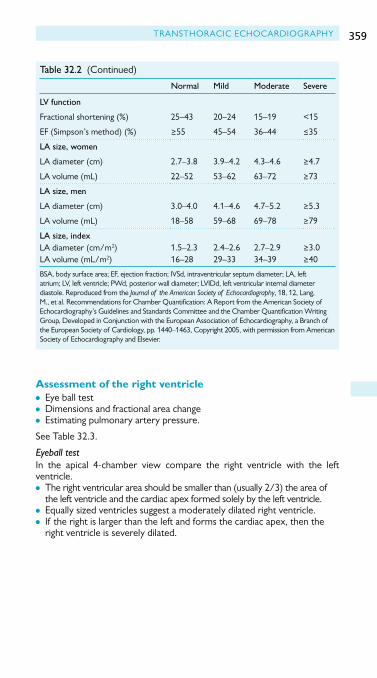
TRAnSTHORACICECHOCARDIOgRAPHy 359
Normal Mild Moderate Severe
LV function
Fractionalshortening (%)
EF(Simpson’smethod)(%)
25–43
≥55
20–24
45–54
5–9
36–44
<5
≤35
LA size, women
LAdiameter (cm)
LAvolume(mL)
2.7–3.8
22–52
3.9–4.2
53–62
4.3–4.6
63–72
≥4.7
≥73
LA size, men
LAdiameter (cm)
LAvolume(mL)
3.0–4.0
8–58
4.–4.6
59–68
4.7–5.2
69–78
≥5.3
≥79
LA size, indexLAdiameter(cm/m2)LAvolume(mL/m2)
.5–2.36–28
2.4–2.629–33
2.7–2.934–39
≥3.0≥40
BSA,bodysurfacearea;EF,ejectionfraction;IVSd,intraventricularseptumdiameter;LA,leftatrium;LV,leftventricle;PWd,posteriorwalldiameter;LVIDd,leftventricularinternaldiameterdiastole. reproduced from the Journal of the American Society of Echocardiography,8,2,Lang,M.,et al.RecommendationsforChamberQuantification: A ReportfromtheAmericanSocietyof Echocardiography’sguidelinesandStandardsCommitteeandtheChamberQuantificationWritinggroup,DevelopedinConjunctionwiththeEuropeanAssociationofEchocardiography,aBranchof theEuropeanSocietyofCardiology,pp. 440–463,Copyright2005,withpermissionfromAmericanSocietyofEchocardiographyandElsevier.
Table 32.2 (Continued)
Assessment of the right ventricle• eye ball test• Dimensions and fractional area change• estimating pulmonary artery pressure.
SeeTable 32.3.
Eyeball testIn the apical 4-chamber view compare the right ventricle with the leftventricle.• Therightventricularareashouldbesmallerthan(usually2/3)theareaof
the left ventricle and the cardiac apex formed solely by the left ventricle.• Equallysizedventriclessuggestamoderatelydilatedrightventricle.• If therightislargerthantheleftandformsthecardiacapex,thenthe
right ventricle is severely dilated.

360 ChapTer 32 Bedside echocardiography
Right ventricular dimensions
Figure 32.28 RVarea.Apical4-chamberview.EstimateRVfunction by measuring area in systole and diastole to calculate fractional area change.
Figure 32.27 RVlength.Apical4-chamberview.normallength7.–7.9cm.
Figure 32.26 BasaldiameterRV.Apical4-chamberview.normaldiameter 2–2.8 cm

TRAnSTHORACICECHOCARDIOgRAPHy 361
Estimating pulmonary artery pressure• useanapical4-chamberorparasternalSAXviewwithavisiblejetof
tricuspid regurgitation.• align CWD through the tricuspid valve regurgitation.• Measurethepeakvelocity.• The tricuspid pressure gradient is calculated from the peak velocity using
amodifiedBernoulliequation:
Pressure gradient = 4 peak velocity2×
• TheRVsystolicpressure,whichisequivalenttopulmonaryarterysystolicpressureintheabsenceof pulmonarystenosis,isequaltothesumof thetricuspidpressuregradientandrightatrialpressure (CVP).
Normal valuesNormal values can be found in the recommendations for chamber quan-tification: a report from the American Society of Echocardiography’sguidelines and Standards Committee and the Chamber QuantificationWritinggroup(seebFurtherreading,p. 36).
There is also an iphone application named echoCalc created by the BritishSocietyof Echocardiography.
Forsomeexamplesof importantpathologiesseeFig. 32.29.
Further readingLangRM,BierigM,DevereuxRB,Flachskampf FA,FosterE,PellikkaPA,et al. recommendations for
chamberquantification: a report fromtheAmericanSocietyof Echocardiography’sguidelinesandStandardsCommitteeandthechamberquantificationwritinggroup,developedinconjunc-tionwiththeEuropeanAssociationof Echocardiography,abranchof theEuropeanSocietyof Cardiology. J Am Soc Echocardiogr2005;8:40–63.
Table 32.3 right ventricular dimensions and function
Normal Mild Moderate Severe
RV dimensions (apical 4 chamber)BasalRVdiameter (cm)Basetoapexlength(cm)
2.0–2.87.–7.9
2.9–3.38.0–8.5
3.4–3.88.6–9.
≥3.9≥9.2
PA diameter (parasternal SAX)MainPA(cm) .5–2. 2.2–2.5 2.6–2.9 ≥3.0
RV areaRVdiastolicarea (cm2)RVsystolicarea(cm2)
–287.5–6
29–327–9
33–3720–22
≥38≥23
RV functionFractionalareachange(%) 32–60 25–3 8–24 ≤7
RV,rightventricle;PApulmonaryartery.ReproducedfromtheJournal of the American Society of Echocardiography,8,2,Lang,M.,et al.RecommendationsforChamberQuantification: A ReportfromtheAmericanSocietyof Echocardiography’sguidelinesandStandardsCommitteeandtheChamberQuantificationWritinggroup,DevelopedinConjunctionwiththeEuropeanAssociationof Echocardiography,aBranchof theEuropeanSocietyof Cardiology,pp. 440–463,Copyright2005,withpermissionfromAmericanSocietyof echocardiography and elsevier.

362 ChapTer 32 Bedside echocardiography
Pos 1: Pericardial e�usion
1
RV RV RV
LV LV
LVLVLV
LV
LALALA
LV
LA
RARA
RA
LALA
RA
RV
RVRV
RVRV
RARA
2 3
Pos 2: Pericardial e�usion Pos 2: Dilated RA + RV Pos 2: Dilated LA + LV
Pos 3: Pericardial e�usion Pos 3: Dilated RV Pos 3: Dilated LV + LA
Pos 3: Dilated LV
Post DC cardiac surgery, following cardiac catheterisation, trauma, renal failure, infection.Pulmonary embolus, RV infraction, pulmonary hypertension, volume overload.Ischemic heart disease, dilated cardiomyopathy, sepsis, volume overload, aorta insu�ciency.Aorta stenosis, arterial hypertension, LV out�ow tract obstruction,hypertrophic cardiomyopathy, myocardial deposits diseases.
PATHOLOGY TO BE CONSIDERED IN PARTICULAR:
Pos 3: Hypertrophy LV +Dilated LA
Pos 3: Hypertrophy LV
Pos 1: Dilated RA + RV
Important pathology
Pos 1: Dilated LA + LV
4 5 6
7 8 9
10 11 12
LV LV
LV
LALV
LV
LA
RV
RV
RV
RV
AO
AO
Figure 32.29 Importantpathologyexamples.ImagetakenfromFATE-cardandreproducedwithpermissionfromProfessorPh.D.MDScErikSloth.<http://www.usabcd.org>(FATE-appavailableforfreedownloadatAppStoreandAndroidMarket).

TRAnSOESOPHAgEALECHOCARDIOgRAPHy 363
Transoesophageal echocardiographyIntroductionThis section will provide an overview of the information that can beobtainedusingTOEinthesettingof theCICu.Themajorityof anyecho-cardiographic examination involves the interpretation of moving images. Therearemanyexamplesof theseavailableviatheInternetwhicharerec-ommended to the reader.
Risks and precautionsTOEisdescribedasan‘invasive,non-invasive’ investigationsincethere ismorbidityandindeedmortalitydescribedfromitsuse.Assuch,safetyisof extremeimportanceandthesonographermustreviewthepatient’shistorytoexcludeanycontraindicationtoTOEbeforeundertakingthestudy.Thepracticeguidelines forperioperativeTOErecommend ‘thatTOEmaybeusedforpatientswithoral,oesophagealorgastricdisease,if theexpectedbenefitoutweighsthepotentialrisk,providedtheappropriateprecautionsareapplied’.Theprecautionsincludeconsideringotherimagingtechniques(e.g.TTE),askingforanuppergIopinionandperhapsendoscopy,usingthesmallestavailableprobe,limitingtheexaminationandavoidingunnecessaryprobemanipulation,andusingthemostexperiencedoperator.
prior to the examination consideration must be given to analgesia and sedation,andthemonitoringof thepatient’svitalsignsmustbedelegatedtoanotherindividual,orthemonitorspositionedsuchthatthesonographerhasaclearviewof themduringtheprocedure.Thisisof particularimpor-tancesinceinthemajorityof casesTOEisbeingperformedtodeterminethe cause of postoperative haemodynamic instability.
The standard viewsTheASE/SCApracticeguidelinessuggest20standardviewstobeobtainedduringacomprehensiveTOEexam.Thereisnogenerallyacceptedorderinwhichtheseviewsareobtained.Fig. 32.30showstheviewswhichmostcommonlyproviderelevantinformationintheCICu,andtheabnormalitieswhichmaybedetected.
Specific indications for TOE in ICU.• Mitralvalveassessment• endocarditis• Embolismof unknown origin• Unexplained hypoxemia• pulmonary thromboembolism• aortic dissection.

364 ChapTer 32 Bedside echocardiography
Mitral valve assessmentThemitralvalveisimmediatelyclosetotheoesophagusandTOEprovidesexcellent quality images for the assessment of the mitral valve. This may be essential for determination of the mitral valve morphology and diagnosis of mechanismsof mitralregurgitation.TOEisalso invaluablefortheassess-mentof theprostheticmitralvalveormitralvalverepair.ThebestviewsareshowninFigs32.3,32.32,and32.33.
(r) desc aortic LAX
(n) TG RV in�ow(m) ME RVin�ow-out�ow
(q) desc aortic SAX
(i) ME AV LAX
(o) ME asc aortic SAX (p) ME asc aortic LAX
(t) UE aortic arch SAX(s) UE aortic arch LAX
(k) deep TG LAX (I) ME bicaval
(h) ME AV SAX
(j) TG LAX
(f ) TG basal SAX (g) ME mitral commissural(e) TG two chamber
(a) ME four chamber (b) ME two chamber (c) ME LAX (d) TG mid SAX
Figure 32.30 Thestandardviewsof acomprehensiveTOEexamination.ReproducedfromShanewiseet al.ASE/SCAguidelinesforPerformingaComprehensiveIntraoperativeMultiplaneTransesophagealEchocardiographyExamination: Recommendationsof theAmericanSocietyof EchocardiographyCouncilforIntraoperativeEchocardiographyandtheSocietyof CardiovascularAnesthesiologistsTaskForceforCertificationinPerioperativeTransesophagealechocardiography. Anesthesia & Analgesia,89,4,p. 870,copyright999,withpermissionfromInternationalAnesthesiaResearchSocietyandWoltersKluwer.

TRAnSOESOPHAgEALECHOCARDIOgRAPHy 365
LA
LV
RA
RV
RVfreewall
Lateral
Septal
AMLPML
Figure 32.3 Midoesophageal4-chamberview(MO4-chamber).Reproducedwithkind permission from Practical Perioperative Transoesophageal Echocardiography,DavidSidebotham,AlanMerry,MalcolmLegget,2003,Figure3.,p.40,Copyrighttheauthors.
LA
LV
AML PMLPML
Figure 32.32 Midoesophagealmitralcommissuralview(MOmitralcommissural)(P3onleftof display,Ponright,A2incentre).Reproducedwithkindpermissionfrom Practical Perioperative Transoesophageal Echocardiography,DavidSidebotham,AlanMerry,MalcolmLegget,2003,Figure3.3,p.4,Copyrighttheauthors.
LA
LV
AML NCC
Ao
Transversesinus
Oblique sinus
RCC
RVOT
LVOT
Figure 32.33 Midoesophagealaorticvalvelongaxisview(MOAVLAX).ThisviewmayrevealdynamicLVOTobstructionsecondarytosystolicanteriormotionofthemitralvalve(SAM).ThisuncommonbutextremelyimportantcauseofhypotensionfollowingcardiacsurgeryischaracterizedbytheanteriorleafletofthemitralvalveprolapsingintotheLVOTandperhapsevencoaptingwiththeinterventricularseptumwhichisusually hypertrophied. There is often associated mitral regurgitation and the velocity of this regurgitant jet may reveal that the pressure inside the left ventricle during systole is higherthanthesystemicbloodpressure.ReproducedwithkindpermissionfromPractical Perioperative Transoesophageal Echocardiography,DavidSidebotham,AlanMerry,MalcolmLegget,2003,Figure3.5,p.37,Copyrighttheauthors.

366 ChapTer 32 Bedside echocardiography
Unexplained hypoxaemiaShuntattheintra-atriallevelmayberevealedeitherasaresultof apatentforamenovaleoranatrialseptaldefect.Bubblecontrasttest(injectionof agitatedsalineintothecentralline)andCFMcanhelpdeterminetheposi-tionanddirectionof anyseptaldefect(Fig. 32.35).Thisimportanttesttocan help to exclude a cardiac cause of hypoxia post cardiac surgery.
Clotcompressionof theatriamayberevealedinthisview.Withdrawalof the central line should be considered if examination reveals the tip to be withintherightatrium.
Pulmonary thromboembolismClot may be seen in the main pulmonary artery in massive pulmonary embo-lism.This is best seen in theMOascending aorta SAXview (Fig. 32.36);however,therightmainbronchusoftenpartiallyobscuresthis view.
Possible findings in endocarditis
Embolism of unknown originTOEissuperiortoTTEintheexaminationof theleftatriumandparticularlytheexaminationof theleftatrialappendagewhichisacommonsourceof arterialembolism.ThisisbestseenintheMO2-chamberview.Alternativeembolic sources can be seen such as left-sided valve vegetations or intracar-diacclotortumour(LVor LA).
LA
LV
LAACS
Inferior Anterior
AMLPML
Figure 32.34 Midoesophageal2-chamberview(MO2-chamber).Reproducedwithkind permission from Practical Perioperative Transoesophageal Echocardiography,DavidSidebotham,AlanMerry,MalcolmLegget,2003,Figure3.4,p.4,Copyrighttheauthors.
• Vegetationson valves• aortic root abscess• Perforatedordestroyedleafletsof any valve• Dehisced mechanical valve• Mechanicalorbioprostheticvalvedysfunction• Fistulabetweenchambers.

TRAnSOESOPHAgEALECHOCARDIOgRAPHy 367
Aortic dissectionTheassessmentof aorticdissectionisbestdonewithTOEratherthanTTE.OftenthepatientwillhavealreadyhadacontrastCTscan.A fullassess-ment of the ascending arch and descending aorta should be performed. Classicallyanendothelialflapwillberevealedintheascendingaortaintypedissection.Inaddition,aorticregurgitationandpericardialhaemopericar-diummayberevealed.Incaseof dissectionintoacoronaryartery,evidenceof awall-motionabnormalitywillbepresent.
Inadditiontothemidoesophagealaorticvalvelong-axisview(MOAVLAX)seenearlier(Fig. 32.33)theviewsinFigs32.37,32.38,and32.39maybe useful.
LA
IVC SVC
RARA appendage
Eustachian valve
Fossa ovalis
Figure 32.35 Midoesophagealbicavalview(MObicaval).Reproducedwithkindpermission from Practical Perioperative Transoesophageal Echocardiography,DavidSidebotham,AlanMerry,MalcolmLegget,2003,Figure3.9,p.38,Copyrighttheauthors.
RPA
MPAAorta
SVC
Figure 32.36 Midoesophagealascendingaorticshort-axisview(MOasc.aortic SAX).ReproducedwithkindpermissionfromPractical Perioperative Transoesophageal Echocardiography,DavidSidebotham,AlanMerry,MalcolmLegget,2003,Figure3.6,p.37,Copyrighttheauthors.

368 ChapTer 32 Bedside echocardiography
LA
RA
RVOT
NCC
LCC
RCC
Figure 32.37 Midoesophagealaorticvalveshort-axisview(MOAVSAX).TheMOAVSAXviewisagoodstartingpointsincetheaorticvalveisofteneasilyidentifiedbytheMercedesBenzsignwhenintheclosedposition.Theleftatriumliesclosesttotheprobeandisdisplayedatthetopof thescreen.Thisviewoffersclearimagesof theleftandrightatriaandtheaorticvalve.TheRVOTisoftenlesswellseensinceitismoredistantfromthetransducerandanycalcificationintheaorticvalvewillcauseultrasounddropout.Compressionof theleftorrightatriabybloodclotwhichisoftenenoughtoleadtopostcardiotomytamponademaybeseeninthisview.Themorphologyof theaorticvalveis seen.ReproducedwithkindpermissionfromPractical Perioperative Transoesophageal Echocardiography, DavidSidebotham,AlanMerry,MalcolmLegget,2003,Figure3.4,p.37,Copyrighttheauthors.
RPA
Asc Ao
Figure 32.38 Midoesophagealascendingaorticlong-axisview(MOasc.aortic LAX).ReproducedwithkindpermissionfromPractical Perioperative Transoesophageal Echocardiography,DavidSidebotham,AlanMerry,MalcolmLegget,2003,Figure3.7,p.36,Copyrighttheauthors.

TRAnSOESOPHAgEALECHOCARDIOgRAPHy 369
Assessment of ventricular function with TOELeft ventricleThetransgastricmidSAXviewisausefulviewfortheassessmentoftheLVfunction.Myocardiumsuppliedbyall threemaincoronaryarteriesareseen.Theviewiscommonlyreferredtoasthebouncingdoughnutview(Fig. 32.39)!
Right ventricleright ventricular systolic function and dimensions can be assessed in this view (Fig. 32.40).Dilatationof the right ventricle isoftenassociatedwithtricuspid regurgitation and determination of the peak velocity of the regurgi-tantjetacrossthetricuspidvalvewillprovidetherightventricularandthere-forethepulmonaryarterysystolicpressure.(RVsystolicpressure=4 ×Vmax2+CVP.)Clotcompressionof theRVmaybeseenbutitisimportanttorememberthattheclassicsignsof tamponade(associatedwithpericardialeffusion)areoftenabsentinthepostcardiacsurgerysettingwith‘regionaltamponade’orlocalizedclotcollectionacommonfinding.
Anterior
LVRV
PosteriorSeptal
Inferior
LateralAntero-septal
AL
PM
Figure 32.39 Transgastricmidpapillaryshortaxisview(Tgmid SAX).ReproducedwithkindpermissionfromPractical Perioperative Transoesophageal Echocardiography,DavidSidebotham,AlanMerry,MalcolmLegget,2003,Figure3.8,p.44,Copyrightthe authors.

370 ChapTer 32 Bedside echocardiography
Clotcompressionof theRVmaybeseenbutitisimportanttorememberthattheclassicsignsof tamponade(associatedwithpericardialeffusion)areoftenabsentinthepostcardiacsurgerysettingwith‘regionaltampon-ade’orlocalizedclotcollectionacommonfinding.
Inferior RV free wall
LA
RA
MPA
RVOTAP
Figure 32.40 Midoesophagealrightventricularinflowoutflowview(MORVinflow-outflow).ReproducedwithkindpermissionfromPractical Perioperative Transoesophageal Echocardiography,DavidSidebotham,AlanMerry,MalcolmLegget,2003,Figure3.8,p.38,Copyrighttheauthors.

371
Index
Aabbreviated scanning
protocols 345–6acetazolamide 284acid–base biochemistry 58, 60
blood gas analysis 6–3mixed effects 63Stewart hypothesis 60
activated clotting time (ACT) 64
Actrapid® 69, 73, 20acute abdomen,
investigation 32–3acute cardiology 239–49acute kidney injury
(AKI) 6–23Acute Kidney Injury
Network (AKIN) 8acute lung injury
(ALI) 08–2post-thoracotomy 77–8respiratory failure,
postoperative 75acute respiratory dis-
tress syndrome (ARDS) 08–2
inflammatory response 50ventilator-induced lung
injury 280adrenaline
circulatory support 289contraindications 27extubation failure 273lactate 64myocardial oxygen
supply 34right ventricular failure 89routine patients 9tracheostomy, bleeding
from a 270upper gastrointestinal
bleeding 34adults with congenital
heart disease (ACHD) 87–200, 205
pregnancy 205afterload reduction (right ven-
tricular failure) 89, 92–3airway management 263–74alcohol hand gel 33alemtuzumab 238alfentanil 37allergies, and antimicrobial
therapy 334
American Heart Association (AHA) 36, 240–
amikacin 337aminoglycosides 337amiodarone 96, 99
atrial fibrillation 69, 05heart failure resistant
to standard medical therapy 28, 29
ventricular tachyarrhythmias 06
amlodipine 70, 28amoxicillin 245, 335anaemia 60anaesthesia
aortic dissection 248pregnancy 206
analgesia see pain reliefannuloplasty ring 02, 03anomalous left coronary
artery from pulmonary artery (ALCAPA) 89
antibiotic prophylaxis 68, 69
cardiac transplantation 23endocarditis 245thoracic patients 73upper gastrointestinal
bleeding 34antibiotics 334–8anticoagulation
heparin-induced thrombocytopenia 55
renal replacement therapy 22
stroke 46antiemesis 0, 68, 69antifibrinolytics 6, 62antimicrobial prescribing/
therapy 33, 334–8antiplatelet therapy 46, 58antiproliferative
immunosuppressants 237antipsychotics 38antitachycardia modes,
epicardial pacing 308antithymocyte globulin
(ATG) 232, 238aortic balloon valvuloplasty,
percutaneous 96aortic dissection 246–9
echocardiography 367, 368–9
aortic regurgitation (AR)acute 98–9
chronic 97–8functional 99
aortic root reconstruction 99
aortic stenosis (AS) 95–6aortic valve implantation,
transcatheter 96aortic valve
replacement (AVR)aortic regurgitation
97, 98, 99aortic stenosis 95, 96
aortography 248APACHE II score 5apical drains 79apical 4-chamber echocardi-
ography 349, 350apical 5-chamber
echocardiography 350apomorphine 72aprotinin 43, 6ARDSnet
recommendations 0argatroban 55arrhythmias
adults with congenital heart disease 96, 99, 200
cardiac transplantation patients 229
heart failure resistant to standard medical therapy 29–20
prophylaxis 69see also bradyarrhythmias;
tachydysrhythmiaaspirin
coagulopathies 58coronary graft
thromboprophylaxis 70
myocardial ischaemia 37platelet function tests 67valve
thromboprophylaxis 7assist control ventilation
(ACV) 276asthma 73atelectrauma 280atrial fibrillation (AF) 05
heart failure resistant to standard medical therapy 29
prophylaxis 69, 05atrial flutter 308

index372 index
atrial septal defect (ASD) 89
atrial tachyarrhythmias 96atrioventricular junctional
tachycardia 308atrioventricular septal defect
(AVSD) 89atropine 04autologous transfusion 60azathioprine 237azithromycin 338
Bbalanced circulation 89barotrauma 279basal drains 79basiliximab 238benzylpenicillin 335Beriplex® 59β-blockers 220β-lactam antibiotics 334bi-level positive airway
pressure (BIPAP) 73, 277
biotrauma 280bisoprolol 70bivalirudin 55, 59biventricular assist devices
(BiVADs) 222Blalock–Taussig shunt 89bleeding 40
cardiac tamponade 46–7cardiac transplantation
patients 229coagulopathies 58concealed 45decreasing intra- and
postoperative blood loss 60
excessive 40, 42gastrointestinal 34–5management 43normal postoperative
patient 6overt 44thoracotomy wounds 327from a tracheostomy 270
blocked tracheostomy tube 268
blood conservation 60–3blood gas analysis 6–3
point-of-care testing 64blood glucose see glucoseblood loss, decreasing intra-
and postoperatively 60blood transfusion 44bradyarrhythmias 04
pacing 04pharmacological
management 04bronchoalveolar lavage 235bronchodilators 73
bronchopleural fistula (BPF) 83
Ccalcineurin inhibitors 237calibrated pulse contour
analysis 260carbamazepine 72carbapenems 336cardiac arrest,
out-of-hospital (OOHCA) 240–2
cardiac depressants, poten-tially reversible 22
cardiac failure see heart failure
cardiac function, normal postoperative cardiac patient 5
cardiac intensive care unit (CICU)
nutrition 36–8obstetric patient 207
cardiac output 84cardiac recovery unit
concept 22, 23cardiac resynchronization
therapy (CRT) 97Cardiac Surgery Advanced
Life Support (CALS) 26–7, 28–9
cardiac surgery prognostic models 8, 9, 20
cardiac tamponade 46–7, 53cardiac transplantation (CTx)
228–32, 232adults with congenital
heart disease 98heart failure resistant
to standard medical therapy 222
heart–lung transplantation 236
cardiogenic pulmonary oedema 28
cardiogenic shock 37cardioPAT 62cardiopulmonary
bypass (CPB)aortic dissection 248, 249bleeding 40coagulopathies 58, 59effects 5–6gastrointestinal
complications 28heparin–protamine
mismatch 58inflammatory
response 50stroke prophylaxis
46, 47cardiovascular drugs 73
cardioversion see synchro-nized DC cardioversion
catecholamines 288–9Caves, Philip 23cefepime 335cefotaxime 335ceftazidime 335, 336ceftobiprole 335ceftriaxone 334, 335, 336cefuroxime 335, 336cell salvage 6central venous
cannulation 254–5CentriMag® 296, 297cephalexin 335cephalosporins 332, 335CESAR trial 2, 300, 30chest drainage 72, 79–82chest X-ray (CXR)
aortic dissection 247heart failure resistant
to standard medical therapy 24, 25
lung transplantation 234childbirth 206children 276, 300chlorhexidine 4cholecystitis, acute 30chronic kidney disease
(CKD) 24–5chronic obstructive pulmo-
nary disease (COPD)lung transplantation 233oxygen therapy 72respiratory
management 28ciclosporin 237ciprofloxacin 332, 337circulatory support
287–302circumflex artery branch
of left coronary artery 33, 35
clarithromycin 338clindamycin 332Clinisan Advance™ 332clonazepam 24clonidine 72, 43, 38clopidogrel 70, 58, 67Clostridium difficile infection
(CDI) 33–2coagulation monitors 64coagulopathies 58–9co-amoxiclav 332, 335co-codamol 320colour flow mapping (CFM)
344, 345communication 76, 79
end of life 223compartment syndrome 326computed tomography
(CT) 247concordance 89

index 373index
Confusion Assessment Method for the ICU (CAM-ICU) 4, 42, 35
congenital heart disease, adults with (ACHD) 87–200, 205
pregnancy 205congenitally corrected
transposition of the great arteries (ccTGA) 89, 92, 99
continuous positive airway pressure (CPAP) 73, 28, 28–2
continuous renal replace-ment therapy (CRRT) 2, 2, 22, 22
continuous veno-venous haemofiltration (CVVH) 2
continuous wave Doppler (CWD) echocardiogra-phy 344, 345
contrast-induced nephropathy 6
cooling during cardiopulmo-nary bypass 5
Cooper, Joel 233coronary angiography 37coronary artery, left
circumflex artery branch 33, 35
left anterior descending (LAD) branch 33, 35
coronary artery bypass graft (CABG) 44, 46
coronary bypass surgery 70coronary graft
spasm 70thrombosis 70
corticosteroidsacute lung injury/acute
respiratory distress syndrome 2
preoperative 72transplantation 237
creatinine 7cyanosis 96cyclosporine 237cystic fibrosis (CF) 233
Ddaclizumab 238dalteparin 69Damus–Kaye–Stansel
procedure 89danaparoid 55, 59DC cardioversion see
synchronized DC cardioversion
DDAVP® 6
DeBakey classification of aortic dissection 246, 247
decannulation 27decolonization therapy,
MRSA 332decompensated heart failure
(DHF) 209–25deep hypothermic
circulatory arrest (DHCA) 249
deep vein thrombosis (DVT)heparin-induced
thrombocytopenia 56prophylaxis 69, 4
delirium 40–3dexamethasone 273dexmedetomidine 42,
43, 38diabetes mellitus 73digoxin 29, 05diltiazem 28discordance 89disequilibrium
syndrome 23displaced tracheostomy
tube 268diuretics 224dobutamine
aortic regurgitation 98cardiac transplantation
patients 230circulatory support 288heart failure resistant
to standard medical therapy 28
myocardial oxygen supply 34
right ventricular failure 9dopamine
acute kidney injury 6aortic regurgitation 98cardiac transplantation
patients 230circulatory support 289right ventricular failure 9routine patients 9
Doppler echocardiography 344, 345
double inlet ventricle 89double outlet ventricle 90dysaesthesia 326dysrhythmia 53
Eechocardiography
bedside 339–69cardiac tamponade 47heart failure resistant
to standard medical therapy 24
modes 343–4, 345
myocardial ischaemia or infarction 34
windows 342, 343see also transoesophageal
echocardiogra-phy; transthoracic echocardiography
ECLS Registry Report 300Eisenmenger syndrome
90, 97pregnancy 205
elderly patients 37electrocardiography (ECG)
aortic dissection 247heart failure resistant
to standard medical therapy 24
hyperkalaemia 9myocardial ischaemia 33right ventricle 88
electroencephalography (EEG) 35
electrolytespostoperative 68, 69routine patients 9
electronic chest drainage systems 8
embolism of unknown origin 366
emphysema 85end of life 223endocarditis 243–5
echocardiography 366prophylaxis 245
endomyocardial biopsycardiac allograft
rejection 23heart failure resistant
to standard medical therapy 25, 26
endotracheal tube (ETT)extubation 9, 272–4intubation 264–5
enhanced recovery (ER) 24
enoximone 290enteral nutrition 36–7epicardial pacing 303–epilepsy 72epoprostenol 89, 92, 93ertapenem 336erythromycin 34, 36, 338erythropoietin 60ESCAPE trial 24esmolol 29European Association for
Cardio-Thoracic Surgery (EACTS) guidelines 26
European Resuscitation Council (ERC) 26
European Society of Cardiology (ESC) 36, 247

index374 index
EuroSCORE 8, 9everolimus 238extracorporeal circuit 59extracorporeal CO2
removal 2extracorporeal life support
294, 299–30extracorporeal lung
support (ECLS) 2, 30–2
extracorporeal membrane oxygenation (ECMO)
acute lung injury/acute respiratory distress syndrome 2
adults with congenital heart disease 98
heart failure resistant to standard medical therapy 22
extubation 9, 272–4
FFallot’s tetralogy 9,
92, 99fast-tracking low-risk
patients 2–4felodipine 28femoral vein
cannulation 255fenestration 90fibrosing alveolitis 0FiO2
(fraction of inspired oxygen) 0,
flecainide 309Flolan® 93flucloxacillin 335, 336
allergy 334endocarditis 244postoperative 69
fluid balance 5fluid management
cardiac transplantation patients 229
routine patients 9thoracic patients 73
focus-assessed transtho-racic echocardiography (FATE) 346
fondaparinux 55Fontan procedure 90,
94, 2004 Ts score, heparin-induced
thrombocytopenia 53fraction of inspired oxygen
(FiO2) 0, Framingham risk factors
25functional capacity 35furosemide 6, 28, 224
Ggabapentin 72gastrointestinal
complications 27–38bleeding 34–5
general anaesthesia, obstet-ric patients 206
gentamicin 336, 337endocarditis 244, 245MRSA decolonization
therapy 332postoperative 69and vancomycin 336
Glenn connection 90, 95glucose
metabolism 58, 65out-of-hospital cardiac
arrest 240parenteral nutrition 37postoperative
control 68, 69glyceryl trinitrate (GTN)
aortic dissection 248aortic regurgitation 98circulatory support 292coronary graft spasm
prevention 70heart failure resistant
to standard medical therapy 28
myocardial ischaemia 37vasoplegic syndrome 55
glycopeptides 336graft dysfunction/failure/
rejectioncardiac transplantation
23–2lung transplantation 234–5
Hhaematology 57–67haematoma 327haemodialysis 2haemodilution
acute normovolaemic 6coagulopathies 58decreasing by pump
prime 62–3haemodynamic
optimization 240haemoglobin
analysers 64increasing preoperative 60
haemoptysis 97haemorrhage see bleedinghaemostatic agents 62haloperidol 43, 38hand hygiene 330–handover 75–9
Hardy, James 233heart dysfunction 83–06heart failure
adults with congenital heart disease 97
differential diagnosis 2echocardiography 354resistant to standard medi-
cal therapy 209–25heart transplantation 228–32
adults with congenital heart disease 98
heart failure resistant to standard medical therapy 222
heart–lung transplantation 236
HeartMate II® 298hemitruncus 90Henderson–Hasselbalch
(bicarbonate-centred) framework 60
heparinbleeding 40extracorporeal lung
support 302heart failure resistant
to standard medical therapy 28
point-of-care testing 64postoperative 69and protamine,
mismatch 58renal replacement therapy
22, 23short-term VADs 296venous thromboembolism
prophylaxis 73heparin-induced throm-
bocytopenia (HIT) 52–6, 59
with thrombosis (HITT) 56
Hepcon® 64HiBiSCRUB® 332high-frequency oscillatory
ventilation (HFOV) , 280
hospital-acquired infections (HAIs) 329–38
hydralazine 248hydrocortisone 73hydrogen ion concentra-
tion see acid–base biochemistry
hyperamylasemia 30hyperbilirubinaemia 30hyperglycaemia,
postoperative 68hyperkalaemia 9hypnotics 37–8

index 375index
hypokalaemia, postoperative 68
hypomagnesaemia, postoperative 68
hypotension 50diagnosis 5echocardiography 353–4management 5–2reversible causes 53–5
hypothermia, therapeutic 24, 242
hypovolaemiaechocardiography 353hypotension 53
hypoxaemia, unexplained 366, 367
Iidiopathic pulmonary
fibrosis (IPF) 233iloprost 92, 89, 93imipenem 336immune system
complications 49–56immunosuppression
235, 237–8induction therapy 237, 238infection control and
prevention 329–38inferior vena cava echocardi-
ography 352 inflammatory response
5, 50–influenza 332information handover and
care planning 75–9inotropes 9, 288–9INR 7insulin 73intensive care unit (ICU)
admission 77discharge 78see also cardiac intensive
care unitintercostal drains (ICDs)
79, 8intermittent renal replace-
ment therapy (IRRT) 2, 2, 22, 22
internal jugular vein (IJV) cannulation 255
International Liaison Committee on Resuscitation (ILCOR) 24
international normalized ratio (INR) 7
intra-aortic balloon pump (IABP)
bleeding, concealed 45
counterpulsation 294, 295
heart failure resistant to standard medical therapy 22
limitations 294myocardial ischaemia 37weaning 225
intrapleural drains 79intubation 264–5invasive monitoring 98ipratropium 73ischaemia
bowel 28radial artery harvest
site 326isoprenaline 04
JJehovah’s Witnesses
60, 62
KKaltostat® 270ketamine 38Kidney Diseases: Improving
Global Outcomes (KDIGO) staging
acute kidney injury 8chronic kidney disease
25kidney dysfunction 5–25Konno procedure 90
Llabetalol 248lactate metabolism 58, 65lamotrigine 72learning difficulties 88left anterior descending
(LAD) branch of left coronary artery 33, 35
left coronary arterycircumflex artery
branch 33, 35left anterior descending
(LAD) branch 33, 35left ventricle (LV)
anatomy 84dimensions and
function 358failure 84–5, 273functional assessment 84physiology 84transoesophageal
echocardiography 369transthoracic echocardiog-
raphy 354–7, 358
ventricular interdependence 86
left ventricular assist devices (LVADs) 222
left ventricular outflow tract (LVOT) obstruction 54
leg wounds 325lepirudin 55levetiracetam 72Levitronix CentreMag™ 92levodopa 72levofloxacin 337, 338levosimendan 200, 29Levotronix CentriMag®
296, 297lidocaine 309liver failure 30low cardiac output
syndrome 54low-molecular-weight
heparin (LMWH)heart failure resistant
to standard medical therapy 28
heparin-induced thrombocytopenia 52
postoperative 69venous thromboembolism
prophylaxis 73low-risk patients,
fast-tracking 2–4lower gastrointestinal
bleeding 35lung dysfunction 07–4lung-protective
strategies 0ventilation 280
lung transplantation (LTx) 233–5
heart–lung transplantation 236
lung volume reduction sur-gery (LVRS) 85
MM-mode echocardiography
344, 345macrolides 338magnesium sulphate 69magnetic resonance imaging
(MRI) 247mannitol 5maximum surgical blood
ordering schedule (MSBOS) 63
mechanical support, right ventricular failure 92
mechanical ventilationmodes 276weaning 283–5

index376 index
mediastinal drains 79mediastinitis 322, 323medications
postoperative 68–7preoperative 72–3
meperidine see pethidinemeropenem 336mesenteric ischaemia 28mesenteric venous
thrombosis 29metabolic acidosis 6, 62, 63metabolic alkalosis 6metformin 73, 6methylene blue 55, 50methylprednisolone
232, 237meticillin 335meticillin-resistant
Staphylococcus aureus (MRSA) 332
metoclopramide 72, 36metronidazole 332, 336midazolam 37mild hand ischaemia 326milrinone
cardiac transplantation patients 230
cardiogenic shock 37circulatory support 290heart failure resistant
to standard medical therapy 28
right ventricular failure 9vasoplegic syndrome 55
mini bypass 62minimally invasive cardiac
output monitoring 260–
minitracheostomy 73mitral regurgitation (MR)
functional 02organic 00–
mitral stenosis (MS) 99–00
mitral valve disease 94mitral valve echocardiogra-
phy 364, 365mitral valve prolapse
(MVP) 0–2mitral valve repair (MVR)
00, 0mobile pleural drainage 82mobilization of thoracic
patients 73monomurab-CD3 232, 238morphine 69, 37, 39
heart failure resistant to standard medical therapy 28, 224
motion (M-mode) echocar-diography 344, 345
moxifloxacin 337, 338
MRSA (meticillin-resistant Staphylococcus aureus) 332
Multiplate® 67mupirocin 323, 332Murray Lung Injury
Score 08Mustard procedure 90,
93, 99myasthenia gravis 72mycophenolate mofetil
(MMF) 235, 237myocardial infarction
(MI) 32diagnosis 34, 36management 37right ventricular failure 93type 5 (post cardiac
surgery) 36myocardial ischaemia 32
diagnosis 33–4hypotension 53management 37
myocardial stunting 93, 230myocardium
coronary arteries 35oxygen supply 33
Nnausea and vomiting
0, 68, 69near-patient testing 63neonates 300neostigmine 72nervous system
complications 39–47Neupro® 72neurally adjusted ventilator
assist (NAVA) 278neuroleptic malignant
syndrome 72neurological complications
of cardiac surgery 44–7nifedipine 28nitric oxide
acute lung injury/acute respiratory distress syndrome 2
cardiac transplantation patients 230
circulatory support 293inflammatory
response 50right ventricular failure
89, 92, 93NMDA-antagonists 38non-calibrated pulse contour
analysis 260non-invasive ventilation
(NIV) 28–2extubation failure 274
non-occlusive mesenteric ischaemia (NOMI) 29
noradrenalinecirculatory support
288, 29extracorporeal lung
support 302post-thoracotomy
acute lung injury, prevention 78
right ventricular failure 9routine patients 9vasoplegic syndrome 55
norfloxacin 337normal postoperative car-
diac patient 3–Norwood procedure 90Novalung® 30NovoSeven® 6nutrition
in the CICU 36–8thoracic patients 74
Oobstetric patients 20–7occlusive mesenteric
ischaemia 29Octaplex® 59oesophageal Doppler 26ofloxacin 338, 337olanzapine 43omeprazole 34ondansetron 0, 69, 73opioids 37organ failure scores 7oxycodone 39oxygen therapy
adults with congenital heart disease 98
heart failure resistant to standard medical therapy 224
thoracic patients 72
Ppacing, epicardial 303–pacing modes 306paediatrics 276, 300pain 5pain relief 33–20
clinical protocols 39–20patient assessment 35–6pharmacology 37–8postoperative 69principles 34routine patients 0thoracic patient 72
palliative care 223pancreatitis, acute 30papaverine 29

index 377index
paracetamol 0, 69, 72, 39parasternal long axis echo-
cardiography 346, 347parasternal short axis
echocardiographyaortic 347, 348mid-papillary 348–9mitral 348
parenteral nutrition (PN) 37
Parkinson’s disease 72paroxysmal re-entrant
supraventricular tachycardia 308
Parsonnet score 8, 20partial pressure of carbon
dioxide (PCO2) 6patient-controlled
analgesia 72penicillins 335
allergy 334endocarditis 245
peptic ulcer prophylaxis 4
percutaneous coronary intervention (PCI)
myocardial ischaemia 37out-of-hospital cardiac
arrest 240perfusion pressure 50pethidine (meperidine) 9,
6, 68, 69, 37PFA-00® 67pH 60phenylephrine 9, 206phenytoin 72, 24phosphodiesterase
inhibitors 290physiotherapy 73piperacillin 335platelet damage 58platelet function tests 67platelet transfusion 55pleural drains 79pleural
echocardiography 353pneumonia,
ventilator-associated (VAP) 3–4
pneumothorax 79, 80, 8point-of-care testing 64–7positive end-expiratory
pressure (PEEP) 0, positive pressure
ventilation 276–80post-perfusion
syndrome 40post-pneumectomy pulmo-
nary oedema 77–8potassium chloride 69Pott shunt 90prednisolone 73, 237
pregnancy 20–7preload
hypotension 5optimization 89
pressure control ventilation (PCV) 276, 277
pressure support ventilation (PSV) 276, 277
prochlorperazine 0prognostic models 4–20prone positioning in acute
lung injury/acute respiratory distress syndrome
propafenone 309propofol
central venous cannulation 254
parenteral nutrition 37postoperative 69sedation and pain relief
37, 39propofol infusion syndrome
(PIS) 37prosthetic valves 245protamine
bleeding management 43heparin–protamine
mismatch 58point-of-care testing 64renal replacement
therapy 22psychiatric disorders 73pulmonary artery catheter
(PAC) 88, 256–9pulmonary effects of surgery
and cardiopulmonary bypass 5
pulmonary embolism (PE) 94pulmonary
hypertension 92, 94pulmonary oedema 28pulmonary thromboembo-
lism 366, 367pulmonary valve
disorders 03pulse contour analysis 260pulsed wave Doppler
(PWD) echocardiogra-phy 344, 345
pump-head syndrome 40pyridostigmine 72
Qquetiapine 43quinolones 337–8
Rradial artery harvest site 326ramipril 70
ranitidine 69rapamycin inhibitors 238Rastelli operation 9recovery room
concept 22, 23recruitment manoeuvres
(RM) red man syndrome 336regional anaesthesia, obstet-
ric patients 206Reitz, Bruce 236remifentail 28, 37, 39renal disease 7renal dose dopamine 6renal failure 7renal replacement therapy
(RRT) 7, 2–3respiratory acidosis 6, 63respiratory alkalosis 6respiratory diseases 72respiratory dysfunction
273respiratory failure,
postoperative 75respiratory
management 275–85resuscitation in the CICU
25–7, 28–9re-warming 5
respiratory acidosis 6routine 9
Richmond Agitation Sedation Scale (RASS) 4, 35
rifampicin 245right coronary artery
(RCA) 33, 35right heart
catheterization 24right ventricle (RV)
anatomy 86dilatation 87dimensions and function
360, 36failure 88–94, 230functional assessment 87physiology 86–7surrogate markers of
contractility 88transoesophageal
echocardiography 369, 370
transthoracic echocardiog-raphy 359–6, 362
ventricular interdependence 86
right ventricle–pulmonary artery conduit 89, 92
right ventricular assist devices (RVADs) 92
risk prediction models 4–20
risperidone 43

index378 index
Ross procedure 9ROTEM® 65, 66rotigotine 72
Ssalbutamol 20, 73, 78saphenous vein site 325SBAR tool 76, 78, 79sedation 33–20
clinical protocols 39patient assessment
35–6pharmacology 37–8postoperative 69principles 34
seizures 24Senning procedure 9, 99sensing threshold, epicardial
pacing 30sepsis 8, 50septic screen 334Serious Hazards of
Transfusion (SHOT) 60severe acute respiratory syn-
drome (SARS) 09severity of illness
scores 4–20shift handover 76, 78, 79shivering 6, 68, 69sildenafil
cardiac transplantation patients 230
right ventricular failure 89, 92, 93
Simpson’s method, left ventricle assessment 356, 357
simvastatin 70single chamber pacing
modes, epicardial pacing 306
single ventricle 9sirolimus 238sodium nitroprusside (SNP)
248, 292–3sodium valproate 72, 24sotalol 309spirometry 234spontaneous breathing
trials 283standard base excess
(SBE) 6standard infection control
precautions (SICPs) 330Stanford classification
of aortic dissection 246, 247
step downanalgesia 320heart failure resistant
to standard medical therapy 225
sternal wounds 322–4Stewart (strong ion)
hypothesis 60stimulation threshold, epi-
cardial pacing 30straddling 9stress ulceration
prophylaxis 68, 69upper gastrointestinal
bleeding 34stridor 273stroke 44–7subaortic obstruction 96–7subclavian aortoplasty 9subclavian vein (SCV)
cannulation 255subcostal echocardiography
35, 360superior vena cava (SVC),
left 90supraventricular
tachycardia (SVT)failed reversion with
rapid ventricular response 308
paroxysmal re-entrant 308surgery, effects of 5–6surgical hand
disinfection 33suxamethonium 266, 274switch operation 9,
94, 99synchronized DC
cardioversionatrial fibrillation 05ventricular tachycardia 06
synchronized intermittent mandatory ventilation (SIMV) 276, 277
Syntocinon® 206systemic inflammatory
response syndrome (SIRS) 50
Ttachydysrhythmia 53
ventricular tachyarrhythmias 06
see also atrial fibrillationtacrolimus 235, 237tamponade, cardiac 46–7, 53tazobactam 335Tazocin® 335TEG® 65teicoplanin 336temperature control 24tension pneumothorax 55tetralogy of Fallot 9,
92, 99thiopentone 46, 266thoracic airway
obstruction 84
thoracic bioimpedance measurement 260
thoracic patient 7–85thoracic surgical drainage
system 8thoracotomy 72, 77–8
wounds 327Thoratec HeartMate II® 298thrombocytopenia,
heparin-induced 52–6thromboelastography 65thromboelastometry 65thromboembolic disease 94thrombosis 52tidal volume ventilation 0time-triggered
ventilation 276tirofiban 58tobramycin 337topiramate 72torsade de point 06total cavopulmonary
connection (TCPC) 9, 95, 200
total parenteral nutrition (TPN) 254
tracheostomy 266–7tramadol 73, 320tranexamic acid 6, 62transbronchial lung biopsy
(TBLB) 235transcutaneous pacing 04transfusion 44
algorithms 63optimizing practice 63triggers 63
transfusion-related acute lung injury (TRALI) 09
transmission-based precau-tions (TBPs) 330
transoesophageal echocardiography 363–9
aortic dissection 248indications 34, 363versus transthoracic
echocardiography 340transplant patients 227–38transposition of the great
arteriesadults with congenital heart
disease 9, 93, 99congenitally connected
(ccTGA) 89, 92, 99
transthoracic echocardiogra-phy (TTE) 342–62
indications 34versus transoesophageal
echocardiography 340transvenous pacing 04tricuspid annular plane
systolic excursion (TAPSE) 88
index 379index
tricuspid balloon valvuloplasty 03
tricuspid regurgitation (TR) 02–3
tricuspid stenosis (TS) 03troponin 36truncus arteriosus 9two-dimensional
echocardiography 344, 345
Uunfractionated heparin
52, 296upper gastrointestinal
bleeding 34–5urine output 5
Vvacuum-assisted closure
(VAC) 323, 324valve heart disease 95–03valve repair or
replacement 53valve surgery
medication following 7stroke following 44target INR following 7
vancomycin 336Clostridium difficile
infection 332endocarditis 244, 245
vasodilatation 353vasodilators 292–3vasoplegia 50–vasoplegic syndrome 54–5vasopressin
circulatory support 292
right ventricular failure 9vasoplegic syndrome 55
vasopressors 9, 29–2veno-arterial extracorporeal
membrane oxygenation (V-A ECMO) 299–300
veno-venous extracorporeal membrane oxygena-tion (V-V ECMO) 299, 300, 30
venous thromboembolism (VTE) prophylaxis 73
ventilationout-of-hospital cardiac
arrest 240routine 9
ventilator-associated pneu-monia (VAP) 3–4
ventilator-induced lung injury (VILI) 276, 279
ventricular arrhythmias 220
ventricular assist devices (VADs) 294
adults with congenital heart disease 98
heart failure resistant to standard medical therapy 22, 222
left (LVADs) 222long-term 298–9right (RVADs) 92short-term 296–7
ventricular dysfunction 200
ventricular fibrillation (VF) 06
ventricular function 369ventricular
tachyarrhythmias 06
ventricular tachycardia (VT) 06
epicardial pacing 308verapamil 29, 309VerifyNow® 67Vigileo™ 260viscoelastic tests 65, 66Visual Analogue Scale
(VAS) 35vitamin K antagonists 54volume controlled ventila-
tion (VCV) 276, 277volutrauma 279vomiting 0, 68, 69
Wwarfarin
bleeding 4coagulopathies 59heparin-induced-
thrombo cytopenia 54
valve thromboprophylaxis 7
viscoelastic tests 65warning signs, normal
postoperative cardiac patient
Waterston shunt 9weaning
endotracheal tube 272heart failure resistant
to standard medical therapy 225
mechanical ventilation 283–5
tracheostomy 27wound management
32–7