Embed Size (px)
Citation preview
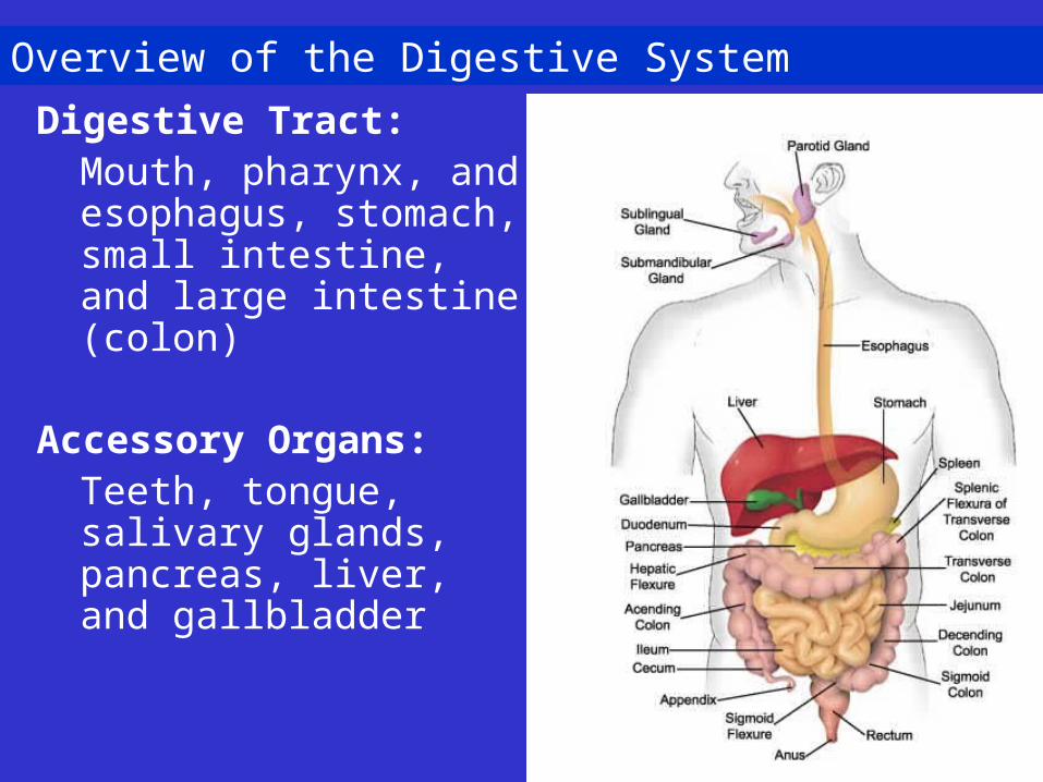
Overview of the Digestive System
Digestive Tract: Mouth, pharynx, and esophagus, stomach, small intestine, and large intestine (colon)
Accessory Organs:Teeth, tongue, salivary glands, pancreas, liver, and gallbladder
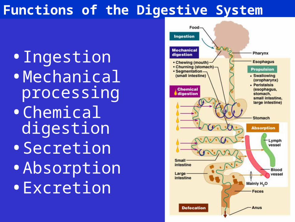
Functions of the Digestive System
• Ingestion• Mechanical
processing• Chemical
digestion• Secretion• Absorption• Excretion
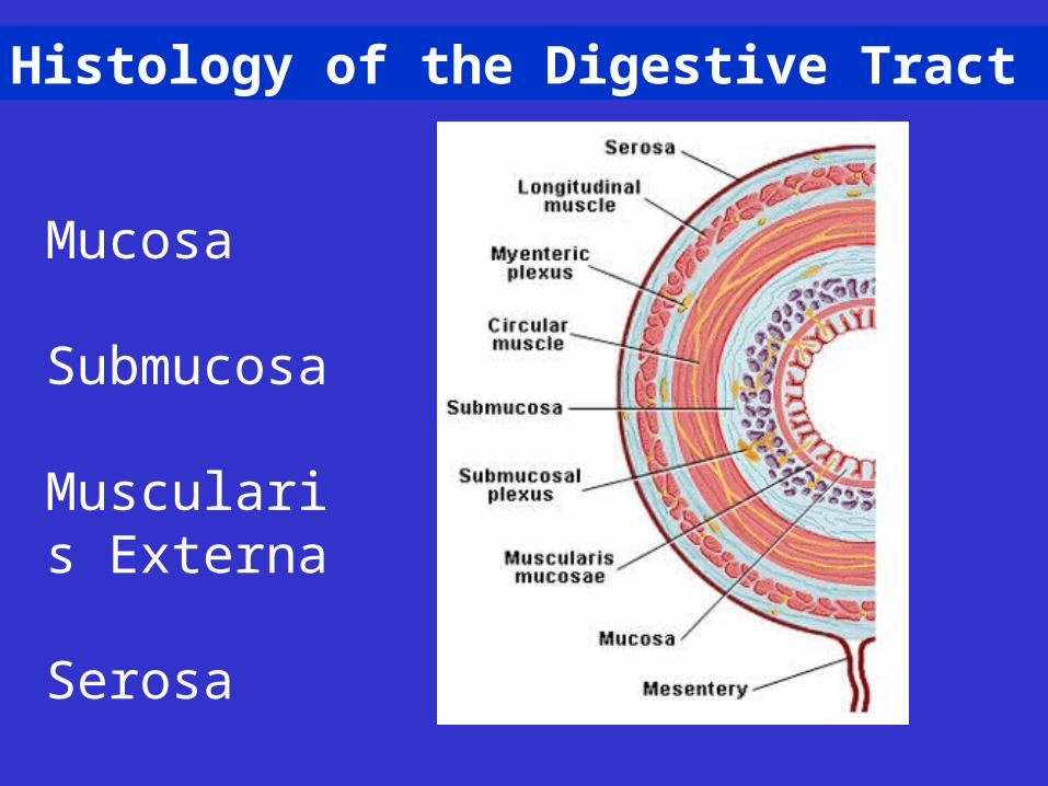
Histology of the Digestive Tract
Mucosa
Submucosa
Muscularis Externa
Serosa
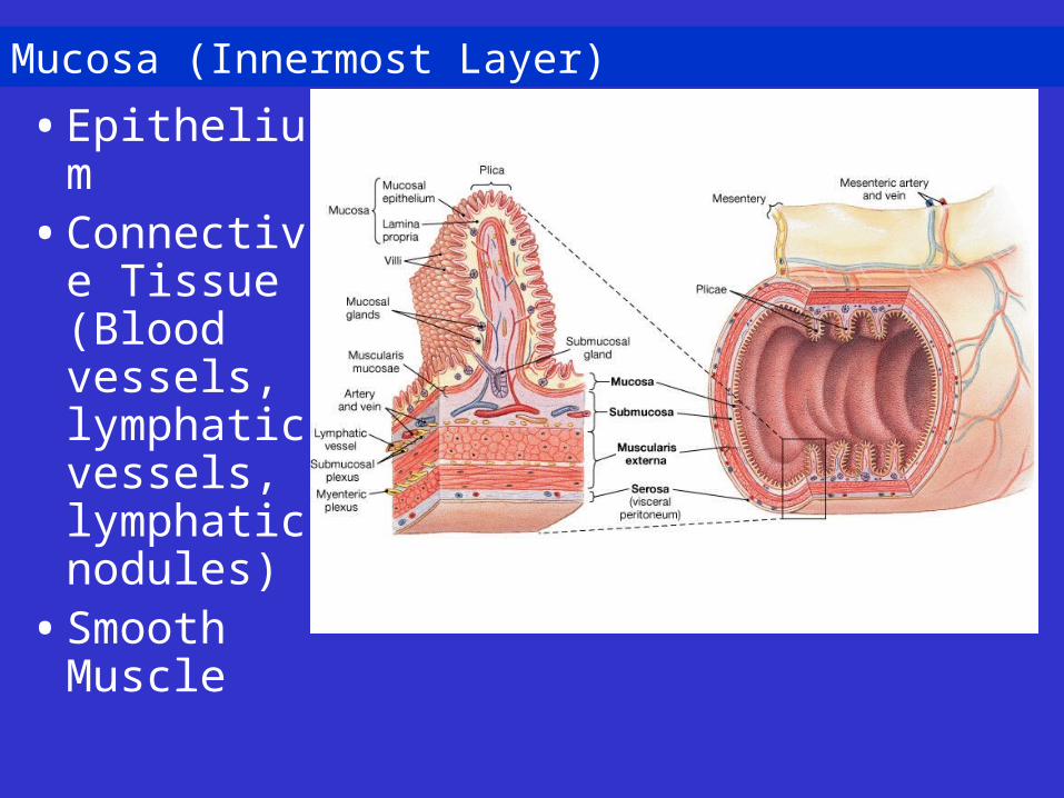
Mucosa (Innermost Layer)
• Epithelium• Connective
Tissue (Blood vessels, lymphatic vessels, lymphatic nodules)
• Smooth Muscle
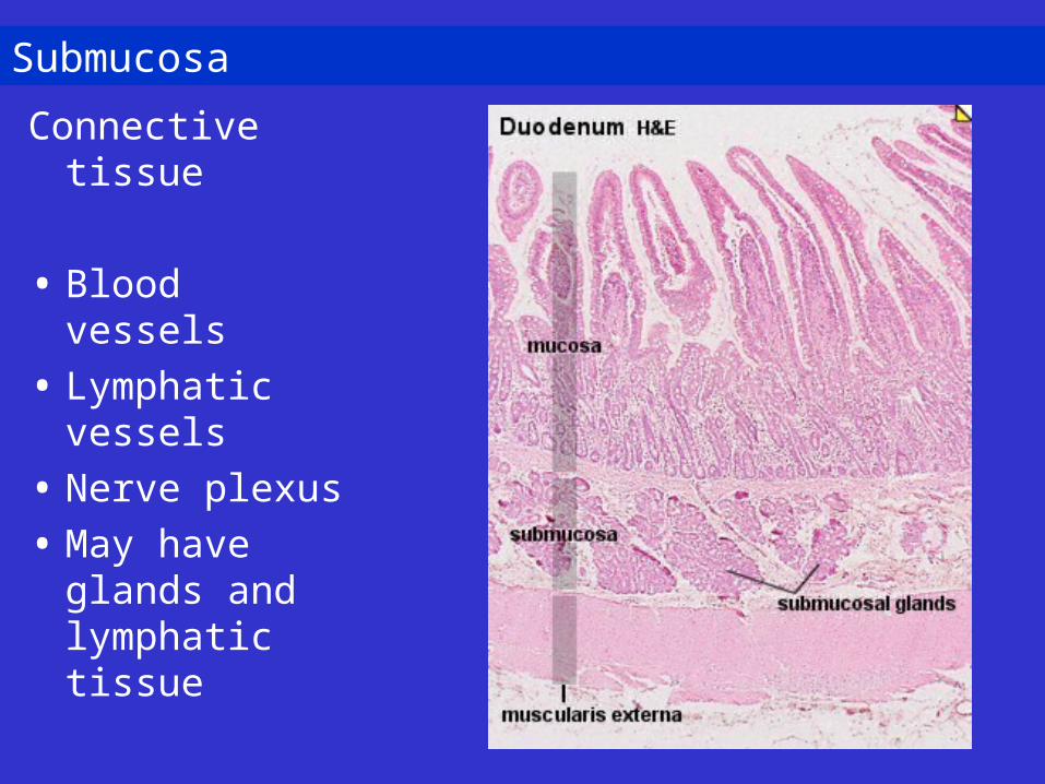
Submucosa
Connective tissue
• Blood vessels
• Lymphatic vessels
• Nerve plexus
• May have glands and lymphatic tissue
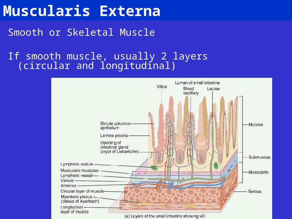
Muscularis ExternaSmooth or Skeletal Muscle
If smooth muscle, usually 2 layers (circular and longitudinal)
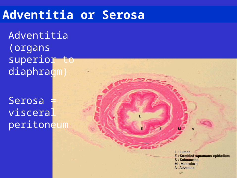
Adventitia or Serosa
Adventitia (organs superior to diaphragm)
Serosa = visceral peritoneum
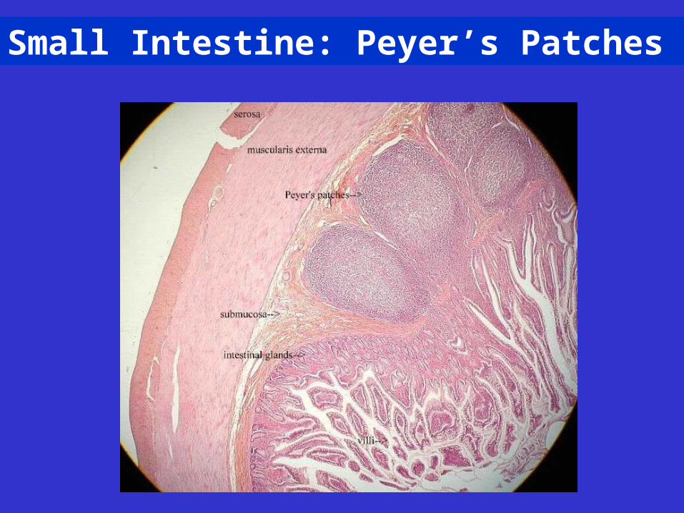
Small Intestine: Peyer’s Patches
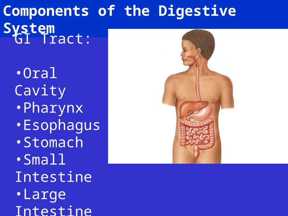
Components of the Digestive System
GI Tract:
•Oral Cavity•Pharynx•Esophagus•Stomach•Small Intestine•Large Intestine
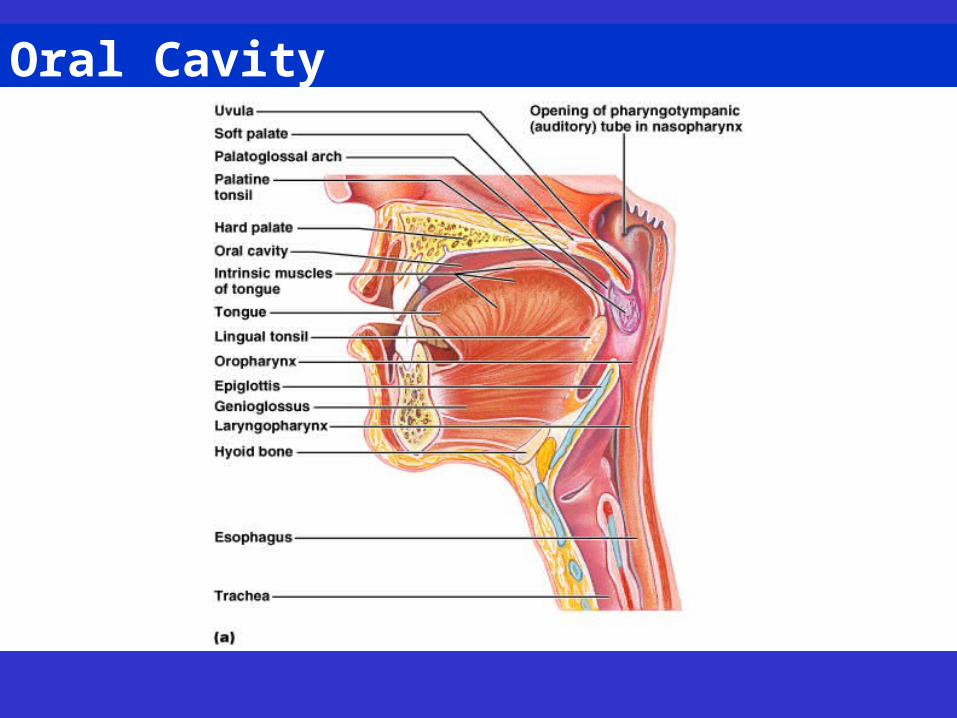
Oral Cavity
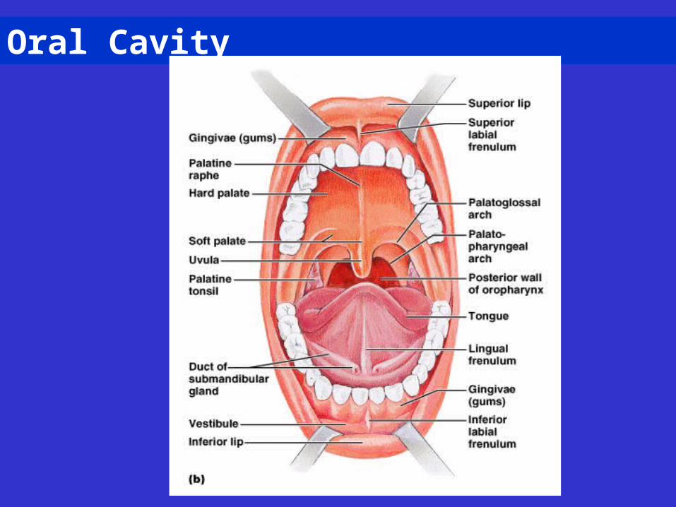
Oral Cavity
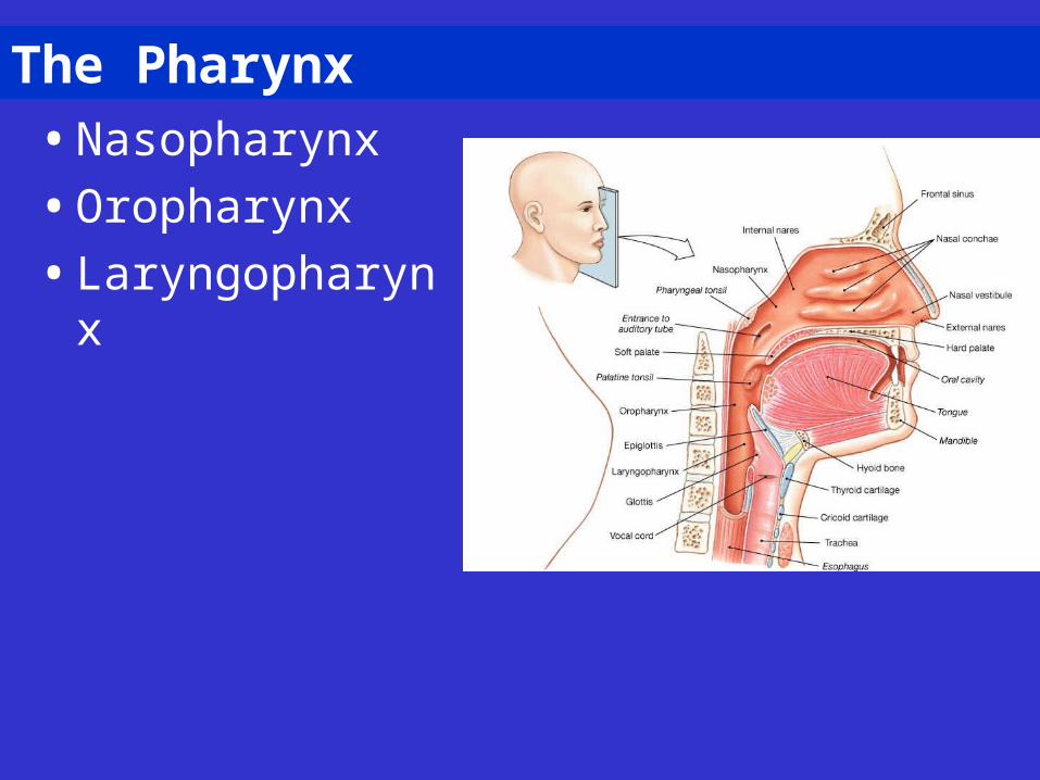
The Pharynx
• Nasopharynx
• Oropharynx
• Laryngopharynx
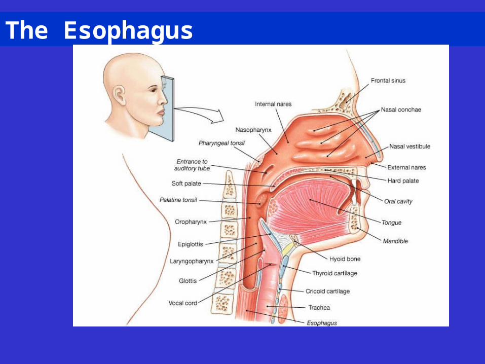
The Esophagus
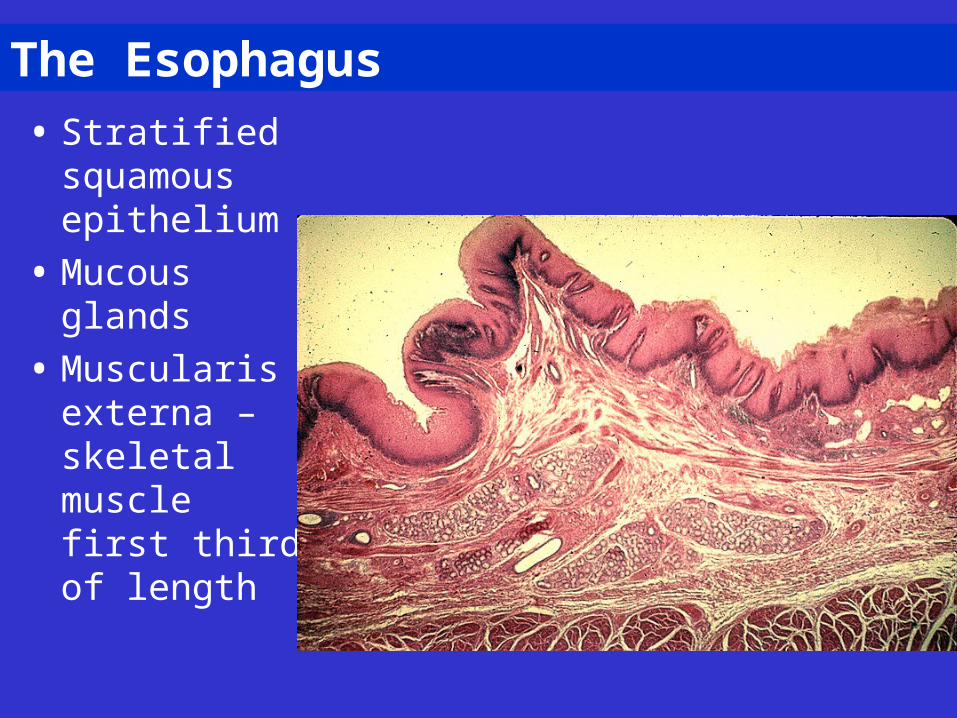
The Esophagus
• Stratified squamous epithelium
• Mucous glands
• Muscularis externa – skeletal muscle first third of length
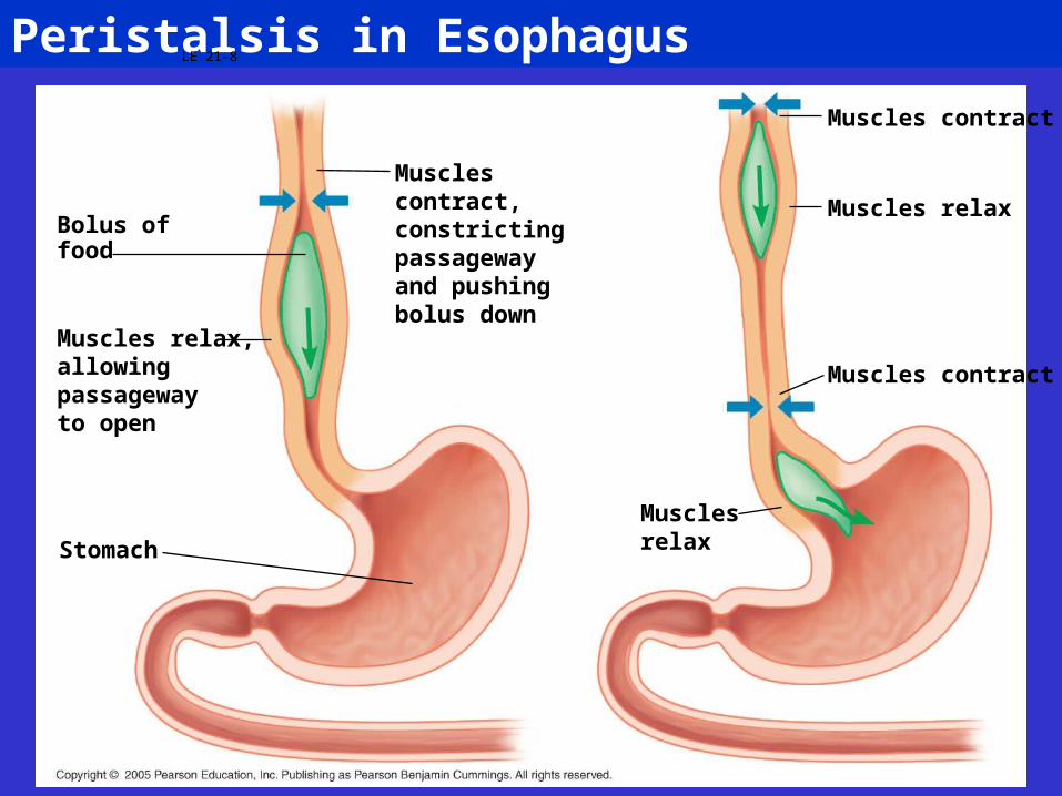
Peristalsis in EsophagusLE 21-8
Bolus offood
Muscles relax,allowingpassagewayto open
Stomach
Musclescontract,constrictingpassagewayand pushingbolus down
Musclesrelax
Muscles contract
Muscles relax
Muscles contract
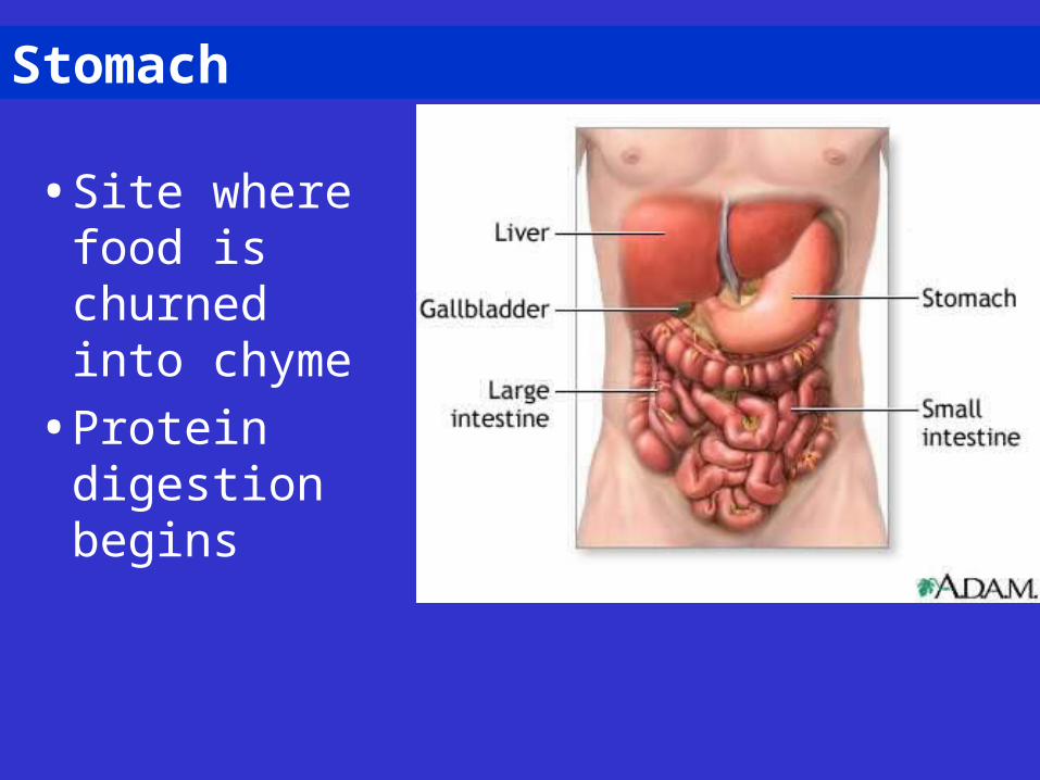
Stomach
• Site where food is churned into chyme
• Protein digestion begins
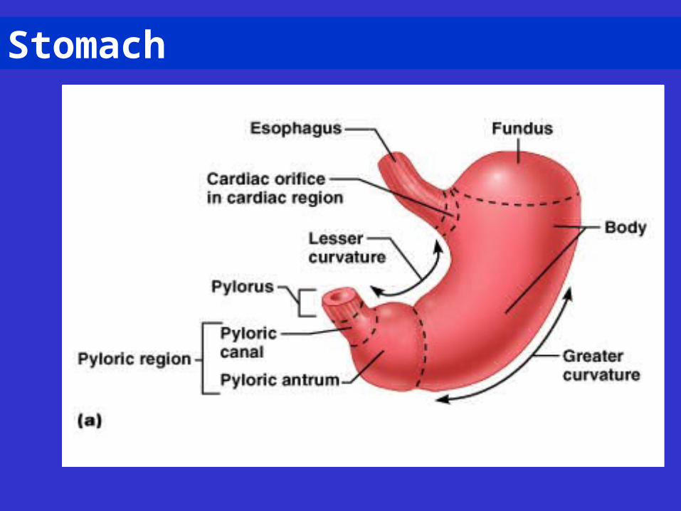
Stomach
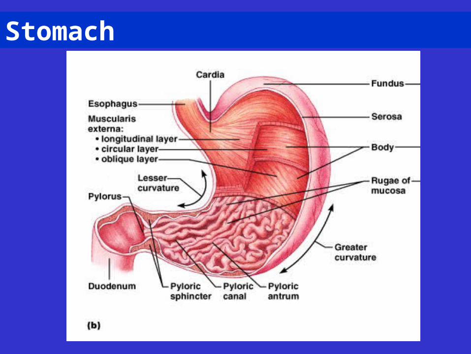
Stomach
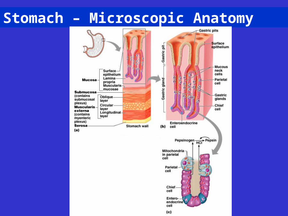
Stomach – Microscopic Anatomy
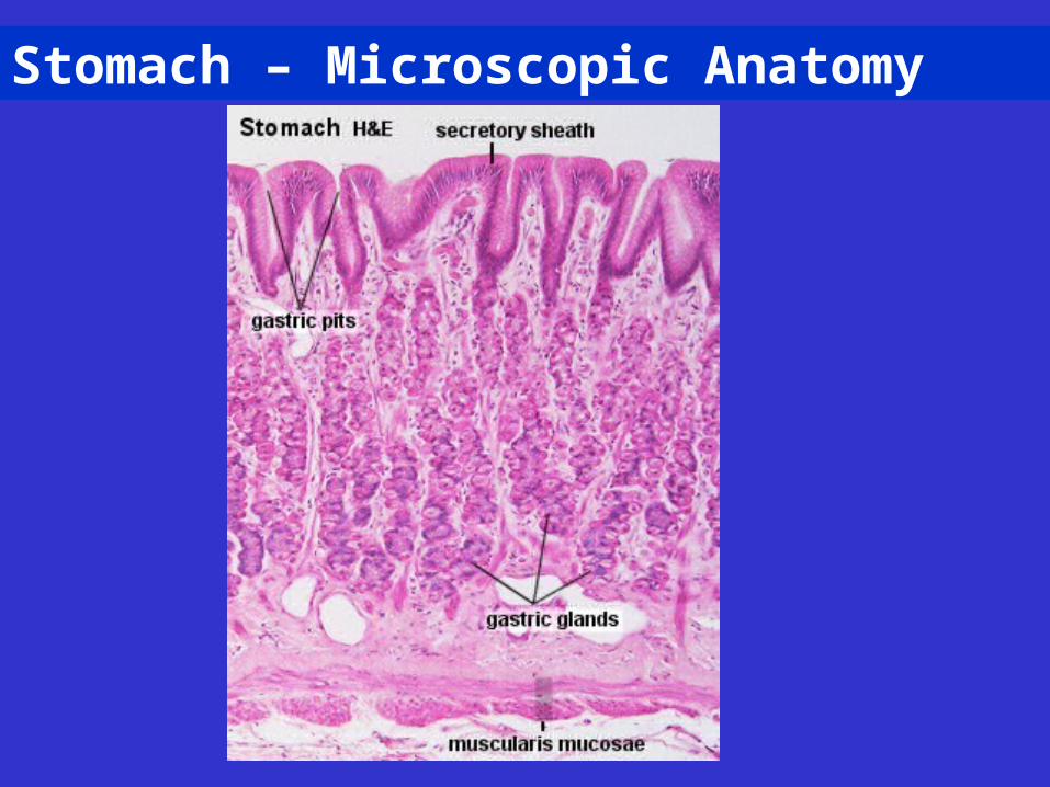
Stomach – Microscopic Anatomy
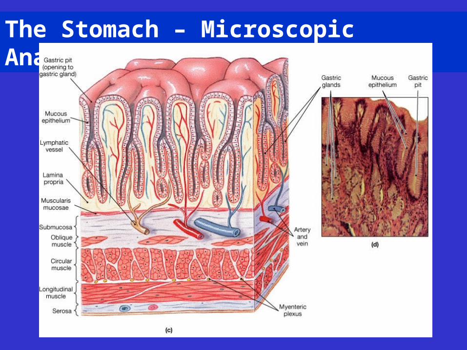
The Stomach – Microscopic Anatomy
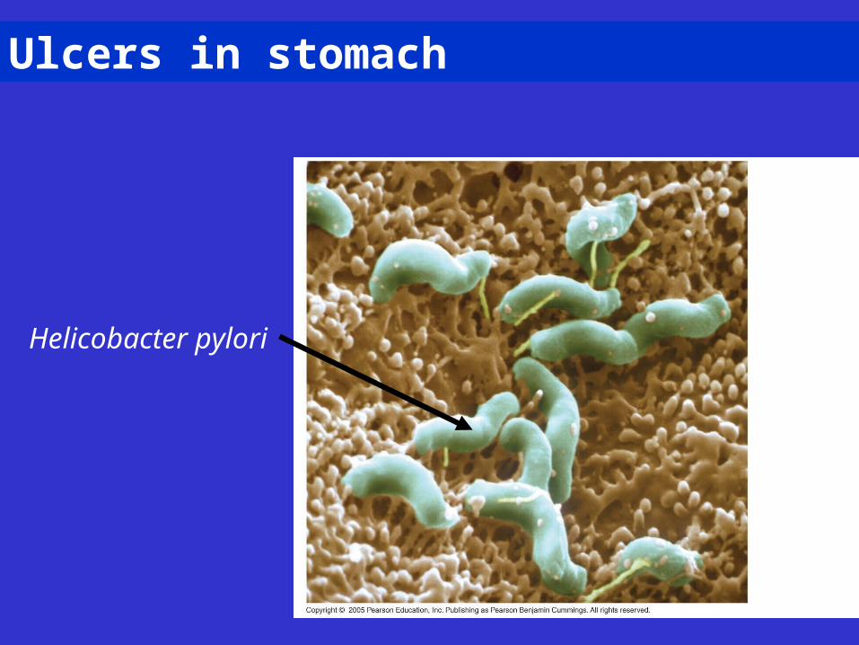
Ulcers in stomach
Helicobacter pylori
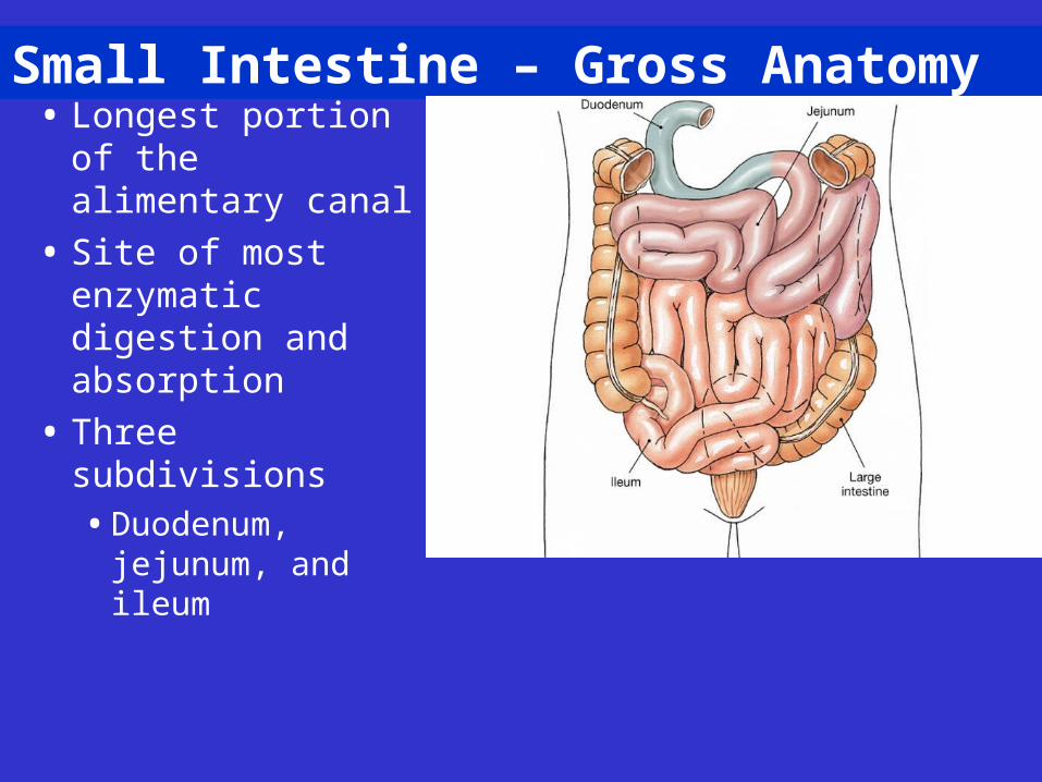
Small Intestine – Gross Anatomy• Longest portion of
the alimentary canal
• Site of most enzymatic digestion and absorption
• Three subdivisions• Duodenum,
jejunum, and ileum
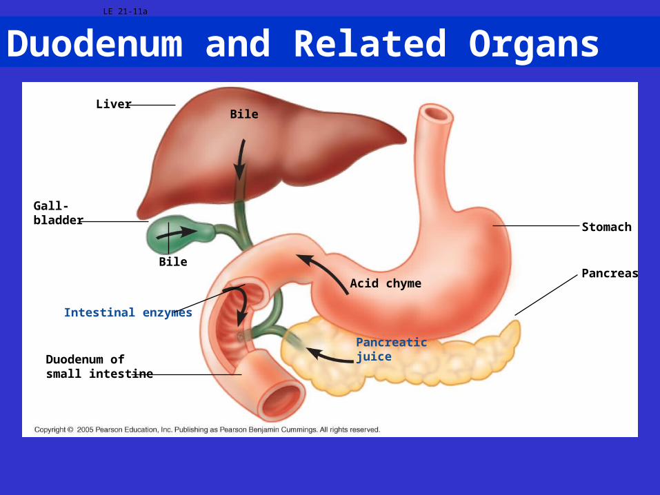
Duodenum and Related OrgansLE 21-11a
LiverBile
Gall-bladder
Bile
Duodenum ofsmall intestine
Acid chyme
Pancreaticjuice
Intestinal enzymes
Stomach
Pancreas
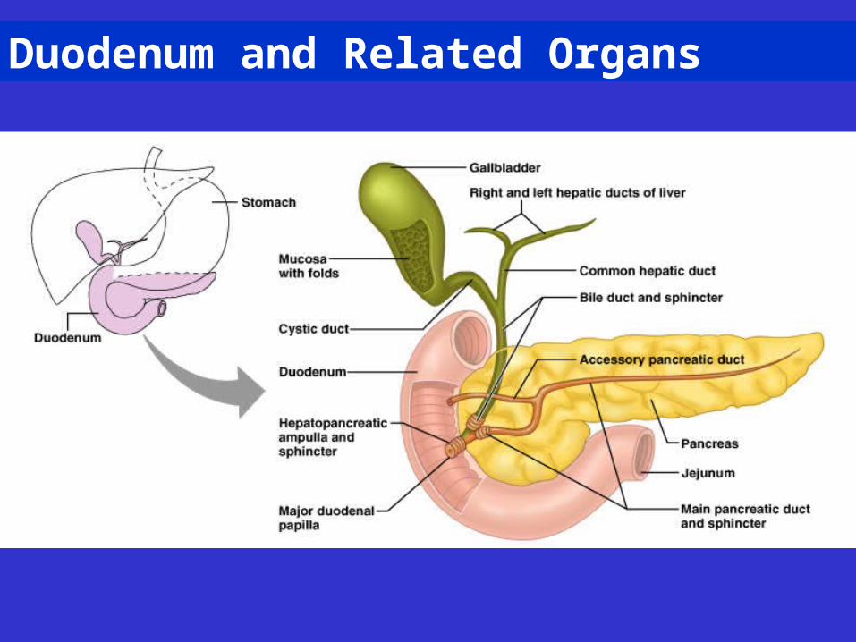
Duodenum and Related Organs
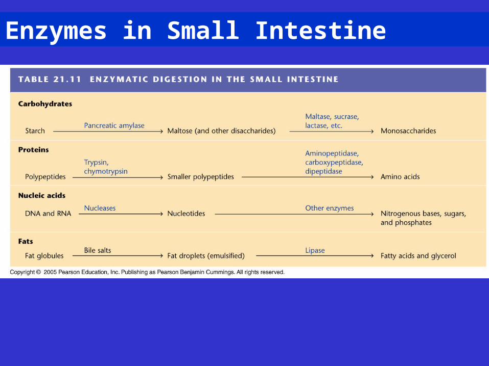
Enzymes in Small Intestine
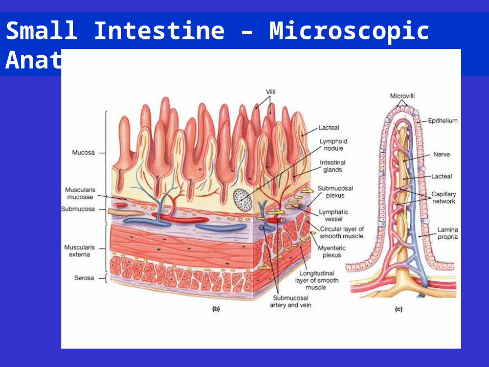
Small Intestine – Microscopic Anatomy

LE 21-11b
Veinwith blooden route tothe liver
Musclelayers
Lumen
Largecircular folds
Villi
Nutrientabsorption
Lymphvessel
Intestinal wall
Villi
Bloodcapillaries
Epithelialcells
Nutrientabsorption
Lumen of intestine
Epithelial cells
Lymph
Blood
Fats
Aminoacidsand
sugars
Fattyacidsand
glycerol
Nutrient absorptioninto epithelial cells
Microvilli

Small Intestine: Duodenum
Br = Brunner glands
V = Villus
G = Goblet cells
Cr = Intestinal glands
MM = Muscularis Mucosae
LP = Lamina Propria
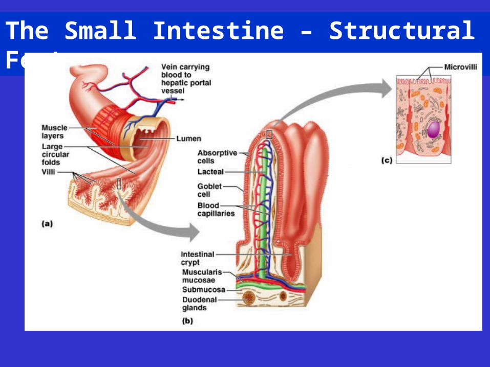
The Small Intestine – Structural Features
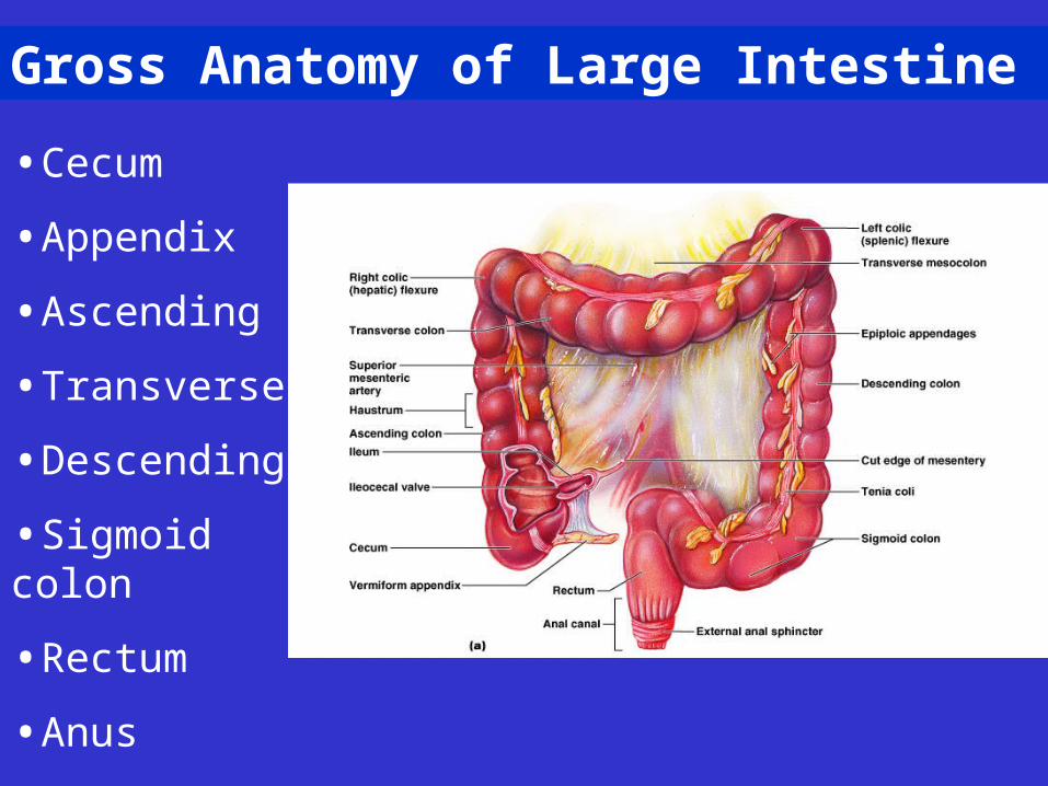
Gross Anatomy of Large Intestine
•Cecum
•Appendix
•Ascending
•Transverse
•Descending
•Sigmoid colon
•Rectum
•Anus
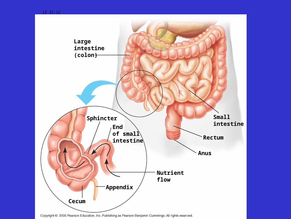
LE 21-12
Largeintestine(colon)
Sphincter
Endof smallintestine
Nutrientflow
Appendix
Cecum
Anus
Rectum
Smallintestine
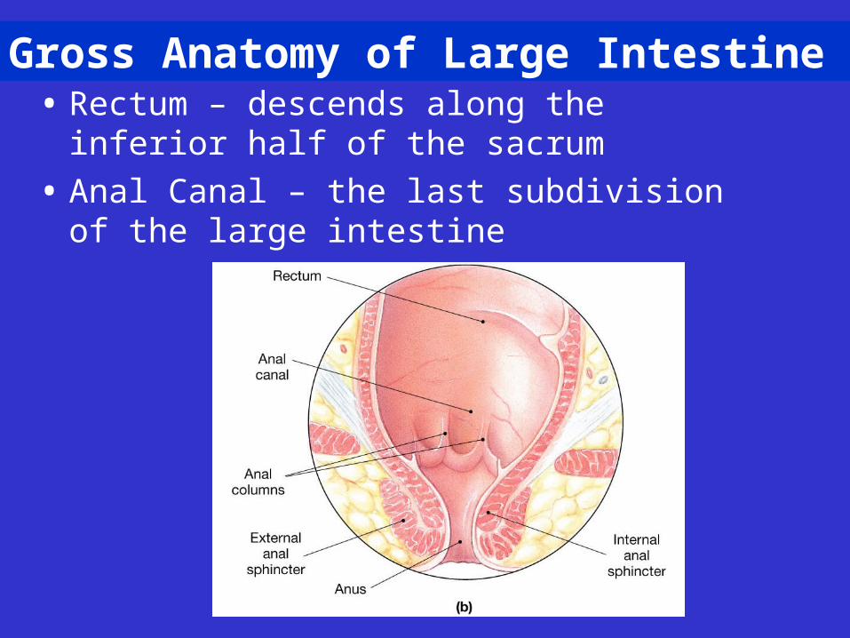
Gross Anatomy of Large Intestine• Rectum – descends along the inferior half of the
sacrum
• Anal Canal – the last subdivision of the large intestine
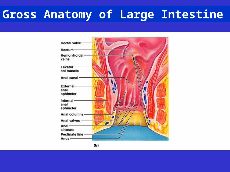
Gross Anatomy of Large Intestine
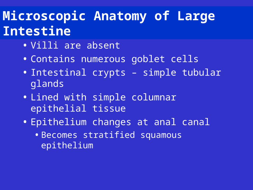
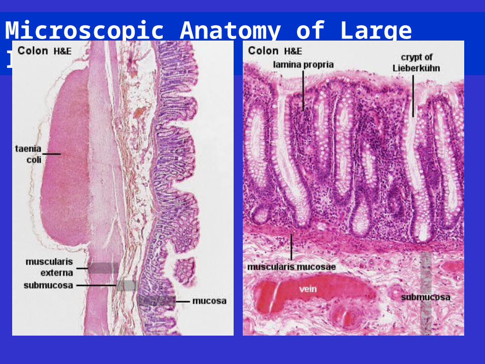
Microscopic Anatomy of Large Intestine
• Villi are absent
• Contains numerous goblet cells
• Intestinal crypts – simple tubular glands
• Lined with simple columnar epithelial tissue
• Epithelium changes at anal canal• Becomes stratified squamous epithelium
Microscopic Anatomy of Large Intestine
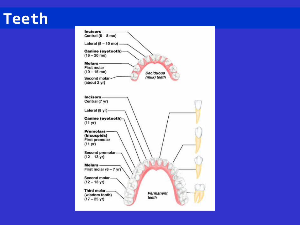
Teeth
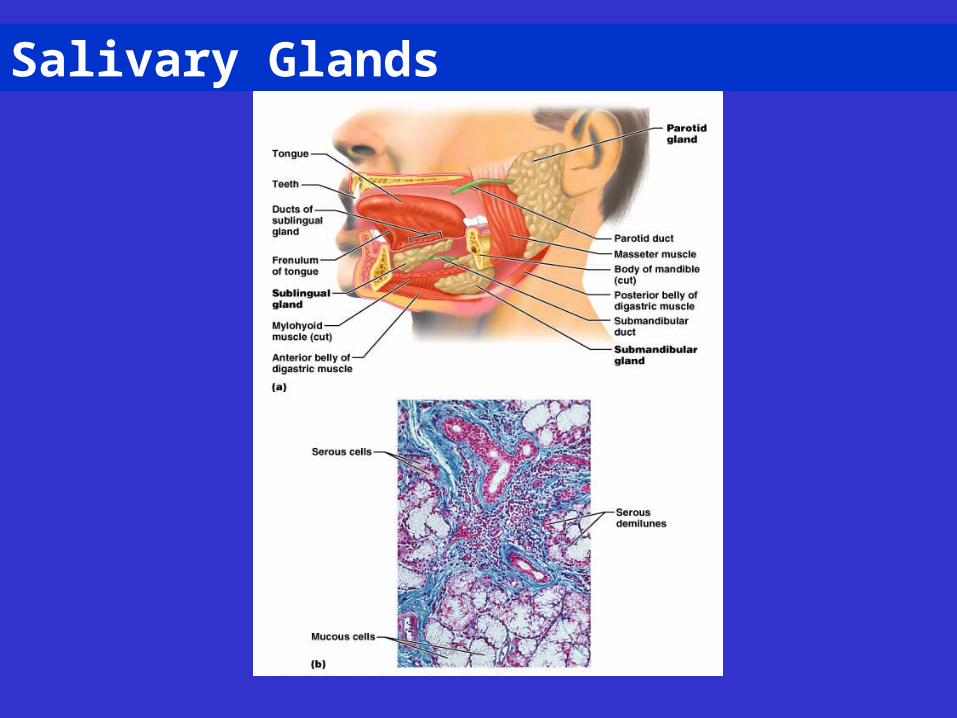
Salivary Glands
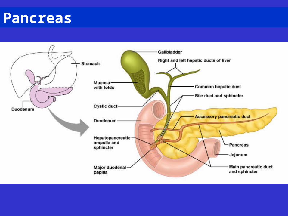
Pancreas
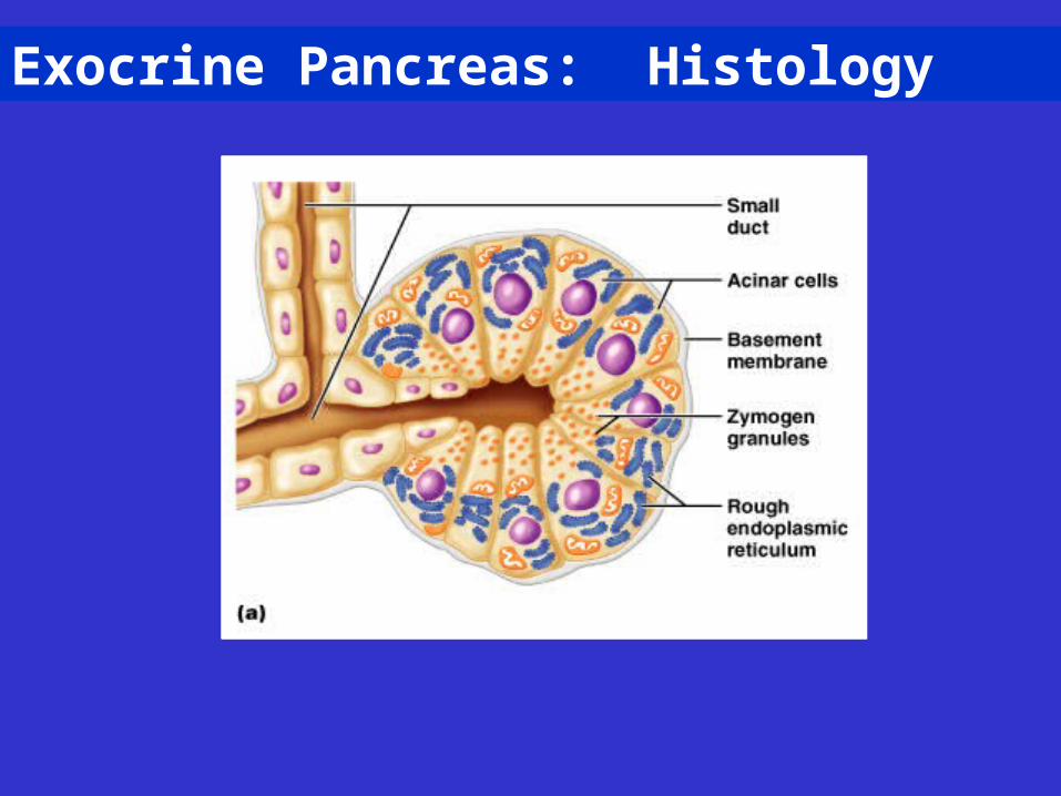
Exocrine Pancreas: Histology
Liver
• Largest gland in the body
• Performs over 500 functions
• Digestive function – bile production
• Performs many metabolic functions
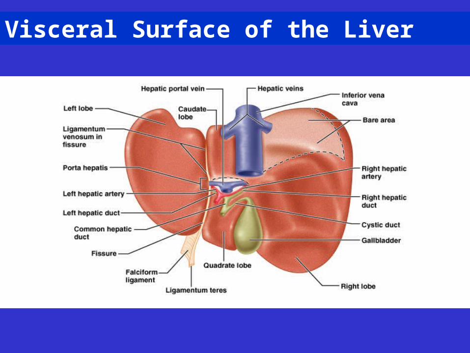
Visceral Surface of the Liver
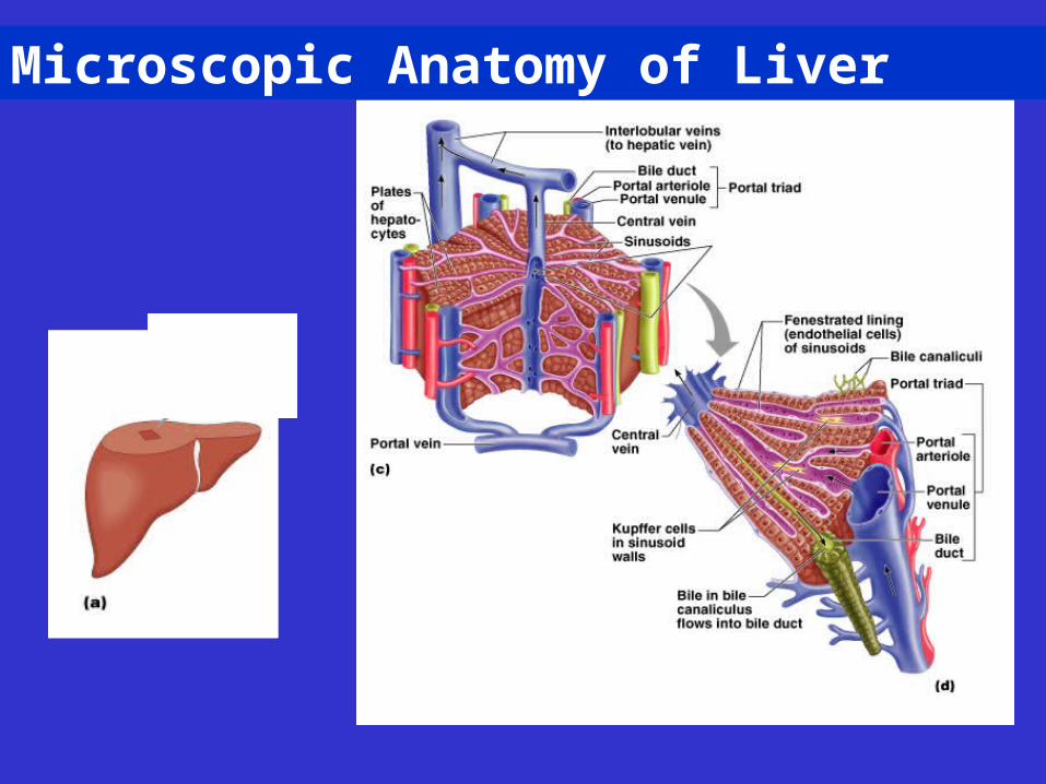
Microscopic Anatomy of Liver
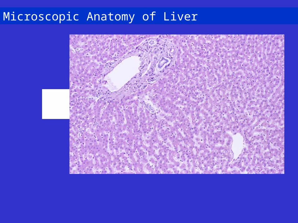
Microscopic Anatomy of Liver
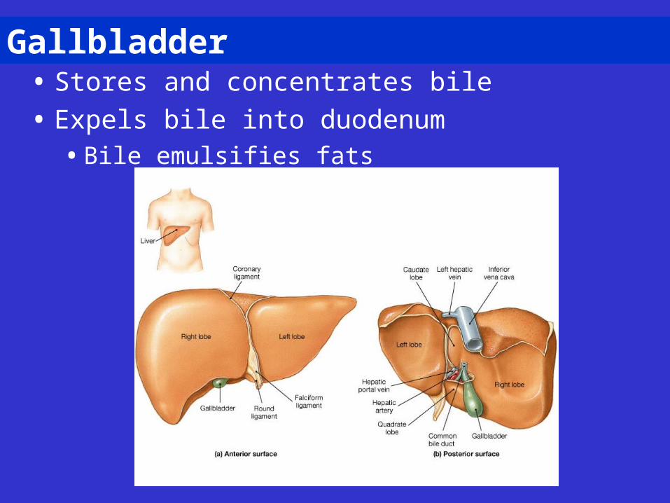
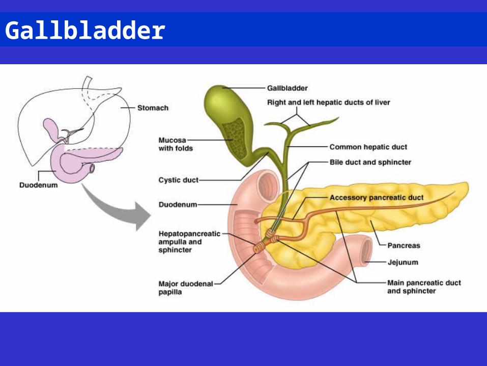
Gallbladder• Stores and concentrates bile
• Expels bile into duodenum• Bile emulsifies fats
Gallbladder

Enzymes in Small Intestine
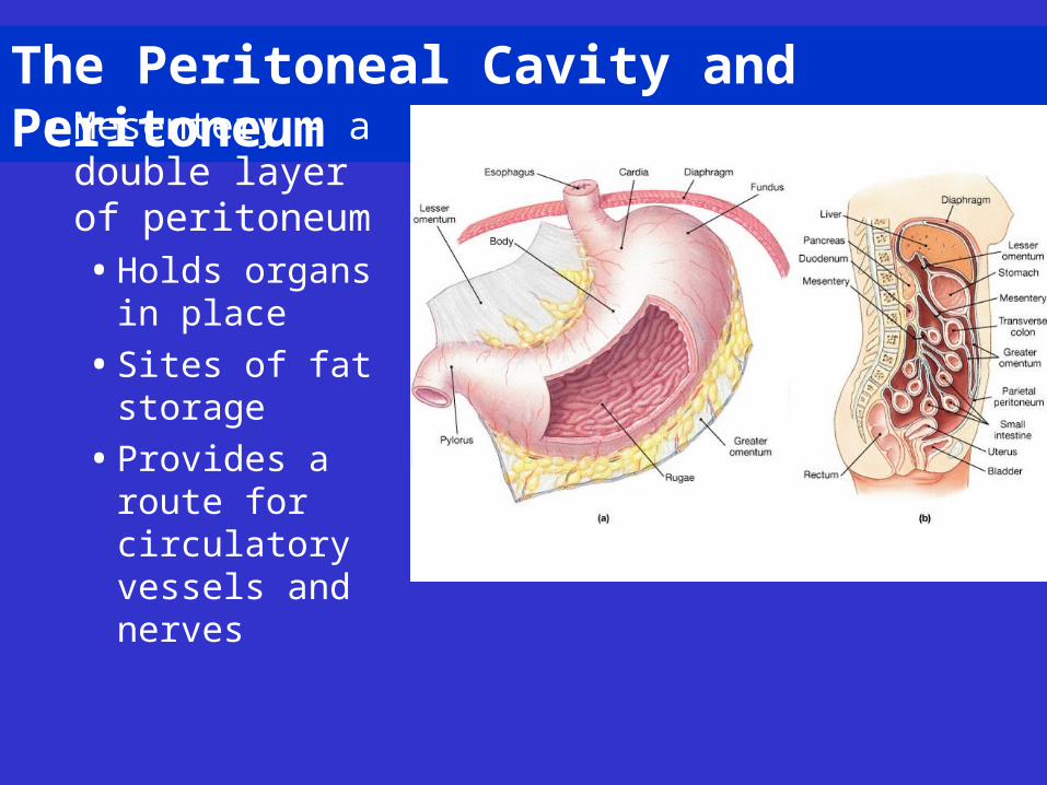
The Peritoneal Cavity and Peritoneum• Mesentery – a
double layer of peritoneum• Holds organs in
place
• Sites of fat storage
• Provides a route for circulatory vessels and nerves
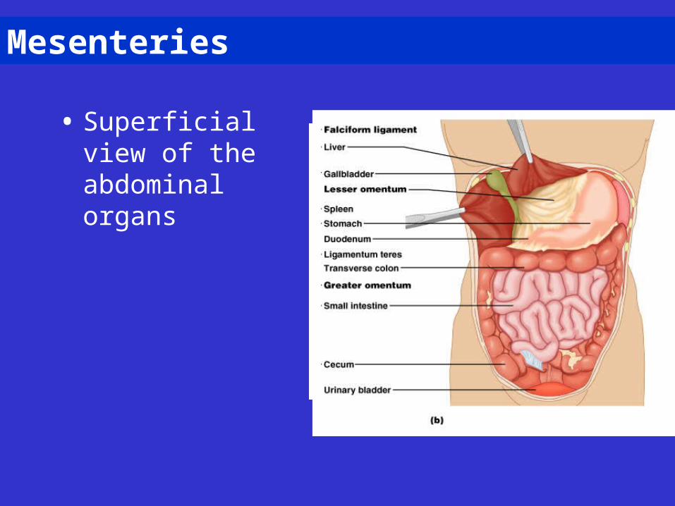
Mesenteries
• Superficial view of the abdominal organs
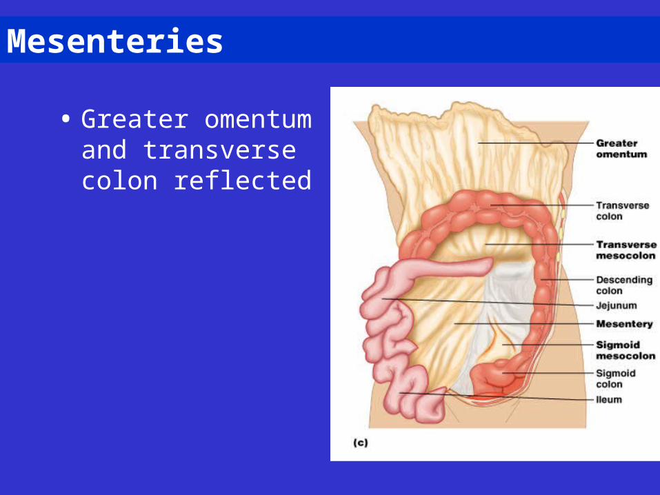
Mesenteries
• Greater omentum and transverse colon reflected
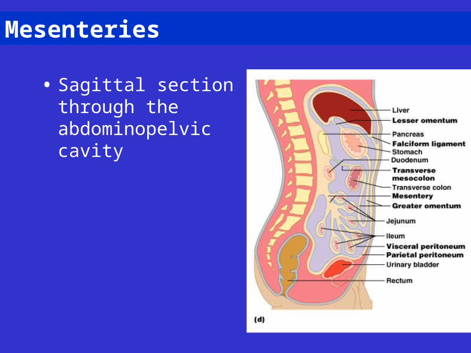
Mesenteries
• Sagittal section through the abdominopelvic cavity
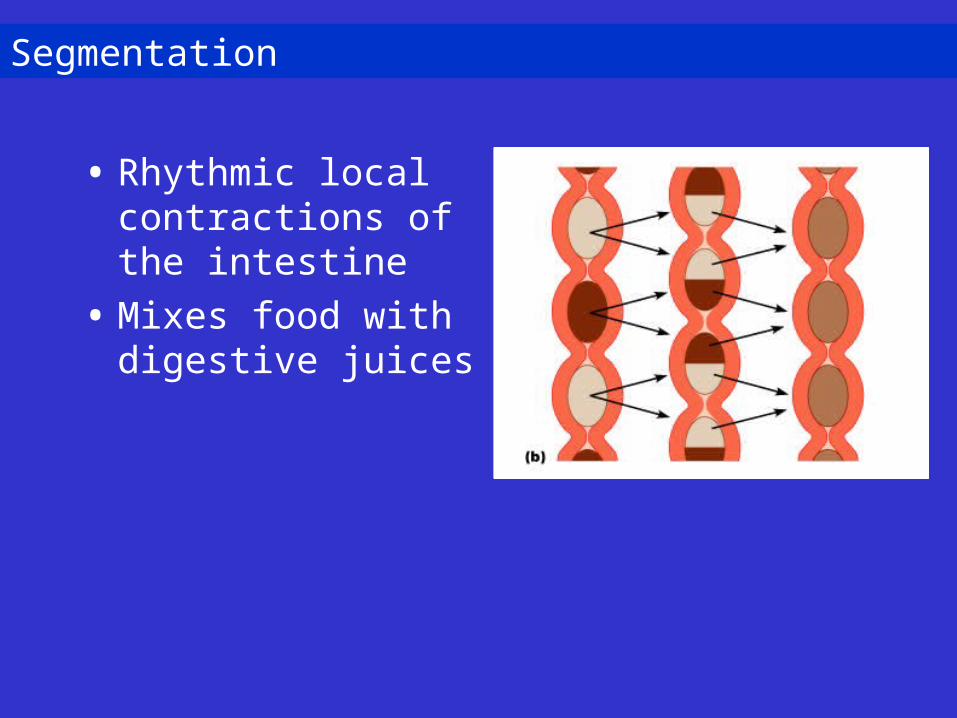
Segmentation
• Rhythmic local contractions of the intestine
• Mixes food with digestive juices
Disorders of the Digestive System
• Intestinal obstruction • Mechanical obstructions
• Adhesions, tumors, or foreign objects
• Nonmechanical obstruction • Halt in peristalsis
• Trauma
• Intestines touched during surgery
Disorders of the Digestive System
• Inflammatory bowel disease • Inflammation of intestinal wall
• Crohn’s disease
• Ulcerative colitis
• Viral hepatitis – jaundice and flu-like symptoms • Major types – A, B, C, and G
• Cystic Fibrosis and the Pancreas
The Digestive System in Later Life
• Middle age – gallstones and ulcers
• Old age – activity of digestive organs decline• Fewer digestive juices and enzymes produced
• Absorption is less efficient
• Dehydration of fecal mass leads to constipation
• Diverticulosis and cancer of digestive organs