Embed Size (px)
Citation preview
Am J Clin Pathol 2006;125:555-581 555555 DOI: 10.1309/UC6KQHLD9LV2KENN 555
© American Society for Clinical Pathology
Anatomic Pathology / OSTEOSARCOMA
Osteosarcoma
Anatomic and Histologic Variants
Michael J. Klein, MD, and Gene P. Siegal, MD, PhD
Key Words: Osteosarcoma; Malignant bone tumors; Bones; Imaging
DOI: 10.1309/UC6KQHLD9LV2KENN
A b s t r a c t
Osteosarcoma is the most common primary tumorof bone, yet its absolute incidence among malignanttumors is low. Within its strict histologic definition,osteosarcoma comprises a family of lesions withconsiderable diversity in histologic features and grade.Its prognosis is dependent not only on these parametersbut also on its anatomic site. It may occur inside thebones (in the intramedullary or intracorticalcompartment), on the surfaces of bones, and inextraosseous sites. Information of diagnostic orprognostic significance has not been elucidated fromstudies of its cytogenetics. This review summarizes theanatomic and histologic variations of osteosarcomaand offers a schema for its subclassification.
At an estimated incidence of 2 cases per million personsper year, osteosarcoma is the most common malignant pri-mary bone tumor excluding hematopoietic intraosseoustumors. Although it occurs at any age, its peak incidence is inthe second and third decades. Its statistical distribution rough-ly parallels skeletal growth; it is more frequent in tall peoplethan in short people and in larger animals than in smaller ani-mals.1 Bones having the fastest rates of growth have the high-est frequency of occurrence. When osteosarcoma appears ear-lier than the second decade or after the cessation of skeletalgrowth, there is often an association with another osseousabnormality. This may be a genetic predisposition such as theLi-Fraumeni or Beckman-Wiederman syndrome,2,3 an under-lying abnormality such as Paget disease4 or fibrous dysplasiathat has a predilection for the development of osteosarcoma,5
or previous radiation of the bone involved.The definition of osteosarcoma is deceptively straightfor-
ward. It is a malignant tumor of connective tissue (mesoder-mal) origin within which the tumor cells produce bone orosteoid (often referred to as “tumor bone” or “tumor osteoid”in the vernacular). Although this makes it seem as though thecells giving rise to osteosarcoma must be of osteoblastic deri-vation, there is no evidence that osteoblasts, once they differ-entiate from osteoprogenitor cells, can actually revert to moreprimitive cells, let alone malignant ones. This implication isinherent in the more arcane term osteogenic sarcoma, whichhas fallen into disuse.
An additional feature often encountered in osteosarcomasis their propensity to produce variable amounts of cartilagematrix and fibrous tissue. In some cases, the cartilage matrixor fibrous tissue may so strikingly dominate the tumor tissuethat the actual production of bone must be carefully sought to
556 Am J Clin Pathol 2006;125:555-581556 DOI: 10.1309/UC6KQHLD9LV2KENN
© American Society for Clinical Pathology
Klein and Siegal / OSTEOSARCOMA
make the correct diagnosis. Because osteosarcoma may differ-entiate along all or any of these pathways of matrix synthesis,it has more of a histologic similarity to fracture callus orfibrous dysplasia than it does to differentiated bonematrix–producing tumors such as osteoid osteoma orosteoblastoma. This has given rise to the 3 traditional subdivi-sions of osteoblastic, chondroblastic, and fibroblasticosteosarcoma. In actuality, most osteosarcomas demonstratevarying amounts of all 3 cell types and matrix. Consequently,division into any one of these types is arbitrary and capriciousbut generally is meant to signify greater than 50% predomi-nance of any histologic type.
Although osteosarcoma usually arises in the medullarycavity of the metaphysis of a growing long tubular bone, italso may arise on the surface of a bone, it may be confined tothe cortex, or it even may arise in an extraskeletal site. Theosteosarcomas arising on the bone surfaces are about 20 timesless frequent than their medullary counterparts. Interestingly,the majority of osteosarcomas of medullary origin are high-grade, whereas most arising on the surfaces of bones are oflower grade. Patients with surface osteosarcomas are often adecade or more older than typical patients with centralosteosarcomas. Occasionally, high- and low-grade elementsmay be present in any given case of osteosarcoma. In thesecases, the biologic behavior is usually that of the histological-ly highest grade area of the neoplasm.
Patients with osteosarcomas usually have nonspecificclinical symptoms, the most common of which is pain for sev-eral weeks or months. The pain often begins insidiously, butby the time patients come to medical attention, their pain ispresent at rest or even disturbs their sleep. The most commonsign is a mass that is almost always firm and tender. Theremay be superficial erythema, venous distension, or other signsof hypervascularity. There may be a limp, loss of function, oreven decreased range of motion. Any of these symptoms orsigns is cause for further clinical investigation.
The imaging studies usually suggest a malignanttumor; at least half the time the specific diagnosis can bemade from conventional radiographs. Especially character-istic is an ill-defined radiodensity occupying the metaphy-seal region of a long bone. The intraosseous density oftenhas a diffusely cloudy or fluffy appearance. Although thereis seldom a fracture or cortical infraction in osteosarcoma,it is usual for the tumor to permeate the extant passagesnot only between osseous trabeculae but also of theHaversian systems and Volkmann canals. As the tumorreaches the outer cortical surface, the periosteum is dis-sected from the bone. The cambium layer of the innerperiosteum reacts to separation from the cortex by produc-ing new bone, which is sometimes visible as an incompletebony shell that appears attached to the bone surface ononly one end and is open or discontinuous in the middle, a
so-called Codman angle. Other kinds of periosteal reac-tions may cause so-called sunburst or hair-on-end radi-ographic densities ❚Image 1❚.
Although periosteal new bone formation sometimes isassociated with benign conditions, periosteal reactions associ-ated with malignant tumors usually are discontinuous, imply-ing that the tumor growth is too rapid for periosteal contain-ment.6 In some cases, conventional radiographs may suggest amalignant tumor that is not osteosarcoma.7 This is especiallytrue when a large proportion of the osteoid matrix in anosteosarcoma is not well mineralized. In rarer cases, the radi-ograph may not even appear to represent a malignant tumor.8
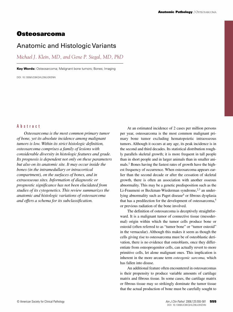
It must be kept in mind that although radiographs usually cor-relate well with histologic studies of bone tumors, radiographsmerely reflect statistical odds ratios and, thus, the likelihoodthat a lesion is, first, a tumor, and second, malignant or benign.Like all statistics, there are some lesions of bone that are out-liers and may not correlate well with the radiographic appear-ance; this is precisely the reason that even classically appear-ing bone lesions must be biopsied ❚Image 2❚.
Historically, the malignant nature of osteosarcomabecame obvious with the appearance of lung metastases, usu-ally in about 18 months to 2 years after first diagnosis, and the
A B
❚Image 1❚ A, Lateral radiograph of the distal femurdemonstrates typical radiographic features of osteosarcoma.Radiodense, cloudy bone lies within an ill-definedradiolucency of the femoral metadiaphysis. The arrows delimita very large soft tissue mass containing bone matrix. ACodman angle periosteal reaction is highlighted with anarrowhead. B, Lateral radiograph in a different casedemonstrating a very large extraosseous, radiodense massthat contains extensively interrupted periosteal “streamers,”forming a “sunburst” periosteal reaction.
Am J Clin Pathol 2006;125:555-581 557557 DOI: 10.1309/UC6KQHLD9LV2KENN 557
© American Society for Clinical Pathology
5-year survival was on the order of 20%. Although its degreeof malignancy has not changed, modern adjuvant chemother-apy has increased this survival more than 3-fold,9 and evenpeople with metastatic disease sometimes can be saved.10,11
Because osteosarcoma may produce various kinds ofextracellular matrix and have different degrees of differen-tiation, its histologic pattern may vary significantly, notonly from case to case but also from area to area in the samecase. Its classification into various subtypes is not only bythe predominant histologic pattern, but also by its anatomiclocation and sometimes by its histologic grade ❚Table 1❚ and❚Table 2❚.
The tendency to subclassify osteosarcomas in this waymakes it seem that there are as many variations of this tumoras any tumor in the oncologic realm. The tendency to subdi-vide so rare a tumor into many such compartments alsomakes straightforward diagnosis more difficult for the gener-al surgical pathologist. In a significant number of cases, how-ever, the subclassification of osteosarcoma matters only if agiven subtype behaves or responds to treatment in a consis-tently different way. The remainder of this review is dedicat-ed to the comparison of these subtypes histologically,anatomically, and biologically.
Conventional Osteosarcoma
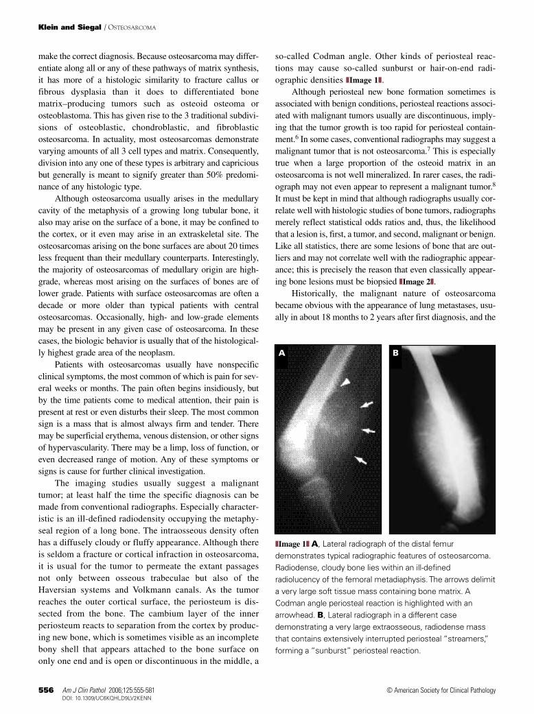
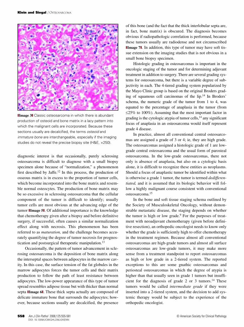
Osteosarcoma is diagnosed most easily when it appearsin its classic, or conventional, form. The tumor cells vary fromspindled to polyhedral; their nuclei are pleomorphic andhyperchromatic. Mitotic figures are easily demonstrable, andatypical mitotic figures also may be identified. The tumor cellsare engaged in the production of extracellular matrix that maybe osseous, cartilaginous, or fibrous in various proportions.The production of bone or osteoid directly by tumor cells atleast somewhere in the tumor is the absolute requirement fordiagnosis ❚Image 3❚.
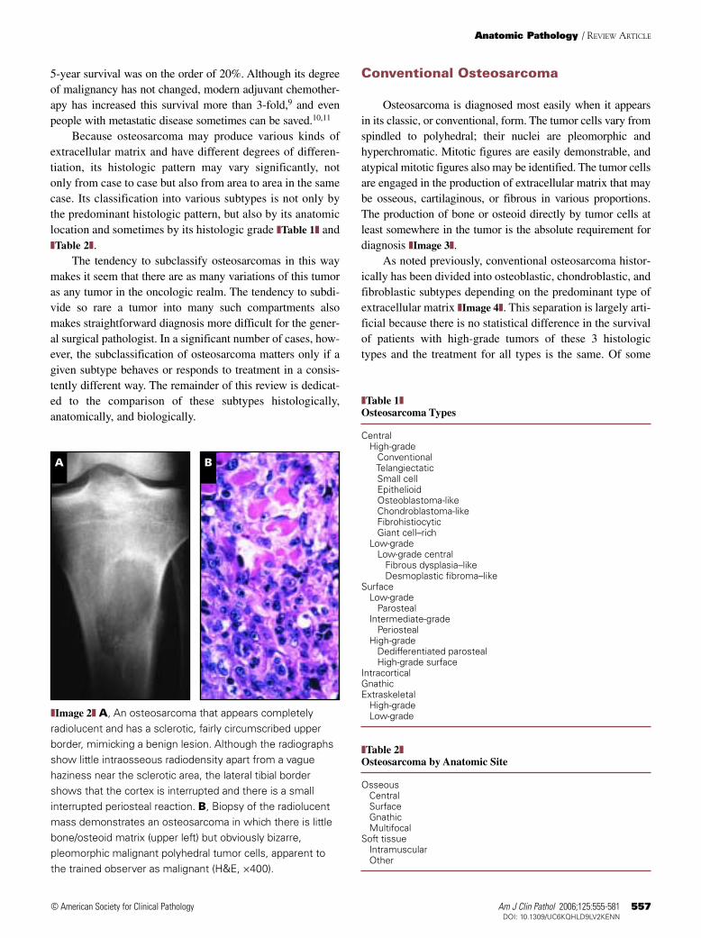
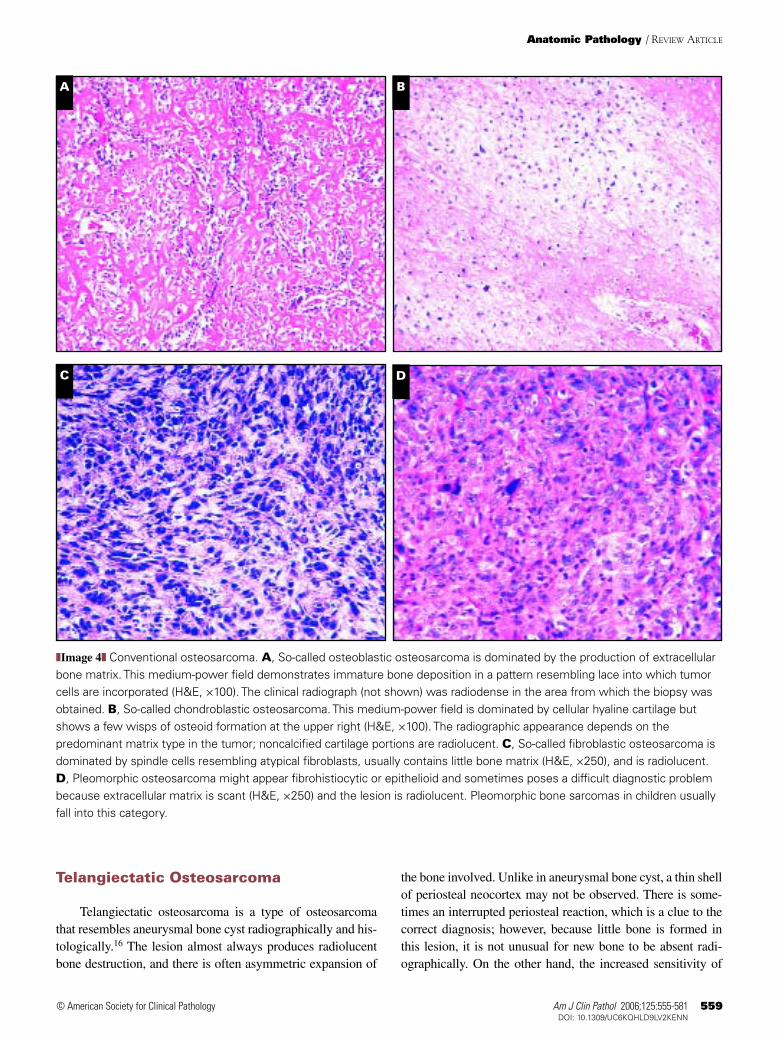
As noted previously, conventional osteosarcoma histor-ically has been divided into osteoblastic, chondroblastic, andfibroblastic subtypes depending on the predominant type ofextracellular matrix ❚Image 4❚. This separation is largely arti-ficial because there is no statistical difference in the survivalof patients with high-grade tumors of these 3 histologictypes and the treatment for all types is the same. Of some
Anatomic Pathology / REVIEW ARTICLE
A B
❚Image 2❚ A, An osteosarcoma that appears completelyradiolucent and has a sclerotic, fairly circumscribed upperborder, mimicking a benign lesion. Although the radiographsshow little intraosseous radiodensity apart from a vaguehaziness near the sclerotic area, the lateral tibial bordershows that the cortex is interrupted and there is a smallinterrupted periosteal reaction. B, Biopsy of the radiolucentmass demonstrates an osteosarcoma in which there is littlebone/osteoid matrix (upper left) but obviously bizarre,pleomorphic malignant polyhedral tumor cells, apparent tothe trained observer as malignant (H&E, ×400).
❚Table 1❚Osteosarcoma Types
CentralHigh-grade
ConventionalTelangiectatic Small cell EpithelioidOsteoblastoma-likeChondroblastoma-likeFibrohistiocyticGiant cell–rich
Low-gradeLow-grade central
Fibrous dysplasia–likeDesmoplastic fibroma–like
SurfaceLow-grade
ParostealIntermediate-grade
PeriostealHigh-grade
Dedifferentiated parostealHigh-grade surface
IntracorticalGnathicExtraskeletal
High-gradeLow-grade
❚Table 2❚Osteosarcoma by Anatomic Site
OsseousCentralSurfaceGnathicMultifocal
Soft tissueIntramuscularOther
558 Am J Clin Pathol 2006;125:555-581558 DOI: 10.1309/UC6KQHLD9LV2KENN
© American Society for Clinical Pathology
Klein and Siegal / OSTEOSARCOMA
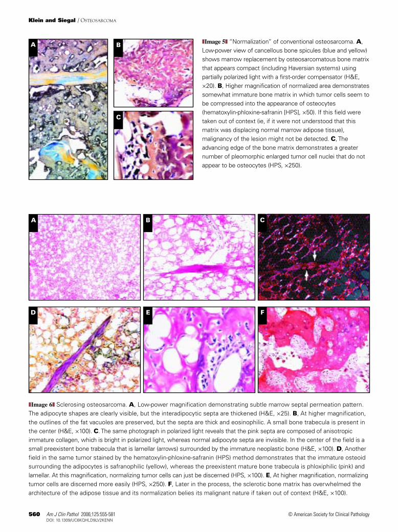
diagnostic interest is that occasionally, purely sclerosingosteosarcoma is difficult to diagnose with a small biopsyspecimen alone because of “normalization,” a phenomenonfirst described by Jaffe.12 In this process, the production ofosseous matrix is in excess to the proportion of tumor cells,which become incorporated into the bone matrix and resem-ble normal osteocytes. The production of bone matrix maybe so excessive in sclerosing osteosarcoma that the cellularcomponent of the tumor is difficult to identify; usuallytumor cells are most obvious at the advancing edge of thetumor ❚Image 5❚. Of additional importance is the knowledgethat chemotherapy given after a biopsy and before definitivesurgery, if successful, often causes a similar normalizationeffect along with necrosis. This phenomenon has beenreferred to as maturation, and the challenge becomes accu-rately quantifying the degree of tumor necrosis for prognos-tication and postsurgical therapeutic manipulation.13
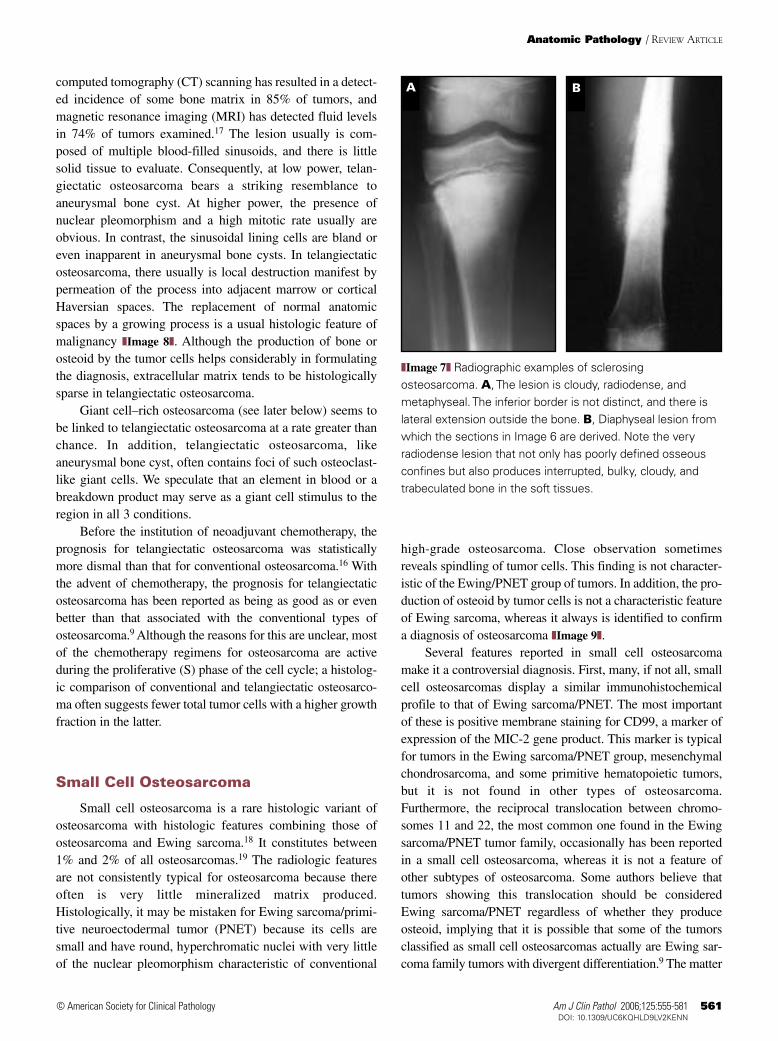
Occasionally, the pattern of tumor advancement in scle-rosing osteosarcoma is the deposition of bone matrix alongthe interseptal spaces between adipocytes in the marrow cav-ity. In this case, the surface tension of the fat globules in themarrow adipocytes forces the tumor cells and their matrixproduction to follow the path of least resistance betweenadipocytes. The low-power appearance of this type of tumorspread resembles adipose tissue but with thicker than normalsepta ❚Image 6❚. These thick septa actually are composed ofdelicate immature bone that surrounds the adipocytes; how-ever, because sections usually are decalcified, the presence
of this bone (and the fact that the thick interlobular septa are,in fact, bone matrix) is obscured. The diagnosis becomesobvious if radiopathologic correlation is performed, becausethese tumors usually are radiodense and not circumscribed❚Image 7❚. In addition, this type of tumor may have soft tis-sue extension on the imaging studies that is not obvious in asmall bone biopsy specimen.
Histologic grading in osteosarcomas is important in theoncologic staging of the tumor and for determining adjuvanttreatment in addition to surgery. There are several grading sys-tems for osteosarcoma, but there is a variable degree of sub-jectivity in each. The 4-tiered grading system popularized bythe Mayo Clinic group is based on the original Broders grad-ing of squamous cell carcinomas of the lip.14 In Broders’schema, the numeric grade of the tumor from 1 to 4, wasequated to the percentage of anaplasia in the tumor (from≤25% to 100%). Assuming that the most important factor ingrading is the cytologic atypia of tumor cells,14 any significantfocus of anaplasia in an osteosarcoma would itself representgrade 4 disease.
In practice, almost all conventional central osteosarco-mas are assigned a grade of 3 or 4; ie, they are high grade.The osteosarcomas assigned a histologic grade of 1 are low-grade central osteosarcoma and the usual form of parostealosteosarcoma. In the low-grade osteosarcomas, there notonly is absence of anaplasia, but also on a cytologic basisalone, it is difficult to recognize these entities as neoplasms.Should a focus of anaplastic tumor be identified within whatis otherwise a grade 1 tumor, the tumor is termed dedifferen-tiated, and it is assumed that its biologic behavior will fol-low a highly malignant course consistent with conventionalosteosarcoma.15
In the bone and soft tissue staging schema outlined bythe Society of Musculoskeletal Oncology, without demon-strable metastatic disease, the staging depends on whetherthe tumor is high or low grade.9 For the purposes of treat-ment with neoadjuvant chemotherapy (given before defini-tive resection), an orthopedic oncologist needs to know onlywhether the grade is sufficiently high to offer chemotherapyin the treatment regimen. Because almost all conventionalosteosarcomas are high-grade tumors and almost all surfaceosteosarcomas are low-grade tumors, it may make moresense from a treatment standpoint to report osteosarcomasas high or low grade in a 2-tiered system. The reportedexceptions to this are some gnathic osteosarcomas andperiosteal osteosarcomas in which the degree of atypia ishigher than that usually seen in grade 1 tumors but insuffi-cient for the diagnosis of grade 2 or 3 tumors.14 Thesetumors would be called intermediate grade if they wereinserted into a 2-tiered system, and the decision to add sys-temic therapy would be subject to the experience of theorthopedic oncologist.
❚Image 3❚ Classic osteosarcoma in which there is abundantproduction of osteoid and bone matrix in a lacy pattern intowhich the malignant cells are incorporated. Because thesesections usually are decalcified, the terms osteoid andimmature bone are interchangeable, especially if the imagingstudies do not reveal the precise biopsy site (H&E, ×250).
Am J Clin Pathol 2006;125:555-581 559559 DOI: 10.1309/UC6KQHLD9LV2KENN 559
© American Society for Clinical Pathology
Telangiectatic Osteosarcoma
Telangiectatic osteosarcoma is a type of osteosarcomathat resembles aneurysmal bone cyst radiographically and his-tologically.16 The lesion almost always produces radiolucentbone destruction, and there is often asymmetric expansion of
the bone involved. Unlike in aneurysmal bone cyst, a thin shellof periosteal neocortex may not be observed. There is some-times an interrupted periosteal reaction, which is a clue to thecorrect diagnosis; however, because little bone is formed inthis lesion, it is not unusual for new bone to be absent radi-ographically. On the other hand, the increased sensitivity of
Anatomic Pathology / REVIEW ARTICLE
C
BA
D
❚Image 4❚ Conventional osteosarcoma. A, So-called osteoblastic osteosarcoma is dominated by the production of extracellularbone matrix. This medium-power field demonstrates immature bone deposition in a pattern resembling lace into which tumorcells are incorporated (H&E, ×100). The clinical radiograph (not shown) was radiodense in the area from which the biopsy wasobtained. B, So-called chondroblastic osteosarcoma. This medium-power field is dominated by cellular hyaline cartilage butshows a few wisps of osteoid formation at the upper right (H&E, ×100). The radiographic appearance depends on thepredominant matrix type in the tumor; noncalcified cartilage portions are radiolucent. C, So-called fibroblastic osteosarcoma isdominated by spindle cells resembling atypical fibroblasts, usually contains little bone matrix (H&E, ×250), and is radiolucent.D, Pleomorphic osteosarcoma might appear fibrohistiocytic or epithelioid and sometimes poses a difficult diagnostic problembecause extracellular matrix is scant (H&E, ×250) and the lesion is radiolucent. Pleomorphic bone sarcomas in children usuallyfall into this category.
560 Am J Clin Pathol 2006;125:555-581560 DOI: 10.1309/UC6KQHLD9LV2KENN
© American Society for Clinical Pathology
Klein and Siegal / OSTEOSARCOMA
A B
C
❚Image 5❚ “Normalization” of conventional osteosarcoma. A,Low-power view of cancellous bone spicules (blue and yellow)shows marrow replacement by osteosarcomatous bone matrixthat appears compact (including Haversian systems) usingpartially polarized light with a first-order compensator (H&E,×20). B, Higher magnification of normalized area demonstratessomewhat immature bone matrix in which tumor cells seem tobe compressed into the appearance of osteocytes(hematoxylin-phloxine-safranin [HPS], ×50). If this field weretaken out of context (ie, if it were not understood that thismatrix was displacing normal marrow adipose tissue),malignancy of the lesion might not be detected. C, Theadvancing edge of the bone matrix demonstrates a greaternumber of pleomorphic enlarged tumor cell nuclei that do notappear to be osteocytes (HPS, ×250).
A B C
D E F
❚Image 6❚ Sclerosing osteosarcoma. A, Low-power magnification demonstrating subtle marrow septal permeation pattern.The adipocyte shapes are clearly visible, but the interadipocytic septa are thickened (H&E, ×25). B, At higher magnification,the outlines of the fat vacuoles are preserved, but the septa are thick and eosinophilic. A small bone trabecula is present inthe center (H&E, ×100). C. The same photograph in polarized light reveals that the pink septa are composed of anisotropicimmature collagen, which is bright in polarized light, whereas normal adipocyte septa are invisible. In the center of the field is asmall preexistent bone trabecula that is lamellar (arrows) surrounded by the immature neoplastic bone (H&E, ×100). D, Anotherfield in the same tumor stained by the hematoxylin-phloxine-safranin (HPS) method demonstrates that the immature osteoidsurrounding the adipocytes is safranophilic (yellow), whereas the preexistent mature bone trabecula is phloxiphilic (pink) andlamellar. At this magnification, normalizing tumor cells can just be discerned (HPS, ×100). E, At higher magnification, normalizingtumor cells are discerned more easily (HPS, ×250). F, Later in the process, the sclerotic bone matrix has overwhelmed thearchitecture of the adipose tissue and its normalization belies its malignant nature if taken out of context (H&E, ×100).
Am J Clin Pathol 2006;125:555-581 561561 DOI: 10.1309/UC6KQHLD9LV2KENN 561
© American Society for Clinical Pathology
computed tomography (CT) scanning has resulted in a detect-ed incidence of some bone matrix in 85% of tumors, andmagnetic resonance imaging (MRI) has detected fluid levelsin 74% of tumors examined.17 The lesion usually is com-posed of multiple blood-filled sinusoids, and there is littlesolid tissue to evaluate. Consequently, at low power, telan-giectatic osteosarcoma bears a striking resemblance toaneurysmal bone cyst. At higher power, the presence ofnuclear pleomorphism and a high mitotic rate usually areobvious. In contrast, the sinusoidal lining cells are bland oreven inapparent in aneurysmal bone cysts. In telangiectaticosteosarcoma, there usually is local destruction manifest bypermeation of the process into adjacent marrow or corticalHaversian spaces. The replacement of normal anatomicspaces by a growing process is a usual histologic feature ofmalignancy ❚Image 8❚. Although the production of bone orosteoid by the tumor cells helps considerably in formulatingthe diagnosis, extracellular matrix tends to be histologicallysparse in telangiectatic osteosarcoma.
Giant cell–rich osteosarcoma (see later below) seems tobe linked to telangiectatic osteosarcoma at a rate greater thanchance. In addition, telangiectatic osteosarcoma, likeaneurysmal bone cyst, often contains foci of such osteoclast-like giant cells. We speculate that an element in blood or abreakdown product may serve as a giant cell stimulus to theregion in all 3 conditions.
Before the institution of neoadjuvant chemotherapy, theprognosis for telangiectatic osteosarcoma was statisticallymore dismal than that for conventional osteosarcoma.16 Withthe advent of chemotherapy, the prognosis for telangiectaticosteosarcoma has been reported as being as good as or evenbetter than that associated with the conventional types ofosteosarcoma.9 Although the reasons for this are unclear, mostof the chemotherapy regimens for osteosarcoma are activeduring the proliferative (S) phase of the cell cycle; a histolog-ic comparison of conventional and telangiectatic osteosarco-ma often suggests fewer total tumor cells with a higher growthfraction in the latter.
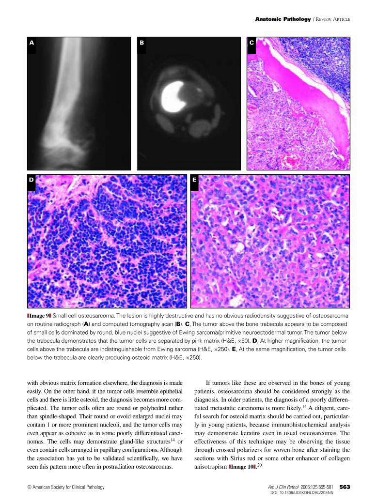
Small Cell Osteosarcoma
Small cell osteosarcoma is a rare histologic variant ofosteosarcoma with histologic features combining those ofosteosarcoma and Ewing sarcoma.18 It constitutes between1% and 2% of all osteosarcomas.19 The radiologic featuresare not consistently typical for osteosarcoma because thereoften is very little mineralized matrix produced.Histologically, it may be mistaken for Ewing sarcoma/primi-tive neuroectodermal tumor (PNET) because its cells aresmall and have round, hyperchromatic nuclei with very littleof the nuclear pleomorphism characteristic of conventional
high-grade osteosarcoma. Close observation sometimesreveals spindling of tumor cells. This finding is not character-istic of the Ewing/PNET group of tumors. In addition, the pro-duction of osteoid by tumor cells is not a characteristic featureof Ewing sarcoma, whereas it always is identified to confirma diagnosis of osteosarcoma ❚Image 9❚.
Several features reported in small cell osteosarcomamake it a controversial diagnosis. First, many, if not all, smallcell osteosarcomas display a similar immunohistochemicalprofile to that of Ewing sarcoma/PNET. The most importantof these is positive membrane staining for CD99, a marker ofexpression of the MIC-2 gene product. This marker is typicalfor tumors in the Ewing sarcoma/PNET group, mesenchymalchondrosarcoma, and some primitive hematopoietic tumors,but it is not found in other types of osteosarcoma.Furthermore, the reciprocal translocation between chromo-somes 11 and 22, the most common one found in the Ewingsarcoma/PNET tumor family, occasionally has been reportedin a small cell osteosarcoma, whereas it is not a feature ofother subtypes of osteosarcoma. Some authors believe thattumors showing this translocation should be consideredEwing sarcoma/PNET regardless of whether they produceosteoid, implying that it is possible that some of the tumorsclassified as small cell osteosarcomas actually are Ewing sar-coma family tumors with divergent differentiation.9 The matter
Anatomic Pathology / REVIEW ARTICLE
A B
❚Image 7❚ Radiographic examples of sclerosingosteosarcoma. A, The lesion is cloudy, radiodense, andmetaphyseal. The inferior border is not distinct, and there islateral extension outside the bone. B, Diaphyseal lesion fromwhich the sections in Image 6 are derived. Note the veryradiodense lesion that not only has poorly defined osseousconfines but also produces interrupted, bulky, cloudy, andtrabeculated bone in the soft tissues.
562 Am J Clin Pathol 2006;125:555-581562 DOI: 10.1309/UC6KQHLD9LV2KENN
© American Society for Clinical Pathology
Klein and Siegal / OSTEOSARCOMA
is not entirely trivial because response to adjuvant chemother-apy for osteosarcoma has been variable and less successful inpatients with small cell osteosarcoma.19 Moreover, Ewing sar-comas, which are exquisitely radiosensitive, may require radi-ation for local control, but osteosarcomas are almost alwaysinsensitive to that approach.
Epithelioid Osteosarcoma
Epithelioid osteosarcoma is an osteosarcoma in which thetumor cells are so poorly differentiated that it is difficult to deter-mine histologically whether the tumor is a sarcoma or a carcino-ma.14 If there are areas of histologically typical osteosarcoma
A B
E F
C D
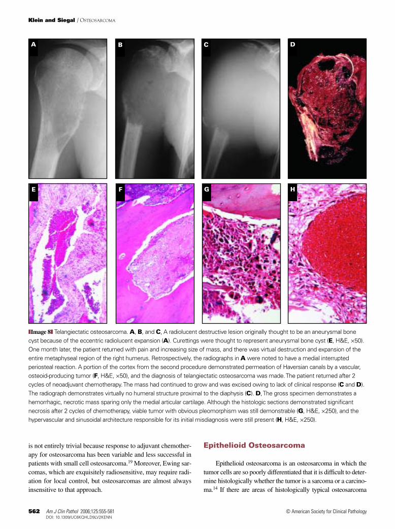
G H
❚Image 8❚ Telangiectatic osteosarcoma. A, B, and C, A radiolucent destructive lesion originally thought to be an aneurysmal bonecyst because of the eccentric radiolucent expansion (A). Curettings were thought to represent aneurysmal bone cyst (E, H&E, ×50).One month later, the patient returned with pain and increasing size of mass, and there was virtual destruction and expansion of theentire metaphyseal region of the right humerus. Retrospectively, the radiographs in A were noted to have a medial interruptedperiosteal reaction. A portion of the cortex from the second procedure demonstrated permeation of Haversian canals by a vascular,osteoid-producing tumor (F, H&E, ×50), and the diagnosis of telangiectatic osteosarcoma was made. The patient returned after 2cycles of neoadjuvant chemotherapy. The mass had continued to grow and was excised owing to lack of clinical response (C and D).The radiograph demonstrates virtually no humeral structure proximal to the diaphysis (C). D, The gross specimen demonstrates ahemorrhagic, necrotic mass sparing only the medial articular cartilage. Although the histologic sections demonstrated significantnecrosis after 2 cycles of chemotherapy, viable tumor with obvious pleomorphism was still demonstrable (G, H&E, ×250), and thehypervascular and sinusoidal architecture responsible for its initial misdiagnosis were still present (H, H&E, ×250).
Am J Clin Pathol 2006;125:555-581 563563 DOI: 10.1309/UC6KQHLD9LV2KENN 563
© American Society for Clinical Pathology
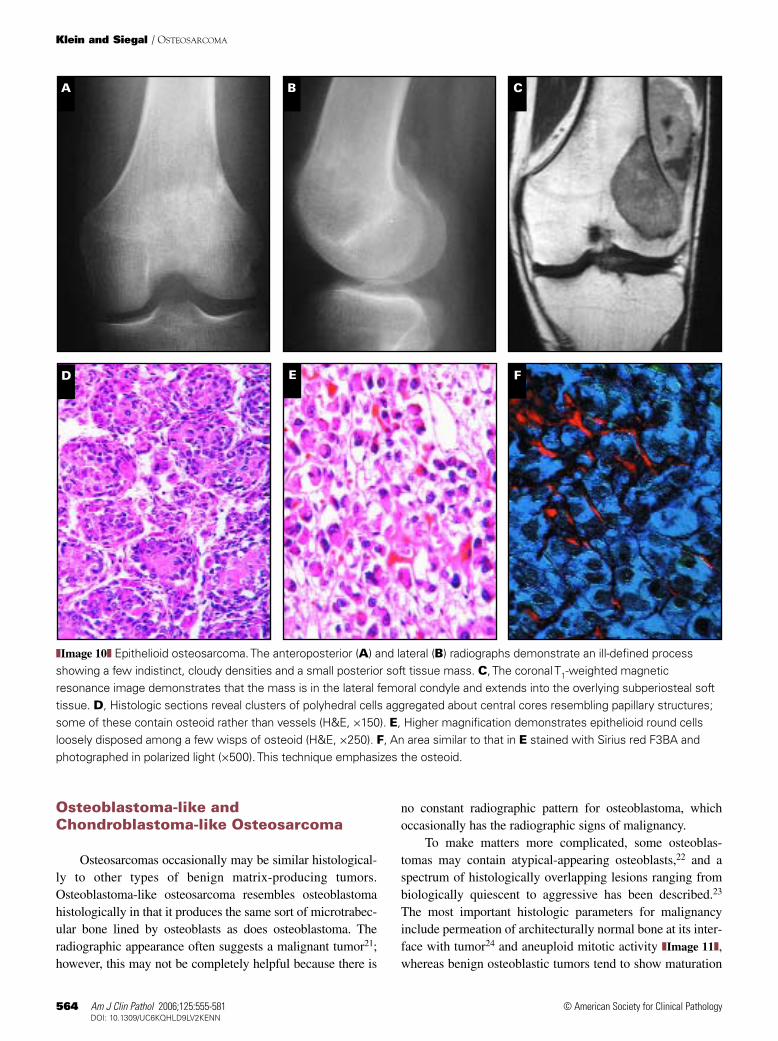
with obvious matrix formation elsewhere, the diagnosis is madeeasily. On the other hand, if the tumor cells resemble epithelialcells and there is little osteoid, the diagnosis becomes more com-plicated. The tumor cells often are round or polyhedral ratherthan spindle-shaped. Their round or ovoid enlarged nuclei maycontain 1 or more prominent nucleoli, and the tumor cells mayeven appear as cohesive as in some poorly differentiated carci-nomas. The cells may demonstrate gland-like structures14 oreven contain cells arranged in papillary configurations. Althoughthe association has yet to be validated scientifically, we haveseen this pattern more often in postradiation osteosarcomas.
If tumors like these are observed in the bones of youngpatients, osteosarcoma should be considered strongly as thediagnosis. In older patients, the diagnosis of a poorly differen-tiated metastatic carcinoma is more likely.14 A diligent, care-ful search for osteoid matrix should be carried out, particular-ly in young patients, because immunohistochemical analysismay demonstrate keratins even in usual osteosarcomas. Theeffectiveness of this technique may be observing the tissuethrough crossed polarizers for woven bone after staining thesections with Sirius red or some other enhancer of collagenanisotropism ❚Image 10❚.20
Anatomic Pathology / REVIEW ARTICLE
ED
A B C
❚Image 9❚ Small cell osteosarcoma. The lesion is highly destructive and has no obvious radiodensity suggestive of osteosarcomaon routine radiograph (A) and computed tomography scan (B). C, The tumor above the bone trabecula appears to be composedof small cells dominated by round, blue nuclei suggestive of Ewing sarcoma/primitive neuroectodermal tumor. The tumor belowthe trabecula demonstrates that the tumor cells are separated by pink matrix (H&E, ×50). D, At higher magnification, the tumorcells above the trabecula are indistinguishable from Ewing sarcoma (H&E, ×250). E, At the same magnification, the tumor cellsbelow the trabecula are clearly producing osteoid matrix (H&E, ×250).
564 Am J Clin Pathol 2006;125:555-581564 DOI: 10.1309/UC6KQHLD9LV2KENN
© American Society for Clinical Pathology
Klein and Siegal / OSTEOSARCOMA
Osteoblastoma-like andChondroblastoma-like Osteosarcoma
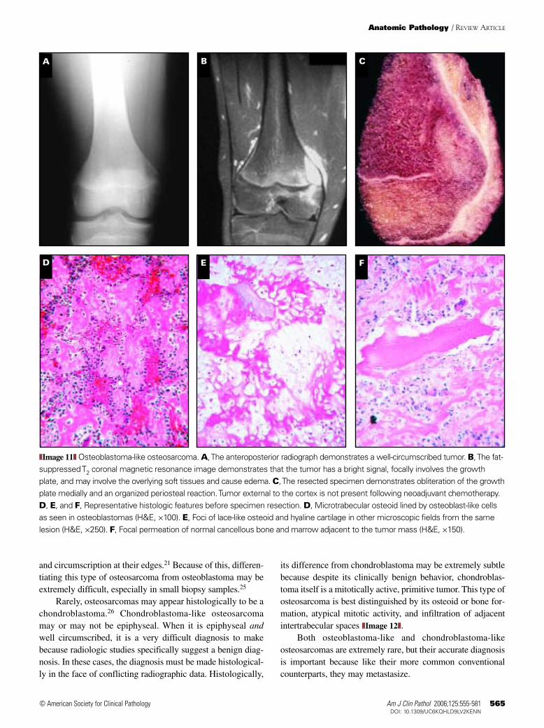
Osteosarcomas occasionally may be similar histological-ly to other types of benign matrix-producing tumors.Osteoblastoma-like osteosarcoma resembles osteoblastomahistologically in that it produces the same sort of microtrabec-ular bone lined by osteoblasts as does osteoblastoma. Theradiographic appearance often suggests a malignant tumor21;however, this may not be completely helpful because there is
no constant radiographic pattern for osteoblastoma, whichoccasionally has the radiographic signs of malignancy.
To make matters more complicated, some osteoblas-tomas may contain atypical-appearing osteoblasts,22 and aspectrum of histologically overlapping lesions ranging frombiologically quiescent to aggressive has been described.23
The most important histologic parameters for malignancyinclude permeation of architecturally normal bone at its inter-face with tumor24 and aneuploid mitotic activity ❚Image 11❚,whereas benign osteoblastic tumors tend to show maturation
A B C
D
❚Image 10❚ Epithelioid osteosarcoma. The anteroposterior (A) and lateral (B) radiographs demonstrate an ill-defined processshowing a few indistinct, cloudy densities and a small posterior soft tissue mass. C, The coronal T1-weighted magneticresonance image demonstrates that the mass is in the lateral femoral condyle and extends into the overlying subperiosteal softtissue. D, Histologic sections reveal clusters of polyhedral cells aggregated about central cores resembling papillary structures;some of these contain osteoid rather than vessels (H&E, ×150). E, Higher magnification demonstrates epithelioid round cellsloosely disposed among a few wisps of osteoid (H&E, ×250). F, An area similar to that in E stained with Sirius red F3BA andphotographed in polarized light (×500). This technique emphasizes the osteoid.
E F
Am J Clin Pathol 2006;125:555-581 565565 DOI: 10.1309/UC6KQHLD9LV2KENN 565
© American Society for Clinical Pathology
and circumscription at their edges.21 Because of this, differen-tiating this type of osteosarcoma from osteoblastoma may beextremely difficult, especially in small biopsy samples.25
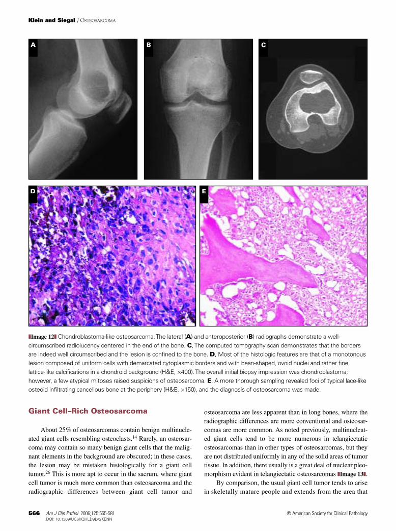
Rarely, osteosarcomas may appear histologically to be achondroblastoma.26 Chondroblastoma-like osteosarcomamay or may not be epiphyseal. When it is epiphyseal andwell circumscribed, it is a very difficult diagnosis to makebecause radiologic studies specifically suggest a benign diag-nosis. In these cases, the diagnosis must be made histological-ly in the face of conflicting radiographic data. Histologically,
its difference from chondroblastoma may be extremely subtlebecause despite its clinically benign behavior, chondroblas-toma itself is a mitotically active, primitive tumor. This type ofosteosarcoma is best distinguished by its osteoid or bone for-mation, atypical mitotic activity, and infiltration of adjacentintertrabecular spaces ❚Image 12❚.
Both osteoblastoma-like and chondroblastoma-likeosteosarcomas are extremely rare, but their accurate diagnosisis important because like their more common conventionalcounterparts, they may metastasize.
Anatomic Pathology / REVIEW ARTICLE
A B C
D E F
❚Image 11❚ Osteoblastoma-like osteosarcoma. A, The anteroposterior radiograph demonstrates a well-circumscribed tumor. B, The fat-suppressed T2 coronal magnetic resonance image demonstrates that the tumor has a bright signal, focally involves the growthplate, and may involve the overlying soft tissues and cause edema. C, The resected specimen demonstrates obliteration of the growthplate medially and an organized periosteal reaction. Tumor external to the cortex is not present following neoadjuvant chemotherapy.D, E, and F, Representative histologic features before specimen resection. D, Microtrabecular osteoid lined by osteoblast-like cellsas seen in osteoblastomas (H&E, ×100). E, Foci of lace-like osteoid and hyaline cartilage in other microscopic fields from the samelesion (H&E, ×250). F, Focal permeation of normal cancellous bone and marrow adjacent to the tumor mass (H&E, ×150).
566 Am J Clin Pathol 2006;125:555-581566 DOI: 10.1309/UC6KQHLD9LV2KENN
© American Society for Clinical Pathology
Klein and Siegal / OSTEOSARCOMA
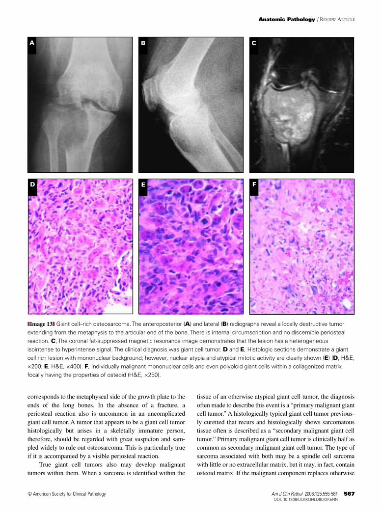
Giant Cell–Rich Osteosarcoma
About 25% of osteosarcomas contain benign multinucle-ated giant cells resembling osteoclasts.14 Rarely, an osteosar-coma may contain so many benign giant cells that the malig-nant elements in the background are obscured; in these cases,the lesion may be mistaken histologically for a giant celltumor.26 This is more apt to occur in the sacrum, where giantcell tumor is much more common than osteosarcoma and theradiographic differences between giant cell tumor and
osteosarcoma are less apparent than in long bones, where theradiographic differences are more conventional and osteosar-comas are more common. As noted previously, multinucleat-ed giant cells tend to be more numerous in telangiectaticosteosarcomas than in other types of osteosarcomas, but theyare not distributed uniformly in any of the solid areas of tumortissue. In addition, there usually is a great deal of nuclear pleo-morphism evident in telangiectatic osteosarcomas ❚Image 13❚.
By comparison, the usual giant cell tumor tends to arisein skeletally mature people and extends from the area that
ED
A B C
❚Image 12❚ Chondroblastoma-like osteosarcoma. The lateral (A) and anteroposterior (B) radiographs demonstrate a well-circumscribed radiolucency centered in the end of the bone. C, The computed tomography scan demonstrates that the bordersare indeed well circumscribed and the lesion is confined to the bone. D, Most of the histologic features are that of a monotonouslesion composed of uniform cells with demarcated cytoplasmic borders and with bean-shaped, ovoid nuclei and rather fine,lattice-like calcifications in a chondroid background (H&E, ×400). The overall initial biopsy impression was chondroblastoma;however, a few atypical mitoses raised suspicions of osteosarcoma. E, A more thorough sampling revealed foci of typical lace-likeosteoid infiltrating cancellous bone at the periphery (H&E, ×150), and the diagnosis of osteosarcoma was made.
Am J Clin Pathol 2006;125:555-581 567567 DOI: 10.1309/UC6KQHLD9LV2KENN 567
© American Society for Clinical Pathology
corresponds to the metaphyseal side of the growth plate to theends of the long bones. In the absence of a fracture, aperiosteal reaction also is uncommon in an uncomplicatedgiant cell tumor. A tumor that appears to be a giant cell tumorhistologically but arises in a skeletally immature person,therefore, should be regarded with great suspicion and sam-pled widely to rule out osteosarcoma. This is particularly trueif it is accompanied by a visible periosteal reaction.
True giant cell tumors also may develop malignanttumors within them. When a sarcoma is identified within the
tissue of an otherwise atypical giant cell tumor, the diagnosisoften made to describe this event is a “primary malignant giantcell tumor.” A histologically typical giant cell tumor previous-ly curetted that recurs and histologically shows sarcomatoustissue often is described as a “secondary malignant giant celltumor.” Primary malignant giant cell tumor is clinically half ascommon as secondary malignant giant cell tumor. The type ofsarcoma associated with both may be a spindle cell sarcomawith little or no extracellular matrix, but it may, in fact, containosteoid matrix. If the malignant component replaces otherwise
Anatomic Pathology / REVIEW ARTICLE
A B C
D E F
❚Image 13❚ Giant cell–rich osteosarcoma. The anteroposterior (A) and lateral (B) radiographs reveal a locally destructive tumorextending from the metaphysis to the articular end of the bone. There is internal circumscription and no discernible periostealreaction. C, The coronal fat-suppressed magnetic resonance image demonstrates that the lesion has a heterogeneousisointense to hyperintense signal. The clinical diagnosis was giant cell tumor. D and E, Histologic sections demonstrate a giantcell rich lesion with mononuclear background; however, nuclear atypia and atypical mitotic activity are clearly shown (E) (D, H&E,×200; E, H&E, ×400). F, Individually malignant mononuclear cells and even polyploid giant cells within a collagenized matrixfocally having the properties of osteoid (H&E, ×250).
568 Am J Clin Pathol 2006;125:555-581568 DOI: 10.1309/UC6KQHLD9LV2KENN
© American Society for Clinical Pathology
Klein and Siegal / OSTEOSARCOMA
typically conventional areas of giant cell tumor withoutreplacing all of the giant cells, it would be easy to mistake thelesion for a giant cell tumor if there is little matrix present. Itis possible that this also is a source of some of the giantcell–rich osteosarcomas reported in the literature.
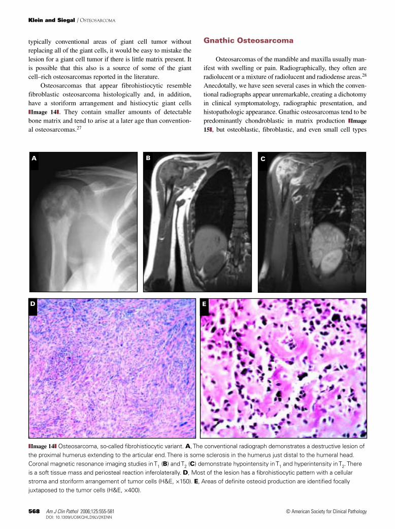
Osteosarcomas that appear fibrohistiocytic resemblefibroblastic osteosarcoma histologically and, in addition,have a storiform arrangement and histiocytic giant cells❚Image 14❚. They contain smaller amounts of detectablebone matrix and tend to arise at a later age than convention-al osteosarcomas.27
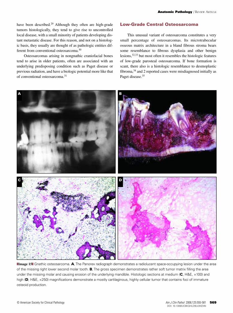
Gnathic Osteosarcoma
Osteosarcomas of the mandible and maxilla usually man-ifest with swelling or pain. Radiographically, they often areradiolucent or a mixture of radiolucent and radiodense areas.28
Anecdotally, we have seen several cases in which the conven-tional radiographs appear unremarkable, creating a dichotomyin clinical symptomatology, radiographic presentation, andhistopathologic appearance. Gnathic osteosarcomas tend to bepredominantly chondroblastic in matrix production ❚Image
15❚, but osteoblastic, fibroblastic, and even small cell types
ED
A B C
❚Image 14❚ Osteosarcoma, so-called fibrohistiocytic variant. A, The conventional radiograph demonstrates a destructive lesion ofthe proximal humerus extending to the articular end. There is some sclerosis in the humerus just distal to the humeral head.Coronal magnetic resonance imaging studies in T1 (B) and T2 (C) demonstrate hypointensity in T1 and hyperintensity in T2. Thereis a soft tissue mass and periosteal reaction inferolaterally. D, Most of the lesion has a fibrohistiocytic pattern with a cellularstroma and storiform arrangement of tumor cells (H&E, ×150). E, Areas of definite osteoid production are identified focallyjuxtaposed to the tumor cells (H&E, ×400).
Am J Clin Pathol 2006;125:555-581 569569 DOI: 10.1309/UC6KQHLD9LV2KENN 569
© American Society for Clinical Pathology
have been described.29 Although they often are high-gradetumors histologically, they tend to give rise to uncontrolledlocal disease, with a small minority of patients developing dis-tant metastatic disease. For this reason, and not on a histolog-ic basis, they usually are thought of as pathologic entities dif-ferent from conventional osteosarcoma.30
Osteosarcomas arising in nongnathic craniofacial bonestend to arise in older patients, often are associated with anunderlying predisposing condition such as Paget disease orprevious radiation, and have a biologic potential more like thatof conventional osteosarcoma.31
Low-Grade Central Osteosarcoma
This unusual variant of osteosarcoma constitutes a verysmall percentage of osteosarcomas. Its microtrabecularosseous matrix architecture in a bland fibrous stroma bearssome resemblance to fibrous dysplasia and other benignlesions,32,33 but most often it resembles the histologic featuresof low-grade parosteal osteosarcoma. If bone formation isscant, there also is a histologic resemblance to desmoplasticfibroma,34 and 2 reported cases were misdiagnosed initially asPaget disease.35
Anatomic Pathology / REVIEW ARTICLE
A
DC
B
❚Image 15❚ Gnathic osteosarcoma. A, The Panorex radiograph demonstrates a radiolucent space-occupying lesion under the areaof the missing right lower second molar tooth. B, The gross specimen demonstrates rather soft tumor matrix filling the areaunder the missing molar and causing erosion of the underlying mandible. Histologic sections at medium (C, H&E, ×100) andhigh (D, H&E, ×250) magnifications demonstrate a mostly cartilaginous, highly cellular tumor that contains foci of immatureosteoid production.

570 Am J Clin Pathol 2006;125:555-581570 DOI: 10.1309/UC6KQHLD9LV2KENN
© American Society for Clinical Pathology
Klein and Siegal / OSTEOSARCOMA
Accurate diagnosis is predicated on satisfactory correlationwith clinical imaging studies and very careful evaluation of his-tologic findings ❚Image 16❚. The imaging features are not invari-ably diagnostic, but they often suggest a lesion of greater localaggressiveness than fibrous dysplasia or other benignlesions.36 These features may include indistinct circumscrip-tion, dense sclerosis, an interrupted periosteal reaction, or corti-cal infraction. Sections through the center of the lesion usually
demonstrate woven microtrabeculae of bone in a moderatelycellular, fibrous stroma. If sections include the interface ofthe lesion with normal bone, fibrous tissue within Haversiancanals or between mature cancellous trabeculae is a telltalesign of malignancy.32 In the absence of these findings, atypi-cal mitotic activity may lead to the correct diagnosis.Although patients usually are treated with surgery alone andtheir prognosis is significantly better than in conventional
A B C
D E F
❚Image 16❚ Low-grade central osteosarcoma. The anteroposterior (A) and lateral (B) radiographs demonstrate a rather mottled,deceptively circumscribed lesion that resembles fibrous dysplasia except for the very focal area of cortical erosion of the lateralfemoral metadiaphysis. C, The fat-suppressed magnetic resonance image in coronal view demonstrates actual extension of thehyperintense mass into the soft tissue. This finding is never seen in uncomplicated fibrous dysplasia. D, The majority of thehistologic features are immature, poorly formed, woven trabeculae without appositional osteoblastic activity in a modestlycellular fibrous stroma (H&E, ×250). This greatly imitates fibrous dysplasia except for the atypical features in the radiographs. E,A search of nuclei at higher magnification revealed not only mitotic activity but also atypical mitoses (H&E, ×787). A diagnosis oflow-grade central osteosarcoma prompted removal of the distal femur. F, A section through the cortex demonstratedpermeation of the Haversian systems by tumor matrix (asterisk) (H&E, ×100).
Am J Clin Pathol 2006;125:555-581 571571 DOI: 10.1309/UC6KQHLD9LV2KENN 571
© American Society for Clinical Pathology
osteosarcoma,37 a certain number of patients develop a sec-ondary high-grade osteosarcoma (dedifferentiation) withinthe original low-grade tumor or at its previous anatomic siteof origin in local recurrences.33,38,39 It is probable that somecases of fibrous dysplasia associated with secondary develop-ment of osteosarcomas are, in fact, low-grade osteosarcomasthat later became higher grade.
Surface Osteosarcomas
Surface osteosarcomas are osteosarcomas whose epicen-ters are outside the cortex of the underlying bone. They usual-ly arise in relation to the periosteum or the cortex of the bonewith minimal or no involvement of the medullary cavity.Unlike conventional osteosarcomas, which are tumors of ado-lescents, these lesions usually arise in patients in the third andfourth decades.
There are several types of surface osteosarcomas, whichhave been characterized by exact anatomic location, by the pre-dominant type of matrix, and by histologic grade. The majori-ty of surface osteosarcomas are low-grade neoplasms with apropensity for local recurrence and a real but very limitedcapacity for distant metastasis. A relatively small number(approximately 10%) of surface osteosarcomas are high-gradetumors. The current classification system identifies indolentand high-grade tumors, so it is able to group the lesions need-ing specific kinds of therapy.40
Parosteal Osteosarcoma
Geschicter and Copeland41 first described the most com-mon form of surface osteosarcoma, parosteal osteosarcoma,although the term currently used for it was formulated byDwinnell et al.42 This lesion often had been confused withosteochondroma and osteoma; however, its local recurrencerate when incompletely excised and its small but definitepropensity for distant spread made it clear that it behaved clin-ically as a uniquely different entity. Parosteal osteosarcomaaccounts for fewer than 1 in 20 osteosarcomas, and its averageincidence is about a decade later than the usual type of centralosteosarcoma. It has a unique presentation and anatomic distri-bution because 75% to 80% of cases arise as denselyradiopaque masses attached to the distal posterior femur.
Although parosteal osteosarcoma may give rise to a sensa-tion of fullness, prevent complete range of flexion, and even beperceived as a mass through the overlying soft tissues, it isunusual for the entity to be spontaneously painful. Its radioden-sity often is not uniform, often being more radiodense centrallyas it approaches the bone surface and less dense at its periphery.In contrast with osteochondroma, there is no usual continuity ofthe exterior of the mass with the adjacent cortex, and its insideis not in continuity with the underlying medullary cavity.
In localized heterotopic ossification (so-called myositisossificans circumscripta) arising near the bone surface, theossification pattern is exactly the opposite radiographically,with the densest bone being outside and the least densebeing inside.
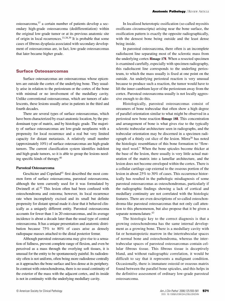
In parosteal osteosarcoma, there often is an incompleteradiolucent line separating most of the sclerotic mass fromthe underlying cortex ❚Image 17❚. When a resected specimenis examined carefully, especially with specimen radiography,this radiolucent line corresponds to the underling perios-teum, to which the mass usually is fixed at one point on theoutside. An underlying periosteal reaction is very unusualbecause to produce such a reaction, the tumor would have tolift the inner cambium layer of the periosteum away from thecortex. Parosteal osteosarcoma usually is not locally aggres-sive enough to do this.
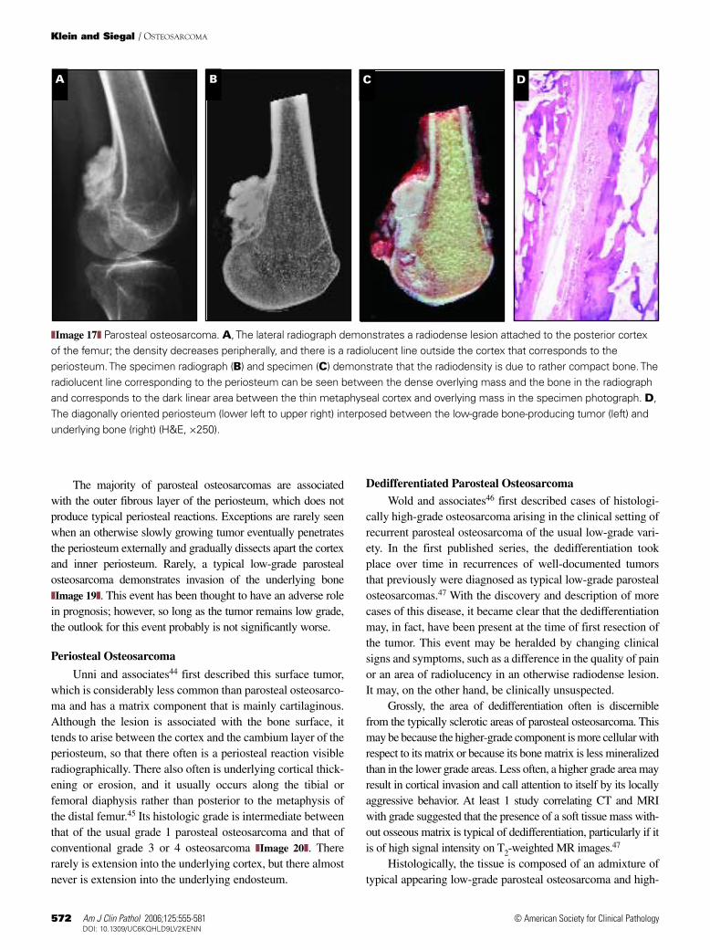
Histologically, parosteal osteosarcomas consist ofstreamers of bone trabeculae that often show a high degreeof parallel orientation similar to what might be observed in aperiosteal new bone reaction ❚Image 18❚. This concentrationand arrangement of bone is what gives rise to the typicallysclerotic trabecular architecture seen in radiographs, and thetrabecular orientation may be discerned in a specimen radi-ograph of a thinly cut slice of the lesion. Mirra26 has notedthe histologic resemblance of this bone formation to “flow-ing steel wool.” When the bone spicules become thicker atthe base of the lesion, there usually is very little actual mat-uration of the matrix into a lamellar architecture, and thelesion does not become enveloped within the cortex. There isa cellular cartilage cap external to the osseous portion of thelesion in about 25% to 30% of cases. This occurrence histor-ically has resulted in the pathologic misdiagnosis of someparosteal osteosarcomas as osteochondromas, particularly ifthe radiographic findings showing a lack of cortical andmedullary continuity are not correlated with the histologicfeatures. There are even descriptions of so-called osteochon-droma-like parosteal osteosarcomas that not only call atten-tion to this phenomenon, but also propose that it be given aseparate nomenclature.43
The histologic key to the correct diagnosis is that agrowing osteochondroma has the same internal develop-ment as a growing bone. There is a medullary cavity withfat or hematopoietic marrow in the intertrabecular spacesof normal bone and osteochondroma, whereas the inter-trabecular spaces of parosteal osteosarcomas contain cel-lular fibrous tissue. This fibrous tissue is deceptivelybland, and without radiographic correlation, it would bedifficult to say that it represents a malignant condition.Occasionally, there is immature osteoid or osseous matrixfound between the parallel bone spicules, and this helps inthe definitive assessment of ordinary low-grade parostealosteosarcoma.
Anatomic Pathology / REVIEW ARTICLE
572 Am J Clin Pathol 2006;125:555-581572 DOI: 10.1309/UC6KQHLD9LV2KENN
© American Society for Clinical Pathology
Klein and Siegal / OSTEOSARCOMA
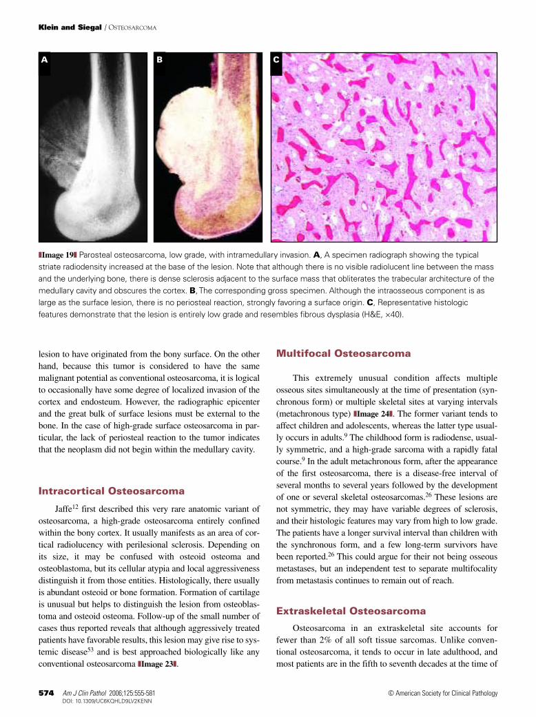
The majority of parosteal osteosarcomas are associatedwith the outer fibrous layer of the periosteum, which does notproduce typical periosteal reactions. Exceptions are rarely seenwhen an otherwise slowly growing tumor eventually penetratesthe periosteum externally and gradually dissects apart the cortexand inner periosteum. Rarely, a typical low-grade parostealosteosarcoma demonstrates invasion of the underlying bone❚Image 19❚. This event has been thought to have an adverse rolein prognosis; however, so long as the tumor remains low grade,the outlook for this event probably is not significantly worse.
Periosteal Osteosarcoma
Unni and associates44 first described this surface tumor,which is considerably less common than parosteal osteosarco-ma and has a matrix component that is mainly cartilaginous.Although the lesion is associated with the bone surface, ittends to arise between the cortex and the cambium layer of theperiosteum, so that there often is a periosteal reaction visibleradiographically. There also often is underlying cortical thick-ening or erosion, and it usually occurs along the tibial orfemoral diaphysis rather than posterior to the metaphysis ofthe distal femur.45 Its histologic grade is intermediate betweenthat of the usual grade 1 parosteal osteosarcoma and that ofconventional grade 3 or 4 osteosarcoma ❚Image 20❚. Thererarely is extension into the underlying cortex, but there almostnever is extension into the underlying endosteum.
Dedifferentiated Parosteal OsteosarcomaWold and associates46 first described cases of histologi-
cally high-grade osteosarcoma arising in the clinical setting ofrecurrent parosteal osteosarcoma of the usual low-grade vari-ety. In the first published series, the dedifferentiation tookplace over time in recurrences of well-documented tumorsthat previously were diagnosed as typical low-grade parostealosteosarcomas.47 With the discovery and description of morecases of this disease, it became clear that the dedifferentiationmay, in fact, have been present at the time of first resection ofthe tumor. This event may be heralded by changing clinicalsigns and symptoms, such as a difference in the quality of painor an area of radiolucency in an otherwise radiodense lesion.It may, on the other hand, be clinically unsuspected.
Grossly, the area of dedifferentiation often is discerniblefrom the typically sclerotic areas of parosteal osteosarcoma. Thismay be because the higher-grade component is more cellular withrespect to its matrix or because its bone matrix is less mineralizedthan in the lower grade areas. Less often, a higher grade area mayresult in cortical invasion and call attention to itself by its locallyaggressive behavior. At least 1 study correlating CT and MRIwith grade suggested that the presence of a soft tissue mass with-out osseous matrix is typical of dedifferentiation, particularly if itis of high signal intensity on T2-weighted MR images.47
Histologically, the tissue is composed of an admixture oftypical appearing low-grade parosteal osteosarcoma and high-
A C DB
❚Image 17❚ Parosteal osteosarcoma. A, The lateral radiograph demonstrates a radiodense lesion attached to the posterior cortexof the femur; the density decreases peripherally, and there is a radiolucent line outside the cortex that corresponds to theperiosteum. The specimen radiograph (B) and specimen (C) demonstrate that the radiodensity is due to rather compact bone. Theradiolucent line corresponding to the periosteum can be seen between the dense overlying mass and the bone in the radiographand corresponds to the dark linear area between the thin metaphyseal cortex and overlying mass in the specimen photograph. D,The diagonally oriented periosteum (lower left to upper right) interposed between the low-grade bone-producing tumor (left) andunderlying bone (right) (H&E, ×250).
Am J Clin Pathol 2006;125:555-581 573573 DOI: 10.1309/UC6KQHLD9LV2KENN 573
© American Society for Clinical Pathology
grade conventional parosteal osteosarcoma ❚Image 21❚. Thededifferentiated component has at least once been reported asbeing telangiectatic,48 and it is possible that other varieties maybe described as larger series are obtained. The prognosis isrelated to that of the least differentiated tumor component; it isworse than that of low-grade parosteal osteosarcoma but seemsto be somewhat better than that of pure high-grade surfaceosteosarcomas.49 At least 1 series suggests that patients withthis disease derive benefit from adjuvant chemotherapy.50
According to the recently published experience from theRizzoli Institute, dedifferentiation occurs in approximately 1 in4 low-grade parosteal osteosarcomas.49
High-Grade Surface Osteosarcoma
This tumor, which manifests as a surface lesion of bone,is entirely high grade histologically.51 It may appear radi-ographically as an ordinary low-grade parosteal osteosarcoma
with dense sclerosis. Alternatively, it may have mixed sclero-sis and radiolucency, or, occasionally, it may form a soft tissuemass with relatively little radiodensity. Because it is a highergrade lesion than ordinary parosteal osteosarcoma, its localgrowth and aggressiveness are more accelerated.Consequently, patients with this disease are more likely tohave more distressing symptoms and signs than those withusually low-grade parosteal osteosarcomas.
Microscopically, the tumor is entirely high grade ❚Image
22❚. It is possible that some high-grade surface osteosarcomasrepresent dedifferentiated parosteal osteosarcomas in whichthe high-grade component has replaced entirely the low-gradecomponent. There usually is very little or no medullary inva-sion by the tumor in conventional radiography, although CTand MRI demonstrate occasional small foci of marrowinvolvement.52 If medullary invasion were a constant feature,it would be more difficult to make a case pathologically for the
Anatomic Pathology / REVIEW ARTICLE
BA C
D ❚Image 18❚ Parosteal osteosarcoma. A, Low-powermagnification demonstrates the typical “pulled steel wool”pattern of bony trabeculae (H&E, ×25). B, The cartilage cap isidentified in approximately 1 in 3 cases of parostealosteosarcoma (H&E, ×40). This is distinguished fromosteochondroma because normal endochondral ossificationgives rise to fatty or hematopoietic bone marrow in theintertrabecular spaces. Parosteal osteosarcoma has fibroustissue between the trabeculae. C, Higher magnificationdemonstrates thickening and maturation of bone spicules at thebase of the tumor (H&E, ×250). D, Although the fibrous tissuebetween the bone spicules is bland, there is no marrow evident.A careful search in the fibrous component sometimes revealsfocal hypercellularity and early immature bone formation, butthis is usually not a frequent feature (H&E, ×400).
574 Am J Clin Pathol 2006;125:555-581574 DOI: 10.1309/UC6KQHLD9LV2KENN
© American Society for Clinical Pathology
Klein and Siegal / OSTEOSARCOMA
lesion to have originated from the bony surface. On the otherhand, because this tumor is considered to have the samemalignant potential as conventional osteosarcoma, it is logicalto occasionally have some degree of localized invasion of thecortex and endosteum. However, the radiographic epicenterand the great bulk of surface lesions must be external to thebone. In the case of high-grade surface osteosarcoma in par-ticular, the lack of periosteal reaction to the tumor indicatesthat the neoplasm did not begin within the medullary cavity.
Intracortical Osteosarcoma
Jaffe12 first described this very rare anatomic variant ofosteosarcoma, a high-grade osteosarcoma entirely confinedwithin the bony cortex. It usually manifests as an area of cor-tical radiolucency with perilesional sclerosis. Depending onits size, it may be confused with osteoid osteoma andosteoblastoma, but its cellular atypia and local aggressivenessdistinguish it from those entities. Histologically, there usuallyis abundant osteoid or bone formation. Formation of cartilageis unusual but helps to distinguish the lesion from osteoblas-toma and osteoid osteoma. Follow-up of the small number ofcases thus reported reveals that although aggressively treatedpatients have favorable results, this lesion may give rise to sys-temic disease53 and is best approached biologically like anyconventional osteosarcoma ❚Image 23❚.
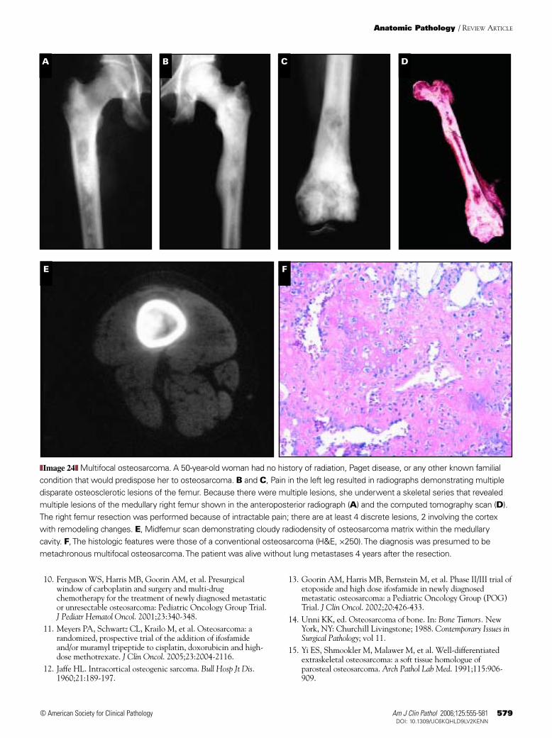
Multifocal Osteosarcoma
This extremely unusual condition affects multipleosseous sites simultaneously at the time of presentation (syn-chronous form) or multiple skeletal sites at varying intervals(metachronous type) ❚Image 24❚. The former variant tends toaffect children and adolescents, whereas the latter type usual-ly occurs in adults.9 The childhood form is radiodense, usual-ly symmetric, and a high-grade sarcoma with a rapidly fatalcourse.9 In the adult metachronous form, after the appearanceof the first osteosarcoma, there is a disease-free interval ofseveral months to several years followed by the developmentof one or several skeletal osteosarcomas.26 These lesions arenot symmetric, they may have variable degrees of sclerosis,and their histologic features may vary from high to low grade.The patients have a longer survival interval than children withthe synchronous form, and a few long-term survivors havebeen reported.26 This could argue for their not being osseousmetastases, but an independent test to separate multifocalityfrom metastasis continues to remain out of reach.
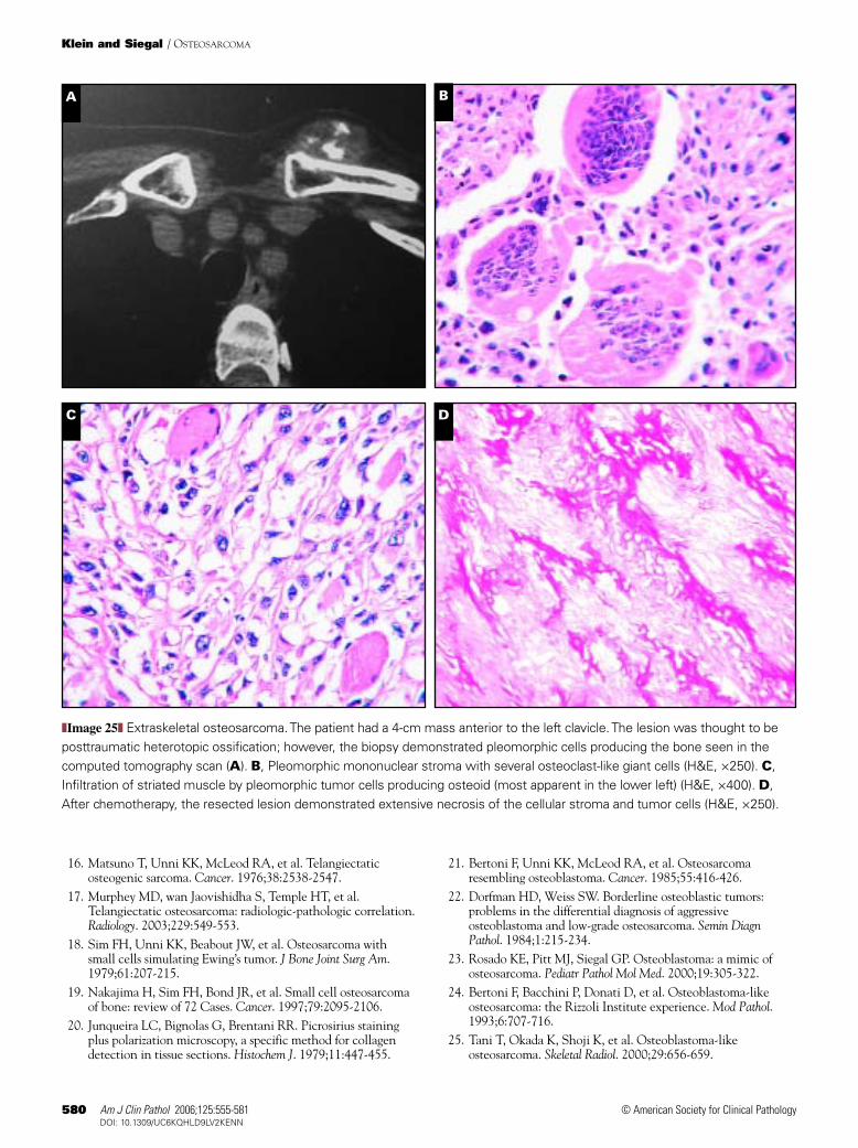
Extraskeletal Osteosarcoma
Osteosarcoma in an extraskeletal site accounts forfewer than 2% of all soft tissue sarcomas. Unlike conven-tional osteosarcoma, it tends to occur in late adulthood, andmost patients are in the fifth to seventh decades at the time of
BA C
❚Image 19❚ Parosteal osteosarcoma, low grade, with intramedullary invasion. A, A specimen radiograph showing the typicalstriate radiodensity increased at the base of the lesion. Note that although there is no visible radiolucent line between the massand the underlying bone, there is dense sclerosis adjacent to the surface mass that obliterates the trabecular architecture of themedullary cavity and obscures the cortex. B, The corresponding gross specimen. Although the intraosseous component is aslarge as the surface lesion, there is no periosteal reaction, strongly favoring a surface origin. C, Representative histologicfeatures demonstrate that the lesion is entirely low grade and resembles fibrous dysplasia (H&E, ×40).
Am J Clin Pathol 2006;125:555-581 575575 DOI: 10.1309/UC6KQHLD9LV2KENN 575
© American Society for Clinical Pathology
diagnosis.54 Most cases arise in the deep soft tissues with apredilection for the thigh followed by the buttocks, upper extrem-ities, and the retroperitoneum.55 Patients usually have an enlarg-ing soft tissue mass, and the mass may be painful. Radiographsshow a mass that may demonstrate mineralization, and CT scansincrease the yield of cases that demonstrate mineralization.
Histologically, the tumors demonstrate all types ofosteosarcoma seen in tumors that arise in bone ❚Image 25❚. Themost unusual histologic subtype is that of low-grade osteosar-coma. When the latter occurs, it has been described as the“soft tissue homologue of parosteal osteosarcoma,”15
although it could as easily have been described as the soft tissue
Anatomic Pathology / REVIEW ARTICLE
FE
DA CB
HG
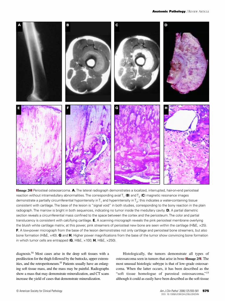
❚Image 20❚ Periosteal osteosarcoma. A, The lateral radiograph demonstrates a localized, interrupted, hair-on-end periostealreaction without intramedullary abnormalities. The corresponding axial T1 (B) and T2 (C) magnetic resonance imagesdemonstrate a partially circumferential hypointensity in T1 and hyperintensity in T2; this indicates a water-containing tissueconsistent with cartilage. The base of the lesion is “signal void” in both studies, corresponding to the bony reaction in the plainradiograph. The marrow is bright in both sequences, indicating no tumor inside the medullary cavity. D, A partial diametricsection reveals a circumferential mass confined to the space between the cortex and the periosteum. The color and partialtranslucency is consistent with calcifying cartilage. E, A scanning micrograph reveals the pink periosteal membrane overlyingthe bluish white cartilage matrix; at this power, pink streamers of periosteal new bone are seen within the cartilage (H&E, ×25).F, A low-power micrograph from the base of the lesion demonstrates not only cartilage and periosteal bone streamers, but alsobone formation (H&E, ×40). G and H, Higher power magnifications from the base of the tumor show convincing bone formationin which tumor cells are entrapped (G, H&E, ×100; H, H&E, ×250).
576 Am J Clin Pathol 2006;125:555-581576 DOI: 10.1309/UC6KQHLD9LV2KENN
© American Society for Clinical Pathology
Klein and Siegal / OSTEOSARCOMA
homologue of low-grade central osteosarcoma. Recently,there was a reported case of high-grade dedifferentiation in alow-grade extraskeletal osteosarcoma analogous to that seenin parosteal and low-grade central osteosarcomas.56
The prognosis is poor in soft tissue osteosarcoma ofthe high-grade type, with about 75% of patients dying ofthe tumor within 5 years of diagnosis.7 Anecdotally,patients with the low-grade variant seem to have a better
A B C
D E F
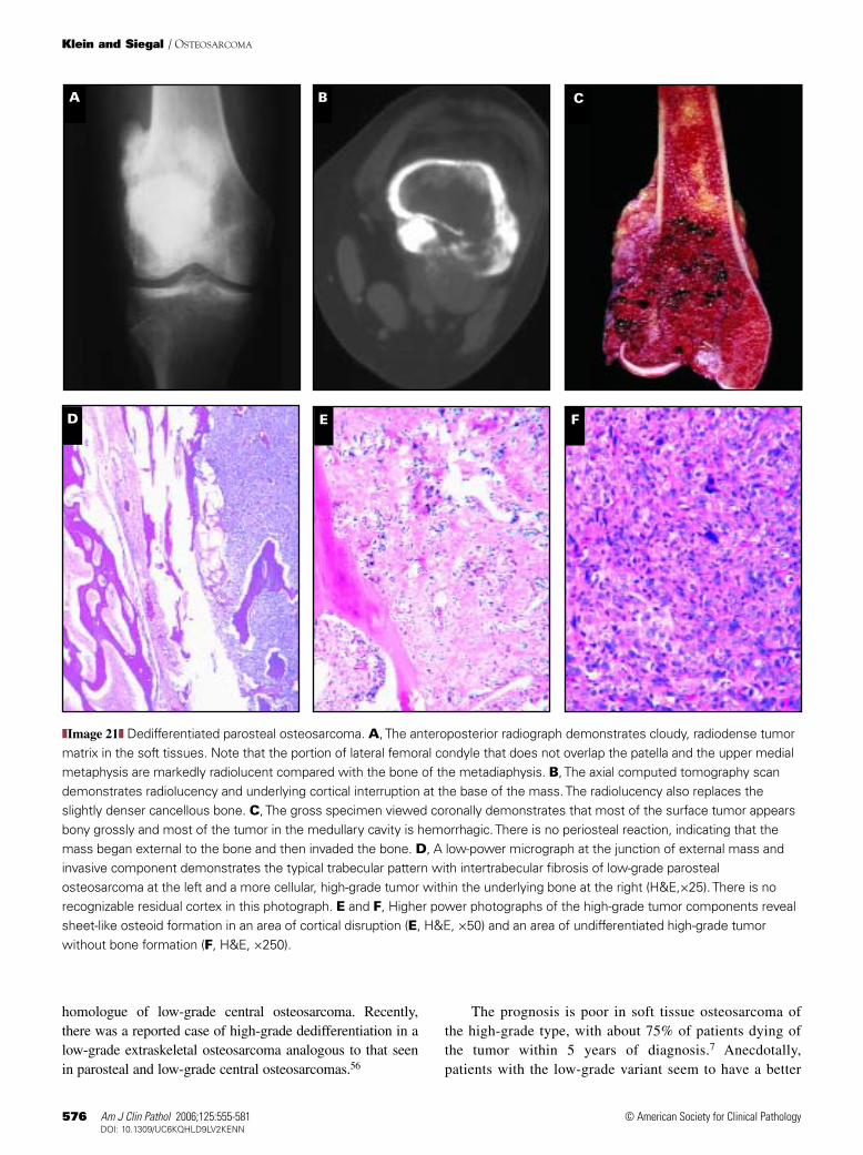
❚Image 21❚ Dedifferentiated parosteal osteosarcoma. A, The anteroposterior radiograph demonstrates cloudy, radiodense tumormatrix in the soft tissues. Note that the portion of lateral femoral condyle that does not overlap the patella and the upper medialmetaphysis are markedly radiolucent compared with the bone of the metadiaphysis. B, The axial computed tomography scandemonstrates radiolucency and underlying cortical interruption at the base of the mass. The radiolucency also replaces theslightly denser cancellous bone. C, The gross specimen viewed coronally demonstrates that most of the surface tumor appearsbony grossly and most of the tumor in the medullary cavity is hemorrhagic. There is no periosteal reaction, indicating that themass began external to the bone and then invaded the bone. D, A low-power micrograph at the junction of external mass andinvasive component demonstrates the typical trabecular pattern with intertrabecular fibrosis of low-grade parostealosteosarcoma at the left and a more cellular, high-grade tumor within the underlying bone at the right (H&E,×25). There is norecognizable residual cortex in this photograph. E and F, Higher power photographs of the high-grade tumor components revealsheet-like osteoid formation in an area of cortical disruption (E, H&E, ×50) and an area of undifferentiated high-grade tumorwithout bone formation (F, H&E, ×250).
Am J Clin Pathol 2006;125:555-581 577577 DOI: 10.1309/UC6KQHLD9LV2KENN 577
© American Society for Clinical Pathology
prognosis, although the total number of cases in the liter-ature may still be too small to make accurate statisticalanalyses.56,57
From the Departments of Pathology, Cell Biology, and Surgery,University of Alabama at Birmingham and the BirminghamVeterans Affairs Medical Center.
Funded in part by grants AR46031, CA93796, and CA98543from the National Institutes of Health, Bethesda, MD, and theHaley’s Hope Memorial Support Fund for OsteosarcomaResearch, University of Alabama at Birmingham.
Address reprint requests to Dr Klein: Section of SurgicalPathology, Department of Pathology, KB 506, University ofAlabama at Birmingham, Birmingham, AL 35233.
References1. Cotterill SJ, Wright CM, Pearce MS, et al. Stature of young
people with malignant bone tumors. Pediatr Blood Cancer.2004;42:59-63.
2. Porter DE, Holden ST, Steel CM, et al. A significantproportion of patients with osteosarcoma may belong to Li-Fraumeni cancer families. J Bone Joint Surg Br. 1992;74:883-886.
Anatomic Pathology / REVIEW ARTICLE
A
DC
B
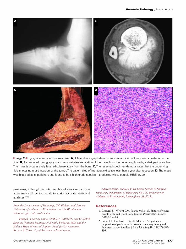
❚Image 22❚ High-grade surface osteosarcoma. A, A lateral radiograph demonstrates a radiodense tumor mass posterior to thetibia. B, A computed tomography scan demonstrates separation of the mass from the underlying bone by a dark periosteal line.The mass is progressively less radiodense away from the bone. C, The resected specimen demonstrates that the underlyingtibia shows no gross invasion by the tumor. The patient died of metastatic disease less than a year after resection. D, The masswas biopsied at its periphery and found to be a high-grade neoplasm producing wispy osteoid (H&E, ×200).
578 Am J Clin Pathol 2006;125:555-581578 DOI: 10.1309/UC6KQHLD9LV2KENN
© American Society for Clinical Pathology
Klein and Siegal / OSTEOSARCOMA
3. Ragland BD, Bell WC, Lopez RR, et al. Cytogenetics andmolecular biology of osteosarcoma. Lab Invest. 2002;82:365-373.
4. McNairn JDK, Damron TA, Landas SK, et al. Inheritance ofosteosarcoma and Paget’s disease of bone. J Mol Diagn.2001;3:171-177.
5. Jhala DN, Eltoum I, Carroll AJ, et al. Osteosarcoma in apatient with McCune-Albright syndrome and Mazabraud’ssyndrome: a case report emphasizing the cytological andcytogenetic findings. Hum Pathol. 2003;34:1354-1357.
6. Ragsdale BD, Madewell JE, Sweet DE. Radiologic andpathologic analysis of solitary bone lesions, part II: periostealreactions. Radiol Clin North Am. 1981;19:749-783.
7. Rosenberg Z, Lev S, Schmahmann S, et al. Osteosarcoma:subtle, rare, and misleading plain film features. AJR Am JRoentgenol. 1995;165:1209-1214.
8. deSantos LA, Edeiken B. Purely lytic osteosarcoma. SkeletalRadiol. 1982;9:1-7.
9. Dorfman HD, Czerniak B. Bone Tumors. St Louis, MO: Mosby;1998.
BA
C D E
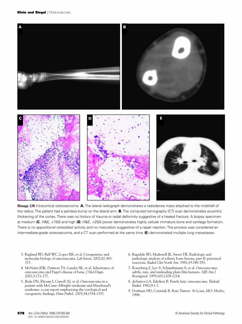
❚Image 23❚ Intracortical osteosarcoma. A, The lateral radiograph demonstrates a radiodense mass attached to the midshaft ofthe radius. The patient had a painless bump on the lateral arm. B, The computed tomography (CT) scan demonstrates eccentricthickening of the cortex. There was no history of trauma or radial deformity suggestive of a healed fracture. A biopsy specimenat medium (C, H&E, ×150) and high (D, H&E, ×250) power demonstrates highly cellular immature bone and cartilage formation.There is no appositional osteoblast activity and no maturation suggestive of a repair reaction. The process was considered anintermediate-grade osteosarcoma, and a CT scan performed at the same time (E) demonstrated multiple lung metastases.
Am J Clin Pathol 2006;125:555-581 579579 DOI: 10.1309/UC6KQHLD9LV2KENN 579
© American Society for Clinical Pathology
10. Ferguson WS, Harris MB, Goorin AM, et al. Presurgicalwindow of carboplatin and surgery and multi-drugchemotherapy for the treatment of newly diagnosed metastaticor unresectable osteosarcoma: Pediatric Oncology Group Trial.J Pediatr Hematol Oncol. 2001;23:340-348.
11. Meyers PA, Schwartz CL, Krailo M, et al. Osteosarcoma: arandomized, prospective trial of the addition of ifosfamideand/or muramyl tripeptide to cisplatin, doxorubicin and high-dose methotrexate. J Clin Oncol. 2005;23:2004-2116.
12. Jaffe HL. Intracortical osteogenic sarcoma. Bull Hosp Jt Dis.1960;21:189-197.
13. Goorin AM, Harris MB, Bernstein M, et al. Phase II/III trial ofetoposide and high dose ifosfamide in newly diagnosedmetastatic osteosarcoma: a Pediatric Oncology Group (POG)Trial. J Clin Oncol. 2002;20:426-433.
14. Unni KK, ed. Osteosarcoma of bone. In: Bone Tumors. NewYork, NY: Churchill Livingstone; 1988. Contemporary Issues inSurgical Pathology; vol 11.
15. Yi ES, Shmookler M, Malawer M, et al. Well-differentiatedextraskeletal osteosarcoma: a soft tissue homologue ofparosteal osteosarcoma. Arch Pathol Lab Med. 1991;115:906-909.
Anatomic Pathology / REVIEW ARTICLE
DCBA
FE
❚Image 24❚ Multifocal osteosarcoma. A 50-year-old woman had no history of radiation, Paget disease, or any other known familialcondition that would predispose her to osteosarcoma. B and C, Pain in the left leg resulted in radiographs demonstrating multipledisparate osteosclerotic lesions of the femur. Because there were multiple lesions, she underwent a skeletal series that revealedmultiple lesions of the medullary right femur shown in the anteroposterior radiograph (A) and the computed tomography scan (D).The right femur resection was performed because of intractable pain; there are at least 4 discrete lesions, 2 involving the cortexwith remodeling changes. E, Midfemur scan demonstrating cloudy radiodensity of osteosarcoma matrix within the medullarycavity. F, The histologic features were those of a conventional osteosarcoma (H&E, ×250). The diagnosis was presumed to bemetachronous multifocal osteosarcoma. The patient was alive without lung metastases 4 years after the resection.
580 Am J Clin Pathol 2006;125:555-581580 DOI: 10.1309/UC6KQHLD9LV2KENN
© American Society for Clinical Pathology
Klein and Siegal / OSTEOSARCOMA
16. Matsuno T, Unni KK, McLeod RA, et al. Telangiectaticosteogenic sarcoma. Cancer. 1976;38:2538-2547.
17. Murphey MD, wan Jaovishidha S, Temple HT, et al.Telangiectatic osteosarcoma: radiologic-pathologic correlation.Radiology. 2003;229:549-553.
18. Sim FH, Unni KK, Beabout JW, et al. Osteosarcoma withsmall cells simulating Ewing’s tumor. J Bone Joint Surg Am.1979;61:207-215.
19. Nakajima H, Sim FH, Bond JR, et al. Small cell osteosarcomaof bone: review of 72 Cases. Cancer. 1997;79:2095-2106.
20. Junqueira LC, Bignolas G, Brentani RR. Picrosirius stainingplus polarization microscopy, a specific method for collagendetection in tissue sections. Histochem J. 1979;11:447-455.
21. Bertoni F, Unni KK, McLeod RA, et al. Osteosarcomaresembling osteoblastoma. Cancer. 1985;55:416-426.
22. Dorfman HD, Weiss SW. Borderline osteoblastic tumors:problems in the differential diagnosis of aggressiveosteoblastoma and low-grade osteosarcoma. Semin DiagnPathol. 1984;1:215-234.
23. Rosado KE, Pitt MJ, Siegal GP. Osteoblastoma: a mimic ofosteosarcoma. Pediatr Pathol Mol Med. 2000;19:305-322.
24. Bertoni F, Bacchini P, Donati D, et al. Osteoblastoma-likeosteosarcoma: the Rizzoli Institute experience. Mod Pathol.1993;6:707-716.
25. Tani T, Okada K, Shoji K, et al. Osteoblastoma-likeosteosarcoma. Skeletal Radiol. 2000;29:656-659.
B
C D
❚Image 25❚ Extraskeletal osteosarcoma. The patient had a 4-cm mass anterior to the left clavicle. The lesion was thought to beposttraumatic heterotopic ossification; however, the biopsy demonstrated pleomorphic cells producing the bone seen in thecomputed tomography scan (A). B, Pleomorphic mononuclear stroma with several osteoclast-like giant cells (H&E, ×250). C,Infiltration of striated muscle by pleomorphic tumor cells producing osteoid (most apparent in the lower left) (H&E, ×400). D,After chemotherapy, the resected lesion demonstrated extensive necrosis of the cellular stroma and tumor cells (H&E, ×250).
A
Am J Clin Pathol 2006;125:555-581 581581 DOI: 10.1309/UC6KQHLD9LV2KENN 581
© American Society for Clinical Pathology
26. Mirra JM, ed. Bone Tumors: Clinical, Radiologic, and PathologicCorrelations. Philadelphia, PA: Lea and Febiger; 1989.
27. Balance WA, Mendelsohn G, Carter JR, et al. Osteogenicsarcoma: malignant fibrous histiocytoma subtype. Cancer.1988;62:763-771.
28. Bertoni F, Dallera P, Bacchini P, et al. The Istituto Rizzoli-Beretta experience with osteosarcoma of the jaw. Cancer.1991;68:1555-1563.
29. Clark JL, Unni KK, Dahlin DC, et al. Osteosarcoma of thejaw. Cancer. 1983;51:2311-2316.
30. Mardinger O, Givol N, Talmi Y, et al. Osteosarcoma of thejaw: the Chaim Sheba Medical Center experience. Oral SurgOral Med Oral Pathol Oral Radiol Endod. 2001;91:445-451.
31. Nora FE, Unni KK, Pritchard DJ, et al. Osteosarcoma ofextragnathic craniofacial bones. Mayo Clin Proc. 1983;58:268-272.
32. Kurt AM, Unni KK, McLeod RA, et al. Low-gradeintraosseous osteosarcoma. Cancer. 1990;65:1418-1428.
33. Unni KK, Dahlin DC, McLeod RA, et al. Intraosseous well-differentiated osteosarcoma. Cancer. 1977;40:1337-1347.
34. Bertoni F, Bacchini P, Fabbri N, et al. Osteosarcoma: low-gradeintraosseous-type osteosarcoma, histologically resemblingparosteal osteosarcoma, fibrous dysplasia, and desmoplasticfibroma. Cancer. 1993;71:338-345.
35. Franchi A, Bacchini P, Della Rocca C, et al. Central low-gradeosteosarcoma with pagetoid bone formation: a potentialdiagnostic pitfall. Mod Pathol. 2004;17:288-291.
36. Andresen KJ, Sundaram M, Unni KK, et al. Imaging featuresof low-grade central osteosarcoma of the long bones andpelvis. Skeletal Radiol. 2004;33:373-379.
37. Choong PF, Pritchard DJ, Rock M, et al. Low grade centralosteogenic sarcoma; a long-term followup of 20 patients. ClinOrthop. 1996;322:198-206.
38. Ogose A, Hotta T, Emura I, et al. Repeated dedifferentiationof low-grade intraosseous osteosarcoma. Hum Pathol.2000;31:615-618.
39. Wenger DE, Sundaram M, Unni KK, et al. Microscopiccorrelation of radiographically disparate appearing welldifferentiated osteosarcoma. Skeletal Radiol. 2002;31:488-492.
40. Raymond AK. Surface osteosarcomas. Clin Orthop.1991;270:140-148.
41. Geschicter CF, Copeland MM. Parosteal osteoma of bone: anew entity. Ann Surg. 1951;133:790-807.
42. Dwinnell LA, Dahlin DC, Ghormley RK. Parosteal(juxtacortical) osteogenic sarcoma. J Bone Joint Surg Am.1954;36A:732-744.
43. Lin J, Yao L, Mirra JM, et al. Osteochondromalike parostealosteosarcoma: a report of six cases of a new entity. AJR Am JRoentgenol. 1998;170:1571-1577.
44. Unni KK, Dahlin DC, Beabout JW. Periosteal osteogenicsarcoma. Cancer. 1976;37:2476-2485.
45. Murphey MD, Jelinek JS, Temple HT, et al. Imaging ofperiosteal osteosarcoma: radiologic-pathologic comparison.Radiology. 2004;233:129-138.
46. Wold LE, Unni KK, Beabout JW, et al. Dedifferentiatedparosteal osteosarcoma. J Bone Joint Surg Am. 1984;66:53-59.
47. Jelinek JS, Murphey MD, Kransdorf MJ, et al. Parostealosteosarcoma: value of MR imaging and CT in the predictionof histologic grade. Radiology. 1996;201:837-842.
48. Wines A, Bonar F, Lam P, et al. Telangiectaticdedifferentiation of a parosteal osteosarcoma. Skeletal Radiol.2000;29:597-600.
49. Bertoni F, Bacchini P, Staals EL, et al. Dedifferentiatedparosteal osteosarcoma: the experience of the Rizzoli Institute.Cancer. 2005;103:2373-2382.
50. Sheth DS, Yasko AW, Raymond AK, et al. Conventional anddedifferentiated parosteal osteosarcoma: diagnosis, treatment,and outcome. Cancer. 1996;78:2136-2145.
51. Wold LE, Unni KK, Beabout JW, et al. High-grade surfaceosteosarcomas. Am J Surg Pathol. 1984;8:181-186.
52. Vanel D, Picci P, De Paolis M, et al. Radiological study of 12high-grade surface osteosarcomas. Skeletal Radiol. 2001;30:667-671.
53. Kyriakos M. Intracortical osteosarcoma. Cancer. 1980;46:2525-2533.
54. Rosenberg AE, Heim SI. Extraskeletal osteosarcoma. In:Fletcher CDM, Unni KK, Mertens F, eds. Pathology andGenetics of Tumours of Soft Tissue and Bone. Lyon, France:IARC Press; 2002. World Health Organization Classification ofTumours.
55. Chung EB, Enzinger FM. Extraskeletal osteosarcoma. Cancer.1987;60:1132-1142.
56. Abramovici L, Hytiroglou P, Klein RN, et al. Welldifferentiated extraskeletal osteosarcoma: report of 2 cases, 1with dedifferentiation. Hum Pathol. 2005;36:439-443.
57. Present D, Bertoni F, Laus M, et al. Low-grade osteosarcoma ofsoft tissues (popliteal fossa). Skeletal Radiol. 1989;18:471-474.
Anatomic Pathology / REVIEW ARTICLE