Embed Size (px)
Citation preview
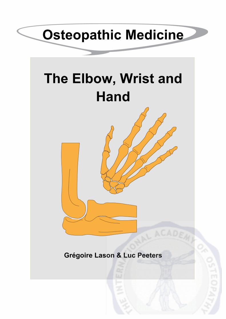
Osteopathic Medicine
The Elbow, Wrist and Hand
Grégoire Lason & Luc Peeters
2
The Elbow, Wrist and Hand
Grégoire Lason & Luc Peeters All rights reserved. Osteo 2000 bvba © 2014. No part of this e-book may be reproduced or made
public by printing, photocopying, microfilming, or by any means without the prior written permission of the publisher.
Contact: Osteo 2000, Kleindokkaai 3-5, B – 9000 Ghent, Belgium
Mail: [email protected]
Web: http://osteopedia.iao.be and www.osteopathie.eu
Tel: +32 9 233 04 03 - Fax: +32 55 70 00 74
ISBN: 9789074400398
The International Academy of Osteopathy – I.A.O.
3
Content
1. Introduction ............................................................................................................ 8
2. Biomechanics and Important Anatomical Features ........................................... 9 2.1. Different Regions ............................................................................................ 9
2.1.1. Elbow .......................................................................................................... 9 2.1.1.1. Joints .............................................................................................. 10 2.1.1.2. Ligaments ....................................................................................... 11 2.1.1.3. Range of Motion - ROM ................................................................. 13
2.1.2. Wrist and Hand ......................................................................................... 15 2.1.2.1. Bones ............................................................................................. 15 2.1.2.2. Distal Radio-Ulnar Joint .................................................................. 16 2.1.2.3. Radio-Carpal Joint .......................................................................... 18 2.1.2.4. Ulno-carpal Joint ............................................................................ 22 2.1.2.5. Mid-Carpal Joint ............................................................................. 22 2.1.2.6. Interphalangeal Joints .................................................................... 26 2.1.2.7. Retinacula of the Wrist and Hand ................................................... 27 2.1.2.8. Absorption and Transmission of Axial Pressure ............................. 27 2.1.2.9. Carpometacarpal Joint ................................................................... 28 2.1.2.10. Arches of the Hand ....................................................................... 28 2.1.2.11. Functional Position of the Hand ................................................... 29
2.2. Development of the Grip in Children ........................................................... 30 2.3. Muscles of the Elbow, Wrist and Hand ....................................................... 31 2.4. Bursae of the Elbow ...................................................................................... 45 2.5. Nerves ............................................................................................................ 46
2.5.1. Brachial Plexus ......................................................................................... 46 2.5.2. Muscular Innervation of the Upper Extremity ........................................... 46 2.5.3. Segments ................................................................................................. 49 2.4.4. Sensation .................................................................................................. 51 2.5.5. Dermatomes ............................................................................................. 52
2.6. Vascularisation .............................................................................................. 53 2.6.1. Arterial ...................................................................................................... 53 2.6.2. Venous ..................................................................................................... 54 2.6.3. Lymphatic Drainage .................................................................................. 54
3. Elbow, Wrist and Hand Pain ............................................................................... 55 3.1. General ........................................................................................................... 55 3.2. Mechanical Problems ................................................................................... 55
3.2.1. Lateral Epicondylitis (Tennis Elbow) ......................................................... 55 3.2.2. Medial Epicondylitis (Golfers Elbow) ........................................................ 58 3.2.3. Rupture or Overstretch of the Ulnar Collateral Ligament ......................... 61 3.2.4. Posterolateral Rotatory Instability ............................................................. 61
4
3.2.5. Dislocation of the Elbow ........................................................................... 62 3.2.6. Terrible Triad of the Elbow ....................................................................... 62 3.2.7. Nursemaid’s Elbow ................................................................................... 63 3.2.8. Bursitis ...................................................................................................... 63 3.2.9. The Stiff Elbow ......................................................................................... 64 3.2.10. Tenosynovitis .......................................................................................... 65 3.2.11. Trigger Finger ......................................................................................... 66 3.2.12. DeQuervain’s Disease ............................................................................ 66 3.2.13. Scapho-lunate Dissociation .................................................................... 67 3.2.14. Mallet Finger ........................................................................................... 68 3.2.15. Boutonniere Deformity ............................................................................ 69 3.2.16. Jersey Finger .......................................................................................... 70 3.2.17. Scaphoid Fracture .................................................................................. 70 3.2.18. Distal Radius Fracture (Colles Fracture) ................................................ 72 3.2.19. Volar Plate Rupture ................................................................................ 73 3.2.20. Central Slip Avulsion .............................................................................. 74 3.2.21. Tears of the Collateral Ligament ............................................................ 74 3.2.22. Ulnar Collateral Ligament Injury of the Thumb (Gamekeeper’s Thumb) 75 3.2.23. Flexor Tendon Avulsion .......................................................................... 75 3.2.24. Intersection Syndrome ............................................................................ 76
3.3. Vascular Problems ........................................................................................ 78 3.3.1. Superior Vena Cava Syndrome ................................................................ 78 3.3.2. Subclavian Steal Syndrome - SSS ........................................................... 78 3.3.3. Raynaud’s Phenomenon .......................................................................... 80 3.3.4. Osteochondritis Dissecans of the Elbow .................................................. 81 3.3.5. Kienbock’s Disease .................................................................................. 82
3.4. Neurological Problems ................................................................................. 83 3.4.1. Cervical Stenosis ...................................................................................... 83 3.4.2. Overstretch or Compression of the Brachial Plexus ................................. 84 3.4.3. Cervicobrachial Neuralgia ........................................................................ 84 3.4.4. Nerve Root Syndromes ............................................................................ 85 3.4.5. Referred Pain Patterns (Sclerotome) ....................................................... 86 3.4.6. Carpal Tunnel Syndrome .......................................................................... 88 3.4.7. Ulnar Nerve Entrapment or Guyon’s Syndrome ....................................... 90 3.4.8. Radial Tunnel Syndrome .......................................................................... 92 3.4.9. Pronator Syndrome .................................................................................. 94 3.4.10. Posterior Interosseus Nerve Syndrome .................................................. 95 3.4.11. Cubital Tunnel Syndrome ....................................................................... 97 3.4.12. Anterior Interosseus Nerve Syndrome ................................................... 98
3.5. Metabolic Problems ...................................................................................... 99 3.5.1. Complex Regional Pain Syndrome (CRPS) ............................................. 99 3.5.2. Ganglion Cyst ......................................................................................... 101 3.5.3. Dupuytren’s Contracture ......................................................................... 102 3.5.4. Paronychia .............................................................................................. 103
5
3.6. Degenerative Problems .............................................................................. 103 3.6.1. Osteoarthrosis ........................................................................................ 103 3.6.2. Osteoarthritis of the First Carpometacarpal ............................................ 105
3.7. Rheumatic Problems .................................................................................. 106 3.7.1. Rheumatoid Arthritis - RA ....................................................................... 106
3.8. Joint Infections ............................................................................................ 108 3.8.1. Septic Arthritis ........................................................................................ 108
4. Examination ....................................................................................................... 110 4.1. Case History ................................................................................................ 110 4.2. Observation ................................................................................................. 111
4.2.1. General ................................................................................................... 111 4.2.2. Observation of the Shortened Structures ............................................... 111 4.2.3. Observation of Body Posture and the Upper Thoracic Region ............... 113
4.3. Provocation Tests ....................................................................................... 116 4.3.1. General ................................................................................................... 116 4.3.2. Palpation of the Posterior Triangle of the Elbow .................................... 116 4.3.3. Provocation Test of the Radio-Humeral Joint ......................................... 117 4.3.4. Traction Test of the Radio-Humeral Joint ............................................... 117 4.3.5. Traction Test on the Humero-Ulnar Joint ............................................... 118 4.3.6. Provocation Test for Lateral Epicondylitis .............................................. 118 4.3.7. Provocation Test for Medial Epicondylitis ............................................... 119 4.3.8. Traction Provocation Test of the Wrist ................................................... 119 4.3.9. Compression Provocation Test of the Wrist ........................................... 120 4.3.10. Compression Provocation Test of the Wrist ......................................... 120 4.3.11. Provocation Test of DeQuervain's Disease .......................................... 121 4.3.12. Provocation Test of the First Carpometacarpal Joint and Scaphoid .... 121 4.3.13. Provocation Test of the Carpals and Intercarpal Joints ........................ 122
4.4. Examples of Muscle Testing ...................................................................... 123 4.4.1. Test for Muscle Strength of the Elbow Extensors ................................... 123 4.4.2. Test for Muscle Strength of the Elbow Flexors ....................................... 123 4.4.3. Test for Muscle Strength of the Supinators ............................................ 124 4.4.4. Test for Muscle Strength of the Pronators .............................................. 124
4.5. Mobility Testing ........................................................................................... 125 4.5.1. Active Tests ............................................................................................ 125 4.5.2. Passive Tests ......................................................................................... 125
4.5.2.1. Mobility Test of the Elbow in Flexion and Extension .................... 125 4.5.2.2. Mobility Test of the Elbow in Adduction and Abduction ................ 126 4.5.2.3. Elasticity Test of the Lateral Capsule ........................................... 127 4.5.2.4. Elasticity Test of the Medial Capsule ........................................... 127 4.5.2.5. Mobility Test of the Radial Head in Anterior and Posterior Rotation (Translation) .............................................................................................. 128 4.5.2.6. Mobility Test of the Radial Head in Anterior and Posterior Translation ................................................................................................. 129
6
4.5.2.7. Mobility Test of the Radius in Cranial and Caudal Translation ..... 129 4.5.2.8. Mobility Test of the Distal Radio-Ulnar Joint ................................ 130 4.5.2.9. Mobility Test of the Wrist in Palmar, Dorsal, Ulnar and Radial Translation ................................................................................................. 130 4.5.2.10. Mobility Test of the Radiocarpal and Intercarpal Joints .............. 131 4.5.2.11. Elasticity Test of the Transverse Ligament ................................ 131 4.5.2.12. Elasticity Test of the Transverse Ligament ................................ 132
5. Techniques ......................................................................................................... 133 5.1. Manipulations .............................................................................................. 133
5.1.1. General ................................................................................................... 133 5.1.2. Manipulation of a Posteriorly Rotated Radial Head ................................ 135 5.1.3. Manipulation of a Posteriorly Rotated Radial Head ................................ 136 5.1.4. Manipulation of a Posteriorly Rotated Radial Head ................................ 137 5.1.5. Manipulation of a Inferiorly Positioned Radial Head ............................... 138 5.1.6. Manipulation of the Medial Collateral Ligaments .................................... 139 5.1.7. Manipulation of the Lateral Collateral Ligaments ................................... 140 5.1.8. Manipulation of a Lunate Lesion in Palmar Translation .......................... 141
5.2. Mobilisations ............................................................................................... 141 5.2.1. General ................................................................................................... 141 5.2.2. Mobilisation of the Elbow into Flexion .................................................... 143 5.2.3. Mobilisation of the Radial Head in Anterior and Posterior Translation ... 143 5.2.4. Mobilisation of the Radial Head in Distal Direction ................................. 144 5.2.5. Mobilisation of the Radial Head in Proximal Direction ............................ 144 5.2.6. Mobilisation of the Wrist in Palmar, Dorsal, Ulnar and Radial Translation .......................................................................................................................... 145 5.2.7. Mobilisation of the Radio-Lunar Joint in Circumduction ......................... 145 5.2.8. Mobilisation of the Radio-Ulnar Joint in Translation ............................... 146 5.2.9. Mobilisation of the Radial Joint Lines ..................................................... 146 5.2.10. Mobilisation of the Ulnar Joint Lines ..................................................... 147 5.2.11. General Mobilisation of the Carpal Joints ............................................. 147 5.2.12. General Mobilisation of the Metacarpophalangeal Joints ..................... 148
5.3. Muscle Energy Techniques – MET ............................................................ 149 5.3.1. General ................................................................................................... 149 5.3.2. Stretching the Elbow Flexors .................................................................. 150 5.3.3. Stretching the Elbow Extensors .............................................................. 151 5.3.4. Stretching the Forearm Supinators ......................................................... 152 5.3.5. Stretching the Forearm Pronators .......................................................... 153 5.3.6. Stretching the Flexors of the Wrist ......................................................... 154 5.3.7. Stretching the Extensors of the Wrist ..................................................... 155 5.3.8. Stretching the Abductors of the Wrist ..................................................... 156 5.3.9. Stretching the Adductors of the Wrist ..................................................... 157
5.4. Strain and Counterstrain Techniques - SCT ............................................. 158 5.4.1. General ................................................................................................... 158
7
5.4.2. Spontaneous Release Technique for a Flexion Lesion of the Elbow ..... 158 5.4.3. Spontaneous Release Technique for Extension Lesion of the Elbow .... 159 5.4.4. Spontaneous Release Technique for Radial Head Dysfunction ............. 159 5.4.5. Spontaneous Release Technique for Flexion Lesions of the Wrist ........ 160 5.4.6. Spontaneous Release Technique for Extension Lesions of the Wrist .... 160 5.4.7. Spontaneous Release Technique for Dysfunction of the First Carpometacarpal Joint ..................................................................................... 161 5.4.8. Spontaneous Release Technique for Flexion Lesions of the Fingers .... 161
6. Bibliography ....................................................................................................... 162
7. About the Authors ............................................................................................. 166
8. Acknowledgment ............................................................................................... 167
9. Osteopathic Terminology ................................................................................. 168 9.1. The Three Anatomical Axes ....................................................................... 168 9.2. The Three Anatomical Planes .................................................................... 169 9.3. Spinal Biomechanics .................................................................................. 170 9.4. General Abbreviations ................................................................................ 172 9.5. Specific Terms ............................................................................................. 173
10. All Videos ......................................................................................................... 174
8
1. Introduction Most movements of the upper extremity involve the elbow and the radioulnar joint.
The elbow is vital for the positioning of the hand during all functional activities. The elbow is also vital for providing a “link” between the powerful movements of the shoulder and the fine motor control of the hand and fingers.
The wrist is a distal joint of the upper limb and allows the hand to assume the optimal position for prehension.
The hand is composed of both complex, and delicately balanced joints. Its function is integral to everyday activities. It is the most active portion of the upper extremity.
The joints of the hand however are less protected than other joints. That is one of the reasons why the hand is so vulnerable to injury.
Hand function is an important feature in humans over other primates who lack fine control and precision. The hands are paired structures dominantly controlled by the opposite brain hemisphere. It is our chief way for physically manipulating our environment and the richest source of tactile feedback.
For those who are not familiar with the typical osteopathic terminology, we refer to chapter 9 at the end of this e-book.

9
2. Biomechanics and Important Anatomical Features (Alcid et al 2004, Bryce & Armstrong 2008, Callaway et al 1997, Floris et al 1998, Fuss 1991, Grant & Boileau 2004, Gray 1995, 2000, Levangie & Norkin 2005, Lockard 2006, Moore & Dalley 1999, Netter 2003, Miyasaka 1999, Modi & Shah, Morrey 1981, 1983, 1997, 2005, Schneck & Bronzino 2002, Sobotta 2001, Ward 2003)
2.1. Different Regions
2.1.1. Elbow The elbow movements can be clearly distinguished from the movements of the radio-ulnar joint and the wrist joint.
The elbow has two interrelated joints:
• The humero-ulnar joint. • The radio-humeral joint.
Elbow movements primarily involve movements between humerus and ulna.
In full extension, the olecranon process is cradled by the olecranon fossa. Full extension is therefore a stable position.
When the elbow is flexed to 20° or more, its bony stability is unlocked which allows more laterolateral laxity.
Stability in a flexed position is therefore not bony but ligamentary. The lateral or radial collateral ligament and the medial or ulnar collateral ligament provide this stability in flexion.
The elbow is a very congruous joint and inherently very stable.
In flexion, the coronoid process locks into the coronoid fossa, while the medial rim of the radial head engages in the trochleocapitellar groove.
In extension, the apex of the olecranon is held in the olecranon fossa.
Elbow stability is enhanced by the perfect congruency between the radial head and the radial notch of the ulna.
Roughly speaking, the bony surfaces contribute to 50% of the mediolateral stability of the elbow, while the other 50% comes from the ligaments.
One important thing to bear in mind is that the role of each of these structures varies with the degree of flexion or extension of the elbow.
10
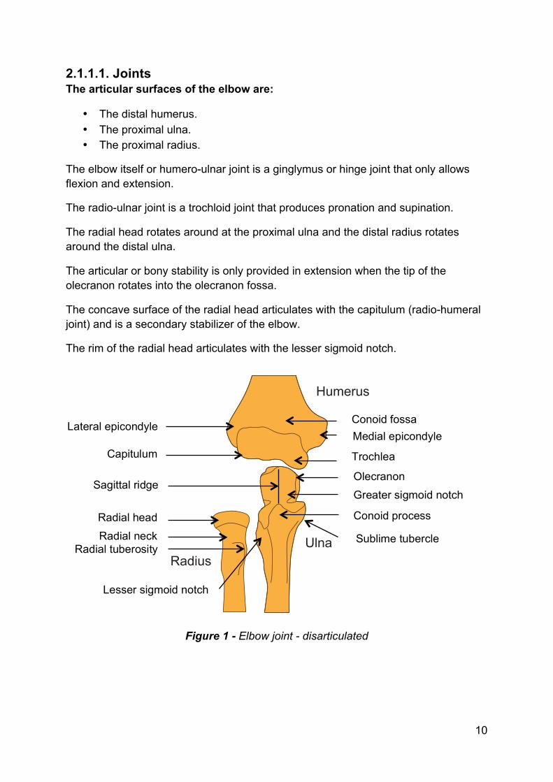
2.1.1.1. Joints The articular surfaces of the elbow are:
• The distal humerus. • The proximal ulna. • The proximal radius.
The elbow itself or humero-ulnar joint is a ginglymus or hinge joint that only allows flexion and extension.
The radio-ulnar joint is a trochloid joint that produces pronation and supination.
The radial head rotates around at the proximal ulna and the distal radius rotates around the distal ulna.
The articular or bony stability is only provided in extension when the tip of the olecranon rotates into the olecranon fossa.
The concave surface of the radial head articulates with the capitulum (radio-humeral joint) and is a secondary stabilizer of the elbow.
The rim of the radial head articulates with the lesser sigmoid notch.
Figure 1 - Elbow joint - disarticulated
Radial tuberosity
Lesser sigmoid notch
Radial neck Radial head
Olecranon
Ulna
Humerus
Radius
Capitulum
Lateral epicondyle Conoid fossa Medial epicondyle
Trochlea
Greater sigmoid notch
Sublime tubercle
Conoid process
Sagittal ridge
11
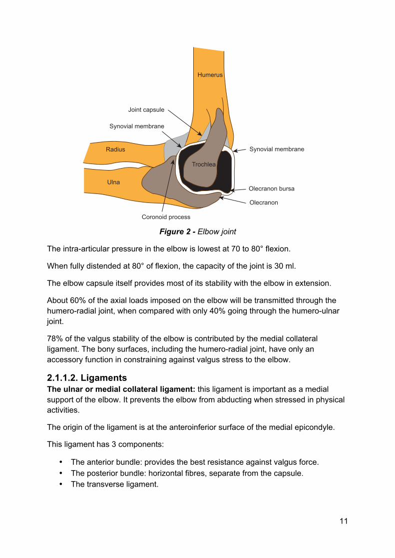
Figure 2 - Elbow joint
The intra-articular pressure in the elbow is lowest at 70 to 80° flexion.
When fully distended at 80° of flexion, the capacity of the joint is 30 ml.
The elbow capsule itself provides most of its stability with the elbow in extension.
About 60% of the axial loads imposed on the elbow will be transmitted through the humero-radial joint, when compared with only 40% going through the humero-ulnar joint.
78% of the valgus stability of the elbow is contributed by the medial collateral ligament. The bony surfaces, including the humero-radial joint, have only an accessory function in constraining against valgus stress to the elbow.
2.1.1.2. Ligaments The ulnar or medial collateral ligament: this ligament is important as a medial support of the elbow. It prevents the elbow from abducting when stressed in physical activities.
The origin of the ligament is at the anteroinferior surface of the medial epicondyle.
This ligament has 3 components:
• The anterior bundle: provides the best resistance against valgus force. • The posterior bundle: horizontal fibres, separate from the capsule. • The transverse ligament.
Humerus
Trochlea
Olecranon
Olecranon bursa
Synovial membrane
Joint capsule
Synovial membrane
Coronoid process
Radius
Ulna
55
3. Elbow, Wrist and Hand Pain (Calliet 1977, Downing 1988, Hertling & Kessler 1996, Kaufmann et al 2010, Nunley & Urbaniak 1996, O’Driscoll et al 2001, Pienimäki et al 2002, Starkey & Ryan 2002, Randip & Bindra 2004)
3.1. General Complaints of the elbow, wrist and hand will be divided into the following possible problems:
• Mechanical. • Vascular. • Neurologic. • Metabolic. • Degenerative. • Rheumatic. • Infectious.
3.2. Mechanical Problems Note: it is not the aim here to explain here all possible fractures in the area. Only common fractures and fractures that can be overlooked are discussed.
3.2.1. Lateral Epicondylitis (Tennis Elbow) Lateral epicondylitis is a chronic tear in the origin of the extensor carpi radialis brevis muscle. It is the most common elbow complaint.
Lateral epicondylitis most commonly involves the extensor carpi radialis brevis, occasionally, the anterior edges of the extensor digitorum communis, the extensor radialis longus, and rarely the origin of the extensor carpi ulnaris.
The lesion may be tenoperiosteal, tendinous or musculotendinous.
It is mostly seen between the ages of 30-50 years old.
Possible causes
• Repetitive micro-trauma or overuse syndrome caused by repeated forceful wrist and finger movements.
Clinical findings
• Pain at the common extensor origin. • Often pain 5 mm distal and slightly anterior to the lateral humeral epicondyle. • Pain with resisted wrist extension and extension of the digits. • Temperature may be slightly elevated over the epicondyle.
56
Pathophysiology
• Micro tears at extensor carpi radialis brevis muscle. • Incomplete healing after trauma. • Granulation tissue formation. • Development of angiofibroblastic hyperplasia. • The extensor carpi radialis brevis is stretched over the radial head when the
elbow is extended and fully pronated. This fulcrum effect may partially explain its susceptibility to chronic inflammation at or near its attachment.
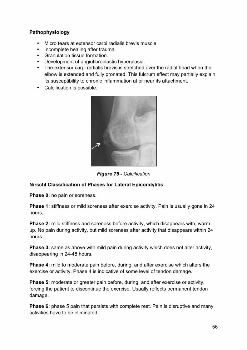
• Calcification is possible.
Figure 75 - Calcification
Nirschl Classification of Phases for Lateral Epicondylitis
Phase 0: no pain or soreness.
Phase 1: stiffness or mild soreness after exercise activity. Pain is usually gone in 24 hours.
Phase 2: mild stiffness and soreness before activity, which disappears with, warm up. No pain during activity, but mild soreness after activity that disappears within 24 hours.
Phase 3: same as above with mild pain during activity which does not alter activity, disappearing in 24-48 hours.
Phase 4: mild to moderate pain before, during, and after exercise which alters the exercise or activity. Phase 4 is indicative of some level of tendon damage.
Phase 5: moderate or greater pain before, during, and after exercise or activity, forcing the patient to discontinue the exercise. Usually reflects permanent tendon damage.
Phase 6: phase 5 pain that persists with complete rest. Pain is disruptive and many activities have to be eliminated.
57
Phase 7: phase 6 pain with disruption of sleep on a consistent basis. Pain is aching in nature and intensifies with activity.
Pain phases 5, 6, and 7 indicate increasing percentages of permanent tendon damage.
Local osteopathic finding: in many cases of lateral epicondylitis, osteopaths find the radial head in a posterior rotation lesion, fixed behind the ulna. This lesion causes strain on the tendon of the extensor carpi radialis brevis muscle.
Treatment
The treatment for lateral epicondylitis is divided into local treatment, treating the inflammation and eventual damage and general treatment taking into account the osteopathic concept.
The osteopathic concept includes optimizing the different tissues of the elbow region in order to give the region optimal conditions to heal.
Local treatment:
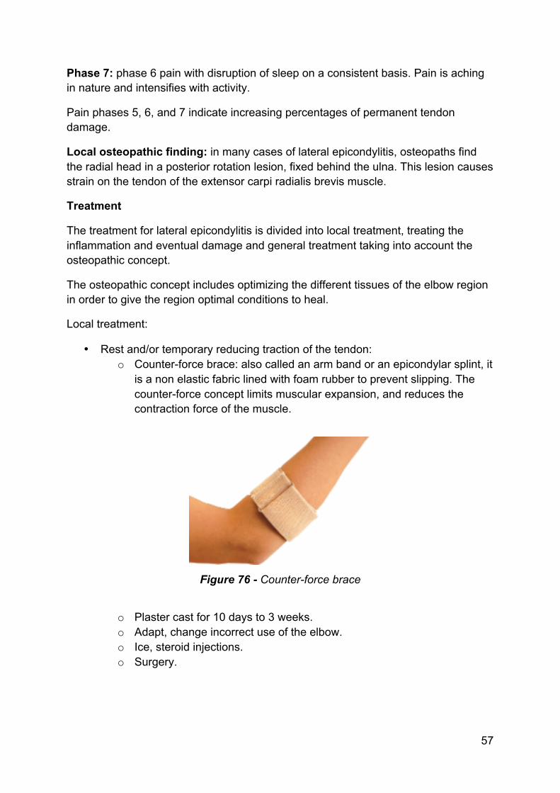
• Rest and/or temporary reducing traction of the tendon: o Counter-force brace: also called an arm band or an epicondylar splint, it
is a non elastic fabric lined with foam rubber to prevent slipping. The counter-force concept limits muscular expansion, and reduces the contraction force of the muscle.
Figure 76 - Counter-force brace
o Plaster cast for 10 days to 3 weeks. o Adapt, change incorrect use of the elbow. o Ice, steroid injections. o Surgery.
110
4. Examination (Good et al 1984, Hoppenfeld 1976, Kuchera 1994, 1996, Peeters & Lason 2005)
The word lesion describes a loss of mobility.
Dysfunction of the elbow, wrist and hand can cause symptoms, which are diverse in nature. Dysfunction can refer to hyper- as well as hypomobility.
The examination of the elbow, wrist and hand requires a sound knowledge of differential diagnosis, and must include an examination of the entire upper kinetic chain, as well as the cervical and thoracic spine.
4.1. Case History The assessment of the elbow, wrist, and hand begins by recording a detailed history. The history helps to focus the examination.
All relevant information must be gathered about the site, nature, behaviour and onset of the current symptoms.
The history should include information about the patient’s age, hand dominance, avocation, and occupation.
During case history taking, the osteopath tries to identify the nature of the pain:
• Aching pain can be from a ligament, especially when occurring in the morning with morning stiffness. Also when it occurs after a longer period of immobility. Ligament complaints are also associated with osteoarthrosis. Transient morning pain that subsides after the patient has moved about, but also reappears with exercise is typical of degenerative nature.
• Sharp pain on specific movements can be caused by muscle strain or inflammation, tendinitis or bursitis.
• Fatigue can be caused by bad posture and poor muscular balance. It can also be associated with arteriosclerosis or rheumatoid arthritis.
• Radiating pain indicates a neurogenic factor and can be radicular or pseudo radicular (referred pain). Detailed neurological tests will have to be done.
• Numbness or muscle weakness indicates compression or damage of a nerve.
• Bilateral pain in the elbow, wrist and hands can be associated with cervical myelopathy or rheumatic disease.
• Nocturnal pain often indicates cancer, inflammation, infection or rheumatic disease.
The type of patient (child, adult, elderly, pregnant, peri-menopausal woman) is helpful for differential diagnosis.
111
The onset of elbow, wrist or hand pain is important. Was there a trauma? How did it happen? Was the onset sudden or progressively worsening? What makes it worse or better?
Is there a popping sensation? Is it painful or not?
Is there any trouble lifting, reaching, throwing, etc.?
Have there been any recent infections?
Have the symptoms increased? Is there psychological distress? (superficial or non-anatomical pain distribution, non-anatomic sensory or motor disturbance, inconsistent neurological signs, inappropriate or excessive verbalization of the pain).
The differential diagnosis should be narrowed down by 80% with a proper history taking.
4.2. Observation
4.2.1. General The purpose of a general observation is to identify:
• Bony deformities. • Muscular contours (asymmetry). • Muscular atrophy. • Swelling and/or erythema. • The arches of the hands. • Scars, masses, and/or localized swelling. • Other deformities. • Differences between the sides. • Location of a the somatic dysfunction? (more details are in the e-book
“Integration and Applied Principles in Osteopathy” by the same authors)
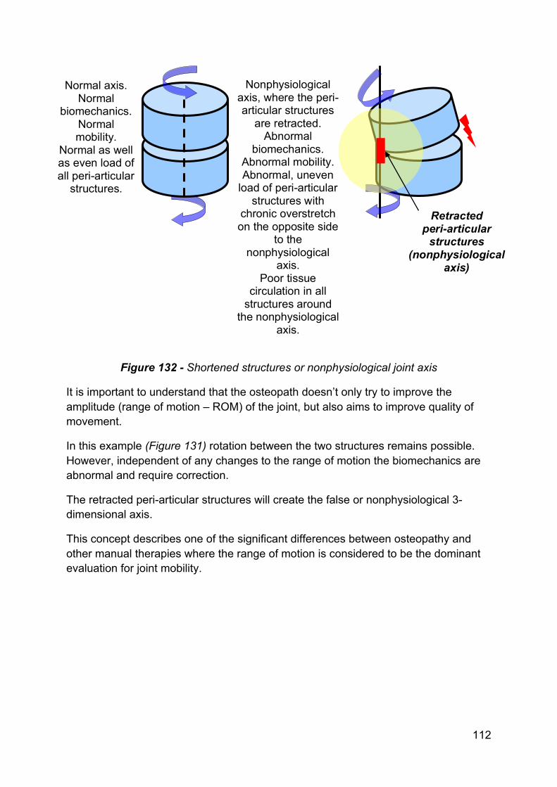
4.2.2. Observation of the Shortened Structures The aim of this observation is to determine where these shortened structures are and to treat them locally. Local treatment can only be done on the shortened side (mobilisation or manipulation).
The patient can complain of symptoms on both the shortened side as well as the overstretched side.
112
Figure 132 - Shortened structures or nonphysiological joint axis
It is important to understand that the osteopath doesn’t only try to improve the amplitude (range of motion – ROM) of the joint, but also aims to improve quality of movement.
In this example (Figure 131) rotation between the two structures remains possible. However, independent of any changes to the range of motion the biomechanics are abnormal and require correction.
The retracted peri-articular structures will create the false or nonphysiological 3-dimensional axis.
This concept describes one of the significant differences between osteopathy and other manual therapies where the range of motion is considered to be the dominant evaluation for joint mobility.
Normal axis. Normal
biomechanics. Normal mobility.
Normal as well as even load of all peri-articular structures.
Nonphysiological axis, where the peri-articular structures
are retracted. Abnormal
biomechanics. Abnormal mobility. Abnormal, uneven
load of peri-articular structures with
chronic overstretch on the opposite side
to the nonphysiological
axis. Poor tissue
circulation in all structures around
the nonphysiological axis.
Retracted peri-articular
structures (nonphysiological
axis)
133
5. Techniques (Cooperstein & Gleberzon 2004, Crow 2010, Danto 2005, Fryette 1954, Haldeman & Dagenais 2004, Johnston et al 2005, Peeters & Lason 2005, Savarese et al 2003, Wyatt 2004)
5.1. Manipulations
5.1.1. General A manipulation or HVLAT (High Velocity Low Amplitude Thrust) is a short, specific and rapid thrust applied to a joint. The aim of a manipulation is variable depending upon the lesion and the joint being treated. The aim of a manipulation is:
• Repositioning of a joint subluxation. • Alleviation of a muscular spasm in short musculature (spine). • Stretching of a capsulo-ligamentous retraction (correction of the
nonphysiological axis – shortened structures). A manipulation is in some situations a necessity, most notably in cases of an articular blockage or subluxation. This is often difficult to differentiate from a restriction (mobility loss with elastic end feel). A manipulation is, in some cases, a more efficient treatment for a restriction. Where elastic end feel is present mobilisations can be used but, if no contra indications exist, then a manipulation is also an option. A manipulation can break down cross-links. Before 20 years of age, “real” articular blockages rarely occur. Contra indications Before an osteopath decides to use a manipulative technique he must be sure that no contra indications are present. The following are examples of contra indications:
• Medication o The osteopath should not manipulate if the patient takes anticoagulants
or corticosteroids. • Trauma
o The osteopath should not manipulate directly after a trauma, without radiological testing showing no osseous lesions or other tissue damage.
o The osteopath should not manipulate after (6-8 weeks) an operation (risk of bleeding).

134
• Lever use o If the patient has pain or neurological symptoms during the positioning
of the body and use of levers for a specific technique, the osteopath should not manipulate.
• Osteoporosis o The osteopath should not manipulate in cases of obvious osteoporosis
such as Sudeck's atrophy. In addition if osteoporosis is suspected, manipulations should not be used without further investigation.
• Children o Real articular blockages do not occur in children, so manipulation is not
necessary. • Pregnancy
o Manipulation of lesions during pregnancy is not an absolute contra indication but does deserve extra caution. Hypermobility is common so any manipulative technique must be carried out very specifically.
• Elderly o In older patients, arthrosis is a frequent reality and changes the joint
surface congruency. Manipulation is not absolutely contra indicated but extra care must be taken. Manipulation is only needed in cases of subluxation.
o When treating arthritic joints the aim is not to drastically improve the range of motion. This will only lead to joint instability. When a joint is arthritic the general loss of mobility is seen as a normal protective mechanism of the body. Therefore the aim is to prevent a nonphysiological axis to develop and to maximise circulatory factors rather than to improve the general loss of mobility.
• Cardiac patients o Manipulations that have a potential autonomic effect upon the heart are
contra indicated. These patients are not the ideal patients for a total osteopathic treatment because osteopathy works so effectively on the circulatory system. Cardiac patients have a faulty ‘motor’ in their circulatory system and an improvement in their circulation may well create an overload for the heart.
• Cancer patients o It is also strongly suggested to avoid manipulation of cancer patients.
Osseous metastasis is always a possibility. o These patients are not the ideal patients for a total osteopathic
treatment because osteopathy works so effectively on the circulatory system, which can allow rapid spread of any metastasis. Post-operative treatment of a complaint is possible if approved by the consulting specialist. This must be considered case by case.
• Psychiatric patients o Great care must be taken with these patients as manipulation can result
in unexpected emotional reactions. With this patient group this is not desired as the appropriate reaction to osteopathic treatment.
• Prosthesis o Joints that have undergone a prosthetic replacement should not be
manipulated.
135
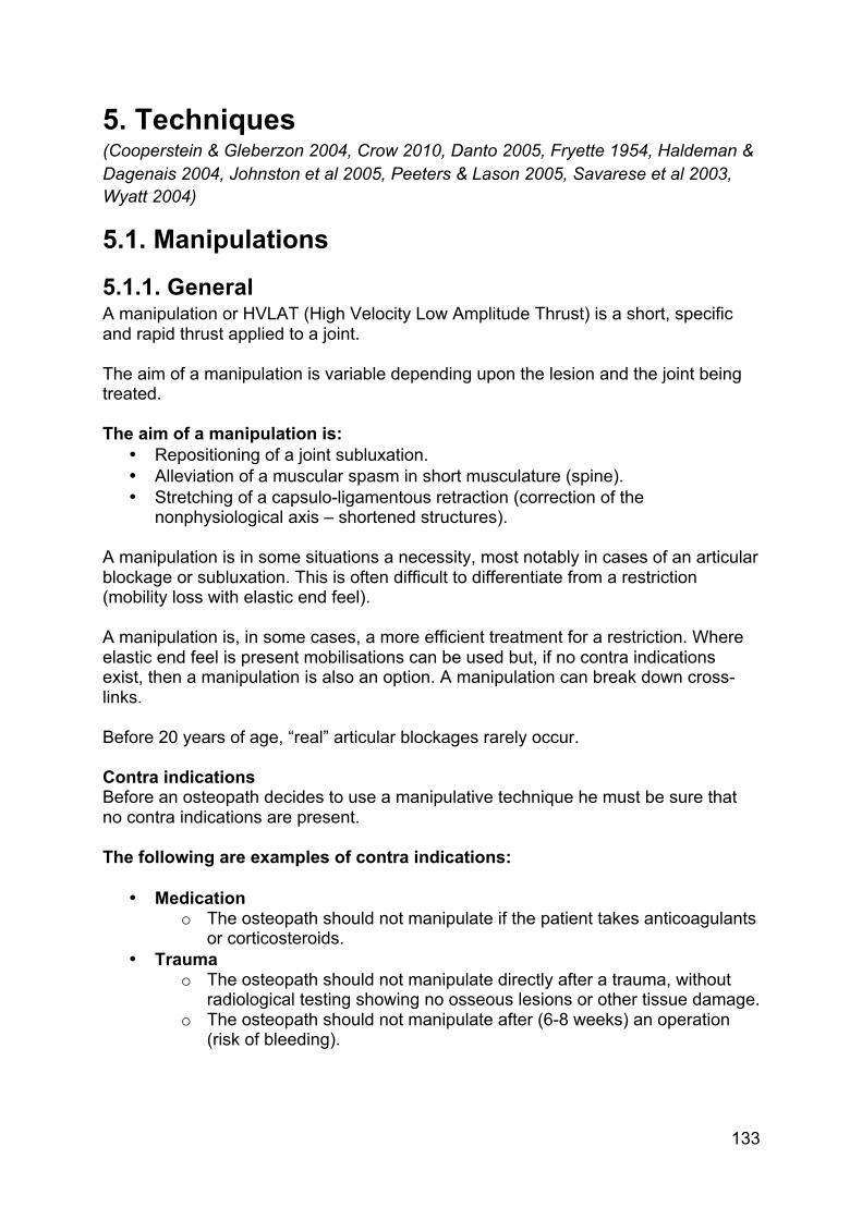
5.1.2. Manipulation of a Posteriorly Rotated Radial Head A posteriorly rotated radial head is often the local mechanical cause for lateral epicondylitis. This is because this lesion causes a fulcrum on the tendon of the extensor carpi radialis brevis muscle.
The patient is supine. The osteopath fixes the patient's shoulder against the table with his elbow.
His thumb contacts the posterior side of the radial head and brings the radial head over the ulna.
With his other hand, he contacts the radius between the thumb and fingers and uses it as a lever applying distal traction.
The thrust is direct (thumb pushes the radial head in anterior direction) as well as indirect (with the lever: supination, abduction and radial traction).
A popping sound shows that the correction is effective.
Note: complete extension of the elbow should be avoided.
Video 29 - Manipulation of a posteriorly rotated radial head
136
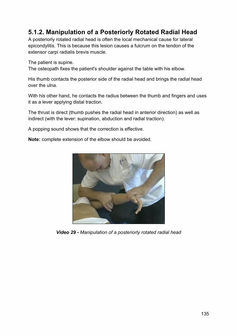
5.1.3. Manipulation of a Posteriorly Rotated Radial Head The patient is supine.
The osteopath fixes the patient's hand between his body and arm.
He contacts the elbow with both hands; the external hand has the index finger posteriorly on the radial head.
While bringing the elbow into extension, he manipulates the radial head in anterior direction.
Video 30 - Manipulation of a posteriorly rotated radial head
137
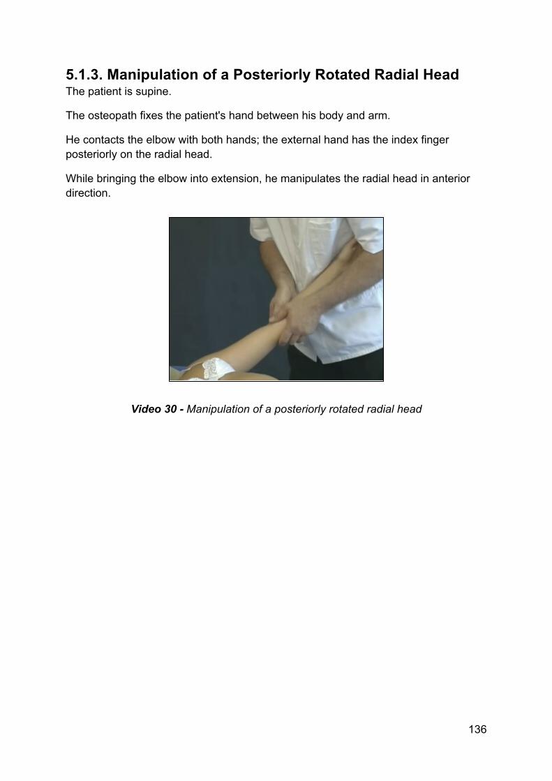
5.1.4. Manipulation of a Posteriorly Rotated Radial Head The patient is sitting.
The osteopath places the thumb posteriorly on the radial head.
The arm is used as a lever by holding onto the hand.
The wrist is brought into flexion to put slight traction on the extensor tendons.
While pushing the radial head in anterior direction, the osteopath brings the elbow into extension. Hyperextension is avoided by the osteopath blocking the arm with his own body.
With this technique the radial head is not only brought into anterior rotation but the technique also stretches possible adhesions in the extensor tendons.
For this reason, the technique is not done on damaged tendons.
Video 31 - Manipulation of a posteriorly rotated radial head

166
7. About the Authors
Grégoire Lason Luc Peeters Gent (B), 21.11.54 Terhagen (B), 18.07.55
Both authors are holders of university degrees, namely the Master of Science in Osteopathy (MSc.Ost. – University of Applied Sciences), and are very active with the promotion and academic structuring of osteopathy in Europe. In 1987 they began The International Academy of Osteopathy (IAO) and are, to this day, the joint-principals of this academy. The IAO is since several years the largest teaching institute for osteopathy in Europe. Both osteopaths are members of diverse professional organizations, including the American Academy of Osteopathy (AAO), the International Osteopathic Alliance (IOA) and the World Osteopathic Health Organization (WOHO), as part of their mission to improve osteopathic development. This osteopathic encyclopaedia aims to demonstrate the concept that a proper osteopathic examination and treatment is based upon the integration of all body systems.

177
This e-book is a product of Osteo 2000 bvba.
If you are interested in publishing an e-book or if you have questions or suggestions, please contact us:
Mail: [email protected]
Fax: +32 55 70 00 74
Tel: +32 9 233 04 03
Web Osteopedia: http://osteopedia.iao.be
Web The International Academy of Osteopathy – IAO: http://www.osteopathie.eu