Embed Size (px)
Citation preview

ESSENTIAL HEALTH TECHNOLOGIES
CLINICAL PROCEDURES
HTP/EHT/CPR 17
Orthopaedic TechniquesOrthopaedic Techniques
Key Points

ESSENTIAL HEALTH TECHNOLOGIES
CLINICAL PROCEDURES
HTP/EHT/CPR 17.117.1 TRACTIONTRACTION
• Use an appropriate method of traction to treat fractures of the extremities and cervical spine
• Apply extremity traction to the skin or to the skeleton using a pin inserted through the bone distal to the fracture
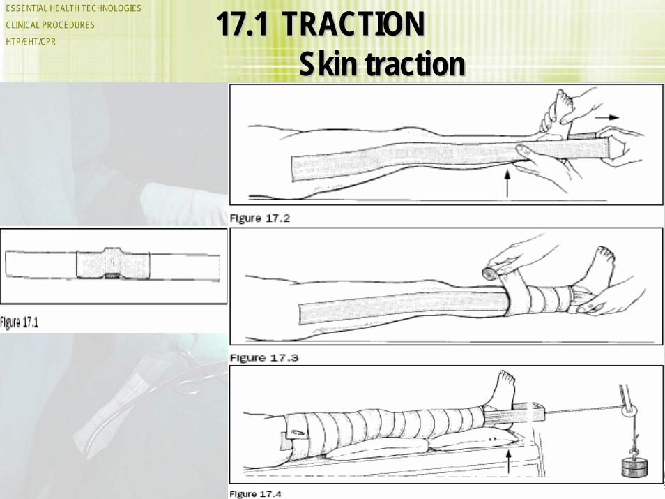
• Apply traction to the cervical spine using a head halter chin sling or skull tongs

ESSENTIAL HEALTH TECHNOLOGIES
CLINICAL PROCEDURES
HTP/EHT/CPR 17.1 TRACTION contd.
• The weight applied through the traction system counteracts the muscle force pulling across the fracture, keeping the bone in proper alignment and length.
• Do not apply traction to skin with:– abrasions,– lacerations,– surgical wounds, – ulcers, – loss of sensation or – peripheral vascular disease.
ESSENTIAL HEALTH TECHNOLOGIES
CLINICAL PROCEDURES
HTP/EHT/CPR17.117.1 TRACTIONTRACTION
Skin tractionSkin traction

ESSENTIAL HEALTH TECHNOLOGIES
CLINICAL PROCEDURES
HTP/EHT/CPR17.117.1 TRACTIONTRACTION
Skeletal TractionSkeletal Traction

ESSENTIAL HEALTH TECHNOLOGIES
CLINICAL PROCEDURES
HTP/EHT/CPR 17.1 TRACTION
ESSENTIAL HEALTH TECHNOLOGIES
CLINICAL PROCEDURES
HTP/EHT/CPR17.117.1 TRACTIONTRACTION
Skull TractionSkull Traction

ESSENTIAL HEALTH TECHNOLOGIES
CLINICAL PROCEDURES
HTP/EHT/CPR 17.117.1 TRACTIONTRACTION

ESSENTIAL HEALTH TECHNOLOGIES
CLINICAL PROCEDURES
HTP/EHT/CPR 17.217.2 CASTS AND SPLINTSCASTS AND SPLINTS

ESSENTIAL HEALTH TECHNOLOGIES
CLINICAL PROCEDURES
HTP/EHT/CPR 17.2 CASTS AND SPLINTS
• Casts and splints provide immobilization of the extremities or spine following injuries, or in cases of other abnormalities of bone or soft tissues
• Use plaster or fibre glass to construct casts and splints
• If necessary, wood and cardboard will serve as temporary splints
• Casts are wrapped circumferentially around the extremity, providing more rigid fixation than splints
• Use a splint for acute injuries to allow room for swelling.

ESSENTIAL HEALTH TECHNOLOGIES
CLINICAL PROCEDURES
HTP/EHT/CPR 17.217.2 CASTS AND SPLINTSCASTS AND SPLINTS

ESSENTIAL HEALTH TECHNOLOGIES
CLINICAL PROCEDURES
HTP/EHT/CPR 17.2 CASTS AND SPLINTS

ESSENTIAL HEALTH TECHNOLOGIES
CLINICAL PROCEDURES
HTP/EHT/CPR 17.2 CASTS AND SPLINTS
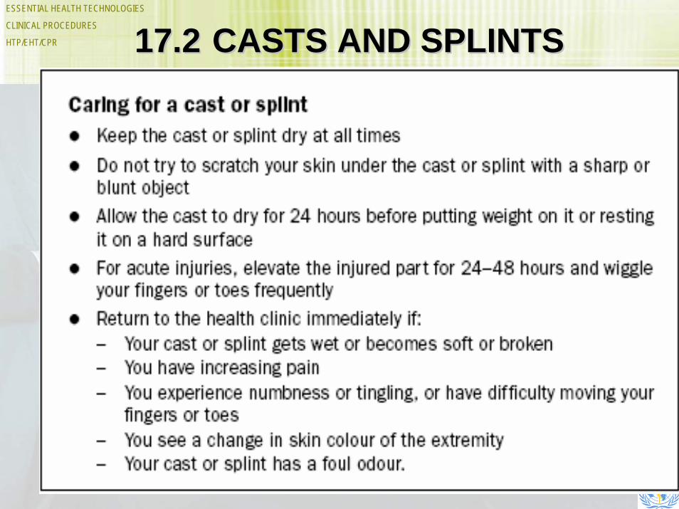
Patient instructions
• Give oral and written instructions to the patient and/or to accompanying relatives or other attendants.
• Give the instructions in non-technical language that the patient can understand.
ESSENTIAL HEALTH TECHNOLOGIES
CLINICAL PROCEDURES
HTP/EHT/CPR 17.217.2 CASTS AND SPLINTSCASTS AND SPLINTS

ESSENTIAL HEALTH TECHNOLOGIES
CLINICAL PROCEDURES
HTP/EHT/CPR 17.217.2 CASTS AND SPLINTSCASTS AND SPLINTSRemoving a castRemoving a cast

ESSENTIAL HEALTH TECHNOLOGIES
CLINICAL PROCEDURES
HTP/EHT/CPR 17.217.2 CASTS AND SPLINTSCASTS AND SPLINTS

ESSENTIAL HEALTH TECHNOLOGIES
CLINICAL PROCEDURES
HTP/EHT/CPR
17.3 APPLICATION OF EXTERNAL FIXATION
• External fixation is a technique for immobilizing fractures by placing pins into the bone above and below the fracture and connecting the pins to an external device
• The fracture position is adjusted by making changes to the external components in an outpatient setting
• Wounds are accessible for dressing changes, debridement and secondary closure or skin grafting.

ESSENTIAL HEALTH TECHNOLOGIES
CLINICAL PROCEDURES
HTP/EHT/CPR
17.3 APPLICATION OF EXTERNAL FIXATION
Materials• Arrange the fixation frame to best
accommodate the fracture pattern and the stability needed
• Partially threaded pins, 3–6 mm diameter, work best but smooth pins will work if threaded ones are not available.

ESSENTIAL HEALTH TECHNOLOGIES
CLINICAL PROCEDURES
HTP/EHT/CPR 17.317.3 APPLICATION OF EXTERNAL APPLICATION OF EXTERNAL FIXATIONFIXATION
Half pins are threaded on the end
Transfixation pins are threaded in the middle

ESSENTIAL HEALTH TECHNOLOGIES
CLINICAL PROCEDURES
HTP/EHT/CPR 17.417.4 DIAGNOSTIC IMAGINGDIAGNOSTIC IMAGING• Diagnostic imaging refers to a variety of graphic techniques:
- routine X-ray images, - ultrasound, - nuclear bone scans, - MRI scans, - CT scans
• X-ray is the most common imaging technique available at the district hospital
• X-ray images are a useful additional aid for diagnosis and treatment, but practitioners must be able to provide care without them
• The most useful and common X-ray examinations include the chest, spine, pelvis and the extremities.

ESSENTIAL HEALTH TECHNOLOGIES
CLINICAL PROCEDURES
HTP/EHT/CPR 17.4 DIAGNOSTIC IMAGING
• Skull radiographs are often of limited value as they neither exclude nor confirm possible life threatening intracranial damage
• In patients with acute abdominal disorders, including trauma injuries, ultrasound examination is the first method of choice, where available
• When performed by well-trained operators, the sensitivity of ultrasound for detecting intraperitonealbleeding is about 90% and the specificity is close to 100%.
ESSENTIAL HEALTH TECHNOLOGIES
CLINICAL PROCEDURES
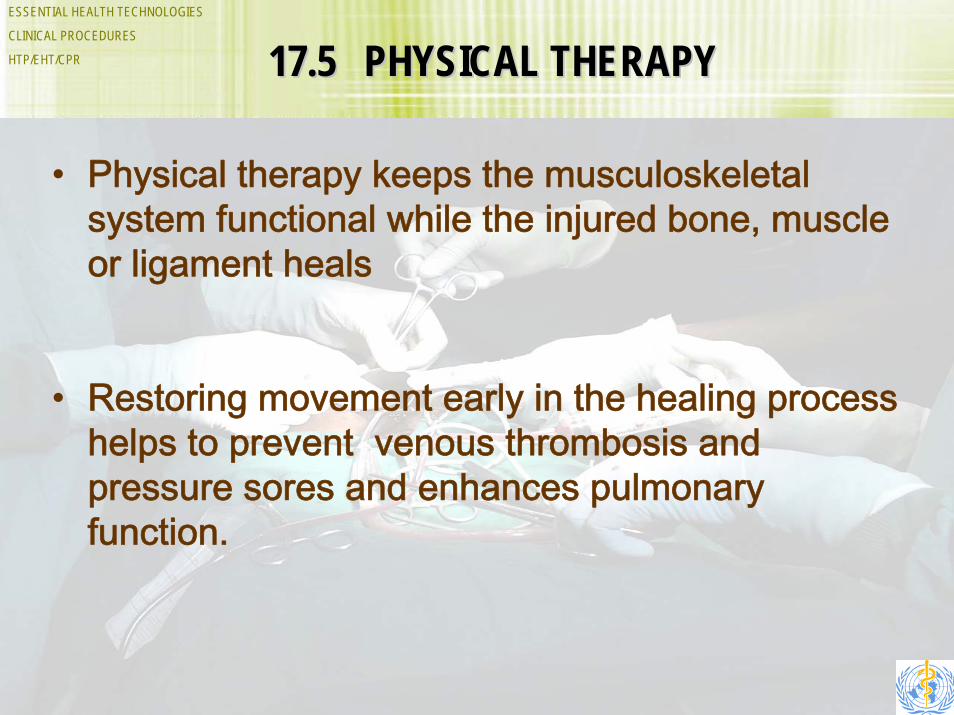
HTP/EHT/CPR 17.517.5 PHYSICAL THERAPYPHYSICAL THERAPY
• Physical therapy keeps the musculoskeletal system functional while the injured bone, muscle or ligament heals
• Restoring movement early in the healing process helps to prevent venous thrombosis and pressure sores and enhances pulmonary function.

ESSENTIAL HEALTH TECHNOLOGIES
CLINICAL PROCEDURES
HTP/EHT/CPR 17.6 CRANIAL BURR HOLES• Traumatic bleeding within the epidural and subdural spaces
increases intracranial pressure and causes neurological impairment
• Clinical features of extremely increased pressure include:– decreased consciousness, – slow pulse rate, – dilated pupils, – seizures and – hemiparesis
• Release of the pressure with cranial burr holes is an emergency and life- saving procedure.

ESSENTIAL HEALTH TECHNOLOGIES
CLINICAL PROCEDURES
HTP/EHT/CPR 17.6 CRANIAL BURR HOLES contd.
• Acute extradural and acute subduralhaematomas are the only two conditions that may benefit from burr holes.
• A history of trauma and a clear clinical diagnosis are essential before undertaking the procedure.

ESSENTIAL HEALTH TECHNOLOGIES
CLINICAL PROCEDURES
HTP/EHT/CPR 17.6 CRANIAL BURR HOLESAcute Extradural Haematoma
• The signs classically consist of:
– Loss of consciousness following an lucid interval, with rapid deterioration
– Middle meningeal artery bleeding with rapid raising of intracranial pressure
– Development of hemiparesis on the opposite side with a dilating pupil on the same side as the impact area, with rapid deterioration.

ESSENTIAL HEALTH TECHNOLOGIES
CLINICAL PROCEDURES
HTP/EHT/CPR 17.6 CRANIAL BURR HOLES
Acute Subdural Haematoma
• Acute subdural haematoma, with clotted blood in the subdural space accompanied by severe contusion of the underlying brain, occurs from the tearing of bridging vein between the cortex and the dura.
• Management is surgical and every effort should be made to do burr-hole decompressions. The diagnosis can be made on history and examination.
• Creating burr holes through the skull to drain the haematoma is often an emergency and life-saving procedure.

ESSENTIAL HEALTH TECHNOLOGIES
CLINICAL PROCEDURES
HTP/EHT/CPR 17.617.6 CRANIAL BURR HOLESCRANIAL BURR HOLESTechniqueTechnique