Embed Size (px)
Citation preview
Zurich Open Repository andArchiveUniversity of ZurichMain LibraryStrickhofstrasse 39CH-8057 Zurichwww.zora.uzh.ch
Year: 2015
Orthodontic material applications over the past century: Evolution ofresearch methods to address clinical queries
Eliades, Theodore
Abstract: The advances in the field of materials as they relate to orthodontics can be divided into theactual evolution of materials applied to daily practice and the changes in research methods to study theperformance and the biologic properties of the materials. Although it is evident that new materials havesaturated the market during the past century, the basic concepts of attaching one appliance to the enamelto use as a grip and inserting wires into that to control the spatial orientation of a tooth are identicalto the original concepts. In contrast to that, the numbers of treatises about those subjects and thecomplexity of instrumentation and analytic tools used in published research have advanced tremendouslyand at a frenetic pace. This highly specialized pattern of research may effectively raise boundaries acrossresearch areas, since the complexity of the issues allows researchers to comprehend the content of journalarticles in a narrow spectrum of disciplines. The purposes of this article were to review the advances inthe research methods for investigating the various properties of orthodontic materials and to assist thereader in navigating this topic. A synopsis of the materials is also provided, listing future applicationsthat already exist at the experimental stage or are yet unavailable but with the relevant technologyalready presented in broader scientific disciplines.
DOI: https://doi.org/10.1016/j.ajodo.2015.03.007
Posted at the Zurich Open Repository and Archive, University of ZurichZORA URL: https://doi.org/10.5167/uzh-118545Journal ArticleAccepted Version
The following work is licensed under a Creative Commons: Attribution-NonCommercial-NoDerivatives4.0 International (CC BY-NC-ND 4.0) License.
Originally published at:Eliades, Theodore (2015). Orthodontic material applications over the past century: Evolution of researchmethods to address clinical queries. American Journal of Orthodontics and Dentofacial Orthopedics,147(5 Suppl):224-231.DOI: https://doi.org/10.1016/j.ajodo.2015.03.007
Eliades, Theodore (2015). Orthodontic material applications over the past
century: Evolution of research methods to address clinical queries. American
Journal of Orthodontics and Dentofacial Orthopedics, 147(5 Suppl):224-231.
Orthodontic material applications over the past century: evolution of research methods to address clinical queries Theodore Eliades Zurich, Switzerland
Professor and Director, Clinic of Orthodontics and Paediatric Dentistry, Center of Dental Medicine, Faculty of Medicine, University of Zurich, Zurich, Switzerland. Correspondence: Theodore Eliades, Plattenstrasse 11, Zurich 8032, Switzerland Email: [email protected]

2
ABSTRACT
The advances in the field of materials as it relates to Orthodontics can be divided in the
actual evolution of materials applied to daily practice; and the change of research methods to
study the performance of the materials as well as their biological properties. Although it is
evident that new materials have saturated the market during the past century, the basic concept of
attaching one appliance to the enamel to use it as a grip and inserting wires into that to control
tooth spatial orientation is identical to the original concept. In contrast to that, the level of treatise
of subjects and the complexity of instrumentation and analytical tools employed in published
research have advanced to a tremendously high extent and at a delirious pace. This highly
specialized pattern of research may effectively raise boundaries across research areas, in the
sense that the complexity of issues allows researchers to comprehend the content of journal
articles in a narrow spectrum of disciplines. The purpose of this text is to review the advances
occurring in the field of research methods of investigating various properties of orthodontic
applications of materials, to assist the reader in navigating into this topic; a synopsis of the future
of materials is also provided listing future applications, which already exist at experimental stage
or are yet unavailable but with the relevant technology already presented in broader scientific
disciplines.

3
INTRODUCTION
The application of dental materials science in orthodontics coincides with the use of gold
wire alloys by E. Angle; the father of the specialty might have not imagined the impact materials
would have had in current orthodontic practice. As the field progressed and grew to receive the
dimensions of a specialty, the incorporation of principles of materials and mechanics of
materials, which are typically taught in the first year of an undergraduate engineering
curriculum, were introduced in postgraduate orthodontic curricula along with accompanying
elements of materials.
In the United States particularly, the disciplines orthodontic mechanics and materials
science, received further emphasis, possibly because of the requirement for a previously earned
degree prior to entering dental school. This prerequisite allowed for the cultivation and growth of
the materials research, since dental graduates entering orthodontic programs, were often
equipped with a bachelor’s level of formal training in natural or engineering sciences, thus
bringing a new perspective to traditional and empirically-taught concepts of the topic.1
The advances in the field of materials as it relates to Orthodontics can be divided in the
actual evolution of materials applied to daily practice; and the change of research methods to
study the performance of the materials as well as their biological properties.
With respect to the first section, although it is profoundly evident that new materials have
saturated the market during the time period examined, the basic concept of attaching one
appliance to the enamel to use it as a grip and inserting wires into that to control tooth spatial
orientation is identical to the original concept. Of course there have been different modes of
attaching this ‘handle’ to the tooth structure along with a large variation of bracket materials and
designs (lingual, self-ligating), and more alloy selection option are available, however the
foregoing changes are within the path of the original concept.
In as much, aligners have revolutionized the conventional appliance configuration and
constitute a new means of tooth movement, nonetheless, they too represent a new version of
removable appliance tooth movement pattern which was popular in the middle of the previous
century. Collectively, the orthodontist practicing in the E. Angle era could adapt with only a few
hours briefing seminar without much of difficulty to the conditions of the profession existing 100
years later.
4
In contrast to that, the level of treatise of subjects and the complexity of instrumentation
and analytical tools employed in published research have advanced to a tremendously high
extent and at a delirious pace. For example, the engineering approaches in materials science and
mechanics, or the methods utilized to study the biological mechanisms of tooth movement, as
well as the data analysis attributes of clinical trials, require a strong background in the respective
sciences involved, which makes it almost unattainable even for the contemporary orthodontist to
follow the developments across fields. This highly specialized pattern of research may
effectively raise boundaries across research areas, in the sense that the complexity of issues
allows researchers to comprehend the content of journal articles in a narrow spectrum of
disciplines. This has been highlighted by investigations on the characteristics of orthodontic
publications, which showed a significantly higher frequency of multi-author teams with
affiliations from different scientific disciplines in orthodontic publications within a decade.2-3
The purpose of this text is to review the advances occurring in the field of research
methods of investigating various properties of orthodontic applications of materials, to assist the
reader in navigating into this topic; a synopsis of the future of materials is also provided listing
future applications, which already exist at experimental stage or are yet unavailable but with the
relevant technology already presented in broader scientific disciplines.
EVOLUTION OF ORTHODONTIC MATERIALS RESEARCH APPROACHES: A LIST
OF PARADIGMS
The next paragraphs describe briefly the shift, which has occurred within the past decades
in the approaches to resolve several issues related to orthodontic materials and their applications.
The subheadings lead the reader from the first steps of intervention beginning with bonding and
covering the appliances and their properties. This section is complemented with an overview of
in vivo-ageing studies or retrieval analyses, which constitute the biggest breakthrough in the area
of simulation of clinical environment.
a) Bonding
1. Roughness of enamel
The alterations in the composition, topography and roughness of enamel has been the topic
of research owing to the interest on potential irreversible changes occurring assigned to
orthodontic bonding; one of the factors assessed is roughness. The traditional approach has

5
been to employ images of enamel (initially microscopic images and later scanning electron
images) to quasi-quantify the effect of bonding on enamel appearance, with apparent lack of
sensitivity since only approximations can be made on the roughness variation of surfaces. Later
on, a stylus-type profilometric analysis of the surface of the substrate before and after bonding
was used to investigate the increase in various roughness parameters, which are used to
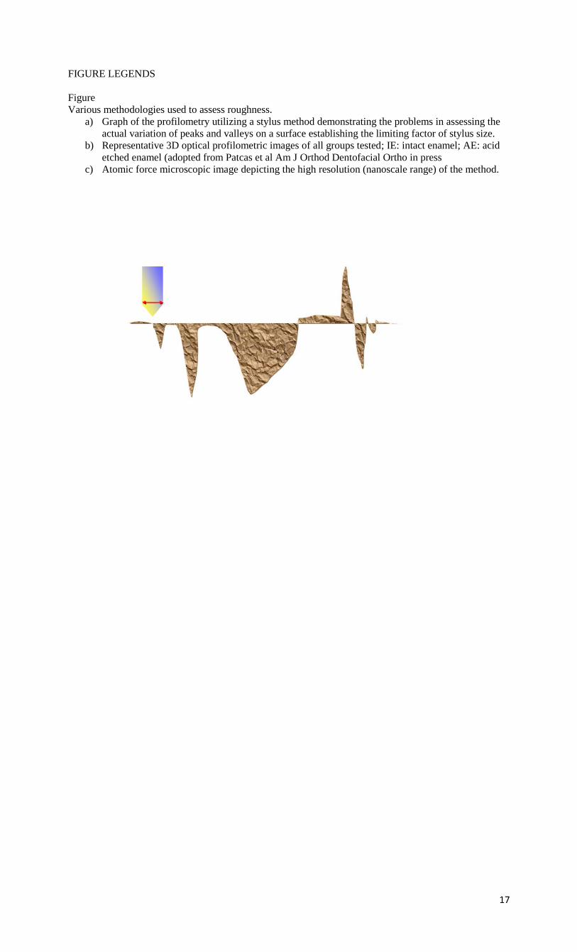
describe the variation of the surface (presence of peaks, valleys etc). As shown in Figure 1, the
conventional approach with the profilometry suffers from various limitations related mostly to
the dimensions of the recording stylus; in the case of deep, narrow valleys, with dimensions
smaller than the order of magnitude of the stylus, this method does not provide an accurate
account of the extent of alterations (Figure 1a). The use of 3D optical profilometry, bypasses
this obstacle (Figure 2b), as does the application of Atomic Force Microscopy (Figure 1c),
which however is essential for studying surface roughness at the nanoscale level, having
resolution far exceeding that of other stylus and optical based methods, and which is somehow
not applicable in large surface variations as those expected to occur following bonding.4-5 In
the past 2 decades we have come to understand far more on the topic than we have ever had and
this is largely due to the involvement of resources, material and human, from various scientific
disciplines.
2. Polymerization efficiency (degree of C=C conversion)
The polymerization efficiency also termed degree of cure (DC) or conversion, is often
mistakenly noted as ‘degree of polymerization’; the latter represents the number of repetitions of
the molecule of monomer to become polymer. The DC is considered a key property for all
polymers. In dental composite resins, this variable has been found to modulate the physical,
mechanical and biological properties of the material since a poor polymer network is susceptible
to release of biologically reactive substances (monomer, additives), predisposes to water
absorption and swelling and hydrolytic degradation, and is associated with reduced mechanical
properties.6-9 The latter had given rise to approaches, which focused on the investigation of the
phenomenon through mechanical testing with the hypothesis that since, higher degree of
conversion is associated with improved mechanical properties profile of the material, evidence
on the polymerization efficiency of the polymer could be extrapolated by measuring the response
to various tests. However, the kinetics of degree of cure and mechanical properties at the time
were not explored, and this resulted in erroneously assigning higher degree of cure in materials
6
which presented better mechanical properties. Since the 1970’s though the pioneering work of I.
Ruyter,10 the degree of cure of orthodontic (and broader dental) resinous adhesives has been
investigated with the actual measuring of the extent (%) of unreacted methacrylate groups,
thereby allowing the percentage of converted double bonds (polymerization) to be accurately
estimated on the surface of materials.11-12
3. Bond strength
Bond strength tests constitute a large portion of bonding-related literature with more than
700 publications listed in the pubmed database during the past decade. Aspects of test
standardization related to direction of loads, morphologic variation of teeth crown, source, age,
and maintenance of teeth in solutions, 13 have been analyzed and the shortcomings of this
preliminary test have been demonstrated.14 The lack of ageing of materials15 and the associated
questionable clinical relevance of this test has shifted the interest to more meaningful research,
which provide an appraisal of the performance of the material complex (adhesive-bracket) in
conditions identical to the actual clinical situation. Therefore, failure rate studies adopting a
prospective study protocol16 or even better, an RCT design,17 have gradually appearing more
often in the literature because of the direct clinical extrapolation. Nonetheless, the generalization
of results in this case, albeit much wider than its bond strength counterparts, depends on the
operator and thus, the results should not be interpreted in their absolute meaning without
caution.18
4. Prevention of demineralization
The attachment of orthodontic appliances to the enamel surface and their prolonged
presence in the oral environment has been associated with the development of some unfovarable
sequelae in the form of demineralization of the hard dental tissue. Fluoride is the most potent
cariostatic agent available that can prevent lesions to develop and dental hygiene plans have this
as a central concept with the use of many materials at different stages such as bonding (glass-
ionomer cements), and during treatment (gels, rinsing solutions and varnishes). For some of
these applications such as the glass-ionomer cements or fluoride-releasing resinous adhesives,
the long-term cariostatic effect of fluoride releasing adhesives has not been sufficiently
established, since most of the fluoride is released within the first few days or weeks.19-21
Therefore, topical fluoride in the form of solutions, varnishes or gels have been an integral part
of hygiene. A revolutionary recent approach targets the ttreatment of enamel following the
7
formation of white spots; which until previously was limited to interventional restorations.
Specifically, peptides such as a statherin-like peptide have been found to reduce the rate of HA
demineralization in caries-simulating solutions by about 50%; research efforts also focus on
salivary proteins, which can bind to HA surfaces and form a selectively permeable pellicle.22
b) Appliance
1. Corrosion
Corrosion of the orthodontic bracket-archwire complex has received attention after the
corrosion products of the bracket base were shown to be diffused into the adhesive.23 The
complexity of the materials and interfaces involved contribute to the development of corrosion in
various elements of the appliance. For example, the bracket in its traditional configuration, is
composed of two phases: a low modulus of elasticity stainless steel alloy for the manufacturing
of the base, which presumably allows for easy debonding after the completion of treatment; and
a high modulus steel alloy for the wings, which intends to minimize deformation arising from the
engagement of the wire and transfer the stresses from the activated archwire or the prescribed
bracket slot, to the tooth. These two alloys are joined with the use of brazing alloys composed of
Ni, Ag or Au and as a result there is a galvanic corrosion formed. In addition, the engagement of
the wire into the slot with the use of stainless steel ligatures formulates an environment where
many forms of corrosion can be developed. For further information, the reader is referred to a
thorough yet simplified review on the topic, which lists all potential forms of corrosion and their
mechanism.24
The advances on the field of orthodontic corrosion are largely due to the work of
engineers, who laid the fundamentals of the understanding of the mechanisms of the
phenomenon . The initial methods of plain examination of the surface under incident light (low
magnification microscopy) or by weighing the material before and after its exposure to the
effector, were substituted by research methods assessing the galvanic potential through
measuring the potential differences. Studies on the field identified that the potential differences
were found to be positive, indicating that the archwires were consistently the cathode and the
brackets were the anode of the galvanic cell. Concurrently, imaging techniques have been
advanced and scanning electron microscopy in conjunction with energy dispersive analysis have

8
contributed information on the topography, morphology and elemental analysis of the areas of
interest of the alloys.25
2. Forces and moments of wire-bracket combinations in various setups
The traditional approach of the majority of articles published in the early stages of
research development of the field of orthodontic materials consisted of bench tests with
simplified laboratory mechanical configurations, which in the majority of cases, were limited to
3-point bending and cantilever tests; tensile tests for the extrapolation of the modulus of the
material were also often employed. An effort to simulate the orthodontic clinical analogue had to
overcome 2 basic obstacles: the complex mechanical profile of multiple brackets bonded to teeth
in an arch form configuration, with wires engaged into the slots and ligated with elastomeric or
stainless steel ligatures; and the ageing of the materials in the intraoral environment and resultant
effects on the properties of the materials. Whereas the latter was not resolved until recently, the
first difficulty was overcome by utilizing a series of approaches:
i) Theoretical analysis with the use of finite element analysis software was
introduced in orthodontics from related sciences as a valid tool to predict the
forces and moments developed during engagement of in cases of insertion of
miniscrews and orthodontic implants.26
ii) Furthermore, experimental instrumentation included advanced analytical tools
such as X-ray diffraction27 and Differential scanning calorimetry,28 which
although did not provide a direct result on the actual mechanical profile of the
archwires, assisted essentially in explaining the performance of materials in
mechanical tests and improved our understanding of the mechanisms underlying
the load-deformation curve of these wires.
iii) Concurrently, new complex mechanical testing configurations were proposed to
simulate a bonded arch with various clinical malocclusion scenarios and selection
of a broad range of bracket and wire types.29-30 These in vitro constructed
analogues of the clinical situation provided information, which may be considered
as the closest to the intraoral situation if the effect of ageing of the materials is
excluded; that was a decisive step in obtaining data on the development of forces
and moments at the 3 planes of space during engagement of wires in brackets.
9
iv) Finally, more recently, brackets with integrated strain gauge-type sensors, were
proposed; these are the most advanced appliances from the onset of specialty.31
These are capable of providing actual force ranges either in real time (in the case
of bulky brackets, which carry a power source) or in a retrospective fashion,
which allows the manufacturing of slim forms of appliances.
3. Effect of intraoral conditions on materials
This area has undergone extensive change, to the point that it constitutes a new field of
research activity. Initially, the materials were subjected to ageing by immersing them into
various liquids (water, saline, artificial saliva, acidic liquids) for various times and then
performing tests (mechanical properties, structural investigations, compositional alterations) to
identify the effects induced by ageing. Modern research relies on retrieval analysis, which was
already a routine approach in the biomedical literature in the 1970’s. The analysis of retrieval
analysis goes beyond the scope of this article and the interested reader is referred to an article
and a book in this topic.15
4. Effectiveness of materials and techniques
After the 1990’s there has been a specific interest on the investigation of the material
performance through clinical trials-as opposed to extrapolation of information on their intraoral
behavior by in vitro tests or simulations. This approach has provided information, which rejected
assumption on various topics such as self-ligating brackets, effectiveness of NiTi wires, or
failure rate of brackets; further information on this topic can be found in this journal issue.
c) Side effects of materials
Within this field, the release of substances (ions from alloys, and monomers, degradation
byproducts and additives from polymers), and the biological properties of materials are
included.
1. Release of substances from orthodontic materials
At the early stages, efforts consisted of measuring in vitro and with primitive techniques
(weight, morphology), the ions or monomers released in immersion media. Later, the same
method was complemented with the introduction of instrumental analysis such as atomic
emission or absorption spectroscopy32 for the case of metals; high performance liquid
chromatography and gas chromatography-mass spectroscopy were also employed for the
10
qualitative and quantitative analysis of immersion media, saliva, blood or urine with respect to
concentration of polymer byproducts.33-37
2. Biological properties of materials
With the establishment of a reliable method for the identification of released substances,
the necessity for assessing the potential biological reactivity emerged. The earliest methods in
the field were usually confined to adding various concentrations of effectors hypothesized to be
released from orthodontic materials in cultures of immortal cell lines and observing the
inhibition zone, which correspond to the lack of level of concentration causing cell death.
Later more advanced techniques allowed for the assessment of action at subtoxic concentrations
to observe the effect on the physiology of cell (or animal) by using markers of oxidative stress or
metabolic function and in 3D cultures.38-40 Then, with the availability of data on the quantity of
species released, these levels of effectors can be added to human gingival or periodontal
ligament fibroblasts to secure two things: first the corresponding mode of action at doses
documented to occur in vivo and secondly to identify potential effects on human cells.
THE FUTURE OF ORTHODONTIC MATERIALS
1. Shape memory polymers
Shape-memory polymers are an emerging class of polymers with applications spanning
various areas of everyday life. These polymers are dual-shape materials belonging to the group
of “actively moving” polymers that can change from one shape to another, the first being a
temporary shape obtained by mechanical deformation and the second obtained from subsequent
fixation of that deformation.41
For shape-memory polymers, heat, light, infrared radiation, electrical and magnetic
fields, and immersion in water have been used to induce this property. The shape-memory effect
depends on the molecular architecture and does not require a specific chemical structure in the
repeating units. In essence, application of external stimuli to these materials introduces a
departure from the as-received shape to a new configuration that is reversible.
In orthodontics, the potential application of these materials involves the manufacturing of
polymeric transparent wires with minimum stiffness, which could then be transformed into
archwires of predetermined elastic modulus upon exposure to a stimulus such as light or heat.
11
Therefore, aesthetics along with a preferable stiffness would be attained during intraoral
application of these materials.
2. Self-healing materials
The design and manufacturing of “smart” synthetic systems that can mimic the behavior
of biological systems which heal themselves has been an objective of intense research during the
past decade. Hybrid materials have been recently created, where micron-scale conduits extend
throughout the material and contain healing fluids or dissolved healing agents.42 When a crack
appears near the network, the fluid can flow to the damaged region and fill the fissure. The
simplest form to achieve this behavior might be the incorporation of bubbles of a
material/substance/precursor in the raw material. This reservoir, upon exposure to air,
polymerizes as a result of crack formation and spontaneously closes the crack, thus maintaining
the structural integrity of the material.
The orthodontic application of this concept may involve polymer brackets and archwires.
The integration of nanosized bubbles filled with autopolymerized monomer in these materials
may result in reduction of wire and bracket breakages. Fracture of the bracket or wire would
induce bursting of the nanobubbles and exposure of the monomer to air, thereby resulting in
polymerization and filling of the crack-induced gap.
3. Biomimetic adhesives
The issue of an enamel-friendly bonding mechanism for orthodontic appliances has been
the subject of investigations since the original introduction of the acid-etching technique. This
intense interest derived from the description of alterations of enamel color and structure
associated with acid-etched-mediated bonding . Although glass-ionomer adhesives have offered
an alternative, their applications remain limited, probably because of the higher failure rates.
The introduction during the past 15 years of a new class of materials that adopt the
paradigms of nature has gradually established the category of biomimetic materials. This term
derives from Greek “bio” (living) and “mimetic” (imitating or resembling), and refers to how
creatures ingenuously employ natural elements to solve problems in the environment.
Geckos, for example, are lizards that belong to the species of gekkonidae are
characterized by a remarkable ability to sustain their weight while upside down. The strong but
temporary adhesion employed by a gecko comes from a mechanical principle known as “contact
splitting”. The foot of a gecko has a flat pad which is densely packed with very fine hairs that are
12
split at the ends, resulting in a greater number of contact points than if the hairs were not split.
More contact points between these hairs and a surface result in a significant increase in adhesion
force. Researchers have discovered that this special nature of the foot pads allows the gecko to
stick to surfaces through the formation of localized van der Waals forces.43 This mechanism has
been employed for high-friction microfibers or carbon nanotubes, which are sprayed on a
surface. Because of their enormous number per unit area, the physical forces developed mimic
the ability of a gecko to attach firmly to surfaces without the use of a chemical substance. While
this mode of bonding may be suitable for dry environments, it fails to provide reliable
performance for wet surfaces. This problem inspired researchers to adopt another natural
example of bonding: that of mussels. Combining the important elements of gecko and mussel
adhesion, the new adhesive material, called “geckel”, functions like a sticky note and exhibits
strong yet reversible adhesion in both air and water.44 Mussel-mimetic polymers have an amino
acid called L-3,4-dihydroxyphenylalanine (DOPA) found in high concentrations in the “glue”
proteins of mussels. Analogously to the gecko-based approach, pillar arrays (400-600 nm in
diameter and length) coated with the mussel-mimetic polymer improved wet adhesion by 15-fold
over uncoated pillar arrays.
The orthodontic application of this innovation is profound. Brackets having bases with
pads mimicking the gecko foot and covered with a layer of DOPA would provide adequate bond
strength to sound enamel without prior enamel conditioning and with minimal color and
structural alterations to the enamel. To the knowledge of the authors, use of this type of
biomimetic adhesive has not yet been adapted by a manufacturer to orthodontic brackets.
4. Self-cleaning materials
The issue of plaque retention on brackets and microbial attachment onto these calcified
biofilms has been a major concern from an enamel prophylactic perspective. The development of
a material that could clean itself not only from inorganic mainly, but also from organic,
precipitations is an application that is very attractive in the materials science areas involving
biomedical, industrial and aeronautical applications.
Early research in this field adopted the paradigm of microscopic bumps on a lotus leaf,
which transform its waxy surface into an extremely water repellent, or superhydrophobic,
material. Synthetic self-cleaning materials have been developed, some of which are based on this
13
“lotus effect”, whereas others employ the opposite property of “superhydrophilicity” as well as
catalytic chemical reactions.
The use of titanium oxide (TiOx) nanocoatings has been found to increase the safety of
aircraft by preventing the buildup of ice and other contaminants. This process creates self-
cleaning “superhydrophobic” surfaces, and involves the application of a hydrofluoric acid
coating on the titanium alloy substrate. A substantially self-cleaning superhydrophobic surface is
created when exposure to ultraviolet light causes the titanium oxide layer to undergo
photocatalytic reaction with oxygen to oxidize any organic contaminants that may be present.
Since superhydrophobic surfaces resist soiling by water-borne contaminants, they are easily
cleaned and useful in directing flow in microfluidic devices.
Photocatalytic activity from the reaction of titanium oxide with ultraviolet light has
recently gained attention in orthodontic materials.45 In particular, there is scientific interest in
inducing photocatalytic reactions on the nickel-titanium archwire alloy. By thickening the
titanium oxide film with electrolytic treatment and then applying heat treatment, the surface film
on NiTi was modified from an amorphous structure to crystalline rutile (TiO2).
14
REFERENCES
1. Eliades T. Dental materials in Orthodontics. In: Graber LW, Vanarsdall RL, Vig KL (eds).
Orthodontics: current principles and techniques. Elsevier, Philadelphia 2012:
2. Kanavakis G, Spinos P, Polychronopoulou A, Eliades T, Papadopoulos MA, Athanasiou AE.
Orthodontic journals with impact factors in perspective: trends in the types of articles and
authorship characteristics. Am J Orthod Dentofacial Orthop 2006;130:516-22
3. Baumgartner S, Pandis N, Eliades T. Exploring the publications in three major orthodontic
journals: a comparative analysis of two 5-year periods. Angle Orthod 2014;84:397-403.
4. Eliades T, Gioka C, Eliades G, Makou M. Enamel surface roughness following debonding using
two resin grinding methods. Eur J Orthod 2004;26:333-8.
5. Patcas R, Zinelis S, Eliades G, Eliades T. Inyerfacial characteristics of bonding to acid-etched
and sandblasted enamel. Am J Orthod Dentofacial Orthod in press
6. Vrochari AD, Eliades G, Hellwig E, Wrbas KT. Curing efficiency of four self-etching, self-
adhesive resin cements. Dent Mater 2009;25:1104-8.
7. Gaintantzopoulou M, Rahiotis C, Eliades G. Molecular characterization of one-step self-etching
adhesives placed on dentin and inert substrate. J Adhes Dent 2008;10:83-93.
8. Rejman DJ, Eliades T, Bradley TG, Eliades G. Polymerization efficiency of glass-ionomer and
resin adhesives under molar bands. Angle Orthod 2008;78:549-52.
9. Niepraschk M, Rahiotis C, Bradley TG, Eliades T, Eliades G. Effect of various curing lights on
the degree of cure of orthodontic adhesives. Am J Orthod Dentofacial Orthop. 2007;132:382-4.
10. Ruyter IE, Györösi PP. An infrared spectroscopic study of sealants. Scand J Dent Res 1976;84:
396-400.
11. Eliades T, Eliades G, Brantley WA, Johnston WM. Polymerization efficiency of chemically
cured and visible light-cured orthodontic adhesives: degree of cure. Am J Orthod Dentofacial
Orthop 1995;108:294-301
12. Eliades T, Eliades G, Bradley TG, Watts DC Degree of cure of orthodontic adhesives with
various polymerization initiation modes. Eur J Orthod 2000;22:395-9.
13. Eliades T, Brantley WA The inappropriateness of conventional orthodontic bond strength
assessment protocols. Eur J Orthod. 2000;22:13-23.
14. Eliades T, Katsavrias E, Zinelis S, Eliades G. Effect of loading rate on bond strength. J Orofac
Orthop 2004;65:336-42.
15. Eliades T, Bourauel C. Intraoral aging of orthodontic materials: the picture we miss and its
clinical relevance. Am J Orthod Dentofacial Orthop 2005;127:403-12.
16. Pandis N, Polychronopoulou A, Eliades T. Failure rate of self-ligating and edgewise brackets
bonded with conventional acid etching and a self-etching primer: a prospective in vivo study.
Angle Orthod 2006;76:119-22.
17. Nandhra SS, Littlewood SJ, Houghton N, Luther F, Prabhu J, Munyombwe T, Wood SR. Do we
need primer for orthodontic bonding? A randomized controlled trial. Eur J Orthod. 2014 pii:
cju024.
18. Pandis N, Fleming PS, Kloukos D, Polychronopoulou A, Katsaros C, Eliades T. Survival of
bonded lingual retainers with chemical or photo polymerization over a 2-year period: a single-
center, randomized controlled clinical trial. Am J Orthod Dentofacial Orthop 2013;144:169-75.
19. Øgaard B, Rølla G, Ruben J, Dijkman T, Arends J. Microradiographic study of demineralization
of shark enamel in a human caries model. Scand J Dent Res 1988;96:209-11.
15
20. Øgaard B, Rølla G, Arends J. Orthodontic appliances and enamel demineralization. Part 1. Lesion
development. Am J Orthod Dentofac Orthoped 1988;94:68-73.
21. Øgaard B, Rølla G, Arends J, ten Cate JM. Orthodontic appliances and enamel demineralization.
Part 2. Prevention and treatment of lesions. Am J Orthod Dentofac Orthoped 1988;94:123-8.
22. Shah S, Kosoric J, Hector MP, Anderson P. An in vitro scanning microradiography study of the
reduction in hydroxyapatite demineralization rate by statherin-like peptides as a function of
increasing N-terminal length. Eur J Oral Sci 2011;119:13-8.
23. Maijer R, Smith DC. Corrosion of orthodontic bracket bases. Am J Orthod. 1982 Jan;81(1):43-8.
24. Eliades, T. and A. Athanasiou . In vivo aging of orthodontic alloys: implications for corrosion
potential, nickel release, and biocompatibility. Angle Orthod 2002;72:222–237.
25. Siargos B, Bradley TG, Darabara M, Papadimitriou G, Zinelis S. Galvanic corrosion of metal
injection molded (MIM) and conventional brackets with Nickel-Titanium and Copper; Nickel-
Titanium archwires. Angle Orthod 2007;77:355-360.
26. Chatzigianni A, Keilig L, Duschner H, Götz H, Eliades T, Bourauel C. Comparative analysis of
numerical and experimental data of orthodontic mini-implants. Eur J Orthod. 2011
Oct;33(5):468-75.
27. Thayer TA1, Bagby MD, Moore RN, DeAngelis RJ. X-ray diffraction of nitinol orthodontic arch
wires. Am J Orthod Dentofacial Orthop 1995;107:604-12.
28. Biermann MC, Berzins DW, Bradley TG. Thermal analysis of as-received and clinically retrieved
copper-nickel-titanium orthodontic archwires. Angle Orthod 2007;77:499-503.
29. Badawi H, Major P. Three-dimensional orthodontic force measurements. Am J Orthod
Dentofacial Orthop 2010;137:299-300.
30. Bourauel C, Drescher D, Thier M. An experimental apparatus for the simulation of three-
dimensional movements in orthodontics. J Biomed Eng 1992;14:371-8.
31. Lapatki BG, Bartholomeyczik J, Ruther P, Jonas IE, Paul O. Smart bracket for multi-dimensional
force and moment measurement. J Dent Res 200;86:73-8.
32. Eliades T, Trapalis C, Eliades G, Katsavrias E. Salivary metal levels of orthodontic patients: a
novel methodological and analytical approach. Eur J Orthod 2003;25:103-6.
33. Eliades T, Hiskia A, Eliades G, Athanasiou AE. Assessment of bisphenol-A release from
orthodontic adhesives. Am J Orthod Dentofacial Orthop 2007;131:72-5.
34. Gioka C, Bourauel C, Hiskia A, Kletsas D, Eliades T, Eliades G. Light-cured or chemically cured
orthodontic adhesive resins? A selection based on the degree of cure, monomer leaching, and
cytotoxicity. Am J Orthod Dentofacial Orthop 2005;127:413-9.
35. Eliades T, Eliades G, Brantley WA, Johnston WM. Residual monomer leaching from chemically
cured and visible light-cured orthodontic adhesives. Am J Orthod Dentofacial Orthop
1995;108:316-21.
36. Eliades T, Voutsa D, Sifakakis I, Makou M, Katsaros C.Release of bisphenol-A from a light-
cured adhesive bonded to lingual fixed retainers. Am J Orthod Dentofacial Orthop 2011;139:192-
5.
16
37. Kloukos D, Taoufik E, Eliades T, Katsaros C, Eliades G. Cytotoxic effects of polycarbonate-
based orthodontic brackets by activation of mitochondrial apoptotic mechanisms. Dent Mater
2013;29::e35-44.
38. Mavrogonatou E, Eliades T, Eliades G, Kletsas D. The effect of triethylene glycol dimethacrylate
on p53-dependent G2 arrest in human gingival fibroblasts. Biomaterials 2010;3:8530-8.
39. Taoufik K, Mavrogonatou E, Eliades T, Papagiannoulis L, Eliades G, Kletsas D. Effect of blue
light on the proliferation of human gingival fibroblasts. Dent Mater 2008;24:895-900.
40. Vande Vannet BM, Hanssens JL.Cytotoxicity of two bonding adhesives assessed by three-
dimensional cell culture. Angle Orthod 2007;77:716-22.
41. Landlein A, Jiang H, Junger O, Langer BR. Light-induced shape memory polymers. Nature
2009;424:879-82.
42. Cordier P, Tournilhac F, Ziakovich-Soullia C, Leibler L. Self-healing and thermoreversible
rubber from supramolecular assembly. Natire 2008;451:977-80.
43. Berengueres J, Saito S, Tadakuma K. Structural properties of a scaled-gecko foot hair. Bioinsp
Biomim 2007;2:1-8.
44. Lee H, Lee BP, Messersmith PB. A reversible wer-dry adhesive inspired by mussles and geckos.
Nature 2007;448:338-41.
45. Sato Y, Miyazawa K, Sato N, Nakano K, Takei Y, Kawai T, Goto S. Study on fabrication of
orthodontic brackets with the photocatalytic function of titanium dioxide. Dent Mater J
2009;28:388-95.
17
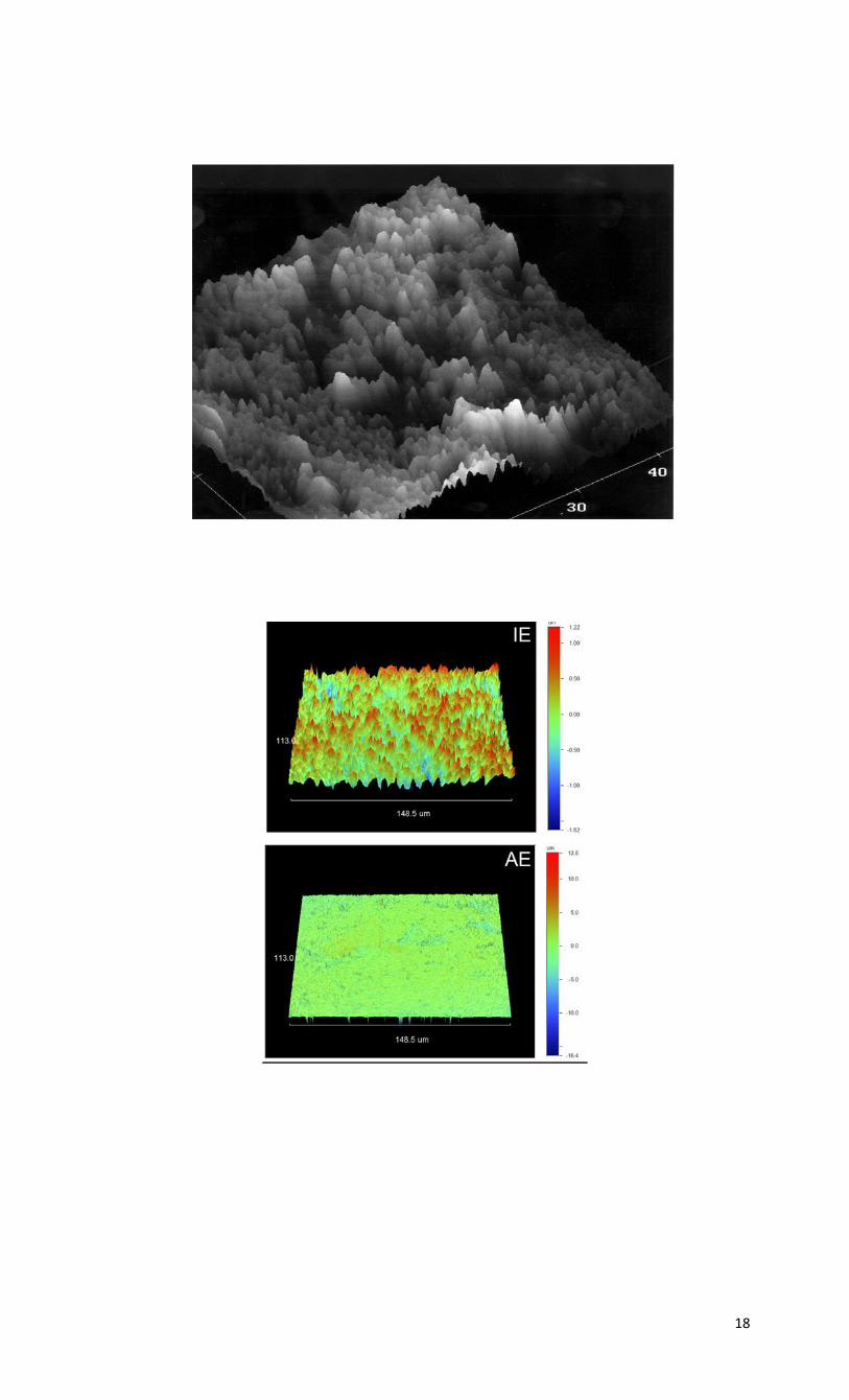
FIGURE LEGENDS
Figure
Various methodologies used to assess roughness.
a) Graph of the profilometry utilizing a stylus method demonstrating the problems in assessing the
actual variation of peaks and valleys on a surface establishing the limiting factor of stylus size.
b) Representative 3D optical profilometric images of all groups tested; IE: intact enamel; AE: acid
etched enamel (adopted from Patcas et al Am J Orthod Dentofacial Ortho in press
c) Atomic force microscopic image depicting the high resolution (nanoscale range) of the method.
18

19