Embed Size (px)
Citation preview
pISSN: 1011-8942 eISSN: 2092-9382
Korean J Ophthalmol 2015;29(2):79-85http://dx.doi.org/10.3341/kjo.2015.29.2.79
© 2015 The Korean Ophthalmological SocietyThis is an Open Access article distributed under the terms of the Creative Commons Attribution Non-Commercial License (http://creativecommons.org/licenses /by-nc/3.0/) which permits unrestricted non-commercial use, distribution, and reproduction in any medium, provided the original work is properly cited.
79
Original Article
Deep Anterior Lamellar Keratoplasty Using Irradiated Acellular Cornea with Amniotic Membrane Transplantation for
Intractable Ocular Surface Diseases
Sung Wook Wee, Sang Uk Choi, Jae Chan Kim
Department of Ophthalmology, Chung-Ang University Hospital, Chung-Ang University College of Medicine, Seoul, Korea
Purpose: To report the clinical outcomes of deep anterior lamellar keratoplasty (DALK) when sterile gamma-ir-
radiated acellular corneal tissues (VisionGraft) are used in combination with amniotic membrane transplanta-
tion (AMT) for intractable ocular surface diseases.
Methods: The medical records of fifteen patients who had DALK with AMT were retrospectively reviewed. Indi-
cations for surgery included ocular burn, bacterial keratitis, herpes simplex virus keratitis, corneal opacity with
Stevens-Johnson syndrome, Mooren’s ulcer, idiopathic myxoid degeneration of corneal stroma, and recurrent
band keratopathy. DALK was performed using partial-thickness acellular corneal tissue and a temporary am-
niotic membrane patch was added at the end of the operation.
Results: All cases that underwent DALK with AMT became epithelialized within 2 postoperative weeks. Twelve
patients showed favorable outcomes without graft rejection, corneal opacification, or neovascularization. The
other three grafts developed corneal opacification and neovascularization, and required additional penetrating
keratoplasty (PK). Unlike the results of previous PKs, there were no graft rejections and the graft clarity was
well-maintained in these three cases for at least 8 months after PK.
Conclusions: DALK using sterile acellular corneal tissues in combination with AMT may be a good therapeutic
strategy for treating intractable ocular surface diseases because of lowered immune rejection, fibroblast ac-
tivation, and facilitation of epithelialization. Furthermore, DALK can help stabilize the ocular surface, prolong
graft survival, and may allow better outcomes when combined with subsequent PK.
Key Words: Amniotic membrane transplantation, Deep anterior lamellar keratoplasty, Intractable ocular surface
disease, Sterile acellular cornea, Visiongraft
Allogeneic corneal transplantation is important; it is very effective, it is the only treatment for blinding corneal disease, and it is a large part of global tissue transplanta-
tion [1]. However, several problems with this method have become apparent in past decades. The supply of donor cor-neas is much lower than the demand. Second, immune-me-diated corneal allograft rejection is likely to develop with high-risk corneal transplantation, including an infectious recipient bed, refractory neovascularization, and limbal stem cell deficiency (LSCD). Although corneal transplan-tation has a high survival rate of 90% when performed in low-risk recipients [2], in cases of high-risk corneal trans-
Received: July 21, 2014 Accepted: October 4, 2014
Corresponding Author: Jae Chan Kim, MD, PhD. Department of Oph-thalmology, Chung-Ang University Hospital, Chung-Ang University College of Medicine, #102 Heukseok-ro, Dongjak-gu, Seoul 156-755, Ko-rea. Tel: 82-2-6299-1665, Fax: 82-2-825-1666, E-mail: [email protected]
80
Korean J Ophthalmol Vol.29, No.2, 2015
plantation, pathologies such as LSCD and autoimmune disease may lead to a poor surgical outcome including de-creased visual acuity (VA) due to recurrent corneal opaci-fication, neovascularization, and persistent epithelial defect (PED) with ulceration.
Deep anterior lamellar keratoplasty (DALK) is advanta-geous over penetrating keratoplasty (PK) because DALK eliminates the possibility of endothelium rejection and us-ing lower-quality tissues [3,4]. However, there is still a pos-sibility of epithelial and stromal rejections after DALK be-cause of f resh corneal tissue cellular components, including the epithelium, keratocytes, and bone mar-row-derived cells, which are sources of major histocompat-ibility complex antigens [5]. One way to overcome this limitation may be by decellularizing corneal substrates. Previous studies reported that glycerin cryopreservation was a practical and effective technique for decellulariza-tion [6]. Decellularizing corneal tissues could prevent an immune transplant rejection pathway because of the ab-sence of antigen-presenting cells, which would reduce the amount of sensitization in the recipient’s T cell levels. In this study, we used the VisionGraft (Tissue Banks Interna-tional, Baltimore, MD, USA), a sterile gamma-irradiated acellular cornea that has recently been made available for use by ophthalmic surgeons.
Amniotic membrane transplantation (AMT) is a widely used method in ocular surface disease. AMT contributes to the restoration of an intact basement membrane, and supports facilitation of epithelialization [7]. Another bene-ficial effect of AMT is that it suppresses T cell prolifera-tion, which can reduce immune-modulatory effects in in-tractable ocular surface disease [8]. Thus, AMT may play an important role in high-risk corneal transplantation by facilitating epithelialization and lowering immune rejec-tion. In this paper, we report our experience with DALK in combination with AMT in a small group of intractable oc-ular surface disease cases, using sterile gamma-irradiated acellular corneal tissues.
Materials and Methods
We retrospectively reviewed the medical records of fif-teen patients operated on at the Chung-Ang University Hospital between November 25, 2010 and October 19, 2012. The study protocol was in accordance with the Declaration
of Helsinki and was approved by the institutional ethics committee of Chung-Ang University Hospital, Seoul, Ko-rea. All participants gave informed consent after a detailed explanation of the study design, ancillary investigations for scientific purposes, and related imaging procedures.
Indications for surgery included ocular burn (n = 3), bac-terial keratitis (n = 3), herpes simplex virus keratitis (n = 3), corneal opacity with Stevens-Johnson syndrome (n = 2), Mooren’s ulcer (n = 2), idiopathic myxoid degeneration of corneal stroma (n = 1), and recurrent band keratopathy (n = 1). The majority of the patients had a history of graft fail-ure despite previous multiple PKs and ocular surface stabi-lization failure despite sole AMT, which resulted in corne-al opacification, neovascularization, PED with ulceration, and infection.
All DALK procedures were performed by the same sur-geon (JK) under retrobulbar anesthesia in a similar fashion using the modified big bubble technique [9]. The Hess-burg-Barron disposable vacuum trephine (Katena Prod-ucts, Denville, NJ, USA) was used and the host cornea was trephined to a level of 70% to 80% of its original thickness. The size of trephination ranged from 7.0 to 8.5 mm, and was 0.5 mm bigger than the size of the recipient bed. The air was injected into the deep stroma of the central cornea with a 30-gauge needle inserted on a 2-mL syringe with the bevel facing down. This procedure formed a large bub-ble between the Descemet’s membrane and stroma, and separated the Descemet’s membrane. Air injection was stopped when the big-bubble reached the trephination line. Air was injected into the anterior chamber after incising the corneal limbus with a diamond blade. Then the anteri-or lamellar stroma (about 80% in thickness) was removed by a 360-degree lamellar dissection technique using Vanna scissors. The remaining anterior stromal fibrous tissue was removed with additional dissection using viscoelastic sub-stance injection and repeated dissections until the Descem-et’s membrane was exposed. The host bed was irrigated to remove any remaining viscoelastic substance using a bal-anced salt solution (Alcon Laboratories, Fort Worth, TX, USA). The sterile acellular corneas were kept in sterile glass bottles and stored in albumin solution. The tissue consisted of a 350-µm partial thickness, clear, corneal lent-icule without endothelium and Descemet’s membrane. The sterile acellular cornea was sutured in place using 24 inter-rupted 10-0 nylon. Air was injected into the anterior cham-ber again to facilitate linkage between the host’s Descem-
81
SW Wee, et al. Keratoplasty with Irradiated Acellular Cornea
et’s membrane and grafted corneal stroma.At the end of the DALK operation, the temporary amni-
otic membrane patch was secured with interrupted 10-0 nylon sutures with episcleral bites in the stromal side up position which covered most of the ocular surface. Pre-served human amniotic membrane (Amnisite; Bioland, Cheonan, Korea) was obtained from donors that were sero-negative for the human immunodeficiency virus, human T cell leukemia virus, hepatitis B virus, hepatitis C virus, and syphilis at delivery. Also administered were subcon-junctival injections of gentamicin and betamethasone.
Topical treatment, administering the third-generation fluoroquinolone (Cravit; Santen, Osaka, Japan) four times per day for 1 month and a 1% prednisolone acetate (Pred-forte; Allergan, Irvine, CA, USA) was administered four times daily for the first week and tapered over 3 months before being discontinued. Oral antibiotic treatment was prescribed for 2 weeks and oral steroid treatment was ad-ministered with a dose of 0.5 mg/kg/day and tapered over 3 weeks.
Temporary amniotic membrane patches were removed at postoperative 1 to 2 weeks, and corneal sutures were re-moved at the end of postoperative 3 to 12 months accord-ing to the state of the grafts. All patients were observed closely for inflammation, infection, graft rejection, epithe-lialization, and clarity. Immunologic stromal rejection was diagnosed clinically and determined by the presence of de-creased vision and stromal edema with or without corneal neovascularization. Graft failure was defined as an irre-versible loss of central graft clarity for a minimum of 3 consecutive months, and required additional PK. The re-sidual acellular cornea was fixed with 10% formalin for 24 hours and stained with hematoxylin-eosin to verify the acellularity of the product.
Results
Patient demographics are summarized in Table 1. There were eight male and seven female patients, with a mean age of 51.0 ± 16.21 years (range, 19 to 78 years). The mean follow-up period was 16.6 ± 3.68 months (range, 10 to 24 months). Ten patients experienced multiple failed PKs even though fresh donor corneas were used. The multiple PK failures exhibited ocular surface stabilization failure with sole AMT, resulting in PED with corneal ulceration. These
cases with failed PK and AMT had previous histories that included ocular burns, herpes simplex virus keratitis, Ste-vens-Johnson syndrome, Mooren’s ulcer, and idiopathic corneal myxoid degeneration. Detailed demographic data, clinical characteristics, and postoperative outcomes of the representative six patients are summarized in Table 2.
All cases that underwent DALK with AMT became epi-thelialized within 2 postoperative weeks. PED was not as-sociated with corneal ulceration. Twelve patients showed favorable outcomes without graft rejection, corneal opaci-fication, or neovascularization (Fig. 1). The other three pa-tients developed corneal opacification, neovascularization, and eventually decreased VA, which required additional PK. These three cases had underlying comorbidities which included Stevens-Johnson syndrome and Mooren’s ulcer. However, unlike the results of previous PKs, corneal opacification and neovascularization did not occur in these three patients during a follow-up period of 8 months (Fig. 2). No mechanical complication developed with the DALK procedure, including Descemet’s membrane microperfora-tion or pseudochamber formation.
Nine patients had preoperative VA that was worse than 20 / 200, and most of the cases showed postoperative VA that was less than 20 / 200 except two cases with hand movement VA.
Hematoxylin-eosin staining of the sterile acellular cor-nea revealed a relatively loose corneal stroma with thinned epithelium that showed significant acellularity. There was
Table 1. Demographic data of fifteen patients with intractable ocular surface diseases
Factor ValueMale : female 8 : 7Age (yr) 51.0 ± 16.21Primary pathogenesis
Ocular burn 3Bacterial keratitis 3Herpes simplex virus keratitis 3Stevens-Johnson syndrome 2Mooren’s ulcer 2Idiopathic corneal myxoid degeneration 1Band keratopathy 1
Follow-up period (mon) 16.6 ± 3.68Values are presented as number or mean ± SD.
82
Korean J Ophthalmol Vol.29, No.2, 2015
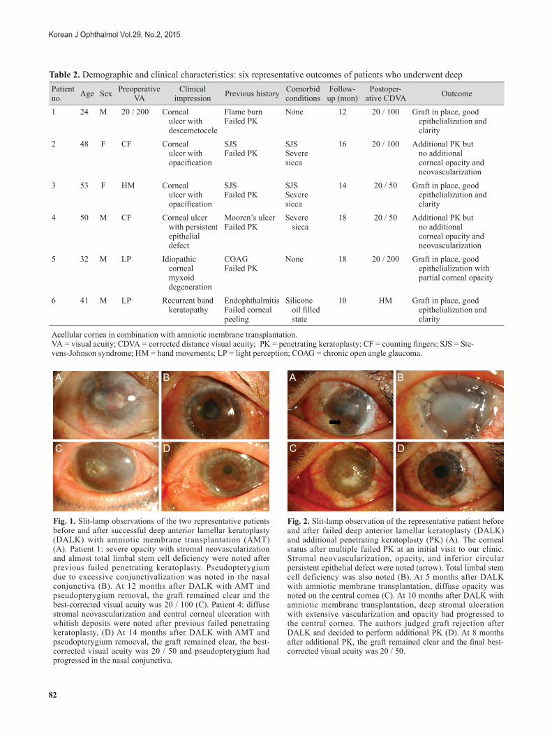
Table 2. Demographic and clinical characteristics: six representative outcomes of patients who underwent deepPatient no. Age Sex Preoperative
VAClinical
impression Previous history Comorbid conditions
Follow-up (mon)
Postoper-ative CDVA Outcome
1 24 M 20 / 200 Corneal ulcer with descemetocele
Flame burn Failed PK
None 12 20 / 100 Graft in place, good epithelialization and clarity
2 48 F CF Corneal ulcer with opacification
SJS Failed PK
SJS Severe sicca
16 20 / 100 Additional PK but no additional corneal opacity and neovascularization
3 53 F HM Corneal ulcer with opacification
SJS Failed PK
SJS Severe sicca
14 20 / 50 Graft in place, good epithelialization and clarity
4 50 M CF Corneal ulcer with persistent epithelial defect
Mooren’s ulcer Failed PK
Severe sicca
18 20 / 50 Additional PK but no additional corneal opacity and neovascularization
5 32 M LP Idiopathic corneal myxoid degeneration
COAG Failed PK
None 18 20 / 200 Graft in place, good epithelialization with partial corneal opacity
6 41 M LP Recurrent band keratopathy
Endophthalmitis Failed corneal peeling
Silicone oil filled state
10 HM Graft in place, good epithelialization and clarity
Acellular cornea in combination with amniotic membrane transplantation.VA = visual acuity; CDVA = corrected distance visual acuity; PK = penetrating keratoplasty; CF = counting fingers; SJS = Ste-vens-Johnson syndrome; HM = hand movements; LP = light perception; COAG = chronic open angle glaucoma.
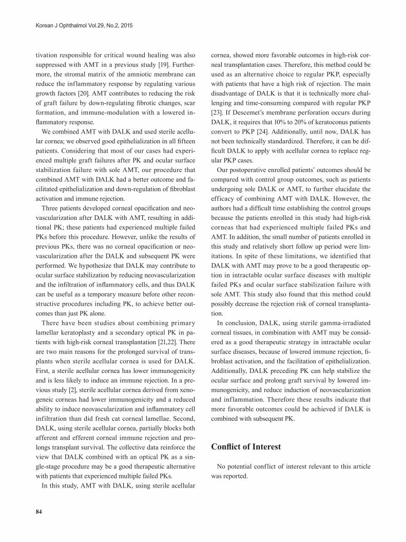
Fig. 1. Slit-lamp observations of the two representative patients before and after successful deep anterior lamellar keratoplasty (DALK) with amniotic membrane transplantation (AMT) (A). Patient 1: severe opacity with stromal neovascularization and almost total limbal stem cell deficiency were noted after previous failed penetrating keratoplasty. Pseudopterygium due to excessive conjunctivalization was noted in the nasal conjunctiva (B). At 12 months after DALK with AMT and pseudopterygium removal, the graft remained clear and the best-corrected visual acuity was 20 / 100 (C). Patient 4: diffuse stromal neovascularization and central corneal ulceration with whitish deposits were noted after previous failed penetrating keratoplasty. (D) At 14 months after DALK with AMT and pseudopterygium remoeval, the graft remained clear, the best-corrected visual acuity was 20 / 50 and pseudopterygium had progressed in the nasal conjunctiva.
A
C
B
D
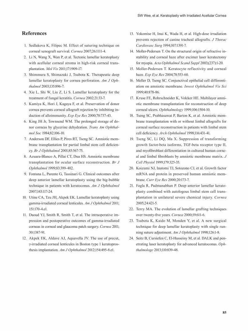
Fig. 2. Slit-lamp observation of the representative patient before and after failed deep anterior lamellar keratoplasty (DALK) and additional penetrating keratoplasty (PK) (A). The corneal status after multiple failed PK at an initial visit to our clinic. Stromal neovascularization, opacity, and inferior circular persistent epithelial defect were noted (arrow). Total limbal stem cell deficiency was also noted (B). At 5 months after DALK with amniotic membrane transplantation, diffuse opacity was noted on the central cornea (C). At 10 months after DALK with amniotic membrane transplantation, deep stromal ulceration with extensive vascularization and opacity had progressed to the central cornea. The authors judged graft rejection after DALK and decided to perform additional PK (D). At 8 months after additional PK, the graft remained clear and the final best-corrected visual acuity was 20 / 50.
A
C
B
D

83
SW Wee, et al. Keratoplasty with Irradiated Acellular Cornea
no endothelium and Descemet’s membrane beneath the stromal tissue in the product (Fig. 3).
Discussion
Fifteen patients with intractable ocular surface diseases were enrolled in this study. We used the sterile acellular cornea (VisionGraft) to perform DALK, this technique is advantageous because it applies decellularization to mini-mize the risk of allograft rejection, and has been associat-ed with good clinical outcomes.
The sterile acellular cornea, which is irradiated human donor corneal tissue, is indicated for use in various corneal procedures that do not require a viable endothelium. Steril-ity is verified via periodic microbiology testing [10] and several previous small-group studies have used the sterile acellular cornea [10-12], which has several advantages com-pared with fresh donor cornea. First, gamma irradiation depletes antigen-presenting cells, thereby minimizing the risk of allograft rejection by preventing direct sensitization [13]. Additionally, this product can be stored at room tem-
perature with a shelf life of 1 year, therefore it can be used in emergency procedures as well as scheduled operations.
Moller-Pedersen [14] and Moller-Pedersen [15] reported that corneal keratocytes support the stroma and presum-ably the stromal clarity, and are also involved in wound healing and collagen formation. According to a previous study, confocal microscopy confirmed that glycerin-cryo-preserved corneas were acellular, while dendrite-like cells and keratocytes were found in the fresh cornea group 2 weeks after DALK. At 3 months after DALK, keratocyte density increased significantly in the glycerin-cryopre-served group, whereas it decreased significantly after sur-gery in the fresh cornea group [10]. Thus, in our study, like glycerin-cryopreserved corneas, keratocytes would repop-ularize and could help to maintain transparency of the transplanted cornea after DALK.
We combined AMT with DALK to prevent the possible complications of DALK, such as delayed epithelialization and inflammatory reaction. In this study, there were ten patients with multiple failed PKs and a sole AMT that re-sulted in PED with corneal ulceration. These patients had partial to complete LSCD because of accompanying ocular burn, herpes simplex virus keratitis, Stevens-Johnson syn-drome, and Mooren’s ulcer. The tendency of PED and cor-neal ulceration may contribute to grafted tissue failure, as in these cases. Therefore, we proposed that combining AMT with DALK could facilitate epithelialization of the corneal graft, and lead to a more favorable surgical out-come.
In a previous study [8], the amniotic membrane promot-ed rapid corneal healing in the majority of patients with PED. The epithelialization effect of the amniotic mem-brane is considered to be due to the restoration of an intact basement membrane. The basement membrane of the am-niotic membrane is an ideal substrate to support the growth of epithelial progenitor cells by prolonging their life span and maintaining their clonogenicity [16,17]. This action may help facilitate epithelialization for PED with corneal ulceration. Even in partial LSCD, AMT was re-ported to produce a beneficial effect to restore the ocular surface in intractable diseases [18].
Another reason why the amniotic membrane supports a successful DALK is that the stromal side of the amniotic membrane contains a matrix component that has an antifi-brotic effect. Several factors, including transforming growth factor β were down-regulated, and fibroblastic ac-
Fig. 3. Histological characteristics of the acellular corneal matrix in hematoxylin-eosin staining of the sterile acellular cornea; relatively loose corneal stroma with thinned epithelium showed significant acellularity when compared with the normal cornea (×100).
84
Korean J Ophthalmol Vol.29, No.2, 2015
tivation responsible for critical wound healing was also suppressed with AMT in a previous study [19]. Further-more, the stromal matrix of the amniotic membrane can reduce the inflammatory response by regulating various growth factors [20]. AMT contributes to reducing the risk of graft failure by down-regulating fibrotic changes, scar formation, and immune-modulation with a lowered in-flammatory response.
We combined AMT with DALK and used sterile acellu-lar cornea; we observed good epithelialization in all fifteen patients. Considering that most of our cases had experi-enced multiple graft failures after PK and ocular surface stabilization failure with sole AMT, our procedure that combined AMT with DALK had a better outcome and fa-cilitated epithelialization and down-regulation of fibroblast activation and immune rejection.
Three patients developed corneal opacification and neo-vascularization after DALK with AMT, resulting in addi-tional PK; these patients had experienced multiple failed PKs before this procedure. However, unlike the results of previous PKs, there was no corneal opacification or neo-vascularization after the DALK and subsequent PK were performed. We hypothesize that DALK may contribute to ocular surface stabilization by reducing neovascularization and the infiltration of inflammatory cells, and thus DALK can be useful as a temporary measure before other recon-structive procedures including PK, to achieve better out-comes than just PK alone.
There have been studies about combining primary lamellar keratoplasty and a secondary optical PK in pa-tients with high-risk corneal transplantation [21,22]. There are two main reasons for the prolonged survival of trans-plants when sterile acellular cornea is used for DALK. First, a sterile acellular cornea has lower immunogenicity and is less likely to induce an immune rejection. In a pre-vious study [2], sterile acellular cornea derived from xeno-geneic corneas had lower immunogenicity and a reduced ability to induce neovascularization and inflammatory cell infiltration than did fresh cat corneal lamellae. Second, DALK, using sterile acellular cornea, partially blocks both afferent and efferent corneal immune rejection and pro-longs transplant survival. The collective data reinforce the view that DALK combined with an optical PK as a sin-gle-stage procedure may be a good therapeutic alternative with patients that experienced multiple failed PKs.
In this study, AMT with DALK, using sterile acellular
cornea, showed more favorable outcomes in high-risk cor-neal transplantation cases. Therefore, this method could be used as an alternative choice to regular PKP, especially with patients that have a high risk of rejection. The main disadvantage of DALK is that it is technically more chal-lenging and time-consuming compared with regular PKP [23]. If Descemet’s membrane perforation occurs during DALK, it requires that 10% to 20% of keratoconus patients convert to PKP [24]. Additionally, until now, DALK has not been technically standardized. Therefore, it can be dif-ficult DALK to apply with acellular cornea to replace reg-ular PKP cases.
Our postoperative enrolled patients’ outcomes should be compared with control group outcomes, such as patients undergoing sole DALK or AMT, to further elucidate the efficacy of combining AMT with DALK. However, the authors had a difficult time establishing the control groups because the patients enrolled in this study had high-risk corneas that had experienced multiple failed PKs and AMT. In addition, the small number of patients enrolled in this study and relatively short follow up period were lim-itations. In spite of these limitations, we identified that DALK with AMT may prove to be a good therapeutic op-tion in intractable ocular surface diseases with multiple failed PKs and ocular surface stabilization failure with sole AMT. This study also found that this method could possibly decrease the rejection risk of corneal transplanta-tion.
In conclusion, DALK, using sterile gamma-irradiated corneal tissues, in combination with AMT may be consid-ered as a good therapeutic strategy in intractable ocular surface diseases, because of lowered immune rejection, fi-broblast activation, and the facilitation of epithelialization. Additionally, DALK preceding PK can help stabilize the ocular surface and prolong graft survival by lowered im-munogenicity, and reduce induction of neovascularization and inf lammation. Therefore these results indicate that more favorable outcomes could be achieved if DALK is combined with subsequent PK.
Conflict of Interest
No potential conflict of interest relevant to this article was reported.
85
SW Wee, et al. Keratoplasty with Irradiated Acellular Cornea
References
1. Sedlakova K, Filipec M. Effect of suturing technique on corneal xenograft survival. Cornea 2007;26:1111-4.
2. Li N, Wang X, Wan P, et al. Tectonic lamellar keratoplasty with acellular corneal stroma in high-risk corneal trans-plantation. Mol Vis 2011;17:1909-17.
3. Shimmura S, Shimazaki J, Tsubota K. Therapeutic deep lamellar keratoplasty for cornea perforation. Am J Oph-thalmol 2003;135:896-7.
4. Xie L, Shi W, Liu Z, Li S. Lamellar keratoplasty for the treatment of fungal keratitis. Cornea 2002;21:33-7.
5. Kamiya K, Hori J, Kagaya F, et al. Preservation of donor cornea prevents corneal allograft rejection by inhibiting in-duction of alloimmunity. Exp Eye Res 2000;70:737-43.
6. King JH Jr, Townsend WM. The prolonged storage of do-nor corneas by glycerine dehydration. Trans Am Ophthal-mol Soc 1984;82:106-10.
7. Anderson DF, Ellies P, Pires RT, Tseng SC. Amniotic mem-brane transplantation for partial limbal stem cell deficien-cy. Br J Ophthalmol 2001;85:567-75.
8. Azuara-Blanco A, Pillai CT, Dua HS. Amniotic membrane transplantation for ocular surface reconstruction. Br J Ophthalmol 1999;83:399-402.
9. Fontana L, Parente G, Tassinari G. Clinical outcomes after deep anterior lamellar keratoplasty using the big-bubble technique in patients with keratoconus. Am J Ophthalmol 2007;143:117-24.
10. Utine CA, Tzu JH, Akpek EK. Lamellar keratoplasty using gamma-irradiated corneal lenticules. Am J Ophthalmol 2011; 151:170-4.e1.
11. Daoud YJ, Smith R, Smith T, et al. The intraoperative im-pression and postoperative outcomes of gamma-irradiated corneas in corneal and glaucoma patch surgery. Cornea 2011; 30:1387-91.
12. Akpek EK, Aldave AJ, Aquavella JV. The use of precut, γ-irradiated corneal lenticules in Boston type 1 keratopros-thesis implantation. Am J Ophthalmol 2012;154:495-8.e1.
13. Yokomise H, Inui K, Wada H, et al. High-dose irradiation prevents rejection of canine tracheal allografts. J Thorac Cardiovasc Surg 1994;107:1391-7.
14. Moller-Pedersen T. On the structural origin of refractive in-stability and corneal haze after excimer laser keratectomy for myopia. Acta Ophthalmol Scand Suppl 2003;(237):1-20.
15. Moller-Pedersen T. Keratocyte ref lectivity and corneal haze. Exp Eye Res 2004;78:553-60.
16. Meller D, Tseng SC. Conjunctival epithelial cell differenti-ation on amniotic membrane. Invest Ophthalmol Vis Sci 1999;40:878-86.
17. Kruse FE, Rohrschneider K, Volcker HE. Multilayer amni-otic membrane transplantation for reconstruction of deep corneal ulcers. Ophthalmology 1999;106:1504-10.
18. Tseng SC, Prabhasawat P, Barton K, et al. Amniotic mem-brane transplantation with or without limbal allografts for corneal surface reconstruction in patients with limbal stem cell deficiency. Arch Ophthalmol 1998;116:431-41.
19. Tseng SC, Li DQ, Ma X. Suppression of transforming growth factor-beta isoforms, TGF-beta receptor type II, and myofibroblast differentiation in cultured human corne-al and limbal fibroblasts by amniotic membrane matrix. J Cell Physiol 1999;179:325-35.
20. Koizumi NJ, Inatomi TJ, Sotozono CJ, et al. Growth factor mRNA and protein in preserved human amniotic mem-brane. Curr Eye Res 2000;20:173-7.
21. Fogla R, Padmanabhan P. Deep anterior lamellar kerato-plasty combined with autologous limbal stem cell trans-plantation in unilateral severe chemical injury. Cornea 2005;24:421-5.
22. Terry MA. The evolution of lamellar grafting techniques over twenty-five years. Cornea 2000;19:611-6.
23. Tsubota K, Kaido M, Monden Y, et al. A new surgical technique for deep lamellar keratoplasty with single run-ning suture adjustment. Am J Ophthalmol 1998;126:1-8.
24. Seitz B, Cursiefen C, El-Husseiny M, et al. DALK and pen-etrating laser keratoplasty for advanced keratoconus. Oph-thalmologe 2013;110:839-48.


![Review Article Lamellar Keratoplasty: A Literature Reviewdownloads.hindawi.com/journals/joph/2013/894319.pdfJournal of Ophthalmology described by Melles et al. [ ] allowing transplantation](https://img.dokumen.tips/doc/110x75/5e39106a1415da08cf09cef9/review-article-lamellar-keratoplasty-a-literature-journal-of-ophthalmology-described.jpg)