Embed Size (px)
Citation preview
872
Scholars Journal of Applied Medical Sciences (SJAMS) ISSN 2320-6691 (Online)
Sch. J. App. Med. Sci., 2016; 4(3D):872-876 ISSN 2347-954X (Print) ©Scholars Academic and Scientific Publisher
(An International Publisher for Academic and Scientific Resources) www.saspublisher.com
Original Research Article
Isolation and Identification of Non Fermenting Gram Negative Bacilli in A
Tertiary Care Hospital Dr. Ruchita Mahajan
1, Dr. Neeraj
2, Dr. Sarika
3, Dr. Bella Mahajan
4
1Senior Resident, Microbiology, GMC, Jammu, India 2Consultant, Orthopaedics, GMC, Jammu, India
3Senior Resident, ASCOMS, Jammu, India 4Professor, Microbiology, GMC, Jammu, India
*Corresponding author Dr. Ruchita Mahajan
Email: [email protected]
Abstract: Non-Fermenting Gram-Negative Bacilli (NFGNB )group includes numerous organisms but the ones which are known to cause nosocomial infections are Pseudomonas aeruginosa, Acinetobacter baumanii, Burkholderia cepacia
complex (BCC) and Stenotrophomonas maltophila .This study was undertaken to identify the various nonfermenters
isolated from patients admitted to our hospital, a tertiary care center, at Jammu. A total of 4585 clinical specimens were
received in the laboratory, which included 1234 urine, 742 pus, and 1864 blood cultures, collected in a brain-heart
infusion broth, 110 endotracheal catheter tips, 529 CSF samples and 115 body fluids. These samples were placed on
blood agar, chocolate agar, and Mac Conkey's agar, and incubated at 37°C for 18-24 hours. The clinical isolates were identified using the conventional biochemical tests as per the standardized protocols. Non-fermenting Gram-negative
bacilli were isolated from 572 out of 4585 clinical specimens accounting for an isolation rate of 12.40%. Pseudomonas
aeruginosa was the most common isolate, accounting for 312 (54.54%), followed by Acinetobacter baumanii 235
(41.08%), Stenotrophomonas maltophilia 15 (2.62%), and Burkholderia cepacia complex 10 (1.70%). In our study, a
total of 13 (86.66%) of the isolates were sensitive to Colistin and a total of 4 (26.60%) were sensitive to Imipenem. Thus
NFGNB are emerging as important opportunistic pathogens and are resistant to commonly used antimicrobials.
Therefore early diagnosis and institution of empirical therapy based on local antibiogram data of the institute would
reduce mortality and improve patient management.
Keywords: Non-Fermenting Gram-Negative Bacilli (NFGNB),Pseudomonas.
INTRODUCTION: Non-Fermenting Gram-Negative Bacilli
(NFGNB) are a group of aerobic, non-sporing,
bacilli/coccobacilli that are either incapable of utilizing carbohydrates as a source of energy or degrade them via
oxidative, rather than fermentative pathway. This group
includes numerous organisms but the ones which are
known to cause nosocomial infections are Pseudomonas
aeruginosa, Acinetobacter baumanii, Burkholderia
cepacia complex (BCC) and Stenotrophomonas
maltophila.
NFGNB were earlier considered to be
commensals or contaminants. But, the pathogenic
potential of these organisms has been established beyond doubt because of their frequent isolation from
clinical specimens and their association with the
disease. These apparently heterogeneous
microorganisms have common traits of clinical
importance that justify their inclusion and study in a
single group. They can be recovered from hospital
environment, commonly cause device related
infections, are often resistant to disinfectants and have
the potential to spread from patient to patient via fomites or the hands of medical personnel.
Also the antimicrobial resistance exhibited by
the NFGNB creates an epidemiologic niche for these
pathogens that facilitates colonization and super
infection in antibiotic-treated patients [1-4].
NFGNB are known to account for about 15%
of all bacterial isolates from a clinical microbiology
laboratory. In recent years, due to the liberal and
empirical use of antibiotics, NFGNB have emerged as important healthcare-associated pathogens [5].
It has always been a tedious task for a routine
microbiological laboratory to identify the NFGNBs and
poor laboratory proficiency in identification of these

Ruchita Mahajan et al., Sch. J. App. Med. Sci., March 2016; 4(3D):872-876
873
NFGNBs prevails worldwide, including in our own
country. For this reason, reports of disease due to these
organisms are rare from India. Identification through
commercial kits and automated systems is not fool-
proof as many non-Burkholderia betaproteobacteria
(Ralstonia picketti and Pandoraea species) are labeled as BCC and some BCC strains as Pseudomonas
aeruginosa [6].
Hence, this study was undertaken to identify
the various nonfermenters isolated from patients
admitted to our hospital, a tertiary care center, at
Jammu. The study was also done to assess their clinical
significance and antimicrobial susceptibility pattern,
and to identify the various healthcare-related infection
they cause.
AIMS AND OBJECTIVES
To isolate, identify and characterize non-
fermenting Gram-negative bacilli from clinical
isolates received from indoor hospital patients
for a period of one year.
To analyze the antibiotic sensitivity patterns of
non-fermenting Gram-negative bacilli.
MATERIAL & METHODS
This prospective study was conducted in the
Department of Microbiology, Government Medical
College and Hospital, Jammu which is an 850 bedded tertiary care hospital, for a period of one year.
A total of 4585 clinical specimens were
received in the laboratory, which included 1234 urine,
742 pus, and 1864 blood cultures, collected in a brain-
heart infusion broth, 110 endotracheal catheter tips, 529
CSF samples and 115 body fluids. These samples were
placed on blood agar, chocolate agar, and Mac Conkey's
agar, and incubated at 37°C for 18-24 hours. The
organisms isolated were identified using the appropriate
biochemical tests [1]. All the organisms giving non-lactose fermenting (NLF) colonies on MacConkey agar
medium. And producing an alkaline reaction (K/K) on
triple sugar iron agar grew on Triple Sugar Iron agar
were provisionally considered to be NFGNB.
ISOLATE SPECIATION / IDENTIFICATION:
The clinical isolates were identified using the
conventional biochemical tests as per the standardized
protocols [19]. The characters assessed included
morphology on Gram-stain, motility, cytochrome
oxidase activity, OF test (Hugh-Leif son medium) for
glucose and mannitol, growth on 10% lactose agar, decarboxylase test, growth on aerobic low peptone
medium, Triple sugar iron fermentation with hydrogen
sulphide production on lead acetate paper and gelatin
liquefaction.
ANTIMICROBIAL SUSCEPTIBILITY TESTING:
The sensitivity test was performed with the
help of the Kirby-Bauer disc diffusion method using commercially available discs (Hi-media). The different
antibiotics tested were Ceftazidime (30mcg),
Ceftriaxone (30mcg), Cefepime (30mcg), Imepenem
(10µg), Piperacillin-Tazobactam (100/10mcg),
Ticarcillin-Clavulinic acid (75/10mcg), Ciprofloxacin
(5mcg), Ofloxacin (5mcg), Amikacin (30mcg),
Gentamycin (10mcg), Tobramycin (10mcg),
Trimethoprim-Sulfamethoxazole (1.25/23.75mcg), and
Colistin (10mcg. The results were interpreted as per the
Clinical and Laboratory Standards Institute (CLSI)
guidelines.7
Following ATCC strains were used as Quality
control strains:
ATCC-25922 E. coli – Gram-negative bacilli
ATCC-25923 S. aureus – Gram-positive cocci
ATCC 27853 P. aeruginosa- Non-Lactose
Fermenters
RESULTS
A total of 4585 clinical samples were received
from the indoor patients in the microbiology laboratory.
Non-fermenting Gram-negative bacilli were isolated from 572 out of 4585 clinical specimens accounting for
an isolation rate of 12.40%. Two ninety seven specimen
(50.16%) showed polymicrobial infection where non-
fermenters were isolated along with other organisms, of
which E. coli and S. aureus were commonly associated.
The remaining two ninety five specimens (49.83%)
showed monomicrobial infection.
The majority of patients (35.13%) belonged to
the age group 40-60 years. 342 NFGNB(59.79%) were
isolated from males and 230 (40.20%) were from females. Regarding invasive devices, 292 (51.04%)
patients had peripheral venous and 229 (40.03%) had
urinary catheter placed. Central venous line and
endotracheal tube were found in 119 (19.93%) and 82
(14.33%) cases respectively. This association was
highly statistically significant. (P value: 0.000001)
History of previous antibiotic therapy was
received from 188 (32.86%) patients, recent
hospitalisation from 275 (48.07%) and recent surgery in
the preceding year from 182 (31.81%). Out of the 572
NFGNB 343 (59.96%) were isolated from pus, 36 (6.29%) from urine, 33 (5.76%) from body fluids, 70
(12.23%) from blood, 48 (8.39%) from CSF, 42
(7.34%) from endotracheal secretions
Ruchita Mahajan et al., Sch. J. App. Med. Sci., March 2016; 4(3D):872-876
874
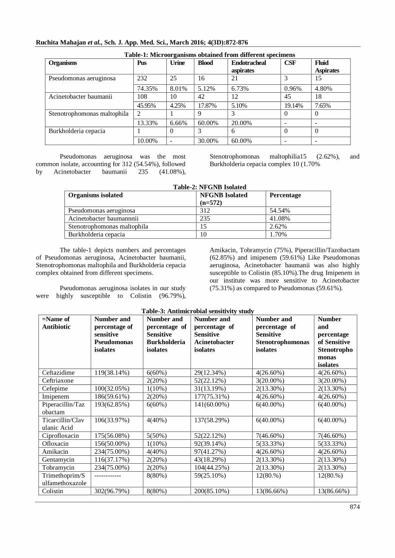
Table-1: Microorganisms obtained from different specimens
Organisms Pus Urine Blood Endotracheal
aspirates
CSF Fluid
Aspirates
Pseudomonas aeruginosa 232 25 16 21 3 15
74.35% 8.01% 5.12% 6.73% 0.96% 4.80%
Acinetobacter baumanii 108 10 42 12 45 18
45.95% 4.25% 17.87% 5.10% 19.14% 7.65%
Stenotrophomonas maltophila 2 1 9 3 0 0
13.33% 6.66% 60.00% 20.00% - -
Burkholderia cepacia 1 0 3 6 0 0
10.00% - 30.00% 60.00% - -
Pseudomonas aeruginosa was the most
common isolate, accounting for 312 (54.54%), followed
by Acinetobacter baumanii 235 (41.08%),
Stenotrophomonas maltophilia15 (2.62%), and
Burkholderia cepacia complex 10 (1.70%
Table-2: NFGNB Isolated
Organisms isolated NFGNB Isolated
(n=572)
Percentage
Pseudomonas aeruginosa 312 54.54%
Acinetobacter baumannnii 235 41.08%
Stenotrophomonas maltophila 15 2.62%
Burkholderia cepacia 10 1.70%
The table-1 depicts numbers and percentages of Pseudomonas aeruginosa, Acinetobacter baumanii,
Stenotrophomonas maltophila and Burkholderia cepacia
complex obtained from different specimens.
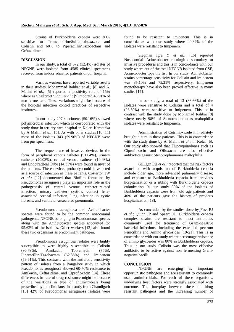
Pseudomonas aeruginosa isolates in our study
were highly susceptible to Colistin (96.79%),
Amikacin, Tobramycin (75%), Piperacillin/Tazobactam (62.85%) and imipenem (59.61%) Like Pseudomonas
aeruginosa, Acinetobacter baumanii was also highly
susceptible to Colistin (85.10%).The drug Imipenem in
our institute was more sensitive to Acinetobacter
(75.31%) as compared to Pseudomonas (59.61%).
Table-3: Antimicrobial sensitivity study
=Name of
Antibiotic
Number and
percentage of
sensitive
Pseudomonas
isolates
Number and
percentage of
Sensitive
Burkholderia
isolates
Number and
percentage of
Sensitive
Acinetobacter
isolates
Number and
percentage of
Sensitive
Stenotrophomonas
isolates
Number
and
percentage
of Sensitive
Stenotropho
monas
isolates
Ceftazidime 119(38.14%) 6(60%) 29(12.34%) 4(26.60%) 4(26.60%)
Ceftriaxone 2(20%) 52(22.12%) 3(20.00%) 3(20.00%)
Cefepime 100(32.05%) 1(10%) 31(13.19%) 2(13.30%) 2(13.30%)
Imipenem 186(59.61%) 2(20%) 177(75.31%) 4(26.60%) 4(26.60%)
Piperacillin/Taz
obactam
193(62.85%) 6(60%) 141(60.00%) 6(40.00%) 6(40.00%)
Ticarcillin/Clav
ulanic Acid
106(33.97%) 4(40%) 137(58.29%) 6(40.00%) 6(40.00%)
Ciprofloxacin 175(56.08%) 5(50%) 52(22.12%) 7(46.60%) 7(46.60%)
Ofloxacin 156(50.00%) 1(10%) 92(39.14%) 5(33.33%) 5(33.33%)
Amikacin 234(75.00%) 4(40%) 97(41.27%) 4(26.60%) 4(26.60%)
Gentamycin 116(37.17%) 2(20%) 43(18.29%) 2(13.30%) 2(13.30%)
Tobramycin 234(75.00%) 2(20%) 104(44.25%) 2(13.30%) 2(13.30%)
Trimethoprim/S
ulfamethoxazole
------------ 8(80%) 59(25.10%) 12(80.%) 12(80.%)
Colistin 302(96.79%) 8(80%) 200(85.10%) 13(86.66%) 13(86.66%)
Ruchita Mahajan et al., Sch. J. App. Med. Sci., March 2016; 4(3D):872-876
875
Strains of Burkholderia cepacia were 80%
sensitive to Trimethoprim/Sulfamethoxazole and
Colistin and 60% to Piperacillin/Tazobactam and
Ceftazidime.
DISCUSSION
In our study, a total of 572 (12.4%) isolates of
NFGNB were isolated from 4585 clinical specimens
received from indoor admitted patients of our hospital.
Various workers have reported variable results
in their studies. Mohammad Rahbar et al.; [8] and A.
Malini et al.; [5] reported a positivity rate of 15%
where as Shailpreet Sidhu et al.; [9] reported 45.91% of
non-fermenters. These variations might be because of
the hospital infection control practices of respective
institutes.
In our study 297 specimens (50.16%) showed
polymicrobial infection which is corroborated with the
study done in tertiary care hospital in Kolar, Karnataka
by A Malini et al.; [5]. As with other studies [10, 11]
most of the isolates 343 (59.96%) of NFGNB were
from pus specimens.
The frequent use of invasive devices in the
form of peripheral venous catheter (51.04%), urinary
catheter (40.03%), central venous catheter (19.93%) and Endotracheal Tube (14.33%) were found in most of
the patients. These devices probably could have acted
as a source of infection in these patients. Costerton JW
et al.; [12] documented that Biofilm formation by
Pseudomonas aeruginosa plays an important role in the
pathogenesis of central venous catheter–related
infection, urinary catheter cystitis, contact lens–
associated corneal infection, lung infection in cystic
fibrosis, and ventilator-associated pneumonia.
Pseudomonas aeruginosa and Acinetobacter species were found to be the common nosocomial
pathogens.. NFGNB belonging to Pseudomonas species
along with the Acinetobacter species accounted for
95.62% of the isolates. Other workers [13] also found
these two organisms as predominant pathogen.
Pseudomonas aeruginosa isolates were highly
susceptible to were highly susceptible to Colistin
(96.79%), Amikacin, Tobramycin (75%),
Piperacillin/Tazobactam (62.85%) and Imipenem
(59.61%). This contrasts with the antibiotic sensitivity
pattern of isolates from a Bangalore study in which Pseudomonas aeruginosa showed 60-70% resistance to
Amikacin, Ceftazidime, and Ciprofloxacin [14]. These
differences in rate of drug resistance might be because
of the variations in type of antimicrobials being
prescribed by the clinicians. In a study from Chandigarh
[15] 42% of Pseudomonas aeruginosa isolates were
found to be resistant to imipenem. This is in
concordance with our study where 40.39% of the
isolates were resistant to Imipenem.
Siegman Igra Y et al.; [16] reported Nosocomial Acinetobacter meningitis secondary to
invasive procedures and this is in concordance with our
study where out of the total NFGNB isolated from CSF,
Acinetobacter tops the list. In our study, Acinetobacter
strains percentage sensitivity for Colistin and Imipenem
was 85.10% and 75.31% respectively. Imipenem
monotherapy have also been proved effective in many
studies [17].
In our study, a total of 13 (86.66%) of the
isolates were sensitive to Colistin and a total of 4
(26.60%) were sensitive to Imipenem. This is in contrast with the study done by Mohamad Rahbar
[8]
where nearly 98% of Stenotrophomonas maltophilia
isolates were resistant to Imipenem.
Administration of Cotrimoxazole immediately
brought a cure in these patients. This is in concordance
with the study done by A. Malini et al.; in Kolar [5].
Our study also showed that Fluoroquinolones such as
Ciprofloxacin and Ofloxacin are also effective
antibiotics against Stenotrophomonas maltophilia
Gilligan PH et al.; reported that the risk factors
associated with acquisition of Burkholderia cepacia
include older age, more advanced pulmonary disease,
and exposure to Burkholderia cepacia from previous
hospitalization or a sibling with Burkholderia cepacia
colonization In our study 30% of the isolates of
Burkholderia cepacia were from old age patients and
40% of the patients gave the history of previous
hospitalization [18].
As concluded by the studies done by Fass RJ et al.; Quinn JP and Speert DP, Burkholderia cepacia
complex strains are resistant to most antibiotics
commonly used for treatment of Gram-negative
bacterial infections, including the extended-spectrum
Penicillins and Amino glycosides [19-21]. This is in
concordance with our study where percentage resistance
of amino glycosides was 80% in Burkholderia cepacia.
Thus in our study Colistin was the most effective
antibiotic to be active against non fermenting Gram-
negative bacilli.
CONCLUSION NFGNB are emerging as important
opportunistic pathogens and are resistant to commonly
used antimicrobials. For each of these organisms,
underlying host factors were strongly associated with
outcome. The interplay between these multidrug
resistant pathogens and the increasing number of
Ruchita Mahajan et al., Sch. J. App. Med. Sci., March 2016; 4(3D):872-876
876
immune-compromised patients poses a challenge for the
microbiologists and clinicians likewise. Early diagnosis
and institution of empirical therapy based on local
antibiogram data of the institute would reduce mortality
and improve patient management.
The present study has sufficient power to
identify factors, organisms and antibiotics influencing
the outcome of infection which will help us to
understand the role of these organisms in human disease
process better. However, use of additional features and
large study sample would have enhanced the value of
study and may have provided greater insight into a
possible link.
REFERENCES
1. Buck AC, Cooke EM; The fate of ingested
Pseudomonas aeruginosa in normal persons. J Med Microbiol 1969; 2: 521–25.
2. Johanson WG Jr, Higuchi JH, Chaudhuri TR;
Bacterial adherence to epithelial cells in bacillary
colonization of the respiratory tract. Am Rev
Respir Dis 1980; 121:55–63.
3. Rebecca Reik, Theodore Spilker, John J; LiPuma:
Distribution of Burkholderia cepacia Complex
Species among Isolates Recovered from Persons
with or without Cystic Fibrosis. Journal of Clinical
Microbiology 2005; 43(6): 2926–28.
4. Richet H, Escande MC, Marie JP; Epidemic Pseudomonas aeruginosa serotype 016 bacteremia
in hematology-oncology patients. J Clin Microbiol
1989; 27: 1992–96.
5. Malini A, Deepa EK, Gokul BN, Prasad SR;
Nonfermenting Gram-negative bacilli infections in
a tertiary care hospital in Kolar, Karnataka. Journal
of lab physicians 2009;1(2):62-66.
6. Gautam V, Ray P, Vandamme P, Chatterjee SS,
Das A, Sharma K, et al.; Identification of lysine
positive non-fermenting Gram negative bacilli
(Stenotrophomonas maltophilia and Burkholderia cepacia complex).Indian Journal of Medical
Microbiology 2009;27(2):128-133.
7. Wayne PA; Clinical and Laboratory Standards
Institute. Performance standards for antimicrobial
susceptibility testing, Fifteenth informational
supplement. Approved Standard 2005; M7-A6.
8. Mohammad Rahbar, Hadi Mehragan, Negar Haji
Ali Akbari; Prevalence of Drug Resistance in
Nonfermenter Gram-Negative Bacilli. Iranian
Journal of Pathology2010; 15(2).
9. Shailpreet Sidhu, Usha Arora, Pushpa Devi;
Prevalence of Nonfermentative Gram Negative Bacilli In Seriously ill Patients With Bacteraemia.
Jk science 2010; 12(4).
10. Yashodara P, Shyamala S; Identification and
characterization of nonfermenters from clinical
specimens. Indian J Med Microbiol 1997; 15: 195-
7.
11. Mishra B, Bhujwala RA, Shriniwas; Nonfermenters
in human infections. Indian J Med Res 1986; 83:
561-66
12. Costerton JW, Stewart PS, Greenberg EP; Bacterial
biofilms: a common cause of persistent infections.
Science 1999; 284: 1318–22. 13. Rao PS, Shivananda PG; Bacteraemia due to non
fermenting Gram negative bacilli in
immunocompromised patients. Ind J Med
Microbiol 1993; 11: 95-99.
14. Veenakumari HB, Nagarathna S, Chandramuki A;
Antimicrobial resistance pattern among aerobic
Gram negative bacilli of lower respiratory tract
specimen of intensive care unit patients in a
neurocentre. Indian J Chest Dis Allied Sci 2007;
49:19-2297.
15. Taneja N, Maharwal S, Sharma M; Imipenem
resistance in nonfermenters causing nosocomial urinary tract infections. Indian J Med Sci 2003; 57:
294-9.
16. Siegman-Igra Y, Bar-Yosef S, Gorea A, Avram J.
Nosocomial acinetobacter meningitis secondary to
invasive procedures: report of 25 cases and review.
Clin Infect Dis 1993; 17: 843–9.
17. Karlowsky JA, Draghi DC, Jones ME; Surveillance
for antimicrobial susceptibility among clinical
isolates of Pseudomonas aeruginosa and
Acinetobacter baumannii from hospitalized patients
in the United States, 1998 to 2001.Antimicrob Agents Chemother 2003; 47: 1681–88.
18. Gilligan PH; Microbiology of airway disease in
patients with cystic fibrosis. Clin Microbiol Rev
1991; 4: 35–51.
19. Quinn JP; Clinical problems posed by
multiresistant nonfermenting Gram-negative
pathogens. Clin Infect Dis1998; 27(1): S117-24.
20. Fass RJ, Barnishan J, Solomon MC; In vitro
activities of Quinolones, β-lactams, Tobramycin,
and Trimethoprim-Sulfamethoxazole against
nonfermentative Gram-negative bacilli. Antimicrob Agents Chemother 1996; 40: 1412–18.
21. Speert DP; Advances in Burkholderia cepacia
complex. Podiatry Respire Rev 2002; 3: 230–235.