Embed Size (px)
Citation preview
Int J Clin Exp Med 2015;8(9):14672-14679www.ijcem.com /ISSN:1940-5901/IJCEM0012332
Original ArticleNew instrument for percutaneous posterolateral lumbar foraminoplasty: case series of 134 with instrument design, surgical technique and outcomes
Zhenzhou Li, Shuxun Hou, Weilin Shang, Keran Song, Hongliang Zhao
Department of Orthopedics, The First Affiliated Hospital of General Hospital of People’s Libertion Army, Beijing 100048, China
Received July 3, 2015; Accepted September 5, 2015; Epub September 15, 2015; Published September 30, 2015
Abstract: Current solutions for treating uncontained lumbar disk herniation include laser assisted endoscopic fo-raminoplasty and Transforaminal Endoscopic Spine System, both of which have some issues in clinical practice. This study aims to report the design of a new instrument for percutaneous posterolateral foraminoplasty. 148 patients with uncontained lumbar disk herniation were treated with percutaneous foraminoplasty followed by trans-foraminal endoscopic discectomy. Follow up were obtained for 134 cases. The VAS scores of pre-operative and post-operative low back pain and sciatica were compared. Oswestry Disability Index (ODI) and MacNab scores were also obtained. Follow-up was up to 5 years postoperatively. There were 75 of excellent, 49 of good and 5 of fair according to MacNab score system, with total successful rate up to 92.5%. 5 cases with L5S1 disc herniation complained about irritation to the dorsal root ganglion. In conclusion, the new transforaminal endoscopic discectomy instrument is safe and effective for percutaneous foraminoplasty.
Keywords: Lumbar disk herniation, minimally invasive treatment, foraminoplasty, transforaminal endoscopic dis-cectomy
Introduction
Since first report of posterolateral endoscopic discectomy in 1992, it is widely used to treat patients with uncontained lumbar disk hernia-tion [1]. The current two techniques used for transforaminal posterolateral endoscopic dis-cectomy were firstly described by Yeung [2] and Hoogland [3]. Dr. Yeung’s technique applied single channel or double channel through which herniation was gradually removed from inner side to outer side of disc. With new generation of transforaminal endoscopic equipment, which allows progressively opening of foramen, surgeons can put tools in space in front of spi-nal dural sac, and resect herniated disc direct-ly. Superior articular process (SAP) is the main obstacle for rod-shaped endoscope when going to the space in front of the spinal dural sac pos-terolateral. One solution is laser assisted endo-scopic foraminoplasty which widens lumbar intervertebral foramen [4, 5]. Using a side firing holmium laser probe, epidural scarring, extrud-
ed and sequestrated disc protrusions or osteo-phytes are ablated with protection of saline solution under direct vision. Problems with laser assisted techniques includes expensive equipment, low working efficiency, and risk of heat-damage to surrounding peripheral nerves. Another solution is THESSYS (Transforaminal Endoscopic Spine System) invented by Hoogland [3] et al. With the help of a cannulat-ed trephines and accompanying instruments, foramen is gradually widened in a step-wise fashion for removal of herniated disc materials. However, cannulated trephines directly reaches para-foramen soft tissue and nerve roots with-out any protection, arising concerns of damage to nerves [6].
To address these issues of existing methods, we invented new instrument for percutaneous posterolateral lumbar foraminoplasty [7]. We combined Dr. Yeung’s posterolateral endoscop-ic system and our new device to perform lum-bar foraminoplasty on 148 patients with uncon-

Instrument for percutaneous posterolateral foraminoplasty
14673 Int J Clin Exp Med 2015;8(9):14672-14679
tained lumbar disk herniation. Outcome of up to 5 years follow-up is summarized in this case series report.
Materials and methods
Patients
This study was approved by institutional review board of the First Affiliated Hospital of General Hospital of People’ Liberation Army. Inclusion and exclusion criteria are shown in Table 1.
From April of 2007 to April of 2009, 148 patients meet inclusion criteria were treated. Follow up data were obtained from 134 cases out of 148, including 14 cases on L3-4, 78 cases on L4-5 and 42 cases on L5S1. Patients ranged in age from 18-78 years (mean age, 41.4 years), including 68 males and 66 females. 108 cases are prolapse type, while 26 cases are sequestration type. Reasons for losing fol-low-up data in some patients includes loss of contact and die from other diseases. Pre-operative symptoms and deficits included nerve root dermatome hypoesthesia in 98 patients (73%), nerve root myotome muscle
weakness in 32 patients (23%), and weakening or disappearance of tendon reflex in 43 patients (32%).
Surgical tools
YESS® Spine Endoscope produced by Richard Wolf Medical Instruments Corporation (Vernon Hills, IL); patented instrument of percutaneous foraminoplasty with a guide-wire, an obturator, a graded duck-mouth cannulas and graded tre-phine (Figure 1). The distal end of duck mouth-like cannulas is 2 cm in length. Half of it is flat, the other half is bevel design. The bevel part is thin, so that it may go through gap between lower half of intervertebral foramen and anteri-or to facet joints. The tip of cannulas may be fixed on vertebrate, preventing cannulas from moving. The trephine works inside the cannulas avoiding any damage to nerves. The dimen-sions of new instrument was listed in Table 2.
Surgical procedures
(1) Anesthesia: Local (0.5% lidocaine solution) plus intravenous strengthen (1 mg of midazol-am and 100 μg of fentanyl).
(2) Position: Lying prone on spinal surgery shelf, avoiding pressure on abdomen.
(3) Location: Positioning the surgical segment by C-arm fluoroscopy.
Surgical protocol
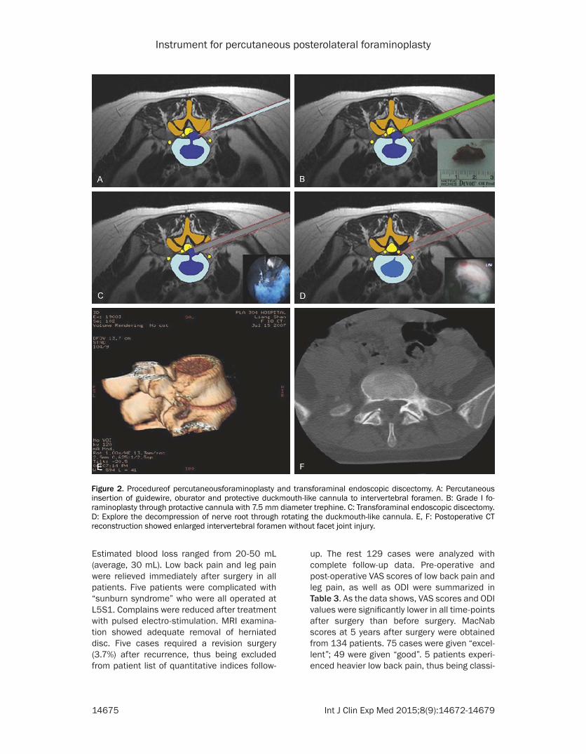
(1) Puncture (Figure 2A): Following Dr. Yeung’s [2] technique, needle was punctured through foramen, 12-15 cm away from posterior longi-tudinal middle line on diseased side. Fluoroscopy was used to confirm that needle went into nucleus pulposus tissue of interverte-bral disc. A mixture of 8 ml of omnipaque and 2 ml of methylene blue was injected for staining purpose. After taking out inner core of the nee-
Table 1. Inclusion and exclusion criteriaInclusion criteria Exclusion criteria(1) Low back pain accompanied by sciatica (1) Cauda equina syndrome
(2) Leg pain is heavier than low back pain (2) Recurrence after discectomy
(3) Non-contained disc herniation as indicated by MRI or CT (3) Central spinal stenosis as shown by radiology
(4) No surgicalhistory of the same segment on lumbar spine (4) Pathological conditions with combined infection, tumor, or fracture
(5) Low efficacy with conservative treatment (5) Foramen and far lateral disc herniation
(6) Single segmental disc herniation or prolapse (6) Segmental instability of lumbar spine
(7) Positive for straight leg raising test
Figure 1. Patented instrument of percutaneous fo-raminoplasty. A: Guidewire; B: Oburator; C: Graded duckmouth-like cannulas; D: Trephine.
Instrument for percutaneous posterolateral foraminoplasty
14674 Int J Clin Exp Med 2015;8(9):14672-14679
dle, guide wire was put in. Following the guide wire, obturator and duck mouth-like cannulas was inserted to protect exiting nerve root, through a 1 cm-incision.
(2) Foraminoplasty (Figure 2B): Through the duck mouth-like cannulas, endoscope was put into foramen. SAP and fat, vessels inside fora-men became visible under endoscope. Along the cannulas, trephine was put to SAP. Part of the ventral portion of SAP was cut and taken out. For patients with simple lumbar disc her-niation, the primary trephine with diameter of 7.5 mm was able to create a tunnel wide enough for surgery. For patients with lateral recess ste-nosis or intervertebral foramen stenosis, the secondary trephine with diameter of 10 mm was needed to create tunnels (Figure 3A, 3B).
(3) Endoscopic discectomy (Figures 2C and 3C): Endoscope was put into spinal epidural space through widened intervertebral foramen. Prolapsed nucleus pulposus was stained with dark blue. Under endoscope, blue nucleus pulposus tissue was removed by a nucleus ron-geur. When part of prolapsed nucleus pulposus was cut off, nerve root might move to ventral side to cover rest of nucleus pulposus. After removal of all prolapsed tissue, a radiofrequen-cy head was used to themo-annuloplast annu-lus rupture.
(4) Turn around the duck mouth-like cannulas again. Nerve root was back in vision.
(5) Intradiscal discectomy: Fix the tip of working channel at the site of annulus rupture. Push rongeur into disc space to take out the loosing nucleus pulposus.
Detailed procedures are described in supple-mentary materials (Supplementary Figures 1, 2, 3, 4, 5, 6, 7, 8), including line drawings and X-ray images.
Outcome assessment
One to two days after surgery, all patients were examined by MRI to check if herniated tissue was removed completely (Figures 2E, 2F, 3E). Outcomes of symptoms were evaluated by fol-low-up interviews at 3 months, 6 months, 1 year and 5 years after surgery. Low back pain and leg pain were measured by Visual Analog Scale (VAS) score (1-100). Functional outcomes were assessed by using Oswestry Disability Index (ODI) [8] and modified MacNab criteria [9, 10]. For MacNab criteria at year 5 after surgery, “excellent” was given to patients who were free of pain and deficit, without restriction of mobil-ity; “good” was given to patients with residual symptoms or deficits not impeding the ability to normal life; “fair” was given to patients with some improvement of functionality but who remained handicapped; “poor” was given to patients with no improvement at all.
Statistical analysis
Statistical analysis was performed with SPSS 11.5 software (SPSS Inc., Chicago, IL). Pre-operative and post-operative (3 month, 6 months, 1 year and 5 years) VAS scores of low back pain and leg pain, as well as ODI values were analyzed with ANOVA. Preoperative and postoperative related nerve root function sta-tus was analyzed with Chi-square test. P<0.01 was considered as significant.
Results
Using new instrument, 148 patients with disk herniation were surgically treated, 134 cases were followed up. No case required conversion to an open procedure during the surgery. No patient needed a blood transfusion. No patients had infections. Operative time ranged from 40-80 minutes (average, 65 minutes).
Table 2. The dimensions of the new instrumentsLength (cm) Diameter (mm) Remarks
Guidewire 25 1Oburator 20 7 OburatorGraded duckmouth-like cannulas Primary 18 7-8 (inner-outer) Tip of duckmouth-like cannu-
las is 2 cm long with half flat and half bevel
Secondary 17 8-9 (inner-outer)Tetiary 16 9-10 (inner-outer)
Quaternary 15 10-11 (inner-outer)Trephine Primary 18 5-7 (inner-outer) 5
Secondary 18 8-10 (inner-outer)
Instrument for percutaneous posterolateral foraminoplasty
14675 Int J Clin Exp Med 2015;8(9):14672-14679
Estimated blood loss ranged from 20-50 mL (average, 30 mL). Low back pain and leg pain were relieved immediately after surgery in all patients. Five patients were complicated with “sunburn syndrome” who were all operated at L5S1. Complains were reduced after treatment with pulsed electro-stimulation. MRI examina-tion showed adequate removal of herniated disc. Five cases required a revision surgery (3.7%) after recurrence, thus being excluded from patient list of quantitative indices follow-
up. The rest 129 cases were analyzed with complete follow-up data. Pre-operative and post-operative VAS scores of low back pain and leg pain, as well as ODI were summarized in Table 3. As the data shows, VAS scores and ODI values were significantly lower in all time-points after surgery than before surgery. MacNab scores at 5 years after surgery were obtained from 134 patients. 75 cases were given “excel-lent”; 49 were given “good”. 5 patients experi-enced heavier low back pain, thus being classi-
Figure 2. Procedureof percutaneousforaminoplasty and transforaminal endoscopic discectomy. A: Percutaneous insertion of guidewire, oburator and protective duckmouth-like cannula to intervertebral foramen. B: Grade I fo-raminoplasty through protactive cannula with 7.5 mm diameter trephine. C: Transforaminal endoscopic discectomy. D: Explore the decompression of nerve root through rotating the duckmouth-like cannula. E, F: Postoperative CT reconstruction showed enlarged intervertebral foramen without facet joint injury.

Instrument for percutaneous posterolateral foraminoplasty
14676 Int J Clin Exp Med 2015;8(9):14672-14679
fied as “fair”. 5 cases with recurrence were given “poor”. Preoperative and postoperative (5 years follow-up) related nerve root function status was summarized in Supplementary Table 1. Sensation and muscle strength recov-ered significantly (P<0.01), while tendon reflex was not changed (P=0.782).
Discussion
Safety of instrument for percutaneous lumbar foraminoplasty
Knight et al. reported a laser assisted method to widen the lumbar foramen by removing part
Figure 3. Case of grade II foraminoplasty. A: Preopera-tive MRI showed L4-5 disc protrusion with compression on right L5 nerve root. B: Postoperative CT reconstruc-tion showed enlarged intervertebral foramen with su-perior part of superior articular process and ventral articular capsule resected. C: Endoscopic disectomy. D: Exploration of nerve root decompression. E: Postopera-tive MRI showed complete resection of herniated disc.
Instrument for percutaneous posterolateral foraminoplasty
14677 Int J Clin Exp Med 2015;8(9):14672-14679
of bone and cartilage tissue surrounding fora-men [4]. However, disadvantage with laser technology is quite obvious, for example, expen-sive equipment, low working efficiency, and risk of heat-damage to surrounding spinal nerves [11]. Hoogland et al. [3], invented THESSYS technique, in which they make use of graded trephine to widen the foramen gradually. But in such surgery, trephine blade makes contact to para-foramen soft tissue and nerve roots, aris-ing concerns of damage to nerves [11]. Based on Dr. Hoogland’s method, we invented new instrument for percutaneous lumbar foramino-plasty. With graded duck mouth-like cannulas which was next to the ventral side of facet joint, excluding the exiting nerve root from the work-ing zone of trephine. Driven by hand, trephine could only cut bone, but not ligament. Meanwhile, patients kept awake under local anesthesia, which marked it possible for sur-geons to get instant feedback from patients. 0.5% lidocaine solution anesthetized sino-ver-tebral nerve surrounding foramen, reducing pain without affecting nerve root. This is impor-tant to ensure safety of foraminoplasty.
Herniation suitable to be treated with forami-noplasty
There’s no need to decompress the foramen and lateral recess. Using primary trephine of 7.5 mm diameter, we could limit the cut to no bigger than 4 mm and make a curved surface on SAP due to protection of duck mouth-like cannulas. On one hand, such cut caused no damages to the articular surface and joint cap-sule of facet joints and no harm to the stability of lumbar segment. On the other hand, such cut ensured the foramen is wide enough to let the cannulas go into the spinal canal, creating working zone for most cases of discectomy [12].
For lateral recess stenosis or intervertebral foramen stenosis, the secondary trephine is
needed to decompress foramen and lateral recess. Using secondary trephine of 10 mm in diameter, we could widen foramen and limit cut to 5 mm due to protection of duck mouth-like cannulas. Upper part of lower SAP and part of ventral SAP of facet joint could be cut, thus decompressed foramen and lateral recess effectively [12].
Influence of foraminoplasty to the stability of lumbar segment
The main function of lumbar facet joint is ori-ented control. Roughly, lumbar facet joint sur-face is vertical to transverse plane and angles 45 to coronal plane. This is good for flexion and extension, but not good for rotation [13]. Osman et al. compared stability of lumbar vertebrae [14]. After transforaminal decompression, the intervertebral foraminal area increased about 45.5%. However, surgical technique used is dif-ferent from what is actually used in clinic. In this study [12], we mimicked posterolateral lumbar foraminoplasty on human lumbar verte-brae specimen, and studied its mechanical properties before and after surgery. Our data showed that there is no damage to joint surface of lumbar facet and joint capsule at all, after primary foraminoplasty. Therefore, no changes of stability were observed. However, secondary foraminoplasty might increase lumbar lateral bending and shift neutral zone without mechan-ical loading, but not affecting the axial rotation flexibility. More data is needed to clarify the influence of increased lumbar flexion to lumbar vertebra.
Outcomes of endoscopic discectomy after percutaneous posterolateral lumbar foramino-plasty
Nellensteijn et al., reported that current evi-dence is not enough to support a better effica-cy of transforaminal endoscopic surgery over open microdiscectomy in patients with symp-
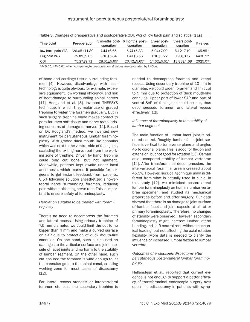
Table 3. Changes of preoperative and postoperative ODI, VAS of low back pain and sciatica (_x±s)
Time point Pre-operation 3 months post-operation
6 months post-operation
1 year post-operation
5years post-oeration F values
low back pain VAS 26.05±11.89 7.44±6.65 5.74±5.83 5.04±7.09 5.12±7.19 165.85*Leg pain VAS 75.89±9.65 3.10±5.84 1.47±3.56 1.16±3.22 0.93±3.17 4436.9*ODI 75.27±9.71 28.51±5.65# 20.42±5.65# 14.62±5.51# 13.83±4.68 2025.0*#P<0.05, *P<0.01, when comparing to pre-operation. P values are calculated by ANOVA.
Instrument for percutaneous posterolateral foraminoplasty
14678 Int J Clin Exp Med 2015;8(9):14672-14679
tomatic lumbar disc herniation or vice versa [15]. Kambin et al. reported an 88.3% of suc-cess rate in case series of 169 patients of lum-bar disc herniation in 24 month follow-up [16]. Meanwhile, open laminectomy and discectomy request patients to use narcotics for a longer duration postoperatively than video-assisted arthroscopic microdiscectomy.
Application of foraminoplasty further improved effectiveness of endoscopic discectomy in treating lumbar disc herniation. Lee et al. reported advantages of foraminoplastic appro- ach in treating extruded disk herniation at L5-S1 level [17]. Out of 25, 22 patients (88%) had favorable outcomes. Only two patients con-verted to open microdiscectomy due to incom-plete decompression and recurrent disk hernia-tion. Using same technique, Choi et al. treated 59 cases of highly migrated intracanal lumbar disc herniation. 91.4% patients experienced satisfactory outcome. Three cases complained about persistent leg pain after surgery. Two patients reported recurrent herniation at same level 6 month post-operation. In present study, we reported case series of 134 patients of lum-bar disc herniation treated with endoscopic dis-cectomy post percutaneous foraminoplasty. 92.5% of cases were given “excellent” or “good” of MacNab scores. Five cases had recurrent herniation at same level. These results are bet-ter than previous studies. One of reasons might be that new instrument not only widened fora-men but also effectively protect nerve root.
Some patients may experience “sunburn syn-drome” which is irritation to dorsal root gangli-on, after discectomy post foraminoplasty, due to retardation or over-sensation of nerve root. It happened in 5-15% of patients, usually at L5S1. Most likely, it is temporary, and disap-peared after conservative treatment2. In our study, 5 patients complained with “sunburn syndrome” were all operated at L5S1. Complains were reduced after treatment of pulsed electro-stimulation.
In conclusion, the advantages of endoscopic discectomy after percutaneous posterolateral lumbar foraminoplasty include: (1) No general anesthesia; (2) Little or no damage to nerves due to surgery; (3) Few infections; (4) Direct removal of herniated discs; (5) No damage to ligament, leaving few scares; (6) No scare tis-sue as obstacle for re-operation after recur-
rence [18]. Our instrument for percutaneous foraminoplasty is effective and safe for transfo-raminal endoscopic discectomy in treating uncontained lumbar disk herniation.
Disclosure of conflict of interest
None.
Address correspondence to: Dr. Zhenzhou Li, Department of Orthopedics, The First Affiliated Hospital of General Hospital of Chinese People’s Liberation Army, Beijing 100048, China. Tel: +86 10 68989121; Fax: +86 10 68989121; E-mail: [email protected]
References
[1] Kambin P. Arthroscopic microdiscectomy. Arth- roscopy 1992; 8: 287-295.
[2] Yeung AT, Tsou PM. Posterolateral endoscopic excision for lumbar disc herniation: Surgical technique, outcome, and complications in 307 consecutive cases. Spine 2002; 27: 722-731.
[3] Hoogland T, Schubert M, Miklitz B, Ramirez A. Transforaminal posterolateral endoscopic dis-cectomy with or without the combination of a low-dose chymopapain: a prospective random-ized study in 280 consecutive cases. Spine 2006; 31: E890-E897.
[4] Knight MT, Vajda A, Jakab GV, Awan S. Endoscopic laser foraminoplasty on the lum-bar spine--early experience. Minim Invasive Neurosurg 1998; 41: 5-9.
[5] No authors listed. Endoscopic laser foramino-plasty. Clin Privil White Pap 2012; 60: 1-13.
[6] Choi G, Lee SH, Lokhande P, Kong BJ, Shim CS, Jung B, Kim JS. Percutaneous endoscopic ap-proach for highly migrated intracanal disc her-niations by foraminoplastic technique using rigid working channel endoscope. Spine 2008; 33: E508-E515.
[7] Yeung AT. The evolution and advancement of endoscopic foraminal surgery: one surgeon’s experience lncorporating adjunctive techolo-gies. SAS J 2007; 1: 108-117.
[8] Fairbank JC, Pynsent PB. The Oswestry Disability Index. Spine 2000; 25: 2940-2952.
[9] Macnab I. Negative disc exploration. An analy-sis of the causes of nerve-root involvement in sixty-eight patients. J Bone Joint Surg Am 1971; 53: 891-903.
[10] Le H, Sandhu FA, Fessler RG. Clinical out-comes after minimal-access surgery for recur-rent lumbar disc herniation. Neurosurg Focus 2003; 15: E12.
[11] Hafez MI, Zhou S, Coombs RR, McCarthy ID. The effect of irrigation on peak temperatures in nerve root, dura, and intervertebral disc dur-
Instrument for percutaneous posterolateral foraminoplasty
14679 Int J Clin Exp Med 2015;8(9):14672-14679
ing laser-assisted foraminoplasty. Lasers Surg Med 2001; 29: 33-37.
[12] Chang X, Chen B, Li HY, Han XB, Zhou Y, Li CQ. The safety and efficacy of minimally invasive discectomy: a meta-analysis of prospective randomized controlled trials. Int Orthop 2014; 38: 1225-1234.
[13] Kenesi C, Lesur E. Orientation of the articular processes at L4, L5, and S1. Possible role in pathology of the intervertebral disc. Anat Clin 1985; 7: 43-47.
[14] Osman SG, Nibu K, Panjabi MM, Marsolais EB, Chaudhary R. Transforaminal and posterior de-compressions of the lumbar spine. A compara-tive study of stability and intervertebral fora-men area. Spine 1997; 22: 1690-1695.
[15] Nellensteijn J, Ostelo R, Bartels R, Peul W, van Royen B, van Tulder M. Transforaminal endo-scopic surgery for symptomatic lumbar disc herniations: a systematic review of the litera-ture. Eur Spine J 2010; 19: 181-204.
[16] Kambin P, O’Brien E, Zhou L, Schaffer JL. Arthroscopic microdiscectomy and selective fragmentectomy. Clin Orthop Relat Res 1998; 347: 150-167.
[17] Lee SH, Kang HS, Choi G, Kong BJ, Ahn Y, Kim JS, Lee HY. Foraminoplastic ventral epidural approach for removal of extruded herniated fragment at the L5-S1 level. Neurol Med Chi (Tokyo) 2010; 50: 1074-1048.
[18] Ahn Y, Lee SH, Park WM, Lee HY, Shin SW, Kang HY. Percutaneous endoscopic lumbar discectomy for recurrent disc herniation: surgi-cal technique, outcome, and prognostic fac-tors of 43 consecutive cases. Spine 2004; 29: E326-E332.
Instrument for percutaneous posterolateral foraminoplasty
1
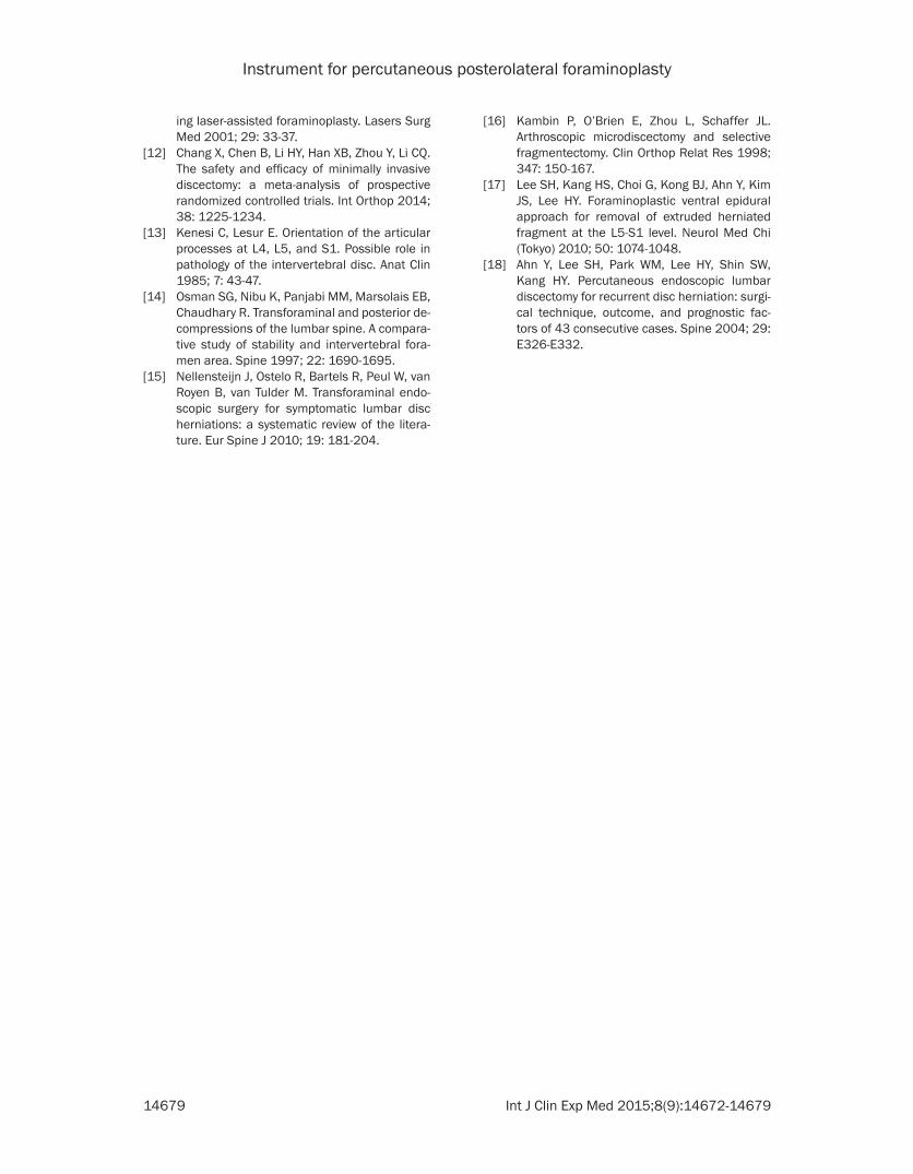
Supplementary Figure 1. Posterolateral insertion of stylet pin and guide wire into low part of intervertebral foramen (A). Lateral view of fluroscopy show the tip arriving the posterior aspect of upper endoplate of caudle vertebral body (B).
Supplementary Figure 2. Oburator insertion into low part of intervertebral fo-ramen over guide wire (A). Lateral view of fluroscopy show the tip of oburator arriving the posterior aspect of upper endoplate of caudle vertebral body (B).
Supplementary Figure 3. Gradual protective cannula insertion into low part of intervertebral foramen over oburator (A, B). Lateral view of fluroscopy show the tip of protective cannula arriving the posterior aspect of upper endoplate of caudle vertebral body (C), Anteroposterior view of fluroscopy show the tip of protective cannula arriving the ventral aspect of superior articular process of caudle vertebral body (D).
Instrument for percutaneous posterolateral foraminoplasty
2
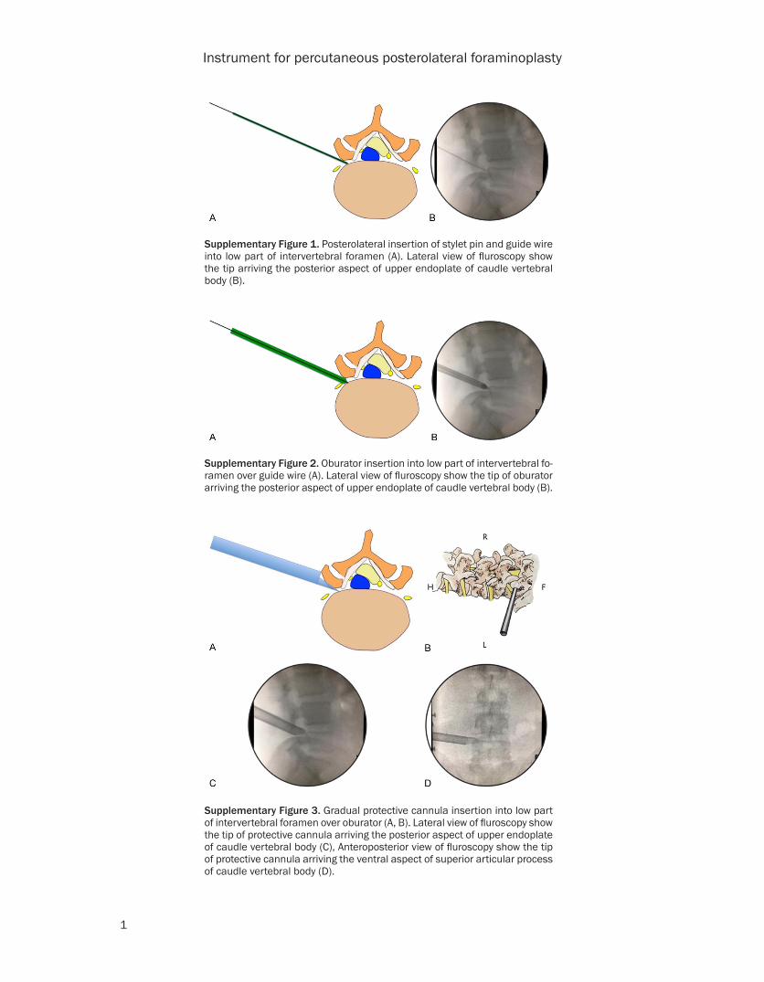
Supplementary Figure 4. Foraminoplasty with trephine rotating and advanc-ing in protective cannula (A), exiting nerve root was kept outside of protective cannula while transversing nerve root was protected by lateral flavum liga-ment. Anteroposterior view of fluroscopy show the tip of trephine arriving the medial border line of pedicle of caudle vertebral body (B).
Supplementary Figure 5. Working cannula insertion into ventral epidural space over oburator which was insertion into lumbar canal through protec-tive cannula (A, B), bevel opening of working cannula was placed toward extrued disc tissue posteriorly.
Supplementary Figure 6. Partial resection of extruded disc tissue (A) until part of transversing nerve root descending into endoscopic field (B). NP-nu-cleus pulposus, NRT-nerve root, D-dorsal, V-ventral, H-head, F-foot.
Instrument for percutaneous posterolateral foraminoplasty
3
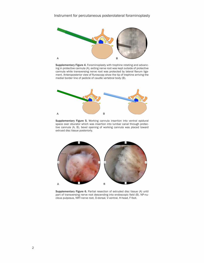
Supplementary Figure 7. Protection of transversing nerve root and further exposure and resection of extruded disc tissue by rotating working cannula 180 degree with bevel opening toward disk space anteriorly (A, B). Position of working cannula should be confirmed by fluroscopy (C, D). NP-nucleus pulpo-sus, NRT-nerve root, D-dorsal, V-ventral, H-head, F-foot.
Supplementary Figure 8. Exploration the decompression of transversing nerve root by rotating working cannula with bevel opening toward transvers-ing nerve root posteriorly (A, B). IVD-intervertebral disc, NRT-nerve root, FL-lavum ligament, PLL-posterior longitudinal ligament, D-dorsal, V-ventral, H-head, F-foot.
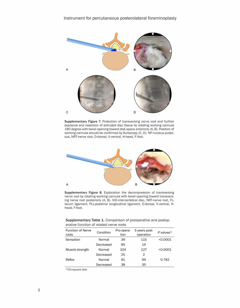
Supplementary Table 1. Comparison of preoperative and postop-erative function of related nerve rootsFunction of Nerve roots Condition Pro-opera-
tion5 years post-
operation P values*
Sensation Normal 39 115 <0.0001Decreased 90 14
Muscle strength Normal 104 127 <0.0001Decreased 25 2
Reflex Normal 91 94 0.782Decreased 38 35
*Chi-square test.