Embed Size (px)
Citation preview
Int J Clin Exp Pathol 2013;6(3):349-357www.ijcep.com /ISSN:1936-2625/IJCEP1209016
Original ArticleHistopathologic study of the rectum in 1,438 consecutive rectal specimens in a single Japanese hospital: I. benign lesions
Tadashi Terada
Department of Pathology, Shizuoka City Shimizu Hospital, Shizuoka, Japan
Received September 20, 2012; Accepted December 15, 2012; Epub February 15, 2013; Published March 1, 2013
Abstract: The author investigated histopathology of 1,438 consecutive rectal specimens in the last 10 years of our pathology laboratory in Japan. A computer review of pathologic reports was done. Observations of pathologic slides were performed, when appropriate. The rectal specimens were composed of 1,022 benign lesions and 416 malignant lesions. The 1,022 benign lesions were composed non-specific proctitis (n=460, 45%), adenoma (n=248, 24%), ulcerative colitis (n=98, 10%), hyperplastic polyp (n=54, 5%), carcinoma in adenoma (n=40, 4%), rectal ulcer (n=37, 4%), serrated adenoma (n=24, 2%), hyperplastic nodule (n=21, 2%), Crohn’s disease (n=9, 1%), ischemic proctitis (n=8, 0.8%), mucosal prolapse syndrome (n=7, 0.6%), juvenile polyp (n=6, 0.6%), lymphoid hyperplasia (n=5, 0.5%), lipoma (n=4, 0.4%) and amebic dysentery (n=2, 0.2%), and mature cystic teratoma (n=1, 0.1%). In this article, histopathological features of these benign lesions were described in details. In particular, adenomas were classified into adenomas with mild, moderate, and severe atypia, serrated adenoma, and carcinoma in adenoma. The later are mainly seen in large adenoma with severe atypia. Ulcerative colitis was characterized by continuous lesion, crypt abscess, abnormal branching, and deletion of goblet cells. Crohn’s disease was characterized by trans-mural inflammation and epithelioid granulomas. Ischemic colitis was characterized by ischemic necrotic changes and pseudomembrane formation. Mucosal prolapse syndrome was characterized by abnormal muscle in the mu-cosa (fibromuscular obliterance). Juvenile polyp was characterized by abnormal dilations of the crypts. Lymphoid hyperplasia must be differentiated from MALT lymphoma. Lipoma was ordinary lipoma without lipoblasts. Amebic dysentery was characterized by ulcer and presence of histiocyte-like entamoeba histolitica. Mature cystic teratoma was characterized by hairs and other elements of skin and mesodermal and endodermal components.
Keywords: Colon, benign lesions, histopathology
Introduction
Benign lesions of the rectum include Hirschsprung’s disease, diverticulosis, ulcer-ative colitis, Crohn’s disease, Ischemic procti-tis, cytomegalovirus proctitis, non-specific proctitis, lymphocytic proctitis, collagenous proctitis, pseudomembranous proctitis, necro-tizing proctitis, amebic proctitis, tuberculosis, Behcet’s disease, graft-versus-host disease, heterotopic gastric mucosa, heterotopic sali-vary glands, melanosis coli, endometriosis, amyloidosis, volvulus, malacoplakia, radiation changes, pneumatosis cystoides intestinalis, mucosal prolapse syndrome (solitary ulcer syn-drome), rectal ulcer, adenoma, familial polypo-
sis, Gardener’s syndrome, Turcot’s syndrome, hyperplastic polyp, juvenile polyp, hyperplastic nodule, Cronkhite-Canada syndrome, Peutz-Jeghers polyp, transitional polyp, dermoid cyst, vascular ectasia, hemangioma, lipoma, lipoma-tosis, leiomyoma, lymphangioma, and angio-myolipoma [1]. In the present study, 1,022 benign conditions of the rectum were described.
Materials and methods
The author investigated histopathology of 1,438 consecutive rectal specimens in the last 10 years of our pathology laboratory. A comput-er review of the pathologic reports was done. Examination of histologic slide was performed,
Rectal benign conditions
350 Int J Clin Exp Pathol 2013;6(3):349-357
when appropriate. The rectal specimens were composed of 1,022 benign lesions and 416 malignant lesions. Clinical records were also reviewed briefly. The age ranged from 21 years to 95 years with a mean of 54 years. In appro-priate cases, an immunohistochemical analysis had performed with the use of Dako Envision method (Dako), as previously described [2-6].
Results
The 1,022 benign lesions were composed of non-specific proctitis (n=460, 45%), adenoma (n=248, 24%), ulcerative colitis (n=98, 10%), hyperplastic polyp (n=54, 5%), carcinoma in adenoma (n=40, 4%), rectal ulcer (n=37, 4%), serrated adenoma (n=24, 2%), hyperplastic
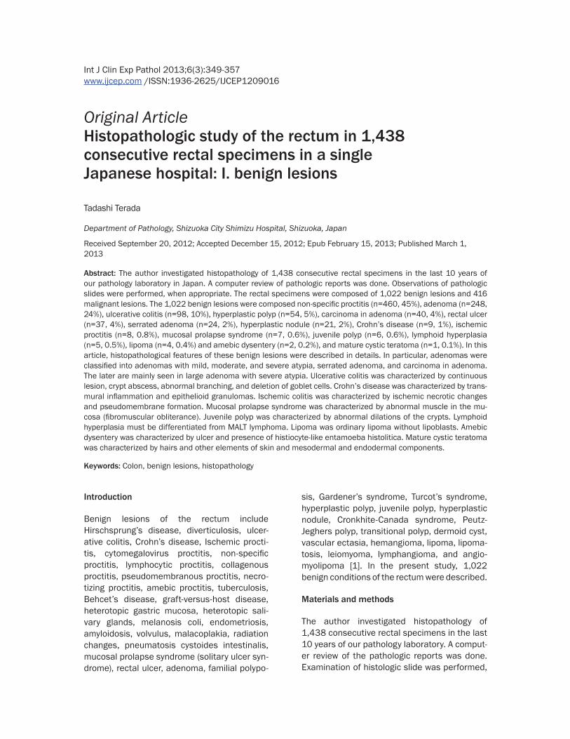
Figure 1. A: Goss features of tubular adenoma. B: Gross features of tubulo-villous adenoma. C: Gross features of large villous adenoma. D: Histology of adenoma with mild dysplasia. HE, x100. E: Histology of adenoma with moder-ate dysplasia. HE, x100. F: Histology of adenoma with severe dysplasia. HE, x100.

Rectal benign conditions
351 Int J Clin Exp Pathol 2013;6(3):349-357
nodule (n=21, 2%), Crohn’s disease (n=9, 1%), ischemic proctitis (n=8, 0.8%), mucosal pro-lapse syndrome (n=7, 0.6%), juvenile polyp (n=6, 0.6%), lymphoid hyperplasia (n=5, 0.5%), lipoma (n=4, 0.4%), amebic dysentery (n=2, 0.2%), and mature cystic teratoma.
Non-specific proctitis (n=460) showed edema and a varying degree of lymphocytic infiltration. The inflammation was non-specific. Infrequently, crypt abscesses were recognized in a small number. Clinically, presenting symptoms were diarrhea, constipation, mucus feces, bleeding, or asymptomatic. Endoscopically, it shows vari-ous features from red erosion to mucosal ulceration.
Adenoma (n=248) was polypoid in appearanc-es (Figure 1A, 1B and 1C) depending on the histological types. The diameter ranged from
1mm to 40mm. Adenoma cases were classified as tubular adenoma (n=182) (Figure 1A), tubu-lo-villous adenoma (n=42) (Figure 1B), and vil-lous adenoma (n=24) (Figure 1C). By the degree of dysplasia, the adenoma cases was classified into mild (n=56) (Figure 1D), moderate (n=107) (Figure 1E) and severe (n=85) (Figure 1F) dys-plasia. In severe dysplasia, the adenoma with severe dysplasia was very difficult to differenti-ate from well differentiated adenocarcinoma, and it distinction was occasionally arbitrary. Multiple adenomas were frequently detected in a person. Clinically, most patients were asymp-tomatic, but some patients showed bleeding and occult blood in the feces. Most of the patients were treated by polypectomy or endo-scopic mucosal resection (EMR).
Ulcerative colitis (n=98) showed, macroscopi-cally, mucosal inflammation consisting of con-
Figure 2. A: Gross features of ulcerative colitis. B: Histology of ulcerative colitis. Crypt abscess is present. HE, x100.
Figure 3. Gross features of hyperplastic polyp. B: Histology of hyperplastic polyp. Serration is present. HE, x100.
Rectal benign conditions
352 Int J Clin Exp Pathol 2013;6(3):349-357
tinuous lesion, mucosal edema, pseudopolypo-sis and ulceration (Figure 2A). Microscopically, it showed mucosal inflammation including lym-phocytic and neutrophilic infiltration, crypt abscess formation, and ulcers (Figure 2B). The histology varied depending on steroid therapy and disease activity. Clinically, it manifested as abdominal pain, bleeding, and diarrhea. The therapy was administration of steroid. Two patients underwent rectectomy because of severe remitting inflammation.
Hyperplastic polyp (n=54) (Figure 3A) showed cryptal hyperplasia, cryptal serration, and mucous hypersecretion. (Figure 3B). The size ranged from 2mm to 10 mm. Most patients were asymptomatic. The therapy was polypec-tomy or EMR.
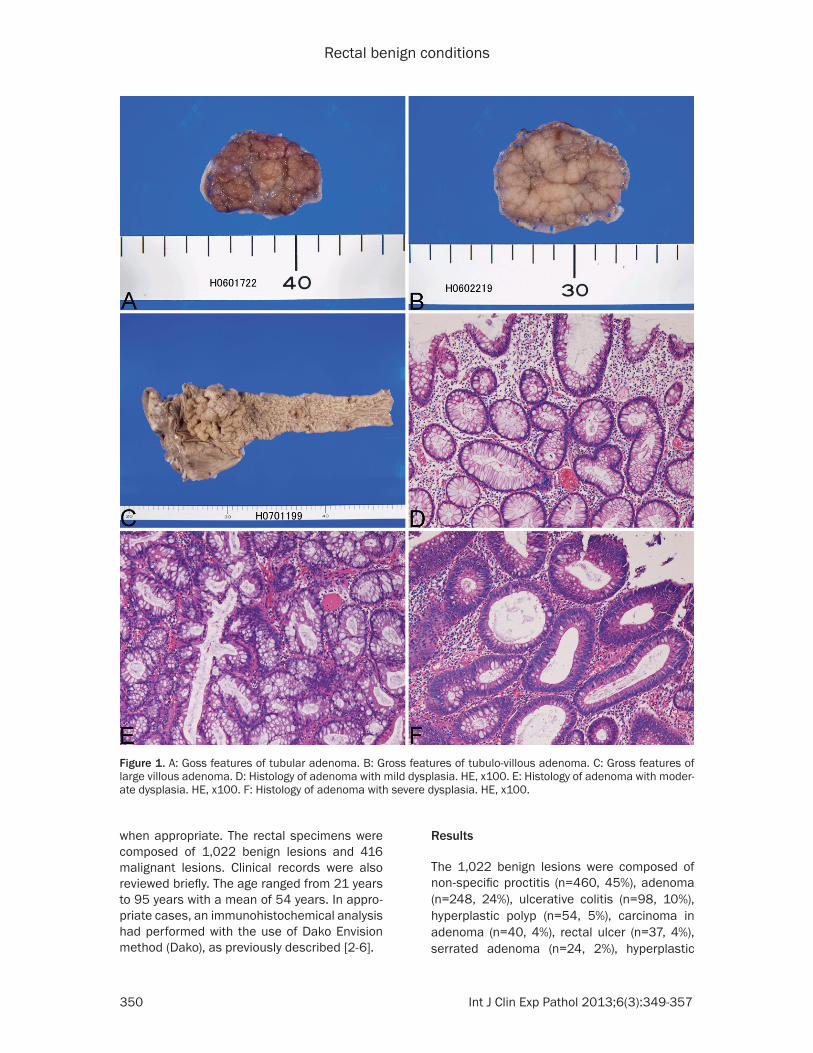
Carcinoma in adenoma (n=40) showed polyp with or without erosion or ulceration (Figure 4A). Most of the carcinoma was well differenti-ated carcinoma without invasion (Figure 4B, 4C
and 4D). However, submucosal invasion was recognized in 8 cases. The background adeno-ma was as follows: tubular adenoma (n=15), tubulo-villous adenoma (n=14), and villous ade-noma (n=11). The adenoma was of severe atyp-ia in almost all cases (Figure 4B and 4C). Immunohistochemically, the carcinoma cells were positive for cytokeratins (6/6) and mostly for p53 protein (5/6). Mean Ki-67 labeling was 48% (n=6). The size ranged from 6mm to 40 mm with a mean of 23 mm. The discovery of carcinoma was incidental in most cases. EMR and polypectomy was performed in 38 cases, and rectectomy was performed in 2 cases.
Rectal ulcer (n=37) was an endoscopic diagno-sis. Pathologically, it consisted of ulcer, granu-lation tissue, and active inflammation. No cyto-megalic inclusions were seen in the present series.
Serrated adenoma (n=24) showed adenoma-tous proliferation of cryptal epithelium with ser-
Figure 4. A: Gross features of carcinoma in villous adenoma. The central ulcer is carcinoma. B: Histology of car-cinoma (center) in adenoma. HE, x100. Histology of carcinoma in adenoma with severe dysplasia. HE, x200. C: Carcinoma area of carcinoma in adenoma. HE, x300.
Rectal benign conditions
353 Int J Clin Exp Pathol 2013;6(3):349-357
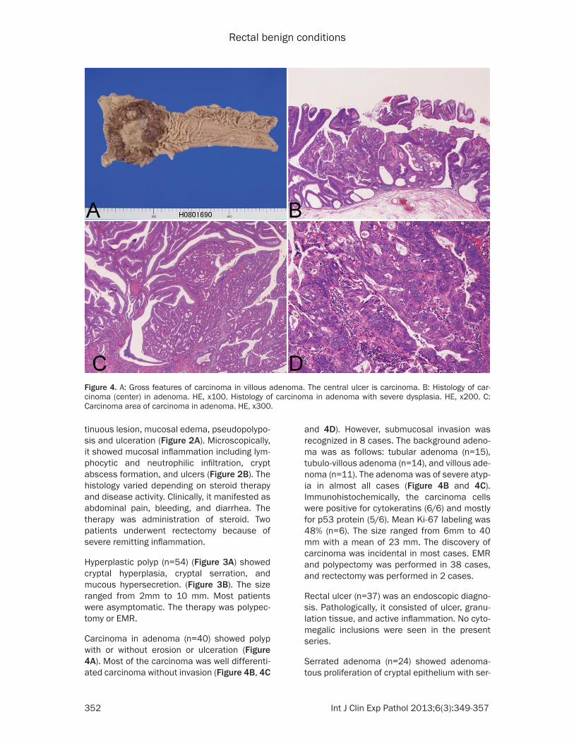
rated appearances (Figure 5A). Macroscopically, it was a simple polyp (Figure 5B). The atypia was mild in 9 cases, moderate in 8 cases, and severe in 7 cases. No coexisting carcinoma was noted in this series. The treatment was pol-ypectomy or EMR. The clinically diagnosis was mostly adenoma. All patients were asymptomatic.
Hyperplastic nodule (n=21), resembled hyper-plastic polyp, but different from hyperplastic polyp in that the hyperplastic nodule was free of serration. The sixe ranged from 1 mm to 4 mm. Most patients were asymptomatic. Polypectomy or EMR was a treatment. Endoscopic diagnosis was mostly adenoma.
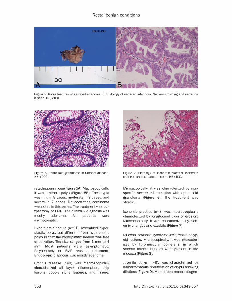
Crohn’s disease (n=9) was macroscopically characterized all layer inflammation, skip lesions, cobble stone features, and fissure.
Microscopically, it was characterized by non-specific severe inflammation with epithelioid granuloma (Figure 6). The treatment was steroid.
Ischemic proctitis (n=8) was macroscopically characterized by longitudinal ulcer or erosion. Microscopically, it was characterized by isch-emic changes and exudate (Figure 7).
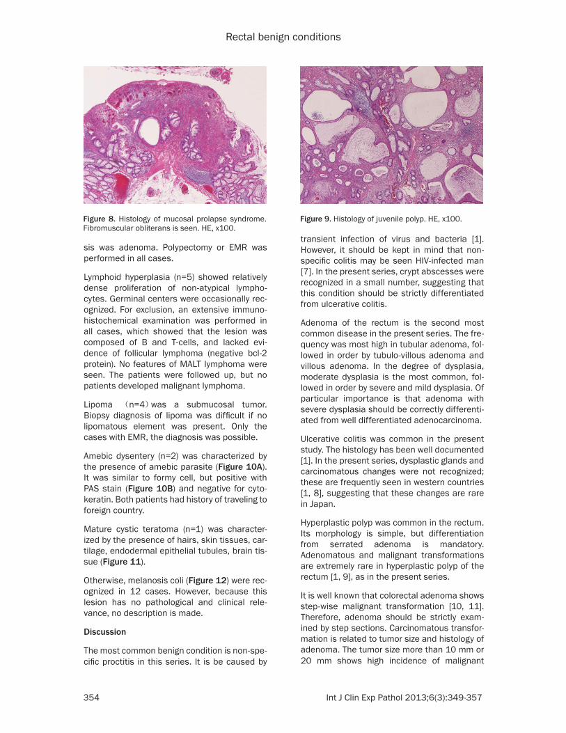
Mucosal prolapse syndrome (n=7) was a polyp-oid lesions. Microscopically, it was character-ized by fibromuscular obliterans, in which smooth muscle bundles were present in the mucosa (Figure 8).
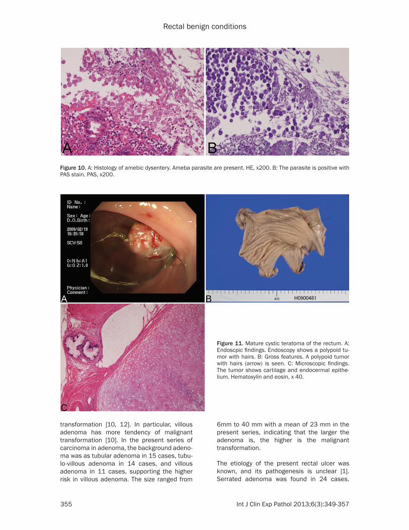
Juvenile polyp (n=6), was characterized by hamartomatous proliferation of crypts showing dilations (Figure 9). Most of endoscopic diagno-
Figure 5. Gross features of serrated adenoma. B: Histology of serrated adenoma. Nuclear crowding and serration is seen. HE, x100.
Figure 6. Epithelioid granuloma in Crohn’s disease. HE, x200.
Figure 7. Histology of ischemic proctitis. Ischemic changes and exudate are seen. HE x100.
Rectal benign conditions
354 Int J Clin Exp Pathol 2013;6(3):349-357
sis was adenoma. Polypectomy or EMR was performed in all cases.
Lymphoid hyperplasia (n=5) showed relatively dense proliferation of non-atypical lympho-cytes. Germinal centers were occasionally rec-ognized. For exclusion, an extensive immuno-histochemical examination was performed in all cases, which showed that the lesion was composed of B and T-cells, and lacked evi-dence of follicular lymphoma (negative bcl-2 protein). No features of MALT lymphoma were seen. The patients were followed up, but no patients developed malignant lymphoma.
Lipoma (n=4)was a submucosal tumor. Biopsy diagnosis of lipoma was difficult if no lipomatous element was present. Only the cases with EMR, the diagnosis was possible.
Amebic dysentery (n=2) was characterized by the presence of amebic parasite (Figure 10A). It was similar to formy cell, but positive with PAS stain (Figure 10B) and negative for cyto-keratin. Both patients had history of traveling to foreign country.
Mature cystic teratoma (n=1) was character-ized by the presence of hairs, skin tissues, car-tilage, endodermal epithelial tubules, brain tis-sue (Figure 11).
Otherwise, melanosis coli (Figure 12) were rec-ognized in 12 cases. However, because this lesion has no pathological and clinical rele-vance, no description is made.
Discussion
The most common benign condition is non-spe-cific proctitis in this series. It is be caused by
transient infection of virus and bacteria [1]. However, it should be kept in mind that non-specific colitis may be seen HIV-infected man [7]. In the present series, crypt abscesses were recognized in a small number, suggesting that this condition should be strictly differentiated from ulcerative colitis.
Adenoma of the rectum is the second most common disease in the present series. The fre-quency was most high in tubular adenoma, fol-lowed in order by tubulo-villous adenoma and villous adenoma. In the degree of dysplasia, moderate dysplasia is the most common, fol-lowed in order by severe and mild dysplasia. Of particular importance is that adenoma with severe dysplasia should be correctly differenti-ated from well differentiated adenocarcinoma.
Ulcerative colitis was common in the present study. The histology has been well documented [1]. In the present series, dysplastic glands and carcinomatous changes were not recognized; these are frequently seen in western countries [1, 8], suggesting that these changes are rare in Japan.
Hyperplastic polyp was common in the rectum. Its morphology is simple, but differentiation from serrated adenoma is mandatory. Adenomatous and malignant transformations are extremely rare in hyperplastic polyp of the rectum [1, 9], as in the present series.
It is well known that colorectal adenoma shows step-wise malignant transformation [10, 11]. Therefore, adenoma should be strictly exam-ined by step sections. Carcinomatous transfor-mation is related to tumor size and histology of adenoma. The tumor size more than 10 mm or 20 mm shows high incidence of malignant
Figure 8. Histology of mucosal prolapse syndrome. Fibromuscular obliterans is seen. HE, x100.
Figure 9. Histology of juvenile polyp. HE, x100.
Rectal benign conditions
355 Int J Clin Exp Pathol 2013;6(3):349-357
transformation [10, 12]. In particular, villous adenoma has more tendency of malignant transformation [10]. In the present series of carcinoma in adenoma, the background adeno-ma was as tubular adenoma in 15 cases, tubu-lo-villous adenoma in 14 cases, and villous adenoma in 11 cases, supporting the higher risk in villous adenoma. The size ranged from
6mm to 40 mm with a mean of 23 mm in the present series, indicating that the larger the adenoma is, the higher is the malignant transformation.
The etiology of the present rectal ulcer was known, and its pathogenesis is unclear [1]. Serrated adenoma was found in 24 cases.
Figure 10. A: Histology of amebic dysentery. Ameba parasite are present. HE, x200. B: The parasite is positive with PAS stain. PAS, x200.
Figure 11. Mature cystic teratoma of the rectum. A: Endoscpic findings. Endoscopy shows a polypoid tu-mor with hairs. B: Gross features. A polypoid tumor with hairs (arrow) is seen. C: Microscopic findings. The tumor shows cartilage and endocermal epithe-lium. Hematoxylin and eosin, x 40.

Rectal benign conditions
356 Int J Clin Exp Pathol 2013;6(3):349-357
Malignant transformation of serrated adenoma is rare [13]. No coexisting carcinoma was noted in this series. Serrated adenoma should be dif-ferentiated from hyperplastic polyp, because both show serrated appearances. In the pres-ent series, hyperplastic nodule resembled hyperplastic polyp, but different from hyper-plastic polyp in that the hyperplastic nodule was free of serration. Hyperplastic nodule may be a precursor of hyperplastic polyp.
The clinical and pathological aspects of Crohn’s disease are well recognized. In practical pathol-ogy, demonstration of epithelioid granuloma is essential for the pathological diagnosis of this disease. In the present series, ischemic procti-tis was characterized by longitudinal ulcer or erosion, and ischemic changes and exudate. Clinician should be aware that this lesion occurs in rectum as well as in the colon. Mucosal prolapse syndrome (n=7) was charac-terized by fibromuscular obliterans in the pres-ent series. However, this entity should be dif-ferentiated from other polyps [14].
The pathologic diagnosis of juvenile polyp is relatively easy, but endoscopic diagnosis is dif-ficult, as shown in the present series. Juvenile polyp is a benign lesion, and malignant trans-formation is almost none [1]. A few cases of lymphoid hyperplasia have been reported [15]. In the present series, 5 case of lymphoid hyper-plasia was recognized. In the present series, extensive immunohistochemical study exclud-ed malignant lymphoma.
Lipoma is a rare submucosal tumor in the rec-tum [1]. Biopsy diagnosis of lipoma is difficult if
no lipomatous element was present. Only the cases with EMR, the diagnosis was possible in the present study. Amebic dysentery was noted in 2 cases in the present series. The demon-stration of entamoeba histolytica is essential for the diagnosis. The parasite resembles mac-rophage, but positive with PAS stain and nega-tive for cytokeratin, as shown in the present study. Both patients had history of traveling to foreign country in the present study. Mature cystic teratoma is extremely rare in the colon [16, 17], and endoscopically characterized by the presence of hairs and Rokitansky protuberans.
Conflict of interest statement
The author has no conflict of interest.
Address correspondence to: Dr. Tadashi Terada, Department of Pathology, Shizuoka City Shimizu Hospital, Miyakami 1231 Shimizu-Ku, Shizuoka 424-8636, Japan. Tel: 81-54-336-1111; Fax: 81-54-336-1315; E-mail: [email protected]
References
[1] Rosai J. Large bowel. In Rosai and Ackerman’s Surgical Pathology. Ninth edition. Mosby, 2004. pp: 776-855.
[2] Terada T, Kawaguchi M, Furukawa K, Sekido Y, Osamura Y. Minute mixed ductal-endocrine carcinoma of the pancreas with predominant intraductal growth. Pathol Int 2002; 52: 740-746.
[3] Terada T, Tanigichi M. Intraductal oncocytic papillary neoplasm of the liver. Pathol Int 2004; 54: 116-123.
[4] Terada T. Primary multiple extragastrointesti-nal stromal tumors of the omentum with differ-ent mutations of c-kit gene. Would J Gastroen-terol 2008; 14: 7256-7259.
[5] Terada T. Gastrointestinal stromal tumor of the uterus: A case report with genetic analyses of c-kit and PDGFRA genes. Int J Gynecol Pathol 2009; 28: 29-34.
[6] Terada T. Large endocervical polyp with carti-laginous and osseous metaplasia: a hitherto unreported entity. Int J Gynecol Pathol 2009; 28: 98-100.
[7] Law CL, Qassim M, Cunningham AL. Mulhall B, Grierson JM. Non-specific proctitis: association with human immunodeficiency virus infection in homosexual man. J Infect Dis 1992; 165: 150-154.
[8] Torres C, Antonioll D, Orze RD. Polypoid dyspla-sia and adenomas in inflammatory bowel dis-ease. Am J Surg Pathol 1998; 22: 275-284.
Figure 12. Histology of melanosis coli. Brown pig-ment is seen in the mucosa. HE, x200.

Rectal benign conditions
357 Int J Clin Exp Pathol 2013;6(3):349-357
[9] O’Brien MJ. Hyperplastic and serrated polyp of the colorectum. Gastroenterol Clin North Am 2007; 36: 947-968.
[10] Hamilton SR, Rubio CA, Vogelstein B, Sobin LH, Kudo S, Fogt F, Riboli E, Winawer SJ, Nakamu-ra S, Goldgar DE, Hainaut P, Jass JR. Carcino-ma of the colon and rectum. In: Hamilton SR, Aaltonen LA eds. WHO Classification of tu-mours. Pathology and genetics of tumours of the digestive system. IARC Press, Lyon, 2000. pp: 105-119.
[11] Vogelstein B, Fearon ER, Hamilton SR, Kern SE, Preisinger AC, Lappert M, Nakamura Y, White R, Smits AM, Bos JL. Genetic alterations during colorectal-tumor development. New Eng J Med 1988; 319: 525-532.
[12] Lawrance IC, Sherrington C, Murray K. Poor correlation between clinical impression, the small colonic polyp and their neoplastic risk. Arch Pathol Lab Med 2006; 21: 563-568.
[13] Toriakovic E, Skovlund E, Snover DC, Torlakovic G, Nesland JM. Morphologic reappraisal of ser-rated colorectal polyps. Am J Surg Pathol 2003; 27: 65-81.
[14] Singh B, Mortensen NJ, Warren BF. Histopatho-logical mimickly in mucosal prolapse. Histopa-thology 2007; 50: 97-102.
[15] Kojima H, Itoh H, Motegi A, Sakata N, Masawa N. Localized lymphoid hyperplasia of the rec-tum resembling polypoid mucosa-associated lymphoid tissue lymphoma: a report of three cases. Pathol Res Pract 2005; 201: 757-761.
[16] Fijita K, Akiyama N, Ishizaki M, Tanaka S, Od-sawa K, Sugiyama H, Kanoh K, Toki F, Asao T, Kuwano H. Dermoid cyst of the colon. Dig Surg 2001; 18: 335-337.
[17] Sakurai Y, Uragoshi T, Imazu H, Hasegawa S, Matsubara T, Ochiai M, Funabiki T. Submuco-sal dermoid cyst of the rectum: report of a case. Surg Today 2000; 30: 195-198.