Embed Size (px)
Citation preview
Int J Clin Exp Pathol 2016;9(1):165-170www.ijcep.com /ISSN:1936-2625/IJCEP0017816
Original ArticleExtraventricular neurocytoma of the sellar region: report of two cases and literature review
Siyuan Chen1, Nan Ji1, Bo Wang1, Junmei Wang2, Shuqing Yu1, Jisheng Wang1
1Department of Neurosurgery, Beijing Tiantan Hospital, Capital Medical University, Beijing, China; 2Department of Neuropathology, Beijing Neurosurgical Institute, Beijing, China
Received October 13, 2015; Accepted November 26, 2015; Epub January 1, 2016; Published January 15, 2016
Abstract: Extraventricular neurocytoma (EVN) of the sellar region is a rare occurrence. To date, merely two reports concerning this tumor have been reported. In this report, we present two cases of EVN that occurred in the sellar region, and reviewed the pathological characteristics and treatment strategies for this disease. Two patients were admitted to our hospital for impaired vision. MRI scans indicated an enhanced invasive mass in the sellar region. One patient was treated via the subfrontal approach, and partial removal was achieved. The other patient was treated via the transsphenoidal approach by microscopy, and subtotal removal was achieved. Histologically, the tumor demonstrated typical features of neurocytoma, which presented nests, islands and strands of neuropil back-ground. Immunohistochemistry (IHC) revealed diffuse Synaptophysin, MAP-2 and neurofilament positivity. Patients were diagnosed with EVN (WHO grade II) and adjuvant radiotherapy was given. In the present report, no loss of 1p/19q or atypical pathological feature was found. Furthermore, some of the molecular pathological characteristics of EVN were explored by literature review. Through these two cases, we confirm that EVN of the sellar region shares similar clinical pathological features with EVN of the other regions. Immunohistochemical examination is an effec-tive method for the diagnosis of EVN. The preferred treatment for EVN of the sellar region is total removal by surgical approaches. Postoperative radiotherapy should be performed for tumors with atypical pathological features or when complete removal is not achieved.
Keywords: Extraventricular neurocytoma, sellar region, pituitary adenoma
Introduction
Extraventricular neurocytoma (EVN) is a rare brain tumor, which shares the same histologi-cal features with central neurocytoma (CN). Thus far, merely 85 cases of intracranial EVN have been reported [1]. In 2007, the World Health Organization (WHO) has described EVN as a distinct neuropathological entity of neuro-nal tumors [2]. EVN can occur to any nervous tissue outside the ventricles. However, most EVN cases have been described within the cerebral hemispheres, followed by localization in the spinal cord, cerebellum, skull base and brain stem. Furthermore, merely two cases of EVN of the sellar region have been reported in the world [3]. In the present case report, we describe two cases of EVN of the sellar region, who were treated and followed up in our hospi-tal. In this report, pathogenesis, clinical fea-
tures, treatment strategies and prognostic fac-tors were presented and discussed.
Case report
Case 1
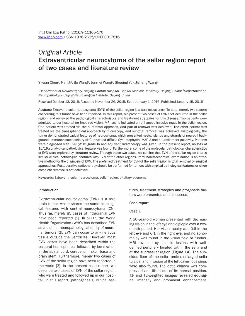
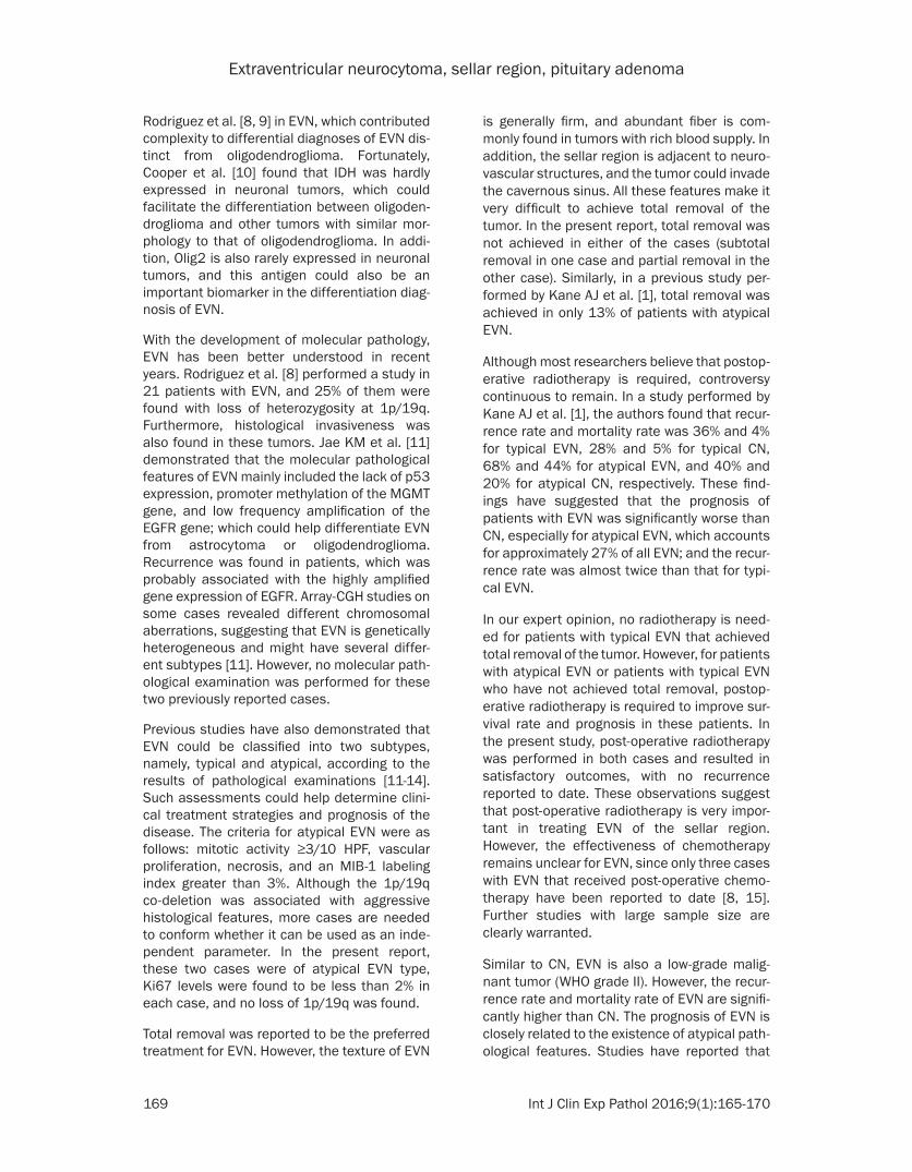
A 50-year-old woman presented with decreas-ing vision in the left eye and diplopia over a two-month period. Her visual acuity was 0.6 in the left eye and 0.1 in the right eye, and no abnor-mality was found in the visual field or fundus. MRI revealed cystic-solid lesions with well-defined periphery located within the sella and at the suprasellar region (Figure 1A). The sub-sided floor of the sella turcica, enlarged sella turcica, and invasion of the left cavernous sinus were also found. The optic chiasm was com-pressed and lifted out of its normal position. T1- and T2-weighted images revealed equisig-nal intensity and prominent enhancement.
Extraventricular neurocytoma, sellar region, pituitary adenoma
166 Int J Clin Exp Pathol 2016;9(1):165-170
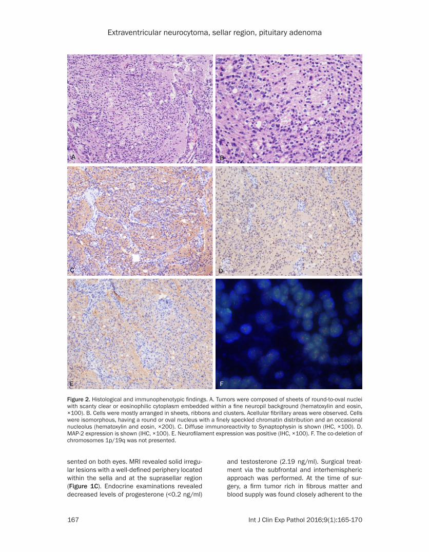
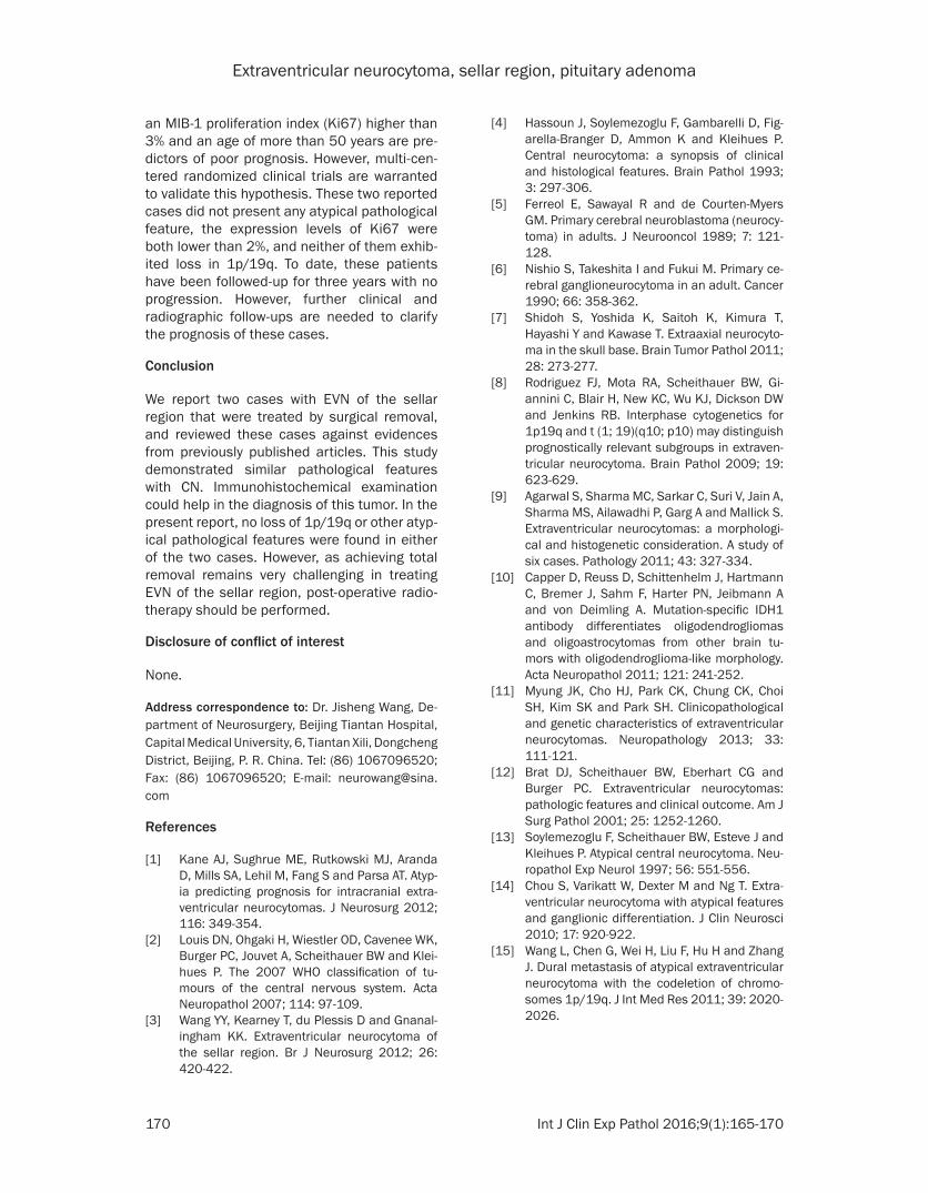
Endocrine examinations revealed no abnormal-ities. Surgical treatment via the transsphenoi-dal approach was performed, and a subtotal removal of the tumor was achieved. The texture of the tumor was dense and firm. The tumor had an abundant blood supply and was rich in fibrous matter. Histologically, these tumors were composed of sheets of round-to-oval nuclei with a scantyclear or eosinophilic cyto-plasm embedded within a fine neuropil back-ground (Figure 2A). Cells were mostly arranged in sheets, ribbons and clusters. Acellular fibril-lary areas were observed. Cells were isomor-phous and had an around or oval nucleus with a finely speckled chromatin and an occasional nucleolus (Figure 2B). Immunohistochemically, these tumor cells revealed diffuse positivity for Synaptophysin (SYN), MAP-2 and neurofilament (NF) in the cytoplasm and fibrillary stroma and diffuse negativity for GFAP, CK8/18, EMA and
NeuN (Figure 2C-E). In addition, the prolifera-tion index of Ki67 was approximately 2%. No loss of heterozygosity at 1p/19q was demon-strated (Figure 2F). The pathological diagnosis was EVN (WHO grade II). MRI revealed tumor regression after post-operative radiotherapy (54Gy total dose/27f/6W; Figure 1B). No recur-rence was reported after a three-year follow-up.
Case 2
A 62-year-old man with diminished vision quali-ty in both eyes and a narrowed visual field was admitted to our hospital. One year prior to admission, he underwent partial tumor removal via the transsphenoidal approach. Physical examinations revealed that visual acuity of the patient was 0.5 in each eye, and homonymous hemianopia of the temporal visual fields pre-
Figure 1. Pre- and post-operative MRI scans. A. The sagittal-T1-weighted enhanced image displays a tumor of the sellar region that invaded into the suprasellar region. B. Enhanced MRI scan was performed after radiotherapy, revealing the regression of the tumor, the pituitary stalk and optic chiasm. C. A coronal-T1-weighted enhanced im-age presenting a tumor located within the sella and the suprasellar region is shown. D. Enhanced MRI scan after radiotherapy showing substantial tumor regression.
Extraventricular neurocytoma, sellar region, pituitary adenoma
167 Int J Clin Exp Pathol 2016;9(1):165-170
sented on both eyes. MRI revealed solid irregu-lar lesions with a well-defined periphery located within the sella and at the suprasellar region (Figure 1C). Endocrine examinations revealed decreased levels of progesterone (<0.2 ng/ml)
and testosterone (2.19 ng/ml). Surgical treat-ment via the subfrontal and interhemispheric approach was performed. At the time of sur-gery, a firm tumor rich in fibrous matter and blood supply was found closely adherent to the
Figure 2. Histological and immunophenotypic findings. A. Tumors were composed of sheets of round-to-oval nuclei with scanty clear or eosinophilic cytoplasm embedded within a fine neuropil background (hematoxylin and eosin, ×100). B. Cells were mostly arranged in sheets, ribbons and clusters. Acellular fibrillary areas were observed. Cells were isomorphous, having a round or oval nucleus with a finely speckled chromatin distribution and an occasional nucleolus (hematoxylin and eosin, ×200). C. Diffuse immunoreactivity to Synaptophysin is shown (IHC, ×100). D. MAP-2 expression is shown (IHC, ×100). E. Neurofilament expression was positive (IHC, ×100). F. The co-deletion of chromosomes 1p/19q was not presented.
Extraventricular neurocytoma, sellar region, pituitary adenoma
168 Int J Clin Exp Pathol 2016;9(1):165-170
hypothalamus, bilateral optic nerve and optic chiasm. Visual acuity of the patient decreased to 0.1 in the left eye, but no change was found in the right eye. Pathological examination dem-onstrated a small round cell tumor with a dif-fuse growth pattern. The nuclei of tumor cells were round or oval, and cytoplasmic vacuoles were found in several cells. Few ganglion-like cells were also observed. A neuropil-like back-ground with vascular endothelial cell prolifera-tion was also found. Immunohistochemical examinations revealed that these tumor cells were positive for SYN, NeuN, Map-2, NF, Chromogranin A and Vimentin. Immunostains for GFAP, CK8/18 and P53 were negative. Ki67 level was less than 2%. No loss of heterozygos-ity at 1p/19q was demonstrated. The patho-logical diagnosis was EVN (WHO grade II). After post-operative radiotherapy for the treat-ment of the residual tumor (50.4Gy total dose/28f/6W), MRI revealed tumor regression (Figure 1D). No progression was found after a three-year follow-up.
Discussion
CN was first described by Hassoun et al. [4] in 1982 and accounts for approximately 0.25% to 0.5% of all central nervous system neoplasms. As a tumor with a generally good prognosis, CN was thought to locate only within the intraven-tricular system (especially at the foramen inter-ventriculae and septum pellucidum). The inci-dence rate of CN is not significantly different between males and females (with a male to female ratio of 1.02:1). However, the young adult subpopulation is affected by CN the most (with a mean age at onset of 29 years). In a study published in 1989, Ferreol and Nishio [5, 6] first described CN located outside the ven-tricular system. The 3rd edition of the WHO Classification of Tumors of the Central Nervous System described EVN as an oligodendroglio-ma-like tumor. However, in the 4th edition issued in 2007, EVN was classified as a neuro-nal tumor with a distinct entity [2].
EVN can occur at any nervous tissue site out-side the ventricles. However, most of the EVN have been described within the cerebral hemi-spheres, followed by the spinal cord, cerebel-lum, skull base and brain stem. The incidence rate of EVN of the sellar region is very low, and merely two cases have been reported to date
[3]. The histopathogenetic mechanisms of EVN of the sellar region remain largely unknown. Since there is no neuronal or neural progenitor cell in existence in the sellar region, investiga-tors postulate that EVN of the sellar region is caused by an abnormal migration of neurons during embryogenesis [7].
Impaired vision, especially changes in visual fields, was present in both cases previously reported and in both cases reported in this report. Endocrine changes caused by a stalk effect could also occur. However, no significant endocrine changes were found in any of these four patients. Through MRI examination, equi-signal intensity in T1-weight images and equi-signal or high signal intensity in T2-weight imag-es were commonly found in these four cases.
Nodular and peripheral enhancement was found in these patients, in which two patients were found with partial cystic degeneration and bleeding. Stippled calcification was also found by CT imaging, which should be distinguished from craniopharyngioma. Since the incidence rate of EVN of the sellar region is very low and no diagnostic criterion is available to date, the diagnosis of this tumor mainly relies on patho-logical examination. The main findings in immu-nohistochemistry assessments included the positive expression of SYN, NeuN and NSE, and the negative expression of GFAP. Sensitivity and specificity of SYN is highest in diagnosing EVN of the sellar region. However, electron microscopy and molecular pathological exami-nations should also be performed to further clarify the diagnosis of this tumor when SYN is found to be negative.
Imaging manifestations of EVN of the sellar region must be carefully reviewed to distinguish these from other tumors located at the sellar regions such as the pituitary adenoma, cranio-pharyngioma and meningioma. Similarly, path-ological examination results should be distin-guished from oligodendroglioma, oligoastrocy- toma, ependymoma, ganglioglioma and dysem-bryoplastic neuroepithelial tumors. In particu-lar, NeuN and SYN could be found to be posi-tively expressed in oligodendroglioma and oligoastrocytoma when neural cellular differen-tiation occurs, which actually makes it much more difficult to distinguish from EVN. Loss of heterozygosity at 1p/19q was reported by
Extraventricular neurocytoma, sellar region, pituitary adenoma
169 Int J Clin Exp Pathol 2016;9(1):165-170
Rodriguez et al. [8, 9] in EVN, which contributed complexity to differential diagnoses of EVN dis-tinct from oligodendroglioma. Fortunately, Cooper et al. [10] found that IDH was hardly expressed in neuronal tumors, which could facilitate the differentiation between oligoden-droglioma and other tumors with similar mor-phology to that of oligodendroglioma. In addi-tion, Olig2 is also rarely expressed in neuronal tumors, and this antigen could also be an important biomarker in the differentiation diag-nosis of EVN.
With the development of molecular pathology, EVN has been better understood in recent years. Rodriguez et al. [8] performed a study in 21 patients with EVN, and 25% of them were found with loss of heterozygosity at 1p/19q. Furthermore, histological invasiveness was also found in these tumors. Jae KM et al. [11] demonstrated that the molecular pathological features of EVN mainly included the lack of p53 expression, promoter methylation of the MGMT gene, and low frequency amplification of the EGFR gene; which could help differentiate EVN from astrocytoma or oligodendroglioma. Recurrence was found in patients, which was probably associated with the highly amplified gene expression of EGFR. Array-CGH studies on some cases revealed different chromosomal aberrations, suggesting that EVN is genetically heterogeneous and might have several differ-ent subtypes [11]. However, no molecular path-ological examination was performed for these two previously reported cases.
Previous studies have also demonstrated that EVN could be classified into two subtypes, namely, typical and atypical, according to the results of pathological examinations [11-14]. Such assessments could help determine clini-cal treatment strategies and prognosis of the disease. The criteria for atypical EVN were as follows: mitotic activity ≥3/10 HPF, vascular proliferation, necrosis, and an MIB-1 labeling index greater than 3%. Although the 1p/19q co-deletion was associated with aggressive histological features, more cases are needed to conform whether it can be used as an inde-pendent parameter. In the present report, these two cases were of atypical EVN type, Ki67 levels were found to be less than 2% in each case, and no loss of 1p/19q was found.
Total removal was reported to be the preferred treatment for EVN. However, the texture of EVN
is generally firm, and abundant fiber is com-monly found in tumors with rich blood supply. In addition, the sellar region is adjacent to neuro-vascular structures, and the tumor could invade the cavernous sinus. All these features make it very difficult to achieve total removal of the tumor. In the present report, total removal was not achieved in either of the cases (subtotal removal in one case and partial removal in the other case). Similarly, in a previous study per-formed by Kane AJ et al. [1], total removal was achieved in only 13% of patients with atypical EVN.
Although most researchers believe that postop-erative radiotherapy is required, controversy continuous to remain. In a study performed by Kane AJ et al. [1], the authors found that recur-rence rate and mortality rate was 36% and 4% for typical EVN, 28% and 5% for typical CN, 68% and 44% for atypical EVN, and 40% and 20% for atypical CN, respectively. These find-ings have suggested that the prognosis of patients with EVN was significantly worse than CN, especially for atypical EVN, which accounts for approximately 27% of all EVN; and the recur-rence rate was almost twice than that for typi-cal EVN.
In our expert opinion, no radiotherapy is need-ed for patients with typical EVN that achieved total removal of the tumor. However, for patients with atypical EVN or patients with typical EVN who have not achieved total removal, postop-erative radiotherapy is required to improve sur-vival rate and prognosis in these patients. In the present study, post-operative radiotherapy was performed in both cases and resulted in satisfactory outcomes, with no recurrence reported to date. These observations suggest that post-operative radiotherapy is very impor-tant in treating EVN of the sellar region. However, the effectiveness of chemotherapy remains unclear for EVN, since only three cases with EVN that received post-operative chemo-therapy have been reported to date [8, 15]. Further studies with large sample size are clearly warranted.
Similar to CN, EVN is also a low-grade malig-nant tumor (WHO grade II). However, the recur-rence rate and mortality rate of EVN are signifi-cantly higher than CN. The prognosis of EVN is closely related to the existence of atypical path-ological features. Studies have reported that
Extraventricular neurocytoma, sellar region, pituitary adenoma
170 Int J Clin Exp Pathol 2016;9(1):165-170
an MIB-1 proliferation index (Ki67) higher than 3% and an age of more than 50 years are pre-dictors of poor prognosis. However, multi-cen-tered randomized clinical trials are warranted to validate this hypothesis. These two reported cases did not present any atypical pathological feature, the expression levels of Ki67 were both lower than 2%, and neither of them exhib-ited loss in 1p/19q. To date, these patients have been followed-up for three years with no progression. However, further clinical and radiographic follow-ups are needed to clarify the prognosis of these cases.
Conclusion
We report two cases with EVN of the sellar region that were treated by surgical removal, and reviewed these cases against evidences from previously published articles. This study demonstrated similar pathological features with CN. Immunohistochemical examination could help in the diagnosis of this tumor. In the present report, no loss of 1p/19q or other atyp-ical pathological features were found in either of the two cases. However, as achieving total removal remains very challenging in treating EVN of the sellar region, post-operative radio-therapy should be performed.
Disclosure of conflict of interest
None.
Address correspondence to: Dr. Jisheng Wang, De- partment of Neurosurgery, Beijing Tiantan Hospital, Capital Medical University, 6, Tiantan Xili, Dongcheng District, Beijing, P. R. China. Tel: (86) 1067096520; Fax: (86) 1067096520; E-mail: [email protected]
References
[1] Kane AJ, Sughrue ME, Rutkowski MJ, Aranda D, Mills SA, Lehil M, Fang S and Parsa AT. Atyp-ia predicting prognosis for intracranial extra-ventricular neurocytomas. J Neurosurg 2012; 116: 349-354.
[2] Louis DN, Ohgaki H, Wiestler OD, Cavenee WK, Burger PC, Jouvet A, Scheithauer BW and Klei-hues P. The 2007 WHO classification of tu-mours of the central nervous system. Acta Neuropathol 2007; 114: 97-109.
[3] Wang YY, Kearney T, du Plessis D and Gnanal-ingham KK. Extraventricular neurocytoma of the sellar region. Br J Neurosurg 2012; 26: 420-422.
[4] Hassoun J, Soylemezoglu F, Gambarelli D, Fig-arella-Branger D, Ammon K and Kleihues P. Central neurocytoma: a synopsis of clinical and histological features. Brain Pathol 1993; 3: 297-306.
[5] Ferreol E, Sawayal R and de Courten-Myers GM. Primary cerebral neuroblastoma (neurocy-toma) in adults. J Neurooncol 1989; 7: 121-128.
[6] Nishio S, Takeshita I and Fukui M. Primary ce-rebral ganglioneurocytoma in an adult. Cancer 1990; 66: 358-362.
[7] Shidoh S, Yoshida K, Saitoh K, Kimura T, Hayashi Y and Kawase T. Extraaxial neurocyto-ma in the skull base. Brain Tumor Pathol 2011; 28: 273-277.
[8] Rodriguez FJ, Mota RA, Scheithauer BW, Gi-annini C, Blair H, New KC, Wu KJ, Dickson DW and Jenkins RB. Interphase cytogenetics for 1p19q and t (1; 19)(q10; p10) may distinguish prognostically relevant subgroups in extraven-tricular neurocytoma. Brain Pathol 2009; 19: 623-629.
[9] Agarwal S, Sharma MC, Sarkar C, Suri V, Jain A, Sharma MS, Ailawadhi P, Garg A and Mallick S. Extraventricular neurocytomas: a morphologi-cal and histogenetic consideration. A study of six cases. Pathology 2011; 43: 327-334.
[10] Capper D, Reuss D, Schittenhelm J, Hartmann C, Bremer J, Sahm F, Harter PN, Jeibmann A and von Deimling A. Mutation-specific IDH1 antibody differentiates oligodendrogliomas and oligoastrocytomas from other brain tu-mors with oligodendroglioma-like morphology. Acta Neuropathol 2011; 121: 241-252.
[11] Myung JK, Cho HJ, Park CK, Chung CK, Choi SH, Kim SK and Park SH. Clinicopathological and genetic characteristics of extraventricular neurocytomas. Neuropathology 2013; 33: 111-121.
[12] Brat DJ, Scheithauer BW, Eberhart CG and Burger PC. Extraventricular neurocytomas: pathologic features and clinical outcome. Am J Surg Pathol 2001; 25: 1252-1260.
[13] Soylemezoglu F, Scheithauer BW, Esteve J and Kleihues P. Atypical central neurocytoma. Neu-ropathol Exp Neurol 1997; 56: 551-556.
[14] Chou S, Varikatt W, Dexter M and Ng T. Extra-ventricular neurocytoma with atypical features and ganglionic differentiation. J Clin Neurosci 2010; 17: 920-922.
[15] Wang L, Chen G, Wei H, Liu F, Hu H and Zhang J. Dural metastasis of atypical extraventricular neurocytoma with the codeletion of chromo-somes 1p/19q. J Int Med Res 2011; 39: 2020-2026.