Embed Size (px)
Citation preview
Optimizing the Periosteal Flap Optimizing the Periosteal Flap for for
Lateral Eyelid ReconstructionLateral Eyelid Reconstruction
W. Thomas McClellan, M.D., WVUClinton McCord, M.D., Emory
Rebecca Neusch, WVU
Nothing to Disclose
Objective of the StudyObjective of the Study
To establish a standardized method to harvest the periosteal flap in order to improve eyelid strength, position, and reduce ectropion.
Materials and Methods - DesignMaterials and Methods - Design
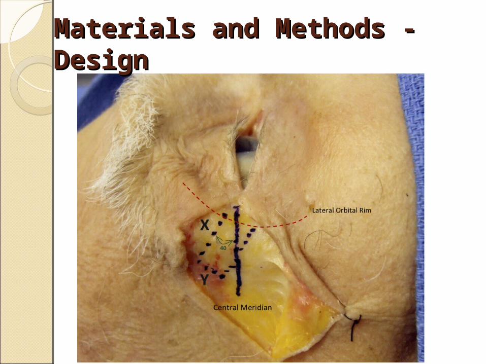
Create a central meridian from the upper puncta to the lateral orbital rim.
This intersection point is where the superior aspect of the flap is started and continues at a 40 degree angle superiorly out a distance X (which is the length needed to repair the defect).
Materials and Methods - DesignMaterials and Methods - Design

The width of the flap should measure 6-7 mm which compensates for primary contracture while maintaining strength of the periosteum. The periosteum is then secured with 5-0 Vicryl to the lateral edge of the tarsal plate with enough tension that the lid approximates the globe.
Materials and Methods - DesignMaterials and Methods - Design
If the flap is extended onto the temporalis fascia up to 50% of the posterior lamella of the lower eyelid can potentially be closed.
Materials and Methods - DesignMaterials and Methods - Design
Materials and MethodsMaterials and Methods
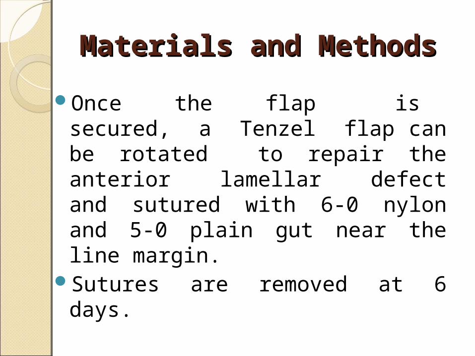
Once the flap is secured, a Tenzel flap can be rotated to repair the anterior lamellar defect and sutured with 6-0 nylon and 5-0 plain gut near the line margin.
Sutures are removed at 6 days.

Surgical ImagesSurgical Images
Figure 1 Pre-Op 80% lower eyelid + canthal Defect
Figure 2 Intra-Op periosteal flap dissection
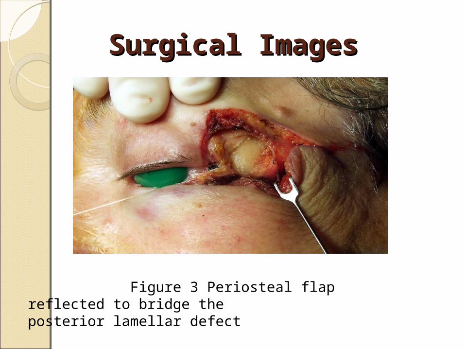
Surgical ImagesSurgical Images
Figure 3 Periosteal flap reflected to bridge the posterior lamellar defect
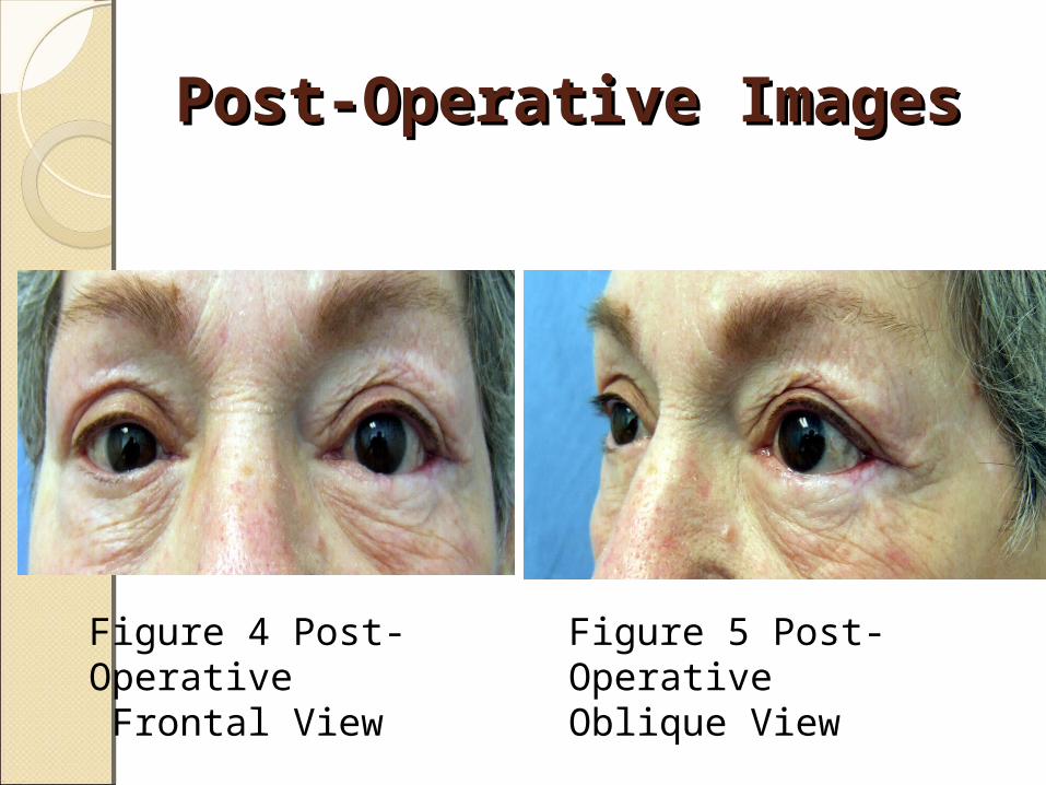
Post-Operative ImagesPost-Operative Images
Figure 4 Post-Operative Frontal View
Figure 5 Post-Operative Oblique View
Results and ConclusionsResults and Conclusions
12 patients all with good outcomes There were no complications such
as canthal dehiscence, cicatrical ectropion or entropion.
Results and Conclusion Results and Conclusion
The periosteal flap is an ideal flap to repair the lateral canthus and lateral posterior lamella due to its simplicity, reliability, and low risk.
Results and ConclusionResults and Conclusion
It can be designed to match the native eyelid contour, originates within the lateral orbit, is strong, and highly vascular2. Additionally if the flap relaxes with time it can be elevated and re-secured within the lateral orbit.

Significance of the FindingsSignificance of the Findings
This standardized method demonstrates a reliable method of reconstructing lateral eyelid defects. It optimizes lid position and reduces post operative ectropion.
ReferencesReferences
1. Smith, B.C., Nesi, F.A. Practical Techniques in Ophthalmic Plastic Surgery. St. Louis: The C.V. Mosby Company, 1981. Pp. 92-95.
2. Weinstein GS, Anderson RL, Tse DT, Kersten RC. The use of a periosteal strip for eyelid reconstruction. Arch. Ophthalmol. 1985 Mar;103(3):357-359.
3. McCord, C.D. Jr., Codner, M.A. Eyelid and Periorbital Surgery, Vol.2, 1st Ed. St. Louis: Quality Medical Publishing, Inc., 2008. Pp. 600.