Embed Size (px)
Citation preview

Proc. Nati. Acad. Sci. USAVol. 83, pp. 7593-7597, October 1986Biochemistry
On the spin trapping and ESR detection of oxygen-derived radicalsgenerated inside cells
(hydroxyl radical/superoxide radical/mammalian cells)
A. SAMUNI*, A. J. CARMICHAEL+, A. Russo, J. B. MITCHELL, AND P. RIESZRadiation Oncology Branch, Clinical Oncology Program, Division of Cancer Treatment, National Cancer Institute, National Institutes of Health,Bethesda, MD 20892
Communicated by W. A. Hagins, June 10, 1986
ABSTRACT Recently several attempts to identify oxygen-derived radicals in whole cells by spin trapping and electronspin resonance have been reported by using 5,5-dimethyl-1-pyrroline-N-oxide as the spin trap. In the present study, thefeasibility of this method is examined. Chinese hamster V79cells and human erythrocytes served as the test systems, whileOH radicals were generated by y radiolysis. Several spin trapswere used to scavenge the radicals and a distinction betweenexo- and endocellular ESR observable species was achievedusing tri(oxalato) chromiate(I) as a line broadening agent. Todistinguish between exo- and endocellular sites of radicalformation, we studied the effects of high molecular weightscavengers (polyethylene glycols), which do not enter the cell.Various possible obstacles associated with trapping and detect-ing the radicals inside the cells were examined. The resultsindicate that the primary radicals react with the spin traps.However, these spin adducts decayed within the cells.Cellularly induced decay of 2-hydroxy-5,5-dimethyl-1-pyrrol-idinyloxyl radical presented the major difficulty in detectingthe endogenous radicals, and potential experimental approach-es to overcome this difficulty are discussed.
Oxygen-derived radicals in various biological systems underphysiological conditions have been identified by using spin-trapping and electron spin resonance (ESR) spectrometrymainly in solutions or in in vitro systems containingsubcellular components as with tissue homogenates (1-3). Bycontrast, few attempts were made to use this technique withwhole cells. With this technique, OH and O° radicals, whichcannot be detected by ESR under physiological conditions,react with spin traps, usually nitrones or nitroso compounds,yielding long-lived radicals observable by ESR. Initially, thespin trapping of radicals within cells might appear inapplica-ble in view of the high concentrations of the DNA, RNA,proteins, amino acids, etc. Analysis of competition kineticsindicated that despite the high concentrations of certaincellular constituents and large rate constants of their reac-tions with free radicals (4), various scavengers, including spintraps, can scavenge at least part of the radicals within wholecells (5). Even though formation under normal physiologicalconditions of oxygen-derived radicals by cells is well estab-lished, their detection by spin trapping might be difficult forseveral possible reasons.
(i) The steady-state concentrations of free radicals withinthe cell might escape detection if their production rate is toolow or decay rate is too high. (ii) A significant fraction ofthese radicals might be produced site specifically near theirbiological targets. Such radicals would therefore be almostnonscavengeable by radical scavengers in general, includingspin traps. (iii) Insufficient as well as slow uptake of the spintrap into the cells or facilitated metabolic removal of the spin
trap might result in a failure to effectively compete for the freeradicals. (iv) The spin trap might accumulate in cellularcompartments other than those in which the radicals areproduced. (v) Spin adducts formed within the cell mightdisappear via metabolic or other pathways that effectivelyshorten their life time. (vi) Restricted motion of the spinadduct inside the cell might cause broadening of the ESRlines and consequently reduce the signal intensity. In spite ofthese potential limitations, several reports have been pub-lished recently describing the cellular generation of OH and°2 *radicals and their subsequent detection and identificationby using spin-trapping techniques (6-11). Spectral charac-teristics of the spin adduct and the competitive effect exertedby radical scavengers were used to identify and locate theradicals. Yet, conclusive evidence establishing endocellular-ly formed radicals is indeed desired.
Various normal metabolic processes are assumed to pro-duce radicals; moreover, under pathogenic conditions ordrug-induced conditions, a distinction between radical spe-cies formed inside or outside the cell (or spilled out) isimportant in determining their role and location in cellularsystems. Although spin trapping is generally a powerful toolfor tracing the participating free radicals (3), we have en-countered difficulties when attempting to reproduce previousexperiments spin trapping OH radicals in erythrocytes. Inview of the experimental barriers listed above and because ofthe wide interest in spin trapping of free radicals within cells,we examined the general feasibility ofapplying this techniqueto whole cells. To that end, we studied several cellular testsystems and various spin traps. The radicals were generatedby y radiation, and the distinction between the endo- andexocellular radicals was achieved by using line broadeningagents and high molecular weight scavengers that are prac-tically excluded from the cell.
MATERIALS AND METHODS
Chemicals. a-Phenyl-N-tert-butylnitrone (PBN), a-(4-py-ridyl-1-oxide)-N-tert-butylnitrone (4-POBN), 5,5-dimethyl-1-pyrroline-N-oxide (DMPO), a-(4-N-methylpyridinium-N-tert-butylnitrone (PyBN), and the stable nitroxide 2,2,6,6-tetramethylpiperidine-1-oxyl (TEMPO) were purchased fromAldrich. Polyethylene glycols (PEG) of various sizes (Sigma),oxalic acid, potassium oxalate, potassium dichromate, andK3[Fe(CN)6] (Fisher) were used without further purification.DMPO was purified before use by distillation or activatedcharcoal. The DMPO concentration in aqueous solutions was
Abbreviations: PBN, a-phenyl-N-tert-butylnitrone; 4-POBN, a-(4-pyridyl-1-oxide)-N-tert-butylnitrone; DMPO, 5,5-dimethyl-1-pyrro-line-N-oxide; PyBN, a-(4-N-methylpyridinium)-N-tert-butylnitrone;TEMPO, 2,2,6,6-tetramethylpiperidine-1-oxyl; Me2SO, dimethylsulfoxide.*On sabbatical leave from the Hebrew University, Jerusalem, Israel.tPresent address: Armed Forces Radiobiology Research Institute,Bethesda, MD 20814.
7593
The publication costs of this article were defrayed in part by page chargepayment. This article must therefore be hereby marked "advertisement"in accordance with 18 U.S.C. §1734 solely to indicate this fact.

Proc. Natl. Acad. Sci. USA 83 (1986)
determined spectroscopically. Tri(oxalato) chromiate(III)[K3[Cr(C204)3]*3H20] (chromium oxalate) was prepared asdescribed (12). Solutions were freshly prepared with milli-Qreagent water (18 MW), and the experiments were conductedaerobically at room temperature.
Radiation. The samples were irradiated at room tempera-ture using a 'Co y ray source with a dose rate of 51 Gy min-1.After irradiation, the samples were scanned for their ESRspectra.ESR Measurements. Unless otherwise stated, spectra were
taken with a Varian E-9 X-band spectrometer (9.5 GHz) witha field modulation frequency of 100 KHz, using microwavepower of 10 mW and modulation amplitude of 0.4 G.
Cells. Chinese hamster V79 cell lines and human erythro-cytes were used. Logarithmic-phase V79 cells were harvest-ed shortly before the experiment from monolayers andwashed four times with and suspended in phosphate-bufferedsaline (PBS). Subsequently, the spin trap was added and thecell suspension was y-irradiated in glass vessels for 1 min.Immediately afterward, aliquots were taken and their ESRspectra were monitored. Fresh erythrocytes were preparedsimilarly.
RESULTSTo distinguish between factors affecting the spin trapping ofradicals within the cell, we chose to produce the primaryradicals radiolytically. This technique has several advantag-es: (i) it ensures the generation of the water-derived primaryfree radicals homogeneously throughout the cell rather thanin certain subcellular compartments; (ii) it is possible tomodify the ratio between endo- and exocellular formation ofthe radicals by varying the cell concentration; (iii) theradiolytic generation of the radicals also makes it possible tocontrol the production rate as well as the spectrum of thepredominant species (by appropriate scavengers).
Effect of Line Broadening Agents. In principle, a distinctionbetween the ESR signals arising from endo- and exocellularspecies can be achieved (13) with selective line broadeningagents that only very slowly enter the cells. In the presentstudy, we have used chromium oxalate, which was previ-ously found to be superior to ferricyanide or Ni(II) (14).Immediately after the addition of 30mM chromium oxalate to29 juM TEMPO, the ESR signal disappeared. Since the linebroadening effect is due to spin-spin interaction, the ESRsignal reappeared upon suitable dilution. Thus, we added toa PBS solution of the stable free radical nitroxide, TEMPO,either erythrocytes, chromium oxalate, or both, and wemonitored the ESR spectra obtained. The results presentedin Fig. 1 indicate that in the absence of cells chromiumoxalate fully abolished the TEMPO signal (Fig. ic). Bycontrast, in the presence of both 5.6 x 10⁹ erythrocytes perml and 30 mM chromium, =50% of the signal persisted (Fig.lb), indicating that this fraction was inaccessible to linebroadening. In the presence ofthe cells the TEMPO radicals,which otherwise survive for days, decayed with a half-life of-2 hr at room temperature. Evidently, as was found previ-ously for bacterial cells (5), the TEMPO radicals entering thecells became shielded from the line broadening effect but alsodecayed faster. Similar results with erythrocytes and chro-mium were observed for the stable 3-carbamoyl-2,2,5,5-tetramethyl pyrrolidine-1-yloxy free radical (data notshown). The same procedure was repeated with a spin adductrather than a stable nitroxide. Since spin adducts of carbon-centered radicals are generally more stable than the adductsof oxygen-derived radicals, we used the methyl adduct of thespin trap 4-POBN. A solution containing both 1 M dimethylsulfoxide (Me2SO) and 0.1 M 4-POBN was -irradiated andsubsequently frozen. Later it was thawed, mixed with either5.1 x 109 erythrocytes per ml or 30 mM chromium oxalate,
.0-N (TEMPO)
CH,(4-POBN-CH3) O-N(QO~CIH-N-C(CH3)3
0
(d) RBC
(eL RBC, Cr (111)
Cr (111)
ll 1. 1 1.,,, ,,,, ,,. *10G-
FIG. 1. The ESR spectra of 29 ,uM TEMPO in PBS measured inthe presence of 5 x 10⁹ erythrocytes (RBC) per ml (a); 30 mMchromium oxalate and erythrocytes (b); 30 mM chromium oxalate(c); the ESR spectra for 0.1 M 4-POBN in PBS y-irradiated for 1 minat room temperature in the presence of 1 M Me2SO, measured afteraddition of 5 x 10⁹ erythrocytes per ml (d); both erythrocytes and 30mM chromium oxalate (e); 30 mM chromium oxalate (f).
or both, and the ESR spectra were recorded. The results inFig. 1 d and e show a six-line spectrum having the hyperfinecoupling constants aN = 15.9 G and aH = 2.65 G, charac-teristic of 4-POBN-CH3. The radiolytically formed OH rad-icals reacted with Me2SO, yielding methyl radicals. A certainfraction of the methyl radicals in turn add to 4-POBN,yielding ESR observable adducts. These relatively stablenitroxides, unlike 4-POBN-OH, which is short-lived (15),persisted as seen in Fig. 1. The presence of the cells did notalter the half-life of the 4-POBN-CH3 (t1/2, =2h) significantly.On the other hand, =30% ofthe ESR signal was shielded fromthe broadening agent when both cells and chromium oxalatewere present. The magnitude of the residual signal is con-sistent with a calculated =44% volume fraction occupied by5.1 x 109 erythrocytes per ml (the mean corpuscular volumeis 86 ± 8 Am3 for erythrocytes). The residual signal of4-POBN-CH3 increased with an increase in cell concentra-tion but did not depend on the incubation time of the cellswith the spin adduct. This indicates the ease with which thespin adducts cross the cell wall. The above results indicate (i)the spin adducts readily partition between the exo- and theendocellular environments; (ii) the width of the ESR spectrallines, beside the high-field doublet, due to endocellularspecies was not broadened; (iii) the relative populations of
7594 Biochemistry: Samuni et al.

Proc. Natl. Acad. Sci. USA 83 (1986) 7595
exo- and endocellular probes were readily accessible; (iv) the4-POBN-CH3 spin adducts maintained their stability withinthe cells.
Similar results (data not shown) were observed when PBNor PyBN were used as spin traps. These results also indicatea limitation of the use of line broadening agents. Chromiumoxalate discriminates between exo- and endocellular freeradicals according to their location rather than site of forma-tion. Spin adducts formed outside can diffuse into the cell andbecome shielded from the line broadening. A residual ESRsignal observable in the presence of a line broadening agentdoes not prove endocellular formation of radicals, whereastotal elimination of an ESR signal by the chromium oxalate,as was found for activated polymorphonuclear leukocytes,does not exclude endocellular formation of the spin adducts(11). The relative contribution ofthe volume of the cells to theoverall volume in dilute suspension is so small that the freeradicals present could escape detection. This may be true,provided that a rapid dynamic exchange of the probe acrossthe cell wall prevails and the partition of the radicals occursaccording to the relative endo- and exocellular total volumes.
Spin Trapping of OH and Superoxide Radicals in Cells.Despite numerous experimental artifacts associated with theuse of DMPO as a "universal" spin trap (16), it is still widelyused. DMPO is a particularly useful probe for trappingoxygen-derived radicals because of the relative stability andthe diverse spectral characteristics ofthe spin adducts (6-11).In addition, DMPO appeared to be an optimal choice in viewof its low cytotoxicity, accessibility to the cell, and high rateconstant of reaction with OH radicals (15, 17). For thesereasons, we examined it in our system. The reaction ofDMPO with superoxide has a low rate constant [-10M-'sec-1 (18)] and yields an unstable spin adduct (19).Nevertheless, with DMPO' the trapping and detection ofsuperoxide radicals appeared feasible since superoxide rad-icals have a longer half-life and the spin adduct is transformedinto the more stable DMPO-OH adduct (19).To compete effectively for the OH or 0- inside the cell, it
was desirable to use high concentrations (-0.1 M) of the spintrap. A comparison of rate constants of H' and hydratedelectrons (e-q) with DMPO [4 x 10⁹ M-'1sec-', 2 x 10¹⁰ M-1(20)] and with oxygen implied that practically all H atoms andhydrated electrons would react with DMPO (100 mM),yielding DMPO-H, rather than with oxygen (0.28 mM) toform superoxide. Therefore, no difference was expectedbetween systems irradiated in the presence or absence of air.Since only 35% of the OH radicals reacting with DMPO leadto an ESR observable spin adduct (21), a buildup of -5 AMDMPO-OH following 60 sec of irradiation was expected,provided this spin adduct would remain long-lived (18, 22).To maximize further the chances of detecting radicals in
the cell, we used concentrated cell suspensions. Chinesehamster V79 cells were suspended in PBS (2.2 x 10⁸ cells perml; mean corpuscular volume, 1100 ,um3) and -irradiated for60 sec in the presence of 0.2 M DMPO. A scan of the cellsuspension (3 min after irradiation) showed no detectableESR signal. The results of an analogous experiment withfresh human erythrocytes are presented in Fig. 2 a-c. TheESR spectra of a 0.2 M DMPO solution obtained 2 min afterirradiation in the absence (Fig. 2a) and in the presence (Fig.2b) of 4.1 x 10⁹ erythrocytes per ml are shown. The ESRsignal consisted of the 1:2:2:1 quartet typical of the DMPO-OH spin adduct with aN = aH = 14.9G (15, 18, 23), and thetriplet of triplets having aN = 16.5G, aH(2) = 22.6G identifiedas the DMPO-H spin adduct (24). Fig. 2b illustrates that in thepresence of erythrocytes the signal ofDMPO-OH was mark-edly decreased when compared to the cell-free system. Inaddition, the experiment was repeated with 30mM chromiumoxalate added to the cells immediately after irradiation.Under these conditions, the cells constituted -35% of the
DMPO(a)
(b)
(c)
RRBC
L___ L _L= - ------ --
I-lOG--
DMPO
PEG, RBC
(e)
--__- - - ~ .. - ---- -----
FIG. 2. The ESR spectra of 0.1 M DMPO in PBS y-irradiated for60 sec at room temperature in the absence (a) and in the presence (b)of 4.1 x 10⁹ erythrocytes (RBC) per ml, and measured 2 min afterirradiation. (c) Same as b, but with 30 mM chromium oxalate addedafter irradiation. (d) ESR spectrum obtained for 0.1 M DMPO in PBSafter y-irradiation of 0.1 M DMPO in PBS for 1 min in the presenceof 0.1 M PEG 3500; (d) with 5.1 x 10⁹ erythrocytes per ml; (e)control. Arrows designate locations of the 1:2:2:1 quartet due toDMPO-OH.
total volume of the sample. Therefore, =35% of the radiolyti-cally formed radicals originate inside the cells. Since chro-mium oxalate (within the time range of the experiment)selectively eliminates the ESR signal of the exocellularspecies, a residual signal due to the endogenous radicals wasanticipated. Yet, even with a 10-fold higher spectrometergain, no ESR signal was detected. This failure to observe anyresidual signal in the presence of the line broadening agentindicates that the ESR signal is due to the exocellular species.In other words, neither DMPO-OH nor DMPO-H adductslocated in or outside the cell were detected.To substantiate the above finding, we used a high molec-
ular weight scavenger of free radicals to distinguish betweenthe endo- and the exocellular formation of the radicals.
Effect of High Molecular Weight Scavengers. Polyethyleneglycols (PEG) rapidly react with OH and H radicals, primar-ily through H abstraction (k = 2.4 x 10⁸ M-1 sec-1), on amonomer basis (25). These polymers, H(OCH2CH2),OH,PEG 3500 (n = 68-84), or PEG 8000 (n = 125-180) areincapable of penetrating into the cell, and therefore onlyscavenge radicals outside the cell. PEG 3500 (0.05 M) was,-irradiated for 2 min in the presence of the spin trap PBN.
Biochemistry: Samuni et al.
MDt$- ur I'or 1111

Proc. Natl. Acad. Sci. USA 83 (1986)
The resulting ESR signal consisted of triplet of doublets,having aN = 15.85G and aH = 2.9G attributable to the PEGspin adduct.The subsequent introduction of 4.1 x 10⁹erythrocytes per
ml had no significant effect on the signal. On the other hand,30 mM chromium oxalate, even in the presence of the cells,fully eliminated the signal. Therefore, the PBN-PEG adductswere confined to the exocellular environment. Thus, in thepresence of DMPO and excess PEG, DMPO is expected totrap OH radicals inside the cell but to react with the PEGradicals outside the cell.
After the y-irradiation of a solution of 0.1 M DMPO in thepresence of 0.1 M PEG 3500, the ESR spectra seen in Fig. 2ewere observed. The nine-line signal of the DMPO-H adduct(Fig. 2 a and b) resulting from H and e- is observed, whereasthe 1:2:2:1 quartet of the DMPO-OH is replaced by thesix-line spectrum of DMPO-PEG spin adduct havinghyperfine coupling constants ofaN = 15.75G and aH = 21.6G.These values were not that different from the DMPO spinadduct of the monomer (CH2OH)2, aN = 15.6G, aH = 22.5G.The ESR spectra of DMPO-PEG and DMPO-OH are easilydistinguishable and offer a measure of the relative contribu-tions of endo- and exocellular spin adducts to the overall ESRsignal. When the concentrated erythrocyte suspension wasy-irradiated in the presence of both 0.1 M DMPO-OH and 0.1M PEG 3500, the ESR signal of DMPO-PEG was observed,but the DMPO-OH could not be detected (Fig. 2 d and e). Infact, even in the absence ofPEG the signal of the DMPO-OHadduct (Fig. 2b) was much smaller than that observed in acell-free system (Fig. 2a). This could be due to a failure of thespin trap to enter the cells or to a decrease in the half-life ofthe spin adduct. To determine whether the spin trap is indeedtaken up by cells without being metabolized, 0.15 M DMPOwas incubated for 40 min in the absence or in the presence of5.1 x 109 erythrocytes per ml (=50% packed cells) andcentrifuged at 800 rpm for 10 min. Subsequently, the twosystems were spectrophotometrically assayed for DMPO.No difference was observed in the shapes and intensities ofthe absorption peaks (227 nm) from DMPO. In addition, thecell pellet was diluted 1:1 with buffer, resuspended, recen-trifuged, and assayed for DMPO. After accounting for dilu-tion, the resulting absorption spectrum of DMPO remainedunchanged, which indicates that DMPO reversibly partitionsbetween the endo- and exocellular environment.
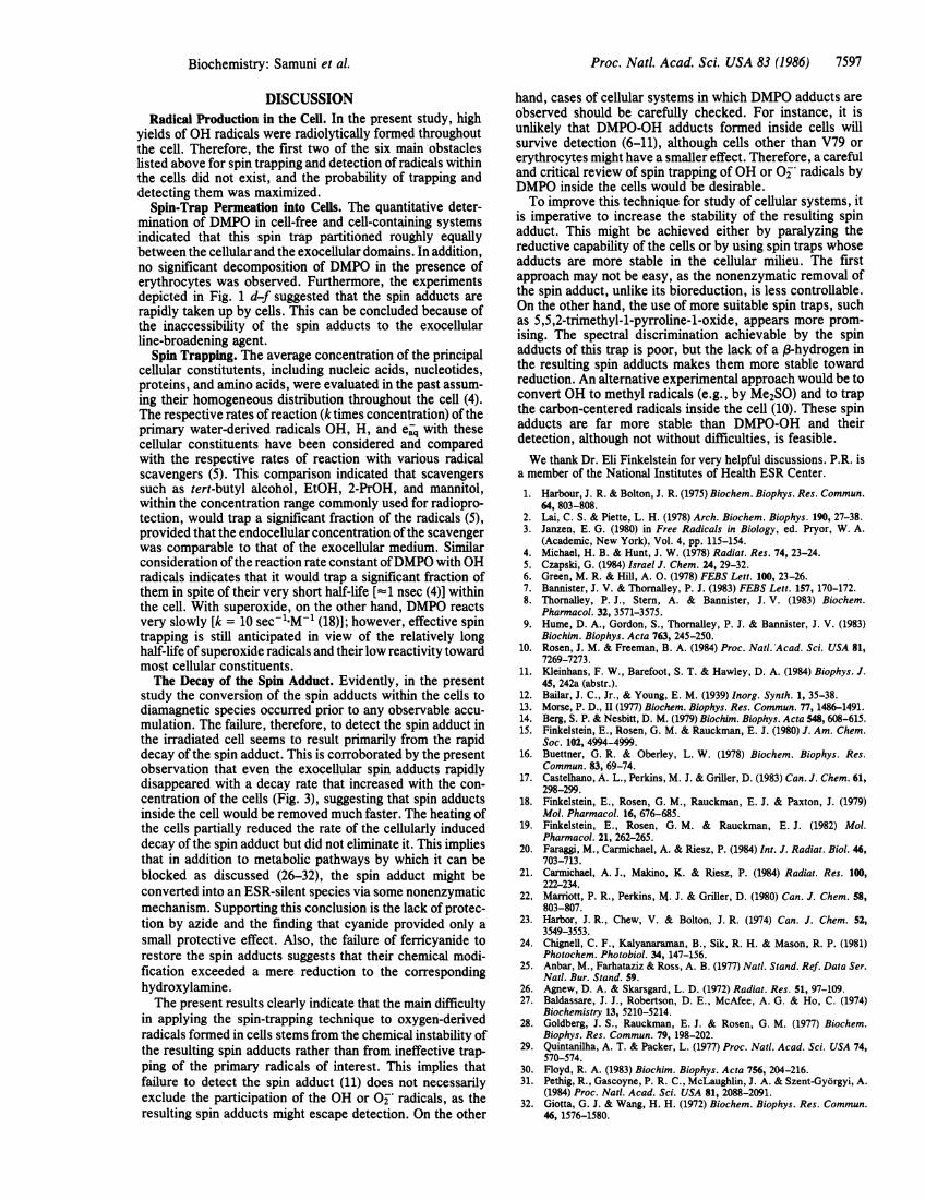
Effect of Cells on the Lifetime of Exoceliular Spin Probes.Cellularly induced decay of spin labels has been previouslyinvestigated because it interferes with their function asradiosensitizers (26) and as cellular probes (27). Probably bymetabolizing the free radicals, various cells or cellularcomponents were reported to hasten the decay of ESRsignals from stable nitroxides and spin adducts (26-31).Therefore, we examined the effect of cells on the DMPO-OHspin adduct. DMPO solutions were -irradiated for 60 sec,diluted to keep the DMPO-OH concentration below 10 AM;then cells were added to the sample and the decay of the ESRsignal was followed. The DMPO-OH adduct at concentra-tions <10 AM and in the absence of impurities is relativelylong-lived (18, 22). By contrast, DMPO-OH decayed rapidlyupon addition of cells. This decay rate increased with cellconcentration. Fig. 3 displays decay curves ofthe ESR signalmonitored for increasing cell concentrations and indicates themarked decrease in the half-life ofDMPO-OH in the presenceof cells.
Factors Affecting the Decay of the Spin Adduct. It haspreviously been suggested for Ehrlich ascites cells that theconversion of spin label into diamagnetic species, probablythrough reduction, might occur at the cell surface (31). Analternative explanation is that the radicals might penetrateinto the cells and there undergo bioreduction (26-28,32). Thechemical reaction in both cases might be mediated enzymat-
zD 4
<3z
cc
9 200.
0~~~~~~~
10 20 30 40 50
TIME AFTER y RADIOLYSIS (min)
FIG. 3. DMPO solutions (0.1 M) were y-irradiated for 1 min andthen diluted to give =10 A.M DMPO-OH spin adduct; erythrocytes atvarious concentrations were added and the decay of the ESR signalat room temperature was followed. *, No cells; *, 0.5%; o, 1%; *,5%; O, 10%; A, 20%; A, 30%; *, 40%; -:, 50% packed cells.
ically. An enzymatic bioreduction would depend on thephysiological state of the cell. Hence, we examined thepossible effects of cyanide, azide, and heat-inactivation of thecells on the decay of the spin adducts. Preincubation of theerythrocytes (=50%) for 10 min with 1-10 mM cyanideincreased the half-life of the spin adducts =2-fold. Converse-ly, exposure of the erythrocytes to 10 mM azide for 10 minhad no effect on the decay rate of DMPO-OH. Preheating theerythrocytes at 60'C for 5 min had no significant effect;however, a prolonged incubation of the cells (50% erythro-cytes) at 60'C for 30 min immediately prior to their additionto the DMPO-OH solution, increased the half-life of the spinadduct 2- to 3-fold as compared to intact cells. This indicatedthat both enzymatic and nonenzymatic pathways contributeto the disappearance of the spin adducts. In addition, sinceprevious studies implicated sulfhydryl groups in the reduc-tion of the radicals (27, 31, 32), we studied the effects ofsulfhydryl blockers. To that end, the erythrocytes werepreincubated with 10mM diamide or N-ethylmaleimide for 10min with hardly any effect on the cell-induced decay ofDMPO-OH. Therefore, no evidence for the involvement ofSH groups in the decay of the spin adducts could beestablished.
In analogous experiments, erythrocytes were preincubatedwith 5 mM diethylenetriaminopentaacetic acid for 20 minbefore adding DMPO-OH to the cells. No effect of thechelating agent on the decay of the spin adduct was observed,indicating that transition metal ions were not mediating thisdecay.
Reversibility of the Reaction. The rapid disappearance, inthe presence of cells, of the ESR signal of the paramagneticspecies could result from the reduction of the spin adduct orby other forms of chemical destruction. The reversibility ofthe process by an oxidizing agent would indicate the formeralternative. To examine this possibility, 4 x 10⁹ erythrocytesper ml were added to a solution of preformed DMPO-OHadduct. After total disappearance of the spin adduct signal, 1mM ferricyanide was added: the signal was not restored.
7596 Biochemistry: Samuni et al.
Proc. Natl. Acad. Sci. USA 83 (1986) 7597
DISCUSSIONRadical Production in the Cell. In the present study, high
yields of OH radicals were radiolytically formed throughoutthe cell. Therefore, the first two of the six main obstacleslisted above for spin trapping and detection of radicals withinthe cells did not exist, and the probability of trapping anddetecting them was maximized.
Spin-Trap Permeation into Cells. The quantitative deter-mination of DMPO in cell-free and cell-containing systemsindicated that this spin trap partitioned roughly equallybetween the cellular and the exocellular domains. In addition,no significant decomposition of DMPO in the presence oferythrocytes was observed. Furthermore, the experimentsdepicted in Fig. 1 d-f suggested that the spin adducts arerapidly taken up by cells. This can be concluded because ofthe inaccessibility of the spin adducts to the exocellularline-broadening agent.
Spin Trapping. The average concentration of the principalcellular constitutents, including nucleic acids, nucleotides,proteins, and amino acids, were evaluated in the past assum-ing their homogeneous distribution throughout the cell (4).The respective rates of reaction (ktimes concentration) of theprimary water-derived radicals OH, H, and e- with thesecellular constituents have been considered and comparedwith the respective rates of reaction with various radicalscavengers (5). This comparison indicated that scavengerssuch as tert-butyl alcohol, EtOH, 2-PrOH, and mannitol,within the concentration range commonly used for radiopro-tection, would trap a significant fraction of the radicals (5),provided that the endocellular concentration of the scavengerwas comparable to that of the exocellular medium. Similarconsideration of the reaction rate constant ofDMPO with OHradicals indicates that it would trap a significant fraction ofthem in spite of their very short half-life [=1 nsec (4)] withinthe cell. With superoxide, on the other hand, DMPO reactsvery slowly [k = 10 sec-1'M-l (18)]; however, effective spintrapping is still anticipated in view of the relatively longhalf-life of superoxide radicals and their low reactivity towardmost cellular constituents.The Decay of the Spin Adduct. Evidently, in the present
study the conversion of the spin adducts within the cells todiamagnetic species occurred prior to any observable accu-mulation. The failure, therefore, to detect the spin adduct inthe irradiated cell seems to result primarily from the rapiddecay of the spin adduct. This is corroborated by the presentobservation that even the exocellular spin adducts rapidlydisappeared with a decay rate that increased with the con-centration of the cells (Fig. 3), suggesting that spin adductsinside the cell would be removed much faster. The heating ofthe cells partially reduced the rate of the cellularly induceddecay of the spin adduct but did not eliminate it. This impliesthat in addition to metabolic pathways by which it can beblocked as discussed (26-32), the spin adduct might beconverted into an ESR-silent species via some nonenzymaticmechanism. Supporting this conclusion is the lack of protec-tion by azide and the finding that cyanide provided only asmall protective effect. Also, the failure of ferricyanide torestore the spin adducts suggests that their chemical modi-fication exceeded a mere reduction to the correspondinghydroxylamine.The present results clearly indicate that the main difficulty
in applying the spin-trapping technique to oxygen-derivedradicals formed in cells stems from the chemical instability ofthe resulting spin adducts rather than from ineffective trap-ping of the primary radicals of interest. This implies thatfailure to detect the spin adduct (11) does not necessarilyexclude the participation of the OH or O°j radicals, as theresulting spin adducts might escape detection. On the other
hand, cases of cellular systems in which DMPO adducts areobserved should be carefully checked. For instance, it isunlikely that DMPO-OH adducts formed inside cells willsurvive detection (6-11), although cells other than V79 orerythrocytes might have a smaller effect. Therefore, a carefuland critical review of spin trapping of OH or O- radicals byDMPO inside the cells would be desirable.To improve this technique for study of cellular systems, it
is imperative to increase the stability of the resulting spinadduct. This might be achieved either by paralyzing thereductive capability of the cells or by using spin traps whoseadducts are more stable in the cellular milieu. The firstapproach may not be easy, as the nonenzymatic removal ofthe spin adduct, unlike its bioreduction, is less controllable.On the other hand, the use of more suitable spin traps, suchas 5,5,2-trimethyl-1-pyrroline-1-oxide, appears more prom-ising. The spectral discrimination achievable by the spinadducts of this trap is poor, but the lack of a (3-hydrogen inthe resulting spin adducts makes them more stable towardreduction. An alternative experimental approach would be toconvert OH to methyl radicals (e.g., by Me2SO) and to trapthe carbon-centered radicals inside the cell (10). These spinadducts are far more stable than DMPO-OH and theirdetection, although not without difficulties, is feasible.We thank Dr. Eli Finkelstein for very helpful discussions. P.R. is
a member of the National Institutes of Health ESR Center.1. Harbour, J. R. & Bolton, J. R. (1975) Biochem. Biophys. Res. Commun.
64, 803-808.2. Lai, C. S. & Piette, L. H. (1978) Arch. Biochem. Biophys. 190, 27-38.3. Janzen, E. G. (1980) in Free Radicals in Biology, ed. Pryor, W. A.
(Academic, New York), Vol. 4, pp. 115-154.4. Michael, H. B. & Hunt, J. W. (1978) Radiat. Res. 74, 23-24.5. Czapski, G. (1984) Israel J. Chem. 24, 29-32.6. Green, M. R. & Hill, A. 0. (1978) FEBS Lett. 100, 23-26.7. Bannister, J. V. & Thornalley, P. J. (1983) FEBS Lett. 157, 170-172.8. Thornalley, P. J., Stem, A. & Bannister, J. V. (1983) Biochem.
Pharmacol. 32, 3571-3575.9. Hume, D. A., Gordon, S., Thornalley, P. J. & Bannister, J. V. (1983)
Biochim. Biophys. Acta 763, 245-250.10. Rosen, J. M. & Freeman, B. A. (1984) Proc. Natl. Acad. Sci. USA 81,
7269-7273.11. Kleinhans, F. W., Barefoot, S. T. & Hawley, D. A. (1984) Biophys. J.
45, 242a (abstr.).12. Bailar, J. C., Jr., & Young, E. M. (1939) Inorg. Synth. 1, 35-38.13. Morse, P. D., II (1977) Biochem. Biophys. Res. Commun. 77, 1486-1491.14. Berg, S. P. & Nesbitt, D. M. (1979) Biochim. Biophys. Acta 548, 608-615.15. Finkelstein, E., Rosen, G. M. & Rauckman, E. J. (1980) J. Am. Chem.
Soc. 102, 4994-4999.16. Buettner, G. R. & Oberley, L. W. (1978) Biochem. Biophys. Res.
Commun. 83, 69-74.17. Castelhano, A. L., Perkins, M. J. & Griller, D. (1983) Can. J. Chem. 61,
298-299.18. Finkelstein, E., Rosen, G. M., Rauckman, E. J. & Paxton, J. (1979)
Mol. Pharmacol. 16, 676-685.19. Finkelstein, E., Rosen, G. M. & Rauckman, E. J. (1982) Mol.
Pharmacol. 21, 262-265.20. Faraggi, M., Carmichael, A. & Riesz, P. (1984) Int. J. Radiat. Biol. 46,
703-713.21. Carmichael, A. J., Makino, K. & Riesz, P. (1984) Radiat. Res. 100,
222-234.22, Marriott, P. R., Perkins, M. J. & Griller, D. (1980) Can. J. Chem. 58,
803-807.23. Harbor, J. R., Chew, V. & Bolton, J. R. (1974) Can. J. Chem. 52,
3549-3553.24. Chignell, C. F., Kalyanaraman, B., Sik, R. H. & Mason, R. P. (1981)
Photochem. Photobiol. 34, 147-156.25. Anbar, M., Farhataziz & Ross, A. B. (1977) Natl. Stand. Ref. Data Ser.
Natl. Bur. Stand. 59.26. Agnew, D. A. & Skarsgard, L. D. (1972) Radiat. Res. 51, 97-109.27. Baldassare, J. J., Robertson, D. E., McAfee, A. G. & Ho, C. (1974)
Biochemistry 13, 5210-5214.28. Goldberg, J. S., Rauckman, E. J. & Rosen, G. M. (1977) Biochem.
Biophys, Res. Commun. 79, 198-202.29. Quintanilha, A. T. & Packer, L. (1977) Proc. Natl. Acad. Sci. USA 74,
570-574.30. Floyd, R. A. (1983) Biochim. Biophys. Acta 756, 204-216.31. Pethig, R., Gascoyne, P. R. C., McLaughlin, J. A. & Szent-Gyorgyi, A.
(1984) Proc. Natl. Acad. Sci. USA 81, 2088-2091.32. Giotta, G. J. & Wang, H. H. (1972) Biochem. Biophys. Res. Commun.
46, 1576-1580.
Biochemistry: Samuni et al.