Embed Size (px)
Citation preview

ON THE ANATOMY AND RELATIONSHIPS OF RECENT MONOPLACOPHORA
by KARL GEORG WINGSTRAND Institute for Cell Biology and Anatomy, University of Copenhagen,
Universitetsparken 15, DK-2100 Copenhagen @ Denmark
ABSTRACT
The original description of monoplacophoran anat- omy, which was based on two somewhat defective specimens of Neopilina galatheae, has been amend- ed and checked on the basis of two immature Vema ewingi and one immature Neopilina galatheae. Some mistakes have been corrected: The evidence for a coiled larval shell has been found to be unreli- able; the "dorsal coelomic sacs" are shown to be en- larged pharyngeal diverticula; it has not been poss- ible to verify the presence of coelomostomes from the kidneys, except perhaps in the heart region. The repetition of pedal retractors, lateropedal connec- tives, nephridia, and other organs has been con-
firmed and compared in the two species. Vema has a more complete set of metameric organs, including six pairs of gills, seven pairs of nephridia, and three pairs of gonoducts (although one is vestigial), but it still has eight pairs of retractors.
The liver is connected with the stomach by a sin- gle, transverse, slitlike opening. The salivary glands of Vema are paired like those of chitons. Other points of the original description have been con- firmed, supplied with new illustrations or notes on variation.
A comparison with other molluscs has been made, and the discussion in the literature inspired by the
To the memory of my late friend
HENNING LEMCHE

descriptions of Neopilina in 1957 and 1959 has been reviewed. with emhasis on the phylogenetical problems . The Monoplacophora are good Con- chifera. and the Conchifera are accepted as a sister group of the Poiyplacophora. within the Testaria . It is concluded that the eight-metameric retractor sys- tem of the Monoplacophora is homologous with the retractor groups of the Polyplacophora; the latter overlap the valve limits . This eight-metamerism is probably a ground-plan feature of ancestral Testa- ria. but has clearly been reduced in most descendent
lines . It is maintained that similar eight-metamerism can have been present in other organ systems of the testarian ancestor and has been reduced in most lines in the same way as has clearly been the case with the muscle metamerism .
The origin of molluscs has been reconsidered . Their derivation from advanced oligomeric Spiralia ("prot-annelids" or "proto-articulates") with dorsal heart. oligomeric coelom. nephridia. gonads and gonoducts is still a possibility. although conclusions in the strict sense cannot be made at present .
CONTENTS
1 . INTRODUCTION . . . . . . . . . . . . . . . . . . . . . . . . . . . . . . . . . . . . . . . . . . . 10
2 . THE SPECIES OF RECENT MONOPLACOPHORA AND THEIR DISTRIBUTION . . . . . . . . . . . . . . . . . . . . . . . . . . . . . . . . . . . . 11
3 . MATERIAL AND METHODS . . . . . . . . . . . . . . . . . . . . . . . . . . . . . . . 12
4 . DESCRIPTIONS . . . . . . . . . . . . . . . . . . . . . . . . . . . . . . . . . . . . . . . . . . . . 13 4 . 1 . Theshell . . . . . . . . . . . . . . . . . . . . . . . . . . . . . . . . . . . . . . . . . . . . . . 13 4 . 2 . The musculature of the body . . . . . . . . . . . . . . . . . . . . . . . . . . . . . 15 4 . 3 . The nervous system . . . . . . . . . . . . . . . . . . . . . . . . . . . . . . . . . . . . . 20 4 . 4 . The gills and the gill nerves . . . . . . . . . . . . . . . . . . . . . . . . . . . . . . 21 4 . 5 . Nephridia and nephridiopores . . . . . . . . . . . . . . . . . . . . . . . . . . . . 23 4 . 6 . Gonads and gonoducts . . . . . . . . . . . . . . . . . . . . . . . . . . . . . . . . . . 24 4 . 7 . Are nephrostomes present? . . . . . . . . . . . . . . . . . . . . . . . . . . . . . . 25
. . . . . . . . . . . . . . . . . . . . . . 4 . 8 . Pericardium, heart and blood vessels 26 4 . 9 . The pharyngeal diverticula . . . . . . . . . . . . . . . . . . . . . . . . . . . . . . . 26 4 . 10 . Oral region, oral cavity, subradular organ,
salivaryglands . . . . . . . . . . . . . . . . . . . . . . . . . . . . . . . . . . . . . . . . . 29 . . . . . . . . . . . . . . . . . . . . . . . . . . . 4 . 10 . 1 . The preoral tentacles 29
. . . . . . . . . . . . . . . . . . . . . . . . . . . . . . 4 . 10 . 2 . The anterior jaw 29 4 . 10 . 3 . The subradular sac and the subradular
. . . . . . . . . . . . . . . . . . . . . . . . . . . . . . . . . . . . . . . glands 32 . . . . . . . . . . . . . . . . . . . . . . . . . . . . 4 . 10 . 4 . The salivary glands 32
. . . . . . . . . . . . . . 4 . 11 . The stomach, the liver and the crystalline style 33 4 . 11 . 1 . The stomach . . . . . . . . . . . . . . . . . . . . . . . . . . . . . . . . . . 33 4 . 11 . 2 . The liver . . . . . . . . . . . . . . . . . . . . . . . . . . . . . . . . . . . . . 33 4 . 11 . 3 . The crystalline style . . . . . . . . . . . . . . . . . . . . . . . . . . . . 35
4 . 12 . The radula apparatus . . . . . . . . . . . . . . . . . . . . . . . . . . . . . . . . . . . 36 4 . 12 . 1 . The radula support . . . . . . . . . . . . . . . . . . . . . . . . . . . . 36 4 . 12 . 2 . The radula ribbon . . . . . . . . . . . . . . . . . . . . . . . . . . . . . 39 4 . 12 . 3 . The radula muscles . . . . . . . . . . . . . . . . . . . . . . . . . . . . 39

. . . . . . . . . . . . . . . . . . . . . . . . . . . . . . . . . . . . . . . . . . . . . . . 5 . DISCUSSION 42 5 . 1 . Morphological features of metamerism in recent Monopla-
. . . . . . . . . . . . . . . . . . . . . . . . . . . . . . . . . . . . . . . . . . . . . . . cophora 42 . . . . . . . . . . . . . . . . . . . . . . . . . . . 5 . 1 . 1 . Introductory remarks 42
. . . . . . . . . . . . . . . . . . 5 . 1 . 2 . The case of Neopilina galatheae 43 . . . . . . . . . . . . . . . . . . . . . . . . . 5 1 . 3 . The case of Vema ewingi 44
. . . . . . . . . . . . . . . . . . . . . . . . . . . . . 5 . 1 . 4 . General viewpoints 46 . . . . . . . . . . 5 . 2 . Muscle metamerism in recent and fossil Conchifera 47 . . . . . . . . . . 5 . 3 . The Tryblidiacea (Monoplacophora) as Conchifera 50
. . . . . . . . . . . . 5 . 3 . 1 . The Conchifera as a monophyletic unit 51 5 . 3 . 2 . The Monoplacophora as a "stem group"
. . . . . . . . . . . . . . . . . . . . . . . . . . . within the Conchifera 52 . . . . . . . . . . . . . . . . . . 5 . 3 . 3 . Radiation within the Conchifera 54
. . . 5 . 3 . 4 . Notes on the Gastropoda (and Bellerophontacea) 55 . . . . . . . . . . . . . . . . . . . . . . . 5 . 3 . 5 . Notes on the Cephalopoda 56
5 . 3 . 6 . Notes on the Diasoma (Bivalvia, . . . . . . . . . . . . . . . . . . Scaphopoda and Rostroconchia) 57
. . . . . . . . . . . . . . . . . . . . . 5 . 4 . Comparison with the Polyplacophora 58 . . . . . . . . . . . . . . . . . . . . . . . . . . . . . . . 5 . 4 . 1 . External features 58
. . . . . . . . . . . . . . . . . . . . . . . . . . . . . . . . . . . . . . 5 . 4 . 2 . Theshell 58 . . . . . . . . . . . . . . . . . . . . . . . . . . . 5 . 4 . 3 . The radula apparatus 61
. . . . . . . . . . . . . . . . . . . . . . . . . . . . . 5 . 4 . 4 . The radula muscles 64 . . . . . . . . . . . . . . . . . . . . . . . . . . . . . . 5 . 4 . 5 . The body muscles 68
5 . 5 . Cladistic relations of Conchifera, Polyplacophora . . . . . . . . . . . . . . . . . . . . . . . . . . . . . . . . . . . . . . . and Aplacophora 73
. . . . . . . . . . . . . . 5 . 6 . General discussion of metamerism in molluscs 76 . . . . . . . . . . . . . . . 5 . 6 . 1 . The metamerism of the musculature 76
. . . . . . . . . . . 5 . 6 . 2 . The metamerism of other organ systems 79 . . . . . . . . . . . . . . . . . . . . . . . . . . . . . . . . . . . . 5 . 6 . 3 . Comments 80
. . . . . . . . . . . . . . . . . . . . . . . . . . . . . . . . . 5 . 7 . The ancestry of molluscs 81 6 . REFERENCES . . . . . . . . . . . . . . . . . . . . . . . . . . . . . . . . . . . . . . . . . . . . . . 90

1. INTRODUCTION
After the anatomical description of Neopilina galatheae Lemche, 1957, had been completed (Lem- che & Wingstrand 1959a, 1959b 1960), Dr. Lemche and I received some additional material of mono- placophorans for further study. The technical work with this material, including also some reconstruc- tions and drawings, was done in the early 1960's, but the publication of the results was unfortunately postponed several times. Not even a preliminary manuscript had been written when Dr.Lemche died in 1977. The material, including section series and reconstructions, is still in my laboratory, but most correspondance related to the material has unfor- tunately been lost.
Judging from numerous publications and letters the hitherto unpublished material is still of consider- able interest, and I have therefore felt it as my duty to publish what I can get out of it.
I have chosen to concentrate on points where the original description was insufficient or directly wrong. I have also paid particular attention to the species Vema ewingi (Clarke & Menzies, 1959) which differs from Neopilina galatheae in the metameric repetition of organs.
Up to now very little original information has been added to the early anatomical accounts of the Monoplacophora which were published between 1957 and 1960. However, the theoretical discussion based on this same material has flourished and resulted in a large and in part confusing literature. Some themes of this discussion are summarized and commented below. Particular emphasis is given to
comparisons between the Monoplacophora and Polyplacophora, because I feel that this important point has been somewhat neglected in the literature.
Acknowledgments
In addition to the new material of Neopilina and Vema provided by Dr. Robert Parker (Coastal Ecosystems Management Inc., Fort Worth, Texas), and the late Dr. Robert J. Menzies (formerly at Duke University, North Carolina), specimens of other molluscs were kindly provided by the Marine Biolo- gical Laboratory, Helsingerr, Denmark, and by Drs. Jnrrgen Knudsen and Jean Just, Zoological Museum, University of Copenhagen.
The following colleagues have seen and critically commented on the entire manuscript or part of it: Drs. Jrargen Liitzen, Arne Nerrrevang and Bjarne Westergaard, Institute of Cell Biology and Anatomy (formerly the Institute of Comparative Anatomy), University of Copenhagen; and Drs. Claus Nielsen and Torben Wolff of the Zoological Museum. Dr. Mary E. Petersen, Zoological Museum, has revised the English text and provided useful criticism and suggestions. I am most grateful to all of the above for their help but take full responsibility for the final result.
To the named persons and institutions, plus sever- al others not specifically mentioned, I express my sincere appreciation for their aid in bringing this manuscript to completion.

2. THE SPECIES OF RECENT MONOPLACOPHORG AND THEIR DISTRIBUTION
In the 27 years which have passed since Lemche's original description of Neopilina galatheae ap- peared in 1957, ten new recent species have been as- signed to the class Monoplacophora (Wenz in Knight 1952) and have been included in the family Neopilinidae Knight & Yochelson, 1958. The num- ber of ¢ species of Monoplacophora is thus brought up to eleven:
Neopilina galatheae Lemche, 1957 N. veleronis Menzies & Layton, 1962 N. adenensis Tebble, 1967 N. bruuni Menzies, 1968 N. oligotropha Rokop, 1972 N. zografi (Deutzenberg & Fischer, 1896) N. (Lemchephyala) rebainsi Moskalev et al., 1983 Vema ewingi (Clarke & Menzies, 1959) I.: bacescui Menzies, 1968 K ((Laevipilina) hyalina McLean, 1979 Monoplacophorus zenkevitchi Moskalev et al., 1983
The species zografi was originally described as an ar- chaeogastropod, Acmaea zografi Deutzenberg & Fischer, 1896, but a recent revision of the type material in the Monaco collections revealed that it
was a neopilinid monoplacophoran (Bouchet, McLean & Warin, 1983). Bouchet et al. therefore' transferred the species to the genus Neopilina. The type material of Acmaea euglypta Deutzenberg & Fischer, 1897, also present in the Monaco collec- tions, was found to be conspecific with that of A. zografi, and the name euglypta is regarded a junior synonym, zografi being preserved by the rules of pri- ority. This old material of monoplacophorans had been collected in the Azores area of the North Atlan- tic in 1888-1896 (see Fig. I).
The distribution of the Monoplacophora appears to be worldwide (Fig. 1) although the records are still far apart. Only one species, K (Laevipilina) hyalina, has been found in moderate depths (174-229 m). All other species are from greater depths, between 1800 and 6480 m.
All the finds and descriptions af recent Monoplacophora up to 1979 are carefully registered by Cesari & Guidastri (1976, 1979) to whom I refer the reader for details (in Italian, for an English text see McLean 1979). Moskalev et al. (1983) summarize all records up to 1983, including recent Russian con- tributions.
Genus Neopilina Genus Vema Genus Monoplacophorus
@ N. galatheae @ N.oligotropha A V.ewingi I M.zenkevitchi 0 N.veleronis @ N.zografi A V.bacescui @ N.adenensis Q N. rebainsi A V.(Laevipilina) hyalina €4 N.bruuni 0 N.sp.indet.
Fig. 1. Rcords o f recent Monoplacophora. For details see Cesari& Guidastri 1976, 1979, Bouchet et al. 1983, and Moskalev et at. 1983.

3. MATERIAL AND METHODS
Neopilina gatatheae Lemche, 1957
New M a t e r i a l Specmen I. Label: VSS - 16.-22. Mar. 59. Off Cape San Lucas
Canyon. Depth 1530-1535 fms. (2780-2810m). Lat. 22" 32.5'N., Long. 109"40.8'W. R.H. Parker.
The specimen was immature, shell measuring 12x11 mm. Original fixation: 8070 alcohol, 40% formalin and 100% acetic acid in the proportions 90:5:5. Stored in 90% alcohol. Decalci- fied in 90% alcohol with 2% hydrochloric acid for 2 days. Embedded in celloidin. 50 pm transverse sections stained in Friedlander-Ehriich's hematoxylin.
The specimen was intact and fixation better than in other specimens seen up to now. However, hard matter in the intestine caused compression and scratching of several sections and made reconstruction of parts of the dorsal pharyngeal sacs ("dorsal coelom") somewhat difficult.
O l d e r m a t e r i a l Specimens IIIand IK The section series of an adult Q (specimen
111) and one adult cr (specimen IV) used for the original description (Lemche & Wingstrand 1959a)' are still available for comparison. They are referred to as specimens I11 and IV as in the publication from 1959.
Specimen XZK Remnants of a crushed and flattened specimen, recently found in the Galathea collections from 1952, are labelled Neopilina galatheae XIV. The intact radula ribbon of this specimen was used for scanning electron microscopy.
Vema ewingi (Clarke & Menzies, 1959).
New m a t e r i a l Specimen 2. Label: 7"30'S. Lat., 81°25'W. Long. 3195-3201 fms
(corrected) (5911-5922 m). R/V Vema 15. Trawl 66, 6.-7. Dec. 1958.
The specimen was immature and was identical with the para- type drawn in fig. 1: A in Clarke & Menzies (1959). Size of shell 9 .2~7 .6 mm. Original fixation formalin. The specimen was refixed for some days in 80% alcohol, 40% formalin and 100% acetic acid (proportions 90:5:5), decalcified, sectioned (30pm) and stained as specimen 1. It was intact but for minor damage to the shell margin (PI. 1). Histological preservation as in the original specimens of N. galatkeae from 1952.
Specimen 3. One minute specimen. Label: Vema 17.XII.1958. South of Milne Edwards Trench. 2972-2976 fms (uncorr.) (5498-5505 m).
The minute specimen was about 1 . 8 ~ 1.3 mm. Original fixa- tion was formalin. Refixed and decalcified in Bouin's solution, embedded in paraffin and sectioned transversely. The 8 pm sec- tions were stained in Friedlander-Ehrlich's haematoxylin and eosin. The shell and dorsal parts had been crushed during cap- ture, but the ventral parts with mouth, foot, radula apparatus, and 6 pairs of gills were almost intact.
1. Lemche & Wingstrand 1959a/1959b/1960 is in the following abbreviated L. & W. 1959a/1959b/1960.
Other molluscs The collection of section series of the Institute of Comparative Anatomy, containing material of all major groups, was availa- ble. Dissection material of various molluscs was kindly sup- plied by the Zoological Museum, Copenhagen. The following species were used extensively:
Polyplacophora Acanthopleura spiniger Sowerby. Alcohol material for dissec-
tion. Collected in the Red Sea by Th. Mortensen in 1937.
Cryptoplax sp. Alcohol material from Port Jackson, collected by Th. Mortensen in 1914.
Tonicella rnarmorea (Fabricius). Alcohol material from Uperna- vik, Greenland, collected by Ryder in 1887. For radula prepa- rations.
Lepidopleurus asellus Spengler. Bouin's material from the Ore- sund, Denmark. Several section series in paraffin and celloidin. Material for dissection.
Lepidockiton cinereus (L.). Bouin's material from the Oresund. Some for dissection, some for section series in celloidin and paraffin, not all including entire animal.
Schizoplax sp. Two specimens alcohol, without locality data, both cut in continuous series in celloidin.
Gastropoda Patella vulgata L. Formalin and alcohol-fixed material for dissec-
tion of radula apparatus and sections of the radula skeleton.
R e c o n s t r u c t i o n s All reconstructions were made on the basis of transverse con- tinous sections. In most cases the structures and organs were projected vertically on to a horizontal plane in which each section was represented by a transverse line. The nerve cords, the rectum, parts of the pallial margin and foot margin served as reference lines from which measurements were taken. The general course of these reference lines was checked by comparing the reconstruction with photographs taken of the entire specimens, enlarged to the magnification of the reconstructions.
Reconstructions in which the animal is seen from the side and the structures are projected on to a sagittal plane are probably less precise, for the reference lines, mainly the ventral body surface, are not as easy to check critically. Some distortion of the recon- struction is therefore difficult to avoid.
The graphic reconstructions were used directly for illustrations in the present paper after some jagged lines had been smoothed and some shading had been added to facilitate understanding. Thus, if nothing else is said in the legends, the relative positions, shape and dimensions of the organs are as in the raw recon- structions.
Diagrams, i.e., simplified figures intending to illustrate structural principles and patterns at the cost of exactness in dimensions and form, involve a certain amount of interpretation. I have kept the number of such diagrams to a minimum in the present account in order to avoid discussions of the kind caused by the diagrams of the previous report (see p. 51). The legends of each figure in the present account indicate whether the figure is a direct graphic reconstruction or if simplifications or other didac- tic changes have been introduced, and if parts have been moved in order to visualize other organs or structures.

4. DESCRIPTIONS
4.1. The shell
The structure of the adult shell of Neopilina galatheae was described in detail by Lemche (1957) and L. & W. (1959a, 1960). Subsequent reports of this and other species of recent Monoplacophora have revealed some variation of the shell with regard to size, shape and surface sculpture, but in all cases the basic structure of the shell was found to be as in Neopilina galatheae. The most significant variation found seems to be the total absence or surface sculp- ture in the minute species Vema (LaevipiEina) hyalina.
Information on shell structure can be found in the following publications:
Lemche (1957), L. & W. (1959a, 1959b, 1960) - On Neopilina galatheae.
Clarke & Menzies (1959) - On Vema ewingi. Menzies & Robinson (1961) - On Neopilina sp. Menzies & Layton (1962) - On Neopilina veleronis. Menzies (1963) - On Neopilina sp. Tebble (1967) - On Neopilina adenensis. Menzies (1968) - On Neopilina bruuni, Vema
bacescui, and Vema ewingi. Filatova & al. (1968, 1969a, 1969b) - Species un-
determined, see Moskalev et al., 1983. Rosewater (1970) - On Neopilina veleronis? Rokop (1972) - On Neopilina oligotropha. Filatova & al. (1974, 1975) - On Neopilina, see
Moskalev et al. 1983. Lowenstam (1977,19781, McLean (1976,1979) -On
Vema (Laevipilina) hyalina. Bouchet et al. (1983) - On Neopilina zografi. Moskalev et al. (1983) - On several species, includ-
ing Monoplacophsrus zenkevitchi, Vema ewingi and Neopilina rebainski.
The finer structure and histochemistry of the shell of recent Monoplacophora is now well known thanks to work of Schmidt (19591, Watabe et al. (1966), Er- ben et al. (1968), Meenakshi et al. (1970), and Pouli- cek & Jeuniaux (1981).
I have nothing to adu to these descriptions of adult monoplacophoran shells but feel it is my duty to make some comments on the much-discussed lar- val shell or protoconch.
T h e larval shell According to the original descriptions of Neopilina galatheae, the dextrally coiled larval shell lies flat on the apex with the aperture backwards (Lemche 1957,
L. & W. 1959a, 1960). The figures indicate a diameter of about 0.1 mm. The coiling and the widely diver- gent axes of the larval shell and the adult shell forced us to assume that the larval shell had been tipped at metamorphosis as in some gastropods, and that the larva - if there is one - is asymmetrical. All this fits very poorly with the pronounced bilateral symmetry of the adult, in which the coiling of the intestine is the only distinct asymmetry.
A coiled larval shell was only reported from one specimen of N. galatheae and was supposed to have been lost in the other specimens. These had instead a somewhat convex apical shield with an oval to cir- cular outline covering the apex (L. & W. 1959a, fig. 35). Such a smooth apical shield without coiling was present in all subsequently examined specimens of this and other species. It was particularly intriguing that small species such as Neopilina veleronis, N. oligotropha, and Vema (Laevipilina) hyalina, show no trace of a coiled protoconch, and that small juvenile animals like the minute specimen 3 of the present report (1.8 x 1.3 mm) only had a simple con- vex apical shield. It is therefore understandable that doubts have arisen about the existence of such a coiled larval shell, and I must admit that I soon shared these doubts.
The matter seemed to be decided when Menzies (1963, 1968) reported on a minute specimen of Ne- opilina sp. with a bulbous, uncoiled protoconch at- tached to the apex (Menzies 1968, fig. 2A). Menzies assumed that the apical shield of larger specimens is the protoconch scar which has healed up in some way when the protoconch is lost in early postlarval stages.
Already before learning of Menzies' discovery I critically examined the evidence for the presence of a coiled larval shell. According to Lemche (1972), Professor Gunnar Thorson was the one who origi- nally discovered the coiled protoconch in a specimen of Neopilina galatheae in 1956, but when asked in the late 1960's Dr. Thorson had forgotten all about :t IL.
In 1956 Lemche made a drawing of the presumed larval shell which he believed to be coiled, and this drawing was used for fig. 34 in L. & W. (1959a). I never saw this original specimen. It could not be re- found later and we therefore believed that the pro- toconch had been destroyed together with specimen 11, which fared badly in an attempt at decalcifi- cation.

Fig. 2. A, symmetrical protoconch on the apex of minute mollusc, probably a newly metamorphosed Neopilina. Drawn after two photographs taken by W. Layton in Menzies (1968, pl. IV). B and C, protoconch on apex of 1.4 mm specimen of Patellid sp. from Thailand, coll. J. Just. Drawn after SEM pictures. Anterior direction marked by arrows. Magnification of Fig. A somewhat uncertain.
When searching for the original coiled pro- toconch we found a similar structure in specimen IV, which had been successfully decalcified and embed- ded in celloidin. The apex of this specimen was pho- tographed and was used for fig. 49 in our monograph (1959a). The figure shows a spiral pat- tern on the apex, of similar dimensions as in the drawing, but it was realized that the picture was a poor piece of evidence. It did not show the third dimension and differed in details from the drawing. Several years later the small celloidin block with the apex of specimen IV was cut into sections and it could be seen that no three-dimensional coiled pro- toconch was present, only a spiral pattern caused by irregularities in staining of the periostracum. Speci- men IV and the fig. 49 of the original account are therefore no evidence of a coiled larval shell.
The only piece of evidence remaining is therefore Dr. Lemche's drawing from 1956, which was reproduced several times (Lemche 1957, L. & W. 1959a, 1960). The drawing was made with a common dissection microscope, in which it may be difficult to . . see the third dimens~on in an object ineasuring absiit 0.1 mm. Dr. Lemche was a very able observer, but it can certainly not be excluded that he may have drawn a two-dimensional colour pattern like that photo- graphed in specimen IV and believed it to be a pro- toconch. No control was possible afterwards, for the specimen could not be refound. I therefore suggest that all evidence in favour of a coiled larval shell is re- jected. This is the only reasonable possibility after
Menzies' description of the true, symmetrical larval shell in Neopilina.
The appearance of the apical shield in the new specimens of Neopilina and Vema was very much as described previously by L. & W. (1959a, fig. 3 9 , Clarke & Menzies (1959) and Menzies (1968, fig 8C and D). Is is slightly convex, almost circular, and is delimited from the surrounding adult shell by the in- nermost (first) concentric ridges of the periostra- cum. These inner ridges are very low and less distinct. They mark a transitional zone in the under- lying mineralized shell.
The microscopic structure was only well preserved in specimen 1 (N. galatheae), which had been cut in a suitable plane (PI. 2). The apical shield differs from the surrounding shell in the absence of the prismatic layer. This begins to appear in the transitional zone and increases in thickness peripherally. The inner prisms near the margin of the apical shield are so low that they are hard to distinguish.
The shield proper thus consists of two layers only, an outer pigmented "periostracum" and an inner & & . - I I n * L ~ I ~ L L layer which has keen mineralized in the intact specimen. The periostracum is thick and uneven on the surface of the shield, but suddenly becomes more even and thinner in the transitional zone along its margin.
The inner, mineralized zone of the shield differs from the nacreous layer in having a distinct radial striation in addition to tangential lamellae, which are directly continuous with the lamellae in the

nacreous layer in the surrounding adult shell. There is no obvious break or discontinuity in these lamel- lae when passing the transitional zone from the api- cal shield into the adult shell. Also the periostracum is continuous in the transitional region, but it is markedly thicker and more uneven within the shield proper (PI. 2).
Comments . The available facts strongly indicate that the original description of a coiled larval shell was a mistake. No real evidence is left to support it, and I suggest that it should be completely forgotten. Menzies' description of a symmetrical bulbous lar- val shell in a minute specimen of Neopilina sp. ap- pears convincing and so is his suggestion that this shell is shed in postmetamorphic specimens (Men- zies 1963, 1968). The scar would then be closed by a regenerated wall, that forms the "apical shield" seen on the top of the apex of larger specimens. The ap- pearance of the shield in sections is not incompatible with this interpretation, for its mineralized wall has a structure of its own, although continuous with the nacreous layer in the periphery. The continuity of the periostracum over the scar seems to speak against the theory for this would require contact with the epidermis when the new periostracum is regenerated. The dilemma is solved if it is supposed that the animal retracts from the larval shell prior to shedding of the protoconch and secretes a new peri- ostracum as in Patella (Smith 1935, fig. 29b).
The remarkable similarity between protoconch of the supposed postalarval Neopilina and the bulbous protoconch of Patella should be noticed. The latter is of course asymmetrical and secondarily "en- dogastric" because of torsion (Figs. 2B and C) .
4.2 The musculature of the body
In the description of Vema ewingi, Clark & Menzies (1959) report that this species has 6 pairs of gills (PI. 1) in contrast to Neopilina galatheae, which has 5 pairs. A comparison of the two species with regard to metameric repetition of interna! structures is there- fore appropriate.
For such comparison I have at my disposal two specimens of K ewingi: the almost full-grown speci- men 2 and the minute and badly damaged specimen 3 (p. 12). In the former the pedal retractor system and the body muscles in general could be reliably recon- structed in most details. In the latter only a general check of pedal retractors and gills was possible, be-
cause the dorsal parts of the body were damaged. For comparison with N. galatheae the old recon-
structions of the large specimens I11 and IV were used (see L. & W. 1959a, fig. 121), but I also recon- structed the musculature of the immature specimen 1, partly because I wanted to check the old recon- struction, partly to check individual variation.
Both N. galatheae and K ewingi have 8 pairs of pe- dal retractor muscles, which look similar in the two species (Figs 3,4,5 and 6). Each individual retractor originates in a single attachment area on the shell and consists of two portions with different fiber directions: 1) musculus mediopedalis, which passes dorsal to the pedal nerve cord and ramifies in the center of the foot, and 2) musculus lateropedalis which passes lateral to the pedal nerve cord and ramifies in the foot margin (L. & W. 1959a, figs 119-127). The two muscle portions of each retractor are visible in the new material (PI. 6: 16), but are not kept apart in the reconstructions, for this would re- quire a very large effort. The two portions, although characterized by different fiber directions, are some- what intermingled both within the attachment area and during their further course, so their contours are difficult to reconstruct precisely in the new, smaller specimens. Instead I have concentrated on the at- tachment areas ("muscle scars"), which are sharply defined and suitable for objective reconstruction.
The small specimen of K ewingi (specimen 3) has the same 8 pairs of pedal retractors as the larger specimens (Fig. 5).
Some irregularities were noted in the new speci- men 1 of N. galatheae. The posterior retractor on the left side is exceptionally small, far smaller than the other retractors. Moreover, the 3rd retractor on the left side has a double head, corresponding to an un- equal bipartition of the muscle itself. The 3rd muscle on the opposite, right side is normal, so this is clearly an accidental variation.
The irregularity of the 3rd muscle is not compara- ble to the variation seen in retractor G in the original large specimen 111, which was interpreted as being bipartite by Salvini-Plawen (1969b, P. 200). In that case the mediopedalis portion had two heads, sepa- rated by the lateropedalis portion, but all three heads are within the same attachment area (L. & W. 1959a, p. 33, fig. 120, 121). The muscle is thus in fact tripar- tite with regard to fiber directions, and all heads originate close together within the same attachment area. This is not very different from other retractors, in which one lateropedalis and one mediopedalis portion originate within the same attachment area.

It should be remarked that the obvious asymme- trical situation of the pedal retractors in specimen 1 (Fig. 3) is in part an artifact for the shell can certainly be tilted and moved in relation to the foot and the body. This was seen in the living specimens of Vema (Laevipilina) hyalina by Lowenstam (1978). If the animal is fixed when the shell is tilted the muscles may of course be more contracted on one side than on the other, as in the posterior part of specimen I (Fig. 3).
If we disregard the obvious individual variations
such as those mentioned above, all specimens of K ewingi and N. galatheae have the same retractor pat- tern with regard to number, structure and relations of the individual retractors: 1) the first retractor on each side is assosiated with the rn. oralis posterior (Figs 3 and 4); 2) the last muscle on each side is lo- cated just lateral to the rectum; and 3) the 6th muscle on each side marks the level at which the large pharyngeal diverticula end and the pericardium be- gins (Figs 10 and 11).
Many other identical relations to other organs
Fig. 3. Neopilinagalatheae, specimen 1. Graphic reconstruction of pedal retractors, nervous system, gill bases and nephridiopores. The sector sequence (see Chapter 4. 2) is indicated to the right. Dorsal view.
A-H, pedal retractors A-H; agn, anterior gill nerve; bg, buccal ganglion: cg, cerebral ganglion; gC, gill C; ipc, interpedal commissure; lc, lateral nerve cord; Ipc 1 and lpc 2, lateropedal connectives 1 and 2; lpc A, lateropedal connective A, mop, musculus oralis posterior; pc, pedal nerve cord; pgn, posterior gill nerve; poc, post oral commissure; np, nephridiopore; r, rectum; st, statocyst; srg, subradular
ganglion.

(long radula muscles, atria of heart, gonads, cross- ing of anterior oblique muscles), help to identify sin- gle retractors in the two species. I therefore regard the retractor patterns in the two genera as homolo- gous, and use the retractor pattern to indicate the situation of other structures, metameric or not, within the animal.
As in L. & W. (1959a) the eight retractors on each side are called "Aq' to "W", "A" being the foremost retractor, associated with the posterior oral muscle, and "W" being the last retractor, situated on the side of the rectum. Other metameric organs have been designated analogously in order to indicate the situ-
ation of the single units in relation to the metamerism of the retractors. Thus, in the nervous system, the lateropedal connective running immedi- ately in front of each retractor is called connective A, B, C ... H.
In the figures I have also indicated the sectors which appear to contain similar sets of metameric organs and have given each sector a letter cor- responding to the contained retractor (Figs 3-6,s-11). The limits of these sectors have been chosen so that, e.g., sector B contains retractor B and reaches from (including) connective B to (not including) connec- tive C.
Fig. 4. Vemaewingi, specimen 2. Graphic reconstruction of muscles, nervous system, gill bases and nephridiopores. The sector sequence (see Chapter 4.2) is indicated to the left. Dorsal view.
A-H, pedal retractors A to H; agn, anterior gill nerve; bg, buccal ganglion; cg, cerebral ganglion; gB, gill B; gC, gill C; ipc, interpedal commissure; Ic, lateral nerve cord; Ipc 1, Ipc 2, lateropedal connective 1 and 2; IpcA-lpcC, lateropedal connectives A to C; mop, musculus oralis posterior; pc, pedal nerve cord; pgn, posterior gill nerve; poc, postoral commissure; npA and npB, nephridiopore A and
B; r, rectum; st, statocyst; srg, subradular ganglion; x, "extra" lateropedal connectives behind retractor H.

Fig. 5. I/emaewingi, specimen 3. Graphic reconstruction of pedal retractors, posterior oral muscles, nervous system, and gill bases. Only gross features could be reconstructed safely in this very small
specimen (1.8 mm). A-H, pedal retractors A to H; cg, cerebral ganglion; g, gill bases;
mop, musculus oralis posterior; r, rectum.
It should be noted that the sector limits are ar- bitrarily chosen and that the introduction of sectors and their designations serves to facilitate compari- son between genera. The sectors do not necessarily presuppose anything about segmental limits or about the nature of the metamerism in these animals.
T h e smaller body muscles such as anterior and posterior oblique muscles, gill retractors and pallial retractors were reconstructed in details in the large specimen I11 of N. galatheae (see L. & W. 1959a, fig. 121). Since a detailed comparison with Vema appeared of interest, I tried to reconstruct some of these smaller muscles also in specimen 2 (K ewingi) and was succesful with the larger ones, but the smallest pallial muscles and some gill muscles had to be given up, as they are nearly invisible in the sma!! specimen 2 a n d somc:imes consist o f a few fibers only.
The mm. ob l iqu i i anter iores were distinct in the larger of the new specimens (1 and 2) as well in the old material of N. galatheae. They were recon- structed completely in specimen 2 (K ewingi, Fig. 6). As in Neopilina, there is a complete series of 8 obli- quii anteriores on each side, one in each of the sec- tors A to H, and the muscles turned out to be
practically identical in the two species (compare Fig. 6 with fig. 121 in L. & W. 1959a). Each obliquus an- terior attaches to the shell peripheral to and some- what behind the pedal retractor of the sector and passes forwards and medially under the pedal retrac- tor to spread out in the circular foot musculature. The only irregularity in this strict metameric series is that m. obliquus anterior A passes transversely over to the opposite side before joining the foot muscula- ture. In both species the attachment areas of these anterior oblique muscles are clearly separate from those of the. foot retractors.
The mm. obl iqui i pos ter iores are not so well developed in any specimen. In the large speci- men 111 of N. galatheae we found only two typical obliquii posteriores on each side, viz. in sectors E
Fig. 6. Vema ewingi, specimen 2. Graphic reconstruction of left side with pedal retractors, oblique muscles and some oral muscles. A-H, pedal retractors A to H; ci, anterior shell insertion of musculus circularis intermedius; g, gill; moa, musculus oralis anterior; mop, m. oralis posterior; mp, m. preoralis; np, nephridiopore; oaA-oaH, obliquus anterior A to H; opF and opH, obliquus posterior F and H; yl, the muscle Y1 (see text).

and F, but the two shell attachments of m. circularis dis and attaching to the shell far anteriorly in the intermedius in sectors A and H look similar and "head region" (Figs 6, 7), is present in both species. couldbeincludedintheseriesofobliquiiposteriores R e t r a c t o r m u s c l e s o f t h e a n t e r i o r b o d y (L. & W 1959a, fig. 121). In K ewingi a typical obli- region. With regard to these retractors, Vema ap- quus posterior was found only in sector F, as seen in pears nearly identical with Neopilina. The following the reconstruction of specimen 2 (Fig. 6). But as in muscles were found in both genera (Figs 6 and 7, Neopilinathe two heads of the m. circularis interme- compared with figs 121 and 137 in L. & W. 1959a): dius were found in sectors A and H and look very
a) musculus preoralis similar to obliquii posteriores.
b) m. oralis anterior The gill r e t r ac to r s could be identified also in
c) m. oralis posterior I.: ewingi but were not reconstructed. In the better preserved gills of specimen 2 there are two in each gill, an outer one and an inner one, as in the large specimen I11 of N. galatheae (see L. & W. 1959a, figs 60, 61, 80, 121).
The c i rcular f o o t muscles, whichwereana- lyzed in detail in the large N. galatheae 111, are shown in L. & W. ((1959a, figs 119 and 121). These muscles were also seen in V; ewingi, but were not recon- structed as the scattered bundles were difficult to delimit clearly in the smaller specimen 2. However, the shell attachments of this muscle system were dis- tinct also in Vema and are included in the reconstruc- tion (Fig. 6).
Muscle Y, , connected with the m. circularis pe-
All three muscles attach to the shell in the head region and pass down to the ventral body wall around the mouth, the velum, the lips and the postoral tentacles (Figs 6 and 7).
In addition, a previously unnoticed muscle is drawn in Fig. 7:
d) m. dilatator oris. It passses from the anterior shell wall to the anterior circumference of the mouth, all the way inside the ventral body wall. This muscle is fairly well defined in specimen 2 of Vema, but similar strands of muscle fibers are present also in the Neopilina specimens although not so clearly defined.
Fig. 7. Vema ewingispecimen 2. Parts of digestive tract and anterior musculature of left side, seen from the left. Graphic reconstruction on sagittal plane. Contour of shell added approximately for didactic purposes.
A-D, pedal retractors A to D; fm, foot margin; rnci, shell attached branch of musculus circularis intermedius; md, dilatator muscle of mouth; moa, musculus oralis anterior; mop, m. oralis posterior; mp, m. praeoralis; mrl, m. radulae longus with its two heads; oaA, oaB and oaC, rn. obliquus anterior A, B and C; oph, opening of pharyngeal diverticula of left side into the pharynx; po, postoral tentacles;
sg, salivary glands; ve, velum; Y the muscle Y,.

4.3 The nervous system The nervous system has been reconstructed in specimen 1 (N. galatheae) and specimen 2 (E! ewingi). Together with the old reconstructions of the large N. galatheae 111 in L. & W. (1959a, figs 135-137), this should be a safe basis for a description.
As can be seen in the reconstructions, the nervous systems of Vema and Neopilina are very similar (Figs 3 and 4). In both there is a circumoral nerve ring with a brainlike swelling on each side. Each "cerebral ganglion" emits a buccal nerve and a subradular nerve. The latter passes up to the dorsal side of the subradular sac and ends in the unpaired subradular ganglion, the posititon of which varies with the degree of protraction or retraction of the subradular sac and organ.
The cerebral ganglion on each side contains two confluent cores of neuropile, surrounded by a com- mon mass of ganglion cells (Pls 2, 4, 5). It emits the lateral and pedal nerve cords, both being bundles of nerve fibers with a continous layer of ganglion cells. The lateral nerve cord on each side follows the roof of the pallial groove, closely attached to the pallial epithelium, and passes media1 to the gill bases to meet its contralateral partner between the foot and the anus, ventral to the rectum.
The pedal nerve cords run backwards to the an- terior foot margin, where they are interconnected by a single pedal commissure, the only complete in- terpedal commissure present except for the one in the posterior region of the foot, where the two pedal cords meet. At the level of the anterior interpedal commissure the pedal cords have a large ganglio- nated swelling, but there is no circumscribed pedal ganglion: there is a continuous transition from the swelling to the ganglionated nerve cords in front and behind.
From the anterior interpedal commissure the pe- dal nerve cords lie in the periphery of the foot at some distance from the margin and fuse posteriorly to form a pedal nerve ring (Pls 5, 6).
The pedal nerve cords are embedded in the ramifi- cations of the pedal retractor muscles. Actually the nerve cord lies in a blood sinus between the ramifica- tions of the musculus mediopedalis and m. laterope- dalis as originally described in N. galatheae by L. & W. (1959a, figs 119, 112, 167).
The small pedal and pallial nerves were not recon- structed in detail in the new material, but great efforts were made to follow the lateropedal connec- tives and the gill nerves, because these are important for the discussion on metamerism. Reconstruction
of the lateropedal connectives was successfull in t.: ewingi (specimen 2), but the new specimen 1 of N. galatheae proved difficult, and three of the an- terior connectives could not be followed completely (hatched in Fig. 3).
The pattern of lateropedal connectives is essen- tially the same in the two genera (Figs 3 and 4). Two connectives on each side are situated in front of the foot. They are not associated with the pedal retractor muscles like the following ones and appear to be in- dependent of other metameric structures. They do not seem to fit into the metameric series of connec- tives following behind them. I have therefore provi- sionally treated them as lying in a premetameric region and have not given them any sector letters.
On the other hand, the two anterior connectives are clearly homologous in Neopilina and Vema. In both genera the first connective is connected with the lateral parts of the cerebral ganglion and passes just behind the m. oralis postrior, whereas the 2nd con- nective is extraordinarily thin and emits a small branch to the statocyst. If there are fibres to the postoral tentacle tuft as in the original specimen I11 of N. galatheae, they cannot be followed in the new smaller specimens.
The statocyst, with a long open duct to the ventral body surface, is situated in the square between the 2nd and 3rd connectives and the lateral and pedal nerve cords. This was seen clearly in E! ewingi (Fig. 4) and in the old material of N. galatheae (see L. & W. 1959a, fig. 136).
The eight following lateropedal connectives (nos. 3-10) have an almost identical course in the two spe- cies, each connective being more or less closely at- tached to the anterior surface of a retractor muscle (Figs 3 and 4). Thus the connectives nos 3 to 10 be- long to the pedal retractors A to H and have been named accordingly.
The larger specimen of I.: ewingi is somewhat different from the Neopilina specimens in having one complete and two "incomplete" connectives be- hind the muscles H in the region of the rectum (Fig. 4). The complete extra connective is situated on the left side of the rectixn and is not associated with a pe- dal retractor. The "incomplete" connectives are situ- ated on either side of the rectum. They are very thin but their relations to the muscle ramification are similar to those of normal connectives and, like these, they are emitted from the inner side of the lateral nerve cord. Both these "incomplete" connec- tives were lost in the muscular parenchyma of the foot margin before making contact with the pedal

nerve cord. Such extra connectives, incomplete or complete, could not be discovered in the specimens of Neopilina.
4.4 The gills and the gill nerves
Neopilina galatheae has 5 pairs of gills, whereas Vema ewingi, even the minute specimen 3, has 6 pairs (Clarke & Menzies 1959). The situation of the gills is similar in the two specimens of Vema (Figs 4,5, and P!. 1).
In both genera the gills are unipectinate of the type described earlier (L. & W. 1959a, figs 58, 59). The number of filaments (lamellae) on each gill seems to vary with the size of the individual and also with the species. Thus, the gills of N. galatheae have 7-8 fila- ments each , both in the big specimen I11 (29 mm) and in the specimen 1 of the present material (12 mm long). Specimen 2 of I/: ewingi is almost as long as the latter specimen (9.2 mm) but has only 4-5 fila- ments (Pl. 3), whereas the minute specimen 3 of I/: ewingi (1.8 mm long) has but 2-3 indistinct filaments on each gill.
In the larger specimen of I/: ewingi (2) the inner filament of each gill appears to be longer than the others and seems to have another direction (Pls 1 and 3). However, the appearance of the soft gills is so dependent upon fixation and handling that com- parisons between the specimens with regard to gill form had to be given up.
In the large original specimens of N. galatheae each gill has a row of small vestigal gill filaments on the side of the gill stem opposite to the larger fila- ments, indicating that the gill is originally bipec- tinate (L. & W. 1959a, figs 58, 59). Similar vestigial lamellae could not be discovered in the new material, but the state of preservation of the gills is hardly good enough for such detailed studies.
The gills are attached to the roof of the pallial groove, and the "gill base" shown in the reconstruc- tions is the line of fusion of the epithelium of the gill stem with the pallial epithelium. The spatial rela- tions betl~leen the gi!! bases, nephridiopores and nerves as shown in the reconstructions are probably as in the living animals, for the nephridiopores open through the pallial epithelium, and the nerves are at- tached to its basement membrane. Artificial disloca- tion of gills, nephridiopores and nerves during fixation and handling is therefore hardly probable. The relations of the gill bases to the retractor muscles and the muscle attachments on the shell are far more
dependent on artificial deformations and muscle contractions, for the shell can be tilted and rotated in relation to the ventral parts. Lowenstam (1978) ob- served this directly in living specimens of Vema (Laevipilina) hyalina.
As the reconstructions show, each gill is asso- ciated with a nephridiopore which opens just medial to the gill base. This metameric repetition of gills and associated nephridiopores is quite regular in sectors B to G (Vema) and C to G (Neopilina) (Figs 3, 4). However, in both forms there is in front of the first gill a supernumerary nephridiopore which opens in- dependently into the pallial groove.
In N. galatheae the 5 gills on each side are located outside the muscle attachments C to G both in the large old specimens and in the new one (specimen 1). Gills D to G are directly outside or somewhat behind the muscle attachments, whereas gill C (the first gill) has a slightly more anterior location, just in front of muscle C and strictly taken within the posterior part of sector B as here defined. On the other hand, if the first gill is to be referred to a particular retractor, it must be retractor C, to which it is closest (Fig. 3). The somewhat exceptional position of gill C was noticed and described also in the old specimen I11 (L. & W. 1959a, p. 66, figs 121, 135) and is obviously charac- teristic of this species.
The aberrant situation of the first gill is also demonstrated by the origin of the two nerves belong- ing to this gill. In sectors D to G both gill nerves are emitted within the sector limits as chosen here (Fig. 3). The nerves of gill C are emitted in the border region between sectors C and B, one nerve falling in front of, the other falling behind, the lateropedal connective C, which is chosen as sector limit.
In I/: ewingi the 6 pairs of gills are located in the sectors B to G, but in this species, as in Neopilina, there is a tendency for the anterior gills to be located more anteriorly in relation to the corresponding pe- dal retractors (Fig. 4). The 5 posterior gills of Vema have a situation which corresponds closely to that of the 5 gills (C to G) of Neopilina. As in this species, gill C is supplied by gill nerves which originate in front of and behind the !zteropeda! connective G , whereas the following gills have both their gill nerves inside the respective sectors. The first gill of Vema clearly lies within sector B and lacks a counterpart in Neopilina. Its gill nerves show the same aberrant origin as those of gill C, on each side of the cor- responding lateropedal connective.
It is thus obvious that the extra gill characteristic of Vema is the gill of sector B. Neopilina lacks this

gill. The other gills (C to G) are clearly homologous The internal structure of the gills in the new, im- in the two genera. mature specimens is remarkably loose and difficult
It should be remarked that the minute specimen 3 to analyse (Pl. 4), not so compact as in the original of E ewingi has 6 pairs of gills situated in the same large specimen I11 (L. & W. 1959a, figs 61, 63). way as in the large specimen 2 (Figs 4, 5). Details such as gill nerves were impossible to reconstruct 4.5 Nephridia and nephridiopores with certainty in this small specimen. The study of the nephridia is as difficult in the new
Fig. 8. Neopilina galatheae, specimen 1, dorsal view of right side, with pedal retractors, nervous system, outline of nephridial complex, nephridiopores, gonads and gill bases. Graphic reconstruction. Sectors indicated as in Fig. 3.
A-H, pedal retractors A to H; g, gill base; gd, gonoduct; goD and goE, gonads of sector D and E; n, nephridial sac complex; na, anterior nephridial diverticulum; np, nephridiopore; nE-nG, separate nephridial sacs of sectors E to G.

material as it was in the original specimens. The nephridial sacs and lobules consist of a single layer of very large vesicular cells which tend to collapse and stain indistinctly. This together with extensive interdigitation of lobules between adjacent nephridia makes it impossible to distinguish separate nephridia in many cases. Reconstruction therefore had to be restricted to the general outline
of the nephridial complex and to the nephridio- pores, which have a low, compact epithelium which is better defined (Pl. 4).
Both Verna and Neopilina have clearly separate nephridia in sectors E, F, and G (Figs 8, 9). The tangle of nephridial lobules in sectors B to D cannot be resolved into separate nephridia in any of the two genera. The anterior termination of the nephridial
Fig. 9. Vema ewingi, specimen 2, dorsal view. Immature specimen. Graphic reconstruction of nephridial complex, nephridiopores, out- line of immature gonads and gonoducts. Gill bases, nervous system and muscles added for orientation. Sectors indicated as in Fig. 4. A-H, pedal retractors A to H; gB, base of gill B; gdC-gdE, gonoducts of sectors C to E; goD and goE, gonads of sectors D and E; na, anterior nephridial diverticulum, probably referable to sector A; nE-nG, separate nephridial sacs of sectors E to G; npA-npD, nephridio-
pores A to D, those of E to G have no lettering.

complex is characteristic on both sides in all speci- mens, the old specimen 111 of N. galatheae included (L. & W. 1959a, figs 144, 145). A distinct branched lobule extends forwards in front of the first pedal retractor muscle and ends at the level of the cerebral ganglion (Figs 8,9). This anterior lobule has a more medial position than the other nephridia and is part- ly medial in relation to the lateral nerve cord.
Each nephridial sac opens into a nephridiopore by a short duct. Both the duct and the pore are easily reconstructed because of their dark-staining, com- pact epithelium (Pl. 4). The series of nephridiopores in the reconstructions thus indicates how many in- dependent nephridia can be expected in the ne- phridial series on each side. The nephridiopores are thus used for the comparison of metameric repeti- tion in the nephridial system rather than the ne- phridial sacs, which are difficult to reconstruct precisely.
In N. galatheae there are 6 nephridiopores on each side, all opening into the pallial groove lateral to the lateral nerve cord. The five posterior nephridiopores are associated with gills C to G, each nephridiopore being attached to the medial margin of the gill base (Fig. 8). Nephridiopore C has the same exceptional anterior location in relation to retractor muscles and nerve commissures as gill C, to which it is associated. The first nephridiopore opens into the pallial groove independently of the gills. It is related to the limit be- tween sectors B and A similar to the way in which nephridiopore C is related to the limit between sec- tors C and B, and it appears to precede nephridio- pore C in the series. It is therefore called ne- phridiopore B, although situated on the very limit between the sectors. Comparison with Vema indi- cates that this is justified.
In I.: ewingi there are 7 distinct nephridiopores on each side (Fig. 9). The six posterior ones are situated as nephridiopores B to G in Neopilina, with the ex- ception that nephridiopore B has the expected as- sociation with gill base B, and that both these structures are clearly contained within sector B in Vema. The supernumerary nephridiopore in Vema opens into the pallial groove independently of gills, medial to and a little in front of nephridiopore B, but still outside the lateral nerve cord (Pl. 4). Lying in front of nephridiopore B it would be expected to belong to sector A. Actually its situation is just on the limit be- tween sectors B and A, but it is called nephridiopore A, for there is actually no alternative if it is a meta- meric structure, and also because it probably comes
from the anterior nephridial complex in front of muscle A, where a nephridium A would be expected to lie. Actual communication between this anterior nephridial complex and nephridiopore A cannot be followed critically, but nephridiopore A connects with the base of the anterior nephridial complex (Fig. 9). The other nephridiopore in this region (nephridiopore B) is clearly associated with gill B and can be referred to the B sector.
It should be recalled that N. galatheae also has a nephridium A, although it lacks a nephridiopore of its own (Fig. 8). It was suspected in our original report on this species that the anterior nephridial lobules in the region of the musculus oralis posterior represent the nephridium of the A sector (L. & W. 1959a, pp. 57-58). This idea is supported by the find- ing of a separate nephridiopore in this region of V: ewingi.
The comparison between the nephridial systems of Vema and Neopilina thus shows that the supranumerary nephridiopore in Vema lies in front of the nephridiopore series seen in Neopilina, i-e., that the series extends forwards to sector B in Neopi- lina but to sector A in Vema. In this as well as in some other respects Vema has a more complete meta- merism than Neopilina.
4.6 Gonads and gonoducts
The gonads of all the new specimens are immature, and those of the minute specimen 3 have not even been identified. In specimen 1 (Neopilina galatheae) and specimen 2 (Vema ewingi) gonadal rudiments are visible as dark masses of small germinal nuclei, situated along the bottom of the perivisceral cavity, below the intestine. The walls of the gonads are thin even in the large mature specimens 111 and IV, and are not discernible at all in the new small specimens. The reconstructions of the gonads in the new speci- mens therefore only show the outline of the clusters of dark germinal cells (Figs 8, 9).
The gonoducts with their low, cell-rich epithelium and darkly staining cells are distinct and could be reconstructed with full certainty in specimens 1 and 2 (Pl. 4). The gonoducts of the old, mature speci- mens 111 and IV of N. galatheae contain germ cells, but germ cells are naturally not present in the gonoducts of the immature specimens.
As in the mature specimens 111 and IV of N. galatheae, the immature specimen 1 has two pairs of gonadal rudiments in sectors D and E, and two pairs of gonoducts pass out to the pallial fold closely be-

hind the pedal retractors D and E, resp. In the pallial fold each gonoduct opens into the nephridium of the same sector: the duct widens into a funnel-like termi- nal part which fuses with the dorsal or dorsomedial wall of the nephridial sac.
The gonadal rudiments are seemingly confluent in the central part of the body of specimen 1 (Fig. 8) and resemble those of the adult female (specimen 111, see L. & W. 1959a, fig. 145). However, this im- pression is probably caused in all cases by extensive interdigitation of the lobules of the gonads, for in the adult male the two strongly lobulated testes could be shown to be distinctly separate (L. & W. 1959a, fig. 123).
In the larger specimen of E ewingi (2) the two pairs of gonadal rudiments are smaller and clearly separate. They are located in the same sectors as in specimen 1 (D and E, Fig. 9). Their gonoducts pass out to the pallial groove and open into the nephridial sacs as in Neopilina.
Thus far specimen 2 is as expected, but to my astonishment this specimen of K ewingi has an addi- tional pair of gonoducts connected with the nephridium C on each side (Fig. 9). The gonoducts of the C sector are somewhat thinner than those of the "normal" genital sectors D and E, but are no doubt correctly identified. These vestigial gono- ducts in sector C connect with the dorsal wall of the nephridial sac in a quite typical way and can be fol- lowed medially to just behind the pedal retractor C, a course analogous to that shown by the gonoducts of the typically fertile sectors D and E. The vestigial gonoduct C tapers and becomes indistinct before making contact with the pedal retractor, where the rudiment of the gonad would be expected to appear. In the present material no gonad could be positively identified here, but the negative statement is made with reservations, for the 30 pm celloidin sections are too lightly stained to reveal small rudiments. It can only be concluded that gonadal rudiments in sector C, if present, must be less differentiated than those of sectors D and E, for the latter are readily identi- fied in nearby slides of the same series.
. . Kea!:z:ng the importznce cf this finding I recheck-
ed several times the location and structure of the gonoducts of sector C, and critically examined the sequence of the sections on the slide. The sections were found in proper order and completely continu- ous although they are somewhat bleached 20 years after the original reconstruction was made.
No doubt, therefore, specimen 2 of T.: ewingi has a third pair of gonoducts located in sector C, in front
of the two "normal" pairs of gonoducts. Whether the extra gonoducts are connected with a functional gonad in the adult is not known, for no adult speci- mens of K ewingi have been sectioned. The small, almost postlarval specimen 3 of the present investi- gation is hardly significant in this connection.
It should be remarked that no gonads or gono- ducts have been discovered in sector C of N. galatheae, not even in the immature specimen 1 of the present investigation.
4.7 Are nephrostomes present?
The original descriptions of the nephridia of Neopi- Eina galatheae included connections between each nephridium and the "dorsal coelom" or, in the posterior sectors, with the pericardium. These sup- posed connections were called nephrostomes and nephrostomic ducts (L. & W. 1959a, p. 58). Artifacts and poor fixation made a precise study difficult, and the observations were mentioned with some reser- vation.
Examination of a nearly intact specimen of Vema in 1959 made us realize that the "dorsal coelom" is a pair of enormouly enlarged pharyngeal diverticula (L. & W. 1959a, p. 56, and 1960, p. 1820). This made us reexamine the connections between the nephridia and the dorsal sacs, for nephrostomes are not ex- pected to come from pharyngeal diverticula. It was obvious that some mistakes had been made in the original material because of poor preservation, but even the new material was not good enough for a definite revised description.
The new material, particularly specimen 1 of Ne- opilina, is better preserved, but artifacts confuse the interpretation in a similar way as in the original specimens. The "nephrostomes" originally describ- ed in the anterior parts (sectors C to E) are probably mistakes, but the posterior ones in sectors F and G seem to be present although interpretation is dif- ficult even in the new material.
In the anterior sectors most nephridia emit one or several lobules in a medial direction, obtaining con- tact with the latera.1 margin of the dorsal pharyngeal sac (sectors A-E). In such contact areas the margin of the pharyngeal sac produces irregular pouches, often fairly long, with a low light-staining epithe- lium completely different from that of the sac proper. Such pouches were described as nephro- stomes and nephrostomic ducts in the original paper. But real communications between such pouches and nephridial lobules were not observed in

the original material - except for one doubtful case (1. c., p. 58) - and are probably not present in the new material either.
In many cases the lateral margin of the dorsal pharyngeal sacs appears to have "exploded" and the pouches mentioned above have opened into the pal- lial fold (PI. 4). This produces very confusing pic- tures, and such cases were obviously interpreted as open communactions between dorsal sacs and the ill-defined nephridial lobules. But in the new mate- rial the intact marginal pouches seem to be blind, and I have not seen a convincing case of open com- munication with a nephridium.
In the pericardial region (sectors F and 6 ) the nephridia do not approach the dorsal sacs which only reach into the anterior part of sector F. Nephridia F and G each sends a long, medial diverticulum which attains contact with the pericardium, and actual continuity appears to exist in the new material of Ne- opilina and Vema between the epithelium of the nephridium and that of the pericardium. However, both the nephridial lobules and the pericardium are collapsed in these specimens and the lumen is difficult to follow in the thick sections. I therefore hesitate to make any statements about open commu- nications in sectors F and G, although the picture is suggestive in the new specimens as well as in the old ones.
It should be mentioned that the histological ap- pearance q.f the presumed nephrostomes and ducts adds to the difficulties, for, if present, they are loose- ly built and stain poorly, quite unlike the homo- logous structures in many other molluscs.
In view of all this uncertainty I suggest that the question of nephrostomes in the heart region is kept open until critically fixed material is available. But I suggest that the description of nephrostomes in the region in front of the heart be forgotten.
Lauterbach (1983a) proposed that the multiple ex- cretory organ of Neopilina has evolved from a single, long sac on each side, lying inside the retractor mus- cles as in some Polyplacophora. The single sac may have been pressed out between the retractor mnscles as diverticles, which hme become Iso!ated zs separate nephridia and in some way aquired nephridiopores. It is even postulated that the origi- nal long nephridium may be preserved as a longitu- dinal duct medially of the retractor muscles but may have been overlooked in previous publications. Such a duct has been looked for in the large specimen I11 and IV and in the new material, with a negative result. If present, the duct should be seen in sections
like that shown in P1. 6:16. A related theory, in itself possible, was considered by Lemche and myself but was given up because of lack of evidence. On the other hand, the presence of separate nephridiopores regularly spaced and associated with gills, is a weak point of the theory since it requires additional as- sumptions. Also the uncertainty about the presence of medial connections from the nephridia ("nephro- stomes") weakens the fundaments of the theory.
4.8 Peri'cardium, heart and blood vessels
The large specimens used in the original studies show the heart, the pericardium and the vessels much better than the new material, in which heart and pericardium are partly collapsed and many tissues have a more loose structure. The descriptions and the documentation given in the original account can therefore not be improved at present.
This is regrettable, for it is still unknown how the pericardial diverticula following the aorta on each side end anteriorly, i.e. whether they have some con- nection with gonads and nephridia or not. The prin- cipal structure of heart and pericard is well documented by the old material (L. & W. 1959a, figs 46-50).
4.9. The pharyngeal diverticula
In the original description of the anatomy of Neopi- lina galatheae (L. & W. 1959a, pp. 28 and 56) we described large, flattened "coelomic sacs'', extending over most of the body under the shell. Other dorsally situated epithelial sacs in the anterior body region were called "pharyngeal diverticula" because their communication with the pharynx could be estab- lished. Interpretations and reconstructions were given with some reservation (stippling), for the ap- pearance of the sacs in the region behind the apex was unknown because of damage to the specimens. The "coelomic sacs" were distinguished from the pharyngeal ones mainly because of their pigmented p,-.;thnl;nm v p l r L l r l L u L , L .
The new specimens 1 and 2 are intact in the postapical area and show that the original interpre- tation was wrong. The so-called coelomic sacs are in open communication with the pharyngeal diverti- cula and the pharynx, making the original interpre- tation as a coelem untenable. Unfortunately this information came too late to be included in the main text in the original papers and was only included as

short notes (L. & W. 1959a, p. 56, and 1960, p. 1820). The system of dorsal sacs has been reconstructed
graphically both in N. galatheae (Fig. 10) and K ewingi (Fig. 11). The anterior parts of the complex including the communication with the pharynx were easily analyzed in the almost intact specimens, but the posterior parts of the sacs were so compressed that the lumen was difficult to see in some places. This appears to be an artifact caused by violent con- traction of the pedal retractors when the animal was caught or fixed. Such compression may also be the reason why the lateral margins of the sacs appear to have "exploded" in some places (see Chapter 4.7 and P1. 4). The small specimen 3 is too damaged in the dorsal regions to show anything about pharyngeal diverticula in the body behind the "head''.
The system of pharyngeal diverticula is practically identical in K ewingi and N. galatheae (Figs 10 and Il), and fits well with the incomplete picture ob- tained from the large specimen I11 (L. & W. 1959a, figs 86, 146). The sacs on each side form a com- municating system, but there are no connections be- tween the sacs across the midline. The sacs of each side communicate with the pharynx by a large, slitlike opening in the region behind and above the radula sheath, where the pharynx is dorsoventrally flattened and broad from side to side (Figs 13, 14, 16-18 and Pls 2 and 5). The opening from the pharynx leads into a kind of central lumen on each side, from which several flat sacs branch off in dif- ferent directions. The constant situation of these secondary sacs in all three reconstructed specimens indicates that they are present in the living animal and not produced artificially as accidental wrinkles during fixation.
There are five pairs of sacs: four in the anterior body region and one in the posterior region. One an- terior sac (I) has a ventral location and fills out the pallial fold in front of the pedal retractor A. This sac has a large open lumen in all the specimens. Another sac (11) is more dorsal and partly covers the first one ('Figs 10 and 11). Both sacs extend into the region in front of the mouth and send branches in between the musc!es cf the radu!a 2nd the cra! zppzrztns (Pi. 7:22).
The dorsal wall of sac I1 is evaginated to form two smaller diverticula on each side, one of them si- tuated medially and directed forwards (111), the other situated more laterally and directed backwards
(IV). The large flattened sacs which extend posteriorly
under the shell (V) cover most of the body in sectors
B to F and end abruptly at the anterior margin of the pericardial sacs. They cover most of the space be- tween the midline and the row of pedal retractors, and their lateral margins tend to bulge out between the retractor muscles and approach the nephridia. The margin of sac V is deeply indented at the level of muscles A and C. This has an obvious reason, for the musculus oralis posterior inserts on the shell medial to muscle A and prevents the sac from extending laterally in this region, and the two heads of the long radula retractor insert medially of muscles C and D and cause an inward bulge of the margin of the sac in that region.
The lateral parts of the large posterior sacs (V) have a relatively low, pigmented epithelium, whereas the medial parts have little or no pigment in their higher, more prismatic cells. The limit between pig- mented and unpigmented epithelium is often sharp and marked by a fold of the dorsal wall of the sac. Sometimes it looks as if pigmented and unpig- mented parts of the sac are separated without com- munication, but this impression is probably artifi- cially produced by the strong compression of the sacs. At other levels the epithelia are distinctly con- tinuous, so I have been convinced that there is one large flat and strongly compressed sac on each side (pls 5:15, 6:26, 7:24, 27). Real subdivisions of these sacs have not been seeen with any certainty. The communication of sac V with the lumen of sac I on each side is beyond doubt in the new specimens.
Pigmented epithelium is also present in the ventral parts of sacs I and I1 in the anterior body region, par- ticularly in the diverticula which penetrate between the radula muscles. Other parts of the sac system are nonpigmented, and there is no reason to use pigmen- tation to distinguish two classes of cavities as was done in our original report.
No doubt the sacs are much compressed in the fixed specimens. In the living animal they are proba- bly filled with fluid, helping to lower the foot and ex- tend it in a stalklike way below the level of the shell margin, so the animal can creep. That the foot is used in this way was shown by observations on living TJma ( L a e v i l hya.!iiia bjr Lsiei istam (1978 j. The sacs in the anterior region of the animal are perhaps also important when the mouth and the radula are brought into contact with the substrate.
4.10. Oral region, oral cavity, subradular organ, salivary glands
The new material mainly confirms the original

descriptions of the oral region, the velum, the postoral tentacles and the oral cavity of Neopilina, but some new observations on the preoral tentacles, the anterior jaw, the subradular sac, and the salivary glands call for some supplementary comments.
4.10.1. The preora l tentacles are poorly de- veloped and hardly visible at all in the new, immature material.
In the large specimens I11 and IV from the Galathea Expedition the preoral tentacles are well
developed, about 0.3 mm long, and could be seen with the dissection microscope as a pair of pear- shaped appendages raised above the surrounding body surface. Each preoral tentacle is situated in the groove between the velum and the body wall, ventral- ly to the cerebral ganglion, to which its small nerve is connected (L. & W. 1959a, figs 66-71, 136).
In the new, immature material, both of Neopilina (specimen 1) and Vema (specimens 2 and 3), the ex- pected site of the preoral tentacles can be easily localized in the sections, but the tentacles themselves
Fig. 10. Neopilina galafheae, specimen 1. Graphic reconstruction of the pharyngeal diverticula, previously mistaken for coelomic sacs. Dorsal view. The different parts of the sac complex are designed I to V. The roof of sacs I1 and IV is removed together with sac 111 on the right side. x indicates the communication with the pharynx, y indicates the communication between sacs I and 11. The outline of
the pericard (pc) is roughly indicated. For other structures see Figs 3 and 8. The level of section shown in P1. 6: 17 is indicated.

are small and indistinct. They would probably es- cape detection among the wrinkles of the epithelium if their location were unknown. The tentacles of the large original specimens have a very high and charac- teristic epithelium, but in the new smaller specimens it is only little higher than that of the surrounding body wall (PIS 3,4:7,5:13). Unexpectedly, the preoral tentacles are easier to find in the minute and poorly fixed specimen 3 of Vema than in the almost mature specimens 1 and 2 (Neopilina and Ema, resp.).
4.10.2. T h e an te r io r jaw There has been some uncertainty about the presence
of a true anterior jaw in the Monoplacophora, par- ticularly whether it can be homologized with a gas- tropod upper jaw or not. Some shrinkage and unsuitable planes of sectioning made the large origi- nal specimens difficult to interpret, and this resulted in a poor and imprecise description (L. & W. I959a, p. 24, figs 75,771. The new specimens (1,2 and 3) are better preserved and are more satisfactory for description and illustration, also because individual variation in the material can be better checked.
In all three new specimens the anterior jaw is un- paired, developed as a well defined, U-shaped thick- ening of the cuticle of the anterior lip, which
Fig. 11. Vema ewingi, specimen 2. Graphic rconstruction of the pharyngeal diverticula. The different parts of the sacs are designed I to V. The roof of sacs I1 and IV is removed together with sac I11 on the right side. x indicates the communication with the pharynx. y indicates communication between sacs I1 and I. For other structures see Figs 4 and 9. The level of sections shown in Pls 2: 3, 5: 13, 5-
14 and 5: 15 is indicated.
sg' Y53&,-7
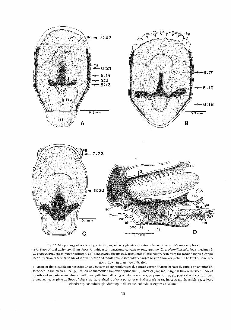
Fig. !2. Morphology of oral cavity, anterior J - iav/, salivary a* .lands and subradular sac in recent Monop!acophora. A-C, floor of oral cavity seen from above. Graphic reconstructions. A, Vema ewingi, specimen 2. B, Neopilina galatheae, specimen 1. C , Vema ewingi, the minute specimen 3. D, Vema ewingi, specimen 2. Right half of oral region, seen from the median plane. Graphic reconstruction. The relative site of radula sheath and radula vesicle somewhat changed to give a simpler picture. The level of some sec-
tions shown in plates are indicated. al, anterior lip; c, cuticle on posterior lip and bottom of subradular sac; cj, pointed corner of anterior jaw; cl, cuticle on anterior lip, sectioned in the median line; ge, section of subradular glandular epithelium; j, anterior jaw; mf, marginal furrow between floor of mouth and subradular membrane, with thin epithelium allowing radula movements; pl, posterior lip; po, postoral tentacle tuft; poc, preoral cuticular plate on floor of pharynx; rss, retained roof over posterior end of subradular sac in A; rv, radula vesicle; sg, salivary
glands; srg, subradular glandular epithelium; sro, subradular organ; ve, velum.
delimites the mouth anteriorly (Fig. 12: D). As described in the original paper from 1959, the an- terior lip is crescent-shaped and separated from the posterior lip by a cleft on each side, through which the mouth cavity communicates with the 'Yeeding furrow" (l.c., figs 66,67,71). The free ventral margin of the lip is only covered by thin cuticle and is wrinkled transversely in the preserved specimens.
The jaw is developed as a shelflike massive thick- ening of the cuticle on the posterior (oral) surface of the anterior lip (Fig. 12D, P1. 8:19, 21). It protrudes into the anterior part of the oral cavity with a distinct concave cutting edge and ends laterally with a point- ed corner in contact with the free lateral corner of the lip. The jaw is obviously fairly rigid, for in fixed specimens it has none of the small transverse wrinkles which characterize the free margin of the lip, where the cuticle is thin (PI: 6).
On the other hand, the jaw can clearly be bent in the middle, forming an anteriorly directed angle, for the specimens differ strikingly with regard to the relative direction of the lateral arms of the jaws. In the small Vema (specimen 3) and the original Neopi- lina (specimen 111) the angle is open so the jaw is crescent-shaped. In the new specimens 1 and 2 (Ne- opilina and Vema, resp.) the angle is much smaller, so the two arms of the jaw are nearly parallel, almost occluding the anterior part of the mouth opening (Fig. 12A-C).
At the anterior angle the anterior jaw is continous with the fairly thick cuticular plate which extends anterodorsally and covers the bottom of the anteri- orly directed part of the pharynx (Fig. 12, P1.6). This plate is touched by the radula ribbon, which is pre- sumed to move forwards and backwards in contact with the corresponding dorsal wall of the pharynx (Fig. 12D).
Histologically the anterior jaw is continuous with the thinner cuticle which covers the free margin of the anterior lip and the bottom of the pharynx. The transition from thin cuticle to jaw structure is so sud- den that the outlines of the jaw are clearly demarcated.
TL- &I-:- ,..+:A ,.CAI- :,.,, ,,, l.., ,a,, +,. r.,.-'.;,+ 1 IIG r l l l L k LuLlC!2 "1 ~ 1 1 2 J a w C.411 UC. JbC11 LV CVIIJIDL
of densely packed, parallel fibers or prisms which are roughly perpendicular to the underlying epithe- lium. This striation can be seen in the light micro- scope at higher magnification.
The total thickness of the jaw from the free cutting edge to the basal epithelium varies with the size of the specimens: 30 pm in the minute specimen 3, 80-100 pm in specimens 1 and 2, and probably more
than 150 pm in the large specimen 111 (which is difficult to measure precisely).
As described earlier (L. & W. 1959a, p. 24) the mouth is delimited posteriorly by a transverse wall (posterior lip) formed by median fusion of the two postoral tentacular ridges. This transverse wall is generally covered by a thin cuticle more laterally. In the large original specimens a median plate with somewhat thickened cuticle protrudes from the an- terior (oral) surface of the posterior lip like an irregu- lar shelf. This was called "lower jaw" in the original description, but after having seen the thin cuticle and the wrinkled appearance of this structure in the new specimens I realized that the term is an over- statement. There is at any rate no clearly demarcated postoral jaw with constant shape in the new speci- mens. However, it could be verified that the cuticle of the posterior lip continues as a thin cuticular plate over the central parts of the bottom of the subradu- lar sgc (Pl. 6:18, Fig. 12).
Comments : It appears to me that the anterior jaw of Monoplacophora, described above, is homo- logous with the upper jaw of the gastropods. It is developed by strengthening of the cuticle of an an- terior lip and consequently situated in the (morpho- logically) anterior wall of the oral cavity, opposite to the radula ribbon. In most gastropods the jaw is paired, and consists of two rodlike structures, which meet with their anterior or anterodorsal ends (Hy- man 1967, p. 211). In other gastropods the two jaws have fused medially as in the Monoplacophora. Ac- cording to Fretter & Graham (1962, p. 168), Star- miihlner (1952) and Nisbeth (1953) the most important role of the jaw in some gastropods is to work together with the radula and to guide and manipulate the radula ribbon. This may well be true in the Monoplacophora also, for in some specimens the radula has remained close to the jaw, and the radula teeth sometimes touch the jaw and the cuticu- lar plate in the bottom of the anterior pharynx (Pl. 6:20, 21).
That the jaw in gastropods as well as Mono- p!acephera cacsists cf packed chiticeus reds may be taken as an additional structural criterion for ho- mology. However, if this is a general way of pro- ducing thick cuticle in molluscs I feel that this support for homology has little weight.
In the Polyplacophora there is generally a cuticle lining the mouth opening and the oral cavity all the way round, and cuticular plates extend into the bot- tom of the pharynx and into the bottom of the
subradular sac. However, no discrete jaws have been found (Plate 1897, p. 19, Fretter 1937, pp 123-125, my own material of Lepidopleurus, Lepidochiton and Schizoplax).
Homologous anterior jaws thus appear to be pre- sent in the Monoplacophora, the Gastropoda and the Scaphopoda (Simroth 1892-94), possibly also the Cephalopoda (the "upper" jaw). I therefore sug- gest that the anterior jaw (or jaws) be listed among the synapomorphies of the Conchifera.
4.10.3. The subradu la r sac a n d t h e subradu la r glands The new material allows some more details to be studied in the subradular complex, partly because of better fixation, partly because individual variation can be better checked.
The subradular sac of the recent Monoplacophora is a flat unpaired diverticulum of the oral cavity, ex- tending backwards below the radula sheath (L. & W. 1959a, fig. 91). In the original specimen 111 the sac is strongly compressed and wrinkled. The new speci- mens give a more reliable picture, and the fact that the different specimens show varying states of pro- traction of the subradular organ indicates that it can function as in chitons (cf. Heath 1903): it can be pro- tracted and exposed in the oral cavity each time the radula ribbon is retracted.
In specimen 1 (Neopilina) the subradular organ and ganglion is somewhat retracted but still at the posterior level of the oral cavity (Fig. 3). In specimen 2 (Vema) the organ and the ganglion are strongly retracted, far behind the level of the mouth (Figs 4, 12D), and the small specimen 3 of Vema has a com- pletely protracted organ, stretched out in the roof of the oral cavity.
In all the new specimens the subradular organ is a cushion of high epithelium formed by the roof of the subradular sac. The lumen of the sac extends under this cushion as a narrow cleft and ends in the posterior blind end of the sac (Fig. 12D). The organ itself looks strictly unpaired in cross sections, not divided into two cushions as described in some Poly- plrcopfiora (?!ate 1897, 1894; FTa!!er l883).
Particular attention was devoted to glandular structures in the subradular sac because of their sug- gested homologies with subradular glandular struc- tures in different Conchifera, Polyplacophora and Aplacophora (Salvini-Plawen 1972, p. 277). The subradular sac has therefore been searched for the presence of glandular epithelia.
No distinct glandular epithelia could be identified
in the roof of the subradular sac. The epithelium which connects the subradular organ with the epithelium supporting the posterior end of the subradular membrane is thin, in some places mo- derately thick, but does not look clearly glandular.
Areas with pronounced glandular epithelium are, however, found in the floor of the subradular sac. Just behind the posterior lip the central parts of this floor are covered by the cuticular plate which extends from this lip. The same is found in the Polypla- cophora (Plate 1899, figs 172, 173). On each side of this cuticular plate there is a strip of high glandular epithelium (PI. 6:18). More posteriorly where the cuticular plate ends the two glandular strips fuse and cover the entire floor of the sac (fig. 12). The glandu- lar epithelium also extends into the blind posterior end of the sac and lines the cleftlike lumen there.
Thus, with the exception of the cuticularized plate in the anterior part of the subradular sac, the entire floor is glandular. This was easily observed because of the characteristic appearance of the glandular cells in all three specimens (1,2 and 3). The cells look like mucous cells, but this could not be verified histochemically for the 30-50 pm sections of speci- mens 1 and 2 did not ailow specific methods.
Comments: The distribution of glandular epithelium in the Monoplacophora appears to be almost identical with that found in the subradular sac of the Polyplacophora. In both the glandular epithelia cover the floor and the posterior end, but in the Polyplacophora the blind end may be produced into paired glandular branches or complicated in other ways (Plate 1897, fig. 17; 1899, figs 173-174; 1901, fig. 346). The situation of the subradular organ and of the cuticular plate adds to the similarity of this complex in the Mono- and Polyplacophora.
4.10.4. The sal ivary glands L. & W. (1959a, p. 26) reported the salivary gland to be unpaired in Neopilina galatheae. The new and better preserved material of Neopilina (specimen 1) confirms this description but specimens 2 and 3 rl~a'lr, ~h,,", that r/emu" has ..,.;,,A ,,I: ..-..-, ..l,-A- v ~ r 1 1 ~ o ~ x v v r ~ 1 1 pallbu J a l l V a l y glallU3,
more comparable to those of the Polyplacophora. In all specimens at hand there are salivary glands
developed from the anterior (dorsal) wall of the pharynx, at the level where the first, anteriorly directed pharyngeal part bends dorsally into the ver- tical part. The glands are situated in front of the margin of the cuticular plate which covers the pharyngeal bottom in front of the jaw (Fig. 12D).

Specimen 1 (N. galatheae) looks very like the 4.11. The stomach, the liver and original large specimens of this species (L. & W. the crystalline style 1959a, figs 81,124). The anterior wall of the pharynx
The orginal description of the stomach and its ad- in front of the cuticular plate is a broad transverse
nexa was somewhat uncertain, for the material was zone of glandular epithelium, reaching from one
deformed and partly disintegrated by crushing and side to the other across the midline. This glandular
autodigestion (L. & W. 1959a, pp 29-30). The new zone is folded into numerous deep parallel wrinkles (Fig. 12B).
material is excellently preserved in these parts and al- lows some more precise descriptions.
It was surprising to find that the specimens of V: ewingi are completely different, having distinctly paired salivary glands in the same zone. In the small specimen 3 of Yema there are two globular glands, evaginated symmetrically on each side of the mid- line. The broad median wall between the two glandu- lar complexes does not look glandular at all (Fig. 12C, P1. 7:23).
The larger specimen (2) of Vema also has two com- plexes of glands, each consisting of 4-5 globular diverticula. The broad zone between the two groups of globular glands has a somewhat thickened epithelium but no diverticula of any kind. In each group are one or two particularly large diverticula, perhaps corresponding to the two which are present in the small specimen (Fig. 12A, P1. 7:22).
Comments: The salivary glands of Vema are thus clearly homologous with the salivary glands or "buccal glands" of the Polyplacophora (see Plate 1897, figs 22,23,25, and Fretter 1937). In both cases they are paired structures, situated in front of the cuticular plate in a comparable and clearly homo- logous pharyngeal region. The most obvious differ- ence is that the salivary glands of the Polypla- cophora form the extreme ends of ciliated grooves, one on each side, which arise high up on the pharyn- geal wall and pass down to the salivary gland on the same side.
The glands present in N. galatheae develop in the same zone and appear homologous with those of Vema and the Polyplacophora but are intercon- nected across the midline to form an unpaired com- plex. It is also possible that the dorsal pharyngeal glands of some gastropods and cephalopods, Sole- nagastres ar;d C - . . A n f n ~ 7 a L ~ U U V I V V L ~ ~ ~ Ln-nInrnn~~~ L L u L z L ~ ~ u 5 u u ~ x A L L A X ,;th
these monoplacophoran salivary glands, but the criteria for homology are not equally strong because the location of the glands is less defined and some- what different in the fairly variable pharynx (cf. Salvini-Plawen 1972, pp 244, 279).
4.11.1. The s tomach was defined as the inflated part of the alimentary canal which is lined by the high prismatic gastric epithelium. Its shape in the "Galathea" animals was described as triangular when seen from above. This holds fairly good also for the new material, for in N. galatheae (1) and I.: ewingi (2) it is pointed anteriorly and has a broad posterior contour (Figs 13, 14). The shape is certain- ly somewhat variable depending on the degree of fill- ing. The oesophagus joins the stomach from an anterodorsal direction, above the attachment of the liver. It can be followed for some distance as a fold or ridge on the roof of the stomach. This could not be distinctly seen in the old material which was badly damaged in this region.
In the new material the stomach proper is nearly symmetrical but the intestine has an asymmetrical origin to the left of the midline from the ven- trolateral wall (Figs 13, 14).
In the new specimen of N. galatheae (1) there is a voluminous and fairly well delimited style sac, evagi- nated from the dorsal part of the hind wall of the stomach. It contains what is believed to be a primi- tive crystalline style. In the original material (speci- men 111) such a sac was present but appeared to be approximately median, above the origin of the in- testine. This certainly depended on artificial disloca- tions, for in the new and practically intact specimen (1) the style sac is clearly to the right of the midline and is clearly separated from the intestine on the left side (Fig. 13).
In I.: ewingi (specimen 2) the style sac is not clearly delimited, but there is a shallow, approximately me- dian evagination containing what seems to be a primitive style (Fig. 14). A small fold seen on the dor- sal surface of this region may be a doubled part of the empty style sac but could also be a simple ar- tifact.
4.11.2. The l iver , attached to the stomach, is strongly lobulated in its peripheral parts. Its walls are characterized by the very high epithelial cells

with spherical secretion granules, described by L. & W. (1959a, p. 30, figs 107, 108). The communication between the liver and the stomach was originally described as "a long slit-like opening", which is cor- rect, but several important features passed unno- ticed in the badly preserved original material (1. c., figs 8, 108).
The new specimens 1 and 2 are practically intact in the liver region. They show clearly the following features:
1. The liver is, strictly speaking, unpaired. The liver complex of the right side is continous with that of the left, connected by an isthmus following the anterior contour of the stomach, below the entrance of the oesophagus. The liver has an extensive zone of contact with the anterolateral margins of the stomach, and the contact is uninterrupted anteriorly where the isthmus is attached to the anterior stomach wall (Figs 13, 14, PI. 7:24).
2. Communication between the liver and the stomach is established by a long, cleftlike opening which extends from the anterolateral margin of the right side, below the entrance of the oesophagus to the anterolateral margin of the left side. The epithe- lium suddenly changes from the gastric type to the
hepatic type, both in the dorsal and ventral walls of the cleft (PI. 7:25).
3. The proximal parts of the liver, attached to the stomach, contain a continuous open lumen, which communicates with the stomach through the cleft. This vestibulum-like proximal lumen is continuous all the way round the anterior half of the stomach. No lobules start directly from the stomach or from the adjacent walls of the vestibulum. The numerous liver lobules grow out from the more peripheral walls of the vestibulum (Fig. 13B).
The sharp limit between gastric and hepatic epithelia excludes confusion of liver parts and of the stomach. Moreover, a furrow on the ventral surface marks the transition from stomach to liver, follow- ing the level of the communicating cleft (Fig. 13B).
Suspecting that the embryonic liver rudiment is paired as in the Polyplacophora 1 carefully checked the "isthmic" zone for signs of this, both in Vema and Neopilina, but found none. The lumen of the vestibulum can be followed from left to right side through the isthmic region, and the cleftlike commu- nication with the stomach passes over the midline without interruption. This can even be seen in single sections (Pl. 7:24). In specimen 1 (Neopilina) two of
Fig. 13. Neopiiina galatheae, specimen 1. Pharynx, oesophagus, stomach, liver and anterior intestine. Graphic reconstruction. Level of sections shown in plates are indicated.
A, dorsal view. B, ventral view. cr, crystalline style; i, intestine; ili, isthmus between right and left liver; li, liver; oe, oesophagus; ph, pharynx; phd; opening into pharynx of pharyngeal diverticula.

the medial lobules are separated by a fairly deep cleft, but this does not cut through to the stomach, so the vestibulum is uninterrupted medially (Fig. 13B). In specimen 2 (Vema) there are several shallow interlobular clefts in the same region but no inter- ruption of the isthmus.
C o m m e n t s : The liver of the Monoplacophora as found in Vema and Neopilina is unpaired but bi- lobed, actually appearing as a broad and flattened pouch from the whole anterior margin of the stomach, and the broad communication between the two organs is not constricted to form a "liver duct" but remains broad. The liver is - as far as can be seen, symmetrical.
The liver of the Polyplacophora, as described by Haller (1882), Plate (1901, pp 439-444) and Fretter (1937) is different in that it is clearly paired. In the adult the right and left liver parts are strongly asym- metrical with regard to size and localization, and their communication with the stomach is asym- metrical (or fused) and constricted to narrow pores.
A symmetrical pair of liver rudiments is present in early ontogenetical stages of Acanthochiton (Ham- marsten & Runnstrom 1925) and young Zschno- chiton (Plate 1901). If embryonic Monoplacophora have paired, pouchlike liver rudiments like the Poly- placophora, one must assume that these rudiments fuse early across the midline below the oesophagus.
4.11.3. The crystal l ine style was originally de- scribed on the basis of material from a single speci- men (L. & W. 1959a, pp 29-30), as the second specimen available at that time for some reason failed to show it. I have paid much attention to this structure in the new material for two reasons: 1) None of the specimens examined possesses a cuticu- lar gastric shield or similar structure, although such a shield is correlated with the presence of a crystal- line style in lamellibranchs and gastropos. 2) Gra- ham (1959) suggested that the style originally described could in fact be a faecal string, for such strings can look like crystalline styles and are present in the same region of many primitive gastropods.
Fig. 14. Pharynx, oesophagus, stomach, and intestine. Dorsal view, reconstructed graphically, to show communication between liver and stomach. The liver is removed.
A, Neopilina galatheae, specimen 1. B. Vema ewingi, specimen 2. c, the dotted line shows approximately the constriction of lumen between stomach and liver; cr, crystalline style; ge, gastric epithelium extending into the roof of the cleft between liver and stomach; i, intestine; lia, cleftlike attachment of liver along the antero-lateral aspects of the stomach, below the oesophagus; oe, oesophagus; ph, pharynx, phd, pharyngeal diverticula; v, vestibulum-like open lu-
men of liver communicating with the stomach; x, dorsal fold of stomach, artificial or remnant of top of style pouch.

None of the new specimens exhibits a crystalline style of the very distinct, strongly stainable type seen in the original specimen 111. But a probably equiva- lent conelike body is present both in N. galatheae (1) and I.: ewingi (2). The outlines and situations of the styles are shown in Figs 13 and 14. The styles of both new specimens have the appearance of somewhat irregular cones; that of specimen 1 distinctly has its narrow posterior end in the presumed style sac. The style of specimen 2 has z similar situation but the style sac is not clearly delimited. Cross sections show an indistinct concentric or spiralized internal struc- ture (Pls 7:26 and 27).
As in the original specimen 111, the style in each of the new specimens follows the roof of the stomach from the style sac in an anterior direction, but it be- comes indistinct and disappears before reaching the level of the oesophagus. The anterior parts contain some food particles embedded between the layers of mucuslike matter.
The style is undoubtedly identical in the original specimen I11 and the two new specimens 1 and 2. The weaker staining in the new specimens can well be a consequence of the different staining techniques: Mallory's phosphotungstic acid hematoxylin and Friedlander-Ehrlich's alum hematoxylin, resp. The style of the new specimens is clearly separated from the exit of the intestine, which is on the left side, whereas the style sac with the style is middorsal or over on the right side (Fig. 14). The "style" can there- fore not be a faecal string (cf. Graham 1959).
The anterior disintegrating part of the style is just behind the entrance of the oesophagus, as would be expected if it were a condensed mucus string from the oesophagus. But this does not explain why all styles seen up to now clearly disintegrate in their an- terior ends, or why they are more solid and contain no food particles in their posterior end. The possibil- ity that the style comes from the oesophagus is also contradicted by the fact that new mucus cornets seem to be added from behind in the strongly stained original specimen 111 (L. & W. 1959a, p. 29).
Studies of the well preserved gastric epithelium in the new specimens confirm that there is no gastric shield or comparable structure. The dorsal gastric wall, along which the style extends, has a particular- ly strong ciliation. Specimen 1 (Neopilina) which is particularly well fixed, seems to show that strong secretion is going on from the style sac epithelium at the level of the posterior end of the style. The peripheral ends of the cells are more stainable than in other parts of the stomach, and their distal ends
bulge as if droplets were about to be detached. This could indicate that the mucuslike style material is secreted in the posterior part of the style sac as ex- pected.
Comments : A mucoid body similar to the sup- posed crystalline style of the old specimen 111 was found also in the new specimens, situated with its posterior end in what seems to be a style sac (in spe- cimen 1). The new well-preserved specimens show definitely that the cone-shaped mucus body is not a faecal string and make its interpretation as a food string from the oesophagus highly improbable. It is therefore probable that it is a true crystalline style, homologous with that of lamellibranchs and some gastropods. However, the absence of a gastric shield and the fact that the style is very short and irregular makes it look less perfected than the style apparatus of lamellibranchs and gastropods. Perhaps it is a primitive issue of a homologous enzyme-secreting apparatus.
4.12. The radula apparatus
The material for the original description of the radu- la apparatus in the Monoplacophora was deficient, particularly with regard to the radula vesicles, which had exploded and collapsed (L. & W. 1959a, pp 25-29, 39-42). Nevertheless the original description proved essentially correct when compared with the three new specimens, which have intact radula vesicles and well-preserved radula cartilages, radula sheaths and muscles (Pls 2, 5, 6).
The radula apparatus of Neopilina is identical in all essential respects with that of Vema, so I have found it unnecessary to r,?peat the verbal description for the latter species. Instead I present a series of figures showing the radula apparatus of Venza, with short comments and references to the original description of Neopilina. The new figures of Vema mostly show the radula apparatus in side view and supplement the original figures, which show dorsal views. Together the new and the original figures should give a better picture of the three dimer?sionc.
4.12.1. The radula suppor t ("odontophore" , "tongue") In all three new specimens the radula vesicles are elongate, ovoid bodies with converging anterior ends (Fig. 15A, B). Their walls consist of thin parallel lamellae, 7-9 in the larger specimens 1 and 2, but only 4 lamellae in the small specimen 3. Each of

rv
1 rnrn
A.vema B. Neopilina
w I rnrn 1 rnrn
C . ~ c h i z o p l a x D. Lepidopleurus
Fig. 15. Radula vesicles, radula cartilages and ventral approximator muscle (m. radulae impar) in A, Vema ewingi, specimen 2, B, Neopi- lina galatheae, specimen 1, C , Schizoplax sp., and D, Lepidopleurus asellus. Graphic reconstructions, seen from the ventral side.
lc, lateral cartilage; mc, medial cartilage; rv, radula vesicle; va, ventral approximator muscle (m. radulae impar); x, small block of car- tilage, partly separate from the medial cartilage.
these lamellae is about 1 pm thick, without apparent structure in the light microscope, and stains lightly with hematoxylin (PI. 8:28). A few scattered cell bodies with nuclei attach to the surface of the lamel- lae, which are densely packed in some parts of the wall but more loosely arranged with large inter- spaces in others. The latter state may be an artifact. No distinct epithelium is seen lining the lumen of the vesicles, but several small nuclei attached to the in- ~lermcxt !2me!!1e indicate that there is 2 very thin cel- lular sheath, although the plasm is invisibel with the staining used (hematoxylin) (PI. 8: 28).
The anterior ends of the radula vesicles are partly covered by vesicular tissue resembling the "pseudo- cartilage" of mollusc odontophores. In all examined specimens of Monoplacophora this pseudocartilage is restricted to the anterior third of the radula vesicles and does not extend to the posterior ends as in the
Polyplacophora (Fig. 15A, B). The morphology of these cartilages could be analyzed in the new speci- mens (1, 2 and 3) but was so badly preserved in the "Galathea" specimens that the original description could not include any details.
Each radula vesicle is associated with a medial and a lateral cartilage (Fig. 15, PI. 6). Both are flattened, bandlike structures closely attached to the surface of the vesicle. The two cartilages meet at the anterior point of the vesic!~, and in that region a sma!! block is partly detached from the medial cartilage (Pl. 6:21). Near their margins the cartilages intermingle with some of the superficial lamellae of the wall, which spread into the vesicular tissue of the pseu- docartilage (Pl. 8:28).
The radula vesicles are quite regular, oval, and seem to be completely empty in the sections. Like the homologous vesicles of chitons they probably con-

tain some fluid in the living animal and act as a kind of rigid skeleton: the turgor pressure of the contents making them hard (see L. & W. 1959a, p. 28).
The medial cartilages of left and right radula vesicles are interconnected over the midline of the animal by a short and strong muscle: musculus radulae impar (Fig. 15; Pi. 6:17). This muscle, the radula cartilages and the anteromedial ends of the radula vesicles form a transverse bar, over which the radula ribbon can slide. The bar is located in the angle between the radula sheath and the ascending pharynx, just above the anterior part of the oral cavi- ty (Fig. 16).
Comments : The homology of the radula vesicles of the Mono- and Polyplacophora was suggested by L. & W. (1959a, p. 31) and seems to have been ac- cepted. This similarity can now be extended to the entire radula support, for the medial and lateral car-
tilages of the Monoplacophora have their counter- parts, in corresponding locations, in the Polypla- cophora (Fig. 15; Pls 6, 8). The only significant difference is that these cartilages are larger in the ex- amined Polyplacophora. In chitons the lateral car- tilage even extends to the posterior end of the vesicle, which is covered by a cup-shaped layer of cartilage (Fig. 15C, D). This cartilaginous cup serves as origin of several important radula muscles, which appear to attach directly to the connective tissue of the vesi- cle in the Monoplacophora.
Except for the Polyplacophora, such a radula sup- port with radula vesicles does not seem to have been reported from other molluscs. In searching for a homologous structure I paid particular attention to the radula of the docoglossan prosobranchs, which is similar to that of Mono- and Polyplacophora in several respects: docoglossan type of dentition, presence of comb-shaped teeth in the Lepetidae
Fig. 16. Vernaewingi, specimen 2. Right half of anterior body, seen from themedian section, to show the principal structure of the radula apparaks and the medial!y si:ua:ed radula iiiiiscles. Based on graphic recoiistmciion, except for the sheii which is added arbitrarily (partly destroyed by decalcification). The pharynx and radula sheath are elevated and separated in relation to the raw reconstruction,
to give more space and avoid mutual covering of muscles and other structures. aj, anterior jaw; mc, medial cartilage; mf, marginal furrow with thin walls along margin of subradular membrane; nl, nacreous layer of shell; p, protoconch scar; ph, pharynx; phd, opening of pharyngeal diverticula into the pharynx; pl, prismatic layer of shell; rd, inner end of radula diverticula; rs, radula sheath; rv, radula vesicle; sg, salivary gland. -Muscles (numbers refer to text): Ia, m. retractor radulae; Ib, m. protractor vesicae major; If, m. vesicae postero-lateralis; Ig, m. vesicae medialis; IIb, "m. protractor radulae"; IId-d, m. radulae longus pars dorsalis; Ild-v, m. radulae longus pars ventralis; IIe, m. pharyngeus marginalis; IIIa, m. radulae impar (= ventral approximator); IIIb, m. protractor cartilaginis profundus; IVg, m. protractor diverticulorum; x, branches of muscle IVg on the dorsal
side of the radula diverticula, equivalent to Plate's muscle 4 in the Polyplacophora.
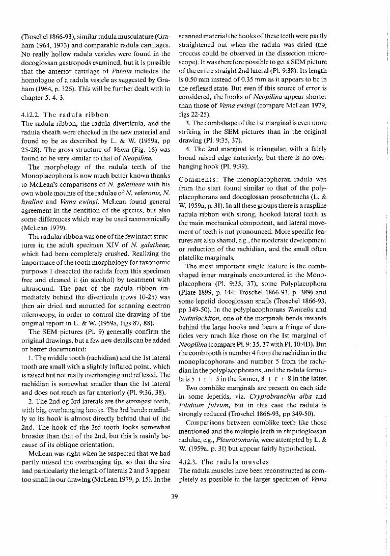
(Troschel1866-93), similar radula musculature (Gra- ham 1964, 1973) and comparable radula cartilages. No really hollow radula vesicles were found in the docoglossan gastropods examined, but it is possible that the anterior cartilage of Patella includes the homologue of a radula vesicle as suggested by Gra- ham (1964, p. 326). This will be further dealt with in chapter 5. 4. 3.
4.12.2. The radu la r ibbon The radula ribbon, the radula diverticula, and the radula sheath were checked in the new material and found to be as described by L. & W. (1959a, pp 25-28). The gross structure of Vema (Fig. 16) was found to be very similar to that of Neopilina.
The morphology of the radula teeth of the Monoplacophora is now much better known thanks to McLean's comparisons of N. galatheae with his own whole mounts of the radulae of N. veleronis, N. hyalina and Vema ewingi. McLean found general agreement in the dentition of the species, but also some differences which may be used taxonomically (McLean 1979).
The radular ribbon was one of the few intact struc- tures in the adult specimen XIV of N. galatheae, which had been completely crushed. Realizing the importance of the tooth morphology for taxonomic purposes I dissected the radula from this specimen free and cleaned it (in alcohol) by treatment with ultrasound. The part of the radula ribbon im- mediately behind the diverticula (rows 10-25) was then air dried and mounted for scanning electron microscopy, in order to control the drawing of the original report in L. & W. (1959a, figs 87, 88).
The SEM pictures (Pl. 9) generally confirm the original drawings, but a few new details can be added or better documented:
1. The middle tooth (rachidian) and the 1st lateral tooth are small with a slightly inflated point, which is raised but not really overhanging and reflexed. The rachidian is somewhat smaller than the 1st lateral and does not reach as far anteriorly (Pl. 9:36, 38).
2. The 2nd og 3rd laterals are the strongest teeth, with big, ~verhanging hacks. The 3rd be:ds mediz!- ly so its hook is almost directly behind that of the 2nd. The hook of the 3rd tooth looks somewhat broader than that of the 2nd, but this is mainly be- cause of its oblique orientation.
McLean was right when he suspected that we had
scanned material the hooks of these teeth were partly straightened out when the radula was dried (the process could be observed in the dissection micro- scope). It was therefore possible to get a SEM picture of the entire straight 2nd lateral (Pl. 9:38). Its length is 0.50 mm instead of 0.35 mm as it appears to be in the reflexed state. But even if this source of error is considered, the hooks of Neopilina appear shorter than those of Vema ewingi (compare McLean 1979, figs 22-25).
3. The combshape of the 1st marginal is even more striking in the SEM pictures than in the original drawing (Pl. 9:35, 37).
4. The 2nd marginal is triangular, with a fairly broad raised edge anteriorly, but there is no over- hanging hook (Pl. 9:39).
Comments : The monoplacophoran radula was from the start found similar to that of the poly- placophorans and docoglossan prosobranchs (L. & W. 1959a, p. 31). In all these groups there is a rasplike radula ribbon with strong, hooked lateral teeth as the main mechanical component, and lateral move- ment of teeth is not pronounced. More specific fea- tures are also shared, e.g., the moderate development or reduction of the rachidian, and the small often platelike marginals.
The most important single feature is the comb- shaped inner marginals encountered in the Mono- placophora (Pl. 9:35, 37), some Polyplacophora (Plate 1899, p. 144: Troschel 1866-93, p. 389) and some lepetid docoglossan snails (Troschel 1866-93, pp 349-50). In the polyplacophorans Tonicella and Nuttalochiton, one of the marginals bends inwards behind the large hooks and bears a fringe of den- ticles very much like those on the 1st marginal of Neopilina (compare PI. 9: 35,37 with P1. 10:4D). But the comb tooth is number 4 from the rachidian in the monoplacophorans and number 5 from the rachi- dian in the polyplacophorans, and the radula formu- la is 5 + r + 5 in the former, 8 + r + 8 in the latter.
Two comblike marginals are present on each side in some lepetids, viz. Cryptobranchia alba and Pilidium fulvum, but in this case the radula is str~ngly rebuceb (Trosche! !866-93, pp 343-50).
Comparisons between comblike teeth like those mentioned and the multiple teeth in rhipidoglossan radulae, e.g., Pleurotomaria, were attempted by L. & W. (1959a, p. 31) but appear fairly hypothetical.
partly missed the overhanging tip, so that the size 4.12.3. The radula muscles and particularly the length of laterals 2 and 3 appear The radula muscles have been reconstructed as com- too small in our drawing (McLean 1979, p. 15). In the pletely as possible in the larger specimen of Gma

ewingi (specimen 2), and nearly complete identity was found with the radula muscles of Neopilina galatheae. Also the minute specimen 3 of Vema was reconstructed, and all the important muscles were re- found. Only some of the smallest muscles were missed - as could be expected in the small (1.8 mm) specimen.
In the following text the radula muscles of Vema are listed with short comments and reference to the drawings (Figs 16, 17, 18), and to the description of the muscles in Neopilina (L. & W. 1959a, pp 39-42). The nomenclature and the abbreviations are the same as in the original description so that the two species can be compared directly. The drawings are all based on lateral reconstructions and are intended to supplement the original description, which was based on dorso-ventral reconstructions only and, probably therefore, has been misunderstood on some points.
The designations of the muscles in the figures in the present paper refer to their place in the text. The muscle Ia is thus m. retractor radulae and is found under the head I, muscle IIc is m. tensor radulae and found under the head 11.
I. Muscles inserting on the posterior tip of the radula vesicles (Figs 16, 17, 18)
Ia) m. retractor radulae (m. re. ra.) - Figs 16, 17 Ib) m. protractor vesicae major (m. pro. ve. ma.) -
Fig. 18 Ic) m. protractor vesicae minor (m. pro. ve. mi.) -
Figs 17, 18 Id) m. vesicae anterolateralis (m. ve. a-1.) - Figs 17,
18 Ie) m. vesicae anteromedialis (m. ve. a.-m.) - Not
found in Vema If) m. vesicae posterolateralis (m. ve. p-1.) - Figs
17, 18 Ig) m. vesicae posteromedialis (m. ve. p-m.) - Figs
16, 17, 18 Ih) m. vesicae ventralis (m. ve. v.) - Figs 17, 18. -
With the single exception of muscle Ie, all these muscles were identified both in Vema and Neopilina and have a very similar course in the two species.
Muscle Ia, m. retractor radulae, inserts on the tip of the radula vesicle and follows a somewhat coiled course, passing as a flat membrane over the medial and lateral surface of the radula vesicle to the posterior margin of the radula diverticulum of the same side. It appears to be the most powerful retrac- tor of the subradular membrane, homologous with that of the Polyplacophora and some gastropods (cf. Graham 1973).
The other muscles of the group appear to manipu-
Fig. 17. Vema ewingi, specimen 2. Pharynx and radula apparatus, including radula vesicles, seen from the left. Some radula muscles of the left side are shown. The pharyngeal diverticula are removed but their opening into the pharynx is shown (phd).
Graphic reconstruction with radula sheath in situ. Muscles: Ia, m. retractor radulae; Ib, m. protractor vesicae major; Ic, m. protractor vesicae minor; Id, m. vesicae anterolateralis; If, m. vesicae posterolateralis; Ig, m. vesicae posteromedialis; Ih, m. vesicae ventralis, IIc, m. tensor radulae; IId, m. radulae longus; IIe, m. pharyngeus marginalis; IIIb, m. protractor cartilaginis profundus; IVg, m. protractor
diverticulorum; x, branches of IVg on the dorsal side of the radula diverticula.

late the tip of the radula vesicles (Id-Ih) and pull the margin of the subradular membrane and thus clearly vesicle forwards (Ib-Ic). Together these muscles seem belong to the series of heads of the m. protractor to be able to control the angle between the radula radulae. In the original Neopilina material these two vesicles and to control the inclination of the odonto- heads were believed to pass to the surface of the car- phore as a whole. tilage and were called "m. protractor cartilaginis
dorsalis". This was clearly a mistake caused by poor 11. Muscles of the subradular membrane preservation of the specimen. (Figs 16, 17, 18) The most remarkable of these muscles is the IIb
IIa) m. retractor radulae (= Ia) mentioned above IIb) m. protractor radulae (m. pro. ra.) - Fig. 18.
Includes many heads on the shell, also two heads which were believed to end on the radula cartilage of Neopilina and were called m. pro- tractor cartilaginis dorsalis. This was clearly wrong (see below!)
IIc) m. tensor radulae (m. te.ra.) - Figs 17, 18 IId) m. radulae longus (m. ra. 1.) - Fig. 16 IIe) m. pharyngeus marginalis (m. ph. m.) - Figs
16, 17, 18.
All these muscles were refound also in Vema ewingi, but a correction is necessary with regard to the two ventrolateral heads of muscle IIb (m. protractor radulae). In the well preserved material of Vema these two muscles can be seen to attach under the
complex, called m. protractor radulae, which seems to lack a homologue in gastropods and polyplaco- phorans. It may act as a protractor of the subradular membrane when the odontophore is in a high situa- tion as in Fig. 18 and the radula ribbon is retracted, whereas some of its bundles could retract the subradular membrane and the entire radula appara- tus when the odontophore is depressed and the subradular membrane is protracted. Only experi- ments can solve this problem, and this is certainly hampered by difficulties involved in getting living material of the largesized species. Muscle IIb is one of the largest muscles of the Monoplacophora.
Muscle IId, m. radulae longus, consists of two almost independent portions, which are partially fused halfway between the origin and the insertion (Fig. 16). The dorsal portion (m. radulae longus pars
Fig. 18. Vemaewingi, specimen 2. As Fig. 17, but practically all radula muscles seen from the left side are included. m, membrana latera- lis, a connective tissue membrane to which some muscles (IVc, IVd) attach; phd, opening of pharyngeal diverticula into the pharynx;
rd, radula diverticulum of left side. Muscles: Ib, m. protractor vesicae major; Ic, m. protractor vesicae minor; Id, m. vesicae anterolateralis; If, m. vesicae posterolateralis; Ig, m. vesicaeposteromedialis; Ih, m. vesicae ventralis; IIb, "m. protractor radulae"; IId, m. radulae longus; IIc, m. tensor radulae; IIe, m. pharyngeus marginalis; IIIb, m. protractor cartilaginis profundus; IIIc, m. cartilaginis anterolateralis; IVb, m. transversalis anterior; IVc, m. tensor membranae lateralis; IVd, m. tensor membranae anterior; IVg, m. protractor diverticulorum; x, branches of IVg, on the
dorsal side of the radula diverticula.

dorsalis, IId-d in the figures) is clearly a retractor of IV. Other muscles of the radula apparatus the radula ribbon, acting on the ventral side of the radula sheath just behind the radula diverticulum (Fig. 16, PI. 5). The ventral portion (IId-v in the figures) passes down to the roof of the oral cavity and is clearly a protractor of the subradular mem- brane; some of its small branches may also retract the subradular sac (Fig. 16). Muscle IId is by far the longest of the radula muscles. The two heads extend caudally to a level far behind the radula vesicles and attach to the dorsal shell medial to pedal retractors B and C (Figs 10,ll).
111. Muscles of the radula cartilages
IIIa) m. radulae impar (m. r. i.) - Figs 16, 15, PI. 6 IIIb) m. protractor cartilaginis profundus (m. pr.
ca. p.) - Fig. 18 IIIc) m. cartilaginis anterolateralis (m. ca. a-I.)
- Fig. 18. Is perhaps a levator more than a retractor
IIId) m. protractor subradularis (m. pr. sr.) - Not found in Vema. Only the small muscle IIId could not be identi- fied in Vema. The other three muscles are easily reconstructed in both genera.
IVa) m. radulae minor (m. ra. mi.). A small muscle, not clearly identified in Vema
IVb) m. transversalis anterior (m. tr. a.) - Fig. 18, PI. 6
IVc) m. tensor membranae lateralis (m. te. m. 1.) - Fig. 18
IVd) m. tensor membranae anterior (m. te. m. a.) - Fig. 18
IVe) m. transversalis posterior (m. tr. p.). Very small in Vema, not reconstructed.
IVf) m. pharyngei preorales (mm. pha.). Present as small strands from pharyngeal roof to anterior shell in Vema. Not shown in the figures.
IVg) m. protractor diverticulorum (m. pr. div.) - Fig. 17.
Muscles IVa-IVg are all small and seem to maintain the radula, the radula sheath, the larger muscle bundles, and pharynx in their proper positions. Muscle IVg is continuous dorsally with its con- tralateral partner, and with small longitudinal muscles on the dorsal side of the radula diverticula. These latter are marked x in Figs.16, 17 and 18 and correspond in part to Plate's muscle 4 (Plate 1897, p. 46, figs 20,25) in the Polyplacophora.
5. DISCUSSION
5.1. Morphological features of metamerism in recent Monoplacophora
The original description of metarism in recent Monoplacophora was founded on a very small material (L. & W. 1959a). Only one somewhat defec- tive specimen (1111, cut transversely, was available for graphic reconstructions, while another specimen (IV), cut horizontally, was used for wax-plate recon- struction as a control. Mistakes caused by deficient preservation and individual variation could there- fore hardly be excluded, and it was thus natural that the results were reluctantly accepted. Mnrcnver, the
that some organs are repeated following a common rhythm. This has to be accepted as a fact, and is essential for the characterization of recent Mo- noplacophora. Its theoretical interpretation - whether the metamerism here is related to the metamerism of articulates or not - is of course still open to discussion but, I hope, on a more stable basis.
5.1.1. In t roduc to ry r emarks The new material revealed some mistakes and mis- interpretations in the first descriptions, which must be considered in 2 ner.?7 disrursinn ef metzzerism.
violent theoretical criticism in many subsequent 1. The "dorsal coelomic sacs" of the original papers cast doubts and uncertainty as to the presence report turned out to be misinterpreted. They are of a metameric organization in these animals and the large pharyngeal diverticula, and they do not show way it was described. metameric subdivision. The latter question was left
Now that more material has accumulated, a open in the original report (op. cit. pp 55-57). general review of the metameric structure of these Whether or not metameric tendencies can be seen in animals seems useful. The new material leaves no the developing mesoderm of embryos and larvae is doubt about the presence of metamerism in the sense still unknown.

2. The "nephrostomes" described with some reservation in the original report are not present in the pre-pericardial region, but it is possible that there are connections between the nephridia and the pericardia in the sectors F and G (Chapter 4.7).
3. A coiled larval shell like that originally described in Neopilinagalatheae (Lemche 1957, L. & W. 1959a) is not present. The true larval shell as described by Menzies (1968) is a bulbous, bilaterally symmetrical structure with some exogastric coiling. This statement removed one of the most intriguing obstacles for understanding monoplacophoran morphology, for a bilateral metameric structure would not be expected in animals which have an asymmetrically coiled shell.
In other respects, e.g., with regard to repetition and spatial relations between muscles, nerves, nephridiopores, gills, atria, gonoducts, and gonads, the originally described metameric pattern of N. galatheae is confirmed in the new material. The
specimens of Vema ewingi show significant differ- ences, however; the species has a more complete metameric repetition of organs, and therefore con- firms the original views.
5.1.2. T h e case of Neopi l ina galatheae The new specimen 1 is immature but in general con- firms the original description. The metamerism of the musculature, nerve connectives, nephridiopores, nephridia, gills, gonads, and gonoducts is practically identical in the new specimen and in the old mature specimen I11 (compare Figs 3,8, 10 of the present ac- count with figs 121, 135, 145 and 146 in L. & W. 1959a).
Even certain irregularities have been refound in the new material. Pedal retractors A and B are closer together than the others, and the gill C appears an- teriorly dislocated in relation to pedal retractor C in the same way as in the old material (Fig. 3).
Comparison of the old material with the new
At ria
@ Vema o Neopilina
Table 1 The metameric repetition of organs in Neopilinagalatheae (open circles) and Vema ewingi (filled circles) plotted into the system of sec- tors in both animals. The sector limits are arbitrarily chosen as the lateropedal nerve connectives (compare Figs 3,4, and 8-11). Arrows
indicate organs not present in Neopilina.

specimen 1 thus indicates that the picture originally given for N. galatheae is typical of the species. In- dividual variation seems to be restricted. In the case of specimen 1 the observed variations are: unilateal duplicity of the head of retractor C on the left side, where it consists of two unequal portions, and a strong asymmetry of the hindmost retractor (H); the left one is much smaller than the right one (Fig. 3).
I have summarized the metameric pattern of N. galatheae in Table 1, using the previously introduced sector system as an arbitrary, descriptive basis. Readers finding this too crude and subjective can compare with the actual reconstructions (Figs 3, 8, 10).
I also maintain that the often used diagram in fig. 165 of our original monograph (1959a) fits in princi- ple with the new material and can be used for orien- tation, if it is remembered that the nephrostomes of kidneys C, D, and E are doubtful and should be re- moved. As indicated on p. 11 in the same mono- graph, diagrams like this are simplified to show as clearly as possible what is meant in the text, and readers must thus accept some loss of detail and ac- curacy of proportions. The legends of fig. 165 ex- pressly state that the figure is a diagram, so we did not expect readers to use it as a piece of documenta- tion for details. There are several figures in the same volume which show actual reconstructions for those readers who want precise information.
I was therefore astonished of the violence with which this diagram was criticized by Salvini-Plawen (1969b, p. 200). He calls it falsified ("ver- f a l sc h t " ), obviously meaning that the meta- merism shown is a result of wishful thinking by the authors. He correctly remarks that the 1st gill is posterior to its proper place (which is immediately in front of the level of the muscle C, not immediately behind this level as in the diagram). This happened when the organs were spread to avoid their covering each other, whereas other correct spatial relations should be maintained. The 1st gill is, for instance, correctly placed in the diagram with regard to the in- terpedal commissure, and it is associated with the 2nd nephropore as in the animal,
Salvini-Plawen's "correction" of the same dia- gram (1969b, p. 200) shows some of the difficulties
base of the 1st instead of the 2nd gill, and the 2nd nephridium therefore erroneously appears to be ly- ing in a hiatus of the gill series. In the animal each of the five gills has a nephridiopore at the base, and these nephridiopores belong to five consecutive kid- neys. This as well as other spatial relations can be seen in the reconstructions of the original report (figs 121, 130, 135, 137, 145) which are accessible to all readers.
Salvini-Plawen's "correction" certainly annihi- lates the spatial correspondance between the gill ser- ies and the nephridiopore series, and produces other irregularities which interfere with the metarism. I do not know what to call this kind of "correction", but if Salvini-Plawen has used this "corrected" diagram I can see why he fails to discover the spatial cor- respondance between the repeated series of organs.
I hope that the present paper, in which unchanged reconstructions allow direct comparisons of the or- gan system in two different species, will eliminate the confusion that has arisen.
5.1.3. T h e case of Vema ewingi V ewingi (specimen 2) is especially important for discussions of the metamerism of Monoplacophora, since it has a more complete series of repeated units than Neopilina.
Its 8 pairs of pedal retractors look very similar to those of Neopilina, and although they appear to form a continuous series of metameric units, the in- dividual retractors in the two species can be homoio- gized because of characteristic positional relations to other organs (Figs 3-4. 8-11):
Muscle A attaches to the shell near m. oralis posterior. Muscles A and B are closer together than the fol- lowing ones. Muscle C is attached anterolaterally to the heads of the long radula retractor. Muscles D and E are in the sectors where the gonads are well developed. Muscle F marks the anterior limit of the heart and pericardium. Muscle G lies between the two pairs of atria. Muscle H lies close to the rectum.
involved: The 1st gill has been "overcorrected" to a The use of the same lettering (A to H) for the single point as far in front of the true site as it was behind retractors and sectors in Vema and Neopilina is thus it in the original (it should be just in front of retrac- justified by well-established homologies. It is also tor C, not in front of B as in the "corrected" version). used in Table 1, where the organ systems of Vema are This disturbes the correspondance between gills and compared with those of Neopilina. kidneys so that the 1st kidney seems to open at the The nervous systems of the two species are very

similiar. There are 8 pairs of lateropedal connectives, corresponding to the 8 pairs of pedal retractors. Each lateropedal connective passes immediately in front of the pedal muscle of the same sector, and it is obvious that the repetition of the connectives is de- pendent on that of the muscles. It has been suggested that this is a functional necessity, i.e., that the repeti- tion of the connectives is not a separate metameric feature but a consequence of that of the muscles. But this argumentation is not completely convincing, for chitons have an 8-metameric repetition in the mus- cles, and yet their lateropedal connectives are consi- dered to be irregular.
The two pairs of connectives in front of the foot region are smail and difficult to follow in the new specimens. One of them could therefore not be com- pletely reconstructed in specimen 1 (Neopilina), but specimen 2 (Vema) was better (Figs 3,4). These con- nectives seem to innervate the statocyst and probably part of the postoral tentacle tufts, which appear to be unique, non metameric organs of the "head" region. Both have a spatial relation to musculus ora- lis posterior and probably innervate it, but this could not be stated with full certainty (see Figs 3, 4).
The situation of m. oralis posterior makes this muscle difficult to compare with the metameric pe- dal retractors, because it ramifies in the sides of the mouth, in the posterior lip, the velum, and other unique structures of the mouth region. M. oralis posterior is therefore more comparable to the other shell-attached retractors of the mouth region, such as m. preoralis, and m. oralis anterior, for which metameric features are absent or at least difficult to find.
For these reasons I regard the m. oralis posterior as a non metameric retractor of the mouth region, and the two prepedal nerve connectives are regarded as speciaIized nerves for the unique structures in the non metameric "head" region.
The nerves of the posterior body region (sectors A to H) are nearly identical in Vema and Neopilina, ex- cept for the small extra nerves in the sector H of Vema. This sector, which is the last in the series, only contains a pedal retractor, the two olilique musc!cs, and the lateropedal connective H, but no other clear- ly metameric structures. Such poor development and variability would be expected in the last member of a metameric series. This is seen in numerous annelids and arthropods, and in the Polyplacophora, where the anal region contains modified pedal retractors (Fig. 25). In the Polyplacophora this end section de- velops late, its muscles may be small and the shell
plate (number 8) may occasionally be reduced in re- cent forms, whereas some fossil forms (Septemchi- ton) probably lacked this plate permanently (Bergenhayn 1955).
The heart region, including sectors F and G, is very similar in the two genera, well equipped with metameric structures: atria and pericardial divertic- ula, gills, nephridia, nephridiopores, nerve connec- tives, pedal retractor muscles, and oblique muscles (Table I).
The most interesting differences between Neopili- na and Vema are found in the prepericardial region, where Vema has a more complete metamerism than Neopilina. Vema has an extra pair of gills, nephridi- opores, and gonoducts, all situated in such a way that they harmonize with the general metameric pat- tern of the animal (Table 1, Figs 8, 9).
When comparing Vema and Neopilina one is struck by the fact that the 8 pedal retractors and the lateropedal nerve connectives remain as a basic, 8-metameric pattern in both. When the number of gills, nephridiopores, and gonoducts is increased in Emu, these organ series are extended anteriorly with one unit, which seems to fit in the next anterior sector (Table 1, Figs 8, 9).
The 6 gills of Vema form a continuous series from sector B to sector 6, and appear to correspond to the pedal retractor muscles B to G (Fig. 4). As in Neopili- nu the posterior gills are more posteriorly located within their sectors (D to G) than the anterior ones. In Vema the two anterior gills (B and C) are actually located on the sector limit, for their two gill nerves connect with the lateral nerve cord on each side of the lateropedal connective. That means that the two first gills have an innervation similar to that of the first gill in Neopilina.
The 7 nephridiopores of Vema appear to belong to nephridia in sectors A to G, and the 6 posterior ones are associated with the gill bases B to 6. There is no gill corresponding to nephridiopores A, which open into the pallial groove independently, not far from pedal retractor A but in an aberrant location. However, its nephridium appears to correspond to the A sector (Fig. 9).
The extra gonoduct in sector C of Vema is also im- portant, for although the gonad of this sector may be rudimentary it shows that also parts of the genital system can be multiplied in accordance with the general metameric rhythm. A simple duplication of gonads has sometimes been suggested for Neopilina, implying that duplication is a simpler process, different from metamerism. For Emu this is not a

satisfactory "explanation", for the genital apparatus of this species extends over 3 sectors and the gonoducts have the same positional relation to the pedal retractors in all these sectors (Fig. 9).
Some important observations can be derived from this comparison of the metameric repetition in the two genera.
1. The number of gills, nephridiopores and gonoducts is higher in Vema than in Neopilina, the extra units are situated in front of each series and are located so as to fit with the repetition of other organs.
2. The 8-metameric pattern of the pedal muscles, oblique anterior muscles and nerve connectives re- mains fixed, when the number of units in the other series varies.
There is thus some reason to believe that the muscles determine the metameric rhythm of the other organs, since their number and situation re- mains constant while there is some variation in the numbers of gills, nephridiopores and gonoducts.
5.1.4. Genera l viewpoints When describing Neopilina Dr. Lemche and I were struck by the metameric organization and suggested that this metamerism could be an ancestral feature in molluscs. We also suggested that it could have been inherited from some metameric ancestors common to annelids, arthropods and molluscs, as once main- tained by Heider (1914), Soderstrom (1925), Naef (1926), and Johansson (1952). This would mean that the metamerism of molluscs is homologous with that of annelids-arthropods.
We realized, however, that terminal proliferation of segments of the type occurring during ontogeny in typical articulates was improbable in the case of Neopilina, in part because this animal ends with a well developed heart region with specialized seg- ments, while the segments of typical arthropods be- come less specialized - "fade out" - posteriorly. Therefore we preferred to compare monoplacopho- ran metamerism, not with the metamerism of ad- vanced articulates ("Tritometameren", Remane !?58), but with the mere simu!tacecusly sccurricg metameres in articulate larvae and embryos ("Deu- tometameren", Remane) or in oligomerous annelids. It was then presumed that the segmentation of the mesoderm seen in oligomerous and polymer- ous articulates is fundamentally the same (homolo- gous), although the latter develop by a more complicated process, by terminal proliferation (L. & W. 1959a, pp 66, 67). Of course the discussion was
hampered by the lack of knowledge of the on- togenetic development and by the erroneous descrip- tion of a coiled larval shell.
Our suggestions obviously touched some very delicate general topics for they were followed by numerous articles pro et contra from different schools and individual scientists. The numerous papers cannot be reviewed extensively here, so the reader is referred to Marcus (1958), Boettger (1959), Beklemischev (1958, 1969), Yonge (1957a, b, 1960), Morton & Yonge (1964), Giinther (1962), Purchon (1968), Ax (19601, Vagvolgyi (1967), Steinbock (1963), Hunter & Brown (1965), Salvini-Plawen (1968, 1969b, 19721, Stasek (1972), Gotting (1980a), and Lauterbach (1983a). Most of these articles are sceptical or critical with regard to the significance of the metamerism of the Monoplacophora, but after seeing the new material, particularly the specimens of Vema, I think there is basis for some recon- sideration.
First of all the new material shows once more that the recent Tryblidiacea are metameric in the sense that different organs are repeated along the body axis following a common rhythm.. The reader can check this by studying the reconstructions of Vema and Neopilina in the present paper (Figs 8-9). It should be noticed that the reconstructions have not been changed to fit ideas of metamerism, but show the metameric structures as they came out of the graphical work.
A number of arguments have been used by authors who deny or doubt the presence of me- tameric repetition of organs in the tryblidians. Some of these arguments depend on different definitions of the concept of metamerism. Other arguments used by different authors restrict the concept so much that many annelids and arthropods, which are generally accepted as metameric, would fall outside the concept. This is clearly not satisfactory. A few such cases will be mentioned.
1. Many authors deny or doubt the presence of metamerism in Neopilina because there are no open, segmented coelomic cavities. But the mesoblast, partic.G!ar!jr the m.G;cuja:.Gre, 'L- I :,I---.- ---' A'--
LIIG I<IUIICYS auu LIIC
gonads, is clearly metameric in the adult specimens, and we do not know if there are open coelomic cavities in the embryo. If open coelomic cavities in the adult are required to recognize articulate metamerism, many arthropods and some annelids would be classified as non-metameric. Of course the presence of segmental coelomic cavities in Mono- placophora would facilitate the identification or

homologization with articulate metamerism, but re- jection of the possibility that monoplacophoran metamerism is of the articulate type, is clearly not justified, as long as ontogeny is unknown.
2. Other authors do not accept the repetition in Monoplacophora as metamerism because the number of repeated units differs from one organ sys- tem to another (in Neopilina: 8 pedal retractors, 6 nephridiopores, 5 gills). Especially the presence of two pairs of units in some organ systems (2 pairs of atria and gonoducts in Neopilina) is often discarded as a metameric repetition and is sometimes directly used to deny a metameric pattern whatever.
But hardly any annelid or arthropod has a com- plete set of organs in all segments, and even reduc- tion to one or two pairs is common (1 or 2 pairs of ostia in many arthropods, 2 pairs of gonads in many oligochaetes). The fact that the number of repeated units is different in the organ systems of Neopilina is therefore not a useful argument against a theory of metamerism. The presence of three pairs of gonoducts in Vema, whereas Neopilina has two, shows the weakness of this argument.
3. A non-metameric pattern is also believed to be indicated by the somewhat different situation of the organs in the "metameres" in different parts of the body (particularly gills and nephridiopores). But organs may have a variable situation in the segments of typically metameric animals: muscles, parapodia, etc., in different parts of annelids; limbs and limb muscles in the head region, thorax and abdomen of arthropods; ventral ganglia of the nerve cord in most articulates.
Clearly such arguments which will make a lot of typically metameric adult animals non-metameric cannot be used in the present discussion and at any rate cannot be used for the categoric conclusion that no metamerism is present. On the whole, I have not seen clear arguments which can prevent me from calling the repetition seen in the Monoplacophora for metamerism, in the sense that there is a cor- related repetition of parts in the different organ systems. The determining factor could well be lacate:! ic the mesaderm, particular!;: in tL- LllL
musculature, for it contains the maximum number of units (8) and it remains constant also in Vema when some other organs vary in number. That the metamerism seems to be induced from a segmented mesoblast is a point of similarity with articulate metamerism, but is perhaps a common feature of many metameric phenomena.
In itself this conclusion, that the Tryblidiida have
a metameric structure, is of limited interest. The im- portant question is whether the monoplacophoran metamerism is homologous with that of other animals, whether it is a feature restricted to and evolved in the Tryblidiida or if it is a common feature of the Mollusca in general, perhaps inherited from some articulate stem forms. It can be hoped that knowledge of the embryology can contribute to this question in the future, but at present it seems difficult to arrive at certain conclusions. The phylo- genetic considerations in the following may con- tribute with some arguments.
5.2. Muscle metamerism in recent and fossil Conchifera
As the reconstructions of the present paper show, there are eight pairs of pedal retractors in Neopilina and Vema. As far as is known the attachments of these muscles cannot be clearly discerned on the in- side of the thin and delicate shells of the recent forms. The number of retractors is therefore difficult to establish without sectioning and reconstructing the specimens, as shown by the following example.
Menzies (1968, pl. 111) distinguished 6 pairs of scars on the inside of the shell of Vema ewingi after total staining of the shell. But in my specimens of the same species there are 8, and comparison with Fig. 4 of the present paper shows that Menzies has missed the posterior muscle (H) and that he has probably not been able to distinguish the two closely set anterior pairs (A and B).
The number of retractors is not established with certainty in the other recent species of Tryblidiacea. "At least six pairs" is mentioned for Neopilina veleronis (Menzies & Layton 1962, p. 404), and "ten- tatively seven pairs" for Vema (Laevipilina) hyalina (McLean 1979, p. 11). I would suppose that the true number is eight in these species, too, but this should of course be checked.
On the other hand, the present investigation has shown that the metameric repetition of some organs (gills, nephridiopores, gonoducts) is somewhat dif- ferent in IhYTeopi/ifia a ~ i d Vema, aalih~iigh the metamerism of the muscles remains constant in these genera. It is of course possible that the varia- tion in the number of internal organs is still greater in other recent forms - also this should of course be investigated!
The typical fossil tryblidians have much thicker shells, and good specimens have distinct muscle scars on the inside so that the number of pedal

retractors is indicated. Pilina unguis (Lindstrom) from Gotlandian, I? cheyennica Peel from Lower Ordovician, Tryblidium reticulatum Lindstom from Gotlandian, and Archaeophiala antiquissima (Wis- inger) from U. Ordovician, all have a retractor pat- tern similar to that of recent Neopilina and Vema. On each side these fossils have a large complex scar, followed by 5 separate single scars. Comparison with
the recent forms (Fig. 19) suggests that the complex anterior scar corresponds to the anterior group of muscles in %ma and Neopilina, which consists of the three formost pedal retractors (A to C), the heads of the long radula retractor, and the posterior oral muscle. These muscles are so closely set in the recent specimens, that one can hardly expect that fossils will show all of them as distinct units.
Vema ewingi Pilina unguis Pilina cheyennica Tryblidium Archaeophiala
Drahomira Lenaella Proplina Bipulvina Moyerokania
Scenella K~rengella Cyrtonella Cyrtolites ornatus Bellerophon
8-5 , / 8 / t ,
j /
~l lDi~ Muhfarites Riberia lucan Babinka
----
Mytilus
Fig. 19. Attachment of pedal retractor muscles (muscle scars) in some recent and fossil Conchifera mentioned in the text. All figures redrawn to fit a standard size. The state of the original specimens ("Steinkern", fossil shell or recent) not indicated.
The left figure of Multifarites shows diagramatically three sets of scars, two younger and one adult, on the peripheral-lateral wall of the shell, hypothetically uncoiled as in Bjaly (1973). The placing of the scars in intact Multifaritesis roughly indicated in the right figure,
which is a theoretical diagram based on Bjaly's text and figures. The figures are based on illustrations and text in: Bjaly (1973), Knight & Yochelson (1960), L. & W. (1959b), McAlester (1965), Pojeta
& Runnegar (1976), Rosov (1970), Peel (1977), Starobogatov (1970), Yochelson (1967), and Yonge (1953).

However, the presence of a separate muscle C is fairly obvious in the complex scar of Pilina unguis and I? cheyennica, whereas the retractor A and B can- not be clearly distinguished (Figs 130-134 in L. & W. 1959a). Thus, if this comparison is correct, there has been 8 pairs of retractors in Pilina, Tryblidium and Archaeophiala, and not only the 6 pairs which can be clearly seen as separate scars in the fossils (Fig. 19).
The interpretation of the complex anterior scar in the fossils depends in part on the homologization of the scar of the long radula retractor. In recent Neopi- lina and Vema this muscle attaches with two heads just medial to pedal retractors B and C (Figs 10, 11). In the fossil Pilina unguis there is a large scar a little in front of this position, medially located within the anterior complex, that is interpreted as the scar of the long radula retractor. This is of course important for the homologization of the patterns of the recent and fossil specimens. The homology of the long radula retractor is supported by the distinctly mottled appearance of this scar in I? unguis, indi- cating that this muscle - like the homologous radula retractor in the Polyplacophora - was sub- divided into smaller units in a unique way (see Chap- ter 5.4.4; P1. 10:44). The attachment area of the long radula retractor in the Polyplacophora is mottled in a way very similar to that of the radula retractor scar in Pilina unguis.
Other fossil Tryblidiacea deviate more from the retractor pattern seen in Neopilina and Vema. In Drahomira Perner from the Upper Silurian, Le- naella, Bjaly from the Lower Ordovician, and Proplina Kobayaschi from the Upper Cambrian and Lower Ordovician there may still be 7 or 8 muscles, but fusion has obviously taken place in different ways, so only 7 or 6 scars remain separate (Fig. 19). In other fossil tryblidians such as Bipulvina Yochel- son from the Lower Ordovician, Moyerokania Rozov from the Lower Ordovician, and Kirengella Rozov from the Upper Cambrian, the reduction of scars to 4 or 5 pairs may partly depend on actual dis- appearance of muscles, but fusion of some scars can hardly he exc!~ded.
The fossil molluscs mentioned up to now are of the same, cupshaped type as the recent tryblidians. Other fossil molluscs are more specialized and ap- pear to belong to separate evolutionary lines. Cyr- tonella Hall from the Silurian and Devonian, with 2 symmetrical pairs of scars on the high, probably ex- ogastric shell is regarded as a "monoplacophoran" with symmetrical, untorted structure. Other forms
with a high, planispiral shell have 1-3 pairs of muscle scars more laterally, sometimes at some distance from the aperture. Some of these forms are referred to the Bellerophontacea (Upper Cambrian to Trias), in which typically a single pair of muscle scars is preserved, as in Bellerophon Montfort (Fig. 19). There has been much discussion about the phyloge- netical affinities and systematics of these planispiral forms, whether they were symmetrical and related to the Monoplacophora or have undergone torsion like gastropods (see Yochelson 1967,1978, Rollins &Bat- ten 1968, Starobogatov 1970, Runnegar & Pojeta 1974, Peel 1972, 1976, 1980, Runnegar & Jell 1976, Pojeta & Runnegar 1976, Berg-Madsen & Peel 1978, Salvini-Plawen 1980a, Runnegar 1981).
Multifarites Bjaly from the Lower Ordovician, is a remarkable, planispiral mollusc with a double row of 4 pairs of muscle scars near the aperture. Similar rows of smaller scars are repeated at two higher (more juvenile) levels on the spire (Bjaly 1973) (Fig. 19).
In the fossil Rostroconchia (Cambrian to Per- mian) the retractor scars seem to have partly fused and moved dorsally towards the apical region, where an irregular and confluent row of 5-7 scars can be seen in some specimens, e.g., Riberia lucan (Walcott) (Fig. 19). The rostroconchians are supposed to be related to recent Scaphopoda and Bivalvia, and a separate taxon Diasoma has been introduced by Pojeta & Runnegar (1976) for this supposedly mono- phyletic unit.
The Ordovician bivalve Babinka has 8 distinct pairs of muscle scars situated high up on the shell in the hinge region, very much as in rostroconchians (Fig. 19; McAlester 1965, 1966). Recent bivalves often have several pairs of retractor units in the same region (Mytilus, nuculids), but 7 pairs appear to be the maximum number, and the scars are usually more irregular (Yonge 1953).
The oldest mollusc with a recognizable tryblidian pattern of muscle scars is Scenella Billings from Middle Cambrian (Fig. 19). It has paired scars which indicate pedal retractors of the Neopilina type, but the scrrs are somewhat irregu!ar and indistinct 2nd the pattern appears to be somewhat different from that of the recent tryblidians. Scenella appears to have 6 or 7 pairs of scars, grouped into 3 large anterior and 3 or 4 smaller posterior on each side, but they seem to form a continuous series (Knight & Yochelson 1960, Runnegar & Pojeta 1974).
Muscle scars are not known from the numerous, usually small molluscs from the Lower Cambrian.

Some of them are classified as "Monoplacophora", reduction of numbers would be expected in Con- because they are cup-shaped or exogastrically coiled like later forms with paired symmetrical scars (see e.g. Wen 1979). However, such classification must be very tentative when no scars show what kind of animal was inside. At any rate the lack of such infor- mation makes these Cambrian molluscs very dif- ficult to use in a phylogenetical discussion.
It may be concluded:
1) Tryblidians with the Neopilina type of pedal retractor pattern and probably 8 pairs of muscles lived in the Ordovician and Silurian (Pilina, Tryb- lidium and Archaeophiala) and no considerable changes have occurred in the tryblidian line from Ordovician to recent Neopilina and Vema.
2) The Neopilina type of muscle scars is known from some upper Cambrian forms (e.g., Pro- plina, Kirengella) and from the Middle Cambrian Scenella, but these forms may have had fewer muscle scars, up to 7-8 in Proplina and 6-7 in Scenella as far as the fossils show. Tryblidians with a well documented 8-metamerism are not known from Cambrian times. Of course this does not show that such forms were absent, for molluscs with distinct muscle scars are rare in the Upper Cambrian, extremely rare in Middle Cam- brian and absent in Lower Cambrian. If 8-metameric forms with well preserved, analyz- able muscle scars were present, it would therefore be almost a miracle if one of them had been preserved. It should be recalled that Neopilina, as a Cambrian fossil, would certainly not show 8 pairs of distinct muscle scars. The accidental character of the fossil record is also shown by the absence of Neopilina-like animals in Post- Devonian deposits although they must have ex- isted through Mesozoic and Cenozoic times.
The fossils thus show that a tryblidian pattern of pedal retractors had developed in the Middle Cambrian, but no clear information is available about its evolution as regards the original num- bers ef scars.
chifera, for it is difficult to see a selective advan- tage of a metameric musculature in animals with a single (or paired) stiff shell covering the dorsal side.
Similarly, it is very difficult to find a selective advantage of a metameric structure in the recent tryblidians. It is therefore improbable that they should have developed a muscle metamerism pro- gressively after the shell had evolved. It is simpler and more "parsimonious" to assume that early pre-tryblidians were 8-metameric and that this metamerism was preserved with very small changes within the tryblidian line. The almost identical appearance of the retractor pattern in Ordovician-Silurian forms and in recent Neo- pilina and Vema gives some support to this in- terpretation and, at least, does not contradict it.
A test of this theory that early pre-tryblidian ancestors had an 8-metameric musculature is ob- tained by cladistic reasoning. If, as will be shown later, the Tryblidiacea and the Polyplacophora appear homologous with regard to their 8-metameric musculature, it follows that such metamerism has been present in their common ancestors. These must have lived in the Cambrian (or earlier), for undoubted tryblidians and poly- placophorans were present in the Ordovician and the Upper Cambrian (Polyplacophora) and Middle Cambrian (Tryblidiacea) (for literature see Knight & Yochelson 1960, Smith 1960, Runne- gar & Pojeta 1974, Bergenhayn 1960).
5.3. The Tryblidiacea (Monoplacophora) as Conchifera
It was pointed out by L. & W. (1959a, p. 68) that the subdivision of the Mollusca into the subphyla Am- phineura and Conchifera could not be upheld after the discovery of Neopilina, which has a shell of con- chiferan type and a nervous system like that of typi- cal Amphineura. It was also soon accepted that the old system, or at least its definitions, had to be re- vised. s ~ h ~ ~ q ~ e n t diSCESrie2s, paft!y based en a
phylogenetical approach, showed beyond doubt that 3) Radiation of conchiferan molluscs resulted in a the tryblidians could be included as primitive mem-
variety of scar patterns within the different Up- bers of the classical Conchifera, since they share per Cambrian to Silurian evolutional lines. The many advanced (apomorphic) features with Gas- specialization was usually accompanied by tropoda, Cephalopoda, Bivalvia, and Scaphopoda reduction in numbers and fusion of scars (Boettger 1959, Ax 1960, Giinther 1962, Gotting (Moyerocania, Multifarites, Cyrtonella, Belle- 1968, 1974, 1980b, Kaestner 1969, Stasek 1972, rophon-like molluscs, Rostrochonchia). Such Salvini-Plawen 1972, 1980a, Lauterbach 1983b).

Runnegar & Pojeta (1974) did not formally sup- port this view. Like most authors mentioned they regarded the Monoplacophora as a primitive group from which the different conchiferan lines had sprung. But they gave up the old taxon Conchifera and divided it into two subclasses, the Diasoma (with classes Rostroconchia, Bivalvia and Scapho- poda) and the Cyrtosoma (with classes Mono- placophora, Gastropoda and Cephalopoda). But their system is in some conflict with their proposed phylogeny when they derive the Diasoma from the Monoplacophora (Cyrtosoma), and still more when they also regard the Monoplacophora (included in the Cyrtosoma) as the stem group of the Poly- placophora, which are not included in the Cyrto- soma. The proposed system thus makes the Monoplacophora paraphyletic in the sense of Hen- nig (1966) and appears unpractical, the more so as the derivation of the Polyplacophora from shelled Monoplacophora appears difficult to maintain (see below). With these exceptions Runnegar and Poje- ta's opinions are not so far from the general tenden- cies in most papers. Boettger (1959) may be mentioned as an example. He stated that the trybli- dians are conchiferans but are closely related also to the Polyplacophora (see also Pojeta 1980).
The classical "Amphineura", including the Poly- placophora and the Solenogastres (+ Caudofo- veata) was maintained for some time as a counter- part of the Conchifera as in the old systems. But the term "Amphineura" was soon found misleading, be- cause Neopilina, although not included in the "Am- phineura", has a good amphineuran nervous system. Salvini-Plawen (1969b, p. 193, footnote) suggested that the name "Aculifera" (Hatschek 1891) should be revived for the Polyplacophora and Solenogastres + Caudofoveata. This term was regarded better, be- cause it alludes to the calcareous spines characte- ristic of these animals.
However, a phylogenetic analysis soon indicated that the Aculifera (Amphineura) is doubtful as a tax- onomical unit, for its two or three subgroups, Poly- placophora, Solenogastres, and Caudofoveata, are pr~bab!y icdepecdect brzcches cf the xc!!usczn main stem. Salvini-Plawen (1969b, 1972, 1980a) therefore discarded the Aculifera (Amphineura) and proposed a new classification, partly based on cladistic phylogeny (1980a, p. 258). The cladogram shown in the present paper (Fig. 20) comes near that proposed by Salvini-Plawen, except for some details, and would probably satisfy the opinions of most neontologists.
5.3.1. The Conch i fe ra as a monophyle t ic unit
The cardinal point of this cladogram is the monophyletic group Conchifera, in which the Tryb- lidiacea are included as a kind of primitive stem group. This arrangement is an unavoidable conse- quence of several synapomorphic characters which support a monophyletic origin of Tryblidiacea and other Conchifera, but are unknown in other mol- luscs (characters "a" in Fig. 20):
1) Presence of a single shell (divided in the Bivalvia).
2) Structure of the pallial margin (with three parallel folds, see L. & W. 1959a).
3) Characteristic location of the periostracum gland (L. & W. 1959b).
4) Structure of the shell (periostracum, prism layer and nacreous layer in primitive forms).
5) Presence of a typical shell gland and larval shell (the larval shell of tryblidians similar to that of Patella, see Chapter 4.1).
6) Presence of paired statocysts.
7) Presence of a well defined anterior jaw (Chapter 4.10.2).
8) Presence of a crystalline style (present though little differentiated in recent tryblidians, see Chapter 4.11.3).
9) Absence of calcareous spines (which are present as plesiomorphic features in the Polyplaco- phora and Solenogastres-Caudofoveata).
10) A subrectal commissure of the pedal nerve cords (instead of suprarectal in the Polyplacophora).
11) Presence of preoral antennae in tryblidians and gastropods (L. & W. 1959a).
A number of undoubted homologies, particularly in the radula dentition, the radula support, and the radula musculature, between recent Tryblidiacea and docoglossan gastropods, are not included in the list of conchiferan synapomorphies above, for they are present also in the Polyplacophora and thus clearly plesiomorphic at the conchiferan level. They were probably present already in the common ances- tc:s cf Cccchiferl zcd Pc!yp!zcnphcrr, zzd thus d ~ \ not show anything about the monophyletic origin of the Conchifera. Nor is the 8-metamerism of the tryb- lidian musculature mentioned, for I regard it as an ancient feature, present in polyplacophorans and probably at still earlier stages of molluscan evolu- tion (see Chapter 5.6.1).
Some of the conchiferan apomorphies mentioned are reduced or lost in some recent Conchifera: shell

in some cephalopods and gastropods, larval shell in cephalopods and other forms with direct develop- ment, crystalline style in cephalopods, scaphopods and many others, anterior jaw in Bivalvia etc. But these features are unknown in non-conchiferan mol- luscs and are therefore classified as synapomorphic, i.e., are supposed to have first appeared in the com- mon conchiferan ancestors (compare Fig. 20).
5.3.2. The Trybl id iacea a s a "stem group" within the Conchifera
Within the Conchifera, practically all authors agree that the Tryblidiacea has been a kind of "stem group" from which Gastropoda, Cephalopoda, Scaphopoda, and Bivalvia have branched off, while the Tryblidiacea themselves survived to the present
days as the Neopilinidae, with little or practically no change. The evidence used to support this belief is partly circumstantial and not clearly defined. Both neontologists and paleontologists have found that the other conchiferan classes can easily be derived from an animal similar to recent or fossil tryblidians. The tryblidians do not have the extreme specializa- tions which justify the independent state of the other classes. A deriviation of all conchiferan subclasses from non-tryblidians, e.g., from Bivalvia, is out of the question, because that would require reduction of their specialized features and return to a standard type before the other classes, e.g., Cephalopoda can be derived.
Paleontologists have found forms which may well be intermediate stages in the development of the
Aplacophora Polyplacophora Conchifera

different classes from tryblidian-like forms (Runne- gar & Pojeta 1974, Pojeta & Runnegar 1976, Pojeta 1980), but in many cases the incomplete preservation and lack of detail hampers the conclusions. For a critical review see, e.g., Yochelson 1978.
Probably the presence of "monoplacophores" of Neopilina type as early as the Upper or Middle Cam- brian has also impressed many authors, but the geo- logical age in itself does certainly not allow the conclusion that the Monoplacophora are ancestral to other Conchifera. All conchiferan classes, with the possible exception of Scaphopoda (Yochelson 1978), were present in the Lower Ordovician, and more or less reliable fossils indicate that most of them had their origin in Cambrian times (Runnegar & Pojeta 1974, Pojeta & Runnegar 1976). But we do not know precisely, when the different classes first appeared, or if typical Tryblidiacea were present at that time (see also Yochelson 1978).
These different arguments for the ancestral state of the Tryblidiacea within the Conchifera of course make the theory probable and certainly do not con- tradict it. But to neontologists like myself it is more conclusive that the tryblidians fit nearly perfectly with the morphotype (Nelson 1970) which can be reconstructed for the ancestral conchiferan, particu- larly if this reconstruction is supported by an out- group comparison with the Polyplacophora.
This hypothetical conchiferan ancestor must have had all the characters which are classified as con- chiferan synapomorphies above (Chapter 5.3.1.).
Moreover, it must have had a number of features, which are shared by the Polyplacophora and at least some of the Conchifera (although they may be lost in some). If these characters are homologous in the Polyplacophora and some Conchifera it is obvious that they must have been present in the conchiferan ancestor ("A" in the diagram, Fig. 20), and of course also in the common polyplacophoran-conchiferan ancestor ("B" in the diagram). For the present pur- pose it is unimportant whether these "Wcharacters are synapomorphic in the Polyplacophora and Con- chifera, i.e., evolved in the ancestor "B", or if they are symplesiomorphic at this fe're! (sensu I-lennig 19551, i.e., evolved in an earlier, placophoran ancestor.
The point is that both sets of characters, the con- chiferan apomorphies ("a") and the common polyplacophoran-conchiferan homologies ("b") must have been present in the ancestor "A'', for if not they would not be present in the recent Conchifera. This, however, gives two fairly detailed sets of characters, which inform us about the features
which must be expected to have been present in the conchiferan ancestor.
Characters of the conchiferan ancestor: ("a" = conchiferan apomorphies, "by' = conchi- feran-polyplacophoran homologies, apomorphic or plesiomorphic).
1) Body bilaterally symmetrical, flattened, with broad creeping sole, pallial groove surrounding foot and head, preoral unpaired fold ("velum") present, anus posteromedian in pallial groove ("b").
2) Heart posterior with two or more pairs of atria ("b").
3) Large pharyngeal diverticula and "liver" present, posterior part of intestine coiled ("b").
4) Gills of ctenidial type without cartilaginous skeleton ("b").
5) Mainly rasping type of radula, with little promi- nent or small rachidian, large hooked laterals, one or two combshaped inner marginals and one or more platelike outer marginals ("b").
6) Typical "amphineuran" type of nervous system ("b").
7) 8 pairs of pedal retractor muscles, maybe sub- divided into mediopedal and lateropedal por- tions. A posterior oral muscle associated with the 1st pedal retractor on each side. Metameric ob- lique muscles probably present ("b").
8) Single shell, with periostracum, prismatic layer and nacreous layers; pallial margin with three folds; periostracum gland ventral to pallial mar- gin; paired statocysts; preoral antennae, larval shell (protoconch), circumscript anterior jaw; crystalline style, nervous system with subrectal commissure; mantle without calcareous spicules ("a").
Taken together, this set of characters is a surprisingly complete description of a recent neopilinid, whereas other Conchifera deviate from this presumably an- cestral morphology in different ways, particularly in the shape and structure of the shell, pallial groove, fmt, metamerism ~f the muscu!ature: radula denti- tion, and nervous system.
This clearly indicates that the recent tryblidians have maintained most features with very little or no change from the common conchiferan ancestor ("A") to recent forms, i.e., during the period when other more specialized conchiferan classes were branching off. It is also of interest for the paleonto- logical as well as for the neontological discussion

that the ancestral conchiferan must have had an 8-metameric musculature. This point as well as the basis for other conchiferan-polyplacophoran ho- mologies is discussed i Chapter 5.4.
The reconstructed conchiferan ancestor is defi- cient on some points where good arguments in the form of homologies between conchiferans and poly- placophorans are wanting. For this reason these points have been omitted in the list of ancestral features, e.g., the number of gonads, nephridia, gills and lateropedal connectives. There is perhaps a reasonable argument for the presence of more than one pair of gills in the ancestor, for it had probably more than one pair of atria (as Nautilus, Mono- placophora, ostia in many Polyplacophora), and the number of gills and atria may show some correlation (Chapter 5.6.2, Hunter & Brown 1965, see also Lau- terbach I953b).
5.3.3. Radia t ion within t h e Conchifera Several ideas about the evolution of the conchiferan classes from tryblidian-like ancestors have been pro- posed, both by neontologists (e.g., Stasek 1972, Yonge 1960, Morton & Yonge 1964) and paleon- tologists (e.g. Runnegar & Pojeta 1974, Pojeta & Runnegar 1976, Pojeta 1980, Yochelson 1963, 1978, 1979, Yochelson et al. 1973). There are reasonable arguments for uniting the Scaphopoda, Bivalvia, and Rostroconchia into one monophyletic taxon, the Diasoma (Runnegar & Pojeta 1974), e.g., the lateral- ly compressed form, the presence of a mantle cavity all around the animal, and the reduction of the head. Gastropods and cephalopods have similarly been regarded as being related (Cyrtosoma) because of their emphasis on the dorsoventral axis, the well developed head and restriction of the pallial groove to the (morphologically) posterior end around the anus. But in my opinion the tryblidians do not fit in any of these groups.
The real difficulties come when the pattern of side branches from a tryblidian-like stem is discussed. This difficulty can be expected, if the stem line has remained unchanged during the branching process, ar if its chacges are ir,car,siderab!e cr ur,kr,awr,. If this is true, all conchiferan classes must have started as side branches from nearly identical stem trybli- dians, and there is not even a theoretical possibility for establishing the sequence of side branches or the relative levels at which they parted from the main stem by using common comparative or cladistic methods. No wonder, then, that details are missing on this point in most phylogenetical diagrams.
What has been said up to now raises the question whether the recent Tryblidiacea are characterized by any clearly apomorphic features, i.e., characters not present in the common conchiferan ancestor. If not, the group is only characterized by plesiomorphies, and can, theoretically, be paraphyletic. As far as I see, the best potential synapomorphies are the almost circular foot, the thin shell, the radula for- mula 5 + 1 + 5, and perhaps the two pairs of gonads. But it is difficult to exclude that the first three of these characters could have been present also in the conchiferan ancestor, particularly if the variation in recent gastropods is considered. It is also possible that the ancestor had two pairs of gonads, particularly if it had a pronounced metamerism in the musculature (Chapter 5.6.).
The metamerism in several organ systems is clear- Iy the most striking feature of recent tryblidians, but this could be a plesiomorphic feature inherited from the conchiferan ancestor. Since most recent con- chiferans - with the exception of the tryblidians and Nautilus - lack metameric structure, this idea does not immediately appear to be "par~imonious~~. But if fossil forms are included in the discussion, it is fair- ly well documented that an ancestral metamerism, particularly in the musculature, has been reduced or lost in several conchiferan lines (see Chapter 5.6. for further discussion).
The lack of clear synapomorphic features in the recent tryblidians makes this group formally doubt- ful as a monophyletic unit, for all the ancestral traits make it look more like a "Primitivgruppe" or paraphyletic assembly in the sense of Hennig (1966). But I trust that more convincing synapomorphies will be found in the future, at least for recent tryblidians.
I have preferred the terms Tryblidiacea or "trybli- dians" in all cases where a more narrow definition is necessary, and have avoided the old term "Mono- placophora", for I agree with Salvini-Plawen (1980a) and Lauterbach (1983b) that this term has become almost impossible to use in a critical discussion. The "Monoplacophora" has "hypertrophied" to include numercus fcssi! farms ir, :.hich metameric mnscle scars are unknown or even forms which seem to fit well in the basic lineage of other classes. This en- larged concept "Monopla~ophora'~ is of course clearly paraphyletic.
Lauterbach (1983a, b) arrived at conclusions very similiar to those of the present paper. He is in doubt about the synapomorphies of the Neopilinida, but nevertheless deals with the neopilinids as a
monophyletic taxon with the other Conchifera as its sister group, which is called Ganglioneura (Hennig 1979). He divides the Ganglioneura into Rhacopoda (Hennig 1979), which includes Gastropoda and Cephalopoda, and Ancyropoda (Hennig 1979) with Scaphopoda and Bivalvia. This is almost completely in agreement with my diagram (Fig. 20), although I have hesitated to accept a clear sister-group relation between the Ganglioneura and the Neopilinida (Tryblidiida), mainly because the latter could be paraphyletic. It may be of some interest that Fig. 20 was drawn some years before I read Lauterbach's analysis, and I have therefore perhaps over- emphasized the difficulties.
I think there are fairly good arguments for a monophyletic group Ancyropoda (Diasoma) and another monophyletic group Rhacopoda (Cyrtoso- ma minus tryblidians), but the critical argument for deriving these two groups together (as Ganglio- neura) from the tryblidian-like ancestors requires some synapomorphies for the Ganglioneura. And I hesitate to accept concentration into ganglia as such a synapomorphy, when the result is so different in Ancyropoda and Rhacopoda, and the general pat- tern of the nervous system is similar to that of the tryblidians.
Salvini-Plawen (1980 p. 258) agrees on some points with the interpretations given here, but his class Galeroconcha is impossible for me to accept, although it may be practical for paleontological work. The Galeroconcha is defined as the Tryblidii- da + the Bellerophontida, and it is true that these two groups are difficult to keep apart. But when Salvini-Plawen directly states that "the cap-shaped (limpet-shaped) Tryblidiina must be considered an- cestral to the other conchiferans", the Galeroconcha becomes clearly paraphyletic. This result is also ob- tained when he admits that the Bellerophontida, the other subdivision of the Galeroconcha, is closely related to the Gastropoda. But neither of us hesitates about the ancestral state of the Tryblidiida or about their relations to the Bellerophontida.
In accordance with my general scepticism I have preferred t s indicate the initial radiaticr, of the Cc3- chifera in a non-committal way in the diagram (Fig. 20).
Up to now the discussion has been focussed on the ancestral state of the Tryblidiida within the Con- chifera, but a few remarks on selected features in other conchiferans are necessary to make the picture more complete.
5.3.4. Notes on t h e Gas t ropoda (and Bel lerophontacea)
Recent gastropods are characterized by the torsion of the shell and visceral hump in relation to the head- foot axis, followed by asymmetry of many organs and more or less pronounced streptoneury of the nervous system. The high dorsoventrally elongated body, the well developed free head, and the restric- tion of the mantle cavity to the ad-anal parts can be apomorphic features shared by cephalopods, gastro- pods, and perhaps Bellerophontacea. Also the eyes of cephalopods and gastropods are said to show similarities (Salvini-Plawen & Mayr 1977), con- tributing as an argument for maintaining the gastro- pods, cephalopods and bellerophonts as a mono- phyletic superclass, called Cyrtosoma in this paper although not including the Monoplacophora which differ in all these respects.
No recent gastropods show distinct and typical metamerism in their retractor musculature, but 2 or 3 pairs of muscle scars are found in some bellerophont-like forms which may be torted or not but in either case are regarded as closely related to ancestral gastropods (see literature in Chapter 5.2.).
The gastropods of course share the common con- chiferan apomorphies with the tryblidians (the character of the shell, mantle, statocyst, jaw, larval shell, subrectal commissure). The preoral antennae are homologous to those of Neopilina.
As mentioned by L. & W. (1959a), Golikov & Starobogatov (1975), MacLean (1979) and others, the Patellacea among the Gastropoda show a num- ber of clearly homologous similarities with the Tryb- lidiacea and the Polyplacophora, particularly in the radula apparatus. The radula dentition is docoglos- sate with strong, hooklike laterals, more or less reduced rachidian, and small, platelike outer margi- nals. Two inner marginal teeth in the Lepetidae are characteristically comblike, quite as the inner margi- nal of the neopilinids (McLean 1979) and the poly- placophorans Tonicella and Nutalochiton (Pls 9, 10). Although the three radula types differ in the number of teeth in each row, it is likely that the pat- <err, of to&!: differecti&icg is hcm~!guniic
0- --. Also the radula support and the radula muscula-
ture of Patella show some characters which appear homologous with those of tryblidians and poly- placophorans, but there are no hollow radula vesicles in the Patellacea (Graham 1964,1973, Chap- ter 5.4.3).
All these similarities between Patellacea and Tryb- lidiacea are clearly plesiomorphic, present also in the

Polyplacophora (see Fig. 20, "c"). They underline the primitive state of the tryblidians within the Con- chifera and of the Patellacea within the Gastropoda. Non-patellacean gastropods are more specialized in these features. In my opinion this comparison allows the conclusion that the docoglossan radula is primi- tive (plesiomorphic) in comparison with the rhipi- coglossan one. This was also the conclusion of Golikov & Starobogatov (1975), whereas Fretter & Graham (1962, p. 170) held the opposite view.
The subdivision of the U-shaped pedal retractor of Patellacea into a series of subequal portions looks superficially like a vestige of an original metame- rism. Also the attachment of this U-shaped retractor in a zone parallel with and not far from the shell mar- gin resembles conditions in the tryblidians, and at any rate is different from that in other gastropods (Golikov & Starobogatov 1975).
However, the patellaceans undergo extensive tor- sion and become secondarily symmetrical during and after metamorphosis, so the evidence for ho- mology of the mentioned features is not strong. Ac- cording to Fretter & Graham (1962, p. 442) and Crofts (1955, p. 748), the right and left larval retrac- tors shift sides during metamorphosis, but both con- tribute to the adult U-shaped muscle. This makes homologization with the primarily symmetrical musculature in tryblidians and polyplacophorans fairly circumstantial and requires complicated sup- plementary assumptions (Golikov & Starobogatov 1975).
The direct connection of the gonad with the kidney in the Docoglossa (not with the nephridio- pericardial duct as in other prosobranchs) can be of importance for comparisons with the Tryblidiacea, in which the gonoducts open directly into the nephridial sacs. This may be a case of true homo- logy, and a similar case of possible homology can perhaps be assumed for the shape of the bulbous lar- val shell in patellaceans and tryblidians (Chapter 4.1.).
5.3.5. Notes o n t h e Cepha lopoda ?l!enctn!ngicz!!y the cephz!epsds 2:e deficed by the septa and the sipho in their high cyrtoconic shells. Recent cephalopods often reduce the shell, but numerous other characters make the group clearly monophyletic (arms, funnel, large coelomic cavity, etc.).
The well developed, free head, the restriction of the pallial cavity to the morphologically posterior end, and the well developed eyes and sense organs are
additional features which are referred to when they are included in the "Cyrtosoma" of Runnegar & Pojeta (1974), together with gastropods and beller- ophontaceans.
Remnants of tryblidian metamerism are not seen in recent cephalopods with the exception of the tetrabranchiate Nautilus, which has a heart complex with two pairs of gills, two pairs of nephridia and two pairs of atria and gill vessels. With regard to the metameric repetition of organs, this complex cor- responds to the heart region (sectors F and G) in Vema and Neopilina in such a way that a homology has to be earnestly considered.
The repetition of kidneys, atria and gills in the heart region of Nautilus was regarded as a primitive feature, perhaps inherited from segmented ancestors by Pelseneer (1899, 1906), Soderstr~m (1925) and Naef (1926), whereas other authors regarded it as the result of secondary duplication of organs (Hescheler 1900, Hoffmann 1937, Boettger 1959, Steinbock 1963, Yonge 1960, Morton & Yonge 1964, Salvini- Plawen 1968, 1969b, 1972). The presence in Neopili- na of a probably homologous heart complex with similar duplication of course favoured the theory that it is an ancient (plesiomorphic) feature, for if not it has to be developed convergently two times.
Gotting (1980a) also argued in favour of a primi- tive metamerism in the heart complex of Nautilus. He found it difficult to imagine a progressive de- velopment of metameric structures in animals like cephalopods, in which the space between mouth and anus is very restricted because of the narrow shell opening. One would expect an opposite effect of selection under these circumstances: a reduction of existing metameric structures, comparable with the reduction of myomeric metamerism seen in many cyrtoconic fossil forms. The presence of two pairs of retractor muscles
matching the two pairs of kidneys, gills and atria of Nautilus would certainly strengthen the homology with the heart complex of the Monoplacophora. The recent Nautilus has only a single paired retractor, but two pairs of retractor muscles were present in some fcssi! nxi:i!omsrphs (&ache :959c, Pduivei 1964b). This is quoted to support the possibility that some metamerism, including also the musculature, may have been preserved to fairly advanced levels of nau- tiloid development.
Mutvei (1964a, pp 90-93) also discusses the possibility that the numerous muscle portions which were situated close together as a more or less con- tinuous muscle sheath in the wall of the domiciliar

cavity of Oncoceratomorphi were remnants of primitive metamerism. At least one oncocerato- morph species has 7 pairs of scars, which would fit well with the 8 pairs of muscles in recent Mono- placophora, but in other species the muscle sheath was divided into 15-25 more or less distinct paired portions. The significance of these oncocerato- morph muscle scars therefore appears questionable.
Several authors have derived the cephalopods hypothetically from cyrtoconic "Monoplacophora" of the Cyrtonella type, with metameric muscle scars. It is, however, difficult to reconstruct a believable morphoseries from such metameric forms to un- doubted cephalopods. More or less typical septation of the apical region of the spire is seen in forms which could be ancestral cephalopods, gastropods or "monoplacophorans", so this characteristic is not enough for critical identification. Therefore the presence of a siphuncle in connection with septa is required for definite classification (Yochelson et al. 1973). Following these criteria, Plectronoceras from the Upper Cambrian would be the earliest cephal- opod, but it has no muscle scars (Holland 1979).
Such fossils as Knightoconus from the Upper Cambrian has multiple septa and is thus a potential cephalopod ancestor, but no clear muscle scars are present in the moderately high, monoplacophoran- like shell. The fact that very similar fossils, e.g., Kirengella from Late Cambrian, have up to 5 pairs of muscle scars is of course suggestive, but the geologi- cal evidence for cephalopod origin from mono- placophorans with metameric muscles is incomplete (see Yochelson et el. 1973).
Cephalopods differ from other recent molluscs in having a very spacious body cavity which includes the pericardium, the gonadal cavities, the nephridio- pericardial connections, the gonoducts, and various other lumina, all of which form a communicating system. This "coelom" is particularly spacious in Nautilus and decapods but is retricted to narrow channels in octopods. In Octopus the system of cavi- ties arises as a schizocoel in a paired compact mesodermal rudiment (Marthy 1968).
The vcscu!lr system cf cephz!gpcds differs frcm that of other molluscs in a presumably advanced feature: some vessels have a continuous endothelial lining, whereas other molluscs (gastropods, bi- valves, polyplacophorans) lack a true endothe- lium (see Rahr 1981, p. 70, Barber & Graziadei (1965-1967), like the majority of invertebrates.
5.3.6. Notes o n the Diasorna (Bivalvia, Scaphopoda and Rost roconchia)
Neontologists have long discussed a possible phylogenetic relation between Bivalvia and Sca- phopoda. Both groups have a poor development of the head and its sense organs, and the head is en- closed in the pallial groove which is continuous all around the body. This complete pallial groove is regarded as a plesiomorphic feature, present also in Monoplacophora and Polyplacophora, and con- trasts with the reduction of the anterior part of the pallial groove in Gastropoda and Cephalopoda. The lateral mantle flaps, developing from the back of the scaphopod larva, grow down along the sides to en- close the body in a tube and look like right and left valves in a bivalve. In the nervous system there are some probably homologous similarities between the two groups, particularly the association or fusion of cerebral and pleural ganglia, the dislocation of the pedal ganglia into the base of the foot, and the situa- tion of the "visceral ganglion" (parietal and visceral ganglia of gastropods) along the sides of the rectum.
The fossil rostroconchs were suggested as primi- tive relatives of the scaphopods and bivalves within a higher taxon, the Diasoma, because they are later- ally compressed, with (bilobed) valves which tend to meet ventrally and (probably) with a poorly deve- loped foot and head (Runnegar & Pojeta 1974, Po- jeta& Runnegar 1976, Pojeta 1980). Development of scaphopod type of tubular shell seems to be nearly completed in some of them. Other rostroconchs seem to be more similar to the bivalve type, with only an unpaired protoconch uniting the two valves (Runnegar & Pojeta 1974, Pojeta & Runnegar 1976).
For comparison with tryblidians, the indications of muscular metamerism are of particular interest. In many recent bivalves, the pedal retractor is sub- divided into several portions, which attach to the shell in a line between the adductor muscles (which are derived from the pallial musculature). The num- ber of paired portions is up to 5 in some nuculids and Mytilus, and 7 in Modiolus (bnge 1953), and they are more or less regularly attached in a straight line betweer, the twe adductcrs. This cause:! C. M. Ycr,ge as early as in 1953 to derive recent bivalves from dor- soventrally flattened ancestral molluscs with 5 pairs of metameric pedal retractors. His reconstruction of the hypothetical ancestor fits very well with a tryb- lidian (op. cit., p. 444). But it should be noted that in 1953 Neopilina had not yet been discovered in the collections of the Galathea Expedition, and accord- ing to a footnote in the same paper (p. 444), the fossil
Monoplacophora were unknown to C. M. Yonge! Some scientists know too much!
For comparison with tryblidians, the fossil Babin- ka from the Ordovician is important, for it has 8 pairs of distinct and regular retractor scars between its adductor muscles (Vokes 1954, McAlester 1965, 1966). Below (lateral to) the pedal retractor scars there are triple groups of small scars (Fig. 19), inter- preted by McAlester as gill retractors because of their position, which is similar to that of the gill retractors of Neopilina. Even if some of the small scars may be attributed to pallial muscles or oblique pedal muscles, McAlester's interpretation appears rea- sonable, for in Neopilina the pallial muscles are small and irregular, and there is one, or maximally two, oblique muscles outside each retractor. It is therefore indicated, with some degree of probability, that Babinka had metameric gills corresponding to the metameric retractors.
In rostroconchs the retractor muscles form a vari- able pattern around the apical region (Pojeta & Runnegar 1976). For the present discussion it is of in- terest that some riberiids, e.g. Riberia lucan (Wal- cott) had a fairly regular linear series of about 6 more or less confluent scars on each side, connected posteriorly by a larger scar (Fig. 19).
Metameric pedal retractors are not known in re- cent scaphopods with certainty.
5.4. Comparison with the Polyplacophora
Some comparisons between Neopilina and the Poly- placophora were made in the original monograph (L. & W. 1959a, pp 31, 45, 56-57, 61, and 65). Many subsequent authors have touched upon this subject, but I think the matter is so important for phyloge- netical discussions on the tryblidians that some sup- plementary remarks should be made.
5.4.1. External fea tures Several external features appear similar in the Poly- placophora and Tryblidiacea, but it is often very difficult to SZ>I if ifhey Ire sy~~pcF,crphic cr sya-
plesiomorphic at this level, for only the rather specialized "Aplacophora" are available for out- group comparison.
The flattened body shape, the broad and flat foot, and the continuous pallial groove, surrounding the foot and the head, are clearly homologous in chitons and tryblidians. The multiple gills look like a homologous feature, but numbers and situations as
well as some structural details are different in con- chiferans and chitons.
A preoral transverse fold with large lateral flaps in Neopilina was called a velum (L. & W. 1959a) and was compared hypothetically with a larval velum in conchiferans. In chitons the corners of the mouth disc are partly free, sometimes reaching far back into the pallial groove on each side of the foot as in Cryp- toplax (Plate 1899, p. 392). These lateral flaps seem to correspond to the lateral flaps of the "velum" in Neopilina, but a clearcut homology can only be seen in Lepidopleurus, where the "velum" is delimited from the anterior lip by a distinct transverse furrow as in Neopilina (PI. 8:29).
Other structures surrounding the mouth of trybli- dians such as preoral tentacles, postoral tentacular tufts, and "feeding groove", are not known in Poly- placophora, but some homologies may be found with structures in non-tryblidian Conchifera (L. & W. 1959a, Salvini-Plawen 1980, p. 268).
5.4.2. The shell It is difficult to find clear synapomorphic features in the shell structure of Polyplacophora and Con- chifera. It is generally assumed that the shell field can be compared in the two groups, but there is some uncertainty about the delimitation of this shell field in the Polyplacophora, if comparison with the Con- chifera is intended. Whether or not the mantle girdle should be included in this shell field has been debated (Beedham & Trueman 1967, Kniprath 1981, Haas et al. 1979).
It is usually agreed that shell secretion as such, i.e., the mineralization process, is homologous in Con- chifera and Polyplacophora. Haas et al. (1979) have shown that the shell groove complexes in chiton lar- vae are roofed over by cell processes in such a way that the first mineralization can take place in a closed chamber similar to that of some conchiferan shell glands.
However, in many features polyplacophoran shells are completely different from conchiferan mes. The Cnnchifer~ hlrie z smr!!, we!! de!imited shell gland, a larval shell, and a single or bivalved adult shell, whereas the Polyplacophora have an ex- tended shell field with 7-8 shell-forming areas, no larval shell, 8 (rarely 7) adult shell plates, and a cuticularized mantle girdle with spicules.
A feature which seems to be unique in the Poly- placophora is the presence of aesthetes in the teg- ment layer of the shell (Plate 1897, 1899, von Knorre

1925). The presence of homologous structures in the Conchifera would of course greatly facilitate the comparison of the shells if they could be found. Lindstrom (1884) described some pores in the shell of Tryblidium reticulatum and believed they had been made by some boring organisms. However, these pores were reinterpreted as aesthete canals cor- responding to those of the Polyplacophora by Knight & Yochelson (1960). If this interpretation could stand for critical analysis, it would greatly facilitate comparison between the shells of these forms, e.g., help to decide if the tryblidian shell has originated by fusion of several plates, similar to those of polyplacophorans. Mutvei (1964b, p. 234) was sceptical for the aesthete pores of Poly- placophora are restricted to the tegmental layer, whereas the canals seen in Tryblidium were said to penetrate all the calcified layers of the shell(?). Er- ben et al. (1968) studied thin ground sections of Tryblidium shells and found that the canals are res- tricted to the middle layer and that their inner ends are ramified. The latter seems to exclude the possibil- ity that the canals of Tryblidium could have anything to do with aesthetes.
Comparison with the canals known in several bivalve shells also gave negative results. Bivalve canals penetrate all the shell layers except the perios- tracum (Odhner 1921, Omori et al. 1962, Omori & Kobayashi 1963, Schroder 1907, Soot-Ryen 1951, 1952). Waller (1980) has shown that in bivalves the canals are formed after the shell is deposited; long processes from the pallial epithelium etch their way through the calcified layers of the shell. A homology with the aesthete canals of the Polyplacophora is thus excluded, for the aesthetes are formed as tissue strings which are embedded in the shell substance along the marginal growth zones of the shells.
The phylogenetical history of the polyplacopho- ran and conchiferan shells has been extensively dis- cussed, recently by Stasek (1972), Salvini-Plawen (1972, 1980a), Beedham & Trueman (1967), Haas et al. (1979), Kniprath (1981), and Pojeta (1980). In my opinion straight forward derivation of the single cccchiferan shell and typical she!! glacc! by Fasicc 9f the 8 polyplacophoran shell plates, formed by 8 for- mative areas on an extensive shell field, appears difficult and is not supported by ontogenetical or other morphological evidence (see Kniprath 1981). The opposite derivation, i.e., that the 8 polyplaco- phoran shell pieces arose by subdivision of a single conchiferan shell, appears still more improbable and is difficult to harmonize with the probable phylo-
geny of other organs, the shell gland, and the larval development.
Actually the conchiferan and polyplacophoran shells share only a few features, and only such fea- tures must be expected to have been present in the common ancestor: a periostracum or its equivalent, a more or less extensive shell field, and a capacity of the shell field to secrete mineralized layers under the periostracum. The conchiferan and polyplacopho- ran shells may then have evolved independently within the two evolutionary lines while preserving some basic features from the common ancestor (Sta- sek 1972, Beedham & Trueman 1967, Kniprath 1981). In conclusion, a direct derivation of the con- chiferan shell from the polyplacophoran one or vice versa is not indicated by the structural patterns in these animals.
A more concrete theory, implying formation of the polyplacophoran (and conchiferan) shells by coalescence of calcareous spines of an aculiferan type in early ancestors, has recently been revived by Salvini-Plawen (1972, 1980a). The idea was ex- pressed by Gegenbaur (1878) and Blumrich (1891, p. 457). Pruvot (1890) described a single larval speci- men of the solenogaster Nematomenia banyulensis and found the back covered by 7 transverse bands of parallel spicules arranged as 7 composite plates. This looks very like the formation of the first 7 shell plates in chitons, particularly in Middendorffia cuprearum (see Kowalewsky 1883), where the initial calcifica- tions are small "granulations" which later coalasce to form the shell plates (Fig. 21).
Pruvot did not show the larva of N. banyulensis in dorsal aspect, so Salvini-Plawen's figure (1972, p. 256) is a later reconstruction, based on Pruvot's lateral figure and a very clear description (Pruvot 1890, pp 691, 692). Pruvot's description had earlier been doubted by Heider (1936, p. 842) and Hyman (1967, p. 69), mainly because only a single larva of this metamorphic stage had been seen by Pruvot, and because a similar arrangement had not been seen in the other species of Solenogastres which had been embryologically investigated (Pruvot 1892, I-Ieat!: 1918, Baba 1938, l940, !95!, Thcmi;;cn 1960). The weakness of Pruvot's es idence was partly amended by Salvini-Plawen (1972, 1980a), who referred to Thiele's (1913, p. 39) description of Nematomeniaprotecta. In this species Thiele found three large plates on the dorsal side of the head, each plate clearly having been formed by fusion of scale- like spicules. Thus, this shows that spicules in Solenogastres can fuse to form larger plates, but

Fig. 21. Figures illustrating possible origin of the valves in the Polyplacophora. A, Septernchiton vermiforrnis Bergenhayn, 1955; B, anterior terminal valve of same, with tubercules ordered in longitudinal "laths"; C, supposed dorsal view of the larva of Nematomenia banyulensis, as drawn by Salvini-Plawen (1972); D, the same larva, in lateral view, as drawn by Pruvot (1890); E and F, Chitonpolii, two
developmental stages showing development of valves (after Kowalevsky 1883).
whether this actually occurred during the evolution of the chitonid shell is of course a different question.
It is certainly suggestive that chiton larvae usually develop 7 primary dorsal plates and that the 8th plate follows after some delay, although there is some variation between species with regard to the order of appearance of plates (Kowalewsky 1883, Christiansen 1954, Smith 1966). To Hyman (1967, p. 121) this common appearance of 7 primary plates in- dicated that the primitive Polyplacophora had been 7-shelled like the Ordovician genus Septemchiton, described by Bergenhayn (1955) and Sanders (1964). Salvini-Plawen (1972, 1980a) suggested that these 7 plates were homologous with the 7 fields on the back of Nematomenia banyulensis, and took the logical consequeilces following from this statement: 1) The common ancestor of the Solenogastres and the Placophora must have had 7 spicule fields on the back like N. banyulensis. 2) In most recent Solenogastres the spicule fields have re-disintegrated and scattered. 3) The 7-shelled Polyplacophora (Septemchiton) are separated as a primitive group, called Heptaplacota, derived by consolidation of the 7 shells from the spicule fields of the Nematomenia- like ancestor. 4) The modern chitons (Placophora) have evolved from the Heptaplacota by addition of an 8th shell piece. 5 ) The Conchifera have arisen from 8-shelled Placophora by fusion of the shell pieces.
This story is supported to some degree by the oc- currence in some recent Polyplacophora of excep- tional specimens with only 7 shell plates. Such 7-shelled individuals may make up 0.5% of the total population, but the significance of such variations is difficult to evaluate, as also 6, 5, 3 and even 9 plates
are found, although more rarely (Hoffmann 1930, p. 173, Langer 1978).
I am inclined to regard the hypothesis of Salvini- Plawen as the most probable one as far as it concerns the derivation of the polyplacophoran shell through fusion of aplacophoran spicules or scales. It has been objected that aplacophoran spicules are un- suitable for such derivation, as they are formed by single cells. However, the formative cells of some polyplacophoran spicules can multiply to form larger shell-secreting complexes, so the difficulty does not seem to be insurmountable (Haas et al. 1979, Pojeta 1980). The pronounced metamerism of the spicule plates of Pruvot's larva, similar to the shell pieces of the Polyplacophora, supports such a derivation, even if the exact number of plates may be uncertain. The tubercles on the shell of some pre- sumed chitons from Late Cambrian (Preacanthochi- ton Bergenhayn) have been interpreted as modified spicules (Pojeta 1980), but personally I think the structure of the shell pieces of Septemchiton ver- micularis, described by Bergenhayn, is more con- vincing. The tegmentum of these shell pieces is said to consist of parallel laths, each lath being marked by a row of tubercles. This gives a picture very similar to that of the plates of larval Nematomenia banyulen- sis, which are said to consist of "spicules rectangu- laries simplement juxtapos6es" (see Fig. 21).
A few comments are necessary on some points where I cannot follow Salvini-Plawen completely.
First of all, the number 7 for the spicule groups and the plates derived therefrom appears to be given too much weight as an argument, particularly when N. banyulensis is discussed. The 7 plates of spicules were found on a larva at the end of the metamorpho-

sis. It could therefore be compared with correspond- muscles) was somewhat unstable, and still is, within
ing stages of young chitons, and these have usually 7 the polyplacophoran line. The opposite view that the
plates on the back, although they get an 8th plate state of the "Heptaplacota" represents a plesio-
later. Therefore the larva of Pruvot cannot be used to morphic state appears less probable because of the
show that ancestral Polyplacophora had precisely 7 complicated additional assumptions it requires'.
plates as adults, but it may indicate that a metameric dorsal skeleton with approximately 7 units was 5.4.3. The r adu la a p p a r a t u s present. The entire radula apparatus of the Polyplacophora
IVeither do I accept the idea that the "ly- shows so many features identical to those of the tryb- ~ l a c o ~ h o r a first developed fidians that a derivation from a similarly shaped ap- pieces which later in their evolution coalesced to paratus in a common ancestor appears unavoidable. form a single monoplacophoran shell. This makes the story much more complicated, and there is noth- The r adu la r i b itself is very similar to that of ing in the development of the conchiferan shell in- the tryblidians: The dentition is of the docog]ossate dicating that 8 shell pieces have ever been present. type as in the patellacean gastropods. ~h~ rhachi-
I prefer to assume that the common ancestor Of dian is small, the laterals are strong, hooked rods, the the P o l ~ ~ l a c o ~ h o r a and the Solenogastres had inner (no. 5) is combshaped in Tonicella
groups and the and Nutalochjton like the inner marginal (4th tooth) musculature. Development of the "leno- of Nfopilina, and the outer marginals are small and gastres then Occur by dispersion Of the platelike. Particularly the morphology of the unique spicules and multiplication of muscles as in Salvini- combshaped tooth supports the homology 9, Plawen's original idea (Fig. 20). The next split of the 10:40, Plate 1899). The numbers of teeth in a row are cladogram would be between the P o l ~ ~ f a c o ~ h o r a + + in the Polyplacophora but only + I +
and the Conchifera. Within the ~ o l ~ ~ l a c o ~ h o r a n in Tryblidiacea (McLean 1979). The ~ocog~ossan line the groups Of spicules were conso1idated to radula in the Patellacea has a similar tooth pattern shell plates. Within the conchiferan line a single shell
+ + 6). Comblike teeth are absent in must have developed, perhaps by fusion of spicules most species, but two of the inner marginals in over a larger area of the back and progressive con- Lepetidae have a similarly fringed margin, although centration of the shell field to a shell gland during the total number ofteeth is reduced, probably in part further embryology in descendents. This is in by fusion (Colikov & Starobogatov 1975, p. 192). cordance with the presence of spicules in the outer The broad adula d iver t icula and the distinct cuticle of many bivalves, as reported by Carter & Al- s u br ad ul a r ane in the Polyplacophora ler (1975) and Aller (1974). and Monoplacophora are similar. These features are
There is no need to consolidate an 8-shelled PO~Y- found also in many gastropods and Nautilus if- placophoran before deriving the Conchifera, for this fin 1900) (Figs 18, 22). complication is obviously introduced in order to ac- The structure of the ad la suppor t is nearly count for the 8-metameric musculature Of the ne- identical in the Polyplacophora and the Tryblidiacea opilinids. But if an 8-metameric musculature is (Fig. 15, p. 37), and differs from that of other mol- assumed to be present in the adenoped luscs in several respects. The paired hollow radula tor, such an ancestor with nonconsolidated spicule vesicles which are kept rigid by internal fluid pres- plates can directly into a conchiferan sure are unique to Mono- and Polyplacophora~ The without making the long way via advanced P o l ~ - accompanying cartilages are smaller and thinner in placophora. With regard to this point my theory is in the tryblidians but are clearly homologous with agieemect xiih the ~ i w s of Pojeta (i9"o). +I- - A ,.c+ - - - z - . - L ----- LF:- *
r11Osc UI ~ h c p u ~ Y y l ~ ~ ~ p ~ ~ u ~ d ~ ~ ~ trig. 15, Pis 6, 8). In The degradation of the "He~ta~lacota" to a side both groups there is a lateral and a medial cartilage
branch Of One Of the early P ~ ~ Y P ~ ~ ~ ~ P ~ ~ ~ ~ ~ lines ap- enwrapping the anterior tip of each radu]a vesicle. pears to me more acceptable. Basing a fairly fun- damental dichotomy on the seven shell pieces appears too daring, when this feature is the only ar- 1. Rolfe (1981) reports that the type material of Septernchifon has
8 valves, and the observation is supported by convincing illustra- gument and Occurs as a frequent tions. The "Heptaplacota" can therefore be discarded and the variation in the presumed sister group. Why not ac- complications it has caused can be forgotten (added after comple-
cept that the number of units (of shell pieces or tion of themanuscript).

Fig. 22. Acanthopleura spiniger. Right half of anterior body region viewed from the median plane. Oral cavity, pharynx, radula apparatus, and some radula muscles are shown. Drawn from photograph of specimen, divided in the midline. Suggested homolo-
gies with monoplacophoran muscles indicated by equal lettering. mc, medial cartilage; mrd, musculus rectus dorsalis; oe, oesophagus; phd, pharyngeal diverticula; phl, levator muscle strands of dorsal pharyngeal wall (diagrammatic = Muscles 18 and 19 of Plate); pm, preoral muscles, in part buccal dilators (Graham 1973), some strings pass to mantle; rd, radula diverticulum; rs, radula sheath; rv, radulavesicle; sg, salivary gland; sro, subradular organ; tr, musculus trans-
versus. Radula muscles: la, m. retractor radulae ("retr" of Plate); Ib, m. protractor vesicae ("protre" and "protri" of Plate); If, Ig, Ih, muscles radiating from posterior end of radula vesicle as in Vema, IId-d, m. radulae longus, pars dorsalis (retr' of Plate, "drr" of Graham); IId-v, m. radulae longus, pars ventralis (no 11 of Plate); IIe, m. pharyngeus marginalis ("31" of Plate, "opm" of Graham); IIIa, m. radulae impar ("m" of Plate, ventral approximator of Graham); 11: muscle 11' of Plate, retractor of subradular sac, coming from lateral wall, not seen in Vema; 12, muscle 12 of Plate, probable protractor of subradular membrane, attached to the radula sheath or wall of arteria
visceralis.
The cartilages are restricted to the anterior third of the vesicle in the Tryblidiacea but extend further back in the Polyplacophora, the lateral one reaching the posterior tip of the vesicle where it forms a thin but distinct cartilaginous cap (Fig. 15). The homo- logy of the medial cartilages is underlined by their connection with the unpaired median radula muscle (m.r.i.1 ir, b ~ t h tryb!idizcs zc:! p~!yp!zc~phcrzr,s.
The hollow radula vesicles are clearly homologous in Tryblidiacea and the Polyplacophora but do not seem to be present in aplacophoran molluscs. They have probably evolved in the common ancestors of Conchifera and Polyplacophora (Testarian ancestor, "B" in the diagram, Fig. 20). One would therefore expect to find some homologous structures also in the radula support of non-tryblidian Conchifera, for
they are descendants of the same ancestor. But hollow vesicles appear to be absent in all conchife- rans other than the tryblidians.
In Patella the radula support has a certain general similarity with that of tryblidians and polypla- cophorans (Pl.8). There are 3 major pairs of car- tilages: The anteromedial, the posterior and the zctere!zterz! czrti!zges (Graham !964), cz!!ed "Ver- derknorpel", "Hinterknorpel" and "Seitenknorpel" by Nowikoff (1912). P1. 8:33 shows a dissected speci- men. Like Graham (1964) I had some difficulties in finding the two minor pairs of "untere Seitenknor- pel" described by Nowikoff, for they are embedded in the musculature and poorly defined.
Comparisons with the Polyplacophora-Trybli- diacea show that the anteromedial cartilages of

Fig. 23. Acanthopleura spiniger. Dissected radula apparatus with attached muscles seen from the left. Drawn after photograph. rd, radula diverticulum with plate of subradular membrane inside.
Muscles: Ia, m. retractor radulae ("retr" of Plate); Ib, m. protractor vesicae ("protre" and "protri" of Plate); Id, If, Ig, muscles radiating from posterior end of radula vesicles as in Vema; IId-d, m. radulae longus, pars dorsalis ("reti" of Plate, "drr" of Graham); IId-v, m. radulae longus, pars ventralis (muscle no. 11 of Plate); IVg, m. protractor diverticulorum (part of Plate's muscle 4); x, longitudinal muscle on dorsal side of radula diverticulum (Plate's muscle 4); 5, Plate's muscle 5, from radula diverticulum to lateral body wall; 12,
Plate's muscle 12, protractor of the subradular membrane, attached to radula sheath.
Patella are the homologues of the medial cartilages and, perhaps, of the radula vesicles, for they are at- tached to the unpaired transverse radula muscle and are part of the "roll'', over which the median part of the subradular membrane glides. The posterior car- tilages of Patella must be the posterior end of the lateral cartilage of chitons, for it is the origin of several homologous muscles: the large retractors of the subradular membrane, the protractors of the rad- ula vesicles and many others. To complete this ho- mology, the posterior cartilages should be continous with the posterior ends of the lateral cartilages of Patella. There is in fact distinct solid connective tis- sue but little or no cartilage in the connecting bridges (PI. 9:33). The sections show - as expected -that the anteromedial and posterior cartilages are clearly separated in Patella, forming what looks like a joint with an open cleft.
Nowikoff (1912) noticed that in Patella the an- teromedial cartilage has distinctly smaller cells and other staining properties than the posterior and an- terolateral cartilages. This was confirmed in my own sections of Patella (Pl. 12:47), and I think it indicates that the anteromedial cartilage has a different evolu- tionary background from that of the other car- tilages. I am therefore inclined to accept Graham's
hypothesis (1964) that the anteromedial cartilage of Patella contains the homologue of the radula ves- icles, together with remnants of the medial cartilage. If this is accepted it means that the originally hollow vesicles have been filled up by invasion of small- celled tissue in Patella. Such a process is indicated in some Polyplacophora and is fairly far advanced in some of my sections of Lepidopleurus (PI. 8:31).
Although Graham (1964) admitted that the an- teromedial cartilages of Patella are the homologues of the radula vesicles, he concluded that, if so, the vesicles and the cartilages must have reversed their position. PIS 8:32 and 8:33 show that such a reversion is not necessary, for in chitons the vesicles reach into the anterior tip of the radula support; moreover, the posterior end of the lateral cartilage, which is the probable homologue of the patellid posterior car- ti!.ge, forms 2 p~sterior rzp np. the tip 9f the vesir!e. This is particularly well chondrified and bulky in Acanthochiton (PI. 8:32).
The finding of a probable homologue of radula vesicles in Patella is important, for it strengthens the previously obtained conclusion that ancestral con- chiferans probably had hollow radula vesicles. At any rate it shows that the hollow vesicles cannot be used as a strong argument for a monophyletic deri-

vation of the Mono- and Polyplacophora, separate from the Conchifera, for the latter can also have shared such an ancestor.
5.4.4. The radu la muscles of several species of Polyplacophora were described by Plate (1897). His results ae summarized and commented by Hoff- mann (1930), Fischer-Piette & Franc (1960) and Hyman (1967). Comparisons with the radula musculature of gastropods and tryblidians were made by Graham (1973), who described the radula muscles of "Lepidochitona cinereus':
In his general review, Graham (1973) found that comparable muscle groups corresponding to the elementary radula movements are present in pro- sobranch gastropods, polyplacophorans and tryb- lidians, whereas more or less different patterns were found in specialized snails. Polyplacophorans and tryblidians are similar in many details, so a consider- able number of individual muscles can be homolo- gized. The patterns of these two groups are also easier to compare, for many radula muscles are at- tached to the shell, giving an extra criterion for homologization. In gastropods the free muscular head is not covered by the shell, so few radula muscles are shell-attached. Only the median protrac- tors of the subradular membrane retain an indirect or direct shell attachment in some gastropods: e.g., in Monodonta, Littorina and Crepidula, where the protractor joins the columellar muscle, and in Patel- la, Cellana, Scutus and Diodora, where the protrac- tor joins the anterior part of the shell muscle (Graham 1973, p. 238).
To facilitate comparisons I present some drawings (Figs 16-18 and 22-24) showing the most prominent radula muscles of a tryblidian (Vema) and those of a polyplacophoran (Acanthopleura spinifera). The latter species was chosen because it is large and easy to dissect, and also because the genus was one of those used by Plate for his descriptions. I found the comparison with Plate's figures fairly difficult, and I noticed that other people have had the same difficulties.
In the following comparison the radula muscles of Vema (and Neopilina) are compared with those of Acanthopleura, using the functional subdivisions of the radula musculature introduced by Graham (1973). Reference is made to the terminology of Plate (1897), Graham (1973) and Hoffmann (1930) (for polyplacophorans) and L. & W. (1959a) (for trybli- dians). For muscles which are homologous in Poly- placophora and Monoplacophora I have used the
same (tryblidian) nomenclature in text and figures, in part in the abbreviated form.
Buccal d i l a to r muscles. In Vemathefollowing muscles probably act as oral dilators, for they con- verge from the shell to the wall around the mouth: m. oralis posterior, m. oralis anterior, m. praeoralis and the newly found dilator muscle (md), see Fig. 7. These muscles correspond in a general way to the polyplacophoran muscles called bd (= buccal dila- tors) by Graham and, probably, to muscles 2: 10, 17, 20, 21, and 23 of Plate (1897, pp 44, 45). Detailed identification is difficult, but the large m. oralis posterior of Vema (Fig. 7, "mop") seems to have a well defined homologue in Acanthopleura ("mop" in Fig. 24). In both species the origin of this muscle on the shell is associated with that of the first pedal retractor and it ramifies in the area around and be- hind the mouth, in the posterior lip and the velum. It may be identical with Plate's muscle 10 (Hofmann's muscle 24).
Buccal c i rcular muscles. These are badly de- fined muscles in the ventral body wall of Vema, not studied in detail. Plate described a transverse muscle (nr. 2) which may act as a sphincter in Acan- thopleura.
Protractors o f t h e odon tophore . In Vema undoubted protractors arise from the posterior tip of the radula vesicles and pass to the anterior shell wall (m. pro. ve. ma., m. pro. ve. mi., m. ve. a.-1.). They correspond very closely to the complex in Poly- placophora called "lpo" by Graham and the muscle complex called "protre" and "protri" by Plate (see Figs 18, 23, Ib-Ic).
In Vema the m. pro. ca. p., passing from the an- terior end of the lateral cartilage to the anterior shell wall, is a typical protractor of the odontophore. A clear homologue seems to be lacking in Acan- thopleura and other Polyplacophora (Fig. 18, IIIb)
'etractors o f t h e odon thophore . Them,ve; posterolateralis of Vema, attached to the posterior end of the radula vesicle, seems to be homologous with the odontophoral retractor ("or") of Le- pidochitona (Graham) and muscle 8 in Acan- thopleura (Plate). In general, the muscles radiating from the posterior point of the radula vesicles show very similar pattern in Vemaand the chitons (see Figs 16-18 and 22-23, Id-Ih).

R e t r a c t o r s o f t h e subradu la r membrane. In Vema there are two regular retractors of the subradular membrane, m. re. ra. and the dorsal por- tion of m. ra. 1 (Figs 16-17, 22-23, Ia, IId-d).
1) M. re. ra., passing from the tip of the radula ves- icles over their inner and outer surfaces to the posterior margin of the radula diverticulum on each side, is clearly homologous with the polyplacopho- ran muscle called "rsm" by Graham. It was designed "lat" by Plate in his Fig. 19, although its function was not realized by him. Hoffmann (1930), who gave portions of this muscle the numbers 2,3, and 4, real- ized its function. The homologous muscle is clearly present in many gastropods and acts as the most im- portant retractor of the subradular membrane. Its attachment on the posterior part of the odon- tophoral cartilages in prosobranchs is similar to that in tryblidians and polyplacophorans.
2) M. radulae longus (m. ra. 1.) of tryblidians was described as one muscle with two distinct portions, one dorsal and one ventral, with clearly different functions (L. & W. 1959a, p. 40, Figs 82, 128). This was obviously unfortunate and has caused some misunderstanding, which I hope will be eliminated by the present Fig. 16. The pars dorsalis of m. ra. 1. is attached to the ventral side of the radula sheath immediately behind the radula diverticulum. It is therefore no doubt a retractor of the radula proper, i.e., the subradular membrane (Fig. 16, IId-d). The m. ra. 1. originates high up on the shell, medial to pe- dal retractor C , the dorsal portion in front of the ven- tral portion (Fig. 11, mrl).
A homologous retractor of the subradular mem- brane is clearly the polyplacophoran "dorsal radula retractor" ("drr") described by Graham and the "retr" described by Plate. In the chitons it is a power- ful muscle originating medially on the 2nd shell plate, just in front of and medial to the 2nd pedal retractor complex (Figs 22, 24, IId). It passes down to the basal end of the radula sheath, just behind the radula diverticula and attaches to the ventral side of the radula sheath. The homology of this muscle with the dorsal portion of the long radula muscle of Vema theref~re seems te be beymc! dsubt. !r, a i?~rr.ber nf the polyplacophorans that I stained after the shells had been removed, this muscle head has shown a characteristic mottled appearance (divided into smaller portions, see P1. 10:44). The same mottled appearance is characteristic of the muscle scar, inter- preted as the scar of the long radula retractor in Pi- lina unguis (L. & W. 1959a, Fig. 42). This scar is situated medial to the anterior pedal retractors also
in the fossil Pilina but is much larger than that of re- cent tryblidians, more like that of chitons. L. & W. (1959a, p. 44) therefore supposed that the mode of feeding of the fossil Pilina has been more like that of recent chitons, whereas the feeding of recent trybli- dians is more problematic.
P ro t rac to r s of the subradu la r membrane . The pars ventralis of the m. ra. 1. of Vema and Neopi- lina is undoubtedly a protractor of the subradular membrane (Fig. 16, IId-v). It originates on the shell just behind the pars dorsalis, passes down under and between the radula vesicle and attaches to the oral margin of the subradular membrane in the roof of the oral cavity. Minute muscle flips are given off to the subradular sac on the way. A homologous muscle can be identified in the Polyplacophora. It is called median protractor muscle by Graham (1973, figs 4, 5, mpm). In Acanthochiton it attaches to the oral margin of the subradular membrane near the median line and joins the hind surface of the dorsal retractor before it attaches to the dorsal shell near the limit between plates 2 and 3 (Fig. 22, IId-v). The muscle is also described by Plate (1897, p. 44, muscle 11). The combined attachment in chitons of a retrac- tor and a smaller protractor of the subradular mem- brane is thus clearly homologous with the as- sociation of the two heads of the m. radulae longus in Vema, although the retractor part is more dominating in the Polyplacophora (Figs 16, 22, 23, IId-d and IId-v).
Graham (1973, p. 344) must have misunderstood our description and figures of Neopilina, for he thinks that them. ra. 1. of this species is an exclusive protractor, corresponding to the mpm protractor in Lepidochitona. As seen in Figs 16 and 22, the muscle is actually double, one part being a protractor, the other a retractor.
M. pharyngeus marginalis is the 2nd clearly de- fined protractor muscle of the subradular membrane in Vema, attaching to the oral margin of this mem- brane and passing caudally along the lateral edge of the oral cavity and spreading in the muscular floor in frnct 9f the fnnt (Fig. !h, IIe). I t clearly corresponds to the "opm" of Lepidochitona (Plate's muscle 31).
The muscle bundle called m. protractor radulae in Neopilina and Vema is problematic (Fig. 18, IIb). Its many portions attach under the lateral margin of the subradular membrane and pass forwards-upwards to the anterior shell where their attachments form a horseshoe-shaped area. Theoretically the muscle can work as a levator of the anterior end of the entire

odontophore, or its different portions may retract or 2. The hollow radula vesicles are present in Tryb- protract the subradular membrane, depending on lidiacea and Polyplacophora. A probable homo- the more or less depressed state of the odontophore. logue is present in the anteromedial cartilage of Comparisons with chitons are not helpful in this Patella but is hardly recognizable in other gastro- case for chitons do not seem to have a clear homo- pods or other conchiferans. logue of this muscle. 3. The lateral and medial cartilages of the odonto-
Approx imato r s of the r adu la vesicles. In Vema the muscle called m. radulae impar (m. ra. i.) is an unpaired, transverse muscle plate connecting the anterior ends of the medial cartilages of the radula vesicles. Its homologue in Lepidochitona was called "ventral approximator" ("va") by Graham (1973), and Plate designs it "m" in Acanthochiton (Figs 15, 16, 22, IIIa).
The m. p ro t rac to r d iver t iculorum ofNeopi- lina and Ems originates near the anterior shell mar- gin together with them. pro. ve. ma., but runs up into the angle between the radula sheath and the oesophagus (Fig. 17, IVg). It ramifies into thin band- like muscles on the dorsal surface of the radula sheath, one following the anterior margin of the diverticule and the other the lateral margin. The latter ends in a short, unpaired band on the dorsal surface of the radula sheath behind the diverticu- lum. This framelike pattern of muscle bands is best seen in L. & W. (1959a, fig. 128), but see also Fig. 17. A clearly homologous muscle pattern is described for polyplacophorans (Figs 22,23). Graham calls the longitudinal string on the upper side of the diverti- culum "retractor muscle of the transverse fold" ("rtf') and Plate uses the number "4" (Figs 16-18, 22-23, IVg, X).
Comments : The Tryblidiacea and the Poly- placophora thus have a series of homologous features in the radula apparatus, but as far as is known up to now, these characters are never or rarely restricted to these two groups. Most are present in a more or less modified state also in conchiferans, par- ticularly in patellaceans and other prosobranchs. The features are thus homologous to tryblidians and polyplacophorans but may have been present origi- cz!!g ir, the pc!yplzcepherzr,-ce~chiferz:: zncester ("B" in Fig. 20).
1. The dentition of the radula ribbon is of a docoglossan type, with poorly developed rachidian, hooked, rodlike laterals and few platelike outer mar- ginal~. The presence of combshaped inner marginals in some forms supports the homology between tryb- lidians and polyplacophorans. But combshaped margi- n a l ~ are also found in some patellacean gastropods.
phore are clearly homologous in tryblidians and polyplacophorans. Homologues can be found in Patella but are difficult to recognize in other con- chiferans, although a general similarity of the odon- tophores is present.
4. The radula diverticula, flat lateral pouches from the distal part of the radula sheath, are clearly homologous in tryblidians and polyplacophorans but are present also in many gastropods and in Nau- tilus among the cephalopods (Griffin 1900, text-fig
8). 5. Many radula muscles are attached to the shell in
polyplacophorans and tryblidians. In gastropods the radula muscles are not shell-attached, with the exception of the median protractor of the subradular membrane in some patellaceans and other proso- branchs.
6. Clearly defined homologies are present between many individual muscles and muscle groups in tryb- lidians and polyplacophorans, but the same muscles or muscle groups can often be identified in gastro- pods (Graham 1973), although often modified in different ways.
These radula characters can thus be used as synapomorphies to keep the Polyplacophora and Conchifera together, but do not support a mono- phyletic origin of the Tryblidiacea-Polyplacophora independent of the remaining Conchifera.
Func t iona l aspects The dorsal retractor of the subradular membrane (m. ra. l., pars dorsalis) of Neopilina and Vema is poorly developed as compared with that of chitons (Graham's muscle "drr" and Plate's "retr"), which indicates it has a different function. Lemche and Wingstrand suggested that the radula of Neopilina caccet he pressed de\x~lir, zg,ir,st the s~bstrate fcr rez! rasping like that of chitons but is used as a transport ribbon for food which has been taken into the mouth cavity by other means, e.g., by way of the feeding grooves. The supposed protractor muscles of the subradular membrane (m. pro. r.) and the retractors of the same membrane (m. re. ra. and m. ra. 1. pars dorsalis) would, then, pull the radula ribbon to and fro, and the required force would be very moderate.

Fig. 24. Acanthopleura spiniger. Right anterior half of the body, drawn from the medial side, after removal of radula apparatus and digestive tract. Attachment of various muscles is shown.
Muscles: lpl-lp3, m. latero-pedalis 1-3, partly hidden in the lateral body wall (the corresponding post-apophysial latero-pedalis muscles are not visible at all); mpl-mp3, pre-apophysial medio-pedalis 1 to 3; mpll-mp3: post-apophysial medio-pedalis 1 to 3.
dm, muscle from mp2' to head diaphragm; mop, m. oralis posterior, the posterior branch may contain a branch of mpl; odl to od4, m. obliquus dorsalis 1 to 4; pm, preoral muscle; rdl to rd4, m. reclus dorsalis 1 to 4; trl to tr3, m. transversus 1 to 3. IId, m. radulae lon-
gus; for other radula muscles see Figs 22 and 23.
Graham discussed this possibility on the basis of his experience with gastropod radulas and was scep- tical (Graham 1964, p. 328, and 1973, p. 344). He ad- mits that the relatively weak muscles of Neopilina make real rasping as in chitons and Patella improb- able, but he points out that much less force is neces- sary if the radula of Neopilina works partly by lateral brushing movements as in rhipidoglossan gastropods (cf. Ankel 1938). As pointed out earlier by L. & W. (1959a, p. 31), the combshaped inner mar- ginal looks like a tooth with such a brushing function.
The remarkable "m protractor radulae" of Vema could be a specialization allowing the radula to be extended very far down to reach the substratum, for both foot and radula in Neopilina are attached high up on the shell and must be extended considerably to reach the level of the shell margin (L. & W. 1959a, Figs 8-10). That the foot of Vema (Laevipilina) hyali- na can be extended to a level far below the shell mar- gin and there be unfolded to a creeping sole was directly observed in living specimens by Lowenstam (1978). No observations were made on the radula, however.
The food of the species Neopilina galatheae, which lives on soft bottom, seems to consist mainly of disintegrated matter derived from the carpet of Xenophyophora. It has also been observed that xenophyophores (Stannophyllum) from the Ne- opilina locality (st. 716) show distinct lesions, prob- ably made by a radula of a Neopilina-like animal (Tendal, 1985). This indicates that the radula can be pushed down to reach the substrate on which the animal creeps. The soft Stannophyllum carpets hardly require violent rasping, and lateral move- ments of the comb teeth may be important for col- lecting the food and bring it into the mouth.
With the exception of the comb teeth, the radula of tryblidians and polyplacophorans is of a rasping type like that of docoglossan gastropods. This cer- tainly indicates that the ancestral radula of gastro- pods was very near the docoglossan type, as maintained earlier in this paper. Graham (1973, 1979) and Fretter & Graham (1962) are more inclined to regard the rhipidoglossan radula as the archaic type in gastropods, but this assumption forces us to admit that the docoglossan type has evolved in- dependently (by convergence) in the Poly-

placophora, the Tryblidiacea and the Patellacea, which makes the theory less probable (see Fig. 20).
5.4.5. The body muscles The body musculature of several species of Poly- placophora is well described by Sampson (1894, 1895), Plate (1897-1901) and Henrici (1913). The results are summarized by Hoffmann (1930), Fischer-Piette & Franc (1960) and Hyman (1967).
As a general rule, a standard set of muscles is
repeated under each of the 8 shell pieces of the Poly- placophora. With the single exception of the m. transversus the muscles are paired, those under the right and left halves of each shell are strict antimeres and are also repeated under the preceding and fol- lowing shells. However, the muscle sets under the foremost and hindmost shells are somewhat differ- ent, so comparison with the standard sets under the shells I1 and VII may cause some difficulties (Samp- son 1895, Plate 1897, p. 87).
Fig. 25. Comparison between the pedal retractor groups (.A-H) of Manoplacophora (top! and of a po!yplacophoran (be!ow). Both dia- grams were supported by horizontal reconstructions of the muscle attachments. The dorso-ventral dimensions are strongly over- emphasized. In the polyplacophoran (below) the attachment area of each transversus complex is marked by dark shadowing, and the pre-apophysial muscle heads which are confluent with the transversus, are black (1, m, p). The crossing of latero-pedalis and medio-
pedalis muscles (see Fig. 26) could not be illustrated. all, attachment areas of m. longitudinalis lateralis (see Henrici 1913); ap, anterior contour of apophysis (hatched); g, gonoducts; h, heart; 1, latero-pedal muscle (pre-apophysial); 1: latero-pedal muscle (post-apophysial), 11, m. longitudinalis lateralis; m, medio-pedal muscle (pre-apophysial); m', medio-pedalis muscle (post-apophysial); mop, m. oralis posterior; np, nephridiopores; od, m. obliquus dorsalis; op, m. obliquus posterior; p, pallial muscles (pre-apophysial); p', pallial muscles (post-apophysial); pc, pericard; rd, m. rectus
dorsalis; rl, m. radulae longus; tr, m. transversus.
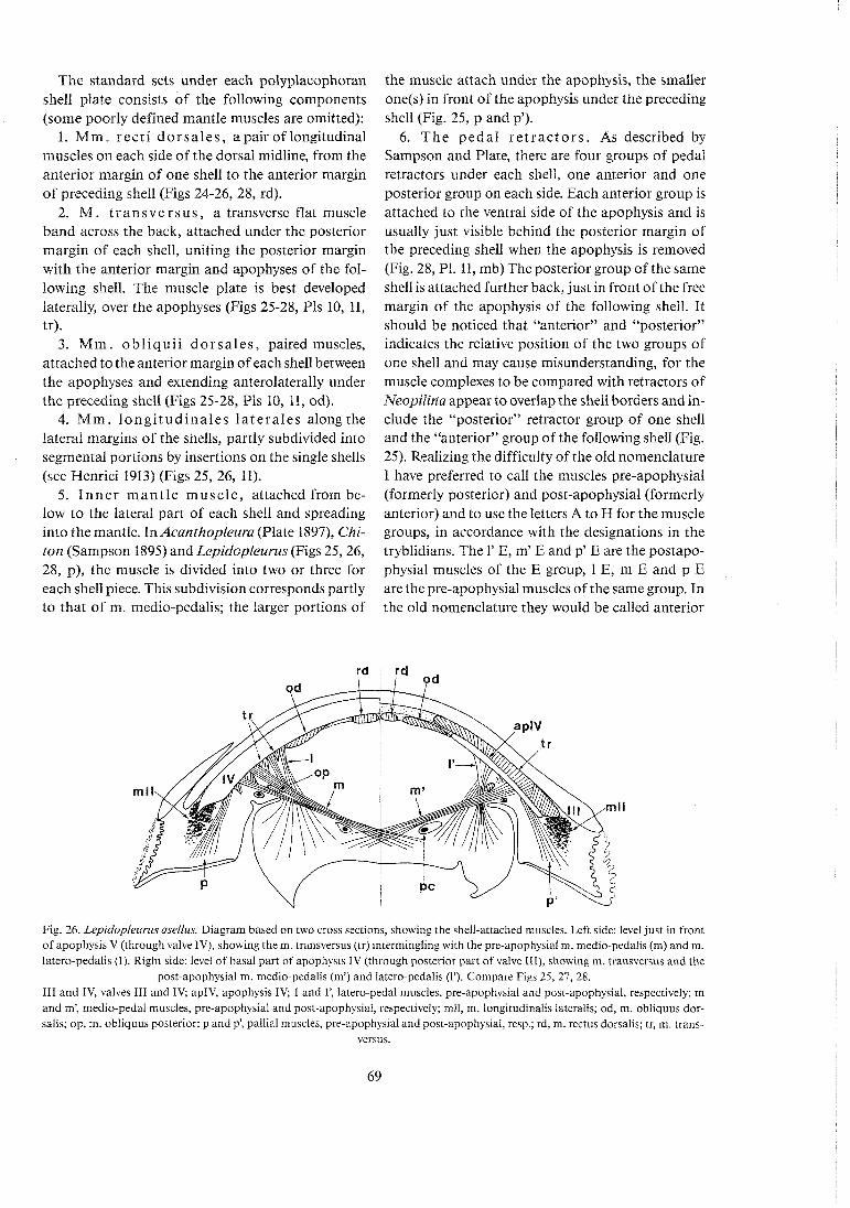
The standard sets under each polyplacophoran shell plate consists of the following components (some poorly defined mantle muscles are omitted):
1. Mm. recti dorsa les , apairoflongitudinal muscles on each side of the dorsal midline, from the anterior margin of one shell to the anterior margin of preceding shell (Figs 24-26, 28, rd).
2. M. t ransve rsus , a transverse flat muscle band across the back, attached under the posterior margin of each shell, uniting the posterior margin with the anterior margin and apophyses of the fol- lowing shell. The muscle plate is best developed laterally, over the apophyses (Figs 25-28, Pls 10, 11, tr).
3. M m . obl iqui i dorsa les , paired muscles, attached to the anterior margin of each shell between the apophyses and extending anterolaterally under the preceding shell (Figs 25-28, Pls 10, 11, od).
4. Mm. longi tudinales la tera les along the lateral margins of the shells, partly subdivided into segmental portions by insertions on the single shells (see Wenrici 1913) (Figs 25, 26, 11).
5. Inne r mant le muscle, attached from be- low to the lateral part of each shell and spreading into the mantle. In Acanthopleura (Plate 1897), Chi- ton (Sampson 1895) and Lepidopleurus (Figs 25,26, 28, p), the muscle is divided into two or three for each shell piece. This subdivision corresponds partly to that of m. medio-pedalis; the larger portions of
the muscle attach under the apophysis, the smaller one(s) in front of the apophysis under the preceding shell (Fig. 25, p and p').
6. The pedal re t rac tors . As described by Sampson and Plate, there are four groups of pedal retractors under each shell, one anterior and one posterior group on each side. Each anterior group is attached to the ventral side of the apophysis and is usually just visible behind the posterior margin of the preceding shell when the apophysis is removed (Fig. 28, P1. 11, mb) The posterior group of the same shell is attached further back, just in front of the free margin of the apophysis of the following shell. It should be noticed that "anterior" and "posterior" indicates the relative position of the two groups of one shell and may cause misunderstanding, for the muscle complexes to be compared with retractors of Neopilina appear to overlap the shell borders and in- clude the "posterior" retractor group of one shell and the "anterior" group of the following shell (Fig. 25). Realizing the difficulty of the old nomenclature I have preferred to call the muscles pre-apophysial (formerly posterior) and post-apophysial (formerly anterior) and to use the letters A to H for the muscle groups, in accordance with the designations in the tryblidians. The 1' E, m' E and p' E are the postapo- physial muscles of the E group, 1 E, m E and p E are the pre-apophysial muscles of the same group. In the old nomenclature they would be called anterior
Fig. 26. Lepidopleurus asellus. Diagram based on two cross sections, showing the shell-attached muscles. Left side: level just in front of apophysis V (through valve IV), showing them. transversus (tr) intermingling with the pre-apophysial m. medio-pedalis (m) and m. latero-pedalis (1). Right side: level of basal part of apophysis IV (through posterior part of valve 111), showing m. transversus and the
post-apophysial m. medio-pedalis (m') and latero-pedalis (1'). Compare Figs 25, 27, 28. 111 and IV, valves 111 and IV; apIV, apophysis IV; 1 and l', latero-pedal muscles, pre-apophysial and post-apophysial, respectively; m and m', medio-pedal muscles, pre-apophysiaf and post-apophysial, respectively; mll, m. longitudinalis lateralis; od, m. obliquus dor- salis; op, m. obliquus posterior; p and p: pallial muscles, pre-apophysial and post-apophysial, resp.; rd, m. rectus dorsalis; tr, m. trans-
versus.
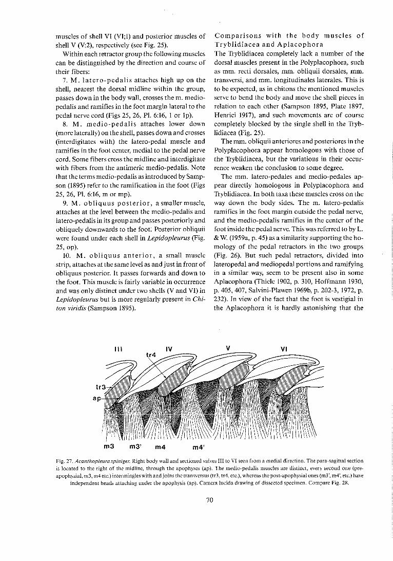
muscles of shell VI (V1;I) and posterior muscles of shell V (V:2), respectively (see Fig. 25).
Within each retractor group the following muscles can be distinguished by the direction and course of their fibers:
7. M . la tero-pedal is attaches high up on the shell, nearest the dorsal midline within the group, passes down in the body wall, crosses the m. medio- pedalis and ramifies in the foot margin lateral to the pedal nerve cord (Figs 25, 26, Pl. 6:16, 1 or lp).
8. M . medio-pedalis attaches lower down (more laterally) on the shell, passes down and crosses (interdigitates with) the latero-pedal muscle and ramifies in the foot center, media1 to the pedal nerve cord. Some fibers cross the midline and interdigitate with fibers from the antimeric medio-pedalis. Note that the terms medio-pedalis as introduced by Samp- son (1895) refer to the ramification in the foot (Figs 25, 26, P1. 6:16, m or mp).
9. M. obl iquus pos ter ior , asmallermuscle, attaches at the level between the medio-pedalis and latero-pedalis in its group and passes posteriorly and obliquely downwards to the foot. Posterior obliquii were found under each shell in Lepidopleurus (Fig.
25, OP). 10. M . obl iquus an te r io r , a small muscle
strip, attaches at the samelevel as and just in front of obliquus posterior. It passes forwards and down to the foot. This muscle is fairly variable in occurrence and was only distinct under two shells (V and VI) in Lepidopleurus but is more regularly present in Chi- ton viridis (Sampson 1895).
Compar isons with the body muscles of Trybl id iacea a n d Aplacophora The Tryblidiacea completely lack a number of the dorsal muscles present in the Polyplacophora, such as mm. recti dorsales, mm. obliquii dorsales, mm. transversi, and mm. longitudinales laterales. This is to be expected, as in chitons the mentioned muscles serve to bend the body and move the shell pieces in relation to each other (Sampson 1895, Plate 1897, Henrici 1917), and such movements are of course completely blocked by the single shell in the Tryb- lidiacea (Fig. 25).
The mm. obliquii anteriores and posteriores in the Polyplacophora appear homologous with those of the Tryblidiacea, but the variations in their occur- rence weaken the conclusion to some degree.
The mm. latero-pedales and medio-pedales ap- pear directly homologous in Polyplacophora and Tryblidiacea. In both taxa these muscles cross on the way down the body sides. The m. latero-pedalis ramifies in the foot margin outside the pedal nerve, and the medio-pedalis ramifies in the center of the foot inside the pedal nerve. This was referred to by L. & W. (1959a, p. 45) as a similarity supporting the ho- mology of the pedal retractors in the two groups (Fig. 26). But such pedal retractors, divided into lateropedal and mediopedal portions and ramifying in a similar way, seem to be present also in some Aplacophora (Thiele 1902, p. 310, Hoffmann 1930, p. 405,407, Salvini-Plawen 1969b, p. 202-3, 1972, p. 232). In view of the fact that the foot is vestigial in the Aplacophora it is hardly astonishing that the
Fig. 27. Acanthopleura spiniger. Right body wall and sectioned valves I11 to VI seen from a medial direction. The para-sagittal section is located to the right of the midline, through the apophyses (ap). The medio-pedalis muscles are distinct, every second one (pre- apophysial, m3, m4 etc.) intermingles with and joins the transversus (tr3, tr4, etc.), whereas the post-apophysial ones (m3: m4: etc.) have
independent heads attaching under the apophysis (ap). Camera lucida drawing of dissected specimen. Compare Fig. 28.
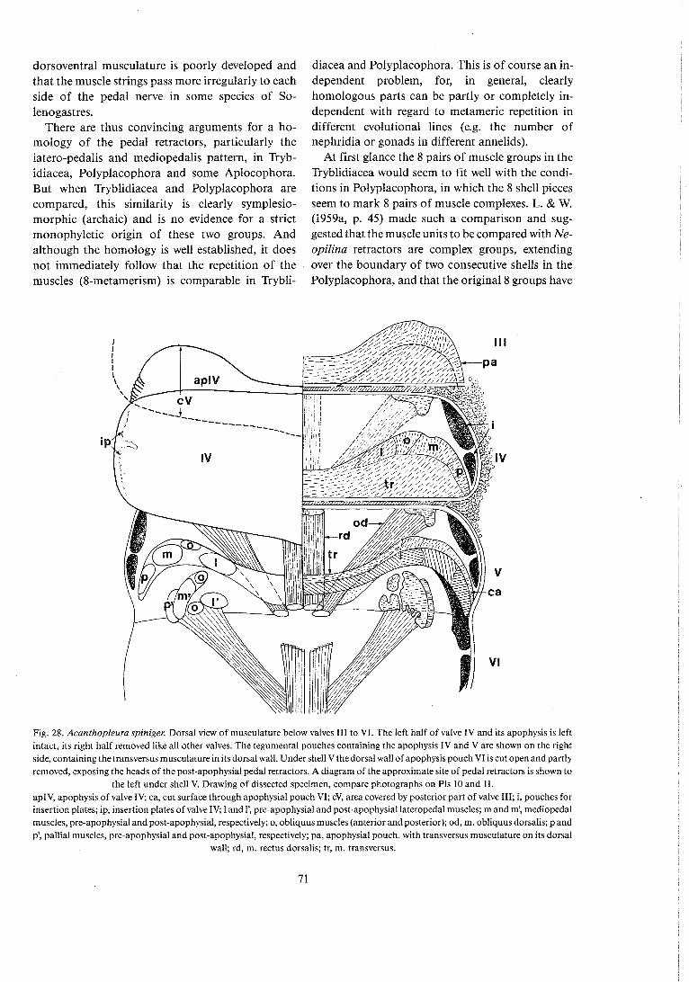
dorsoventral musculature is poorly developed and that the muscle strings pass more irregularly to each side of the pedal nerve in some species of So- lenogastres.
There are thus convincing arguments for a ho- mology of the pedal retractors, particularly the latero-pedalis and mediopedalis pattern, in Tryb- idiacea, Polyplacophora and some Aplocophora. But when Tryblidiacea and Polyplacophora are compared, this similarity is clearly symplesio- morphic (archaic) and is no evidence for a strict monophyletic origin of these two groups. And although the homology is well established, it does not immediately follow that the repetition of the muscles (8-metamerism) is comparable in Trybli-
diacea and Polyplacophora. This is of course an in- dependent problem, for, in general, clearly homologous parts can be partly or completely in- dependent with regard to metameric repetition in different evolutional lines (e.g. the number of nephridia or gonads in different annelids).
At first glance the 8 pairs of muscle groups in the Tryblidiacea would seem to fit well with the condi- tions in Polyplacophora, in which the 8 shell pieces seem to mark 8 pairs of muscle complexes. L. & W. (1959a, p. 45) made such a comparison and sug- gested that the muscle units to be compared with Ne- opilina retractors are complex groups, extending over the boundary of two consecutive shells in the Polyplacophora, and that the original 8 groups have
Fig. 28, kcanihopieura spiniger. Dorsai view of muscuiaiure beiow vaives i i i to Vi. Tne ieft haif of vaive iV and its apophysis is left intact, its right half removed like all other valves. The tegumental pouches containing the apophysis IV and V are shown on the right side, containing the transversus musculature in its dorsal wall. Under shell V thedorsal wall of apophysis pouch VI is cut open and partly removed, exposing the heads of the post-apophysial pedal retractors. A diagram of the approximate site of pedal retractors is shown to
the left under shell V. Drawing of dissected specimen, compare photographs on Pls 10 and 11. apIV, apophysis of valve IV; ca, cut surface through apophysial pouch VI; cV, area covered by posterior part of valve 111; i, pouches for insertion plates; ip, insertion plates of valve IV; 1 and 1: pre-apophysial and post-apophysiai lateropedal muscles; m and m', mediopedal muscles, pre-apophysial and post-apophysiai, respectively; o, obliquus muscles (anterior and posterior); od, m. obliquus dorsalis; p and p', pallial muscles, pre-apophysial and post-apophysial, respectively; pa, apophysial pouch, with transversus musculature on its dorsal
wall; rd, m. rectus dorsalis; tr, m. transversus.

been partly subdivided into two by the development of the apophysis from the anterior margin of the fol- lowing shell, as indicated in Fig. 27. The apophysis would, then, cut into each retractor group, dividing it into a post-apophysial group attached to the apophysis, a m. transversus between the apophysis and the next anterior shell, and a pre-apophysial group attached in front of the apophysis to the preceding shell (Figs 25, 27, 28, P1. 11).
Some authors accepted in a general way this sug- gested homology between the 8-metamerism in the musculature of Tryblidiacea and chitons (Boettger 1959, Yonge 1960, p. 19, Ax 1960, Gotting 1980a). But objections were raised particularly by Salvini- Plawen (1968, 1969b, 1972, 1980a), who proposed that polyplacophoran musculature is in fact 16-metameric. He regarded this as a step in a con- tinuous process of reductions of the dorsoventral muscle strings, beginning with hypothetical turbel- larian ancestors (numerous pairs), through the Solenogastres (numbers somewhat reduced) and the Polyplacophora (16? pairs) to the Monoplacophora (8 pairs) and ending with still stronger reduction in the Conchifera, e.g., in bivalves (7 pairs or less) and gastropods (one pair or even M pair). This series is of course beautiful and mentally satisfactory in a way but to me it does not seem to be convincingly sup- ported by facts for the different steps. At any rate I doubt that the polyplacophoran musculature can be regarded as 16-metameric. The reasons for my opinion are:
1. In the Polyplacophora there are no indisputable series of 16 comparable muscle units along the axis of the animal. The main pedal retractors, m. medio- pedalis and m. latero-pedalis, are divided into an an- terior and a posterior portion under each shell border, giving a total of about 16 along the animal. But the anterior and posterior portions are different with regard to attachment, size, fiber direction and, in some species, with regard to fusion with the 7-metameric m. transversus (Figs 25, 27, 28). Every second component in the proposed "16-metameric" series is thus different from the other eight, and ccunticg such 2 hetercgenecus sequence t c get the number 16 is hardly relevant.
The mm. obliquii posteriores may be repeated twice under some shells of Chiton viridis (Sampson 1895), but only 8 or 7 single pairs are present in Lepidopleurus. The inner mantle muscle may be divided into two or three very dissimilar portions un- der some shells of, e.g., Lepidopleurus (Fig. 25), but I do not think that the repetition is regular
enough to be accepted as evidence for a 16- metamerism. To me these series - because of the deviating features of every second unit - look more like an 8-metamerism, where the original muscles have been separated into two unequal portions on each side of the joints between the shell plates.
It should be remarked that the theory of a 16-metameric pattern of muscles in the Poly- placophora stands or falls with the recognition of a convincing series of 16 comparable muscle units. No other structures in the animal itself support a 16-metameric pattern. The shell and the other muscles are clearly repeated 8 times and the many gills show nonstabilized repetition into many units. No support for a 16-metamerism can be obtained from outgroup comparison, for the tryblidiacean retractor muscles are clearly 8-metameric, and those of the Solenogastres appear nonstabilized, mul- tiplied alternating with intestinal pouches.
2. Muscles of Polyplacophora other than those mentioned above are clearly 8- or 7-metameric, repeated one time for each shell piece: m. rectus, m. obliquus dorsalis, m. transversus, and m. longi- tudinalis lateralis. The muscle groups, of which the pedal retractors form an integral part, are also repeated 8 times. The attachment areas of these muscle groups are more or less completely covered by the broad and flat mm. transversi, present in a number of 7 (absent above the last group under the posterior margin of shell VIII). The heads of m. latero-pedalis and m. medio-pedalis of the posterior groups, although sometimes regarded as 16-meta- meric, usually fuse and intermingle with the m. transversus, which is a typical 8(7)-metameric muscle (Figs 25, 27, 28).
3. Comparison with the Monoplacophora indi- cates that the 8-metameric pattern of the pedal retractors is homologous in the two groups. The presence of the 8 paired muscle groups in both can- not be ignored as a numerical support for the idea that the muscle patterns are homologous also with regard to the number of units. An analysis of the re- lations to other organs gives some support to this ho- 1 l l 0 ! 0 0 ' 7 : a J
In both taxa the muscle complex A is associated with the m. oralis posterior, which can be identified by its spreading in the ventral wall of the mouth region. The pericardium begins at the level of the muscle complex F. The atria of Neopilina are si- tuated in the intervals between complexes F-6 and 6-H. Two pairs of ostia are present in Polypla- cophora (most species), they have the same localiza-

tion (Fig. 25, see also Plate 1901, p. 480, and Hoffmann 1930, pp. 281-286). If only one pair of ostia is present it is located under shell VII (between muscle groups F and G) as in Lepidopleurus (Fig 25), and this is the location of the first ostium in the few forms which have more pairs of ostia. It should also be mentioned that muscle group H, in addition to its location on each side of the rectum, has a more specific similarity in the Tryblidiacea and the Poly- placophora. Its medio-pedalis crosses the midline ventral to the rectum and ramifies in the opposite side of the foot. This group (H) is not subdivided in the polyplacophorans, and there is no m. transver- sus, obviously because there is no apophysis (Fig. 25, Pi. 10:41, 43).
I consider the evidence for a 16-metamerism of the musculature in the Polyplacophora very doubtful, for it relies exclusively on the supposed 16- repetition of pedal retractors. Such results can be obtained only if one ignores the fact that every second muscle is different and forgets that 16 non-comparable units are counted.
No such objections can be raised against the presence of an 8-metamerism, for this interpretation is supported by several sets of 8-metameric struc- tures (retractor muscles, muscle groups, shell pieces of the animal). Only this interpretation is supported by outgroup comparison, for the series of 8 muscle groups is clearly homologous with the 8 retractor groups in tryblidiaceans, in which even some of the single units of the series can be individually homo- logized because of comparable relation to other structures. Also the comparison of the 7 spicule plates of Nematomenia with the 7 or 8 plates of the Polyplacophora supports an 8- or 7-metamerism in the latter rather than a 16-metamerism.
Salvini-Plawen (1968, p. 193) pointed out that the situation of the nephridioduct between muscles VII, and VII, in Polyplacophora is imcompatible with our hypothesis that the 8 pedal retractors in Tryblidiacea have been divided to form two units each in Polyplacophora. But this is a mistake prob- ably caused by the (confusing) classical numbering =f tho nm r\r\nhxr ; * A 0 7 m - . o n l a
r l x r y ~ r - 2 ~ v y ~ l j ~ ~ 2 ! 211u pvo~-8Yv~~~J i )~8! I I L L L ~ ) ~ ~ ~
portions in Polyplacophora, for the original idea (L. & W. 1959a, p. 45) is in good agreement with the facts Salvini-Plawen uses for his criticism.
To avoid further misunderstanding I have in- troduced the previously used letters A to H for the 8 muscle groups on each side in Tryblidiacea, and also for the muscle groups which are believed to be de- rived from them in Polyplacophora (Fig. 25). The
nephridioduct of the Polyplacophora is situated be- tween muscle complexes F and 6, which means, be- tween VII, and VII, in the classical annotation. In Tryblidiacea this corresponds to the interval between muscles F and G, where nephridium F is situated, so there is no trouble. Both in tryblidians and poly- placophorans nephridium F corresponds to the level of the foremost atrium (or ostium), so the identifica- tion is quite convincing (Figs 8, 9, 25).
The gonoduct of the Polyplacophora, which lies between VI, and VII, (classical annotation), causes some difficulties, but this problem is not mentioned by Salvini-Plawen. A comparison between Mono- placophora and Tryblidiacea (Fig. 25) makes it necessary to assume that the posterior gonoduct has changed its position when the monoplacophoran muscles divided to form the pattern of the poly- placophorans. If not, the posterior gonoduct would be expected to penetrate muscle F in the Mono- placophora, which is not the case. In view of the variability of the gonoducts in molluscs, such a redisposition does not seem improbable. It is recalled that the connections of the gonoducts are variable between different Aplacophora (Salvini- Plawen 1972, p. 250) and that the gonoduct in the Chitonida runs dorsal to the lateral nerve in most species but ventral to it in Ischnochiton ruber, Tonicella marmorea and Katharina tunicata (Plate 1901, p. 467).
It should be noticed that Salvini-Plawen's deriva- tion of 8 pairs of pedal retractors in Monopla- cophora from the hypothetical 16 pairs in the Polyplacophora leads to similar unpleasant results, if the two muscles under each shell in the Poly- placophora are supposed to fuse to form single muscles in the Monoplacophora. Then the poly- placophoran nephridium, lying between the two muscles under shell VII, would be expected to pene- trate the muscle VII (6) in tryblidians, and it does not. This was referred to as a serious obstacle of our original theory, but on false premises (Salvini- Plawen 1968, p. 193).
5.5 Ziadistic reiations between Conchifera, Polyplacophorn and Aplacophorn
The situation of the Conchifera and Polyplacophora was, in a more general way, discussed in Chapter 5.3. It was concluded that the Conchifera, including the Tryblidiacea, are a good monophyletic unit, and the main synapomorphic features supporting this con- clusion were mentioned.

It is also obvious and generally accepted that the Conchifera and Polyplacophora are closely related, forming a monophyletic unit of higher order, some- times called "Testaria" (Salvini-Plawen 1972, 1980a, Lauterbach 1983b). The testarian synapomorphies supporting this relationship are:
1. Many features in the radula apparatus, e.g., radula dentition of docoglossan rasping type with characteristic differentiation, hollow radula vesicles, homologous lateral and medial cartilages of the odontophore, homologous radula diverticula, many homologous radula muscles (pp 66-67).
2. The "velum", the subradular organ, the large pharyngeal diverticula ("Zuckerdriisen"), the large digestive gland ("liver"), and the coiled intestine.
3. The 8 pairs of pedal retractor groups, the an- terior pair associated with a posterior oral muscle (pp 70-73).
4. Heart with 2 pairs of atria, situated between retractors F-G and G-H. Polyplacophorans have only two pairs of ostia in corresponding sites, and the number of ostia is somewhat variable. Among Conchifera only Tryblidiacea and Nautilus have 2 pairs of atria, so this apomorphy is somewhat un- certain.
Some of the mentioned synapomorphic features are reduced in the advanced conchiferan classes and are typically developed only in the Polyplacophora and Tryblidiacea. But in view of the fact that the Conchifera, including the Tryblidiacea, is a well es- tablished monophyletic unit, there can be no doubt about the phylogenetical consequences (see dia- gram, Fig. 20). Theoretically some of these features could be plesiomorphic, i.e., could have been present already in some Aplacophoran ancestors (e.g., point 3), but other characters, particularly those of the radula apparatus, appear sufficient to show that the Conchifera and Polyplacophora are strictly mono- phyletic.
Most autapomorphic characters for the Poly- placophora are restricted to shell morphology: the 8 (or 7) shell plates, the articulamentum, the aesthetes, and the apophyses (in recent forms). The many,gills ccu!c! be 2:: zutzpcmcrphy, but this i s true m ! y i f the multiplication of gills has been independent in Poly- placophora on one side and in neopilinids and Nau- tilus on the other. The characteristic girdle with cuticle and spicules is a polyplacophoran apomor- phy, but not the presence of spicules as such, for spicules are a plesiomorphic feature present also in the Aplacophora. Only the restriction of the cuticle and spicules to the mantle margin is significant as an
autapomorphic character of the Polyplacophora, and this character is obviously correlated with ap- pearance of the middorsal shell plates.
The common testarian ancestor "B" in the dia- gram (Fig. 20) must have had the testarian apomor- phies, also the 8 pairs of pedal retractor groups. I do not think that it had consolidated shell plates on the back, for nothing in the development of the con- chiferan shell supports the theory that it has arisen by fusion of separate consolidated shell plates in an ancestor. But it is very probable that the back was covered by a cuticle and separate spicules, the latter perhaps even arranged into 8 transverse fields cor- responding to the 8 retractors. By fusion of the spi- cules this ancestor may have evolved into a poly- placophoran with 8 shell plates or a conchiferan with a single shell (p. 61). The 8 pairs of retractors are sup- posed to be preserved in both sister groups.
In my diagram (Fig. 20) the Ordovician genus Septemchiton (Bergenhayn 1955) with 7 shell plates is placed as a side branch from the main stem of 8-shelled Polyplacophora, developed by the loss of one plate. Salvini-Plawen (1980a) proposed a new subclass, Heptaplacota, for Septemchiton and regarded it as ancestral to other Polyplacophora, ob- viously because a similar 7-metamerism seems to be present in Pruvot's famous larva of the solenogaster Nematomenia (cf. pp 59-61). The Heptaplacota are then supposed to have given rise to the 8-shelled Poly- placophora, from which the Conchifera are believed to have evolved by fusion of the shells. I hesitate to give the 7-shelled condition of the Heptaplacota so much weight as an argument in the phylogenetical discussion (see p. 61) for 7 or 8 shells may be an un- stable character, as indicated by the intraspecific variation in recent chitonids. It is admitted that the plesiomorphic state of 7 shells gets some support by ontogeny, for recent Polyplacophora seem to form the 8th shell after some delay and thus exist as 7-shelled larvae for some time, although there is some variation (Smith 1966). And only a distinct in- terruption of shell formation at the 7-shell stage can be interpreted as a kind of ontogenetic repitulation, fcr i t i s hzrdly remzrkzb!e that the most pcxtericr shell plate is the last to be formed.
The geological age of Septemchiton is a poor ar- gument for its ancestral state in relation to other Polyplacophora. Is was found in the Lower Ordovi- cian (Bergenhayn 1955) and Upper Ordovician (Sanders 1964), and we do not know the number of valves in the oldest, Upper Cambrian, Polypla- cophora (Bergenhayn 1960).

The evidence for a primary 7-metamerism derived from Pruvot's description of Nernatomenia is cer- tainly circumstantial, for the 7 fields of spicules were found in a post-metamorphosis specimen. Chitons at a corresponding stage may also have 7 plates, although they have 8 as adults.
Although it is possible that the Heptaplacota can have had the ancestral relation to Polyplacophora suggested by Salvini-Plawen, I regard this idea as an unnecessary complication. The Heptaplacota have consolidated shell pieces; Salvini-Plawen's theory and the said idea, in itself uncertain, thus forces us to accept a formation of the conchiferan shell from separate consolidated shell plates, a hypothesis which lacks real support.
Down to the level of the "Testarian ancestor" ("B" in Fig. 20) the phylogeny can be reconstructed with reasonable probability because of evidence from anatomy, embryology and paleontology. The real difficulties come when the relations of the Aplacophora to the Testaria are considered. No modern zoologists will deny that the Aplacophora are good molluscs and share many homologous fea- tures with the Testaria, e.g., presence of a radula, dorsal cuticular cover with spicules, a ventral creep- ing sole (reduced in some Aplacophora) and a pedal gland (in some). No doubt, therefore, there has been a common ancestor of all the molluscs, an "Urmol- lusk" as indicated by "En in Fig. 20. The morpho- logical features of this ancestor can, of course, only be theoretically deduced, and this is difficult, be- cause two of the earliest derived lines, the Solenogas- tres and the Caudofoveata, are admitted to be strongly specialized.
The simplest interpretation would be to regard the Testaria and the Aplacophora as sister groups, de- rived from the "Urmollusk" as indicated by the solid lines in the cladogram (Fig. 20). This would be the best alternative if it can be shown that the Caudo- foveata and the Solenogastres are monophyletic, i.e., share some good synapomorphic features showing that all Aplacophora had a common ancestor ("D" in the cladogram) different from the testarian an- cestor "B".
Potential apomorphic similarities between the Caudofoveata and the Solenogastres are the worm- like, almost cylindrical shape, and the extension of the dorsal cuticle down the sides towards the ventral midline, with total or partial reduction of the creep- ing sole as a consequence. This is usually accepted as a synapomorphic feature, for practically all zoolo- gists suppose that the ancestral mollusc had a well
developed creeping sole. But is has been stressed, particularly by Salvini-Plawen (1972) that the final reduction of the foot and pallial groove has taken place in different ways in Caudoveata and Soleno- gastres, and that the mentioned similarity therefore may be convergent. On the other hand, the initial stages of this development can have taken place in a common ancestor ("D" in Fig. 20) and the differ- ences evolved after the Caudofoveata and the Solenogastres s. str. parted from each other. As iong as this possibility is open, the cylindrical shape, the downward extension of the mantle and part of the reduction of the foot can still be a synapomorphic feature, supporting a monophyletic group Apla- cophora.
That there are profound differences between the Caudofoveata (Chaetodermatoida) and the So- lenogastres s. str. (Ventroplicata) has been known for a long time (Wirkn 1892, Odhner 1919, Hoffmann 1949, Boettger 1955, 1959), but only Salvini-Plawen (1969a, b, 1972, 1980a) went so far as to suggest that the Aplacophora are directly paraphyletic and to propose an alternative cladogram. According to his hypothesis, the Caudofoveata and Solenogastres are completely independent branches of the molluscan main stem. The Caudofoveata are regarded as the first branch, and are regarded as a sister group of all non-caudofoveate molluscs. These are regarded as the other monophyletic sister group called Adenopoda. The adenopod ancestor ("C") has, in a dichotomous fashion, evolved into Solenogastres and the Testaria (dotted line in Fig. 20).
This alternative possibility can be made probable if Solenogastres and Testaria share synapomorphic features, showing that they are more closely related to each other than either of them is to Caudofoveata. Salvini-Plawen emphasizes the preoral extension of the pallial groove and the presence of a pedal gland. But these are good pieces of evidence only, if it can be presumed that they were absent in the molluscan ancestor ("EM) and there is in fact no possibility to show with certainty that this condition is satisfied. Caudofoveata certainly lack the preoral pallial gmo.ie 22d the ped,! gl~c.1, but beth car, haxve bee:: reduced during specialization within the Caudo- foveate line and may have been preserved in the Solenogastres and Polyplacophora as a plesio- morphic feature (the pedal gland is absent in adult Polyplacophora but is present in early ontogenetic stages).
Analogously, it is very difficult to get a strong case for synapomorphy in other features of Sole-

nogastres and Caudofoveata, or for Solenogastres and Testaria, the result being so dependent on the structure of the molluscan ancestor. And since this ancestor is really unknown and dependent on each author's private views on relationships and origin of molluscs, the conclusions must be fairly uncertain. I have therefore left open both alternative ideas about the Aplacophora in the diagram, but can admit that I think there is somewhat stronger support for a monophyly of the Aplacophora, mainly because some characters (extension of mantle, reduction of foot), although possibly convergent, are at least clearly apomorphic.
In either case the significance of the Nema- tomenia larva for the early phylogeny of molluscs is interesting. If the 7-metameric structure, as ex- pressed in the spicules on the back, is homologous with the 7- or 8-metamerism of polyplacophorans, such metameric structure must have been present in the common ancestor. This means that a metameric structure of this kind must have been present in an- cestor "C" or ancestor "E" depending on which phylogenetic interpretation is preferred. In both cases the metamerism is brought down to very near the origin of molluscs. Salvini-Plawen (1980a), sup- porting the diphyletic origin of Aplacophora, logi- cally concludes that the adenopod ancestor ("C") had such metamerism similar to that of Nematome- nia. It also follows that the spicule fields on the back must have scattered again during further develop-
. ment of the Solenogastres. If 7 pairs of retractor muscles, matching the structure of the back, have been present in the ancestor "C" they must have multiplied in recent Solenogastres, for these usually have many muscle strings, which alternate with in- testinal pouches (Fig. 20). It is even questionable if the metamerism of Solenogastres is homologous with that of other molluscs. Such alternation of in- testional pouches and retractor muscles is not seen in other molluscs but looks more like the typical "pseudometamerism" seen in several non-mol- luscan groups like Nemertini and Turbellaria. The multiplication in the Solenogastres could therefore be comparable to that in other animals with elon- gated body shape.
5.6. General discussion of metamerism in molluscs
The repetition of organs in Vema and Neopilina is reviewed and compared in Chapter 5.1. It is conclud- ed that the repetition in different organ systems shows reasonable spatial correlation in the two spe-
cies, although the absolute number of repeated units is greater in Vema. The latter has the most synor- ganized rnetameric pattern seen in a mollusc. The term "metamerism" is then used in a descriptive way to denote repetition of organs or units along the lon- gitudinal axis of the animal.
The significance of this metamerism remains to be discussed: whether there is reason to assume that it is a fundamental feature in molluscs, and, if so, whether it is related to other kinds of metamerism seen in other protostomians, e.g., in turbellarians and articulates.
5.6.1. The metamer ism of the muscula ture The low, flattened Tryblidiacea have clearly had a distinctly metameric musculature in the early Paleozoic time, and the recent Neopilina and Vema have preserved the 8 pairs s f pedal retractors. The number of muscle pairs was probably 8 also in some Ordovician and Silurian forms such as Tryblidium, Pilina and Araeophiala, although the number is obscured by partial fusion in the fossils. Even the Upper Cambrian and Lower Ordovician Proplina seems to have had 7 or 8 pairs, whereas the Middle Cambrian Scenella may have had 6 or 7 (Fig. 19, Chapter 5.2.).
Those "monoplacophorans" which have a high cyrtoconic shell and therefore are perhaps related to the Cyrtosoma (Rhacopoda) have fewer muscle pairs: Kirengella has 4 or 5 pairs. Cyrtonella 2 pairs, and Moyerokania 5 pairs. Such reduction would be expected if the animal is higher than long, with a dorsoventral axis much longer than the antero- posterior one. This tendency is more pronounced in the high, plainispiral forms like Cyrtolithes (2 pairs) or more typical bellerophontids, in which only one pair of scars is preserved (Chapter 5.2., Fig. 19). No recent gastropods or cephalopods have preserved multiple pedal retractors but more than one pair of retractors were present in some fossil nautiloids (Mutvei 1964a, Chapter 5.3.5.).
That some fossil Diasoma had about 8 pairs of pe- dal retractors can hardly be doubted (Rostroconchs, 6-7 pairs, Rabinka; 8 pairs) and recent bivalves have preserved 1-7 pairs (Chapter 5.3.6.). In the Diasoma it is characteristic that the retractor scars on each side move up to form a line on each side of the dorsal midline and the reduction of the number of muscles seems to have been slow in some cases. No meta- meric retractors are known from recent scaphopods, but the situation in some proposed ancestral ro- stroconchs is uncertain (Pojeta & Runnegar 1976).

When these facts are considered in the light of the probable phylogeny of the Conchifera (Fig. 20), it may be stated that multiple retractor muscles or muscle scars (8 or less pairs) are known from all three evolutional lines, and were most common in older Paleozoic times. Reduction of the metameric mus- culature is obvious in the Cyrtosoma and the Bival- via, and only Tryblidiacea and Bivalvia have preserved a metameric musculature to recent times. The idea accepted by most recent zoologists, that the metameric musculature is an ancient feature, re- tained from ancestral conchiferans, is therefore well established.
This conclusion is also acceptable if functional aspects are considered. It is certainly difficult to im- agine a functional advantage of a metameric retrac- tor system in the Conchifera, which have a rigid, inflexible shell. As maintained by Gotting (1980a) for Nautilus, a progressive development of muscle metamerism under such circumstances appears im- probable. On the contrary, such animals would be expected to reduce whatever metamerism present, and that is actually what was stated above in most conchiferan lines. It can also be expected that animals with a long dorsal attachment zone such as rostroconchs, bivalves and tryblidiaceans should preserve the metameric musculature better than the high cyrtosomic gastropods, bellerophonts and cephalopods, in which the space for muscle attach- ments becomes more restricted. These functional considerations, although not directly conclusive, support the idea that the metameric retractors must have been present in the conchiferan ancestors, and that later changes only have been in the direction of reduction.
The musculature of the Polyplacophora is of deci- sive interest for this discussion. In Chapter 5.3.5. it was concluded that the series of 8 paired retractors in Tryblidiida is homologous with the series of 8 paired groups of muscles in the Polyplacophora. In the Polyplacophora each of the first 7 muscle groups corresponds to and overlaps the 7 limits between the shell plates, whereas an eighth group extends along the posterior border of the last she!! p!ate. Sa!vini- Plawen's (1969, 1972, 1980a) interpretation of poly- placophoran musculature as 16-metameric is difficult to accept, for all the shell-attached body muscles are parts of the 8 well-defined muscle groups. Some of these muscles are present as 8 units along the side of the animal, and one of them, the m. transversus, covers the heads of the other muscles of the group and is confluent with some of them. This
makes the definition of the 7 anterior groups parti- cularly clear (Figs 25, 28, P1. 10) - the 8th group lacks a transversus.
The m. medio-pedalis and m. latero-pedalis are duplicated within each group except the 8th one, so that a pre-apophysial and a post-apophysial portion has been formed within each group. This appears to be a secondary subdivision, caused when the apo- physes evolved in recent Polyplacophora. A count along the side of the animal will thus give 16 units of these subdivided muscles, but regarding this as a 16- (or 15) metamerism is justified only if the units counted are comparable. And in this case every sec- ond unit differs from the others (pre-apophysial and post-apophysial portions are different). Moreover, the heads of the post-apophysial components are fused and sometimes indistinguishable from the clearly 7-metameric rn. tranversus (Fig. 28, Pls 10, 11). I therefore maintain that 8-metamerism rather than 16-metamerism is by far the most probable in- terpretation (pp 72-73).
Presence of 8 homologous pairs of muscle groups in Tryblidiacea and Polyplacophora leads to the con- clusion that such 8-metameric muscle groups must have been present in the commmon ancestor of Tryb- lidiacea and Polyplacophora (Testarian ancestor, "B" in Fig. 20). This hypothetical animal must have lived already in Middle Cambrian, for tryblidians with metameric muscles were present in Middle and Upper Cambrian (Chapter 5.2.) and isolated poly- placophoran shells are known from Upper Cam- brian (Bergenhayn 1960). The presence of a metameric musculature can thus be followed down to the level of the testarian ancestor with reasonable certainty.
There is even some evidence of a 7- or 8-meta- merism earlier in molluscan development, in the adenopod ancestor ("C") or in the ancestor of all molluscs ("E"). But this hypothesis depends on ac- ceptance of Pruvot's old description of the larval Nematomenia banyulensis (Chapter 5.4.2.), in which 7 transverse fields of spicules were present on the back. I find it very probable that a corresponding m ~ t a m ~ r i c m xxmc nrprpnt in the r n ~ ~ ~ ~ ~ ~ l a t ~ ~ r o nf th;c '-'-LU.-'-"U". "UU y L ' U " ' C "' C"' " ' Y U ' U ' U C U ' ' "I L l l l U
larva, inducing the metameric pattern on the back. If comparable with the retractor metamerism of the Polyplacophora and Monoplacophora, such meta- merism of the retractor musculature would be ex- pected to have been present also in the common adenopod ancestor, or in the "Urmollusk" if the Aplacophora is regarded as monophyletic (Fig. 20). This theory that the metamerism of the dorsal shell

plates in the Polyplacophora and the spicule groups of the Nematomenia larva are induced by the under- lying musculature will probably meet with objec- tions, for many scientists have more or less clearly expressed the opposite opinion, viz., that the 8 shell plates of chitons are the primary feature, and that the musculature adjusts to this by developing an 8-metamerism. But I consider the arguments for a primary metamerism in the musculature stronger or at least equally good: 1) It has been shown that the main musculature appears in larval chitons before the shell plates or even the shell fields are formed (Heath 1899, Hammarsten & Runnstrom 1925, p. 276). Although not strong, this argument speaks in favour of a muscular metamerism as the primary factor. 2) The opposite possibility, that the metamerism of the musculature is dependent on the dorsal shell plates is contradicted by the Trybli- diacea, in which the musculature is clearly meta- meric although the shell is undivided.
It is admitted that conditions in adult Aplaco- phora complicate the theoretical interpretation, for many Solenogastres s. str. have numerous strings of pedal retractors, alternating with a corresponding series of gastric pouches (Fig. 20). Their spicules do not show the 7-metameric pattern reported for Pruvot's larva but are more irregularly scattered. It is therefore understandable and logical that Salvini- Plawen (1980a), while admitting that the 7-meta- meric dorsal spiculation must have been present in the adenopod ancestor, suggests that the Solenogas- tres early lost these spicule fields by disintegration and scattering of the spicules.
The history of the pedal retractor musculature on the Aplacophoran level is thus highly hypothetical and is open to numerous theories, for example:
1) The multiplication of musculature into nu- merous strings in the Solenogastres can be an auta- pomorphic feature in this group, for a similar muscular metamerism, synorganized with repetition of intestinal pouches, is not known in other molluscs.
3) The r ~ p ~ t i t i ~ ~ ~f ~ G S C ! ~ S i~ S n ! ~ ~ n g a ~ t ~ e ~ -/ ---- --
also be an ancient feature, inherited from pre- molluscan ancestors with typical pseudometame-
* rism, where numerous retractor strands and intesti- nal pouches alternate. This was the alternative preferred by Salvini-Plawen (1972, 1980a). However, this implies that the adenopod ancestor, with 7 spi- cule fields on the back, should lack synorganization between these plates and the numerous retractor
strand inside, and this does not appear immediately probable.
3) It is also possible that the adenopod ancestor with 7 or 8 spicule fields had 7 or 8 pairs of retractor muscles. This could be an ancient feature, eventually inherited from non-molluscan ancestors with 7 or 8 pairs of intestinal pouches, perhaps also with a repe- tition in other organs. This 7-8 pattern could have passed further directly to Poiyplacophora and Monoplacophora without changes, whereas deriva- tion of the Solenogastres would require secondary multiplication of muscles and pouches, and, of course, disintegration of the spicule fields.
The choice between these three alternatives could be favoured by outgroup comparison with the Caudofoveata if these are an independent line separate from the Adenopoda, but the information obtained in this way is doubtful in part because the embryology is unknown. According to Salvini- Plawen (1975) there are 3 to 6 pairs of muscle bundles near the anterior end of some species, but they pass down and attach to the cerebrally innervated "oral shield", called "pedal shield" by Salvini-Plawen (1972). It is uncertain whether the muscles in the Caudofoveata are homologous with true pedal retractors, for the "oral shield" is at any rate not identical with the ciliated, ventrally innervated foot of other molluscs (see SaEvini-Plawen 1972). The "oral shield" and the foot proper are regarded as derivatives of a hypothetical common locomotory surface of molluscan ancestors, but nevertheless the two parts are regarded separate enough to be used as major arguments when the phylogenetic indepen- dence of the Caudofoveata is discussed. The ho- mology of the retractorlike muscles is therefore uncertain, but this does not change much, for the numbers of these muscles in Caudofoveata do not indicate a clear choice between the alternatives men- tioned above.
It may be concluded:
1) that an 8-metameric pattern of pedal retractors is an original feature in conchiferan molluscs, present z!:eady ir, the car,chife:ar, ancestar, a l th~ugh the number of muscle pairs has been reduced in most re- cent forms.
2) that a homologous 8-metamerism is present in the Polyplacophora, in which the muscle groups overlap the limits between the shell plates and some compo- nent muscles of the groups are subdivided. It is con- cluded that the 8-metamerism is a synapomorphic

feature in Conchifera and Polyplacophora and must gans, including pedal retractors (8 pairs), posterior have been present in the testarian ancestor. oblique muscles (8 pairs), lateropedal connectives (8
3) the 7-metameric pattern of spiculation on the back of the Solenogaster Nematomenia banyulensis might indicate that this animal had a homologous 7- or 8-metamerism in the musculature. If so, the origin of the rnetameric pattern must be moved further back in the molluscan history, to the adenopod an- cestor or to the common ancestor of all molluscs (the latter if the Aplacophora are monophyletic). The strength of the conclusion under this point is, unfor- tunately, dependent on acceptance of Pruvot's description of the larval Nematomenia.
5.6.2. T h e metamer ism in o the r systems The multiplication of soft organs other than muscle- s, so striking in the recent Ilkyblidiacea, is difficult to evaluate with regard to its general significance in the phylogeny of molluscs. First of all, few other recent molluscs show signs of a similar metamerism; sec- ondly, fossil molluscs usually show nothing, and, fi- nally, the embryology of recent Tryblidiacea is unknown.
Chapter 5.1. summarizes the new results relating to the metameric repetition of organs in Neopilina and Vema, particularly how the repetition of other organ systems matches that of the musculature. It is hoped that the new reconstructions and descriptions eliminate some of the doubts and misunderstand- ings which hampered previous discussions.
The most important new result is that the metamerism of the 8 pairs of pedal retractors re- mains unchanged in the two species, although the number of gills, nephridiopores, and gonoducts is greater in Vema.
For the theoretical discussion it is important that Vema has 3 pairs of gonoducts, and probably also 3 pairs of gonads, although only 2 pairs of gonads could be clearly seen in the immature specimen. The presence of only 2 pairs of gonads in the originally examined N. galatheae was sometimes quoted as a negative argument in discussions on metamerism. It x x n c r v u o ; n t ~ v n m t ~ A ~ ~ G - L ~ X U L U U as 2 case Gf simp!e dup!ication
which, for some reason, was regarded as a simpler process than metameric repetition. The three gonoducts have a corresponding situation within each of the three sectors C, D, and E in Vema, and although no distinct gonad could be found in sector C, there is no doubt about the identification of the gonoduct (Fig. 9).
K ewingi has the most extensive repetition of or-
pairs), nephridiopores (and ?nephridia, 7 pairs), gills (6 pairs), gonoducts (3 pairs) and atria (2 pairs). For documentation see Chapter 5.1.
This repetition in several organ systems, together with the fact that the units in the different systems are disposed so as to conform with a common meta- meric pattern, is characteristic of both Tryblidiacea examined. In my opinion the irregularities observed with regard to number and precise location of units in the different series are not larger than those seen in numerous articulates, which are accepted as meta- meric animals. I therefore object strongly to the numerous authors who a priori reject the term me- tamerism for the Tryblidiacea using arguments which would make many typical articulates non- metameric. The presence of only two pairs of atria or two or three pairs of gonads in Tryblidiacea is thus no argument against metamerism, for the same is found in many arthropods and annelids (see also Chapter 5.1.4.).
The structures of Vema would not be in conflict with a hypothesis that its ancestors had developed from larvae with a paired, serially divided meso- derm, perhaps even with open coelomic sacs. This would be in agreement with the fact that many of the metameric organs in Vema are mesodermal, e.g., muscles, kidneys, nephridiopores, gonads, gono- ducts, and heart atria.
This hypothesis, which is like the one originally suggested by L. & W. (1959a, pp 66, 67), receives doubtful support from a cladistic-type comparison with other molluscs. Actually most recent molluscs show no or only few features which can be regarded as remnants of tryblidiacean-type metamerism. If we except the pedal retractors, which have been 8-metameric during a considerable part of the mol- luscan history, only the atria of the heart, the gills, and with some reservation the nephridia show any trace of multiplication in other molluscs.
The atr ia . Two pairs of skematical atria and pericarctial sacs are present in Trj..b:idiacea and lirrii- tilus. In chitons the atria are fused on each side and the two atria thus formed unite to an unpaired sac behind the unpaired ventricle (Plate 1901, Hoff- mann 1930, p. 281ff). In the majority of poly- placophorans there are two pairs of ostia, located at the same levels as the atria in Tryblidiacea (between retractors F-G and G-H). Those chitons in which the ostia are reduced to one pair have this single pair lo-

cated at the level where the anterior atrium is found in Neopilina (between F and G).
Two possibilities are clearly present (cf. Fig. 20): 1) The two pairs of atria (or ostia) have developed
independently by convergence in tryblidiaceans, polyplacophorans and Nautilus, or
2) the two pairs are a strict homology, present al- ready in ancestral molluscs or at any rate not later than in the common testarian ancestor. In the latter case the two pairs must have been reduced by conver- gence to one pair at least 3-4 times.
It is not immediately obvious which of these alter- natives is the simplest one.
The g i 11 s. Two pairs of gills are present in Nautilus, and a functional connection with the 2 pairs of atria is obvious. Tryblidiacea have 5-6 pairs of gills and 2 pairs of atria, with the anterior atrium on each side being fed by the foremost 4 (or 5) gills. The multiple gills of the Polyplacophora are usually regarded as a secondary multiplication within this group, and this is certainly probable. However, Hunter & Brown (1965) suggest that the original number of gills has been 4 (2 pairs), based on the 2 pairs of ostia. That ancestral molluscs may have had 2 pairs of gills and atria was also mentioned as a possibility by Pantin in a discussion with Yonge.
It should also be mentioned that small indistinct scars located outside the retractor scars in such a way as to suggest attachment of gill muscles are present in the tryblidiacean Pilina unguis and some other fossil forms. But in Neopilina some other muscles, mm. obliquii and pallial muscles, attach in the same region so the small muscle scars are not absolute evi- dence for the presence of gills in the fossil. Rows of 4-5 small scars were also seen outside the pedal scars C to G in the fossil bivalve Babinka. As suggested by McAlester (1965, 1966) some of them may be marks of gill muscles, but again it is uncertain if they can be regarded as convincing evidence for multiple gills in this species.
The nep hridia. Six or seven pairs of nephridia ap- pezr t9 be presect ir, Nogpt!t..za 2nd Erma, respec- tively, as judged from the number of nephridiopores opening into the pallial cavity. The lobulated nephridia are difficult to count for they interdigitate in such a way as to make delimitation between differ- ent kidneys difficult in some cases. The number of pores, however, is definitely established in both spe- cies. It is also quite definitely established that the nephridia D and E receive the gonoducts in the D
and E sectors and thus serve as terminal parts of the functional gonoducts. The examined specimen of Vema also has a smaller gonoduct in sector C, con- nected with the nephridium C, i.e., 3 pairs in all, but the gonad C is perhaps vestigial.
Among other recent molluscs only Nautilus has more than one pair of nephridia. In front of the 2 kidneys on each side there is a gonoduct with its own opening to the pallial cavity (Griffin 1900, Hoff- mann 1937). The two pairs of nephridia, two pairs of gills, and two pairs of atria seen in Nautilus appear to form a complex very similar to the heart region of Tryblidiacea.
The gonad. The two pairs of gonads present in the examined recent Tryblidiacea must be mentioned once more, partly because there is some uncertainty and misunderstanding in the literature. Neopilina galatheae was described by L. & W. (1959a, pp 59-61) as having two pairs of gonads and gonoducts. No doubt was possible on this point as far as the horizontally sectioned male (spec. IV) was con- cerned, for an open cleft between the two testes on each side could be followed all the.way through the animal (op. cit., fig. 123). In the transversely sec- tioned female (spec. 111) the separation of the two ovaries was more difficult, because of extensive intermingling of lobules from the two ovaries and because of the plane of the sections, but there was no reason to doubt that two separate ovaries were present on each side. Two pairs of nephridia served as gonoducts (D and E), as shown by the presence of mature eggs or sperm in the duct systems (op. cit., figs 123, 157).
In the new material the two gonads on each side are difficult to delimit clearly in the immature speci- men of N. galatheae (specimen I ) , but the two pairs of gonoducts and their connections with the proper nephridia are distinct (Fig. 8). In the immature speci- men (2) of Vema ewingi (specimen 2) the two gono- dal rudiments on each side are well separated, but a third gonad, expected to be present in sector C, could not be clearly seen in the thick sections, although a sm2!! gc\r,ndt?ct is present in this sector (Fig. 9).
5.6.3. Comments The simple accumulation of facts above, where mul- tiplication of organs contra non-multiplication is recorded, gives the result that the majority of recent molluscs are non-metameric. Reconstruction of the phylogeny of the organ systems in the usual way, using this material and the cladogram in Fig. 20, will

therefore give ample evidence for a non-metameric structure of most organs in the ancestral molluscs. The appearance of the supposed ancestors is then deduced from the homologous features of the off- spring. Such methods have clearly been used by many authors and have resulted in a categorical statement that the molluscs are non-metameric.
I doubt that such a procedure, which works ap- proximately like a majority vote, leads to reasonable results in a case like this, for there are important complications:
1) The metameric repetition in different organ sys- tems is rarely independent. On the contrary, a meta- meric repetition in one organ system is usually correlated - spatially and numerically - with metameric repetition in other organs. This is a gen- eral rule in annelids and also in "pseudometameric" ani~nals like many turbellarians. 2) The pedal retractor muscles are the best known metameric structures of molluscs, in part because they can be seen (as scars) also in many fossil forms. The available evidence strongly indicates that an 8-metamerism was present in the retractors of ances- tral Testaria and was progressively reduced in many lines, ieading to absence of metamerism in most re- cent forms (see p. 105).
3) The metamerism of non-muscular organs relies on much less convincing evidence. With the excep- tion of some probable gill muscle scars in Pilina and Babinka, no information about the non-muscular organs can be derived from the fossils. The state of metamerism can therefore only be deduced from re- cent anatomy. This is clearly unsatisfactory, for we know that the metamerism of the pedal retractors has been reduced within several lines. A correlated reduction can therefore be expected to have occurred in other organ systems, which were metameric in the ancestors.
Absence of metamerism in the nephridia of gas- tropods is, for example, not a good argument for non-metamerism of nephridia in conchiferan ances- tors, for we know that the multiple retractors have been reduced to one pair within this line, and the fate of the nephridia can have been the same. Massing such negative arguments from recent Conchifera as proof for a non-metameric structure of conchiferan ancestors is thus a doubtful method.
Conversely, the presence of metameric repetition of pedal retractors and the observed reduction of the metamerism within different lines support the idea that metamerism was an original feature in the Con- chifera. It is even probable that other soft parts such
as gills, atria, nephridia, gonads, etc. had a more complete metamerism in the ancestral Conchifera, and that this metameric pattern has been preserved in recent Neopikina and Vema. This seems to be a more reasonable hypothesis than assuming that the metamerism of tryblidians developed progressively within this very line, an idea which is in conflict with the general trend for reduction of metameric struc- tures, which would be expected in univalved animals.
The 8-metameric Polyplacophora and the testa- rian ancestor may originally have had a more com- plete metamerism, but recent chitons show a metamerism only in the shell plates and the muscles. The history of the metamerism at the aplacophoran level can only be guessed by extrapolation and the result is dependent on the weight given to arguments coming from the uncertain 7-metamerism of Pru- vot's larva and from the multiple muscle strings of the Solenogastres. The muscle strings of the latter al- ternate with intestinal pouches in a way which makes comparison with other molluscs difficult.
The comparative data from recent and fossil mol- luscs are thus still insufficient for a well-supported theory about the history of molluscan metamerism. But it can be concluded with considerable certainty that a muscular metamerism is an original feature in the Conchifera and Polyplacophora and must have been present in the testarian ancestors. It is even pos- sible that this metamerism of early testarians was correlated with repetitions in other organs as it is in recent Tryblidiacea. I maintain this possibility, for which there are some good arguments, as a contrast to the often repeated categorical statement that "the molluscs are non-metameric". Without further specification the latter statement is clearly unjusti- fied and, in the case of testarians, grossly mislead- ing. It is misleading also at the aplacophoran level, for metameric tendencies are shown by the So- lenogastres, particularly by the larval Nematomenia described by Pruvot, but there is some uncertainty about the presence of metamerism in the ancestral aplacophorans and about the kind of metamerism originally present in ancestral Mollusca.
5.7. The ancestry of the molluscs
It was concluded above that a 7- or 8-metameric structure was probably an ancestral feature of the shell-bearing molluscs, Testacea. Metameric fea- tures of another type are present also in many recent Solenogastres, but the material of recent molluscs does not clearly show whether the common ancestor

of all molluscs was metameric or what kind of meta- meric structures it had.
To get further with these questions it is necessary to consider the phylogenetic origin of molluscs, i-e., to find a probable sister group of molluscs among non-molluscan invertebrates. This has been dealt with by numerous authors during the last 100 years and a review covering all conflicting ideas cannot be given here. Vagvolgyi's well-written paper from 1967 may be used as an introduction.
Although different in many details, the many theories can be roughly grouped as follows:
1. Turbel lar ian theory. Molluscs are said to have evolved from turbellarians or plathelminth an- cestors as a separate line, independent of annelids or more advanced spiralian groups: Lang (1894, p. 858), Nierstrasz (1922), Steinbock (1963), Salvini-Plawen (1969b, 1972), Morton & Yonge (1964), Fretter & Graham (1962).
2. Modif ied turbel lar ian theory. The mol- luscs are said to be derived, together with annelids, from a common stem with its root in Turbellaria or turbellarian ancestors. The state of the coelom and metamerism within the common stem is usually not precisely commented: Hammarsten & Runnstrom (1925), Boettger (1959), Beklemishev (1958, 1969), Vagvolgyi (1967), Stazek (1972).
3. Coelomat e theory. The molluscs are clear- ly a sister group of annelids. The common annelid- mollusc is equipped with some kind of coelom, usually also with some metamerism: L. & W. (1959a), Ax (1960), Reisinger (1970), Siewing (1976), Gotting (1980b).
4. The or ig inal annel id theory. The mol- luscs evolved from the annelid stem after develop- ment of eumetamerism (typical annelid metame- rism): Pelseneer (1899), Heider (1914), Soderstrom (1925), Naef (1926), Johansson (1952). This theory in its original form is now largely abandoned.
The molluscs clearly belong to the complex called Spiralia, which is characterized mainly by some on- togenetical features supposed to be apomorphic: spiral 4d cleavage, trochophore-like larvae, an8 an orthogonal nervous system. The typical Spiralia are the Plathelminthes s. str., Nemertini, Gnatho- stomulida, Entoprocta, Mollusca, Sipuncoloida, Echiuroida, and Annelida. The Arthropoda, al- though not typical Spiralia with regard to ontogene- sis, are usually regarded as related to the Annelida. The relation to the typical Spiralia of the different "Scolecid" groups such as Rotatoria, Nematoda, Gastrotricha, Kinorhyncha, and Priapulida is un-
certain, but this problem is left open here and may be irrelevant to the present discussion.
That the Mollusca belong to the spiralian complex is generally accepted today. The critical question is whether they are an independent spiralian lineage, i.e., a sister group of all other recent Spiralia, or whether they are specifically related to one or a few other spiralian groups.
The Mollusca of course share with Turbellaria some general spiralian features such as spiral 4d cleavage (Reisinger 1970), trochophore-like larvae (Salvini-Plawen 1972) and an orthogonal nervous system (Reisinger 1970), but such characters certain- ly give no or little information on relations within the Spiralia. The flattened shape and the develop- ment of a ventral, postoral creeping sole, often with a ciliated surface, have often been referred to as specific characters common to Turbellaria and Mol- lusca, but they are clearly plesiomorphic spiralian features preserved extensively in Turbellaria and most Mollusca. The plesiomorphic state of the foot is shown by the presence of a ciliated ventral locomo- tory organ in some adult annelids (Dinophilus, Pro- todrilus, Diurodrilus) and in many larval Spiralia, where it is sometimes called neurotroch (see Jager- sten 1972). Many entoproct larvae have such a ven- tral ciliated field developed as an elongate flattened foot similiar to that of molluscs (Nielsen 1971). It is obvious that the ventral ciliated organs are homolo- gous in larval and adult Spiralia and are homologous with the molluscan foot. This character is therefore no good argument for a specific sister group rela- tionship between molluscs and turbellarians. But of course, if no other clear synapomorphic features can be found between molluscs and other specified spiralian groups, the conclusion will be that mol- luscs are an independent line from turbellarian-like spiralian ancestors.
But Mollusca have a series of supposedly ad- vanced characters lacking in recent Turbellaria: presence of an anus, a heart with a pericardium, metanephridia, gono-pericardial connections, me- tamerism and some larval features. The general or- ganization of Mollusca thus seems to be more advanced than that of the Piatheiminthes. Most authors therefore derive the molluscs from the com- mon spiralian stem above the Plathelminthes, at a level where an anus, a gono-pericardial complex, metanephridia, and a typical trochophore larva are supposed to be present. Some authors even suppose that the spiralians developed a more elaborate coe- lom and some kind of metamerism before the mol-

luscs branched off. A few of these features will be discussed briefly below.
Ontogeny. The spiral4d cleavage of the molluscs in general conforms with that of the other Spiralia, both with regard to morphological patterns and prospective potencies of the blastomeres. That the cell configurations at the animal pole of the blastula, once called "molluscan cross" and "annelid cross", are not comparable has long been known (Korschelt & Heider 1936, p. 866). The cleavage pattern and the potencies of the blastomeres are largely the same in the two groups, but small differences in relative size and positions of the blastomeres caused the old em- bryologists to include non-homologous cells in the two crosses. If we forget the man-made crosses and redefine annelid and molluscan crosses with the cell lineage in mind the real differences turn out to be moderate (Siewing 1969, pp 65-67). Salvini-Plawen appears to have overemphasized these differences (1969b, p. 207): "der tiefgreifende Unterschied in den sogenannten Kreuz-Bildungen ... welcher nur uber so neutrale und nicht-determinierte Zustande wie bei der Turbellaria erklarbar ist". It is enough to suppose that the last common ancestor of annelids and mol- luscs had these "neutral conditions".
For many years the trochophore theory of Hat- schek (1878, p. 80) gave a kind of system in the jungle of larval forms in spiralian invertebrates. Features satisfying a more or less defined trochophore con- cept were found in larvae of Annelida, Echiuroida, Sipunculoida, Mollusca, Entoprocta, and perhaps Myzostomida. Other larvae of the same groups were regarded as specialized from a primary trochophore type, e.g., the veliger larva of many molluscs (with enlarged ciliary tracts) and the strongly deviating types called pericalymma larvae (Salvini-Plawen 1973,1980b), in which the prototroch and epispheral epithelium grows down and covers hypospheral lar- val parts.
The ciliary tracts of the trochophore were regard- ed as the most characteristic common feature: the prototroch (often double) and the metatroch both ---- :..* -r -------- a 21:- --a --- ------ A-a L-- -- L U I I S I ~ L UI L U I I I ~ U U I I U u l l a a i l u ale a ~ ~ p a l a ~ c u uy a11
adoral zone of smaller, simple cilia and a neurotroch (or gastrotroch) passing posteriorly from the oral ciliation in the ventral midline. This ciliary appara- tus was regarded as an advanced feature in compari- sons with the larvae of polyclad Turbellaria (Muller's and Goette's larvae) and Nemertini (Pilidum and Desor's larva) in which the ciliation is almost homogeneous. For this reason a phylogetic relation
between these plathelminth larvae and the trocho- phore was only supported by some general features and was accepted with great hesitation (compare Salvini-Plawen 1980b).
The many structural details appear to make ho- mology of the ciliary apparatus well founded in the typical trochophore, and the absence of these fea- tures in plathelminth larvae makes it look like a good synapomorphy of the "higher Spiralia". This indi- cates that Annelida, Echiuroida, Sipunculoida, Mollusca, Entoprocta and Myzostomida form a monophyletic unit, derived from a common stem separate from that of Plathelminthes and Nemer- tini.
It has been maintained that molluscs never or rare- ly have a metatroch and that the evidence of the ciliary apparatus should be weakened in this group (Hatschek 1878, p. 84, Salvini-Plawen 1980b, pp 393, 403). The trochophore-like larva of Teredo (Kor- schelt & Heider 1936, p. 937) is sometimes men- tioned as the only mollusc larva with a metatroch, but several veliger larvae could be added. Waller (1981) has published SEM figures of Ostrea, showing a distinct postoral metatroch with compound cilia, and a similar structure is said to be common in other veliger larvae (Strathmann et al. 1972). C. Nielsen (personal communication) has shown me SEM figures of several prosobranch veligers showing both prototroch and metatroch with typical compound cilia. That many molluscan larvae lack the me- tatroch is certainly no argument against the ho- mology of the trochophore ciliary apparatus in cases where it is complete.
Salvini-Plawen (1969b, 1972, 1980b) has criticized the trochophore theory and advanced as an alterna- tive the "pericalymma" larva as the primary larva of Annelida, Echiuroida, Sipunculoida, and Mollusca. Larvae of this type are the "Hullglocken" larvae of Solenogastres, scaphopods and some bivalves among molluscs, the "endolarva" of Polygordius and some annelids, and the "serosa larva" of Echiu- rus (Salvini-Plawen 1980b), The "calymma" is a cap of tissue, growing down from the episphere and the *..- L I.l...4." ..- ---,-...-- -....A- -r &L L -.-- --I.--.. ~ l u C 1 1 B v l a a ~ a a d c u v ~ l ; l l ~ p a l m ul ~~~e ~ l ~ p u a ~ l ~ ~ l a :
parts of the larva. To me it seems doubtful whether these very different pericalymma types are homolo- gous throughout the system. The different relations of the calymma to the mouth opening clearly speaks against a homology in annelids, where the mouth opens through the calymma, and in molluscs, where the mouth remains in a normal position under the calymma. This rather indicates that the calymma

has arisen by convergence - independently in these two groups, as has sometimes been suggested.
According to Salvini-Plawen (1980b, p. 403) the pericalymma is not "gleichartig" in the recent representatives, and its primary morphological ap- pearance in ancestral forms is therefore not appar- ent. Under such circumstances I feel it is difficult to argue for the homology of this larval type or for its presumed role as an original larva in the groups con- cerned. Further, when so little can be said about the ancestral state of the pericalymma larva, it is difficult to see how it can be said that the trocho- phore type has evolved by convergence from this type in Annelida, Sipunculoida and Mollusca. This, I think, is the main weakness of the pericalymma theory as now formulated, and that it fails to explain the obvious similarities of the trochophores, which hardly can be explained as a case of wholesale con- vergence.
Under these circumstances I am reluctant to ac- cept the pericalymma and prefer a trochophore theory, because the homologies are better supported for the trochophore than for the pericalymma. This means that there is some fairly good evidence in the trochophore structure for a monophyletic origin of the "higher" Spiralia, including Annelida, Echiu- roida, Sipunculoida, Mollusca, Entoprocta, and perhaps Myzostomida.
T h e anus. The evidence of the anus is controversial because of the conflicting ideas about the phyloge- netical evolution of the spiralian ancestor. Many authors believe that the presumed turbellarian-like ancestral Spiralia lacked an anus ("planula the- ories"). The anus must then be evolved as an apo- morphic achievement during further specialization of the spiralianline. If so, the anus is an argument for the derivation of molluscs together with some ad- vanced Spiralia. But this conclusion is somewhat confused by the statement by Karling (1965, 1966) that one or several "anal" openings are present and seem to have evolved convergently in different Tur- bellaria, mainly in polyclads.
If aii anus was preseiit already iii aiicestral Bi- lateria, as supposed in the different variations of the enterocoel theory (Remane 1950, 1958, 1963, 1967, Jagersten 1955, 1959), it follows that there has been convergent reduction of this structure in Plathel- minthes, and the presence of an anus in Mollusca be- comes a more circumstantial argument. Of course the absence of an anus in the Plathelminthes s. str. makes them poorly suited as ancestors for derivation
of molluscs. But according to the enterocoel theory this character is an advanced feature in recent turbel- larians, which are excluded as direct ancestors of molluscs also because of other specializations, mainly in the genital system. A derivation of mol- luscs from a generalized plathelminth ancestor with anus is thus possible, also in this case, but not prob- able for other reasons.
The Gono-per icardia l complex. The argu- ments derived from the heart, the pericardial sacs, the metanephridia and the gono-pericardial connec- tions of molluscs are briefly summarized by Gotting (1980a). It is tempting to compare the molluscs with echiuroids and annelids, which have a comparable dorsal vessel (heart) and a paired coelom, connected with metanephridia and gonads. A central - and much discussed - point is the significance of the heart and the pericardium, and the following discus- sion of this problem is strongly influenced by discus- sions with Nrarrevang, Rahr and other colleagues at this institute (see Rahr 1981, pp 69-71).
The blood spaces of invertebrate vessels are gener- ally derived from the embryonic blastocoel, a fact known for 100 years: Biitschli (1883, "blastocoel theory"), and Lang (1903, "haemocoel theory"). Some blastocoelic channels remain as open, non- delimited clefts, communicating with other similar spaces in the mesenchyme which fills the blastocoel in adult animals. Defined vessels are formed when blastocoelic channels are surrounded or closed up between epithelial organs such as intestine, coelomic sacs, ectoderm, etc. Epithelial walls when present in such defined vessels are obviously derived by apposi- tion of the epithelial walls of the surrounding or- gans. It is consequently the basal side of the epithelium, more precisely the basal lamina, which is the innermost layer in contact with the blood in such vessels.
Hearts and contractile vessels are surrounded by contractile coelomic epithelium with the myofila- ments localized in the epithelial cells of coelomic ori- gin outside the basal lamina. For illustrations of the priiiciple see RBhr (:98:, B ~ ~ f i ~ h i ~ ~ i ~ i i i r ? j , Laiig (1903, Taf. 3) and Anderson (1966, pp 27-30, an- nelids).
It may thus be summarized that a basal lamina is the innermost component of the wall of invertebrate vessels, and the epithelium is located outside this lamella. Hearts are usually contractile because of contractility of surrounding coelomic (or pericar- dial) epithelium. An endothelium in the vertebrate

meaning, situated inside the basal lamina, is not present, and does not form a morphological and functional closed barrier as in vertebrates. Actually the blood vessels of invertebrates have no epithelial walls of their own; as it was expressed by Lang (1903, p. 194), the walls are supplied from surrounding organs.
Ultrastructural investigations have confirmed this for a number of invertebrate groups: Acrania, tuni- cates, echinoderms, enteropneusts, annelids, mol- luscs (see Rahr 1981, p. 70). The hirundineans, in which the original vascular system is partly or com- pletely replaced by coelomic channels, are of course specialized. The arthropod heart and pericardium seem to develop in early stages following the general model. A basal lamina followed by a pericardium- derived muscular epithelium forms the wall of the heart, but great changes, perhaps including forma- tion of a "mixocoel", occur later. Some scattered cells (blood cells, often vagile) occur inside the basal lamina in Brunchiostomu and some annelids (Han- son 1949, p. 152 ff, Nakao 1974, Rahr 1981) and may form a non-continuous layer. A completely unique "vascular system" is present in the Nemertini (see Hyman 1951, p. 486 ff). It is said to be closed by walls consisting of an inner endothelium-like layer, a thick intermediate "basal lamella7' and an outer muscular epithelium. It does not look like anything in other in- vertebrates, so like Lang (1903, p. 355) I have to give it up. Maybe it should be regarded as a coelom (Rup- pert & Carle 1963).
Among molluscs the hearts of the Polyplaco- phora, Gastropoda and Bivalvia have been shown ultrastructurally to conform the principles outlined above (see literature in Rahr 1981, p. 70, 0kland 1980). The heart is surrounded by a pericardial epithelium, which has its basal lamina on the lumi- nal (blood) side (PI. 12). The musculature is partly detached as free trabecules in the heart lumen below the pericardial epithelium, but contact between these trabecules and the basal lamina seems to indi- cate that the origin of the musculature is basi- epithelial (compare 0kland 1980).
The cephr!opods rre so f ~ r differer?? i ~ . t h ~ t there is an endothelium-like layer of cells on the inner side of the basal lamina in some vessels (Barber & Gra- ziadei 1965-1967, Gray 1969). But this "endothe- lium" is discontinuous in some places and is not a barrier to solutes in the same way as vertebrate en- dothelia (Abott et al. 1981, 1982). It is, functionally at least, more comparable to the scattered and often clearly vagile cells attached to the inner side of the
basal lamina of Bruchiostomu (Rahr 1981) and some annelids (Nakao 1974, see also Ruppert & Carie 1983). The location of the musculature in cephalo- pod blood vessels appears typical: In the basal part of the presumed coelomic cells (pericytes) next to the basal lamina (Barber et al. 1965).
These considerations hardly lead directly to a definite homologization of molluscan pericardial sacs with the paired body coelom of echiuroids and annelids, for vessels with contractile walls have obvi- ously been formed several times by similiar inclusion of blastocoelic channels between coelomic walls in invertebrates, for instance in Brunchiostomu (Rahr 1981).
But it is certainly tempting to compare the mollus- can heart complex with the dorsal contractile vessel present in many annelids and echiuroids. In annelids the walls of this vessel are formed by the contractile epithelia of the paired coelomic sacs around a lon- gitudinal vascular lumen formed by incomplete fu- sion of the coelomic walls dorsally of the intestine (see Anderson 1966, p. 27). The blood space is direct- ly lined by a basal lamina. A homologization along these lines would, of course, involve that the pe- ricardial sacs of molluscs are homologous with the much larger coelomic cavities of echiuroids and most annelids. That both types of cavities are con- nected with metanephridia and are invaded by ger- minal cells to form localized gonads must be quoted as a similarity and is certainly no argument against a homology. This is also true of the embryonic de- velopment, for the material in which the pericardial and coelomic cavities are formed is in both cases the progeny of the 4d cell. A basic condition for this comparison is that the pericardial sacs of molluscs are originally paired. This is obvious in Neopilina (L. & W. 1959a), and in embryos of cephalopods (Mar- thy 1968), some gastropods (Raven 1958) and poly- placophorans (Hammarsten & Runnstrom 1925, p. 278).
Objections to the homology of the pericardia of molluscs with paired annelid coeloms are of various kinds, although hardly conclusive. The 4d cell gives rise te z !zrge nurr,her sf orgzns acc! structures ic zc- nelids, so the gono-pericardial apparatus of mol- luscs can be homologous only with part of these, and the evidence provided by development can only be general and unprecise (see Vagvolgyi 1967, Salvini- Plawen 1968, 1972).
It has also been stressed that the theory involves a hypothesis that the molluscan coelom, if originally of an annelid or prot-annelid type, must have been

reduced to a small pericardium during the evolution of most molluscs. It has been regarded as a weakness of the theory that no such reduction is evidenced during the ontogeny of molluscs or by comparative evidence (Vagvolgyi 1967, Salvini-Plawen 1968, 1972, Clark 1964, p. 253). But the weight of such negative arguments can be doubted, and the large gono-pericardial cavities of cephalopods, for which a good alternative explanation is lacking, may be a potential argument for a larger coelom in ancestral molluscs (Naef 1926).
Clark (1964, 1980) has repeatedly correlated the development of the coelom with locomotary specializations of different animals. He has shown that the extensive development (and metamerism) of the coelom in annelids has been strongly favoured by its involvement as a hydroskeleton in peristaltic bur- rowing. This functional role is lacking in molluscs, where no peristaltic movement is present, and it is therefore understandable that the coelom remains small. Clark doubts the homology of the pericardial sac of molluscs with the coelom of annelids for his definition of the annelid coelom is strongly influ- enced by its function as a hydroskeleton.
Salvini-Plawen rejects the homology more cate- gorically and maintains that the annelid coelom and the molluscan gono-pericardium have arisen in- dependently, the annelid cselom favoured by its function as hydrostatic skeleton, the molluscan pericardium favoured by its functional importance for the heart activity. The main argument is thus the different functions (see Salvini-Plawen 1968, p. 205). But this argument is deficient, for the heart function of the coelom in molluscs is matched by a closely corresponding function in the annelids, where the contraction of the dorsal vessel depends on the ap- posed muscular walls of the coelom. I agree that this significance for blood circulation must be important as a selective advantage (see Salvini-Plawen 1968). As it is a feature shared by the coelom of annelids, echiuroids and the pericardium of molluscs it speaks in favour of a homology of the two cavities rather than against it.
E&jici-P{21~re~ is c,g~fxji~ced thzt the pericrdiurr.
of molluscs is a structure sui generis, developed within the early molluscs, more or less as a result of a functional "need" for free mobility and protection of the heart (1968, p. 201). But it seems unnecessary to assume an independent origin of this apparatus in molluscs when a very similar propulsory apparatus has been formed from parts of the coelomic wall in annelids and echiuroids.
In a kind of model Salvini-Plawen (1968, p. 195) has tried to show how a continuous development of the pericardium can have taken place in acoelic, turbellarian-like molluscs: The compact molluscan ancestor with open blood spaces in the mesenchyme is supposed to have evolved a pulsating heart out of one of these channels. The subsequent evolution of a pericardium as an open space around this heart is then thought to have been induced (or favoured) under the influence of a functional need for free mo- bility and protection of the heart.
I find this model little probable, for the initial con- tractile heart is an open mesenchymal space without tight walls and will probably be inefficient as a pump. It should be stressed that no heart or heart function will remain in a form like Lepidochiton (Pl. 12:48), if the pericardium is removed. Only a space, perhaps with some muscle fibers, remains in the mesenchymatic tissue, with free communications to surrounding mesenchymal spaces. Pumping of blood and pressure filtration of urine would, under these circumstances, be impossible. Even the con- tractility of the presumed heart primordium is ques- tionable, for in other animals the heart musculature lies in the pericardial epithelium.
I find it difficult to accept this model as a start for a progressive development of the molluscan heart, and regard the alternative model - that a blood space is locked up between preexisting contractile epithelia - for much simpler and in reasonable agree- ment with observed facts.
Altogether I still maintain that the evidence is good for a homology of the heart-gono-pericardial complex of molluscs with the dorsal vessel-coelomic complex of the annelids and echiuroids. The struc- ture and the situation of heart and pericardium is the same, although in molluscs the entire complex is short and restricted to the hind part of the body. The connection with the metanephridia and the gonads is an additional support to the homology, and the functional significance of this apparatus for blood circulation, excretion and gonad function is partly identical in both groups.
This piece nf svidencp theref~re indicates 2 deriva- tion of molluscs from some prot-annelid level of the spiralian stem. The common ancestors of molluscs and annelids-echiuroids must have had some kind of body cavity with muscular walls which could be specialized as walls of the contractile vessels when a blood circulation evolved. It is not necessary for this interpretation that the body cavity was very large or metamerically divided, only that it extended on both

sides of the intestine. Once formed, the selective ad- vantage of a regular blood circulation can be ex- pected to have made this mechanism relatively stable from a phylogenetical point of view. It has been preserved in molluscs and also been maintained in annelids, in spite of the fact that the latter have been specialized for peristaltic movement by excess de- velopment of coelom and metameric organization.
It is very possible that the body cavity (coelom) of the common ancestor of annelids and molluscs had established contact with metanephridia and gonads before the stage when a heart was developed. This would represent a condition similiar to that found in myzostomids and sipunculoids, in which no organ- ized blood pump is developed.
Met a m e r i c s tructure. The phylogenetical sig- nificance of the metamerism has been dealt with by several recent authors, with particular reference to Spiralia: Beklemischev (1958, 1969), Ax (1960), Clark (1963, 1964, 1980), Schmidt (1966), Salvini- Plawen (1968, 1972, 1980a), Vagvolgyi (1967), Stasek (1972), and Gotting (1980a). Again, the excellent review of Vagvolgyi (1967) is recommended as in- troductory reading.
Serial repetition of organs is a common feature in most spiralians: different turbellarian groups, Ce- stoda, Nemertini, Myzostomida, Annelida, Ar- thropoda, and Mollusca, but there are great differ- ences with regard to numbers of repeated units and the degree of synorganization between the repeated organs.
It is therefore hardly astonishing that the mol- luscs, if being a branch of the spiralian stem, show metameric repetition. But this general statement is hardly informative in a phylogenetical sense.
Vagvolgyi, Stasek, and Beklemischev suggest that the common ancestors of Annelida, Arthropoda and Mollusca, being Spiralia of a "super-turbel- larian" level, developed repetition of different organ systems, and that the repeated series of organs were primarily unorganized. In articulates, including an- nelids and arthropods, a synorganized metamerism inc!uc!i_n_g most cgrr! systems develcped, great!y favoured by selectional forces because of the func- tion of the segmented coelom as a fluid skeleton in peristaltic movements (Clark 1964, 1980). This ulti- mately lead to teloblastic production of a long seg- mented body in some not precisely defined way.
The molluscs were also supposed to have inherited an uncoordinated repetition of some organs. The serial organs are then supposed to have become syn-
organized, reduced or stabilized within the different molluscan lines. The obvious independent multipli- cation of gills in the Polyplacophora is quoted as a kind of proof, showing that such a story can be true. Of course this theory gives a kind of phylogenetical background for the presence of metameric recapitu- lation of molluscs but hardly says more than that such repitition is common within the Spiralia. The critical question, whether molluscan metamerism has any features in common with that of other de- fined Spiralia, for instance turbellarians or annelids, is thus avoided.
The original " annel id theory " compared molluscan metamerism directly with the meta- merism of advanced annelids (or was said to do so by opponents). This idea, originally proposed by Pel- seneer (1899), Heider (1914), Soderstrom (1925), and Johansson (1952) was subjected to crushing attacks by many opponents and can actually not be upheld in the original formulation. It should be remarked that Naef (1926, p. 41) was one of the first to realize that segmentation in molluscs is different from that in annelids; the molluscs lack the teloblastic produc- tion of segments so typical of the annelids.
L. & W. (1959a, pp 66,67) also made some reserva- tions with regard to the metamerism of Neopilina. We noted that the posterior position of the heart complex, particularly the complete development of metameric structures in the "telson", is incompatible with teloblastic production of segments. We there- fore preferred a comparison with the larval segments of annelids, which tend to be produced simultane- ously (op. cit., p. 67). Unfortunately our more cate- gorical statement, made as an introductory remark, was the one most quoted (op. cit., p. 66). I agree com- pletely with the majority of recent authors, that mol- luscan metamerism cannot be compared with the teloblastically produced "eumetamerism" of ad- vanced annelids.
Steinbock (1963), like Boettger (1959) and Be- klemischev (1958), classifies the repetition of organs in molluscs as "pseudometamerism" of a similar type as that seen in turbellarians and nemerteans. This is ccntrasted tc the "eumetamerism" ~f ar,- nelids and arthropods, characterized by more strict and orderly recapitulation of organs, teloblastic for- mation of segments and metameric subdivision of the coelom. If the two concepts "pseudometame- rism" and "eumetamerism" are regarded as mutual- ly exclusive, it is hardly astonishing that the said authors could find no (eu-)metamerism in Neopilina and other molluscs.

Clark (1964, 1980) for practical reasons suggests that the terms "pseudometamerism" and "eumeta- merism" be maintained, but admits that transitional forms may occur. He also warns against the use of "metamerism" in the undefined form as a separate argument in phylogenetical discussion. He did not find that the multiplication of organs in molluscs satisfies the concept of "eumetamerism" and is in- clined to regard it as a specialization of its own, in- dependent of that of annelids. The "eumetamerism" of annelids, including the large metameric coelom, is regarded as a unique specialization for peristaltic movement and appears to have been favoured phylogenetically by the selective advantage given by this mechanism.
Salvini-Plawen (1969b, 1972) supports the view that molluscan metamerism is of the "pseudo- metameric" kind and has formulated a coherent the- ory. Deriving the molluscs directly from turbel- larians he considers the regular alternation of numerous retractor muscles and gastric pouches in many Solenogastres as a turbellarian feature, well known for 100 years, particularly in the Turbellaria Seriata (Lang 1881, "Gunda" theory). Such "pseu- dometamerism" is usually believed to have arisen convergently several times in different lines of plathelminths and nemertians, usually in larger forms with an elongate shape. The primary meta- meric structure in such "pseudometameric" turbel- larians is usually believed to be the gastric pouches, which facilitate the distribution of nutrients.
This theory is supported by some morphological evidence, particularly by the similar alternation of retractor muscles and gastric pouches in some tur- bellarians and Solenogastres. But this "pseudo- metamerism" in the more or less wormlike So- lenogastres could also have arisen as a convergent specialization within this group in the same way as it obviously has in some Turbellaria. It is therefore not necessary to regard the muscle metamerism of Solenogastres as homologous with that of other molluscs (Testaria), which appears to be different. The metamerism of the retractors of the Poly- plzcephera and Menep!acephe~a i s fret cerre!ated with a repetition of gastric pouches, and the number of metameres is stabilized, and is usually 8 (Chapter 5.6.1.). Positive arguments for comparing testarian metamerism with the "pseudometamerism" of Solenogastres and turbellarians are thus lacking. On the contrary, the correlation of the muscle metamerism in Tryblidiacea with repetitions in other mesodermal organs indicates that this is a more fun-
damental rhythmicity in the mesoderm and may be completely different, also with regard to the origin.
Salvini-Plawen's (1969b, 1980a) theory that the metamerism of the retractor muscles has been re- duced stepwise through the molluscs, was mentioned on pp 72 and 73. He suggested that numerous pseu- dometameric muscle pairs, derived from supposed turbellarian ancestors, were preserved in the So- lenogastres, and had been reduced to 16 pairs in the Polyplacophora, to 8 pairs in the Monoplacophora and to 8 or less than 8 pairs (8,7,5, 1 or %) in differ- ent Conchifera. I certainly agree that reductions have taken place in the Conchifera, but I maintain that the musculature of chitons is 8-metameric; 16 muscle pairs can only be obtained by very optimistic counting (pp 72-73). In general I hesitate to homologize the metamerism in Solenogastres with that of other molluscs in the way necessary for the theory (see above), but admit that the idea is beautiful and theo- retically possible. One complication should be men- tioned, however. Salvini-Plawen (1980) supposed that the adenopod ancestor ("C" in Fig. 20) had been an animal with a 7-metameric spicule pattern on the back, similiar to that of Pruvot's famous lar- va. This pattern was supposed to be a kind of "pre- adaptation" for the development of the Poly- placophora. But if the Solenogastres have taken over the "pseudometamerism" from turbellarians it fol- lows that the adenopod ancestor must have had mul- tiple pairs of retractors inside, at least if the story has been as suggested by Salvini-Plawen, and there would be no synorganization with the 7 plates in the dorsal skeleton. This does not appear immediately probable.
It was concluded above that the presence of a gono-pericardial complex is an argument for deriva- tion of molluscs together with the annelids from the spiralian stem (pp 84-86), i.e., that the Annelida and Mollusca are sister groups. The common ancestor must have been a kind of prot-annelid before ap- pearance of peristaltic burrowing and evolution of teloblastic metamerism, probably with paired coe- lom, heart and gono-pericardial complex. It is difficu!t te find I recent mima! c!ear!y re!ated te such a prot-annelid, but I feel sure that it has existed, because I do not believe that annelid eumetamerism, with all locomotory specializations in different or- gan systems, was introduced as a single phylogeneti- cal event, a macromutation as suggested by Vag- volgyi (1967, p. 166).
In this connection it is a highly relevant question whether the molluscan segmentation was originally
coelomic or not. Gotting (1980a) summarized the ar- guments indicating that the metamerism was origi- nally coelomic: the two pairs of gonads, atria and pericardial diverticula of Neopilina, the 2 pairs of atria (ostia) of chitons, the 2 pairs of gonoducts and the 6 pairs of nephridia in Neopilina support an ori- gin from coelomates. The relations of the nephridia to the coelom are uncertain in Neopilina, but they are clearly homologous with typical, coelom- connected nephridia in other molluscs. Metameric metanephridia are admittedly connected with meta- meric coelomic sacs in other invertebrates, so this can be taken as an argument for a metameric struc- ture of the coelom in ancestral molluscs. The num- ber of gonoducts (3 pairs) and nephridia (7 pairs) of Verna gives some additional support to these argu- ments (see also Lauterbach 1983a, b).
Negative arguments can easily be obtained from recent molluscs. But as discussed on pp 80-81 the re- cent Conchifera are poor witnesses when the state of metamerism in ancestors is discussed, for if original- ly present, it can hardly by expected to have been preserved when the metamerism of the retractors can be shown to have been reduced during phylo- genetical development.
Conc lus ions As expected, the review of theories in this chapter can hardly result in definite conclusions about mol- luscan ancestry or about the prehistory of molluscan metamerism. But in my opinion some arguments are strong enough to favour the probability of some of the numerous ideas proposed.
I. The molluscs belong to the Spiralia. This can hardly be doubted.
2. The trochophore larva, particularly its ciliary apparatus, favours a derivation of molluscs from the spiralian stem above the level of Plathelminthes and Nemertini, for these groups have not developed a clear trochophore ciliation.
3. The presence in molluscs of a gono-pericardial complex with heart, pericardium, metanephridia azc! peritardinm-csnzecte:! gocads icdicztes a deri- vation from Spiralia with a dorsal contractile vessel formed by paired coelomic cavities functioning as a pericardium with metanephridia, and with gonads lying in coelomic (pericardial) diverticula. The gono-pericardial complex is thus a potential syn- apomorphy supporting a sister group relation be- tween molluscs and articulates (Annelida, Ar- thropoda).
4. Absence of teloblastic production of segments and of typical coelomic metamerism in recent mol- luscs makes it difficult to argue for a direct articulate origin. The common ancestor of molluscs and arti- culates is therefore supposed to have been a prot- annelid with heart and gono-pericardial complex, but still lacking the advanced coelomic metamerism characteristic of the annelids. The coelom (peri- cardium) of this ancestor can perhaps have been oligomerically subdivided as indicated by the mul- tiple nephridia of some recent molluscs. Such prot- annelids are hypothetical, but if they did not exist we are forced to assume that annelid evolution of a syn- organized metameric coelom, teloblastic production of segments, and full specialization of peristaltic creeping was a single unique evolutional event (ma- cromutation). In my opinion this is hardly probable.
5. The metamerism of molluscs is in itself hardly unexpected, for many features support their incor- poration within the Spiralia, a group in which differ- ent kinds of metameric repetition are common. But this statement is not very informative from a phylogenetical point of view. Metamerism in recent molluscs, when present, is of two fairly different types:
A. An oligomeric repetition, probably 7- or 8-metamerism, is present in Polyplacophora and some recent and fossil Conchifera but is obviously reduced in many recent forms. This type of metamerism seems to be the original type in the Testaria. More uncertain arguments indicate that it was present also in the common molluscan ances- tors. Multiplied gastric pouches are not involved in this type of metamerism.
B. A typical pseudometamerism, including mul- tiple pedal retractors alternating with gastric pouches, is present in some Solenogastres, but such metamerism is unknown in other molluscs.
6. Other features, independent of metamerism, were used when the origin of molluscs from a prot- annelid level of the spiralian stem was discussed above. Such prot-annelids may well have had an oligomeric type of organization, with few segments aiid a subdivided cse:oir~ (pericai-diii~~lj; fi-0x1 these elements oligomeric body muscles, nephridia, gonads, and gonoducts were formed. They would therefore fit with the theoretical ancestors of mol- luscs, and make an original presence of oligomeric repetition of organs clearly possible. They would also satisfy the somewhat uncertain indications that the molluscan metamerism was originally associated with some subdivision of the coelom.

7. Derivation of molluscs from prot-annelids does L. & W. (1959a), i.e., that molluscs can have been not favour the idea that pseudometamerism of the originally oligomeric and in this respect comparable Solenogastres type was present in ancestral mol- to larval articulates. While these considerations are luscs. The fact that this type of repetition is corre- certainly not conclusive in a strict sense, they show lated with multiplication of gastric pouches rather that presence of oligomerism in ancestral molluscs is indicates that it is an independently developed fea- a possibility, the categorical denial of which cannot ture (sui generis) within the Solenogastres. be justified by any presented facts.
I thus return to an idea similar to that expressed by
6. REFERENCES
Abbot, N. J., M. Bundgaard & H. F. Cserr, 1981: Fine-structural evidence for a glial blood-brain barrier to protein in the cuttle- fish, Sepia officinalis. - J. Physiol., Lond. 316: 52-53P.
--- 1982: Experimental study of the blood-brain barrier in
the cuttle-fish, Sepia officinalis L. - Ibid. 326: 43-44P. Aller, R., 1974: Prefabrication of shell ornamentation in the
bivalve Laternula. - Lethaia 7: 43-56. Anderson, D. T., 1966: The comparative embryology of the Poly-
chaeta. - Acta zool., Stockh. 47: 1-42. Ankel, W. E., 1938: Erwerb und Aufnahme der Nahrung bei den
Gastropoden. - Verh. dt. zool. Ges. 1938. Zool. Anz., Suppl. 11: 223-295.
Ax, P., 1960: Die Entdeckung neuer Organisationstypen im Tier- reich. - Neue Brehm-Biicherei 258, 115 pp. Ziemsen, Wit- tenberg.
Baba, K., 1938: The later development of a solenogastre, Epi- menia verrucosa (Nierstrasz). - J. Dep. Agric. Kyushu imp. Univ. 6: 21-40.
- 1940: The early development of a solenogastre, Epimenia ver- rucosa (Nierstr.). - Annotnes zool. jap. 19: 107-113.
- 1951: General sketch of the development in a solenogastre, Epimenia verrucosa (Nierstr.). - Misc. Rep. Res. Inst. nat. Resour., Tokyo 19-21: 38-46.
Barber, V. C. & F. Graziadei, 1965-1967: The fine structure of cephalopod bloodvessels. - Z. Zellforsch. mikrosk. Anat. 66: 765-781 (1965). Ibid. 77: 147-161 (1967). Ibid. 77: 162-174 (1967).
Beedham, G. & E. Trueman, 1967: The retationship of the mantle and shell of the Polyplacophora in comparison with that of other Mollusca. - Proc. zool. Soc. Lond. 151: 215-231.
- 1968: The cuticle of the Apiacophora and the evolutionary significance in the Mollusca. - Ibid. 154: 443-451.
Beklemishev, W. N., 1958: On the early evolution of the Molluscs. - Zool. Zh. 37: 518-522. (In Russian with English summary).
- 1969: Principles of comparative anatomy. Vols. 1 and 2. Eng- lish edition. - Oliver and Boyd, Edinburgh.
Bergenhayn, J. R. M., 1930: Kurze Bemerkungenzur Kenntnis der Schalenstruktur und Systematik der Loricaten. - K. svenska - - VetenskAkad. Hanai. Ser. 3, 9, nr. 3: i-54.
- 1955: Die fossilen schwedischen Loricaten nebst einer vor- laufigen Revision des Systems der ganzen Klasse. - Lunds Univ. ~ r s sk r . , N. F., Avd. 2, 51, Nr 8: 1-43.
- 1960: Cambrian and Ordovician loricates from North Ame- rica. - J. Paleont. 34: 168-178.
Berg-Madsen, V., & J. S. Peel, 1978: Middle Cambrian monoplacophorans from Bornholm and Australia, and the systematic position of the bellerophontoform Mollusca. - Lethaia 11: 113-125.
BjaIy, U. N., 1973: New early Ordovician monoplacophorans of Siberia. - Paleont. Zh. 1973, no 3: 47-52.
Blumrich, J. 1891: Das Integument der Chitonen. - Z. wiss. Zool. 52: 404-476.
Boettger, C. B., 1955: Beitrage zur Systematik der Urmollusken (Amphineura). - Zool. Anz. Suppl. 19: 223-256.
- 1959: Diskussion after papers of H. Lemche and K. 6. Wing- strand. - Proc. XVth Int. Congr. Zool., London, 16-23 July 1958, pp 386-389.
Bouchet, Ph., J. H. Mclean & A. Waren, 1983: Mono- placophorans in the North Atlantic. - Oceanologica Acta 6: 117-118.
Butschli, 0. 1883: ~ b e r eine Hypothese beziiglich der phylogenetischen Herleitung des Blutgefassapparates eines Teils der Metazoen. - Morph. Jb. 8: 474-482.
Carter, J. G. & R. Aller, 1975: Calcification in the bivalve periostracum. - Lethaia 8: 315-320.
Cesari, P., & R. Guidastri, 1976: Contributo alla conoschenza dei monoplacophori recenti. - Conchiglie, Milano 12: 223-250.
- 1979: I monoplacophori recenti: Nota di aggiornarnente. - Boll. Malacologico 13: 301-304.
Christiansen, M. E., 1954: The life history of Lepidopleurus asellus (Spengler). - Nytt Mag. Zool. 2: 52-72.
Clark, R. B., 1963: The evolution of the coelom and metameric segmentation. - In: The Lower Metazoa (Ed. E. G. Dougher- ty), pp 91-107. University of California Press, Berkeley and Los Angeles.
- 1964: Dynamics in metazoan evolution. The origin of the coe- lom and segments. - Clarendon Press, Oxford, 313 pp.
- 1980: Natur und Entstehung der metameren Segmentierung. - Zool. Jb., Anat. 103: 169-195.
Clarke, A. H. & R. J. Menzies, 1959: Neopilina (Vema) ewingi, a second living species of the paleozoic class Monoplacophora. - Science, N.Y. 129: 1026-1027.
Crofts, D. R., 1955: Muscle morphogenesis in primitive gastropods and its relation to torsion. - Proc. zool. Soc. Lond. 125: 711-750.
Deutzenberg, P., & H. Fischer, 1896: Dragages effectuees par i'Hirondeiie ei Princesse-Alice: Moiliisces Gasieropodes. - MCm. Soc. zool. Fr. 9: 395-498.
- 1897: Dragages effectukes par 1'Hirondelle et par Princesse- Alice, 1888-1896. - Ibid. 10: 139-234.
Erben, H. K., G. Fjals & A. Siehl, 1968: ~ b e r die Schalenstruktur der Monoplacophoren. - Abh. math.-naturw. K1. Akad. Wiss. Mainz 1968 (1): 1-24.
Fiiatova, Z. A., M. N. Sokolova & R. Ya. Levenstein, 1968: Mol- luscs of the Cambro-Devonian class Monoplacophora found in the Northern Pacific. - Nature, Lond. 220: 1114-1115.

Filatova, Z. A., N. G. Vinogradova and L. I. Moskalev, 1974: New finding of the ancient primitive mollusc Neopilina in the At- lantic part of the Antarctic. - Ibid. 219: 675.
- 1975: The mollusc Neopilina (Monoplacophora) in the Ant- arctic. - Okeanologija 15 (1): 101-102.
Fischer-Piette, E. &A. Franc, 1960: Classes des Polyplacophores. - In: Traitt de Zoologie (Ed. P.-P. Grasse) 5 (2), pp 1701-1785. Masson, Paris.
Fretter, V., 1937: The structure and function of the alimentary canal of some species of Polyplacophora (Mollusca). - Trans. R. Soc. Edinb. 59: 119-164.
- & M. A. Graham, 1962: British prosobranch molluscs. - Ray Society, London, 735 pp.
Gegenbaur, K., 1878: Grundrisse der vergleichenden Anatomie. - W. Engelmann, Leipzig.
Gotting, K., 1968: Mollusca (in book review). - Fortschr. Zool. 20: 1-128.
- 1974: Malacozoologie. - G. Fischer, Stuttgart, 120 pp. - 1980a: Arguments concerning the descendence of Mollusca
from metameric ancestors. - Zool. Jb., Anat. 103: 211-218. - 1980b: Origin and relationships of the Mollusca. - Z. zool.
Syst. & Evolutionsforsch. 18: 24-27. Golikov, A. N. & Y. I. Starobogatov, 1975: Systematics of pro-
sobranch gastropods. - Maiacologia 15: 185-232. Graham, A., 1959: Discussion after papers of H. Lemche and K.
G. Wingstrand. - Proc. XVth Int. Congr. Zool., London, 16-23 July 1958, p. 384.
- 1964: The functional anatomy of the buccal mass of the limpet (Patella vulgata). - Proc. 2001. Soc. Lond. 143: 301-329.
- 1973: The anatomical basis of function in the buccal mass of prosobranch and amphineuran molluscs. - Ibid. 169: 317-348.
- 1979: Gastropoda. - In: The Origin of Major Invertebrate Groups (Ed. M. R. House), pp. 359-365. Academic Press, London.
Gray, E. G., 1569: Electron microscopy of the glio-vascular organization of the brain of Octopus. -Phil. Trans. R. Soc. B 255: 13-32.
Griffin, L. E., 1900: The Anatomy of Nautilus pompilius. - Mem. natn. Acad. Sci. 8, no. 5.
Gunther, K. 1962: Systematik und Stammesgeschichte der Tiere. - Fortschr. Zool. 14: 268-547.
Haas, W., K. Kriesten & N. Watabe, 1979: Notes on shell forma- tion in the larvae of the Polyplacophora (Mollusca). - Biomineralization 10: 1-8.
Haller, B., 1883: Die Organisation der Chitonen der Adria. - Arb. 2001. Inst. Univ. Wien 5 (1): 1-32.
Hammarsten, 0. D. & J. Runnstrom, 1925: Zur Embryologie von Acanthochiton discrepans Brown. - Zool. Jb., Anat. 47: 261-318.
Hanson, H., 1949: The histology of the blood system in O!igoch.etz zzb Pe!:r~haet2. - %e!. Re:,. 24: 127-173.
Harry, H., 1968: An alternative view of the phylogeny of the Mol- lusca. - Mar. biol. Ass. India, Proc. Symp. (Mollusca) 3.
Hatschek, B., 1878: Studien uber Entwicklungsgeschichte der An- neliden. - Arb. 2001. Inst. Univ. Wien 1 (3): 277-404.
- 1891: "Vorbemerkung" in Blumrich 1891.
Heath, H., 1899: The development of Zschnichiton. - Zool. Jb., Anat. 12: 567-656.
- 1903: The function of the Chiton subradular organ. - Anat. Anz. 23: 92-95.
- 1918: Solenogastres from the eastern coast of North America. - Mem. Mus. comp. Zool. Harv. 45 (2): 183-263.
Heider, K., 1914: Phylogenie der Wirbeilosen. - In: Die Kultur der Gegenwart, Theil3, Abt. 4, 4, pp 453-529. Leipzig.
- 1936: Vergleichende Entwicklungsgeschichte der Tiere. - See Korschelt & Heider 1936.
Hennig, W., 1966: Phylogenetic Systematics. - Univ. of Illinois Press, Urbana-Chicago-London, 263 pp.
- 1979: Taschenbuch der speziellen Zoologie. Teil 1, Wirbellose. 4. Aufl. - G. Fischer, Jena.
Henrici, P., 1913: ~ b e r die Muskulatur und die Fussdriisen bei Tonicella marmorea (Fabr.). - Ark. Zool. 7, No. 35: 1-17.
Hescheler, K., 1900: Lehrbuch der vergleichenden Anatomie der wirbellosen Thiere von A. Lang. 1. Lief.-G. Fischer, Jena.
Hoffmann, H., 1930: Amphineura und Scaphopoda. Nachtrag. - In: Bronn's Klassen und Ordnungen des Tier-Reichs 3, Abt. 1. Leipzig.
- 1937: iJber die Stammesgeschichte der Weichtiere. - Zool. Anz. Suppl. 10: 33-69.
Hoffman, S., 1948: Studien iiber das Integument der Soleno- gastrennebst Bemerkungen iiber die Verwandtschaft zwischen Solenogastren und Placophoren. - Zool. Bidr. Upps. 27: 293-427.
Holland, C. H., 1979: Early Cephalopoda. - In: The Origin of Major Invertebrate Groups (Ed. M. R. House) pp. 367-379. Academic Press, London - N. Y. - San Fransisco.
Hunter, W. B., & S. C. Brown, 1965: Ctenidial number in relation to size in certain chitons, with a discussion of its phylogenetic significance. - Biol. Bull. mar. biol. Lab., Woods Hole 128: 508-521.
Hyman, L. H., 1951: The Invertebrates, Vol. 2, Plathelminthes and Rhynchocoela - McGraw Hill Book Co., N. Y., London, 550 pp.
- 1967: The Invertebrates, Vol. 6, Mollusca 1. - Ibid., 792 pp.
Jagersten, G., 1955: On the phylogeny of the Metazoa. - Zool. Bidr. Upps. 30: 321-354.
- 1959: Further remarks on the early phylogeny of the Metazoa. Ibid. 33: 79-108.
- 1972: Evolution of the metazoan life cycle. -Academic Press, London.
Johansson, J., 1952: On the phylogeny of the Mollusca. - Zool. Bidr. Upps. 29: 277-291.
Kaestner, A., 1969: Lehrbuch der speziellen Zoologie 1, Teil 1. - 2. Aufl. Gustav Fischer, Stuttgart, 898 pp.
Karling, T. G., 1965: Haplopharynx rostratus Meizner (Turbel- laria) mit den Nemertinen verglichen. - Z. 2001. Syst. & Evolutionsforsch. 3: 1-18.
- 1966: On the defacation apparatus in the genus Archimono- celia (Turbellaria, Monocoelidae). - Sarsia 24: 37-44.
Knight, J. B., 1952: Primitive fossil gastropods and their bearing on gastropod classification. - Smithson. misc. Collns 117, No. !3, 56 pp.
- & E. L. Yochelson, 1958: A reconstruction of the relationships of the Monoplacophora and the primitive Gastropoda. - Proc. malac. Soc. Lond. 33: 37-48.
- - 1959: Occurrence and classification of fossil Mono-
placophora. - Proc. XVth Int. Congr. Zool., London, 16-23 July 1958, pp. 376-378.
- - 1960: Monoplacophora. - In: Treatise on Invertebrate
Paleontology (Ed. R. C. Moore), Part 1 (Mollusca I), pp. 177-184. Univ. of Kansas Press.

Kniprath, E., 1981: Ontogeny of the molluscan shell field. - Zool. Scripta 10: 61-79.
Knorre, H. von, 1925: Die Schale und die Riickensinnesorgane von Trachydermon (Chiton) cinereus L. - Jena. Z. Naturw. 61: 469-632.
Korschelt, E. & K. Heider, 1936: Vergleichende Entwick- lungsgeschichte der Tiere 2. - Gustav Fischer, Jena.
Kowalevsky, M. A,, 1883: Embryogenie du Chiton polii (Philip- pi). - Annls Mus. Hist. nat. Marseille, 1 (mkm 5): 1-54.
Lang, A,, 1881: Der Bau von Gunda segmentina und die Ver- wandtschaft der Plathelminthen mit Coelentheraten und Hirudineen. -Mitt. zool. Stn Neapel3: 187-251.
- 1894: Lehrbuch der vergleichenden Anatomie der wirbellosen Tiere 7. Kapitel Mollusca. - Gustav Fischer, Jena.
- 1903: Beitrage zu einer Trophocoeltheorie. - Jena. Z. Naturw. 38: 1-373.
Langer, P. D., 1978: Abnormality of shell plates in three chitons from New England. - Veliger 21: 274-275.
Lauterbach, K.-E., 1983a: Gedanken zur Entstehung der mehrfach paarigen Exkretionsorgane von Neopilina (Mol- lusca, Conchifera). - Z. zool. Syst. & Evolutionsforsch. 21: 38-52.
- 1983b: Erorterungen zur Stammesgeschichte der Mollusca, insbesondere der Conchifera. - Ibid. 21: 201-216.
Lemche, H., 1957a: A new living deep-sea mollusc of the Cambro- Devonian class Monoplacophora. - Nature, Lond. 179: 413-416.
- 1957b: Urtids-blotdjuret, som dok upp ur Stilla Havet. - Zool. Revy, No. 1, 1957: 3-6.
- 1957c: Uraltes Leben entsteigt dem Meer. - Umschau 57 (3): 266-267.
- 1958a: Neuer Tiefsee-Fund eines rezenten Vertreters der kambro-silurischen Molluskengruppe Tryblidiacea. - Geol. Rdsch. 47: 249-252.
- 1958b: A new living deep-sea mollusc of the Cambro- Devonianclass Monoplacophora. - Zool. Zh. 37: 511-517. (In Russian, with English summary).
- 1959a: Molluscan phylogeny in the light of Neopilina. - Proc. XVth Int. Congr. Zool., London, 16-23 July 1958, pp. 380-381.
- 1959b: Protostomian interrelationships in the light of Neopi- lina. - Ibid., pp. 381-389.
- 1959c: Neopilina. - Revta Univ. Madr. 8: 41 1-442. - 1962: The primitive mollusc that emerged from our oceans. -
Scientia, Ser. 6, 56ime Annee, Jan. 1962: 1-8. - 1966: Presidential address: The place of Mollusca amongst in-
vertebrates. - Malacologia: 5: 7-10. - 1972: The discovery of Neopilina. - In: M. M. Jenkins: The
Curious Molluscs, pp. 195-201. Holiday House, N. Y.
Lemche, H. & K. G. Wingstrand, 1959a: The anatomy of Neopi- lina galatheae Lemche, 1957. - Galathea Rep. 3: 9-71..
- - 1959b: The comparative anatomy of Neopilina galatheae
iemche, i957 jivioiiusca Xonopiacophoraj. - Proc. XVth Int. Congr. Zool., London, 16-23 July 1958, pp. 378-380.
- - 1960: Classe des Monoplacophores. - In: Traite de
Zoologie (Ed. P.-P. Grasse) 5, Fasc. 2 pp. 1787-1821. Masson, Paris.
Lindstrom, G., 1880: Fragmenta Silurica e dono Caroli Henrici Wegelin (N. P. Angelin). - Vol. 15, Holmiae.
- 1884: On the Silurian Gastropoda and Pteropoda of Gotland. - K. svenska VetenskAkad. Handl. 19, No. 6: 1-250.
Lowenstam, H. A,, 1977: First recovery of live Monopla-
cophorans. - Geol. Soc. of America, 1977, Annual Meetings, Abstracts with Programs 9(7): 1076.
- 1978: Recovery, behaviour, and evolutionary implications of live Monoplacophora. - Nature, Lond. 213: 231-232.
Marcus, E., 1958: On the evolution of animal phyla. - Quart. Rev. Biol. 33: 24-58.
Marthy, H.-J., 1968: Die Organogese des Coelomsystems von Oc- topus vulgaris Lam. - Revue suisse Zool. 75: 723-763.
McAlester, A. L., 1965: Systematics, affinities, and life habits of Babinka, a transitional Ordovician lucinid bivalve. - Palaeontology 8: 231-246.
- 1966: Evolutionary and systematic implications of a transi- tional Ordovician lucinid bivalve. - Malacologia 3: 433-439.
McLean, J. H., 1976: A new genus and species of Monoplacophora from the continental shelf of Southern California (Abstract). - Bull. Malac. Union for 1975: 60.
- 1979: A new monoplacophoran limpet from the continental shelf of Southern California. - Contr. Sci. nat. Hist. Mus., Los Angeles, 307: 1-19.
Meenakshi, V. R., P. E. Hare, N. Watabe, K. M. Wilbur & R. J. Menzies, 1970: Ultrastructure, histochemistry, andamino acid composition of the shell of Neopilina. - Anton Bruun Rep. 2: 1-12.
Menzies, R. J., 1963: Investigations on the deep-sea fauna made on the U. S. N. S. Eltanin (U. S. A. R. P.) during cruise 3 be- tween Panama and Valparaiso, Chile, in 1962. - Int. Revue ges. Hydrobiol. Hydrogr. 48 (2): 185-200.
- 1968: New species of Neopilinaof the Cambro-Devonian Class Monoplacophora from the Milne-Edwards Deep of the Peru- Chile Trench, R/V Anton Bruun. - Mar. biol. Ass. India, Proc. Symp. (Mollusca) held at Conchin from Jan. 12 to 16, 1968. Mandapam Camp, India. Symp. Ser. 3: 1-19.
- & E. Chin, 1966: Cruise Report, Research Vessel Anton Bruun, Cruise 11. - Special Report No. 1. Texas A. & M. University. Galveston, Texas.
- & W. Layton, 1962: A new species of monoplacophoran mol- lusc, Neopilina (Neopilina) veleronis from the slope of the Cedros Trench, Mexico. - Ann. Mag. nat. Hist. Ser. 13, 5: 401-406.
- & D. Robinson, 1961: Recovery of the living fossil mollusc, Neopilina, from the slope of the Cedros Trench, Mexico. - Science, N. Y. 134: 338-339.
Morton, 3. E. &C. M. Yonge, 1964: Classification and structure of the Mollusca. - In: Physiology of Mollusca (Ed. K. M. Wil- bur & C. M. Yonge) l , pp. 1-58. Academic press, N. Y. - London.
Moskalev, L. I., Z. A. Starobogatov & 2. A. Filatova, 1983: New data on the abyssal Monoplacophora from the Pacific and South Atlantic oceans. - Zoo). Zh. 62: 961-996. (In Russian, with English summary).
Mutvei, H., 1964a: Remarks on the anatomy of recent and fossil Cephaiopoda. - Si~ckh. Coiiir. Geo:. 11: 79-23.
- 1964b: On the shells of Nautilus and Spirula with notes on the shell secretion in non-cephalopod molluscs. - Ark. 2001. 16 (14): 223-278.
Nakao, T., 1974: An electron microscopic study of the circulatory system in Nereis japonica. - J. Morph. 144: 217-236.
Naef, A., 1926: Studien zur generellen Morphologie der Mol- lusken. 3. Theil. - Ergebn. Fortschr. Zool. 6: 27-124.
Nelson, G. J., 1970: Outline of a theory of comparative biology. - Syst. Zool. 19: 373-384.
Nielsen, C., 1971: Entoproct life-cycles and the entoproct/ec- toproct relationship. - Ophelia 9: 209-341.
Nierstrasz, H. E, 1922: Die Verwandtschaftsbeziehungen zwischen Mollusken und Anneliden. - Bijdr. Dierk. 22: 33-43.
Nisbeth, R. H., 1953: The structure and function of the buccal mass in some gastropod molluscs. 1. Monodonta lineata (Da Costa). - Ph. D. thesis, University of London.
Nowikoff, M., 1912: Studien iiber das Knorpelgewebe von Wirbel- losen. - Z. wiss. Zool. 103: 661-717.
Odhner, N., 1919: Norwegian Solenogastres. - Bergens Mus. Ar- bog 1918-19, Naturvid. Rzekke, No. 3: 1-86.
- 1921: On some species of Pisidium in the Swedish State Mu- seum. - J. Conch., Lond. 16 (7): 218-223.
- 1961: Some notes on the classification of Gastropoda. - Proc. malac. Soc. Lond. 34: 250-254.
Okland, S., 1980: The heart ultrastructure of Lepidopleurusasel- lus (Spengler) and Tonicella marmorea (Fabricius) (Mollusca: Polyplacophora). - Zoomorphology 96: 1-19.
Omori, M., & I. Kobayashi, 1963: On the micro-canal structures found in the shell of Arca navicularis Broguiere and Spondy- lus barbatus Reeve. - Venus, Kyoto 22: 274-280.
- - & M. Shibata, 1962: Preliminary report on the shell struc- ture of Glycorneris vestita (Dunker). - Sci. Rep. Tokyo Kyoi- kn Daig. Sec. C, 8: 192-202.
Peel, J. S., 1972: Observations on some lower paleozoic tremanoti- form Bellerophontacea (Gastropoda) from North America. - Palaeontology 15: 412-422.
- 1976: Musculature and systematic position of Megalomphala taeniata (Bellerophontacea, Gastropoda) from the silurian of Gotland. - Bull. geol. Soc. Denmark 25: 49-55.
- 1977: Relationship and internal structure of a new Pilina (Monoplacophora) from the late ordovician of Oklahoma. - J. Paleont. 51: 116-122.
- 1980: A new silurian retractile monoplacophoran and the ori- gin of the gastropods. - Proc. Geol. Ass. 91: 91-97.
Pelseneer, P., 1899: Recherches morphogCnetiques et phylogeneti- ques sur les mollusces archaiques. - MCm. cour. Acad. r. Sci. Belg. 57: 1-113.
- 1906: Mollusca. - In: Treatise on Zoology (Ed. E. Ray Lan- caster), Part 5. Adam & Charles Black, London.
Plate, L. H., 1897: Die Anatomie und Phylogenie der Chitonen, A. Fauna chilensis 1. - Zool. Jb. Suppl. IV, pp. 1-243.
- 1899: Die Anatomie und Phylogenie der Chitonen, B. Fauna chilensis 11. - Ibid. Suppl. IV, pp. 15-216.
- 1901: Die Anatomie und Phylogenie der Chitonen, C. - Ibid. Suppl. V, pp. 282-600.
Pojeta, J., 1980: Molluscan phylogeny. - Tulane Stud. Geol. Paleont. 16: 55-80.
- & B. Runnegar, 1976: The paleontology of rostroconch mol- luscs and the early history of the phylum Mollusca. - Prof. Pap. U. S. geo:. Siirv. 968: 1-84.
Poulicek, N., & C. Jeuniaux, 1981: Matrice organique de la co- quille de Neopilina galatheae (MolIusques, Monoplaco- phores). - Annls. Soc. r. zool. Belg. 111: 143-160.
Pruvot, G., 1890: Sur le dheloppement d'un solenogastre. - C.r. hebd. SCanc. Acad. Sci. Paris 111: 689-695.
- 1892: Sur l'embryologie d'un Proneomenia. - Ibid. 114: 1211-1214.
Purchon, R. D., 1968: The Biology of the Mollusca. - Pergamon Press, N. Y., 596 pp.
Rlhr, H., 1981: The ultrastructure of the blood vessels of Bran- chiostoma lanceolatum (Pallas) (Cephalochordata). - Zoomorphology 97: 53-74.
Raven, C., 1958: Morphogenesis: The analysis of molluscan de- velopment. - Int. Series. Monogr. Pure appl. Biol. Zoology 2. Univ. of Washington Press.
Reisinger, E., 1970: Zur Problematik der Evolution der Coelomaten. - Z. Zool. Syst. & Evolutionsforsch. 8: 81-109.
Remane, A., 1950: Die Entstehung der Metamerie der Wirbel- losen. - Verh. dt. 2001. Ges., Anat. Anz., Suppl. 14: 16-23.
- 1958: Zur Verwandtschaft und Ableitung der niederen Metazoen (Stellungnahme zu den Hypothesen Steinbocks und Hadzis). - Ibid., Suppl. 21: 179-196.
- 1963. The enterocoelic origin of the coelom. - In: The Lower Metazoa (Ed. E. C. Dougherty), pp. 78-90. University of- California Press, Berkeley and Los Angeles.
- 1967: Die Geschichte der Tiere. - In: Die Evolution der Or- ganismen (Ed. Herberer), 3. Aufl. 1, pp. 589-677. G. Fischer, Stuttgart.
Rokop, F. J., 1972: A new species of monoplacophoran from the abyssal North Pacific. - Veliger 15: 91-95.
Rolfe, W. D. I., 1981: Septemchiton - a misnomer. - J. Paleont. 55: 675-678.
Rollins, H. B. & R. L. Batten, 1968: A sinus-bearing mono- placophoran and its role in the class identification of primitive molluscs. - Palaeontology 11: 132-140.
Rosewater, J., 1970: Monoplacophora in the South Atlantic. - Science, N. Y. 167: 1485-1486.
Rosov, S. N., 1970: Moyerokania, a Siberian early Ordovician monoplacophoran. - Paleont. Zh. 1970 (2): 109-112.
Runnegar, B., 1981: Muscle scars, shell form and torsion in Cam- brian and Ordovician univalved molluscs. - Lethaia 14: 311-322.
- & P. A. Jell, 1976: Australian Middle Cambrian molluscs and their bearing on early molluscan evolution. - Alcheringa 1: 109-138.
- & J. Pojeta, 1974: Molluscan Phylogeny: The paleontological viewpoint. - Science, N. Y. 186: 311-317.
Ruppert, E. E. & J. Carle, 1983: Morphology of metazoan circula- tory systems. - Zoomorphology 103: 193-208.
Salvini-Plawen, L. von, 1967: ~ b e r einige Beobachtungen an Solenogastres (Mollusca, Aculifera). - Sarsia 31: 131-142.
- 1968: Die Funktions-Coelomtheorie in der Evolution der Mol- lusken. - Syst. Zool. 17: 192-208.
- 1969a: Beitrage zur Systematik der niederen Mollusken. - Mar. biol. Ass. India, Proc. Symp. (Mollusca), Ser. 3, Part 1: 248-256.
- 1969b: Solenogastres und Caudofoveata (Mollusca, Acu- lifera): Organisation und phylogenetische Bedeutung. - Malacologia 9: 191-216.
- 1972: Zzr MrJrphrJ!ngie z-(! ?hy!egenie der hta!!u;ca: Fie Beziehungen der Caudofoveata und der Solenogastres als Aculifera, als Mollusca und als Spiralia. - Z. wiss. Zool. 184: 205-394.
- 1973: Zur Klarung des Trochophora-Begriffes. - Experientia 29: 1434-1435.
- 1975: Mollusca Caudofoveata. - Marine Invertebrates of. Scandinavia, No. 4. Universitetsforlaget, Oslo, pp. 1-55.
- 1980a: A reconsideration of systematics in the Moliusca. (Phylogeny and higher classification). - Malacologia 19: 249-278.

Salvini-Plawen, L. von, 1980b: Was ist eine Trochophora? Eine analyse der Larventypen mariner Protostomier. - Zool. Jb., Anat. Ont. 103: 389-423.
- & E. Mayr, 1977: On the evolution of photoreceptors and eyes. - Evol. Biol. 10: 207-263.
Sampson, L. V., 1894: Die Muskulatur von Chiton. - Jena. Z. Naturw. 28: 460-468.
- 1895: The musculature of Chiton. - J. Morph. 11: 595-624. Sanders, R., 1964: A new species of Septemchiton. - Proc. Okla.
Acad. Sci. 1964: 94-98. Scheltema, A. H., 1978: Position of the class Aplacophora in the
phylum Mollusca. - Malacologia 17: 99-109. - 1981: Comparative morphology of the radulae and the alimen-
tary tracts in the Aplacophora. - Ibid. 20: 361-383. Schmidt, P. L., 1966: Evolutionare Ontogenie der Tiere. Kap. 8.
Metamerie. - Akademie-Verlag, Berlin. (Original in Russian). Schmidt, W. J., 1959: Bemerkungen zur Schalenstruktur von
Neopilina galatheae. - Galathea Rep. 3: 73-77. Schroder, O., 1907: Beitrage zur Histologie des Mantels von
Calyculina (Cyclas) lacustris Miiller. - Zool. Anz. 31: 506-510.
Siewing, R., 1976: Probleme und neuere Erkenntnisse in der Gross-systematik der Wirbellosen. - Verh. dt. zool. Ges., 1976: 59-83.
- 1969: Lehrbuch der vergleichenden Entwicklungsgeschichte der Tiere. - Paul Farey, Hamburg, Berlin, 531 pp.
Simroth, H., 1892-1894: Amphineura und Scaphopoda. - In: Bronn's Klassen und Ordnungen des Thier-Reichs 3, Abt. 1, 467 pp. Leipzig.
Smith, A. G., 1960: Mollusca. - In: A Treatise on Invertebrate Paleontology (Ed. C. Moore), 1. Mollusca, pp. 141-176.
- 1966: The larval development of chitons (Amphineura) - Proc. Calif. Acad. Sci. 32: 433-346.
Smith, F. G., 1935: The development of Patella vulgata. - Phil. Trans. R. Soc. B225: 95-125.
Soderstrom, A,, 1925: Die Verwandtschaftsbeziehungen der Mol- lusken. - Uppsala.
Soot-Ryen, T., 1951: Antartic Pelycopoda. - Scient. Results Norw. Antarct. Exped. 32: 1-46.
- 1952: Verticipronus triastensis n. sp. - Proc. malac. Soc. Lond. 29: 200-201.
Starmuhlner, F., 1952: Zur Anatomie, Histologie und Biologie einheimischer Prosobranchier. - 0st. zool. Z. 3: 546-590.
Starobogatov, Y. I., 1970: Systematics of early paleozoic Monoplacophora. - Paleont. Journal 4: 293-302 (Transl. from Russian): Paleont. Zh. 1970 (3): 6-7.
Stasek, C. R., 1972: The molluscan framework. - In: Chemical Zoology (Ed. Florkin, Marcel& Scheer), 7, pp. 1-44. Academic Press, N. Y.
Steinbock, O., 1963: iJber die Metamerie und das Zolom der Neopilina galatheae Lemche 1957. - Verh. dt. zool: Ges., 1962: 385- 403.
Strathman, R. R., T. L. John& J. R. C. Fonesca, 1972: Suspension feeding by marine invertebrate larvae: Clearance of particles by ciliated bands of a rotifer, pluteus and trochophorae. - Biol. Bull. mar. biol. Lab., Woods Hole 142: 505-519.
Tebble, N., 1967: A Neopilina from the Gulf of Aden. - Nature, Lond. 215: 663-664.
Tendal, 0 . S., 1985: Xenophyophoria in the diet of Neopilina galatheae, - Galathea Rep. 16: 95-98.
Thiele, J., 1895: Uber die Verwandtschaftsbeziehungen der Am- phineuren. - Biol. Zbl. 15: 859-869.
- 1902: Die systematische Stellung der Solenogastres und die Phylogenie der Mollusken. - Z. wiss. Zool. 72: 249-406.
- 1913: Antarktische Solenogastren. - Deutsche Sudpolarexpe- dition 14 (Zoologie) 6: 35-65. Georg Reimer, Berlin.
Thompson, T. E., 1960: The development of Neomenia carinata (Mollusca Aplacophora). - Proc. 2001. Soc. Lond. (B) 153: 263-278.
Troschel, F. H., 1866-1893: Das Gebiss der Schnecken. - Nicholaische Verlags-Buchhandlung, Berlin, 408 pp.
Vagvolgyi, J., 1967: On the origin of molluscs, the coelom, and coelomic segmentation. - Syst. Zool. 16: 153-168.
Vokes, H. E., 1954: Some primitive fossil Pelycopoda and their possible significance. - J. Wash. Acad. Sci. 44: 233-236.
Waller, Th., 1980: Scanning electron microscopy of shell and mantle in the order Arcoida (Mollusca: Bivalvia). - Smith- son. Contr. Zool. 313: 1-58.
- 1981: Functional morphology and development of veliger lar- vae of the European oyster, Ostrea edulis Linn6. - Ibid. 328: 1-70.
Watabe, N. P., E. Meenakshi, R. J. Hare, R. J. Menzies & K. M. Wilbur, 1966: Ultrastructure, histochemistry and amino-acid composition of the shell of Neopilina sp. - Physiologist, Wash. 9 (3).
Wen, Y., 1979: Earliest Cambrian monoplacophorans and gastro- pods from western Hubei with their biostratigraphical sig- nificance. - Acta palaeont. sin. 18: 233-266. (In Chinese with English summary).
Wirkn, A., 1892: Studien uber die Solenogastres, I & 11. - K. svenska VetenskAkad. Handl. 24, No. 12: 1-66 and 25, No. 6: 1-100.
Yochelson, E. L., 1963: Problems of the early history of Mollusca. - Proc. 16th Int. Congr. Zool., Washington, p. 187.
- 1967: Quo vadis, Belierophon? - In: R. C. Moore Com- memorative Volume (Ed. C. Teichert and E. L. Yochelson), pp. 141-161. Univ. of Kansas, Special Publ. 2. Univ. of Kansas Press.
- 1978: An alternative approach to the interpretation of phy- logeny of ancient molluscs. - Malacologia 17: 165-191.
- 1979: Early radiation of Mollusca and mollusc-like groups. - In: The Origin of the Major Invertebrate Groups (Ed. M. R. House) pp. 323-358, Academic Press, London - N. Y.
- , R. H. Flower & G. E Webers, 1973: The bearing of the new late Cambrian monoplacophoran Knightoconus upon the origin of the Cephalopoda. - Lethaia 6: 275-309.
Yonge, C. M., 1953: The monomyarian condition in the Lamel- libranchia. -Trans. R. Soc. Edinb. 62 (Part 11): 443-478.
- 1957a: Ne~~uilina: Survival from the Paleozoic. - Discovery. Lond. June 1957: 255-256.
- 1957b: Reflexions on the monoplacophoran Neopilina galatheae Lemche. - Nature, Lond. 179: 672-673.
- 1960: General characteristics of Mollusca. - In: Treatise on Invertebrate Paleontology (Ed. R. C. Moore), Part 1 (Mollusca l), pp. 3-36.