Embed Size (px)
Citation preview

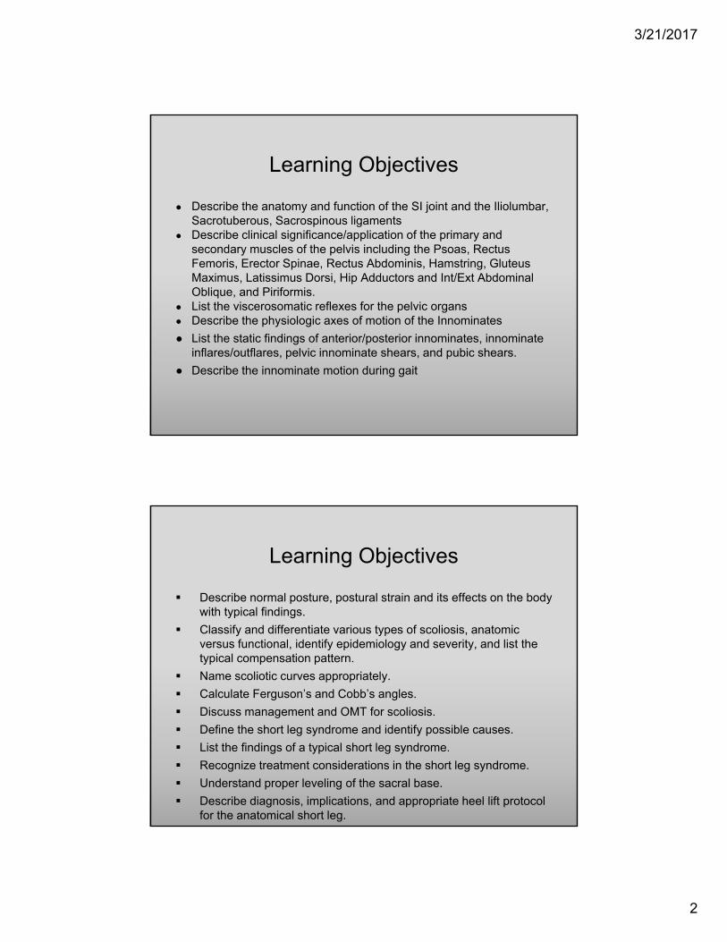
3/21/2017
1
David Shoup, D.O. Associate Professor, OPP
ATSU-SOMA
A.T. Still University Osteopathic Medicine Center Arizona
480-248-8198
OMM WorkshopPelvis and a Level
Foundation
Disclosures
2
I have no financial disclosures that would be a potential conflict of interest with this presentation
3/21/2017
2
● Describe the anatomy and function of the SI joint and the Iliolumbar, Sacrotuberous, Sacrospinous ligaments
● Describe clinical significance/application of the primary and secondary muscles of the pelvis including the Psoas, Rectus Femoris, Erector Spinae, Rectus Abdominis, Hamstring, Gluteus Maximus, Latissimus Dorsi, Hip Adductors and Int/Ext Abdominal Oblique, and Piriformis.
● List the viscerosomatic reflexes for the pelvic organs ● Describe the physiologic axes of motion of the Innominates
● List the static findings of anterior/posterior innominates, innominate inflares/outflares, pelvic innominate shears, and pubic shears.
● Describe the innominate motion during gait
Learning Objectives
Describe normal posture, postural strain and its effects on the body with typical findings.
Classify and differentiate various types of scoliosis, anatomic versus functional, identify epidemiology and severity, and list the typical compensation pattern.
Name scoliotic curves appropriately.
Calculate Ferguson’s and Cobb’s angles.
Discuss management and OMT for scoliosis.
Define the short leg syndrome and identify possible causes.
List the findings of a typical short leg syndrome.
Recognize treatment considerations in the short leg syndrome.
Understand proper leveling of the sacral base.
Describe diagnosis, implications, and appropriate heel lift protocol for the anatomical short leg.
Learning Objectives
3/21/2017
3
AnatomyRelevant Clinical
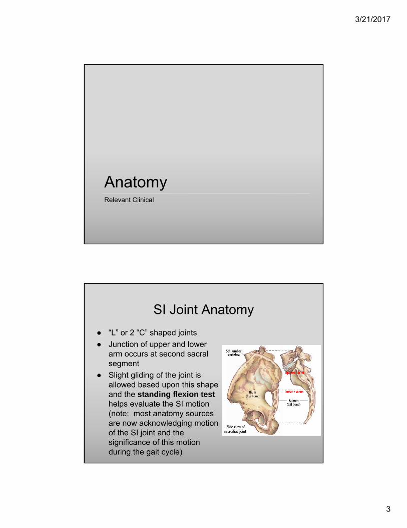
SI Joint Anatomy
● “L” or 2 “C” shaped joints
● Junction of upper and lower arm occurs at second sacral segment
● Slight gliding of the joint is allowed based upon this shape and the standing flexion test helps evaluate the SI motion (note: most anatomy sources are now acknowledging motion of the SI joint and the significance of this motion during the gait cycle)
upper arm
lower arm
3/21/2017
4
● Capable of small motion of 2-18 degrees which is not a “range” of motion, but rather more of a give to the joint.
● Sacral surface covered by hyaline cartilage● Ilial surface covered by fibrocartilage● Early in life the SI joint is flat, but with walking, the SI joint develops
distinct angular surfaces providing stabilization during the stance phase but allowing some gliding motion.
● Functions of the SI joint (which require slight motion)● Shock absorption● Torque conversion allowing transverse rotations of the lower
extremity to transmit up the spine● Self-locks or is pinned during the stance phase● Contraction of the ipsilateral Gluteus Max and contralateral
Latissiumus Dorsi provides a stabilizing force at the SI joint during the stance phase
Clinical application: Somatic dysfunction with SI joint restriction causes excessive joint friction and could lead to surrounding muscle strain.
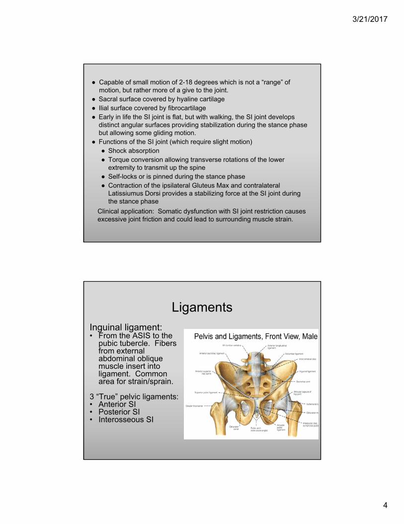
Ligaments
Inguinal ligament:• From the ASIS to the
pubic tubercle. Fibers from external abdominal oblique muscle insert into ligament. Common area for strain/sprain.
3 “True” pelvic ligaments:• Anterior SI• Posterior SI• Interosseous SI
3/21/2017
5
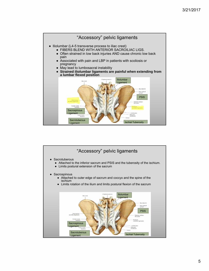
“Accessory” pelvic ligaments
● Iliolumbar (L4-5 transverse process to iliac crest)● FIBERS BLEND WITH ANTERIOR SACROILIAC LIGS. ● Often strained in low back injuries AND cause chronic low back
pain● Associated with pain and LBP in patients with scoliosis or
pregnancy● May lead to lumbosacral instability● Strained iliolumbar ligaments are painful when extending from
a lumbar flexed positionIliolumbarLigament
PSIS
SacrospinousLigament
SacrotuberousLigament Ischial Tuberosity
“Accessory” pelvic ligaments
● Sacrotuberous● Attached to the inferior sacrum and PSIS and the tuberosity of the ischium. ● Limits postural extension of the sacrum
● Sacrospinous ● Attached to outer edge of sacrum and coccyx and the spine of the
ischium● Limits rotation of the ilium and limits postural flexion of the sacrum
IliolumbarLigament
PSIS
SacrospinousLigament
SacrotuberousLigament Ischial Tuberosity
3/21/2017
6
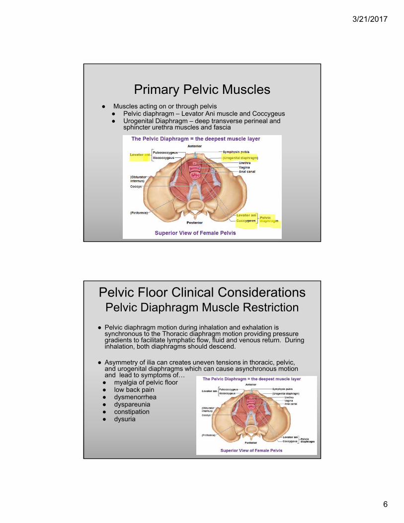
Primary Pelvic Muscles● Muscles acting on or through pelvis
● Pelvic diaphragm – Levator Ani muscle and Coccygeus● Urogenital Diaphragm – deep transverse perineal and
sphincter urethra muscles and fascia
Pelvic Floor Clinical ConsiderationsPelvic Diaphragm Muscle Restriction
● Pelvic diaphragm motion during inhalation and exhalation is synchronous to the Thoracic diaphragm motion providing pressure gradients to facilitate lymphatic flow, fluid and venous return. During inhalation, both diaphragms should descend.
● Asymmetry of ilia can creates uneven tensions in thoracic, pelvic, and urogenital diaphragms which can cause asynchronous motion and lead to symptoms of…● myalgia of pelvic floor● low back pain● dysmenorrhea● dyspareunia● constipation● dysuria
3/21/2017
7
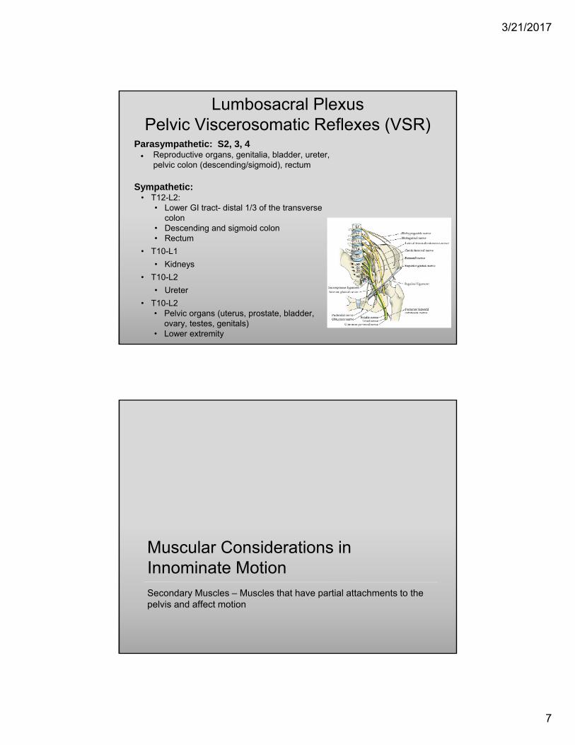
Parasympathetic: S2, 3, 4● Reproductive organs, genitalia, bladder, ureter,
pelvic colon (descending/sigmoid), rectum
Sympathetic:• T12-L2:
• Lower GI tract- distal 1/3 of the transverse colon
• Descending and sigmoid colon• Rectum
• T10-L1
• Kidneys
• T10-L2
• Ureter
• T10-L2• Pelvic organs (uterus, prostate, bladder,
ovary, testes, genitals)• Lower extremity
Lumbosacral PlexusPelvic Viscerosomatic Reflexes (VSR)
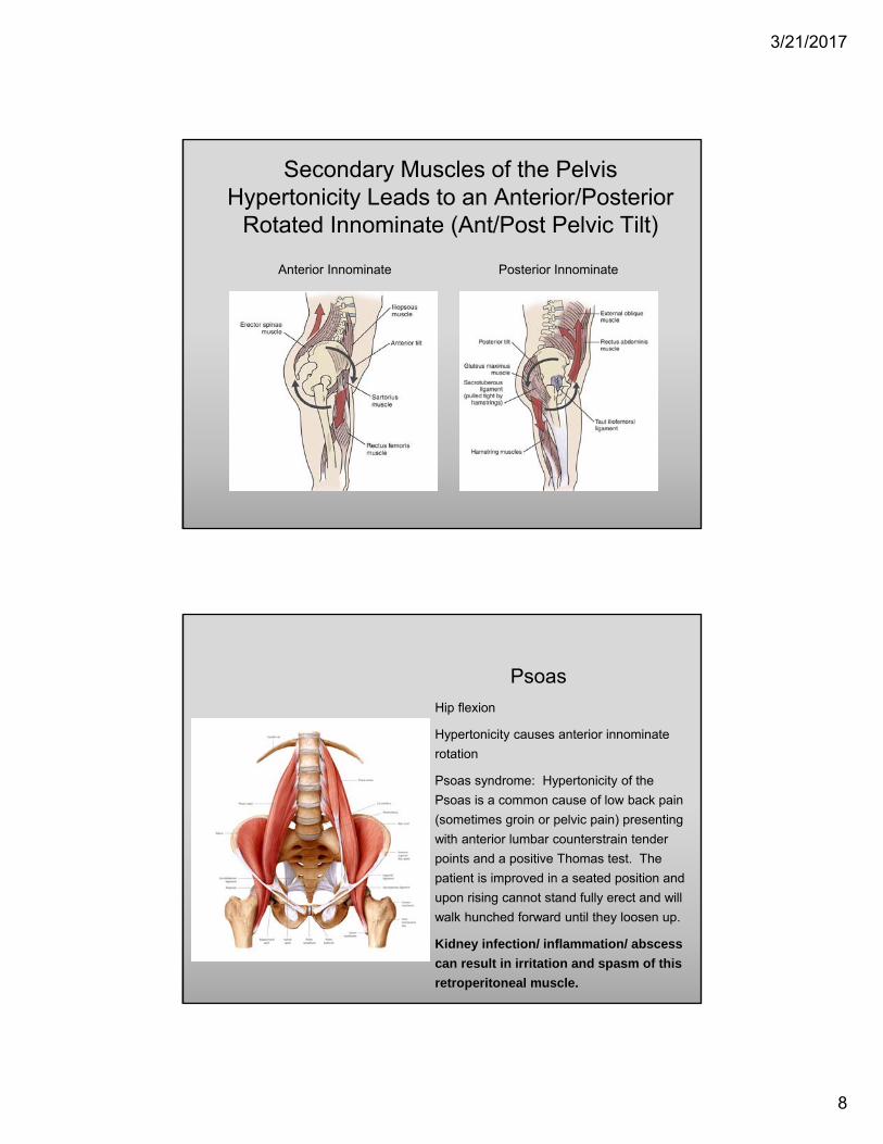
Muscular Considerations in Innominate MotionSecondary Muscles – Muscles that have partial attachments to the pelvis and affect motion
3/21/2017
8
Secondary Muscles of the Pelvis Hypertonicity Leads to an Anterior/Posterior
Rotated Innominate (Ant/Post Pelvic Tilt)
Anterior Innominate Posterior Innominate
Psoas
Hip flexion
Hypertonicity causes anterior innominate
rotation
Psoas syndrome: Hypertonicity of the
Psoas is a common cause of low back pain
(sometimes groin or pelvic pain) presenting
with anterior lumbar counterstrain tender
points and a positive Thomas test. The
patient is improved in a seated position and
upon rising cannot stand fully erect and will
walk hunched forward until they loosen up.
Kidney infection/ inflammation/ abscess
can result in irritation and spasm of this
retroperitoneal muscle.
3/21/2017
9
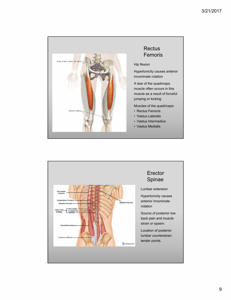
Rectus Femoris
Hip flexion
Hypertonicity causes anterior
innominate rotation
A tear of the quadriceps
muscle often occurs in this
muscle as a result of forceful
jumping or kicking
Muscles of the quadriceps:
• Rectus Femoris
• Vastus Lateralis
• Vastus Intermedius
• Vastus Medialis
Erector Spinae
Lumbar extension
Hypertonicity causes
anterior innominate
rotation
Source of posterior low
back pain and muscle
strain or spasm.
Location of posterior
lumbar counterstrain
tender points.
3/21/2017
10
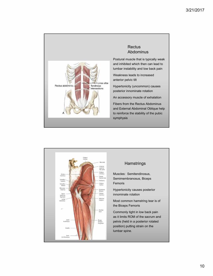
Rectus Abdominus
Postural muscle that is typically weak
and inhibited which then can lead to
lumbar instability and low back pain
Weakness leads to increased
anterior pelvic tilt
Hypertonicity (uncommon) causes
posterior innominate rotation
An accessory muscle of exhalation
Fibers from the Rectus Abdominus
and External Abdominal Oblique help
to reinforce the stability of the pubic
symphysis
Hamstrings
Muscles: Semitendinosus,
Semimembranosus, Biceps
Femoris
Hypertonicity causes posterior
innominate rotation
Most common hamstring tear is of
the Biceps Femoris
Commonly tight in low back pain
as it limits ROM of the sacrum and
pelvis (held in a posterior rotated
position) putting strain on the
lumbar spine.
3/21/2017
11
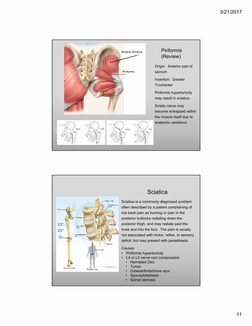
Piriformis (Review)
Origin: Anterior part of
sacrum
Insertion: Greater
Trochanter
Piriformis hypertonicity
may result in sciatica.
Sciatic nerve may
become entrapped within
the muscle itself due to
anatomic variations.
Sciatica
Sciatica is a commonly diagnosed problem
often described by a patient complaining of
low back pain as burning or pain in the
posterior buttocks radiating down the
posterior thigh, and may radiate past the
knee and into the foot. The pain is usually
not associated with motor, reflex, or sensory
deficit, but may present with parasthesia.
Causes• Piriformis hypertonicity• L4 or L5 nerve root compression
• Herniated Disc• Tumor• Osteoarthritis/bone spur• Spondylolisthesis• Spinal stenosis
3/21/2017
12
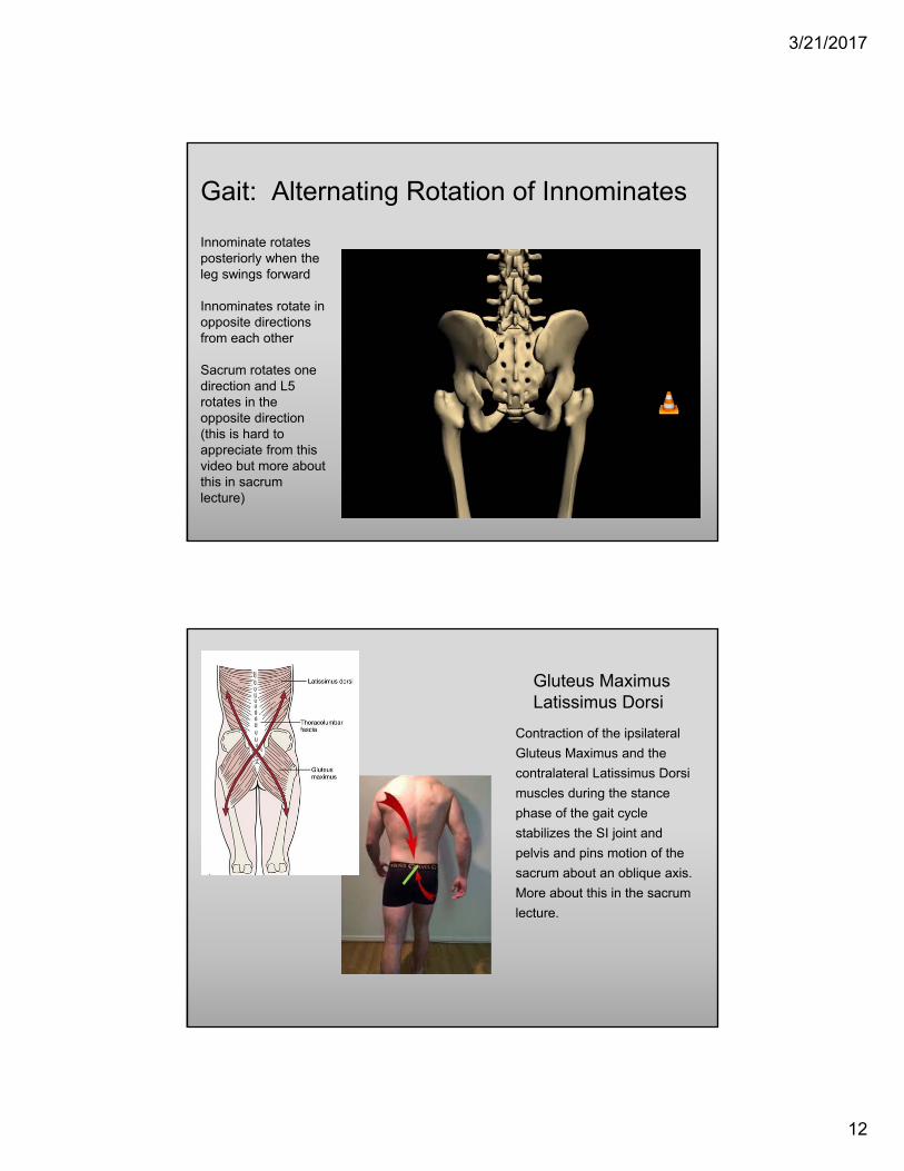
Gait: Alternating Rotation of Innominates
Innominate rotates posteriorly when the leg swings forward
Innominates rotate in opposite directions from each other
Sacrum rotates one direction and L5 rotates in the opposite direction (this is hard to appreciate from this video but more about this in sacrum lecture)
gait2.avi
Gluteus MaximusLatissimus Dorsi
Contraction of the ipsilateral
Gluteus Maximus and the
contralateral Latissimus Dorsi
muscles during the stance
phase of the gait cycle
stabilizes the SI joint and
pelvis and pins motion of the
sacrum about an oblique axis.
More about this in the sacrum
lecture.
3/21/2017
13
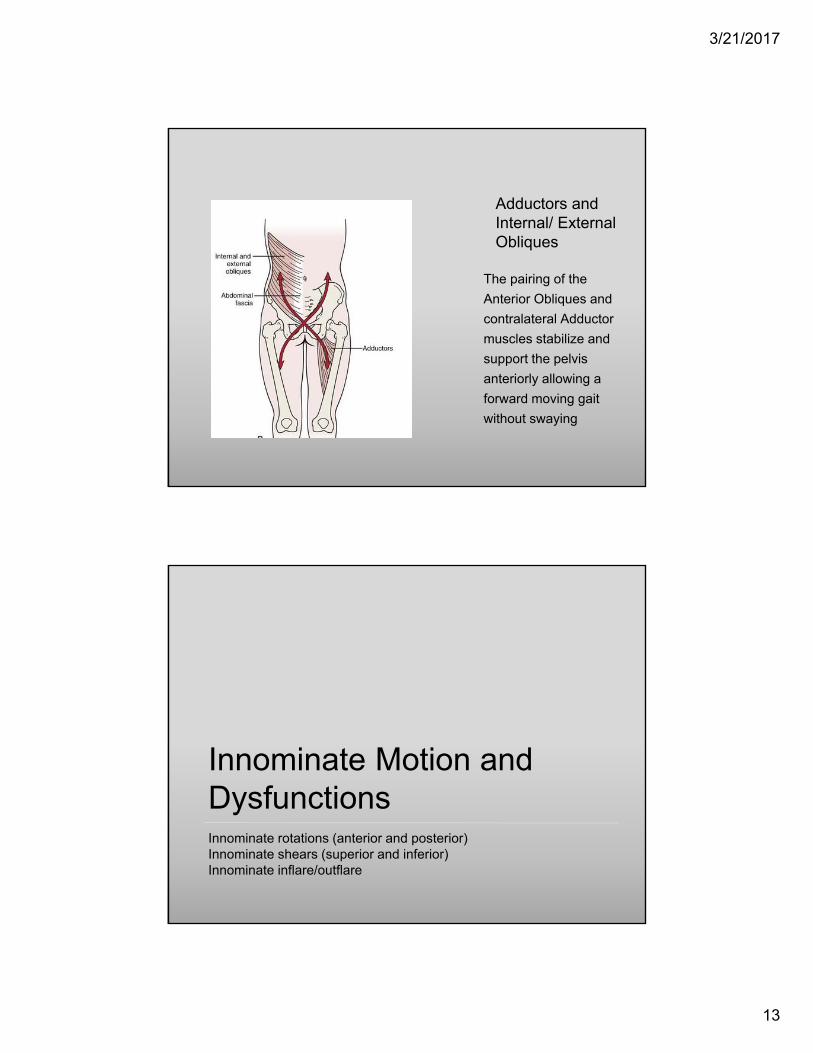
Adductors and Internal/ External Obliques
The pairing of the
Anterior Obliques and
contralateral Adductor
muscles stabilize and
support the pelvis
anteriorly allowing a
forward moving gait
without swaying
Innominate Motion and DysfunctionsInnominate rotations (anterior and posterior)Innominate shears (superior and inferior)Innominate inflare/outflare
3/21/2017
14
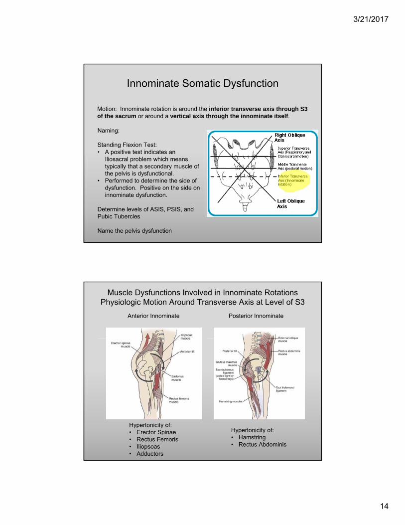
Motion: Innominate rotation is around the inferior transverse axis through S3 of the sacrum or around a vertical axis through the innominate itself.
Innominate Somatic Dysfunction
Naming:
Standing Flexion Test: • A positive test indicates an
Iliosacral problem which means typically that a secondary muscle of the pelvis is dysfunctional.
• Performed to determine the side of dysfunction. Positive on the side on innominate dysfunction.
Determine levels of ASIS, PSIS, and Pubic Tubercles
Name the pelvis dysfunction
Muscle Dysfunctions Involved in Innominate RotationsPhysiologic Motion Around Transverse Axis at Level of S3
Anterior Innominate Posterior Innominate
Hypertonicity of:• Erector Spinae• Rectus Femoris• Iliopsoas• Adductors
Hypertonicity of:• Hamstring• Rectus Abdominis
3/21/2017
15
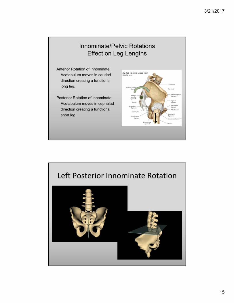
Innominate/Pelvic Rotations Effect on Leg Lengths
Anterior Rotation of Innominate:
Acetabulum moves in caudad
direction creating a functional
long leg.
Posterior Rotation of Innominate:
Acetabulum moves in cephalad
direction creating a functional
short leg.
Left Posterior Innominate Rotation
3/21/2017
16
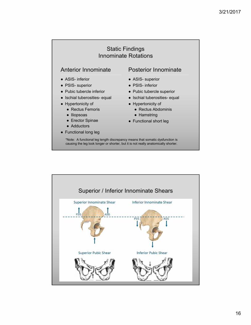
Anterior Innominate
● ASIS- inferior
● PSIS- superior
● Pubic tubercle inferior
● Ischial tuberosities- equal
● Hypertonicity of● Rectus Femoris● Iliopsoas● Erector Spinae● Adductors
● Functional long leg
● ASIS- superior
● PSIS- inferior
● Pubic tubercle superior
● Ischial tuberosities- equal
● Hypertonicity of ● Rectus Abdominis● Hamstring
● Functional short leg
Static Findings Innominate Rotations
Posterior Innominate
*Note: A functional leg length discrepancy means that somatic dysfunction is causing the leg look longer or shorter, but it is not really anatomically shorter.
Superior / Inferior Innominate Shears
3/21/2017
17
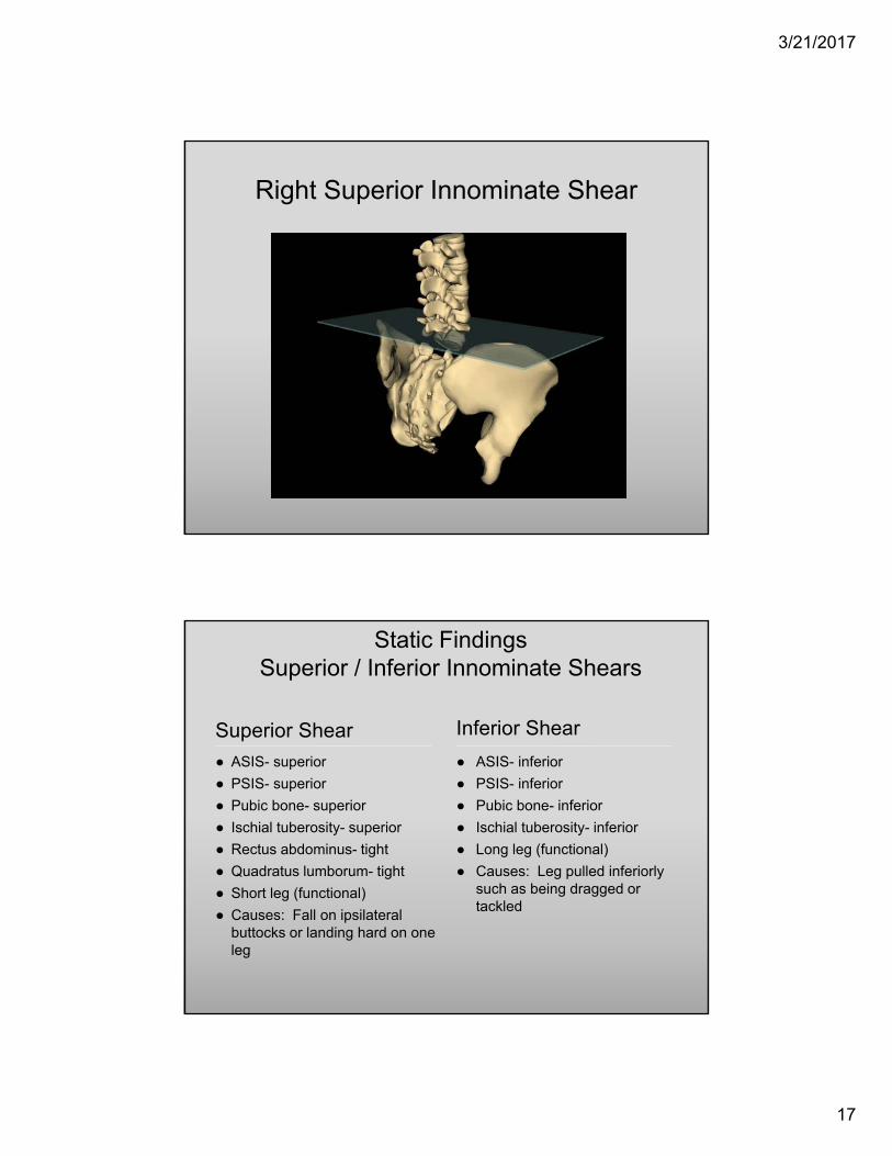
Right Superior Innominate Shear
Superior Shear
● ASIS- superior
● PSIS- superior
● Pubic bone- superior
● Ischial tuberosity- superior
● Rectus abdominus- tight
● Quadratus lumborum- tight
● Short leg (functional)
● Causes: Fall on ipsilateral buttocks or landing hard on one leg
● ASIS- inferior
● PSIS- inferior
● Pubic bone- inferior
● Ischial tuberosity- inferior
● Long leg (functional)
● Causes: Leg pulled inferiorly such as being dragged or tackled
Static Findings Superior / Inferior Innominate Shears
Inferior Shear
3/21/2017
18
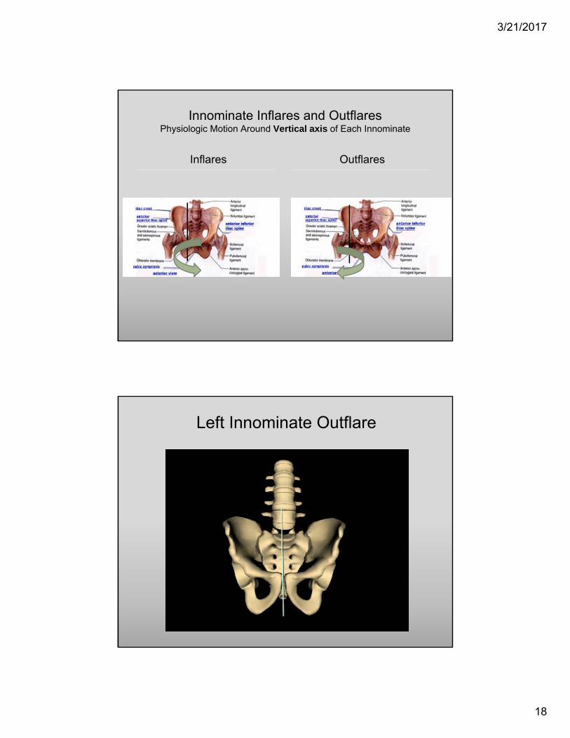
Inflares
Innominate Inflares and OutflaresPhysiologic Motion Around Vertical axis of Each Innominate
Outflares
Left Innominate Outflare

3/21/2017
19
Inflared Innominate
● ASIS- medial or closer to umbilicus
● PSIS- lateral
● Ischial tuberosity- lateral
● ASIS- lateral or further away from umbilicus
● PSIS- medial
● Ischial tuberosity- medial
Static FindingsInnominate Inflares and Outflares
Outflared Innominate
Note: Measure the distance from the ASIS to the umbilicus and compare bilaterally for a quick assessment
Anterior/Posterior
Pubic Non-Physiologic Motion Pubic Shears
Superior/Inferior
3/21/2017
20
Superior Pubic Shear
● Pubic bone measured at the pubic tubercle- superior
● Pubic bone measured at the pubic tubercle- inferior
Static Findings Pubic Shears
Inferior Pubic Shear
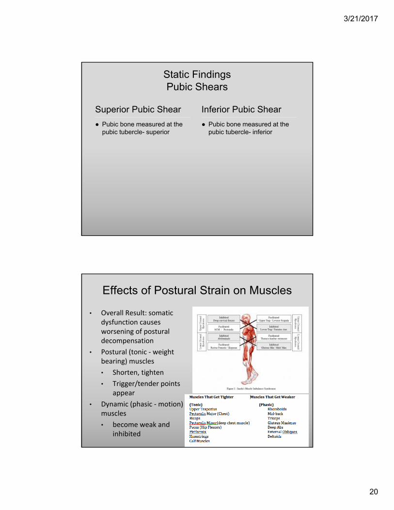
Effects of Postural Strain on Muscles
• Overall Result: somatic dysfunction causes worsening of postural decompensation
• Postural (tonic ‐ weight bearing) muscles
• Shorten, tighten
• Trigger/tender points appear
• Dynamic (phasic ‐motion) muscles
• become weak and inhibited
3/21/2017
21
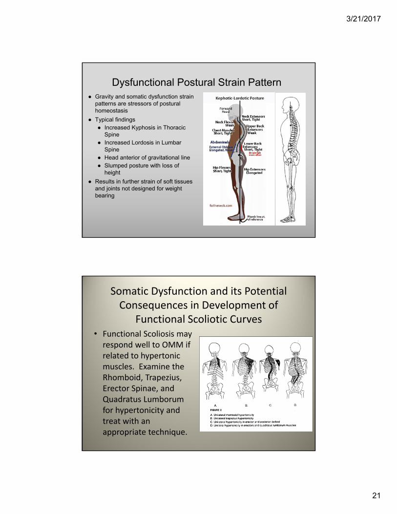
Dysfunctional Postural Strain Pattern● Gravity and somatic dysfunction strain
patterns are stressors of postural homeostasis
● Typical findings● Increased Kyphosis in Thoracic
Spine● Increased Lordosis in Lumbar
Spine● Head anterior of gravitational line● Slumped posture with loss of
height
● Results in further strain of soft tissues and joints not designed for weight bearing
Somatic Dysfunction and its Potential Consequences in Development of
Functional Scoliotic Curves• Functional Scoliosis may respond well to OMM if related to hypertonic muscles. Examine the Rhomboid, Trapezius, Erector Spinae, and Quadratus Lumborumfor hypertonicity and treat with an appropriate technique.
3/21/2017
22
Short Leg Syndrome Often Associated with Scoliosis
• Somatic dysfunction which causes a functional scoliosis may lead to an unleveling of the sacral base. The leg may appear to be a short leg (functional short leg) but may not actually be short as in a true anatomic short leg.
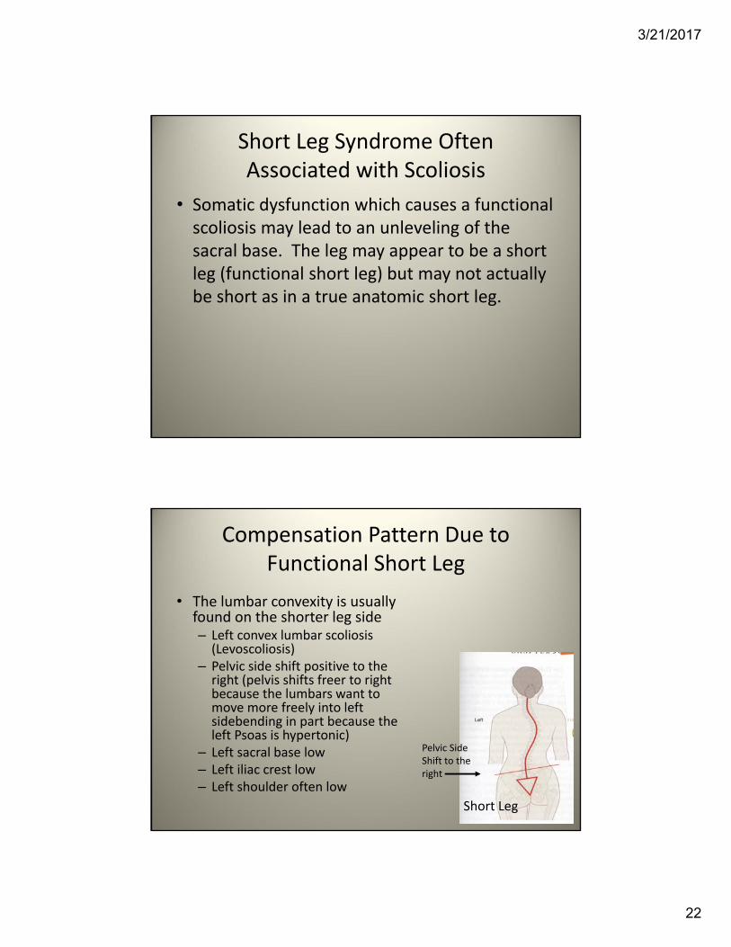
Compensation Pattern Due to Functional Short Leg
• The lumbar convexity is usually found on the shorter leg side– Left convex lumbar scoliosis (Levoscoliosis)
– Pelvic side shift positive to the right (pelvis shifts freer to right because the lumbars want to move more freely into left sidebending in part because the left Psoas is hypertonic)
– Left sacral base low– Left iliac crest low– Left shoulder often low
Short Leg
Pelvic Side Shift to the right
3/21/2017
23
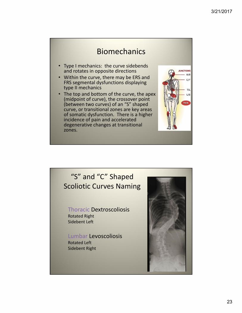
Biomechanics
• Type I mechanics: the curve sidebendsand rotates in opposite directions
• Within the curve, there may be ERS and FRS segmental dysfunctions displaying type II mechanics
• The top and bottom of the curve, the apex (midpoint of curve), the crossover point (between two curves) of an “S” shaped curve, or transitional zones are key areas of somatic dysfunction. There is a higher incidence of pain and accelerated degenerative changes at transitional zones.
“S” and “C” Shaped Scoliotic Curves Naming
Thoracic DextroscoliosisRotated RightSidebent Left
Lumbar LevoscoliosisRotated LeftSidebent Right
3/21/2017
24
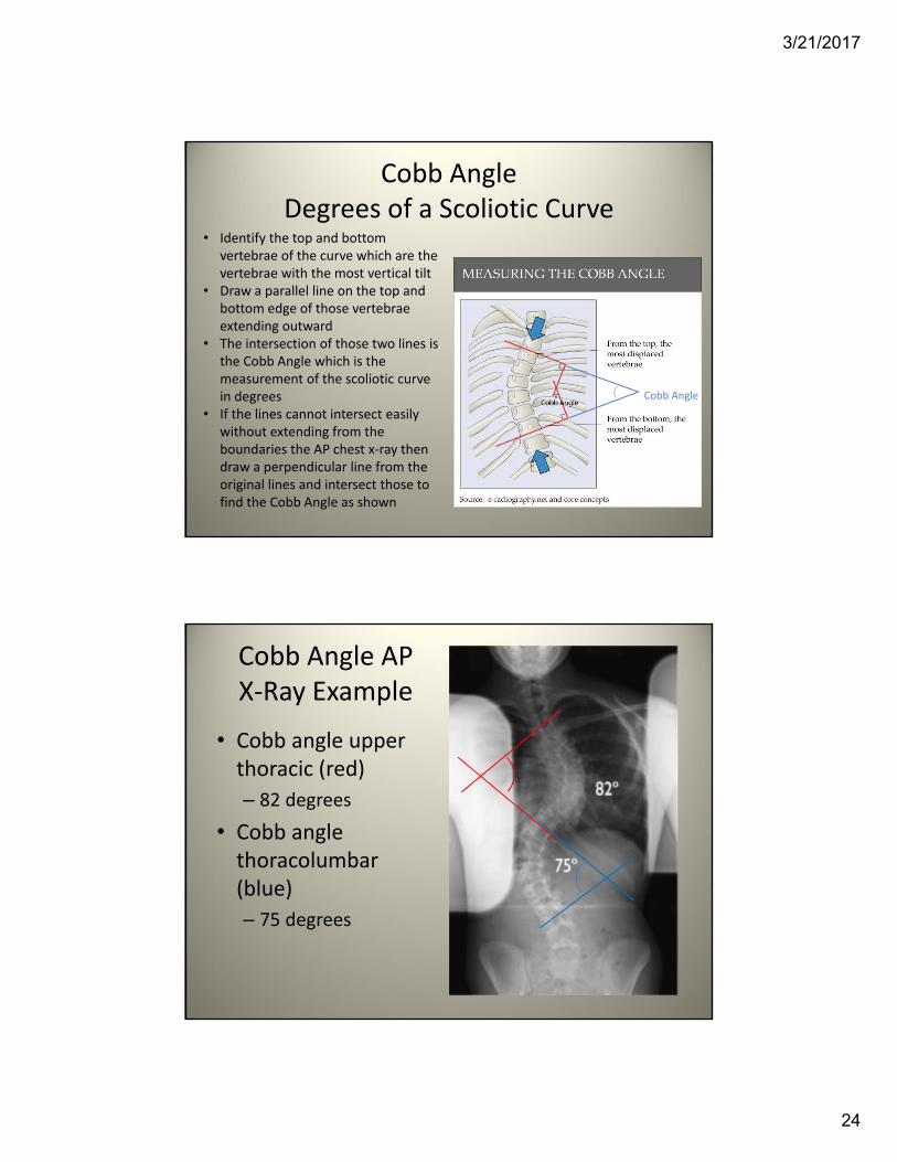
Cobb AngleDegrees of a Scoliotic Curve
Cobb Angle
• Identify the top and bottom vertebrae of the curve which are the vertebrae with the most vertical tilt
• Draw a parallel line on the top and bottom edge of those vertebrae extending outward
• The intersection of those two lines is the Cobb Angle which is the measurement of the scoliotic curve in degrees
• If the lines cannot intersect easily without extending from the boundaries the AP chest x‐ray then draw a perpendicular line from the original lines and intersect those to find the Cobb Angle as shown
Cobb Angle AP X‐Ray Example
• Cobb angle upper thoracic (red)
– 82 degrees
• Cobb angle thoracolumbar (blue)
– 75 degrees
3/21/2017
25
Structural Versus Functional Scoliosis Classification
• Structural– Inflexible or fixed– Muscles and ligaments shortened on the concave side– Fail to correct with side to side motion in a forward bending position
– Curve is present regardless of the patient position and don’t improve with sidebending, rotation, flexion, or heel lift therapy
• Functional– Flexible– Improves when patient lays down. The scoliosis may be secondary to an injury, muscle spasm, or leg length inequality. Responds well to OMT.
– May become structural
Treatment of Scoliosis
• Early screening and intervention is important• An early osteopathic structural exam and imaging are lead to early intervention. Abnormal stresses eventually lead to structural changes (Wolff's law)
• Mild Scoliosis: If < 20 degrees or if near skeletal maturity treat using conservative management – OMT, PT, home exercise prescription
• Goal: improve flexibility, strengthen trunk and abdominal muscles and balance muscle tone
– Traction may also be useful – Orthotics/lift therapy– Periodic monitoring for progression
3/21/2017
26
OMT Goals in Functional Scoliosis
• Optimize function of entire person– Balance muscle/fascial tension in an effort to straighten curves with a goal of maintaining a curve of < 40 degrees during skeletal maturation
– Stabilize foundation of spine, evaluate for leg length inequalities, and level the sacral base
– Relieve pain
– Remove somatic dysfunction (be careful some of these might be compensatory)
– Increase range of motion
– Enhance fluid and tissue motion
Short Leg Syndrome (Leg Length Inequality) and Sacral Base Unleveling
• The syndrome is directly related to an un‐leveling of the sacral base – which can be caused by multiple factors.
3/21/2017
27
Definition
• The term “Short Leg Syndrome” is recognized as a “misnomer” because the cause of the condition may not be related to an actual leg length difference. Other diagnoses that are common used are “Leg Length Inequality/anatomic short leg” or “Leg Length Discrepancy/functional short leg.”
• It is called a syndrome because it is associated with a variety of biomechanical findings and symptoms. The syndrome is directly related to an un‐leveling of the sacral base which can be caused by multiple factors, somatic dysfunction being one of the most common.
Anatomic versus Functional Short Leg
• Anatomic Short Leg
– One leg is anatomically shorter than the other
• Functional Short Leg
– One leg appears shorter than the other but is secondary to pelvic dysfunction or other structural imbalance or scoliotic curve.
3/21/2017
28
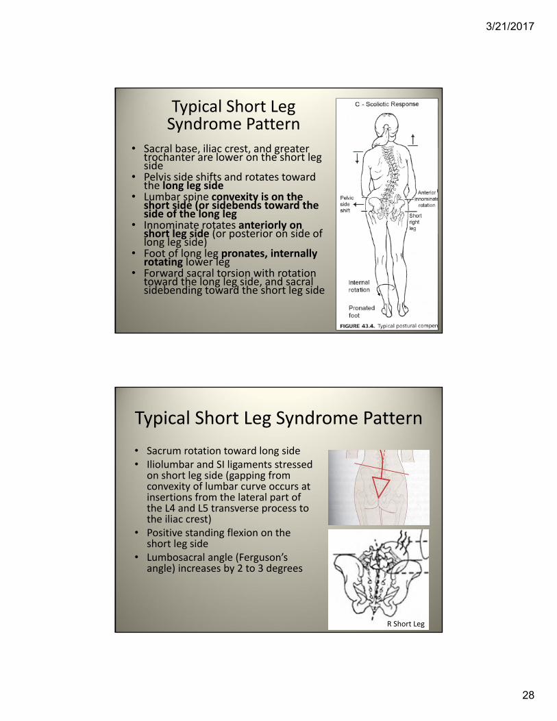
Typical Short Leg Syndrome Pattern
• Sacral base, iliac crest, and greater trochanter are lower on the short leg side
• Pelvis side shifts and rotates toward the long leg side
• Lumbar spine convexity is on the short side (or sidebends toward the side of the long leg
• Innominate rotates anteriorly on short leg side (or posterior on side of long leg side)
• Foot of long leg pronates, internally rotating lower leg
• Forward sacral torsion with rotation toward the long leg side, and sacral sidebending toward the short leg side
Typical Short Leg Syndrome Pattern
• Sacrum rotation toward long side• Iliolumbar and SI ligaments stressed
on short leg side (gapping from convexity of lumbar curve occurs at insertions from the lateral part of the L4 and L5 transverse process to the iliac crest)
• Positive standing flexion on the short leg side
• Lumbosacral angle (Ferguson’s angle) increases by 2 to 3 degrees
R Short Leg
3/21/2017
29
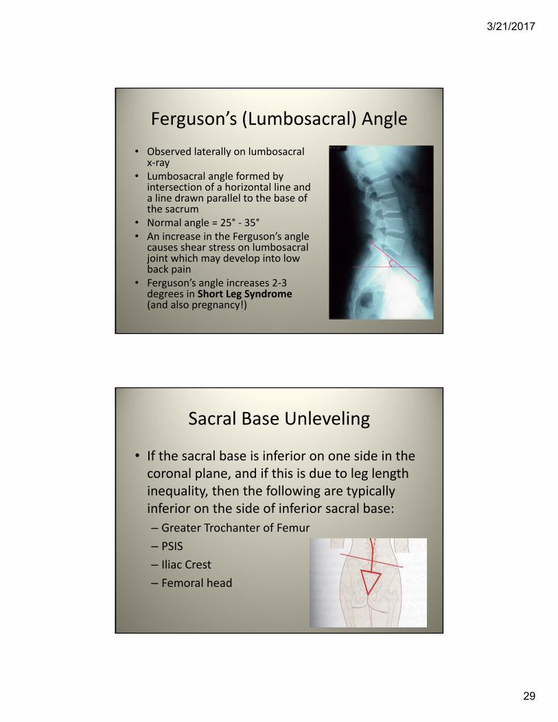
Ferguson’s (Lumbosacral) Angle
• Observed laterally on lumbosacral x‐ray
• Lumbosacral angle formed by intersection of a horizontal line and a line drawn parallel to the base of the sacrum
• Normal angle = 25° ‐ 35°• An increase in the Ferguson’s angle
causes shear stress on lumbosacral joint which may develop into low back pain
• Ferguson’s angle increases 2‐3 degrees in Short Leg Syndrome(and also pregnancy!)
Sacral Base Unleveling
• If the sacral base is inferior on one side in the coronal plane, and if this is due to leg length inequality, then the following are typically inferior on the side of inferior sacral base:
– Greater Trochanter of Femur
– PSIS
– Iliac Crest
– Femoral head
3/21/2017
30
Diagnosis and Treatment Considerations
• Compensatory mechanisms are sometimes so effective that a single landmark measurement may provide inaccurate information regarding the total biomechanics. (ex: medial malleolus levels)
• Care must be taken to not “fix” a single somatic dysfunction without considering the possible ramifications in other regions that may have compensated for the dysfunction.
• OMT should be directed to all related somatic dysfunctions prior to diagnosing a short leg.
Diagnosis and Treatment Considerations Continued
• Consider recurrent patterns of somatic dysfunction.
• Comparing levels of the medial malleoli and ASIS levels in the supine position offers a quick leg length assessment.
• In the standing position measurement of the levels of the greater trochanters may be more reliable than the supine evaluation when looking for a short leg.
• Standing AP pelvis with plumb bob are the most accurate way to assess leg lengths.
3/21/2017
31
Short Leg Diagnosis
• Standing
– Observe iliac crest height, greater trochanter levels, degree of scoliotic curves, angle of the scapula and shoulder levels, PSIS/ASIS levels
– Obtain standing postural x‐ray – to evaluate sacral base unleveling
Short Leg Diagnosis
• Supine/Prone– Check leg lengths at medial malleolus
– Look at ASIS levels
– Check for pelvic dysfunctions (innominate shears and rotations), and sacral dysfunctions
– Examine lumbar musculature and segmental dysfunctions
– Check for counterstrain tenderpoints of hips and anterior lumbar/pelvis
– Perform orthopedic tests
3/21/2017
32
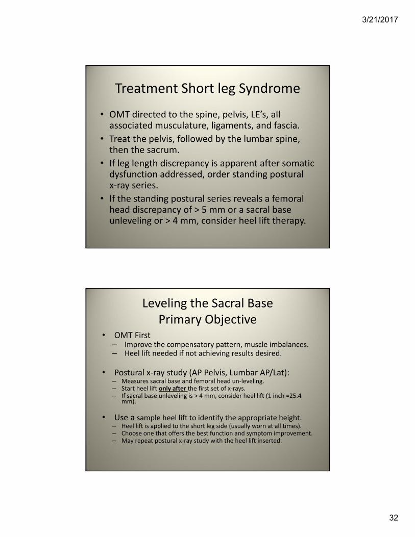
Treatment Short leg Syndrome
• OMT directed to the spine, pelvis, LE’s, all associated musculature, ligaments, and fascia.
• Treat the pelvis, followed by the lumbar spine, then the sacrum.
• If leg length discrepancy is apparent after somatic dysfunction addressed, order standing postural x‐ray series.
• If the standing postural series reveals a femoral head discrepancy of > 5 mm or a sacral base unleveling or > 4 mm, consider heel lift therapy.
Leveling the Sacral BasePrimary Objective
• OMT First– Improve the compensatory pattern, muscle imbalances.– Heel lift needed if not achieving results desired.
• Postural x‐ray study (AP Pelvis, Lumbar AP/Lat): – Measures sacral base and femoral head un‐leveling. – Start heel lift only after the first set of x‐rays.– If sacral base unleveling is > 4 mm, consider heel lift (1 inch =25.4
mm).
• Use a sample heel lift to identify the appropriate height.– Heel lift is applied to the short leg side (usually worn at all times).– Choose one that offers the best function and symptom improvement. – May repeat postural x‐ray study with the heel lift inserted.
3/21/2017
33
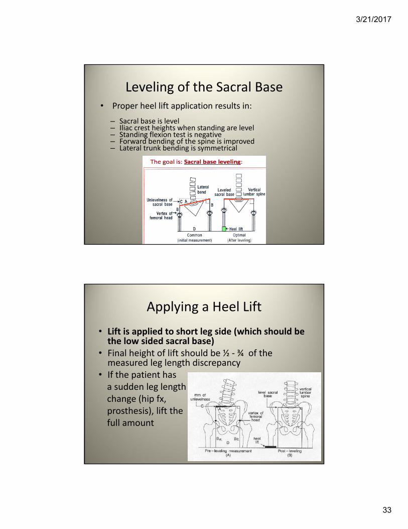
Leveling of the Sacral Base• Proper heel lift application results in:
– Sacral base is level– Iliac crest heights when standing are level– Standing flexion test is negative– Forward bending of the spine is improved– Lateral trunk bending is symmetrical
Applying a Heel Lift
• Lift is applied to short leg side (which should be the low sided sacral base)
• Final height of lift should be ½ ‐ ¾ of the measured leg length discrepancy
• If the patient has a sudden leg length change (hip fx, prosthesis), lift the full amount
3/21/2017
34
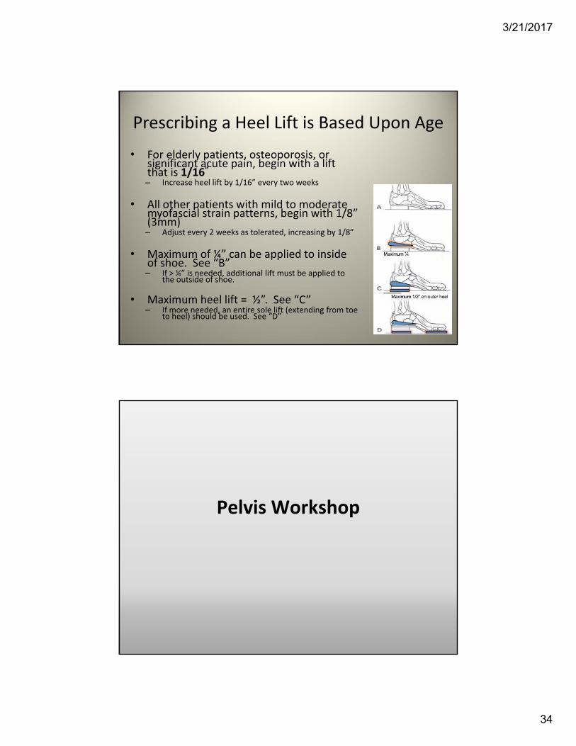
Prescribing a Heel Lift is Based Upon Age
• For elderly patients, osteoporosis, or significant acute pain, begin with a lift that is 1/16”– Increase heel lift by 1/16” every two weeks
• All other patients with mild to moderate myofascial strain patterns, begin with 1/8” (3mm)– Adjust every 2 weeks as tolerated, increasing by 1/8”
• Maximum of ¼” can be applied to inside of shoe. See “B”– If > ¼” is needed, additional lift must be applied to
the outside of shoe.
• Maximum heel lift = ½”. See “C”– If more needed, an entire sole lift (extending from toe
to heel) should be used. See “D”
Pelvis Workshop
3/21/2017
35
Learning Objectives
• Diagnose the pelvis using appropriate landmarks and performing the Trendelenburg test, the hip drop test, and the standing/seated flexion tests
• Perform muscle energy treatments for a pubic compression, an anterior/posterior innominate and an inflared/outflaredinnominate
• Perform HVLA Leg Tug on a superior innominate shear
• Perform Counterstrain on Iliopsoas (A3‐4L), Pectineus/Inguinal Ligament, Rectus Abdominus (A5L) muscle tender points
• Identify and treat Chapman’s points of pelvis (Ant & Post)
• Synchronize thoracic and pelvic diaphragms by performing a pelvic diaphragm release
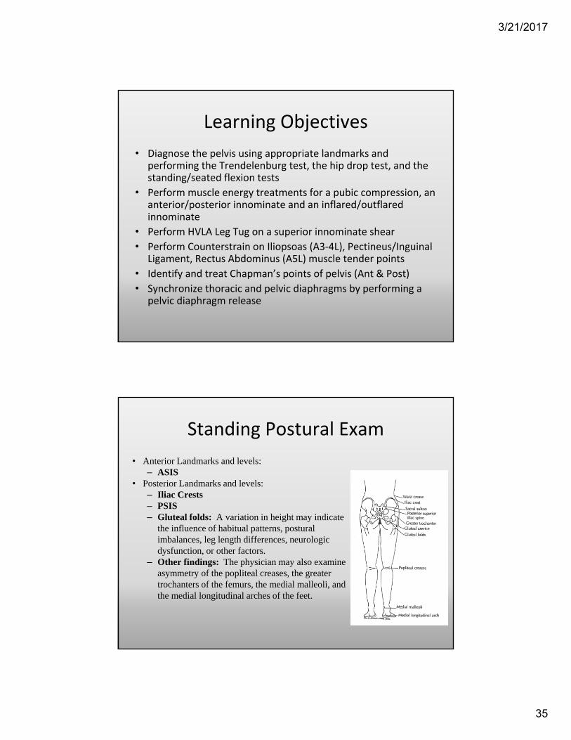
Standing Postural Exam
• Anterior Landmarks and levels:– ASIS
• Posterior Landmarks and levels:– Iliac Crests– PSIS– Gluteal folds: A variation in height may indicate
the influence of habitual patterns, postural imbalances, leg length differences, neurologic dysfunction, or other factors.
– Other findings: The physician may also examine asymmetry of the popliteal creases, the greater trochanters of the femurs, the medial malleoli, and the medial longitudinal arches of the feet.
3/21/2017
36
Pelvis Diagnosis Overview
• Determine lateralization– Standing flexion test (testing innominate/iliosacraldysfunctions)
– Seated flexion test (testing sacroiliac dysfunctions)
– ASIS compression test (Hip flop first!)
• Screen for leg length discrepancy – medial malleolus
• Anterior– ASIS
– Pubic tubercles
• Posterior– PSIS
– Ischial tuberosity
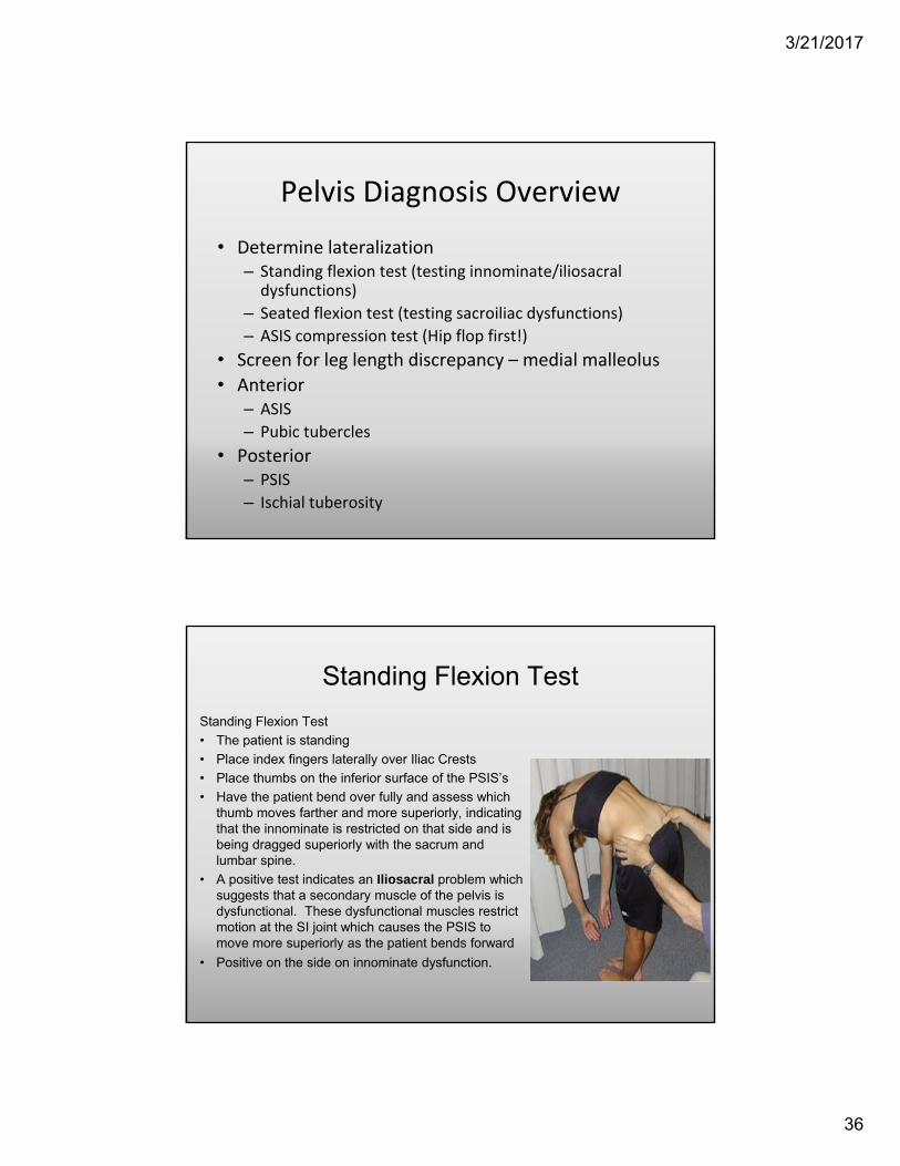
Standing Flexion Test
Standing Flexion Test
• The patient is standing
• Place index fingers laterally over Iliac Crests
• Place thumbs on the inferior surface of the PSIS’s
• Have the patient bend over fully and assess which thumb moves farther and more superiorly, indicating that the innominate is restricted on that side and is being dragged superiorly with the sacrum and lumbar spine.
• A positive test indicates an Iliosacral problem which suggests that a secondary muscle of the pelvis is dysfunctional. These dysfunctional muscles restrict motion at the SI joint which causes the PSIS to move more superiorly as the patient bends forward
• Positive on the side on innominate dysfunction.
3/21/2017
37
Seated Flexion Test
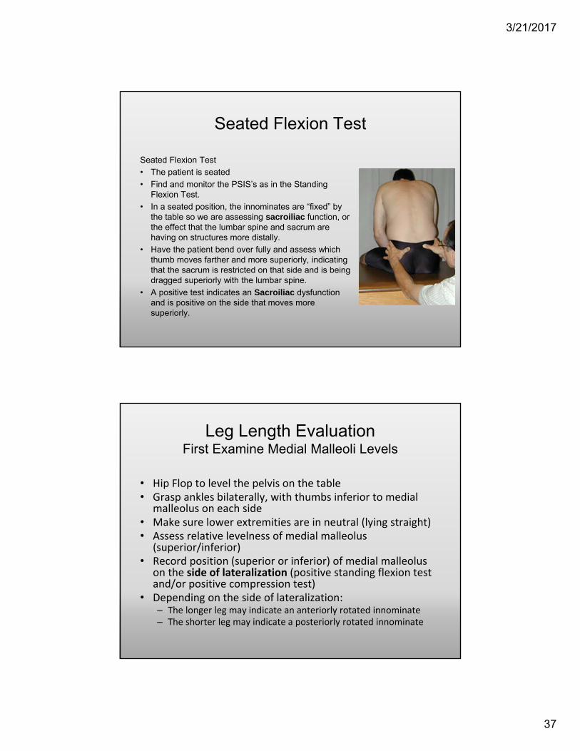
Seated Flexion Test
• The patient is seated
• Find and monitor the PSIS’s as in the Standing Flexion Test.
• In a seated position, the innominates are “fixed” by the table so we are assessing sacroiliac function, or the effect that the lumbar spine and sacrum are having on structures more distally.
• Have the patient bend over fully and assess which thumb moves farther and more superiorly, indicating that the sacrum is restricted on that side and is being dragged superiorly with the lumbar spine.
• A positive test indicates an Sacroiliac dysfunction and is positive on the side that moves more superiorly.
Leg Length EvaluationFirst Examine Medial Malleoli Levels
• Hip Flop to level the pelvis on the table• Grasp ankles bilaterally, with thumbs inferior to medial
malleolus on each side• Make sure lower extremities are in neutral (lying straight)• Assess relative levelness of medial malleolus
(superior/inferior)• Record position (superior or inferior) of medial malleolus
on the side of lateralization (positive standing flexion test and/or positive compression test)
• Depending on the side of lateralization:– The longer leg may indicate an anteriorly rotated innominate– The shorter leg may indicate a posteriorly rotated innominate
3/21/2017
38
Examine Tibia and Femur Lengths
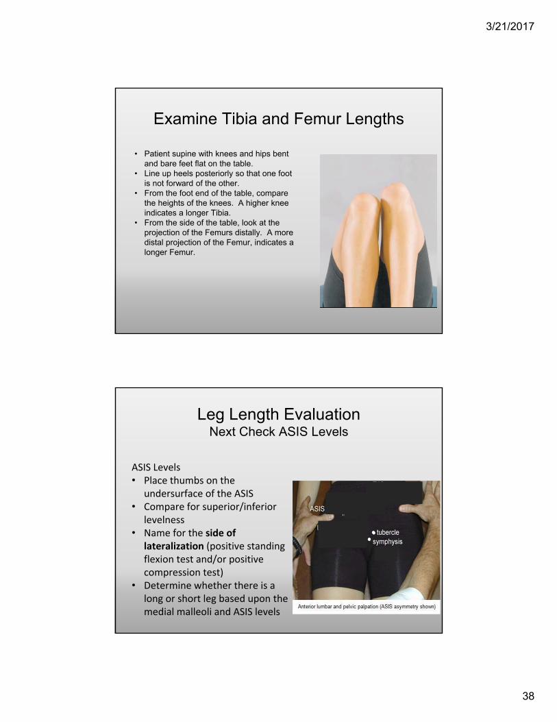
• Patient supine with knees and hips bent and bare feet flat on the table.
• Line up heels posteriorly so that one foot is not forward of the other.
• From the foot end of the table, compare the heights of the knees. A higher knee indicates a longer Tibia.
• From the side of the table, look at the projection of the Femurs distally. A more distal projection of the Femur, indicates a longer Femur.
Leg Length EvaluationNext Check ASIS Levels
ASIS Levels• Place thumbs on the
undersurface of the ASIS• Compare for superior/inferior
levelness• Name for the side of
lateralization (positive standing flexion test and/or positive compression test)
• Determine whether there is a long or short leg based upon the medial malleoli and ASIS levels
3/21/2017
39
PSIS Levels
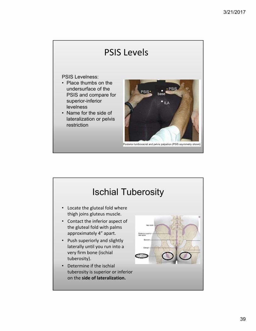
PSIS Levelness:• Place thumbs on the
undersurface of the PSIS and compare for superior-inferior levelness
• Name for the side of lateralization or pelvis restriction
Ischial Tuberosity
• Locate the gluteal fold where thigh joins gluteus muscle.
• Contact the inferior aspect of the gluteal fold with palms approximately 4” apart.
• Push superiorly and slightly laterally until you run into a very firm bone (ischial tuberosity).
• Determine if the ischial tuberosity is superior or inferior on the side of lateralization.
3/21/2017
40
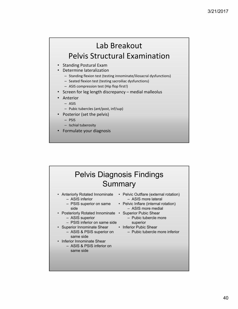
Lab Breakout Pelvis Structural Examination
• Standing Postural Exam• Determine lateralization
– Standing flexion test (testing innominate/iliosacral dysfunctions)
– Seated flexion test (testing sacroiliac dysfunctions)
– ASIS compression test (Hip flop first!)
• Screen for leg length discrepancy – medial malleolus
• Anterior– ASIS
– Pubic tubercles (ant/post, inf/sup)
• Posterior (set the pelvis)– PSIS
– Ischial tuberosity
• Formulate your diagnosis
Pelvis Diagnosis Findings Summary
• Anteriorly Rotated Innominate– ASIS inferior– PSIS superior on same
side• Posteriorly Rotated Innominate
– ASIS superior– PSIS inferior on same side
• Superior Innominate Shear– ASIS & PSIS superior on
same side• Inferior Innominate Shear
– ASIS & PSIS inferior on same side
• Pelvic Outflare (external rotation)– ASIS more lateral
• Pelvic Inflare (internal rotation)– ASIS more medial
• Superior Pubic Shear– Pubic tubercle more
superior• Inferior Pubic Shear
– Pubic tubercle more inferior
3/21/2017
41
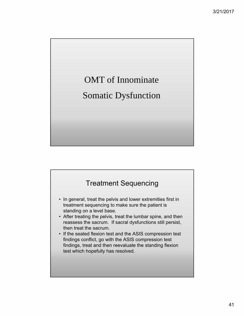
OMT of Innominate
Somatic Dysfunction
Treatment Sequencing
• In general, treat the pelvis and lower extremities first in treatment sequencing to make sure the patient is standing on a level base.
• After treating the pelvis, treat the lumbar spine, and then reassess the sacrum. If sacral dysfunctions still persist, then treat the sacrum.
• If the seated flexion test and the ASIS compression test findings conflict, go with the ASIS compression test findings, treat and then reevaluate the standing flexion test which hopefully has resolved.
3/21/2017
42
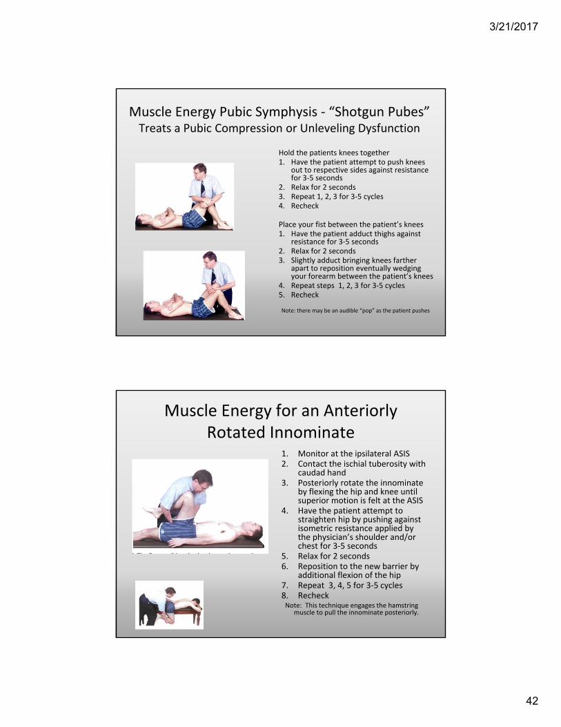
Muscle Energy Pubic Symphysis ‐ “Shotgun Pubes”Treats a Pubic Compression or Unleveling Dysfunction
Hold the patients knees together1. Have the patient attempt to push knees
out to respective sides against resistance for 3‐5 seconds
2. Relax for 2 seconds3. Repeat 1, 2, 3 for 3‐5 cycles4. Recheck
Place your fist between the patient’s knees1. Have the patient adduct thighs against
resistance for 3‐5 seconds2. Relax for 2 seconds3. Slightly adduct bringing knees farther
apart to reposition eventually wedging your forearm between the patient’s knees
4. Repeat steps 1, 2, 3 for 3‐5 cycles5. Recheck
Note: there may be an audible “pop” as the patient pushes
Muscle Energy for an Anteriorly Rotated Innominate
1. Monitor at the ipsilateral ASIS2. Contact the ischial tuberosity with
caudad hand3. Posteriorly rotate the innominate
by flexing the hip and knee until superior motion is felt at the ASIS
4. Have the patient attempt to straighten hip by pushing against isometric resistance applied by the physician’s shoulder and/or chest for 3‐5 seconds
5. Relax for 2 seconds6. Reposition to the new barrier by
additional flexion of the hip7. Repeat 3, 4, 5 for 3‐5 cycles8. RecheckNote: This technique engages the hamstring
muscle to pull the innominate posteriorly.
3/21/2017
43
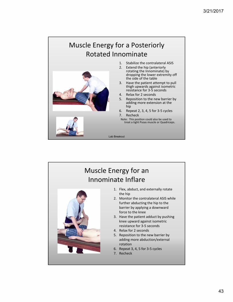
Muscle Energy for a Posteriorly Rotated Innominate
1. Stabilize the contralateral ASIS 2. Extend the hip (anteriorly
rotating the Innominate) by dropping the lower extremity off the side of the table
3. Have the patient attempt to pull thigh upwards against isometric resistance for 3‐5 seconds
4. Relax for 2 seconds5. Reposition to the new barrier by
adding more extension at the hip
6. Repeat 2, 3, 4, 5 for 3‐5 cycles7. RecheckNote: This position could also be used to
treat a tight Psoas muscle or Quadriceps.
Lab Breakout
Muscle Energy for an Innominate Inflare
1. Flex, abduct, and externally rotate the hip
2. Monitor the contralateral ASIS while further abducting the hip to the barrier by applying a downward force to the knee
3. Have the patient adduct by pushing knee upward against isometric resistance for 3‐5 seconds
4. Relax for 2 seconds5. Reposition to the new barrier by
adding more abduction/external rotation
6. Repeat 3, 4, 5 for 3‐5 cycles7. Recheck
3/21/2017
44
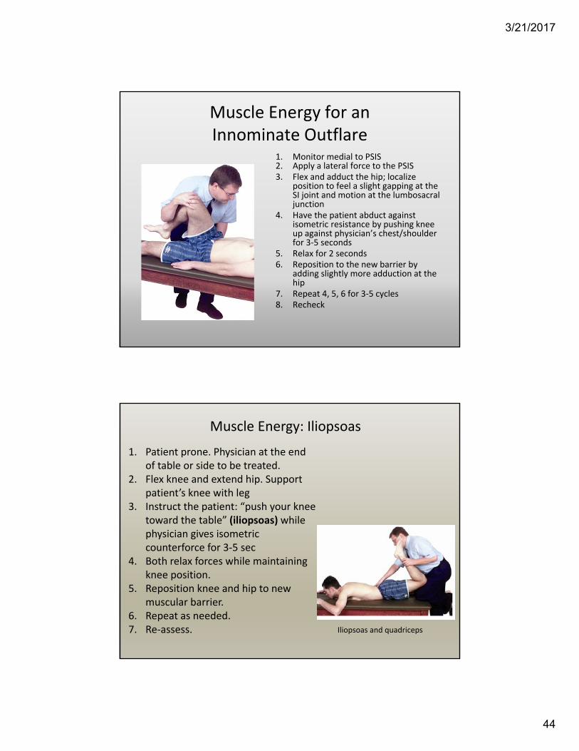
Muscle Energy for an Innominate Outflare
1. Monitor medial to PSIS2. Apply a lateral force to the PSIS3. Flex and adduct the hip; localize
position to feel a slight gapping at the SI joint and motion at the lumbosacral junction
4. Have the patient abduct against isometric resistance by pushing knee up against physician’s chest/shoulder for 3‐5 seconds
5. Relax for 2 seconds6. Reposition to the new barrier by
adding slightly more adduction at the hip
7. Repeat 4, 5, 6 for 3‐5 cycles8. Recheck
Muscle Energy: Iliopsoas
Iliopsoas and quadriceps
1. Patient prone. Physician at the end of table or side to be treated.
2. Flex knee and extend hip. Support patient’s knee with leg
3. Instruct the patient: “push your knee toward the table” (iliopsoas) while physician gives isometric counterforce for 3‐5 sec
4. Both relax forces while maintaining knee position.
5. Reposition knee and hip to new muscular barrier.
6. Repeat as needed.7. Re‐assess.
3/21/2017
45
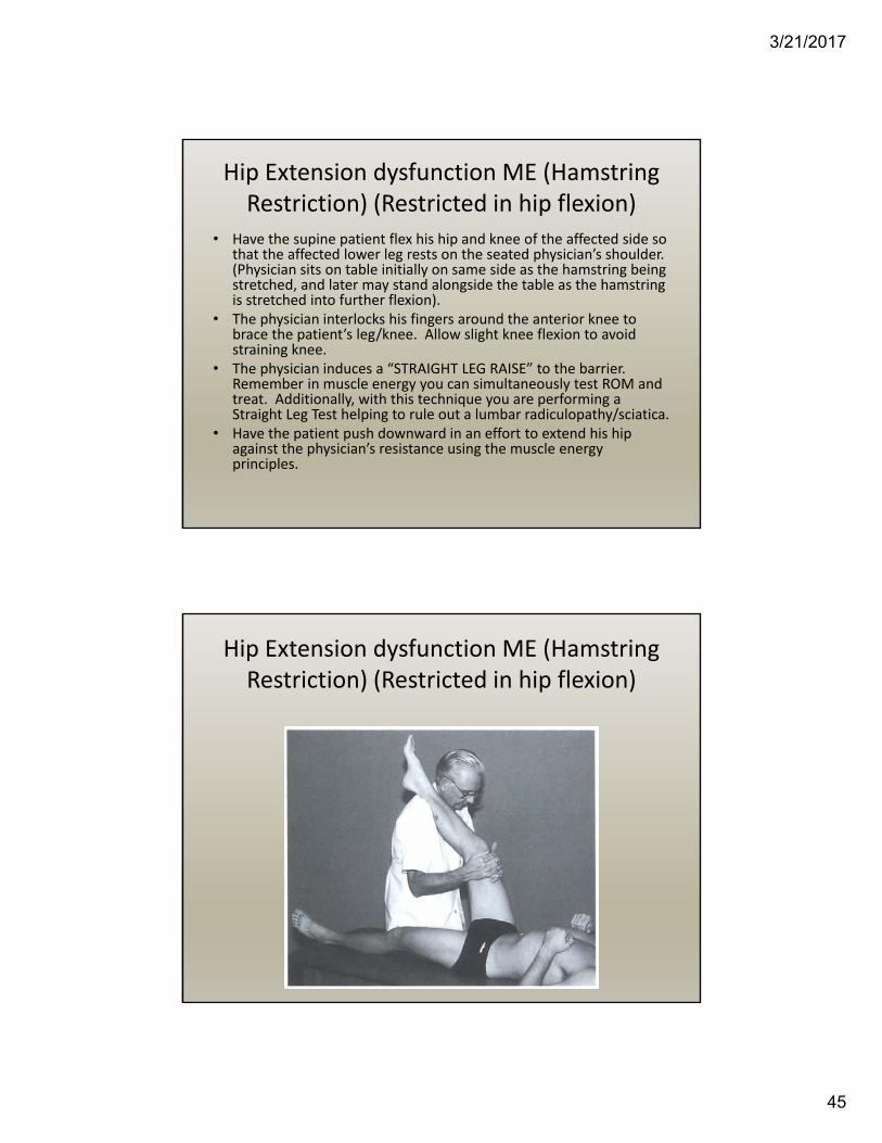
Hip Extension dysfunction ME (Hamstring Restriction) (Restricted in hip flexion)
• Have the supine patient flex his hip and knee of the affected side so that the affected lower leg rests on the seated physician’s shoulder. (Physician sits on table initially on same side as the hamstring being stretched, and later may stand alongside the table as the hamstring is stretched into further flexion).
• The physician interlocks his fingers around the anterior knee to brace the patient’s leg/knee. Allow slight knee flexion to avoid straining knee.
• The physician induces a “STRAIGHT LEG RAISE” to the barrier. Remember in muscle energy you can simultaneously test ROM and treat. Additionally, with this technique you are performing a Straight Leg Test helping to rule out a lumbar radiculopathy/sciatica.
• Have the patient push downward in an effort to extend his hip against the physician’s resistance using the muscle energy principles.
Hip Extension dysfunction ME (Hamstring Restriction) (Restricted in hip flexion)
3/21/2017
46
Piriformis Muscle Energy
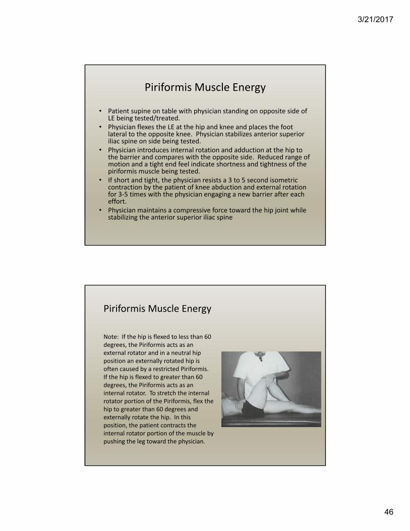
• Patient supine on table with physician standing on opposite side of LE being tested/treated.
• Physician flexes the LE at the hip and knee and places the foot lateral to the opposite knee. Physician stabilizes anterior superior iliac spine on side being tested.
• Physician introduces internal rotation and adduction at the hip to the barrier and compares with the opposite side. Reduced range of motion and a tight end feel indicate shortness and tightness of the piriformis muscle being tested.
• If short and tight, the physician resists a 3 to 5 second isometric contraction by the patient of knee abduction and external rotation for 3‐5 times with the physician engaging a new barrier after each effort.
• Physician maintains a compressive force toward the hip joint while stabilizing the anterior superior iliac spine
Piriformis Muscle Energy
Note: If the hip is flexed to less than 60 degrees, the Piriformis acts as an external rotator and in a neutral hip position an externally rotated hip is often caused by a restricted Piriformis. If the hip is flexed to greater than 60 degrees, the Piriformis acts as an internal rotator. To stretch the internal rotator portion of the Piriformis, flex the hip to greater than 60 degrees and externally rotate the hip. In this position, the patient contracts the internal rotator portion of the muscle by pushing the leg toward the physician.
3/21/2017
47
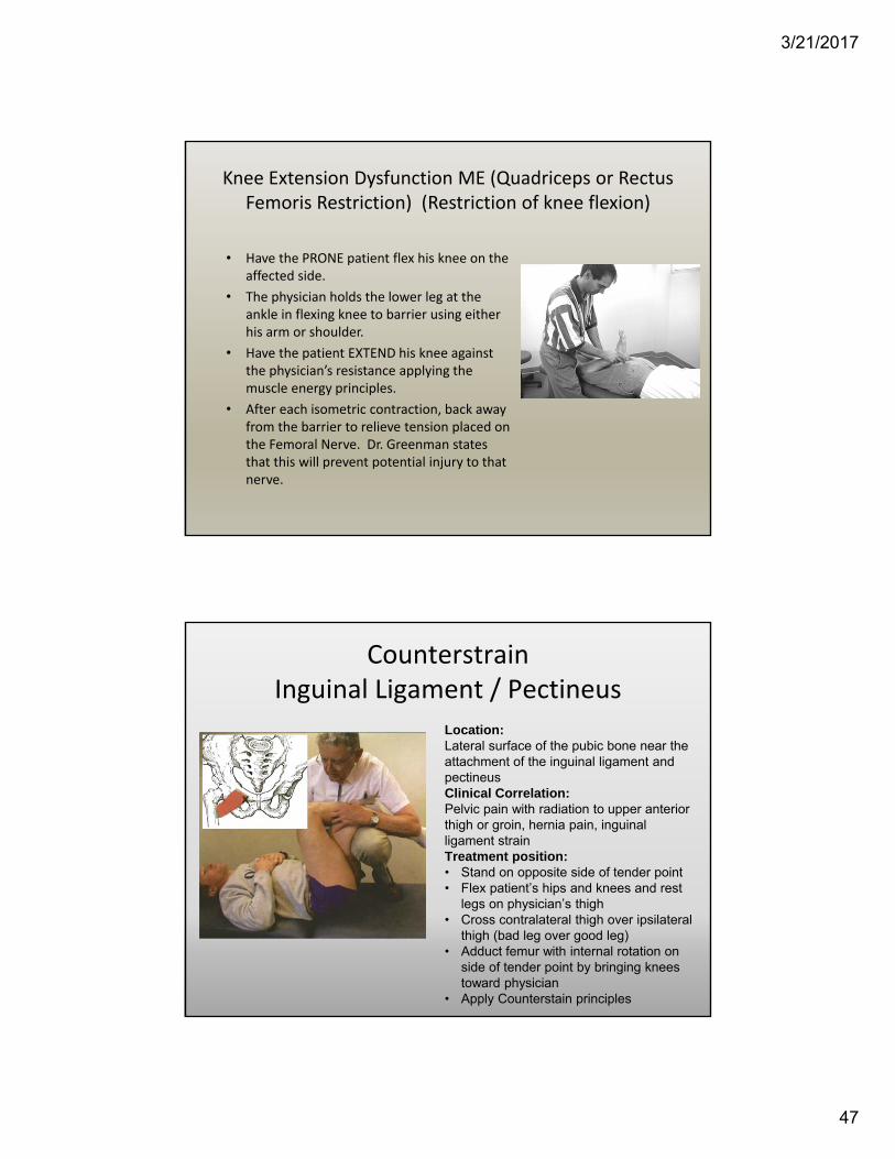
Knee Extension Dysfunction ME (Quadriceps or Rectus Femoris Restriction) (Restriction of knee flexion)
• Have the PRONE patient flex his knee on the affected side.
• The physician holds the lower leg at the ankle in flexing knee to barrier using either his arm or shoulder.
• Have the patient EXTEND his knee against the physician’s resistance applying the muscle energy principles.
• After each isometric contraction, back away from the barrier to relieve tension placed on the Femoral Nerve. Dr. Greenman states that this will prevent potential injury to that nerve.
CounterstrainInguinal Ligament / Pectineus
Location:Lateral surface of the pubic bone near the attachment of the inguinal ligament and pectineus Clinical Correlation:Pelvic pain with radiation to upper anterior thigh or groin, hernia pain, inguinal ligament strainTreatment position:• Stand on opposite side of tender point• Flex patient’s hips and knees and rest
legs on physician’s thigh• Cross contralateral thigh over ipsilateral
thigh (bad leg over good leg)• Adduct femur with internal rotation on
side of tender point by bringing knees toward physician
• Apply Counterstain principles
3/21/2017
48
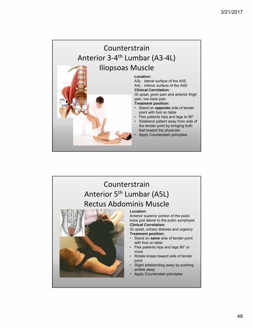
CounterstrainAnterior 3‐4th Lumbar (A3‐4L)
Iliopsoas MuscleLocation:A3L: lateral surface of the AIISA4L: inferior surface of the AIISClinical Correlation:GI upset, groin pain and anterior thigh pain, low back painTreatment position:• Stand on opposite side of tender
point with foot on table• Flex patients hips and legs to 90°• Sidebend patient away from side of
the tender point by bringing both feet toward the physician
• Apply Counterstain principles
A3L
A4L
CounterstrainAnterior 5th Lumbar (A5L)Rectus Abdominis Muscle
Location:Anterior superior portion of the pubic bone just lateral to the pubic symphysisClinical Correlation:GI upset, urinary distress and urgencyTreatment position:• Stand on same side of tender point
with foot on table• Flex patients hips and legs 90° or
more• Rotate knees toward side of tender
point• Slight sidebending away by pushing
ankles away• Apply Counterstain principles
3/21/2017
49
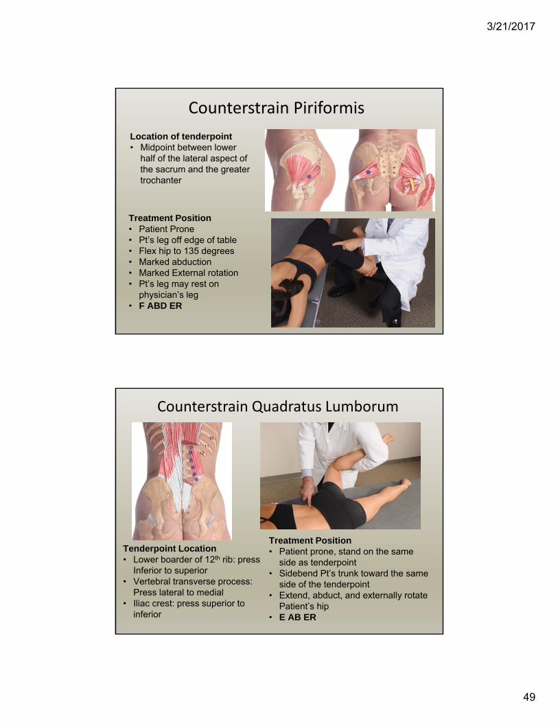
Counterstrain Piriformis
Location of tenderpoint• Midpoint between lower
half of the lateral aspect of the sacrum and the greater trochanter
Treatment Position• Patient Prone• Pt’s leg off edge of table• Flex hip to 135 degrees• Marked abduction• Marked External rotation• Pt’s leg may rest on
physician’s leg• F ABD ER
Counterstrain Quadratus Lumborum
Tenderpoint Location• Lower boarder of 12th rib: press
Inferior to superior• Vertebral transverse process:
Press lateral to medial • Iliac crest: press superior to
inferior
Treatment Position• Patient prone, stand on the same
side as tenderpoint• Sidebend Pt’s trunk toward the same
side of the tenderpoint• Extend, abduct, and externally rotate
Patient’s hip• E AB ER
3/21/2017
50
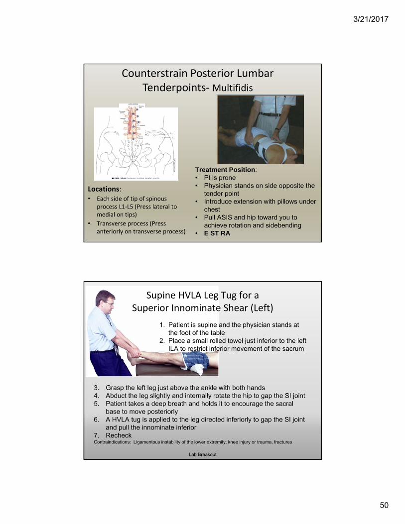
Counterstrain Posterior Lumbar Tenderpoints‐Multifidis
Locations:• Each side of tip of spinous
process L1‐L5 (Press lateral to medial on tips)
• Transverse process (Press anteriorly on transverse process)
Treatment Position: • Pt is prone• Physician stands on side opposite the
tender point• Introduce extension with pillows under
chest• Pull ASIS and hip toward you to
achieve rotation and sidebending• E ST RA
Supine HVLA Leg Tug for aSuperior Innominate Shear (Left)
3. Grasp the left leg just above the ankle with both hands4. Abduct the leg slightly and internally rotate the hip to gap the SI joint5. Patient takes a deep breath and holds it to encourage the sacral
base to move posteriorly6. A HVLA tug is applied to the leg directed inferiorly to gap the SI joint
and pull the innominate inferior7. RecheckContraindications: Ligamentous instability of the lower extremity, knee injury or trauma, fractures
1. Patient is supine and the physician stands at the foot of the table
2. Place a small rolled towel just inferior to the left ILA to restrict inferior movement of the sacrum
Lab Breakout
3/21/2017
51
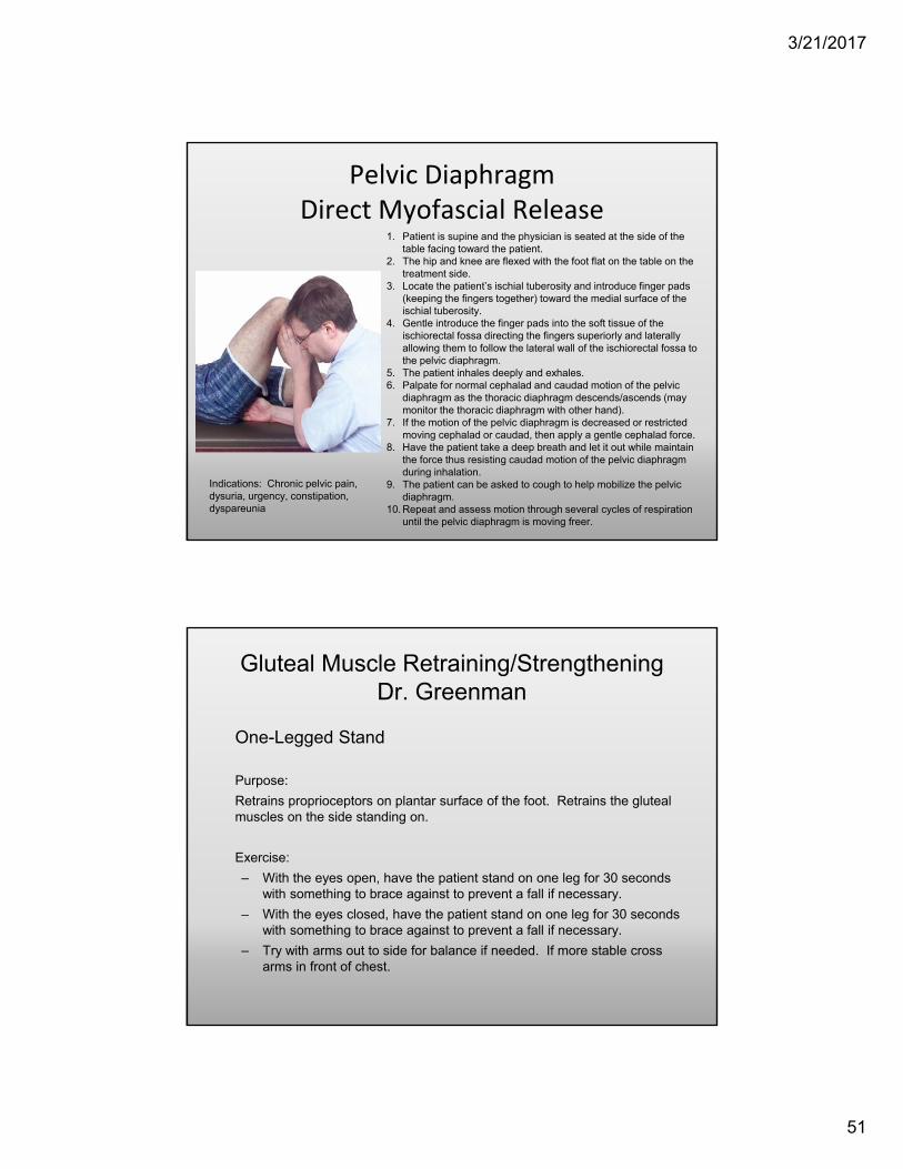
Pelvic Diaphragm Direct Myofascial Release
1. Patient is supine and the physician is seated at the side of the table facing toward the patient.
2. The hip and knee are flexed with the foot flat on the table on the treatment side.
3. Locate the patient’s ischial tuberosity and introduce finger pads (keeping the fingers together) toward the medial surface of the ischial tuberosity.
4. Gentle introduce the finger pads into the soft tissue of the ischiorectal fossa directing the fingers superiorly and laterally allowing them to follow the lateral wall of the ischiorectal fossa to the pelvic diaphragm.
5. The patient inhales deeply and exhales.6. Palpate for normal cephalad and caudad motion of the pelvic
diaphragm as the thoracic diaphragm descends/ascends (may monitor the thoracic diaphragm with other hand).
7. If the motion of the pelvic diaphragm is decreased or restricted moving cephalad or caudad, then apply a gentle cephalad force.
8. Have the patient take a deep breath and let it out while maintain the force thus resisting caudad motion of the pelvic diaphragm during inhalation.
9. The patient can be asked to cough to help mobilize the pelvic diaphragm.
10. Repeat and assess motion through several cycles of respiration until the pelvic diaphragm is moving freer.
Indications: Chronic pelvic pain, dysuria, urgency, constipation, dyspareunia
Gluteal Muscle Retraining/StrengtheningDr. Greenman
One-Legged Stand
Purpose:
Retrains proprioceptors on plantar surface of the foot. Retrains the gluteal muscles on the side standing on.
Exercise:
– With the eyes open, have the patient stand on one leg for 30 seconds with something to brace against to prevent a fall if necessary.
– With the eyes closed, have the patient stand on one leg for 30 seconds with something to brace against to prevent a fall if necessary.
– Try with arms out to side for balance if needed. If more stable cross arms in front of chest.
3/21/2017
52
References
1. Foundations for Osteopathic Medicine, 3rd Ed., pg. 437‐480. “Postural Considerations in Osteopathic Diagnosis and Treatment”
2. Essentials of Musculoskeletal Care, 4th ed., by Sarwark, Pages 964‐966 Chapter “Spine” and Section “Scoliosis in Adults”
3. Steinberg, Akins, Baran, Orthopaedics in Primary Care, 3rd
ed., LWW, Philadelphia, 1998, Pg. 154‐1564. Kuchera and Kuchera, Osteopathic Principles in Practice,
2nd ed., revised. Greyden Press, Columbus, Ohio, 1994. 5. Ward, editor, Foundations for Osteopathic Medicine., 2nd
ed., LWW, Philadelphia, 2003
Related Research
• John Henry Juhl, Tonya M. Ippolito Cremin, and George RussellPrevalence of Frontal Plane Pelvic Postural Asymmetry—Part 1J Am Osteopath Assoc, Oct 2004; 104: 411 ‐ 421.
• RB THOMASPostural dynamicsJ Am Osteopath Assoc, Oct 1954; 54: 92 ‐ 95. ...Articles Postural dynamics RB THOMAS Postural dynamics. | Journal Article | Posture | POSTURE...
• HW BaileyTheoretical significance of postural imbalance, especially the "short leg"J Am Osteopath Assoc, Feb 1978; 77: 452.
• Christopher M. RancontChronic Psoas Syndrome Caused by the Inappropriate Use of a Heel LiftJ Am Osteopath Assoc, Sep 2007; 107: 415 ‐ 418.

![Tracheo-Innominate Fistula diagnosis and treatment: A …Tracheo-Innominate Fistula [TIF] is a rare lethal complication following tracheostomy occurring approximately 1% of cases](https://img.dokumen.tips/doc/110x75/60ad42be92879e62c24d0267/tracheo-innominate-fistula-diagnosis-and-treatment-a-tracheo-innominate-fistula.jpg)