Embed Size (px)
Citation preview
Olfactory Neuroblastoma is a Peripheral Primitive Neuroectodermal Tumor Related to EwingSarcomaAuthor(s): Poul H. B. Sorensen, John K. Wu, Ken W. Berean, Jerian F. Lim, Wayne Donn,Henry F. Frierson, C. Patrick Reynolds, Dolores López-Terrada and Timothy J. TricheSource: Proceedings of the National Academy of Sciences of the United States of America,Vol. 93, No. 3 (Feb. 6, 1996), pp. 1038-1043Published by: National Academy of SciencesStable URL: http://www.jstor.org/stable/38743 .
Accessed: 02/05/2014 08:20
Your use of the JSTOR archive indicates your acceptance of the Terms & Conditions of Use, available at .http://www.jstor.org/page/info/about/policies/terms.jsp
.JSTOR is a not-for-profit service that helps scholars, researchers, and students discover, use, and build upon a wide range ofcontent in a trusted digital archive. We use information technology and tools to increase productivity and facilitate new formsof scholarship. For more information about JSTOR, please contact [email protected].
.
National Academy of Sciences is collaborating with JSTOR to digitize, preserve and extend access toProceedings of the National Academy of Sciences of the United States of America.
http://www.jstor.org
This content downloaded from 62.122.73.77 on Fri, 2 May 2014 08:20:34 AMAll use subject to JSTOR Terms and Conditions

Proc. Natl. Acad. Sci. USA Vol. 93, pp. 1038-1043, February 1996 Medical Sciences
Olfactory neuroblastoma is a peripheral primitive neuroectodermal tumor related to Ewing sarcoma
(EWS/FLIH/t(11;22)/translocation/solid tumor)
POUL H. B. SORENSEN*t, JOHN K. Wut, KEN W. BEREAN?, JERIAN F. LIM*, WAYNE DONN?, HENRY F. FRIERSONT, C. PATRICK REYNOLDSII, DOLORES L6PEZ-TERRADAII, AND TIMOTHY J. TRICHEII
Departments of *Pathology and *Pediatrics, British Columbia's Children's Hospital, Vancouver, BC, Canada V6H-3V4; ?Department of Pathology, Vancouver Hospital and Health Sciences Centre, Vancouver, BC, Canada V5Z-lM9; 1Department of Pathology, University of Virginia Health Sciences Center, Charlottesville, VA 22908; and I"Department of Pathology, Childrens Hospital of Los Angeles, Los Angeles, CA 90027
Communicated by Alfred G. Knudson, Fox Chase Cancer Center, Philadelphia, PA, October 13, 1995 (received for review May 31, 1995)
ABSTRACT Olfactory neuroblastoma (ONB) is a malig- nant tumor of the nasal mucosa whose histogenesis is unclear. A relationship to neuroblastoma (NB), a pediatric tumor of the sympathetic nervous system, is based on morphologic similarities and the expression of similar neural antigens. However, the clinical presentation of ONB differs from that of NB, and MYCN amplification characteristic of NB is not observed. We have therefore examined the relationship of this malignancy to other classes of neural tumors. In previous studies, two ONB cell lines demonstrated cytogenetic features and patterns of protooncogene expression suggestive of a relationship to the Ewing sarcoma family of childhood pe- ripheral primitive neuroectodermal tumors (pPNETs). The pPNETs show t(11;22) (q24;ql2) or t(21;22) (q22;ql2) chromo- somal translocations fusing the EWS gene from 22q12 with either the FLH gene on 11q24 or the ERG gene on 21q22. We therefore analyzed ONBs for the presence of pPNET- associated gene fusions. Both cell lines showed rearrangement of the EWS gene, and fluorescence in situ hybridization (FISH) of each case demonstrated fusion of EWS and FLH genomic sequences. Moreover, both lines expressed EWS/FLIJ fusion transcripts with in-frame junctions between exon 7 of EWS and exon 6 of FLH as described for pPNETs. We identified similar gene fusions in four of six primary ONB cases. None of the cases expressed tyrosine hydroxylase, a catecholamine biosynthetic enzyme widely expressed in NB. Our studies indicate that ONB is not a NB but is a member of the pPNET family.
Primitive sarcomas often lack phenotypic features of their cell of origin and are therefore very difficult to classify. This has important clinical implications as accurate diagnosis in large part determines the chosen therapeutic strategy for these malignancies. The class of poorly differentiated malignancies known as the peripheral primitive neuroectodermal tumors (pPNETs) occurs most frequently in adolescents and young adults and includes Ewing sarcoma, peripheral neuroepithe- lioma, and Askin tumor. These tumors are thought to have a neural cell of origin based on limited neuroectodermal devel- opment (reviewed in ref. i). In addition, neural differentiation can be induced in vitro by treatment of cultured tumor cells with retinoic acid and nerve growth factors (2, 3). Greater than 95% of pPNETs show t(11;22)(q24;ql2) or t(21;22)(q22;ql2) chromosomal translocations which are considered specific for this tumor class (4). These translocations fuse the 5' portion of the EWS gene on chromosome 22q12 in-frame to either of the two Ets family transcription factor genes, FLI] on l1q24 (5, 6) or ERG on 21q22 (7-9). Resultant fusion genes express chimeric proteins which are capable of cell transformation and
The publication costs of this article were defrayed in part by page charge payment. This article must therefore be hereby marked "advertisement" in accordance with 18 U.S.C. ?1734 solely to indicate this fact.
appear to act as aberrant transcription factors (10). Identifi- cation of EWS/FLI] and EWS/ERG fusion transcripts by reverse transcription PCR (RT-PCR) has formed the basis of a sensitive and specific diagnostic test for pPNETs (11).
Olfactory neuroblastoma (ONB) is a tumor of the nasal cavity that was first described in 1924 by Berger et al. (12). Since that time the histogenesis of this tumor has been vigorously debated, giving rise to a variety of other names, including esthesioneuroblastoma, olfactory neuroepithe- lioma, and others (13). A neural derivation is indicated ultrastructurally by the presence of dense core neurosecre- tory granules and neuritic processes within tumor cells and immunohistochemically by expression of neurofilament pro- tein, neuron-specific enolase, and S-100 protein (14-17). Several anatomical origins of ONB have been suggested, including the sympathetic fibers of the anterior nasal cavity, the neuroectodermal cells of the olfactory placode, the sphenopalatine ganglion, and the organ of Jacobson (13). The proposed relationship to neuroblastoma (NB) implied in the name is based on morphologic similarities to primitive forms of NB, the identification of norepinephrine and dopamine f3-hydroxylase (a key enzyme in the metabolic pathway of catecholamine synthesis) in a case of ONB (18), and a possible origin from sympathetic fibers of the nervus terminalis located in the anterior nasal cavity. Unlike NB, however, ONB rarely occurs in early childhood, tends to behave less aggressively than NB, and is rarely associated with catecholamine secretion (13, 15). Moreover, the supe- rior location of ONB within the nasal vault favors an origin from neuroectodermally derived cells of the olfactory pla- code. These clinicopathologic considerations have led to the alternative hypothesis that ONB is related to the pPNET family of primitive sarcomas.
Although morphologically similar, a number of molecular features distinguish pPNETs and NBs. The t(11;22) and t(21;22) chromosomal translocations and gene fusions de- scribed for pPNETs have never been observed in NBs. Instead, these malignancies demonstrate amplification of the MYCN protooncogene in about 30% of the cases (19, 20), and a similar proportion have allelic losses of the distal short arm of chromosome 1 (21, 22). NBs are associated with catecholamine secretion, and expression of tyrosine hydroxylase, the rate- limiting enzyme in catecholamine biosynthesis, has been used diagnostically for the detection of residual NB cells (23). In contrast, pPNETs do not secrete catecholamines (13), and tyrosine hydroxylase is not expressed in these tumors (24). Moreover, pPNETs do not demonstrate MYCN amplification
Abbreviations: ONB, olfactory neuroblastoma; NB, neuroblastoma; FISH, fluorescence in situ hybridization; pPNET, peripheral primitive neuroectodermal tumor; RT-PCR, reverse transcription PCR; MEM, malignant ectomesenchymoma. tTo whom reprint requests should be addressed.
1038
This content downloaded from 62.122.73.77 on Fri, 2 May 2014 08:20:34 AMAll use subject to JSTOR Terms and Conditions
Medical Sciences: Sorensen et al. Proc. Natl. Acad. Sci. USA 93 (1996) 1039
but express the c-MYC protooncogene (25). Alterations of chromosome 1 in pPNETs have been described but these primarily involve chromosome lq (26).
We previously reported the establishment of two ONB cell lines, TC-268 and JFEN, derived from young adult patients with ONB (27). Both patients presented with olfactory mu- cosal tumors showing classical pathologic features of ONB. Characterization of the cell lines demonstrated a protoonco- gene expression pattern typical of pPNETs (25), with expres- sion of c-MYC but not of MYCN. Moreover, both cell lines demonstrated cytogenetic abnormalities of chromosomes llq and 22q (27); JFEN showed a t(11;22)(q21;q12) while TC-268 displayed deletions involving both llq and 22q. These cyto- genetic findings are suggestive though not diagnostic of pPNETs (28). A t(11;22)(q24;q12) has also been described by others in a case of ONB (29). To further study the relationship of ONB with the pPNET family of neural tumors, we have now tested TC-268 and JFEN for the presence of the EWS/FLI1 gene fusion. We report that both of these cell lines and four of six primary ONB tumors expressed EWS/FLIJ fusion tran- scripts indistinguishable from those observed in pPNETs. These data indicate that ONB is not a NB but is a member of the pPNET class of neural tumors.
MATERIALS AND METHODS
Cell Lines and Primary Tumor Tissue. The ONB cell lines TC-268 and JFEN have been described (27) and were estab- lished from metastatic lesions in a 22-year-old female (TC- 268) and a 22-year-old male (JFEN) diagnosed with ONBs. The cell lines were maintained in tissue culture using Iscoves's modified Dulbecco's medium supplemented with 20% fetal calf serum and insulin/transferrin/sodium selenite (TC-268) or RPMI 1640 medium supplemented with 20% fetal calf serum (JFEN). Frozen primary tumor tissue was available for analysis from both cases, and from four additional ONB cases (PL, male, 36 years of age; MP, male, 45 years of age; DL, male 50 years of age; RS, male, 54 years of age). Growth and maintenance of the pPNET cell lines TC-32, TC-71, and TC-106 (30), the NB cell lines IMR-32, LAN-1, and SAN-2 (31), as well as the rhabdomyosarcoma cell lines CTR and TC-487 (32) have also been reported. All reagents were from the Sigma unless otherwise stated.
RNA and DNA Analysis. Total RNA was isolated from cell lines (33) and from primary tumor tissue (11) as described. Conventional Southern blot analysis was performed using 10 ,ug of genomic DNA per sample isolated from cell lines (34). Northern analysis was performed according to standard meth- ods (34), using 20 ,tg of total RNA for each sample. Comple- mentary DNA probes for EWS and for FLI1 have previously been reported (7). Probes for Northern and Southern analysis were radiolabeled by random primer extension.
Fluorescence in Situ Hybridization (FISH). Slides contain- ing metaphase and interphase spreads of TC-268 and JFEN were used for dual-color FISH studies. Slides were denatured at 70?C for 2 min and then hybridized overnight at 37?C under low stringency conditions (50% formamide/2X SSC), using EWS/FLII gene fusion probes flanking the t(11;22)(q24;q12) breakpoint. These consisted of three cosmid contigs from the EWS locus and another three contigs from the FLI] locus, cloned into the pWE15 cosmid vector (Stratagene) and labeled with biotin and digoxygenin, respectively (35). Detection was carried out using fluorescein-labeled avidin and rhodamine- labeled anti-digoxigenin (Oncor). Chromosomes were coun- terstained with 4',6-diamidino-2-phenylindole and visualized using an Olympus BH2 microscope equipped with Olympus BP495 filters and a triple bandpass filter (Omega BP 405).
RT-PCR. RT-PCR detection of EWS/FLII or EWS/ERG fusion transcripts was performed as reported, using total RNA as starting material (7, 11). Oligonucleotide primers used for~
PCR included the FLII-specific primer ESBP-2 (7), 11.3, which detects FLI1 and ERG sequences (5, 11), and the EWS-specific primer ESBP-1 (7). RT-PCR detection of ty- rosine hydroxylase expression was carried out using previously reported methods and primer pairs (23, 36). Amplified prod- ucts were analyzed by electrophoresis using 2% agarose gels and stained with ethidium bromide.
Cloning and Sequencing of PCR Products. PCR products were purified from 2% low-melting point agarose gels using 3-agarase (New England Biolabs). Purified products were then
cloned using the Invitrogen TA cloning system (version 1.3) as described by the manufacturer. Direct sequencing of purified recombinant plasmid DNA was performed on an ABI Applied Biosystems 373A DNA sequencer, using Sp6, T7, m13 reverse, and m13 forward (-20) sequencing primers (Invitrogen). Sequences were analyzed using DNASTAR Sequence Analysis software.
RESULTS
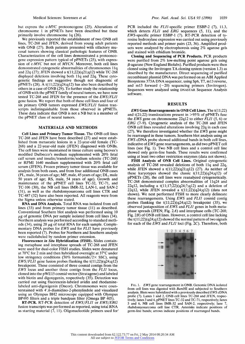
EWS Gene Rearrangements in ONB Cell Lines. The t(11;22) and t(21;22) translocations present in >95% of pPNETs fuse the EWS gene on chromosome 22q12 to either FLI1 (5, 6) or ERG (7-9). Cytogenetic analysis of the TC-268 and JFEN ONB cell lines revealed alterations involving 22q in each case (27). We therefore investigated whether the EWS gene might be rearranged in these tumors. Southern blot analysis using an EWS cDNA probe showed additional bands in both cell lines, indicative of EWS gene rearrangements, as did two pPNET cell lines (see Fig. 1). Two NB cell lines and a control cell line showed only germ-line bands. These results were confirmed using at least two other restriction enzymes (data not shown).
FISH Analysis of ONB Cell Lines. Original cytogenetic analysis of TC-268 revealed deletions of both llq and 22q, while JFEN showed a t(11;22)(q21;ql2) (27). As neither of these karyotypes showed the classic t(11;22)(q24;ql2) of pPNETs (28), the cell lines were reanalyzed cytogenetically. TC-268 demonstrated complex abnormalities of 11q24 and 22q12, including a t(11;17;22)(q24;?;ql2) and a deletion of 22q12, while JFEN revealed a t(11;22)(q24;ql2) (data not shown). We next performed FISH to more clearly delineate these rearrangements. Using EWS and FLI1 cosmid contig probes flanking the t(11;22)(q24;ql2) breakpoint (35), we observed juxtaposition of EWS and FLI] genes in both meta- phase spreads (JFEN; Fig. 2A) and interphase cells (TC-268; Fig. 2B) of ONB cell lines. However, a control cell line lacking the t(11;22)(q24;ql2) showed the normal pattern of two signals for each of the EWS and FLI1 loci (Fig. 2C). Therefore, both
1 2 3 4 56 7
FIG. 1. EWS gene rearrangement in ONB. Genomic DNA isolated from cell lines was digested with BamHI and subjected to Southern analysis. Blots were hybridized with a previously described EWS cDNA probe (7). Lanes 1 and 2, ONB cell lines TC-268 and JFEN, respec- tively; lanes 3 and 4, pPNET lines TC-32 and TC-71, respectively; lanes 5 and 6, NB cell lines IMR-32 and SAN-2, respectively; lane 7, rhabdomyosarcoma cell line CTR. Asterisks indicate positions of germ-line bands; arrows indicate positions of rearranged bands.
This content downloaded from 62.122.73.77 on Fri, 2 May 2014 08:20:34 AMAll use subject to JSTOR Terms and Conditions

1040 Medical Sciences: Sorensen et al. Proc. Natl. Acad. Sci. USA 93 (1996)
FIG. 2. Dual-color FISH analysis demonstrating juxtaposition of EWS and FLI] genes in JFEN and TC-268. (A) Metaphase spread of JFEN showing normal copy of EWS (green color; curved arrow) and FLI] loci (red color; small arrow) as well as fusion of the EWS locus to FLI] (yellow color; thick arrow). (B) Interphase cells of TC-268 showing normal copy of EWS (green color; curved arrow) and FLI] (red color; small arrow) as well as EWS/FLI] gene fusion (yellow or green/red color; thick arrow). (C) Control cells showing the normal pattern of two copies of EWS locus (green color; curved arrow) and FLI] locus (red color; small arrow).
cases demonstrate fusion of EWS and FLI] sequences, con- sistent with pPNET-associated translocations. These results were corroborated using whole chromosome 11 and 22 paint- ing probes, revealing derivative chromosomes in each case containing juxtaposed chromosome 11 and 22 sequences (data not shown).
Expression of EWS/FLI1 Fusion Transcripts in ONB. To determine whether the observed EWS gene rearrangements in the two ONB cell lines represented EWS/FLI1 or EWS/ERG gene fusions characteristic of pPNETs, we screened cell lines for expression of chimeric transcripts by RT-PCR using EWS- specific and FL!]/ERG-specific oligonucleotide primers. As shown in Fig. 3, RT-PCR analysis using ELI]-specific ESBP-1
A23 A0 p 1 2 3 4 5 6 7 8 603b-
310 bp-
B 1 2 3 4 5 6 7 8 603 bp- _____________ 310 bp-
FIG. 3. Expression of EWS/FLII in ONB cell lines and primary tumors identified by RT-PCR. Total RNA extracted from cell lines or frozen primary tumor tissue was analyzed by RT-PCR using EWS (ESBP-1) and FLI] (ESBP-2) primers. Products were detected by electrophoresis using 2% agarose gels. (A) Analysis of EWS/FLI] expression in TC-268 and JFEN cell lines. Lanes 1 and 2, pPNET cell lines TC-32 and TC-71, respectively; lanes 3 and 4, ONB cell lines TC-268 and JFEN, respectively; lanes 5-7, NB cell lines IMR-32, LAN-1, and SAN-2, respectively; lane 8, alveolar rhabdomyosarcoma cell line TC-487. (B) Analysis of EWS/FLII expression in ONB primary tumors. Lanes 1 and 2, pPNET cell lines TC-32 and TC-71, respectively; lanes 3 and 4, TC-268 and JFEN primary tumors; lanes 5-8, additional ONB cases PL, MP, DL, and RS, respectively. The arrows indicate the positions of EWS/FLIJ amplified products. Mark- ers are indicated on the left.
and EWS-specific ESBP-2 revealed amplified products for TC-268 and JFEN that were identical to those observed for the two pPNET cell lines, TC-32 and TC-71, known to express EWS/FLII gene fusions. Three NB cell lines and a rhabdomy- osarcoma cell line were negative for products; the presence of intact cDNA for these cases was confirmed by amplification using control primer sets. The above results were confirmed by hybridizing the observed PCR products with an EWS oligo- nucleotide probe (11) (data not shown) and could be repro- duced when several independently derived cryopreserved pas- sages of each cell line were tested. These results therefore indicate fusion of EWS to FLI] in TC-268 and JFEN. To confirm the identity of the observed amplification products, we cloned and sequenced PCR products from TC-268 and JFEN. Sequence analysis revealed in-frame fusions between exon 7 of EWS and exon 6 of FLI] that were identical to those described for type 1 EWS/FLI1 fusions of pPNETs (5, 8, 11) (data not shown). These data confirm that RT-PCR products detected in ONB represent amplification of EWS/FLI] fusion tran- scripts with junctional sequences identical to those observed in pPNETs.
To independently demonstrate the expression of the EWSI FLI] gene fusion in ONB, we performed Northern analysis of total RNA isolated from TC-268 and JFEN. Northern blots hybridized with an EWS cDNA probe revealed, in addition to a 2.2-kb germ-line EWS band, a 3.1-kb species in both cell lines that comigrated with an EWS/FLII fusion band present in TC-32 cells (see Fig. 4). This aberrant band was not detected in a NB control cell line. When the blot was stripped and reprobed with a FLI] cDNA probe, only the 3.1-kb band present in TC-268, JFEN, and TC-32 hybridized the probe (see Fig. 4). These data confirm the identity of the 3.1-kb species as the EWS/FLI] fusion transcript.
RT-PCR Analysis of Primary Tumors. To demonstrate that EWS/FLI] fusion transcripts were present in primary tumors, we performed RT-PCR using RNA isolated directly from original frozen tumor tissue used to establish TC-268 and JFEN. As shown in Fig. 3B, results identical to those in Fig. 3A were obtained using ESBP-1 and ESBP-2 oligonucleotide primers. These results were confirmed using ESBP-1 in com- bination with another ELI] -specific primer, 11.3 (5, 1 1), and by hybridizing the observed PCR products with an EWS oligo- nucleotide probe (11) (data not shown). We also tested four
This content downloaded from 62.122.73.77 on Fri, 2 May 2014 08:20:34 AMAll use subject to JSTOR Terms and Conditions

Medical Sciences: Sorensen et al. Proc. Natl. Acad. Sci. USA 93 (1996) 1041
1 2 3 4 1 2 3 4
-28S
EWS/FLI-]I_
EWSS-
-18S
EWS probe FLI-I probe
FIG. 4. Expression of EWS/FLII in ONB cell lines identified by Northern analysis. Total RNA extracted from cell lines was probed with an EWS cDNA probe (Left). The blot was then stripped and reprobed with a FLI] cDNA probe (Right). Lanes 1, pPNET cell line TC-32; lanes 2, ONB cell line TC-268; lanes 3, ONB cell line JFEN; lanes 4, NB cell line SAN-2. Arrows indicate the positions of the 2.2-kb germ-line EWS transcript and the 3.1-kb EWS/FLII fusion transcript in TC-32 (7). The positions of the 18S and 28S markers are indicated.
additional ONB cases that had frozen primary tumor tissue available for RT-PCR analysis. Clinical information for these cases is presented in Materials and Methods. As shown in Fig. 3B, two of the four cases demonstrated amplification products using EWS- and FLI1-specific primers. Cloning and sequenc- ing of the PCR products revealed type I EWS/FLI1 gene fusions in each case (data not shown). The two cases that failed to demonstrate amplification products were also negative using ESBP-1 in combination with the 11.3 primer, which detects both EWS/FLI] and EWS/ERG fusion transcripts (7). We therefore investigated whether other variant translocations involving EWS were present in these cases. As genomic DNA was not available for Southern blot analysis of EWS gene rearrangements, we performed Northern blot analysis of total RNA from each case using an EWS cDNA probe. Neither case demonstrated an aberrant EWS transcript (data not shown). Moreover, 3' rapid amplification of cDNA ends failed to detect aberrant amplification products using several EWS oligonu- cleotides as 5' primers (data not shown). These data strongly argue against the presence of EWS gene fusions in the two negative cases, although they do not rule out this possibility. Expression of EWS/FLII fusion transcripts could therefore be detected in four of six primary ONB cases analyzed in this study.
To further compare ONB with classical NB, we tested TC-268, JFEN, and all six primary tumors for expression of tyrosine hydroxylase, the rate-limiting enzyme in catechol- amine biosynthesis. This enzyme is known to be widely ex- pressed in NBs but not in pPNETs (23, 36). As shown in Fig. 5, the IMR-32 NB cell line demonstrated tyrosine hydroxylase expression, while neither the TC-268 and JFEN cell lines nor the six primary ONB tumors expressed this gene. These data are consistent with our hypothesis that ONB is not related to classical NB but is instead a member of the pPNET family.
DISCUSSION In the present study, we have shown that two ONB tumors previously shown to have karyotypic abnormalities of llq and 22q both express the EWS/FLI1 gene fusion generated by the t(11;22)(q24;q12). Two of four additional primary ONBs tested demonstrated identical gene fusions using original tumor tissue for analysis. EWS/FLI1 or EWS/ERG chimeric proteins [expressed by the EWS/ERG gene fusion of t(21;22)(q22;q12) translocations] have been detected in tu- mors with the t(1 1;22) or the t(21;22) by immunoprecipitation using specific antisera (6, 7). The EWS/FLI1 protein trans- forms NIH 3T3 cells in culture (6), and transfected NIH 3T3 cells form tumors when injected into nude mice (D.L.-T., T.J.T., and C. T. Denny, unpublished data). The chimeric oncoprotein localizes to the nucleus and appears to act as an aberrant transcription factor (10). EWS/FLI1 fused to a Gal-4 DNA-binding domain is a much more potent transcriptional activator of reporter constructs containing Gal-4 binding sites than is the same Gal-4 domain fused to FL11 (10). In the present study, Northern analysis of RNA from TC-268 and JFEN revealed an aberrant band hybridizing to both EWS and FLI1 cDNA probes that comigrated with the EWS/FLI1 fusion transcript of the TC-32 pPNET cell line. Moreover, sequence analysis of cloned RT-PCR fusion products from each of the ONBs tested showed in-frame fusions between exon 7 of EWS and exon 6 of the FLI1 gene. This pattern is identical to that previously described for type I fusions of pPNETs, the most commonly detected EWS/FLI1 fusion in this class of tumors (8). These data suggest that the EWS/FLI1 chimeric protein plays a similar oncogenic role in olfactory neuroblastomas.
Our data fail to support the notion of a direct relationship between ONB and NB. In concordance, the primary ONB cases in this study all failed to express tyrosine hydroxylase, a key enzyme in the biosynthetic pathway of catecholamines. Our studies have demonstrated that ONB cell lines and primary tumors express EWS/FLI1 gene fusions, suggesting instead a relationship to pPNETs. Consistent with this is the clinical presentation of ONB, which is more in keeping with the pattern of pPNETs (13), and the observation that ONBs express the c-MYC protooncogene as opposed to MYCN (25, 27). Furthermore, it has been demonstrated that tumors of neuroectodermal origin with the t(11;22) express insulin-like growth factor I (IGF-I) (37). In that study, the JFEN cell line strongly expressed IGF-I, while all 15 neuroblastoma cell lines tested were negative (37).
Two of the six primary ONB cases failed to demonstrate EWS/FLI] or EWS/ERG gene fusions (see Fig. 3B). Our studies also failed to reveal evidence of other variant translo- cations involving the EWS gene in these two cases, although we have not unequivocally ruled out this possibility. Alternatively, there may be heterogeneity among tumors diagnosed as ONB (38). This tumor is known to have a bimodal age distribution, with one peak in late adolescence or early adulthood and another at 55 years of age (39). Interestingly, both negative cases were patients aged 50 years or older. It is therefore
M 1 2 3 4 5 6 7 8 9 10 11 12 310 bp
271,281 bp
234 bp '. 194 bp-r- -0 1 18 bp-| 722pb-
FIG. 5. Analysis of tyrosine hydroxylase expression in ONB cell lines and primary tumors. RT-PCR using primers spanning separate exons of the human tyrosine hydroxylase gene was performed on total RNA as described (23, 36). These primers detect major bands at 149 and 161 bp. Lane M, markers; lane 1, NB cell line IMR-32; lane 2, TC-268 primary tumor; lane 3, JFEN primary tumor; lanes 4-7, cases PL, MP, DL, and RS, respectively; lane 8, TC-268 cell line; lane 9, JFEN cell line; lanes 10-12, pPNET cell lines TC-32, TC-71, and TC-106.
This content downloaded from 62.122.73.77 on Fri, 2 May 2014 08:20:34 AMAll use subject to JSTOR Terms and Conditions

1042 Medical Sciences: Sorensen et al. Proc. Natl. Acad. Sci. USA 93 (1996)
possible that cases diagnosed as ONB in older patients rep- resent yet another phenotypically neural tumor unrelated to pPNETs, and the two negative cases in the present study may be examples of this entity.
The finding of the same cytogenetic and molecular genetic abnormalities in each member of the pPNET family provides the most compelling evidence to date of a common histogen- esis for this group of tumors. The pPNETs are hypothesized to originate from neural crest-derived embryonic neuroecto- derm, which is known to be widely dispersed in human tissues (40). Consistent with this, Ewing sarcoma and peripheral neuroepithelioma occur in bone and soft tissues of a variety of different anatomic sites. We have recently provided evidence that primitive forms of another childhood sarcoma, malignant ectomesenchymoma (MEM), express the EWS/FLI] gene fusion and should also be included in the pPNET family (41). MEMs show very primitive neural features in addition to a malignant mesenchymal (principally rhabdomyoblastic) com- ponent (reviewed in ref. 41). The origin of MEMs is thought to be the ectomesenchymal cell (40), which is a widely distrib- uted pluripotential neuroectodermal derivative with the ability to differentiate not only into neural tissue but also along various mesenchymal lineages, including skeletal muscle (40, 42). Askin tumor, defined as a soft tissue pPNET of the chest wall region, and ONB are more limited in their locations, possibly reflecting particularly high concentrations of neuro- ectodermal cells at these sites. The wide distribution of pPNETs brings up the interesting possibility that members of this tumor family are actually derived from postganglionic parasympathetic nerve cells, as has been suggested by others (25). Consistent with this is the absence of tyrosine hydroxylase expression in pPNETs, including ONBs, but expression instead of choline acetyltransferase, an enzyme involved in cholinergic transmitter biosynthesis (25, 41). The finding of pPNET- associated gene fusions in ONB is intriguing for several reasons. First, while ONB shows the most overt neural differ- entiation of pPNETs, there is also evidence for epithelial differentiation in these tumors (43, 44). Therefore, as with primitive MEMs (41), our results provide evidence for similar gene fusions in phenotypically diverse tumors. Second, the finding of epithelial features in some of these tumors calls into question the notion that the cells of origin of pPNETs have only neural or mesenchymal differentiation capacity and sug- gest a possible derivation of at least some tumors from neuroepithelium, as is suggested for neuroendocrine tumors such as small cell carcinoma of the lung. An alternative hypothesis, given the increasingly widespread phenotypic di- versity seen in pPNETs, is that the neural (including parasym- pathetic) or neuroepithelial phenotypes observed in the tumor cells of origin are the result of local inductive effects on primitive precursor cells with the capacity for more widespread differentiation than previously appreciated.
The theme is therefore emerging that widely distributed but histogenetically related cells with the capacity for neuroecto- dermal differentiation may acquire common genetic alter- ations and give rise to a family of morphologically primitive neural tumors. Whether these cells are particularly susceptible to chromosomal rearrangements that lead to EWS/FLI1 or EWS/ERG gene fusions remains to be determined. ONB therefore joins Ewing sarcoma, peripheral neuroepithelioma, Askin tumor, and primitive MEM as a member of the pPNET family. As a tumor family, pPNETs are now the second most common soft tissue tumors in adolescents and young adults (1). The number of tumors in this class may continue to increase as the histogenesis of peripheral neural tumors is more clearly understood, and as more nosologic entities are tested for EWS/FLI] or EWS/ERG gene fusions.
We thank Dr. Julius Peters for his contributions to the study and Drs. William May and Chris Denny for providing t(11;22) cosmid
contigs for use as FISH probes. This work was supported in part by a grant from the British Columbia Health Research Fund and by the Las Madrinas Endowment for Molecular Pathology at Childrens Hospital of Los Angeles.
1. Triche, T. J. (1993) in Pathology of Pediatric Malignancies, eds. Pizzo, P. A. & Poplack, D. G. (Lippincott, Philadelphia), Vol. 2, 115-152.
2. Linnoila, R. I., Tsokos, M., Triche, T. J. & Chandra, R. S. (1986) Am. J. Surg. Pathol. 10, 124-133.
3. Cavazzana, A. O., Miser, J. S., Jefferson, J. & Triche, T. J. (1987) Am. J. Pathol. 127, 507-518.
4. Delattre, O., Zucman, J., Melot, T., Garau, X. S., Zucker, J.-M., Lenoir, G. M., Ambros, P. F., Sheer, D., Turc-Carel, C., Triche, T. J., Aurias, A. & Thomas, G. (1994) N. Engl. J. Med. 331, 294-299.
5. Delattre, O., Zucman, J., Ploustagel, B., Desmaze, C., Melot, T., Peter, M., Kovar, H., Joubert, I., de Jong, P., Rouleau, G., Aurias, A. & Thomas, G. (1992) Nature (London) 359, 162-165.
6. May, W. A., Gishizky, M. L., Lessnick, S. L., Lunsford, L. B., Lewis, B. C., Delattre, O., Zucman, J., Thomas, G. & Denny, C. T. (1993) Proc. Natl. Acad. Sci. USA 90, 5752-5756.
7. Sorensen, P. H. B., Lessnick, S. L., Lopez-Terrada, D., Liu, X. F., Triche, T. J. & Denny, C. T. (1994) Nat. Genet. 6, 146-151.
8. Zucman, J., Melot, T., Desmaze, C., Ghysdael, J., Plougastel, B., Peter, M., Zucker, J. M., Triche, T. J., Sheer, D., Turc-Carel, C., Ambros, P., Combaret, V., Lenoir, G., Aurias, A., Thomas, G. & Delattre, 0. (1993) EMBO J. 12, 4481-4487.
9. Giovannini, M., Biegel, J. A., Serra, M., Wang, J.-Y., Wei, Y. H., Nycum, L., Emmanuel, B. S. & Evans, G. A. (1994)J. Clin. Invest. 94, 489-496.
10. May, W. A., Lessnick, S. L., Braun, B. S., Klemsz, M., Lewis, B. C., Lunsford, L. B., Hromas, R. & Denny, C. T. (1993) Mol. Cell. Biol. 13, 7393-7398.
11. Sorensen, P. H. B., Liu, X. F., Thomas, G., DeLattre, O., Row- land, J. M., Biggs, C. A. & Triche, T. J. (1993) Diag. Mol. Pathol. 2, 147-157.
12. Berger, L. & Richard, L. (1924) Bull. Assoc. Fr. Etude Cancer 13, 410-421.
13. Enzinger, F. M. & Weiss, S. W. (1988) Soft Tissue Tumors (Mosby, St. Louis).
14. Trojanowski, J. K., Lee, V., Pillsbury, N. & Lee, S. (1982) N. Engl. J. Med. 307, 159-163.
15. Mills, E. M. & Frierson, J. H. F. (1985) Am. J. Surg. Pathol. 9, 317-327.
16. Choi, H. S. & Anderson, P. J. (1986) J. Neuropathol. Exp. Neurol. 45, 576-587.
17. Schmidt, J. L., Zarbo, R. J. & Clark, J. L. (1990) Laryngoscope 100, 1052-1058.
18. Micheau, C., Guerinot, F., Bohuon, C. & Brugere, J. (1975) Cancer 35, 1309-1312.
19. Brodeur, G. M., Seeger, R. C., Schwab, M., Varmus, H. E. & Bishop, J. M. (1984) Science 224, 1121-1124.
20. Brodeur, G. M. & Fong, C. T. (1989) Cancer Genet. Cytogenet. 41, 153-174.
21. Fong, C. T., Dracopoli, N. C., White, P. S., Merrill, P. T., Grif- fith, R. C., Housman, D. E. & Brodeur, G. M. (1989) Proc. Natl. Acad. Sci. USA 86, 3753-3757.
22. Martinsson, T., Weith, A., Cziepluch, C. & Schwab, M. (1989) Genes Chromosomes Cancer 1, 67-78.
23. Naito, H., Kuzumaki, N., Uchino, J., Kobayashi, R., Shikano, T. & Matsumoto, S. (1991) Eur. J. Cancer 27, 762-765.
24. Crabbe, D. C. G., Peters, J., Reynolds, C. P., Seeger, R. C. & Triche, T. J. (1992) Med. Pediatr. Oncol. 20, 383 (abstr.).
25. Thiele, C. J., McKeon, C., Triche, T. J., Ross, R. A., Reynolds, C. P. & Israel, M. A. (1987) J. Clin. Invest 80, 804-811.
26. Mugneret, F., Lizard, S., Aurias, A. & Turc, C. C. (1988) Cancer Genet. Cytogenet. 32, 239-245.
27. Cavazzana, A. O., Navarro, S., Noguera, R., Reynolds, P. C. & Triche, T. J. (1988) Adv. Neuroblast. Res. 2, 463-474.
28. Turc-Carel, C., Aurias, A., Mugneret, F., Lizard, S., Sidaner, I., Volk, C., Thiery, J. P., Olschwang, S., Philip, I. & Berger, M. P. (1988) Cancer Genet. Cytogenet. 32, 229-238.
29. Whang-Peng, J., Freter, C. E., Knutsen, T., Nanfro, J. J. & Gazdar, A. (1987) Cancer Genet. Cytogenet. 29, 155-157.
This content downloaded from 62.122.73.77 on Fri, 2 May 2014 08:20:34 AMAll use subject to JSTOR Terms and Conditions

Medical Sciences: Sorensen et al. Proc. Natl. Acad. Sci. USA 93 (1996) 1043
30. Whang-Peng, J., Triche, T. J., Knutsen, T., Miser, J., Kao-Shan, A., Tsai, S. & Israel, M. A. (1986) Cancer Genet. Cytogenet. 21, 185-208.
31. Jalava, A. M., Heikkila, J. B. & Akerman, K. E. (1988) Exp. Cell Res. 179, 10-17.
32. Whang-Peng, J., Knutsen, T., Thiel, K., Horowitz, M. E. & Triche, T. J. (1992) Genes Chromosomes Cancer 5, 299-310.
33. Chomczynski, P. & Sacchi, N. (1987) Anal. Biochem. 162, 156-159. 34. Sambrook, J., Fritch, E. F. & Maniatis, T. (1989) Molecular
Cloning: A Laboratory Manual (Cold Spring Harbor Lab. Press, Plainview, NY), 2nd Ed.
35. Lopez-Terrada, D., Cui, M. Y., Denny, C. T., Dietz-Band, J., Airhart, S. & Triche, T. J. (1994) Modern Pathol. 7, 147 (abstr.).
36. Coker, G. T., Strudelska, D., Harmon, S., Burke, W. & O'Malley, K. L. (1990) Mol. Brain Res. 8, 93-98.
37. Yee, D., Favoni, R. E., Lebovic, G. S., Lombana, F., Powell, D., Reynolds, C. P. & Rosen, N. (1990) J. Clin. Invest. 86, 1806-1814.
38. Frierson, H. F., Mills, S. E., Fechner, R. E., Taxy, J. B. & Levine, P. A. (1986) Am. J. Surg. Pathol. 10, 771-779.
39. Elkon, D., Hightower, S. I., Lim, M. L., Cantrell, R. W. & Constable, W. C. (1979) Cancer 44, 1087-1094.
40. Le Douarin, N. (1980) Curr. Top. Dev. Biol. 16, 31-91. 41. Sorensen, P. H. B., Shimada, H., Liu, X. F., Lim, J. S., Thomas,
G. & Triche, T. J. (1995) Cancer Res. 55, 1385-1392. 42. Stemple, D. L. & Anderson, D. J. (1992) Cell 71, 973-985. 43. Taxy, J. B., Bharani, N. K., Mills, S. E., Frierson, H. F. J. &
Gould, V. E. (1986) Am. J. Surg. Pathol. 10, 687-695. 44. Frierson, H. F., Ross, G. W., Mills, S. E. & Frankfurter, A. (1990)
Am. J. Clin. Pathol. 94, 547-553.
This content downloaded from 62.122.73.77 on Fri, 2 May 2014 08:20:34 AMAll use subject to JSTOR Terms and Conditions