Embed Size (px)
Citation preview
Neuron, Vol. 28, 15–18, October, 2000, Copyright 2000 by Cell Press
Olfactory Ensheathing Cells Minireviewand CNS Regeneration:The Sweet Smell of Success?
with OECs. These are the cells that ensheath the axonswithin the olfactory nerve and constitute the major glialcomponent of the superficially located nerve fiber layerof the olfactory bulb. This property of the OEC to providea permissive boundary between the PNS and CNS is
Robin J. M. Franklin*‡ and Susan C. Barnett†
*Department of Clinical Veterinary MedicineUniversity of CambridgeMadingley RoadCambridge, CB3 OESUnited Kingdom most clearly demonstrated by replacing the nonpermis-
sive astrocytes of the DREZ with OECs, a manipulation†Departments of Neurology and Medical OncologyUniversity of Glasgow that then allows regenerating crushed dorsal root axons
to enter the spinal cord (Ramon-Cueto and Nieto-Garscube EstateSwitchback Road Sampedro, 1994).
OECs: A Unique Class of Glial CellGlasgow, G61 1BDUnited Kingdom The OEC, originally described by the Spanish histologist
Blanes Viale at the end of the nineteenth century, sharesproperties with both Schwann cells and astrocytes (re-viewed by Doucette, 1990). They have therefore tradi-Elegant experiments are often based on the simplest oftionally been regarded as a cell type that straddles thepropositions. Nowhere is this truer than in the litany ofdivide between the glia of the PNS and CNS. However,studies, spawned by Cajal and Tello’s classic peripheralwhether it is still reasonable to regard CNS and PNS glianerve transplant experiment, which suggested that theas entirely separate and exclusive lineages is uncertainregeneration of damaged CNS axons can be promotedgiven recent reports of the differentiation of CNS pro-by replacing an environment that inhibits regenerationgenitors into Schwann cells (Keirstead et al., 1999). Mor-with one that encourages it. For the most part, thesephologically, OECs bear a close resemblance to nonmy-studies have involved providing damaged CNS axonselinating Schwann cells. Both cells ensheath smallwith Schwann cells, the glial cells of the PNS wherediameter axons (,1 mm) without forming myelin sheaths,axon regeneration occurs with much greater efficiencyalthough there is much greater separation of individualthan in the CNS. However, in recent years there hasaxons by Schwann cell cytoplasmic processes in pe-been a gathering interest in using olfactory ensheathingripheral nerve than by OECs in the olfactory nerve. Simi-cells (OECs), the principal glial cell of the peripherallarly, in tissue culture the OEC expresses a distinctiveolfactory system where axon growth not only occurs inantigenic profile that is most similar to that of the nonmy-response to injury but also occurs as a normal physio-elinating Schwann cell (Barnett et al., 1993; Heredia etlogical process throughout the lives of healthy individu-al., 1998).als. In this short article, we review the remarkable regen-
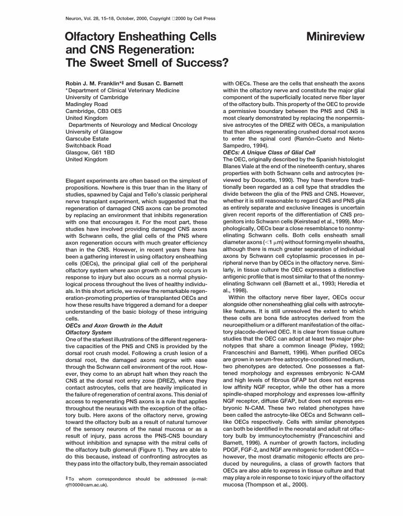
Within the olfactory nerve fiber layer, OECs occureration-promoting properties of transplanted OECs andalongside other nonensheathing glial cells with astrocyte-how these results have triggered a demand for a deeperlike features. It is still unresolved the extent to whichunderstanding of the basic biology of these intriguingthese cells are bona fide astrocytes derived from thecells.neuroepithelium or a different manifestation of the olfac-OECs and Axon Growth in the Adulttory placode-derived OEC. It is clear from tissue cultureOlfactory Systemstudies that the OEC can adopt at least two major phe-One of the starkest illustrations of the different regenera-notypes that share a common lineage (Pixley, 1992;tive capacities of the PNS and CNS is provided by theFranceschini and Barnett, 1996). When purified OECsdorsal root crush model. Following a crush lesion of aare grown in serum-free astrocyte-conditioned medium,dorsal root, the damaged axons regrow with easetwo phenotypes are detected. One possesses a flat-through the Schwann cell environment of the root. How-tened morphology and expresses embryonic N-CAMever, they come to an abrupt halt when they reach theand high levels of fibrous GFAP but does not expressCNS at the dorsal root entry zone (DREZ), where theylow affinity NGF receptor, while the other has a morecontact astrocytes, cells that are heavily implicated inspindle-shaped morphology and expresses low-affinitythe failure of regeneration of central axons. This denial ofNGF receptor, diffuse GFAP, but does not express em-access to regenerating PNS axons is a rule that appliesbryonic N-CAM. These two related phenotypes havethroughout the neuraxis with the exception of the olfac-been called the astrocyte-like OECs and Schwann cell–tory bulb. Here axons of the olfactory nerve, growinglike OECs respectively. Cells with similar phenotypestoward the olfactory bulb as a result of natural turnovercan both be identified in the neonatal and adult rat olfac-of the sensory neurons of the nasal mucosa or as atory bulb by immunocytochemistry (Franceschini andresult of injury, pass across the PNS-CNS boundaryBarnett, 1996). A number of growth factors, includingwithout inhibition and synapse with the mitral cells of
the olfactory bulb glomeruli (Figure 1). They are able to PDGF, FGF-2, and NGF are mitogenic for rodent OECs—do this because, instead of confronting astrocytes as however, the most dramatic mitogenic effects are pro-they pass into the olfactory bulb, they remain associated duced by neuregulins, a class of growth factors that
OECs are also able to express in tissue culture and thatmay play a role in response to toxic injury of the olfactory‡ To whom correspondence should be addressed (e-mail:
[email protected]). mucosa (Thompson et al., 2000).
Neuron16
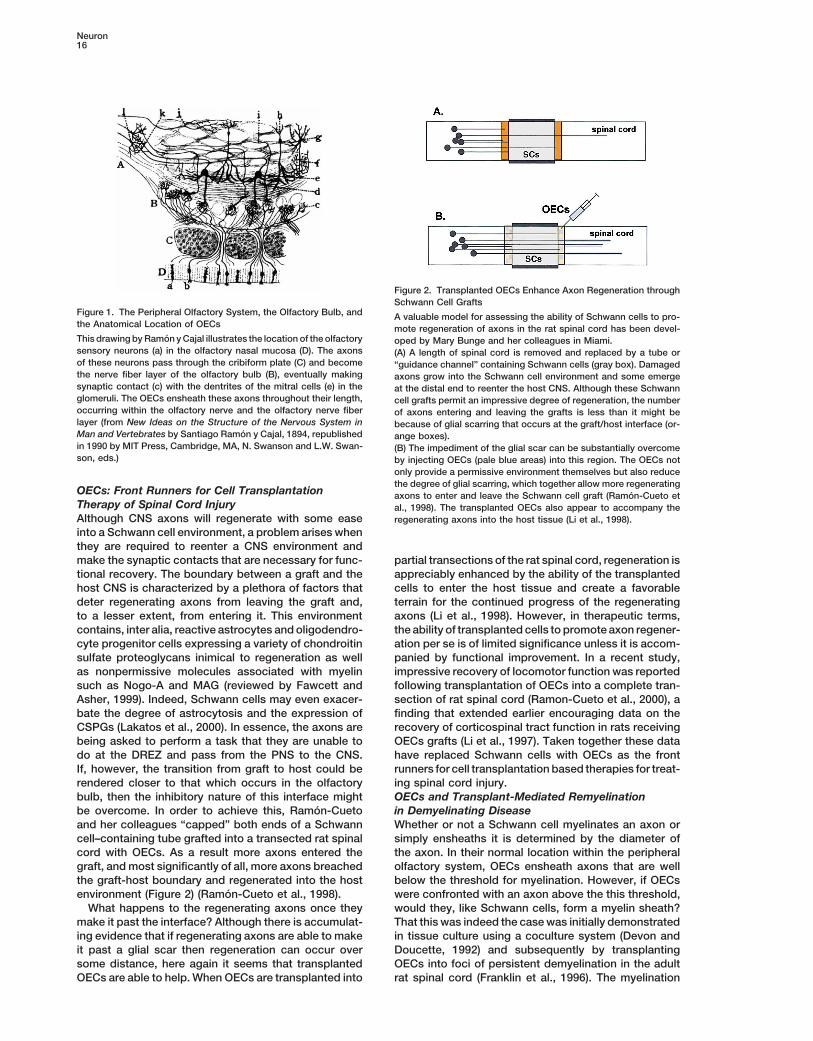
Figure 2. Transplanted OECs Enhance Axon Regeneration throughSchwann Cell Grafts
Figure 1. The Peripheral Olfactory System, the Olfactory Bulb, and A valuable model for assessing the ability of Schwann cells to pro-the Anatomical Location of OECs mote regeneration of axons in the rat spinal cord has been devel-This drawing by Ramon y Cajal illustrates the location of the olfactory oped by Mary Bunge and her colleagues in Miami.sensory neurons (a) in the olfactory nasal mucosa (D). The axons (A) A length of spinal cord is removed and replaced by a tube orof these neurons pass through the cribiform plate (C) and become “guidance channel” containing Schwann cells (gray box). Damagedthe nerve fiber layer of the olfactory bulb (B), eventually making axons grow into the Schwann cell environment and some emergesynaptic contact (c) with the dentrites of the mitral cells (e) in the at the distal end to reenter the host CNS. Although these Schwannglomeruli. The OECs ensheath these axons throughout their length, cell grafts permit an impressive degree of regeneration, the numberoccurring within the olfactory nerve and the olfactory nerve fiber of axons entering and leaving the grafts is less than it might belayer (from New Ideas on the Structure of the Nervous System in because of glial scarring that occurs at the graft/host interface (or-Man and Vertebrates by Santiago Ramon y Cajal, 1894, republished ange boxes).in 1990 by MIT Press, Cambridge, MA, N. Swanson and L.W. Swan- (B) The impediment of the glial scar can be substantially overcomeson, eds.) by injecting OECs (pale blue areas) into this region. The OECs not
only provide a permissive environment themselves but also reducethe degree of glial scarring, which together allow more regenerating
OECs: Front Runners for Cell Transplantation axons to enter and leave the Schwann cell graft (Ramon-Cueto etTherapy of Spinal Cord Injury al., 1998). The transplanted OECs also appear to accompany theAlthough CNS axons will regenerate with some ease regenerating axons into the host tissue (Li et al., 1998).into a Schwann cell environment, a problem arises whenthey are required to reenter a CNS environment andmake the synaptic contacts that are necessary for func- partial transections of the rat spinal cord, regeneration is
appreciably enhanced by the ability of the transplantedtional recovery. The boundary between a graft and thehost CNS is characterized by a plethora of factors that cells to enter the host tissue and create a favorable
terrain for the continued progress of the regeneratingdeter regenerating axons from leaving the graft and,to a lesser extent, from entering it. This environment axons (Li et al., 1998). However, in therapeutic terms,
the ability of transplanted cells to promote axon regener-contains, inter alia, reactive astrocytes and oligodendro-cyte progenitor cells expressing a variety of chondroitin ation per se is of limited significance unless it is accom-
panied by functional improvement. In a recent study,sulfate proteoglycans inimical to regeneration as wellas nonpermissive molecules associated with myelin impressive recovery of locomotor function was reported
following transplantation of OECs into a complete tran-such as Nogo-A and MAG (reviewed by Fawcett andAsher, 1999). Indeed, Schwann cells may even exacer- section of rat spinal cord (Ramon-Cueto et al., 2000), a
finding that extended earlier encouraging data on thebate the degree of astrocytosis and the expression ofCSPGs (Lakatos et al., 2000). In essence, the axons are recovery of corticospinal tract function in rats receiving
OECs grafts (Li et al., 1997). Taken together these databeing asked to perform a task that they are unable todo at the DREZ and pass from the PNS to the CNS. have replaced Schwann cells with OECs as the front
runners for cell transplantation based therapies for treat-If, however, the transition from graft to host could berendered closer to that which occurs in the olfactory ing spinal cord injury.
OECs and Transplant-Mediated Remyelinationbulb, then the inhibitory nature of this interface mightbe overcome. In order to achieve this, Ramon-Cueto in Demyelinating Disease
Whether or not a Schwann cell myelinates an axon orand her colleagues “capped” both ends of a Schwanncell–containing tube grafted into a transected rat spinal simply ensheaths it is determined by the diameter of
the axon. In their normal location within the peripheralcord with OECs. As a result more axons entered thegraft, and most significantly of all, more axons breached olfactory system, OECs ensheath axons that are well
below the threshold for myelination. However, if OECsthe graft-host boundary and regenerated into the hostenvironment (Figure 2) (Ramon-Cueto et al., 1998). were confronted with an axon above the this threshold,
would they, like Schwann cells, form a myelin sheath?What happens to the regenerating axons once theymake it past the interface? Although there is accumulat- That this was indeed the case was initially demonstrated
in tissue culture using a coculture system (Devon anding evidence that if regenerating axons are able to makeit past a glial scar then regeneration can occur over Doucette, 1992) and subsequently by transplanting
OECs into foci of persistent demyelination in the adultsome distance, here again it seems that transplantedOECs are able to help. When OECs are transplanted into rat spinal cord (Franklin et al., 1996). The myelination
Minireview17
achieved was remarkably similar both morphologically OEC Heterogeneity: Implicationsfor CNS Regenerationand biochemically to that achieved by Schwann cells.
In the transplantation study, a clonal OEC line was used It is clear from both tissue culture and morphologicalstudies that OECs can exist in a number of differentto be certain that the myelination achieved was attribut-
able to OECs and not due to Schwann cell contaminants. guises. In both axonal injury and demyelination para-digms, at least two distinctive morphologies are ob-A later study using the same transplant model showed
that extensive myelination could also be achieved by served. In addition to a myelinating Schwann cell–likemorphology seen when transplanted OECs remyelinateprimary rat OECs and, significantly, that this myelination
led to an enhancement of conduction by the demyelin- demyelination axons (Franklin et al., 1996; Imaizumi etal., 1998) or myelinate regenerating axons (Li et al.,ated axons (Imaizumi et al., 1998). This capacity to remy-
elinate demyelinated axons has pushed the OECs not 1998), other cells that have been variously called as-trocyte-like or meningeal-like are also observed (Frank-only to the forefront of spinal cord injury research but
also to the forefront of cell transplantation based thera- lin et al., 1996; Li et al., 1998; Barnett et al., 2000). It istempting to believe that the myelinating cell is akin topies for demyelinating diseases like multiple sclerosis.
Although both Schwann cells and oligodendrocyte lin- the Schwann cell–like, p75-expressing cell, while theother cells are related to the astrocyte-like, embryoniceage cells are able to achieve remyelination following
transplantation into a variety of laboratory animal mod- NCAM-expressing cell described in tissue culture, al-though there is currently no formal proof that this is so.els of demyelinating disease, significant problems cur-
rently exist in translating this progress into clinical appli- The functional implications of this heterogeneity withinthe olfactory system are unclear, although there maycation (reviewed by Blakemore and Franklin, 2000). Put
simply, oligodendrocyte precursor cells, the cells most be important implications for using OECs in transplant-mediated therapies. In the context of providing an envi-likely to lead to widespread remyelination, are extremely
difficult to obtain from the adult human CNS, while ronment favorable to axon regeneration, the differentmorphologies adopted seem to become arranged in anSchwann cells, obtainable from adult human peripheral
nerve with comparative ease, are unlikely to achieve advantageous manner, with some cells myelinating theregenerating axons while others form perineurial-likewidespread remyelination in chronically demyelinated
plaques characterized by extensive astrocyte scarring. sheathes around them. However, when the objective isto remyelinate demyelinated axons, heterogeneity mayThe candidacy of the OEC rests on it being obtainable
from adult human nervous system and being able to be disadvantageous since cells that associate with ax-ons without forming myelin sheaths will decrease theremyelinate axons adequately despite the presence of
astrocytes. The first issue has been largely resolved with remyelinating efficiency of the transplant. A crucialquestion facing OEC biologists, therefore, is whethertwo recent studies reporting the isolation of OECs from
human olfactory bulbs resected during surgery and tissue culture manipulation of OEC phenotype prior totransplantation determines the behavior of the celldemonstrating that human OECs will remyelinate axons
in rodent models of demyelination in a manner very within a lesion environment, or whether environmentalcues to which the transplanted cells are exposed aresimilar to that achieved by rodent OECs (Barnett et al.,
2000; Kato et al., 2000). Several important points have so powerful that they override any such manipulation.OECs: Now that We Know What They Can Do,emerged from these studies regarding the extrapolation
of information obtained using rodent material to the hu- Lets Find Out Who They AreIt is our current ignorance about fundamental questionsman situation. It is clear from these studies that tech-
niques for purifying and expanding human OECs are not such as these that expose the deficiencies in the OECfield, where the contribution these cells can make toidentical to those used for rodent cells. The in vitro
biology of the human cell therefore needs studying in regenerating the CNS has raced ahead of an in-depthevaluation of their basic biology. There are a number ofits own right and cannot simply be inferred from rodent
studies. On the other hand, the similarity in the behavior pressing clinically related issues for which this funda-mental knowledge is required. For example, althoughof human and rodent cells after transplantation validates
using rodents to model what might happen in humans. OECs can be obtained from the human olfactory system,it is nevertheless a somewhat inaccessible region. ByThe second issue of whether the OEC will achieve exten-
sive remyelination in the face of a heavy astrocyte pres- contrast, its close relative, the Schwann cell, can beobtained much more easily from peripheral nerve. If oneence has yet to be fully resolved. However, several lines
of evidence suggest that this may well be the case. knew precisely how these cells differ from Schwanncells, then it might be possible by means of relativelyFirst, OECs exist “cheek-by-jowl” with astrocytes in the
olfactory nerve fiber layer of the olfactory bulb in a natu- small steps to convert Schwann cells into “OECs,”thereby providing sufficiently large numbers of theserally occurring cohabitation that never occurs between
Schwann cells and astrocytes (Doucette, 1990), al- cells from a more accessible source and increasing thefeasibility of autologous transplantation. Alternatively, ifthough the OEC compatibility of these astrocytes may
be very different from those associated with the dam- more were known about the mechanisms of OEC devel-opment, then we would be better placed to exploit ad-aged CNS. Second, OECs and Schwann cells interact
very differently with astrocytes when cocultured, OECs vances in stem cell biology to generate the numbersand purity of cells required for transplantation. Thus,and astrocytes tending to intermingle while Schwann
cells and astrocytes segregate from one another (La- the realization of the enormous potential of these cellsin overcoming the regenerative limitations of the CNSkatos et al., 2000). Third, transplanted OECs appear to
migrate further within the CNS than transplanted will require the combined efforts of many branches ofthe neurosciences.Schwann cells (Ramon-Cueto et al., 1998).
Neuron18
Selected Reading
Barnett, S.C., Alexander, C.L., Iwashita, Y., Gilson, J.M., Crowther,J., Clark, L., Dunn, L.T., Papanastassiou, V., Kennedy, P.G.E., andFranklin, R.J.M. (2000). Brain 123, 1581–1588.
Barnett, S.C., Hutchins, A.-M., and Noble, M. (1993). Dev. Biol. 155,337–350.
Blakemore, W.F., and Franklin, R.J.M. (2000). Cell Transplant. 9,289–294.
Devon, R., and Doucette, R. (1992). Brain Res. 589, 175–179.
Doucette, R. (1990). Glia 3, 433–449.
Fawcett, J.W., and Asher, R.A. (1999). Brain Res. Bull. 49, 377–391.
Franceschini, I.A., and Barnett, S.C. (1996). Dev. Biol. 173, 327–434.
Franklin, R.J.M., Gilson, J.M., Franceschini, I.A., and Barnett, S.C.(1996). Glia 17, 217–224.
Heredia, M., Gascuel, J., Ramon-Cueto, A., Santacana, M., Avila,J., Masson, C., and Valverde, F. (1998). Glia 24, 352–364.
Imaizumi, T., Lankford, K.L., Waxman, S.G., Greer, C.A., and Kocsis,J.D. (1998). J. Neurosci. 18, 6176–6185.
Kato, T., Honmou, O., Uede, T., Hashi, K., and Kocsis, J.D. (2000).Glia 30, 209–218.
Keirstead, H.S., Ben-Hur, T., Rogister, B., O’Leary, M.T., Dubois-Dalcq, M., and Blakemore, W.F. (1999). J. Neurosci. 19, 7529–7536.
Lakatos, A., Franklin, R.J.M., and Barnett, S.C. (2000). Glia, in press.
Li, Y., Field, P.M., and Raisman, G. (1997). Science 277, 2000–2002.
Li, Y., Field, P.M., and Raisman, G. (1998). J. Neurosci. 18, 10514–10524.
Pixley, S.K. (1992). Glia 5, 269–284.
Ramon-Cueto, A., Cordero, M.I., Santos-Benito, F.F., and Avila, J.(2000). Neuron 25, 425–435.
Ramon-Cueto, A., and Nieto-Sampedro, M. (1994). Exp. Neurol. 127,232–244.
Ramon-Cueto, A., Plant, G.W., Avila, J., and Bunge, M.B. (1998). J.Neurosci. 18, 3802–3815.
Thompson, R.J., Roberts, B., Alexander, C.L., Williams, S.K., andBarnett, S.C. (2000). J. Neurosci. Res. 61, 172–185.