Embed Size (px)
Citation preview
OITE Foot and Ankle Review
Anatomy and Biomechanics
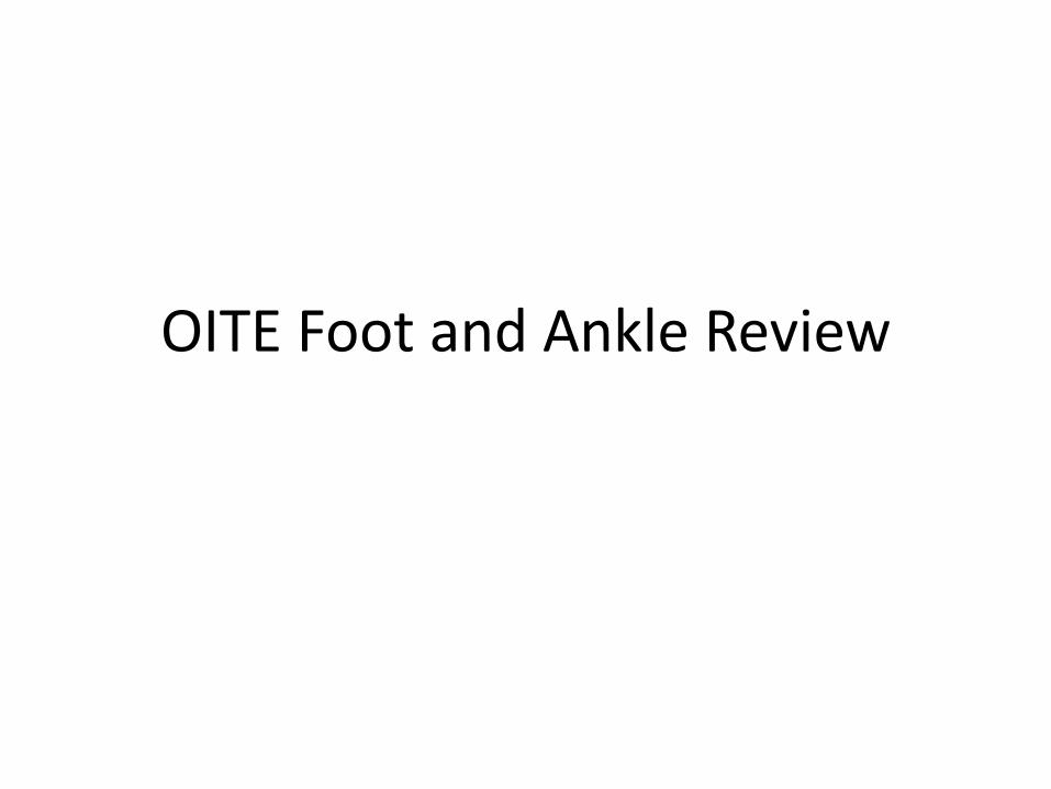
• Bones and Ligaments – The Ankle Joint
• Includes the Tibia, talus and fibula
• Joint is trapezoidal and wider anteriorly
• Talus only tarsal bone without muscular or ligamentous insertions
– Lateral Ankle Ligaments • Anterior Talofibular Ligament
(ATFL) – Under strain in plantar flexion,
inversion, and Internal rotation
• Calcaneofibular Ligament (CFL) – Under strain in dorsiflexion
and inversion
• Posterior Talofibular Ligament (PTFL)
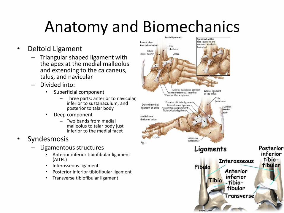
Anatomy and Biomechanics • Deltoid Ligament
– Triangular shaped ligament with the apex at the medial malleolus and extending to the calcaneus, talus, and navicular
– Divided into: • Superficial component
– Three parts: anterior to navicular, inferior to sustanaculum, and posterior to talar body
• Deep component – Two bands from medial
malleolus to talar body just inferior to the medial facet
• Syndesmosis – Ligamentous structures
• Anterior inferior tibiofibular ligament (AITFL)
• Interosseous ligament • Posterior inferior tibiofibular ligament • Transverse tibiofibular ligament
Anatomy and Biomechanics
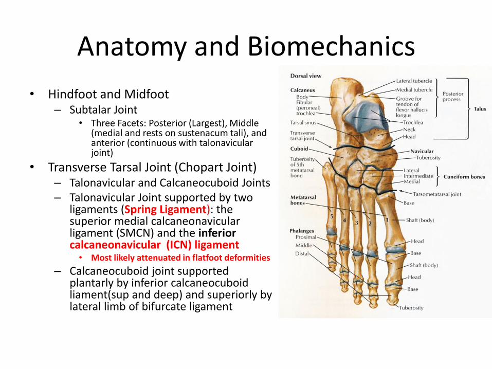
• Hindfoot and Midfoot – Subtalar Joint
• Three Facets: Posterior (Largest), Middle (medial and rests on sustenacum tali), and anterior (continuous with talonavicular joint)
• Transverse Tarsal Joint (Chopart Joint) – Talonavicular and Calcaneocuboid Joints – Talonavicular Joint supported by two
ligaments (Spring Ligament): the superior medial calcaneonavicular ligament (SMCN) and the inferior calcaneonavicular (ICN) ligament
• Most likely attenuated in flatfoot deformities
– Calcaneocuboid joint supported plantarly by inferior calcaneocuboid liament(sup and deep) and superiorly by lateral limb of bifurcate ligament
Anatomy and Biomechanics
• Tarsometatarsal Joint – Osseous anatomy serves as a transverse Roman arch in the axial plane
with dorsal surface wider than plantar surface – Second MT base serves as keystone – Ligamentous support of TMT in three layers
• Interosseous layer (strongest, includes Lisfranc ligament), plantar layer, dorsal layer (weakest)
• Lisfranc ligament from plantar aspect of medial cuneiform to base of 2nd MT
• Forefoot – The plantar fascia runs from medial calcaneal tuberosity and inserts on
base of 5th MT, plantar plate (plantar aspect of 1st MT joint), and bases of 5 proximal phalanges
– Conjoined tendon of the Adductor Hallucis inserts on the lateral proximal first metatarsal and lateral sesamoid
222) What does the main component of the Lisfranc Ligament connect?
1 The first metatarsal base to the medial cuneiform
2 The first metatarsal base to the second metatarsal base
3 The medial cuneiform to the base of the second metatarsal
4 The medial cuneiform to the lateral cuneiform
5 The middle cuneiform to the second metatarsal base
This is just one of those questions that you have to know. The main component of the Lisfranc ligament connects the medial cuneiform to the base of the second metatarsal. Hence, if this is ruptured, you get widening between the first and second tarso-metatarsal joint spaces.
Anatomy and Biomechanics
• Compartments of the leg – Anterior Compartment
• Tibialis Anterior (TA), Extensor Hallicus Longus (EHL), Extensor Digitorum Longus (EDL), and Peroneus Tertius
• Anterior Tibial Artery • Deep Peroneal Nerve
– At extensor retinaculum of ankle, Anterior tibial artery and DPN lie between TA and EHL tendons
– Superficial Posterior Compartment • Gactrocnemius Soleus Comlex and Plantaris Muscle • Gastroc and soleus meet to form Achilles tendon which
twists medially 90 degrees so that superificial fibers at myotendinous junction insert laterally on calcaneus
Anatomy and Biomechanics
• Deep Posterior Compartment – Posterior Tibial Tendon (PT), Flexor Digitorum Longus (FDL), and Flexor
Hallicus Longus (FHL) – Oriented from anteromedial to posterolateral in the tarsal tunnel: PTT,
FDL, Posterior Tibial Artery, Tibial nerve, and FHL tendon – FHL and FDL are interconnected at the knot of Henry in the plantar
midfoot
• Lateral Compartment – Peroneus longus (inserts base of 1st TMT joint), Peroneus Brevis
(inserts base of 5th MT), Superficial Peroneal Nerve (SPN), and the Peroneal Artery
– The peroneus brevis tendon lies superior and the peroneus longus tendon lies inferior to the peroneal tubercle in the inferior peroneal retinaculum
Anatomy and Biomechanics
• Muscles of the Plantar Foot – First layer(superficial)
• Flexor Digitorum Brevis(FDB)
• Abductor Halicus(AbH)
• Abductor Digiti Minimi (ADM)
– Second Layer • Quadratus Plantae
• Tendons of Lumbricals
• FDL Tendons
• FHL Tendon
• Medial and Lateral Plantar arteries and nerves
– Third layer
– Oblique and transverse heads of Adductor Hallicus (AdH)
– Flexor Hallicus Brevis (FHB)
– Flexor Digiti Minimi brevis (FDM) muscles
– Fourth Layer (deepest)
– Peroneus long
– Tibialis posterior
– Four dorsal interossei
– Three plantar interossei
Anatomy and Biomechanics
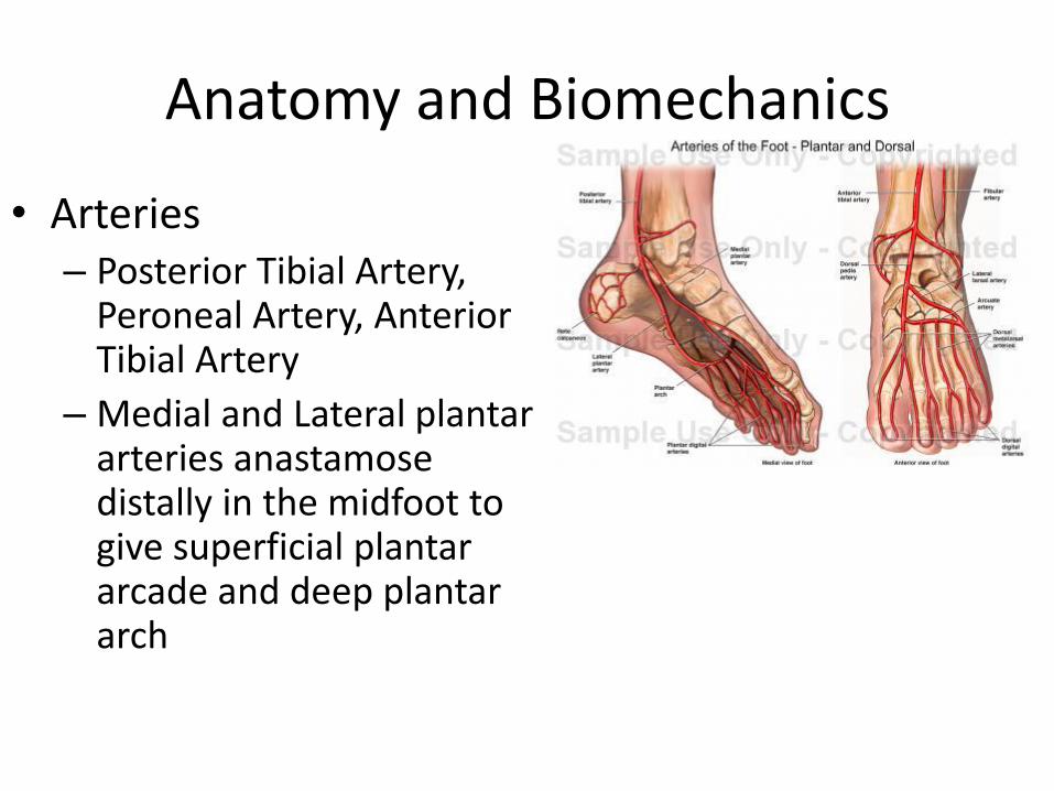
• Arteries – Posterior Tibial Artery,
Peroneal Artery, Anterior Tibial Artery
– Medial and Lateral plantar arteries anastamose distally in the midfoot to give superficial plantar arcade and deep plantar arch
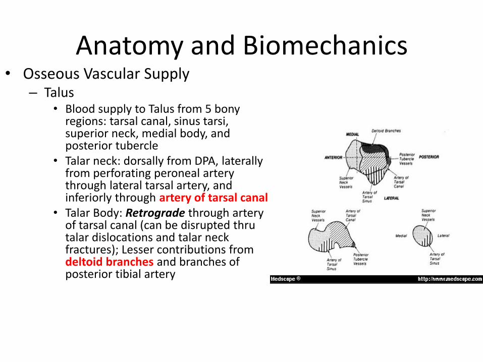
Anatomy and Biomechanics • Osseous Vascular Supply
– Talus • Blood supply to Talus from 5 bony
regions: tarsal canal, sinus tarsi, superior neck, medial body, and posterior tubercle
• Talar neck: dorsally from DPA, laterally from perforating peroneal artery through lateral tarsal artery, and inferiorly through artery of tarsal canal
• Talar Body: Retrograde through artery of tarsal canal (can be disrupted thru talar dislocations and talar neck fractures); Lesser contributions from deltoid branches and branches of posterior tibial artery
Anatomy and Biomechanics
• Fifth Metatarsal
– Penetrating at junction of proximal and middle thrids, the main nutrient vessel divides into proximal and distal vessels
– Proximal blood supply through the tuberosity, creating a watershed area at proximal metaphyseal/diaphyseal junction, which is prone to stress fractures and nonunion (Jone’s Fractures)
Anatomy and Biomechanics
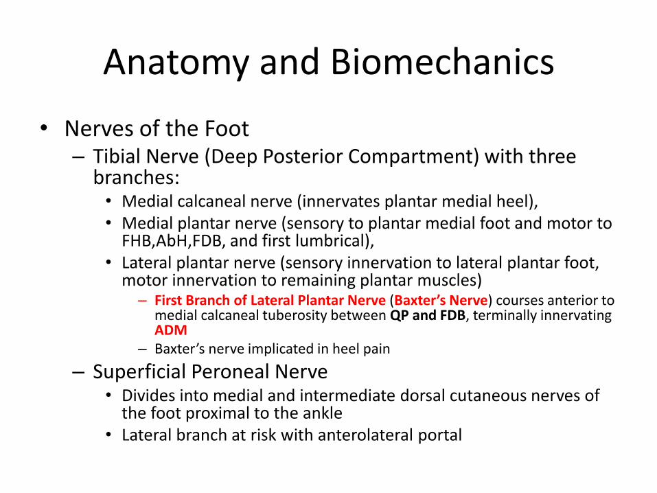
• Nerves of the Foot – Tibial Nerve (Deep Posterior Compartment) with three
branches: • Medial calcaneal nerve (innervates plantar medial heel), • Medial plantar nerve (sensory to plantar medial foot and motor to
FHB,AbH,FDB, and first lumbrical), • Lateral plantar nerve (sensory innervation to lateral plantar foot,
motor innervation to remaining plantar muscles) – First Branch of Lateral Plantar Nerve (Baxter’s Nerve) courses anterior to
medial calcaneal tuberosity between QP and FDB, terminally innervating ADM
– Baxter’s nerve implicated in heel pain
– Superficial Peroneal Nerve • Divides into medial and intermediate dorsal cutaneous nerves of
the foot proximal to the ankle • Lateral branch at risk with anterolateral portal
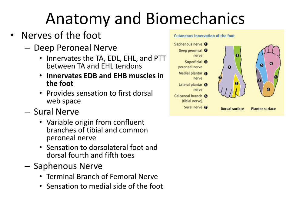
Anatomy and Biomechanics • Nerves of the foot
– Deep Peroneal Nerve • Innervates the TA, EDL, EHL, and PTT
between TA and EHL tendons • Innervates EDB and EHB muscles in
the foot • Provides sensation to first dorsal
web space
– Sural Nerve • Variable origin from confluent
branches of tibial and common peroneal nerve
• Sensation to dorsolateral foot and dorsal fourth and fifth toes
– Saphenous Nerve • Terminal Branch of Femoral Nerve • Sensation to medial side of the foot
Anatomy and Biomechanics
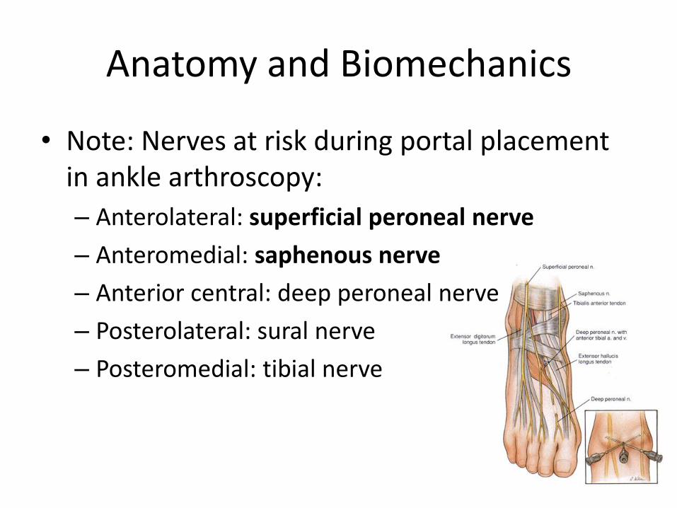
• Note: Nerves at risk during portal placement in ankle arthroscopy:
– Anterolateral: superficial peroneal nerve
– Anteromedial: saphenous nerve
– Anterior central: deep peroneal nerve
– Posterolateral: sural nerve
– Posteromedial: tibial nerve
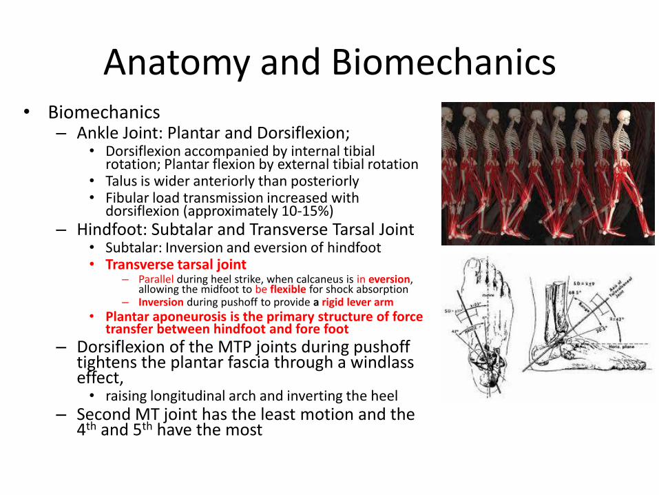
Anatomy and Biomechanics • Biomechanics
– Ankle Joint: Plantar and Dorsiflexion; • Dorsiflexion accompanied by internal tibial
rotation; Plantar flexion by external tibial rotation • Talus is wider anteriorly than posteriorly • Fibular load transmission increased with
dorsiflexion (approximately 10-15%) – Hindfoot: Subtalar and Transverse Tarsal Joint
• Subtalar: Inversion and eversion of hindfoot • Transverse tarsal joint
– Parallel during heel strike, when calcaneus is in eversion, allowing the midfoot to be flexible for shock absorption
– Inversion during pushoff to provide a rigid lever arm
• Plantar aponeurosis is the primary structure of force transfer between hindfoot and fore foot
– Dorsiflexion of the MTP joints during pushoff tightens the plantar fascia through a windlass effect,
• raising longitudinal arch and inverting the heel – Second MT joint has the least motion and the
4th and 5th have the most
58. During the normal gait cycle, at the pushoff phase of stance the hindfoot
1- inverts and the transverse tarsal joints lock
2- inverts and the transverse tarsal joints unlock
3- everts and the transverse tarsal joints unlock
4- everts and the transverse tarsal joints lock
5- remains neutral and the transverse tarsal joints lock
Answer:
1- inverts and the transverse tarsal joints lock
- Ahh the gait cycle. A little more involved than “60%/40%”. Dr. Deasla wrote a great chapter in the
Orthopedic Surgery Essentials F&A book that you should read. Briefly, when the heel is everted the
transverse tarsal joints (talonavicular and calcaneocuboid) are parallel to one another allowing for motion.
As the heel inverts this parallel relationship changes and they become oblique thus locking the transverse
tarsal joint. This is important for push-off as it gives both the gastrocsoleus, posterior tib and forefoot
flexors a stable base.

OITE 2006- Foot & Ankle
6. Arch height is maintained during the stance phase of gait primarily by
1. Achilles tendon contraction
2. Posterior tibial tendon contraction
3. Bony and ligamentous structures
4. Unlocking of the transverse tarsal joints
5. Balanced contraction of the peroneus longus and anterior tibialis
Discussion
• The plantar aponeurosis is the major support of the medial longitudinal arch. It originates on the plantar medial aspect of the calcaneus and passes distally, inserting into the base of the flexor mechanism of the toes. This question stated this answer in a not so nice way by calling it “bony and ligamentous structures.” No, the OITE does not want to trick you!
• One author described the function of the plantar aponeurosis as being analogous to a windlass mechanism, in which the arch of the foot elevates by winding of the plantar aponeurosis around the heads of the metatarsals during toe extension. Various studies of cadavera have revealed that release of the plantar aponeurosis decreases arch height, confirming the arch-supporting function of the plantar aponeurosis.
• The posterior tibialis is the main invertor of the subtalar joint during heel rise which initiates the subtalar inversion.
• Unlocking of the transverse tarsal joint occurs from heel strike to foot flat when there is progressive eversion of the subtalar joint.
• The Achilles tendon contracts concentrically at heel rise/push-off while the tibialis anterior is quiescent.
• The tibialis anterior and peroneus longus contract eccentrically at heel strike (controlled dorsiflexion) while achilles is quiescent.
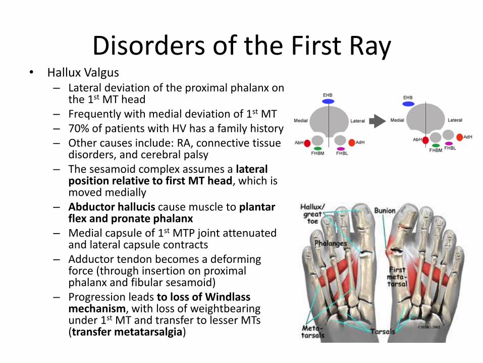
Disorders of the First Ray • Hallux Valgus
– Lateral deviation of the proximal phalanx on the 1st MT head
– Frequently with medial deviation of 1st MT – 70% of patients with HV has a family history – Other causes include: RA, connective tissue
disorders, and cerebral palsy – The sesamoid complex assumes a lateral
position relative to first MT head, which is moved medially
– Abductor hallucis cause muscle to plantar flex and pronate phalanx
– Medial capsule of 1st MTP joint attenuated and lateral capsule contracts
– Adductor tendon becomes a deforming force (through insertion on proximal phalanx and fibular sesamoid)
– Progression leads to loss of Windlass mechanism, with loss of weightbearing under 1st MT and transfer to lesser MTs (transfer metatarsalgia)
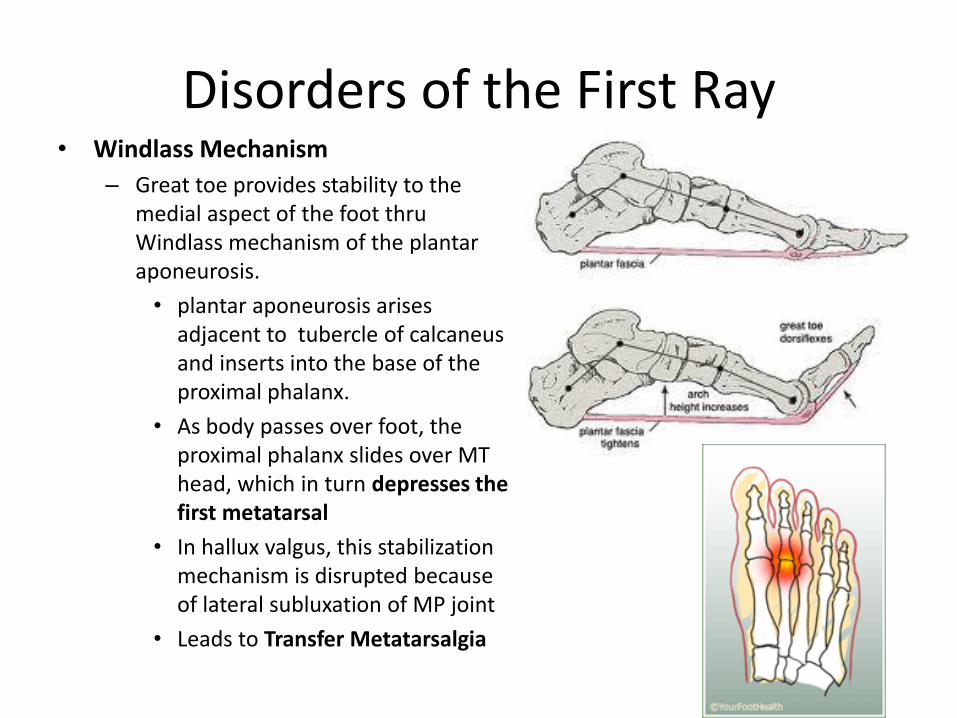
Disorders of the First Ray • Windlass Mechanism
– Great toe provides stability to the medial aspect of the foot thru Windlass mechanism of the plantar aponeurosis.
• plantar aponeurosis arises adjacent to tubercle of calcaneus and inserts into the base of the proximal phalanx.
• As body passes over foot, the proximal phalanx slides over MT head, which in turn depresses the first metatarsal
• In hallux valgus, this stabilization mechanism is disrupted because of lateral subluxation of MP joint
• Leads to Transfer Metatarsalgia
Disorders of the First Ray
• Evaluation – Bunion noted along medial aspect of 1st MTP joint – Swelling and redness due to bursal inflammation – Note associated hammer toes and calluses from stress
transfer laterally – Weight bearing AP and lateral views most commonly
obtained
• Treatment – Nonsurgical: Shoewear modifications (low heeled shoes,
wide toe box), pads, orthoses to alleviate pes planus or metatarsalgia symptoms
– Surgical

Disorders of the First Ray • Important Radiographic
Angles
– HVA: Hallux Valgus Angle
• Between long axis of proximal phalanx and first MT
• Degree of deformity at MTP
• Normal <15 degrees
– IMA: Intermetatarsal Angle
• Between long axis of first and second MT
• Normal <9 degrees
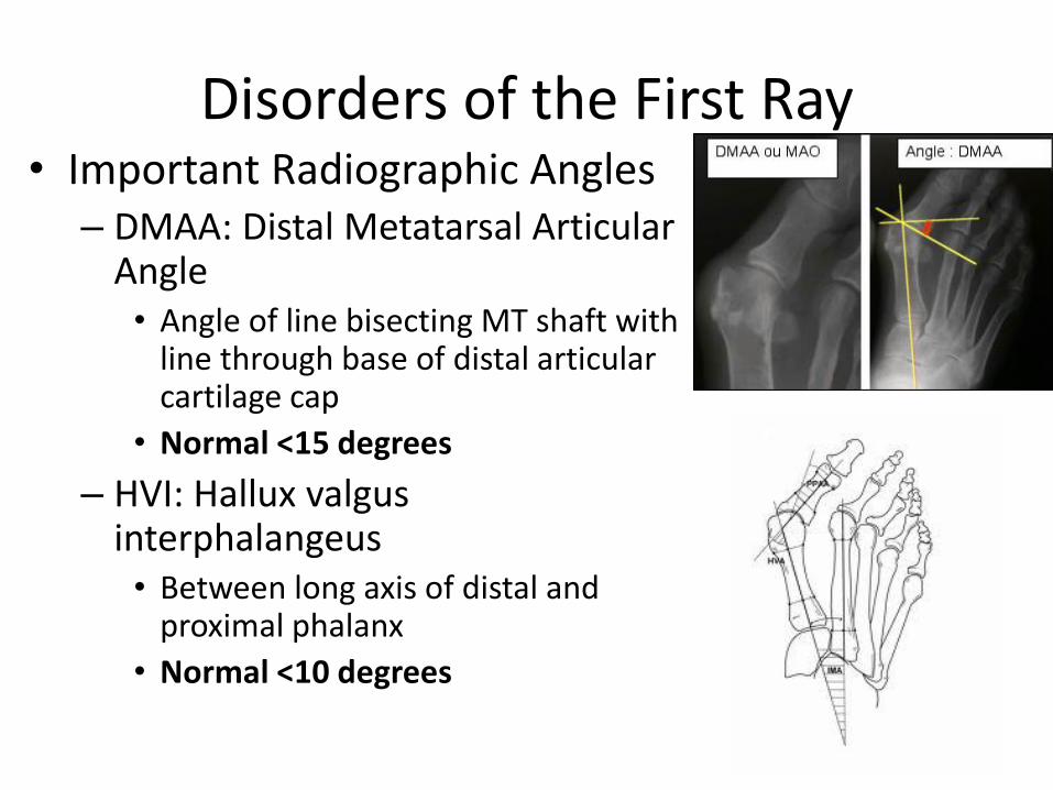
Disorders of the First Ray • Important Radiographic Angles
– DMAA: Distal Metatarsal Articular Angle
• Angle of line bisecting MT shaft with line through base of distal articular cartilage cap
• Normal <15 degrees
– HVI: Hallux valgus interphalangeus
• Between long axis of distal and proximal phalanx
• Normal <10 degrees
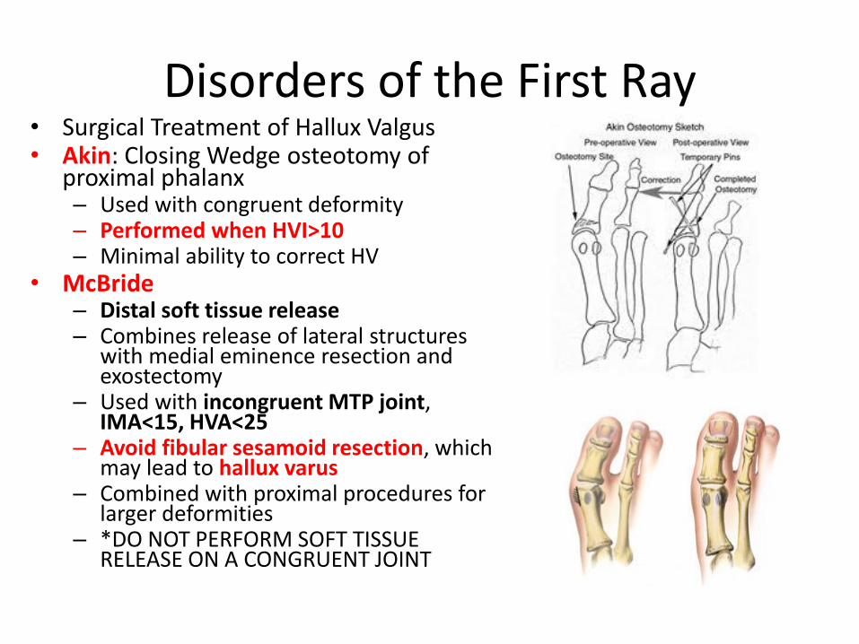
Disorders of the First Ray • Surgical Treatment of Hallux Valgus • Akin: Closing Wedge osteotomy of
proximal phalanx – Used with congruent deformity – Performed when HVI>10 – Minimal ability to correct HV
• McBride – Distal soft tissue release – Combines release of lateral structures
with medial eminence resection and exostectomy
– Used with incongruent MTP joint, IMA<15, HVA<25
– Avoid fibular sesamoid resection, which may lead to hallux varus
– Combined with proximal procedures for larger deformities
– *DO NOT PERFORM SOFT TISSUE RELEASE ON A CONGRUENT JOINT
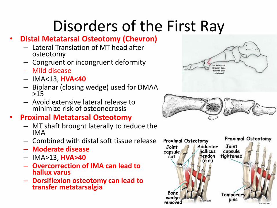
Disorders of the First Ray • Distal Metatarsal Osteotomy (Chevron)
– Lateral Translation of MT head after osteotomy
– Congruent or incongruent deformity – Mild disease – IMA<13, HVA<40 – Biplanar (closing wedge) used for DMAA
>15 – Avoid extensive lateral release to
minimize risk of osteonecrosis • Proximal Metatarsal Osteotomy
– MT shaft brought laterally to reduce the IMA
– Combined with distal soft tissue release – Moderate disease – IMA>13, HVA>40 – Overcorrection of IMA can lead to
hallux varus – Dorsiflexion osteotomy can lead to
transfer metatarsalgia
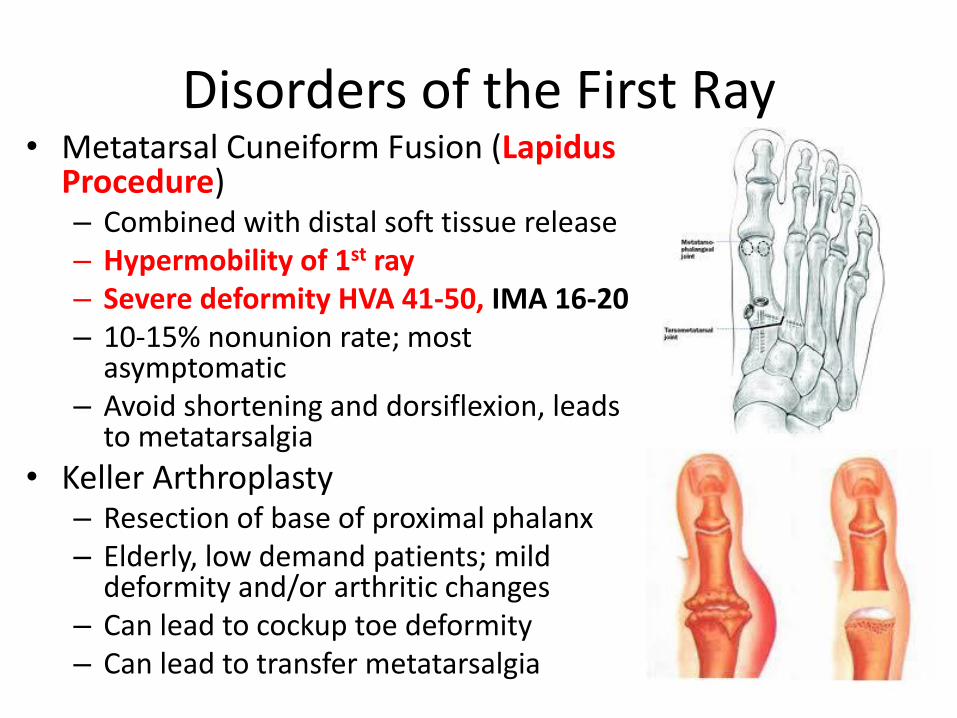
Disorders of the First Ray • Metatarsal Cuneiform Fusion (Lapidus
Procedure) – Combined with distal soft tissue release – Hypermobility of 1st ray – Severe deformity HVA 41-50, IMA 16-20 – 10-15% nonunion rate; most
asymptomatic – Avoid shortening and dorsiflexion, leads
to metatarsalgia
• Keller Arthroplasty – Resection of base of proximal phalanx – Elderly, low demand patients; mild
deformity and/or arthritic changes – Can lead to cockup toe deformity – Can lead to transfer metatarsalgia
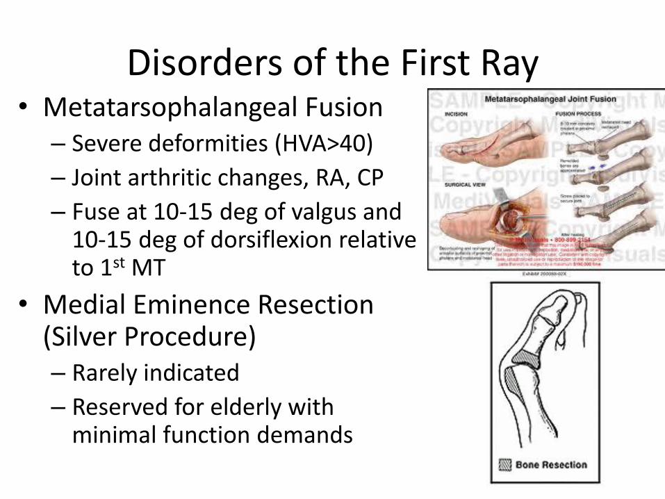
Disorders of the First Ray • Metatarsophalangeal Fusion
– Severe deformities (HVA>40)
– Joint arthritic changes, RA, CP
– Fuse at 10-15 deg of valgus and 10-15 deg of dorsiflexion relative to 1st MT
• Medial Eminence Resection (Silver Procedure) – Rarely indicated
– Reserved for elderly with minimal function demands
OITE 2006 - Foot/Ankle
267. A 44-year-old woman has a symptomatic bunion and a painful callus under the second metatarsal head that continues to limit her activity and shoe wear despite the use of shoe modifications. Radiographs show an intermetatarsal angle of 18°, a hallux valgus angle of 38°, and a first metatarsal that is shorter than both the second and third metatarsals. When considering surgical options, each of the following first metatarsal procedures are appropriate for this patient EXCEPT
1. Z osteotomy (Scarf). 2. oblique proximal osteotomy (Ludloff). 3. distal chevron osteotomy. 4. proximal crescentic osteotomy. 5. Lapidus procedure.
OITE 2006 - Foot/Ankle - 267
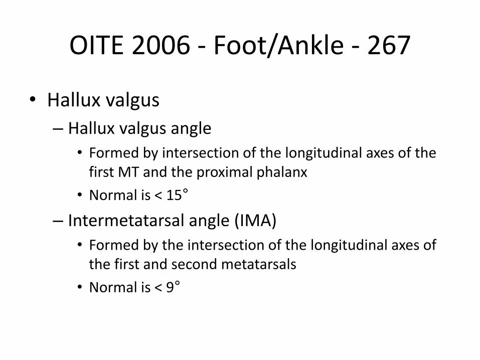
• Hallux valgus
– Hallux valgus angle
• Formed by intersection of the longitudinal axes of the first MT and the proximal phalanx
• Normal is < 15°
– Intermetatarsal angle (IMA)
• Formed by the intersection of the longitudinal axes of the first and second metatarsals
• Normal is < 9°
OITE 2006 - Foot/Ankle - 267
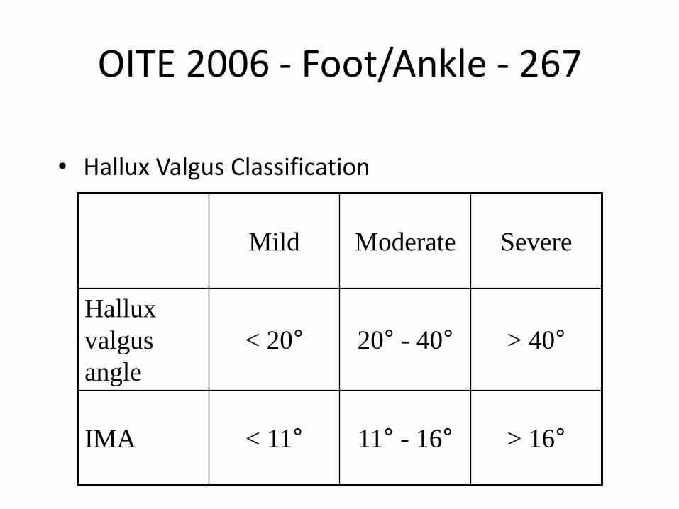
• Hallux Valgus Classification
Mild Moderate Severe
Hallux
valgus
angle
< 20° 20° - 40° > 40°
IMA < 11° 11° - 16° > 16°
OITE 2006 - Foot/Ankle - 267
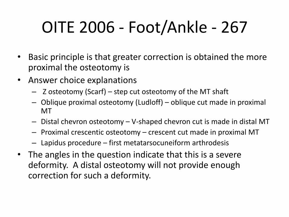
• Basic principle is that greater correction is obtained the more proximal the osteotomy is
• Answer choice explanations – Z osteotomy (Scarf) – step cut osteotomy of the MT shaft
– Oblique proximal osteotomy (Ludloff) – oblique cut made in proximal MT
– Distal chevron osteotomy – V-shaped chevron cut is made in distal MT
– Proximal crescentic osteotomy – crescent cut made in proximal MT
– Lapidus procedure – first metatarsocuneiform arthrodesis
• The angles in the question indicate that this is a severe deformity. A distal osteotomy will not provide enough correction for such a deformity.
Foot and Cankle
Foot and Cankle
Answer 4
Foot and Cankle
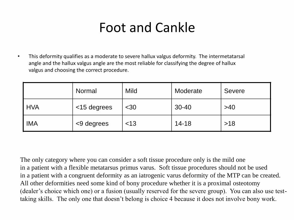
• This deformity qualifies as a moderate to severe hallux valgus deformity. The intermetatarsal angle and the hallux valgus angle are the most reliable for classifying the degree of hallux valgus and choosing the correct procedure.
Normal Mild Moderate Severe
HVA <15 degrees <30 30-40 >40
IMA <9 degrees <13 14-18 >18
The only category where you can consider a soft tissue procedure only is the mild one
in a patient with a flexible metatarsus primus varus. Soft tissue procedures should not be used
in a patient with a congruent deformity as an iatrogenic varus deformity of the MTP can be created.
All other deformities need some kind of bony procedure whether it is a proximal osteotomy
(dealer’s choice which one) or a fusion (usually reserved for the severe group). You can also use test-
taking skills. The only one that doesn’t belong is choice 4 because it does not involve bony work.
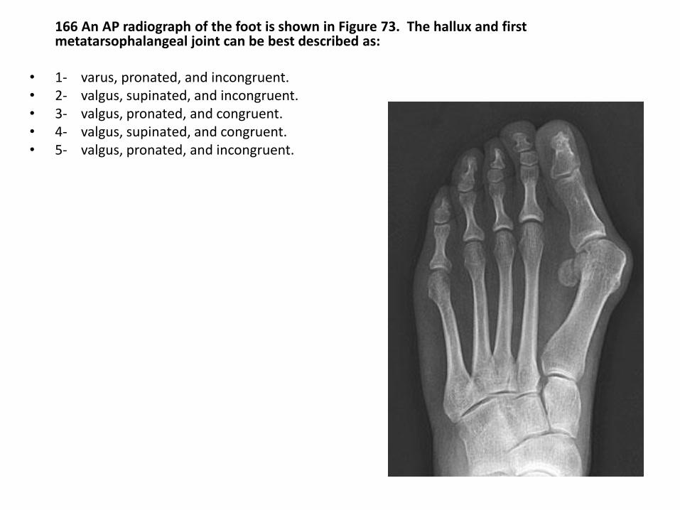
166 An AP radiograph of the foot is shown in Figure 73. The hallux and first metatarsophalangeal joint can be best described as:
• 1- varus, pronated, and incongruent. • 2- valgus, supinated, and incongruent. • 3- valgus, pronated, and congruent. • 4- valgus, supinated, and congruent. • 5- valgus, pronated, and incongruent.
PREFERRED RESPONSE: 5 valgus, pronated, and incongruent. - Case of hallux “valgus”, hope you didn’t pick answer 1, the MTP is always in pronation, there is a great picture in Miller but
basically given progression of deformity the EHB is pulled medially and the FHB, sesamoids, AbH are all pulled laterally causing a pronation deformity. Joint congruence refers to the relationship between the articular surfaces of the 1st MT head and proximal phalanx:
Congruent: 2 articular surfaces are parallel Incongruent: not parallel RECOMMENDED READINGS: Mann RA, Coughlin MJ: Adult hallux valgus, in Coughlin MJ, Mann RA (eds): Surgery of the Foot and
Ankle, ed 7. St Louis, MO, Mosby, 1999, pp 150-269. Coughlin MJ: Hallux valgus. J Bone Joint Surg Am 1996;78:932-966.
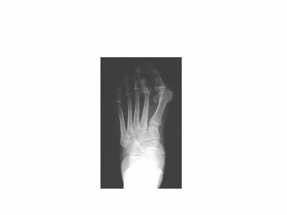
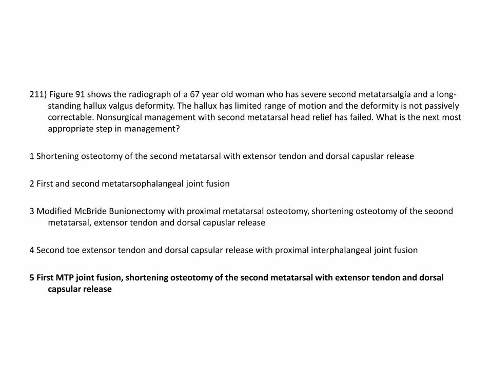
211) Figure 91 shows the radiograph of a 67 year old woman who has severe second metatarsalgia and a long-standing hallux valgus deformity. The hallux has limited range of motion and the deformity is not passively correctable. Nonsurgical management with second metatarsal head relief has failed. What is the next most appropriate step in management?
1 Shortening osteotomy of the second metatarsal with extensor tendon and dorsal capuslar release
2 First and second metatarsophalangeal joint fusion
3 Modified McBride Bunionectomy with proximal metatarsal osteotomy, shortening osteotomy of the seoond metatarsal, extensor tendon and dorsal capuslar release
4 Second toe extensor tendon and dorsal capsular release with proximal interphalangeal joint fusion
5 First MTP joint fusion, shortening osteotomy of the second metatarsal with extensor tendon and dorsal capsular release.
211) Figure 91 shows the radiograph of a 67 year old woman who has severe second metatarsalgia and a long-standing hallux valgus deformity. The hallux has limited range of motion and the deformity is not passively correctable. Nonsurgical management with second metatarsal head relief has failed. What is the next most appropriate step in management?
1 Shortening osteotomy of the second metatarsal with extensor tendon and dorsal capuslar release
2 First and second metatarsophalangeal joint fusion
3 Modified McBride Bunionectomy with proximal metatarsal osteotomy, shortening osteotomy of the seoond metatarsal, extensor tendon and dorsal capuslar release
4 Second toe extensor tendon and dorsal capsular release with proximal interphalangeal joint fusion
5 First MTP joint fusion, shortening osteotomy of the second metatarsal with extensor tendon and dorsal capsular release
This patient has a a HVA of > 13 degrees, and an IMA of at least 20 degrees, they are over the age of 50, with what looks like degenerative changes at the first and second MTP joints. With this information alone you know that the patient needs more than just soft tissue work, they need a combination of boney and soft tissue work. This eliminates #3 (A Modified McBride bunionectomy is soft tissue work only). With treatment of bunions, it is important to examine the whole foot. If you only treat one metatarsal that appears the most deformed, and ignore the rest, the patient will still have pain, and not be happy. Your procedure will have been useless. Looking at the diagram, you can tell that the first and second MTP joints are involved. So, to adequately treat the patient you need to treat both joints. This eliminates 1 and 4 because they deal with the second MTP joint only, and ignore the first. Arthrodesis is an effective solution for pain control, especially if the patient has begun to have degenerative changes. Both of these are offered in 2 and 5. The second MTP joint helps you choose between the two. If you look at the second MTP joint carefully, you note that it is subluxated laterally. You should not fuse this incongruent joint in this position. Rather, try to realign it and then fuse it. To realign it you need a distal soft tissue procedure and a proximal osteotomy. This is answer 5. Could something more be done to correc the Hallux Valugus deformity of the first metatarsal? Possibly, but that is not given as an answer choice here. So, the best answer is 5.
Disorders of the First Ray
• Juvenile Hallux Valgus
– Look for generalized ligamentous laxity
– Nonsurgical: shoewear modification,education
– Surgical treatments similar to adults, but an increased IMA is corrected with medial opening wedge cuneiform osteotomy rather than proximal metatarsal osteotomy or fusion
– A congruent joint with an elevated DMAA more common in juvenile hallux valgus
– Recurrence rates up to 50% with surgical treatment
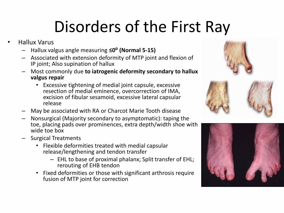
Disorders of the First Ray • Hallux Varus
– Hallux valgus angle measuring ≤0⁰ (Normal 5-15) – Associated with extension deformity of MTP joint and flexion of
IP joint; Also supination of hallux – Most commonly due to iatrogenic deformity secondary to hallux
valgus repair • Excessive tightening of medial joint capsule, excessive
resection of medial eminence, overcorrection of IMA, excision of fibular sesamoid, excessive lateral capsular release
– May be associated with RA or Charcot Marie Tooth disease – Nonsurgical (Majority secondary to asymptomatic): taping the
toe, placing pads over prominences, extra depth/width shoe with wide toe box
– Surgical Treatments • Flexible deformities treated with medial capsular
release/lengthening and tendon transfer – EHL to base of proximal phalanx; Split transfer of EHL;
rerouting of EHB tendon • Fixed deformities or those with significant arthrosis require
fusion of MTP joint for correction
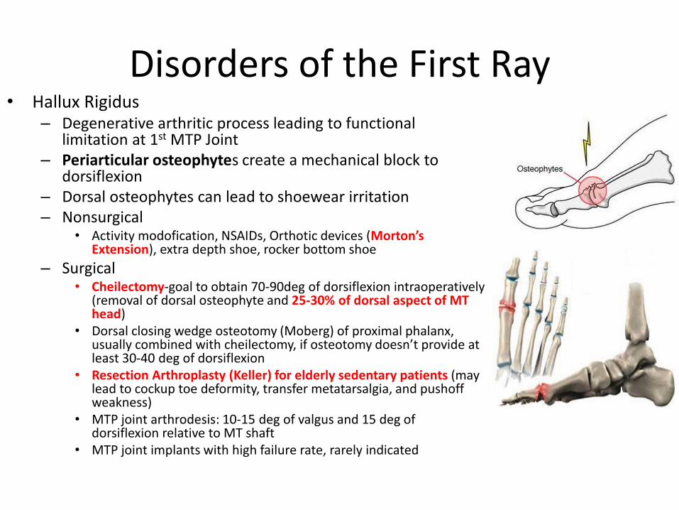
Disorders of the First Ray • Hallux Rigidus
– Degenerative arthritic process leading to functional limitation at 1st MTP Joint
– Periarticular osteophytes create a mechanical block to dorsiflexion
– Dorsal osteophytes can lead to shoewear irritation – Nonsurgical
• Activity modofication, NSAIDs, Orthotic devices (Morton’s Extension), extra depth shoe, rocker bottom shoe
– Surgical • Cheilectomy-goal to obtain 70-90deg of dorsiflexion intraoperatively
(removal of dorsal osteophyte and 25-30% of dorsal aspect of MT head)
• Dorsal closing wedge osteotomy (Moberg) of proximal phalanx, usually combined with cheilectomy, if osteotomy doesn’t provide at least 30-40 deg of dorsiflexion
• Resection Arthroplasty (Keller) for elderly sedentary patients (may lead to cockup toe deformity, transfer metatarsalgia, and pushoff weakness)
• MTP joint arthrodesis: 10-15 deg of valgus and 15 deg of dorsiflexion relative to MT shaft
• MTP joint implants with high failure rate, rarely indicated
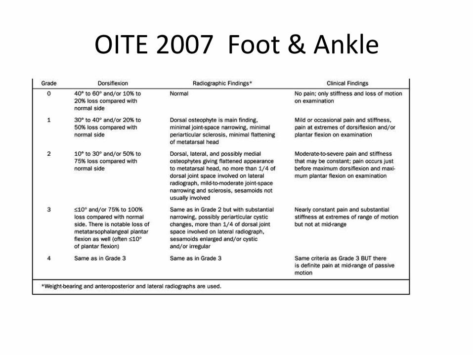
Disorders of the First Ray • Treatment Algorithm Hallux Rigidus
– Grade 1: minor joint space narrowing w/ dorsal osteophyte -> Rx: non op, cheilectomy/debridement
– Grade 2: more extensive narrowing with only the plantar joint space preserved
-> Rx: cheilectomy/debridement +/- Moberg procedure (dorsal closing wedge osteotomy of proximal phalanx for younger more active patients who require more dorsiflexion)
– Grade 3: complete joint space narrowing/arthrosis
-> Rx: arthrodesis, Keller resection arthroplasty, interpositional arthroplasty, implant arthroplasty (bad idea)
OITE 2007 Foot & Ankle
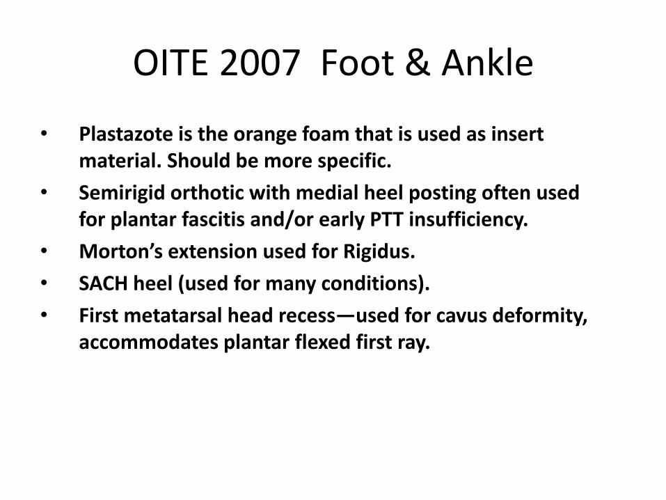
248.Which of the following orthotic features is recommended for a patient with hallux rigidus?
1. Plastazote insert
2. Semirigid orthotic with medial heel posting
3. Morton’s extension
4. SACH heel
5. First metatarsal head recession
OITE 2007 Foot & Ankle
248.Which of the following orthotic features is recommended for a patient with hallux rigidus?
1. Plastazote insert
2. Semirigid orthotic with medial heel posting
3. Morton’s extension
4. SACH heel
5. First metatarsal head recession
OITE 2007 Foot & Ankle
• Plastazote is the orange foam that is used as insert material. Should be more specific.
• Semirigid orthotic with medial heel posting often used for plantar fascitis and/or early PTT insufficiency.
• Morton’s extension used for Rigidus.
• SACH heel (used for many conditions).
• First metatarsal head recess—used for cavus deformity, accommodates plantar flexed first ray.
OITE 2007 Foot & Ankle
• A briefing on Hallux Rigidus: • Painful affliction of the first MTP joint secondary to arthrosis and is
associated with restriction of dorsiflexion. • Marginal osteophytes are typically present dorsally and laterally. Forced
dorsiflexion will usually reproduce the patient’s pain, as will lateral deviation if a lateral osteophyte is present.
• Often, the dorsal medial cutaneous nerve is sensitive. • Conservative management consists of use of a shoe with adequate width
and depth to accommodate the increased bulk of the joint and with a rigid rocker sole to diminish joint motion.
• If there is significant bone proliferation or pain with dorsiflexion, a cheilectomy or debridement of the MTP joint should be considered.
OITE 2007 Foot & Ankle
Quoted Article: Coughlin et al, Hallux Rigidus: Grading and Long term results of operative treatment, JBJS (Am) 2003; 85: 2072-2088.
• “Ninety-seven percent (107) of the 110 patients had a good or excellent subjective result, and 92% (eighty-six) of the ninety-three cheilectomy procedures were successful in terms of pain relief and function. Cheilectomy was used with predictable success to treat Grade-1 and 2 and selected Grade-3 cases. Patients with Grade-4 hallux rigidus or Grade-3 hallux rigidus with <50% of the metatarsal head cartilage remaining at the time of surgery should be treated with arthrodesis.”
OITE 2007 Foot & Ankle
OITE 2007 Foot & Ankle
Morton’s extension used for Rigidus.
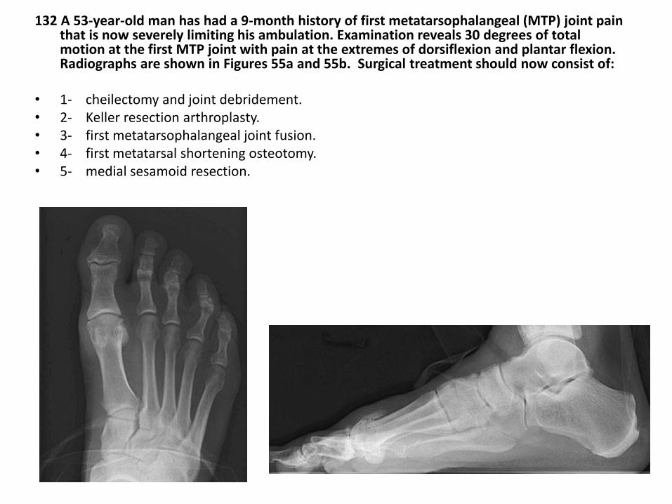
132 A 53-year-old man has had a 9-month history of first metatarsophalangeal (MTP) joint pain that is now severely limiting his ambulation. Examination reveals 30 degrees of total motion at the first MTP joint with pain at the extremes of dorsiflexion and plantar flexion. Radiographs are shown in Figures 55a and 55b. Surgical treatment should now consist of:
• 1- cheilectomy and joint debridement. • 2- Keller resection arthroplasty. • 3- first metatarsophalangeal joint fusion. • 4- first metatarsal shortening osteotomy. • 5- medial sesamoid resection.
PREFERRED RESPONSE: 1 cheilectomy and joint debridement. - Vignette describes a guy with hallux rigidus. Normal ROM is about 30 degrees plantar flexion and 100 degrees dorsiflexion
with about 60 degrees needed for ADLs. Need to look at the radiographs to determine what to do: Grade 1: minor joint space narrowing w/ dorsal osteophyte -> Rx: non-op, cheilectomy/debridement Grade 2: more extensive narrowing with only the plantar joint space preserved -> Rx: cheilectomy/debridement +/- Moberg
procedure (dorsal closing wedge osteotomy of proximal phalanx for younger more active patients who require more dorsiflexion)
Grade 3: complete joint space narrowing/arthrosis -> Rx: arthrodesis, Keller resection arthroplasty, interpositional
arthroplasty, implant arthroplasty (bad idea) RECOMMENDED READINGS: Coughlin MJ, Shurnas PS: Hallux rigidus: Grading and long-term results of operative treatment. J
Bone Joint Surg Am 2003;85:2072-2088. Mann RA, Clanton TO: Hallux rigidus: Treatment by cheilectomy. J Bone Joint Surg Am 1988;70:400-406.

9. A sedentary 65 yo women has pain and swelling localized to the first MTP joint for the past 12 months. She underwent a
Silastic implant arthroplasty for hallux rigidus 12 years ago. Examination reveals that the first MTP joint is swollen and
warm and has less than 20 degrees of total motion. The overall alignment and length of the great toe are acceptable, and she
has no transfer lesions. Most of her erythema resolves with elevation. She is afebrile and her CRP and ESR are within
normal limits. What is the most appropriate surgical treatment for this patient?
1- implant removal and joint debridement
2- dorsiflexion phalangeal osteotomy
3- first metatarsal shortening osteotomy
4- first MTP fusion with bone block autograft
5- revision Silastic arthroplasty
Answer:
1- Implant removal and joint debridement
- Just as in the hand these silastic implants have not had a good track record in the foot. They often dislodge
and/or present with chronic synovitis. They tell you that it is painful, swollen, warm with limited ROM but
not infected by telling you that the ESR/CRP are wnl. They also tell you that the alignment is good and that
there are no transfer lesions -> this should eliminate answers 2 and 4. There is not much in the literature
regarding revision Silastic arthroplasty. There is a small series with short f/u (Koenig) but 5 is not a good
answer because most patients don’t want another one, this lady obviously has had a reactive synovitis and
revision can be difficult secondary to subsidence of previous implant and bone stock issues. Answer 4 is a
great answer but only for someone who is younger and more active. Therefore 1 is the right answer.
Disorders of the First Ray • Turf Toe Injuries
– Injury to periarticular structures around hallux MTP joint
– Due to hyperextension of the MTP with an axial load applied to plantar flexed foot
– Flexible shoes on artificial turf
– AP radiograph with proximal migration of the sesamoids indicates complete rupture of the plantar plate
– Intrinsic minus position of the hallux, with MTP joint extended and IP joint flexed indicates a severe injury
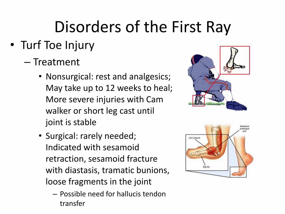
Disorders of the First Ray • Turf Toe Injury
– Treatment
• Nonsurgical: rest and analgesics; May take up to 12 weeks to heal; More severe injuries with Cam walker or short leg cast until joint is stable
• Surgical: rarely needed; Indicated with sesamoid retraction, sesamoid fracture with diastasis, tramatic bunions, loose fragments in the joint
– Possible need for hallucis tendon transfer
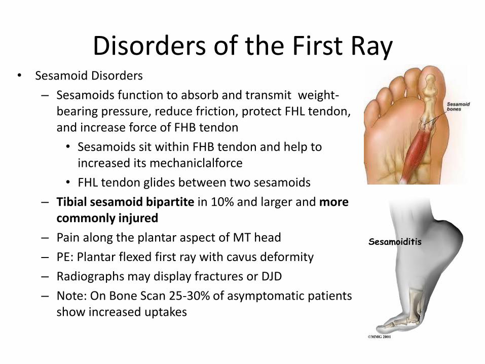
Disorders of the First Ray • Sesamoid Disorders
– Sesamoids function to absorb and transmit weight-bearing pressure, reduce friction, protect FHL tendon, and increase force of FHB tendon
• Sesamoids sit within FHB tendon and help to increased its mechaniclalforce
• FHL tendon glides between two sesamoids
– Tibial sesamoid bipartite in 10% and larger and more commonly injured
– Pain along the plantar aspect of MT head
– PE: Plantar flexed first ray with cavus deformity
– Radiographs may display fractures or DJD
– Note: On Bone Scan 25-30% of asymptomatic patients show increased uptakes
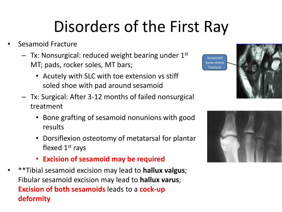
Disorders of the First Ray • Sesamoid Fracture
– Tx: Nonsurgical: reduced weight bearing under 1st MT; pads, rocker soles, MT bars;
• Acutely with SLC with toe extension vs stiff soled shoe with pad around sesamoid
– Tx: Surgical: After 3-12 months of failed nonsurgical treatment
• Bone grafting of sesamoid nonunions with good results
• Dorsiflexion osteotomy of metatarsal for plantar flexed 1st rays
• Excision of sesamoid may be required
• **Tibial sesamoid excision may lead to hallux valgus; Fibular sesamoid excision may lead to hallux varus; Excision of both sesamoids leads to a cock-up deformity
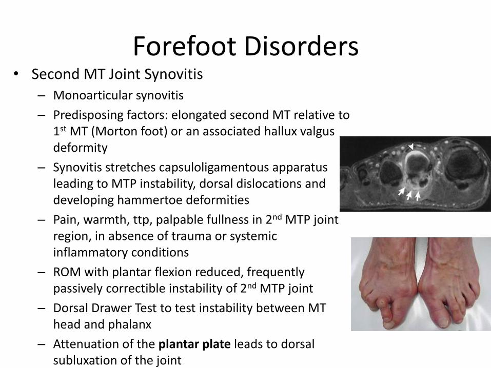
Forefoot Disorders • Second MT Joint Synovitis
– Monoarticular synovitis
– Predisposing factors: elongated second MT relative to 1st MT (Morton foot) or an associated hallux valgus deformity
– Synovitis stretches capsuloligamentous apparatus leading to MTP instability, dorsal dislocations and developing hammertoe deformities
– Pain, warmth, ttp, palpable fullness in 2nd MTP joint region, in absence of trauma or systemic inflammatory conditions
– ROM with plantar flexion reduced, frequently passively correctible instability of 2nd MTP joint
– Dorsal Drawer Test to test instability between MT head and phalanx
– Attenuation of the plantar plate leads to dorsal subluxation of the joint
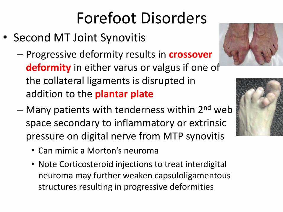
Forefoot Disorders • Second MT Joint Synovitis
– Progressive deformity results in crossover deformity in either varus or valgus if one of the collateral ligaments is disrupted in addition to the plantar plate
– Many patients with tenderness within 2nd web space secondary to inflammatory or extrinsic pressure on digital nerve from MTP synovitis
• Can mimic a Morton’s neuroma
• Note Corticosteroid injections to treat interdigital neuroma may further weaken capsuloligamentous structures resulting in progressive deformities
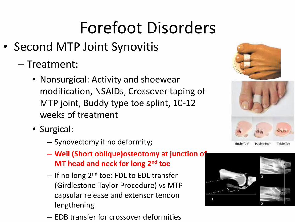
Forefoot Disorders • Second MTP Joint Synovitis
– Treatment:
• Nonsurgical: Activity and shoewear modification, NSAIDs, Crossover taping of MTP joint, Buddy type toe splint, 10-12 weeks of treatment
• Surgical: – Synovectomy if no deformity;
– Weil (Short oblique)osteotomy at junction of MT head and neck for long 2nd toe
– If no long 2nd toe: FDL to EDL transfer (Girdlestone-Taylor Procedure) vs MTP capsular release and extensor tendon lengthening
– EDB transfer for crossover deformities
Forefoot Disorders
• Freidberg Infraction
– Infarction and Fracture of MT head; Most commonly in 2nd MT dorsally
– Recurrent microtrauma vs osteonecrosis of MT head leading to subchondral collapse
– MTP joint stiffness and swelling worse with weightbearing
– Initial radiographs with flattening of MT head and subchondral sclerosis progressing to MTP joint arthritis
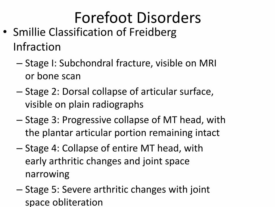
Forefoot Disorders • Smillie Classification of Freidberg
Infraction
– Stage I: Subchondral fracture, visible on MRI or bone scan
– Stage 2: Dorsal collapse of articular surface, visible on plain radiographs
– Stage 3: Progressive collapse of MT head, with the plantar articular portion remaining intact
– Stage 4: Collapse of entire MT head, with early arthritic changes and joint space narrowing
– Stage 5: Severe arthritic changes with joint space obliteration
Forefoot Disorders
• Friedberg Infraction
– Nonsurgical: uploading and protecting 2nd MT head; SLC extended to toes for 4-6 weeks followed by several months of stiff soled shoe with MT bar
– Surgical: Dorsal closing wedge osteotomy of MT head vs isolated debridement vs partial head resection (DuVries arthroplasty-for stage4-5)
Deformities of the Lesser Toes
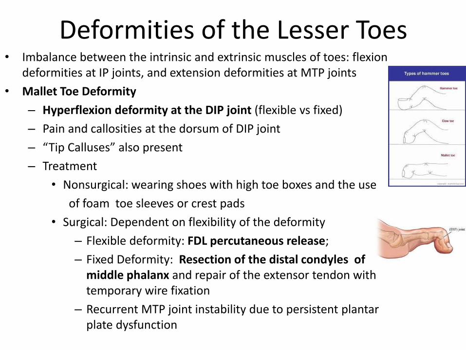
Deformities of the Lesser Toes • Imbalance between the intrinsic and extrinsic muscles of toes: flexion
deformities at IP joints, and extension deformities at MTP joints
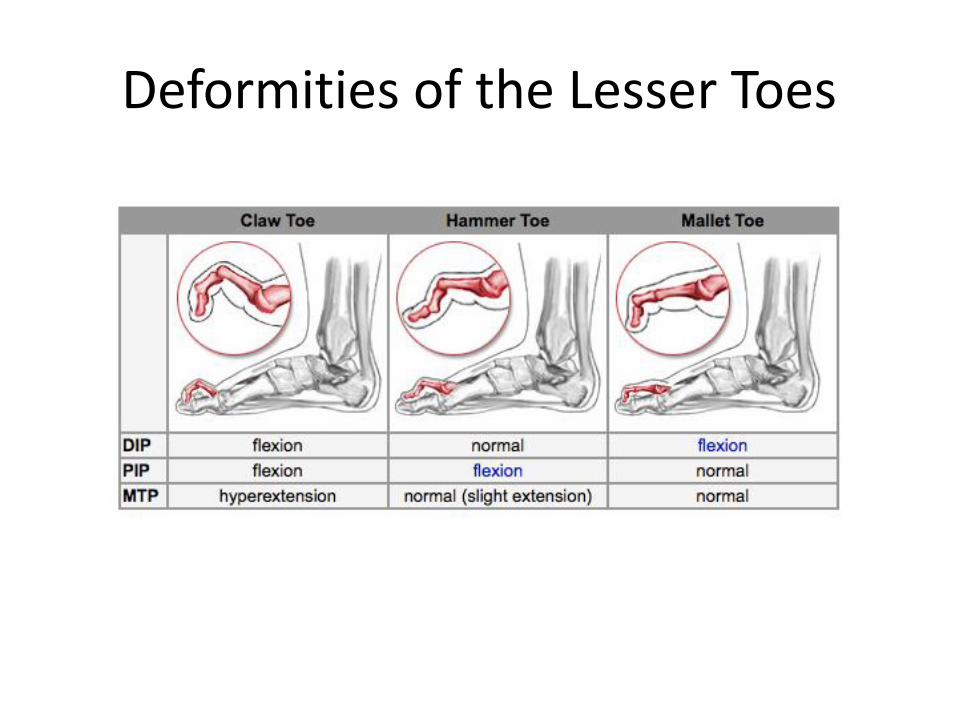
• Mallet Toe Deformity
– Hyperflexion deformity at the DIP joint (flexible vs fixed)
– Pain and callosities at the dorsum of DIP joint
– “Tip Calluses” also present
– Treatment
• Nonsurgical: wearing shoes with high toe boxes and the use
of foam toe sleeves or crest pads
• Surgical: Dependent on flexibility of the deformity
– Flexible deformity: FDL percutaneous release;
– Fixed Deformity: Resection of the distal condyles of middle phalanx and repair of the extensor tendon with temporary wire fixation
– Recurrent MTP joint instability due to persistent plantar plate dysfunction
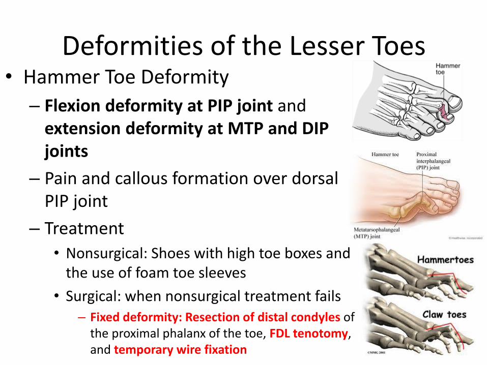
Deformities of the Lesser Toes • Hammer Toe Deformity
– Flexion deformity at PIP joint and extension deformity at MTP and DIP joints
– Pain and callous formation over dorsal PIP joint
– Treatment
• Nonsurgical: Shoes with high toe boxes and the use of foam toe sleeves
• Surgical: when nonsurgical treatment fails – Fixed deformity: Resection of distal condyles of
the proximal phalanx of the toe, FDL tenotomy, and temporary wire fixation
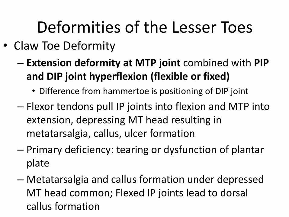
Deformities of the Lesser Toes • Claw Toe Deformity
– Extension deformity at MTP joint combined with PIP and DIP joint hyperflexion (flexible or fixed)
• Difference from hammertoe is positioning of DIP joint
– Flexor tendons pull IP joints into flexion and MTP into extension, depressing MT head resulting in metatarsalgia, callus, ulcer formation
– Primary deficiency: tearing or dysfunction of plantar plate
– Metatarsalgia and callus formation under depressed MT head common; Flexed IP joints lead to dorsal callus formation
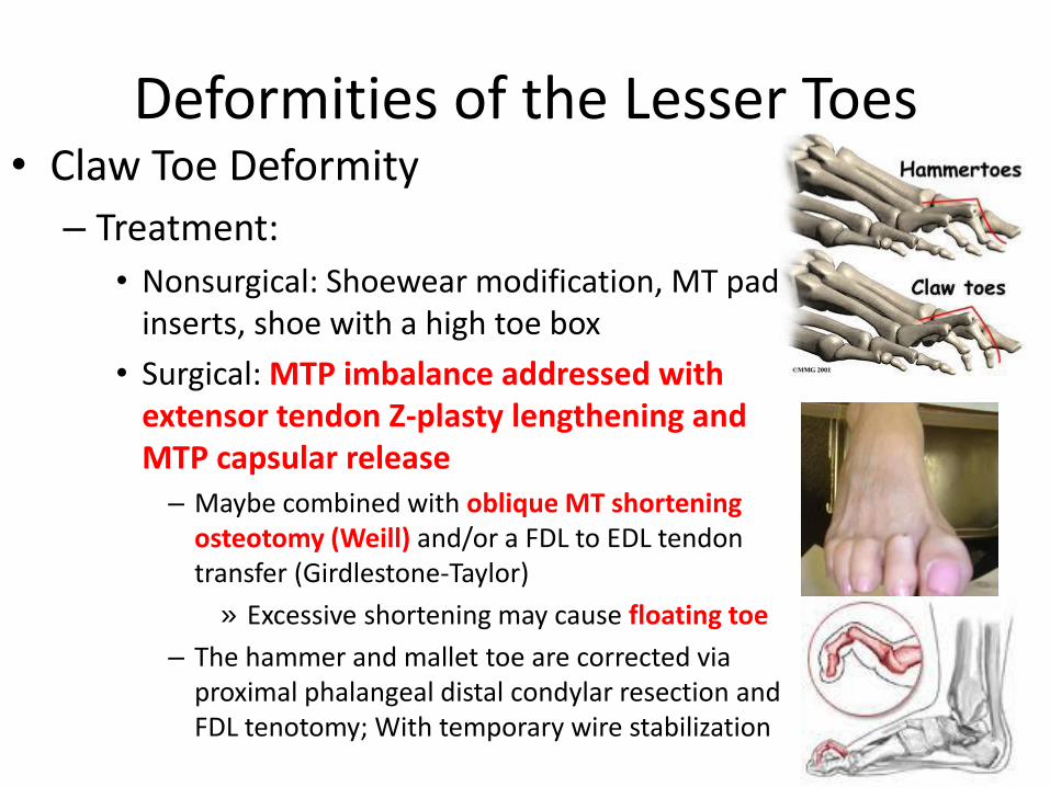
Deformities of the Lesser Toes • Claw Toe Deformity
– Treatment:
• Nonsurgical: Shoewear modification, MT pad inserts, shoe with a high toe box
• Surgical: MTP imbalance addressed with extensor tendon Z-plasty lengthening and MTP capsular release
– Maybe combined with oblique MT shortening osteotomy (Weill) and/or a FDL to EDL tendon transfer (Girdlestone-Taylor)
» Excessive shortening may cause floating toe
– The hammer and mallet toe are corrected via proximal phalangeal distal condylar resection and FDL tenotomy; With temporary wire stabilization
21. What complication is frequently associated with a Weil lesser metatarsal osteotomy (distal, oblique) in
treating a lesser toe deformity
1- excessive shortening
2- dorsal displacement of the metatarsal head
3- osteonecrosis of the metatarsal head
4- nonunion
5- extended of “floating toe”
Answer:
5- Extended or “floating toe”
- A weil osteotomy is a distal dorsal to proximal plantar osteotomy of the distal MT that shortens the bone
and often indicated in severe hammer toe deformities and metatarsalgia. Excessive shortening is possible
(anything is when you are cutting bone) but not the best answer. Answer 2 is wrong b/c the metatarsal is
shortened with plantar displacement of the head. The blood flow to the distal MT is general is good so
osteonecrosis nor nonunion are not issues. Extended of “floating toe” is the main complication.
Question 43
Claw toe deformities of the lesser toes, following a severe closed calcaneal fracture, are the result of which of the following?
1. Weakness of the tibialis anterior 2. Contracture of the intrinsic flexor muscles of the
foot 3. Tethering of the flexor hallucis longus tendon by
fracture fragments 4. Lateral plantar nerve neuropathy 5. Medial plantar nerve neuropathy
Citing Article: Myerson M and Quill GE. “Late complications of fractures of the
calcaneus.” JBJS(A)1993; 75:331-341.
The above reference article is not particularly useful. The correct answer has to
involve either the FDL, intrinsic muscles or the nerves that supply them. That rules out
Tib Ant weakness (dorsiflexor), and tethering of FHL because that would only result in
IP flexion of the great toe alone.
The only answer that would affect all the toes is number 2, contracture of the intrinsics
which is a result of foot compartment syndrome in the setting of calcaneal fracture.
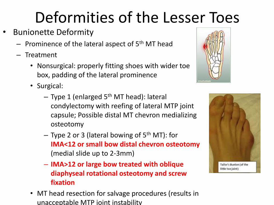
Deformities of the Lesser Toes • Bunionette Deformity
– Prominence of the lateral aspect of 5th MT head
– Treatment
• Nonsurgical: properly fitting shoes with wider toe box, padding of the lateral prominence
• Surgical:
– Type 1 (enlarged 5th MT head): lateral condylectomy with reefing of lateral MTP joint capsule; Possible distal MT chevron medializing osteotomy
– Type 2 or 3 (lateral bowing of 5th MT): for IMA<12 or small bow distal chevron osteotomy (medial slide up to 2-3mm)
– IMA>12 or large bow treated with oblique diaphyseal rotational osteotomy and screw fixation
• MT head resection for salvage procedures (results in unacceptable MTP joint instability
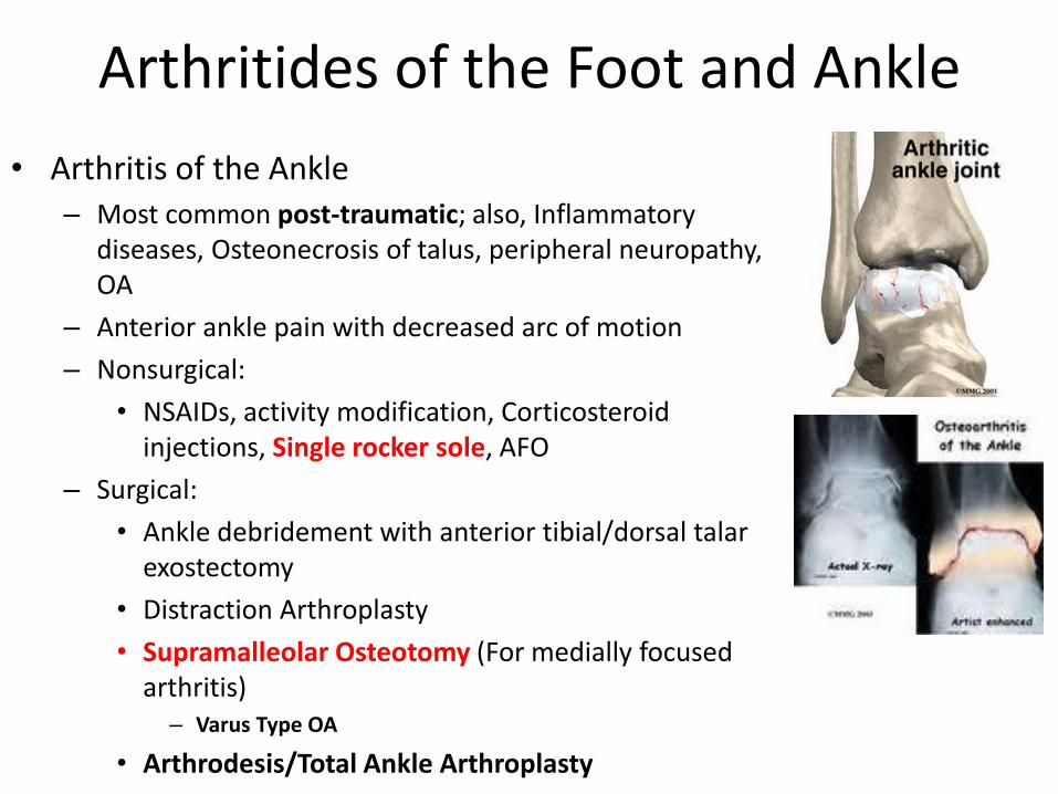
Arthritides of the Foot and Ankle
• Arthritis of the Ankle – Most common post-traumatic; also, Inflammatory
diseases, Osteonecrosis of talus, peripheral neuropathy, OA
– Anterior ankle pain with decreased arc of motion
– Nonsurgical:
• NSAIDs, activity modification, Corticosteroid injections, Single rocker sole, AFO
– Surgical:
• Ankle debridement with anterior tibial/dorsal talar exostectomy
• Distraction Arthroplasty
• Supramalleolar Osteotomy (For medially focused arthritis)
– Varus Type OA
• Arthrodesis/Total Ankle Arthroplasty
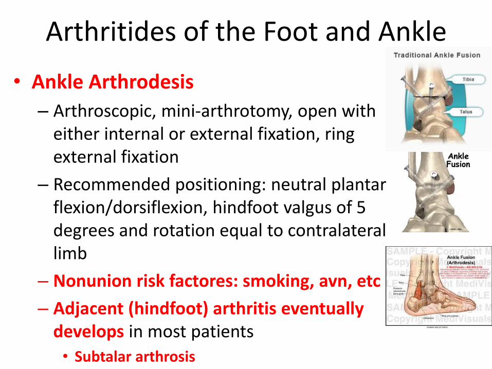
Arthritides of the Foot and Ankle
• Ankle Arthrodesis
– Arthroscopic, mini-arthrotomy, open with either internal or external fixation, ring external fixation
– Recommended positioning: neutral plantar flexion/dorsiflexion, hindfoot valgus of 5 degrees and rotation equal to contralateral limb
– Nonunion risk factores: smoking, avn, etc
– Adjacent (hindfoot) arthritis eventually develops in most patients
• Subtalar arthrosis
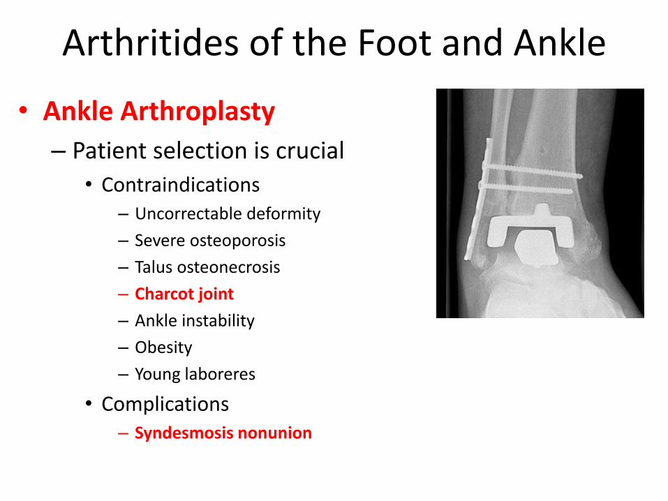
Arthritides of the Foot and Ankle
• Ankle Arthroplasty
– Patient selection is crucial
• Contraindications – Uncorrectable deformity
– Severe osteoporosis
– Talus osteonecrosis
– Charcot joint
– Ankle instability
– Obesity
– Young laboreres
• Complications – Syndesmosis nonunion
Question 60
What is the optimal positioning of the foot when performing a tibiotalar joint arthrodesis?
1. Neutral flexion, 0 degrees to 5 degrees hindfoot valgus, 5 degrees to 10 degrees external rotation
2. Neutral flexion, 0 degrees to 5 degrees hindfoot valgus, 0 degrees external rotation
3. Neutral flexion, 20 degrees hindfoot valgus, 5 degrees to 10 degrees external rotation
4. 10 degrees dorsiflexion, 0 degrees to 5 degrees hindfoot valgus, 5 degrees to 10 degrees external rotation
5. 5 degrees to 10 degrees plantar flexion, 20 degrees hindfoot valgus, 5 degrees to 10 degrees external rotation
Citing Article: Buck P et al. “The optimum position of arthrodesis of the ankle. A gait
Study of the knee and ankle.” JBJS(A) 1987;69:1052-1062.
Unfortunately, this question you just either know or you don’t. It is based on the above
article in which they found the ideal position to be neutral flexion, slight (0-5 degrees)
valgus and 5-10 degrees of external rotation.
-In this study they evaluated 19 patients who had a solid ankle arthrodesis at least 4
years prior
-A gait analysis was done to evaluate foot ROM and knee ROM
-Results showed:
-fusion in plantar-flexion leads to more abnormal gait as well as increased
Genu recurvatum (therefore fuse in neutral)
-fusion in slight valgus allows for more varus-valgus motion of hind part of foot
-fusion in internal rotation causes decreased ROM for dorsiflexion/plantarflexion
and therefore you want to fuse in slight external rotation
115 Long-term follow-up of ankle arthrodesis has demonstrated a significant incidence of: • 1- tibial stress fracture. • 2- subtalar osteoarthritis. • 3- ipsilateral knee osteoarthritis. • 4- metatarsalgia. • 5- deltoid ligament insufficiency.
PREFERRED RESPONSE: 2 subtalar osteoarthritis. - Ankle fusion refers to fusion of the tibiotalar joint. Similar to the spine where you see adjacent segment degeneration
cranial/caudal to a fusion mass the same can happen after ankle fusion. Usually the subtalar joint (next adjacent joint) is first involved but the midfoot can also progress to degenerative arthritis.
Thomas R, Daniels TR, Parker K: Gait analysis and functional outcomes following ankle arthrodesis for isolated ankle arthritis. J
Bone Joint Surg Am 2006;88:526-535.
• Twenty-six patients who had undergone ankle arthrodesis for the treatment of isolated unilateral ankle arthritis were
identified and retrospectively assessed clinically and radiographically. The mean age at the time of surgery was fifty-four years, and the mean interval between surgery and assessment was forty-four months. A gender and age-matched control group of twenty-seven individuals was recruited for comparison. All subjects were evaluated with gait analysis, the American Orthopaedic Foot and Ankle Society (AOFAS) Ankle-Hindfoot scale, the Musculoskeletal Outcomes Data Evaluation and Management Systems (MODEMS) questionnaire, and the Ankle Osteoarthritis Scale (AOS).
• Results: On preliminary review, twenty of the twenty-six patients were completely satisfied or satisfied with their surgical outcome. All patients but one stated that they would undergo the surgery again. Five patients stated that they did not notice a gait abnormality. Twelve patients wore orthotics, and all believed that the use of the orthotics improved their gait. When the functional outcome scores in the arthrodesis group were compared with those in the control group, specific scores assessing hindfoot pain and satisfaction were similar. However, scores focusing on ankle-hindfoot function and disability revealed significant differences. Gait analysis also identified significant differences between the two groups with regard to cadence and stride length. In addition, there was significantly decreased sagittal, coronal, and transverse range of motion of the hindfoot and midfoot during the stance and swing phases of gait in the arthrodesis group. Radiographic review demonstrated that four of the twenty-six patients had development of moderate to severe arthritis of the subtalar joint.
• Conclusions: In the intermediate term following an arthrodesis for the treatment of end-stage ankle arthritis, pain is reliably
relieved and there is good patient satisfaction. However, there are substantial differences between patients and the normal
population with regard to hindfoot function and gait. On the basis of these results, patients should be counseled that an ankle fusion will help to relieve pain and to improve overall function; however, it is a salvage procedure that will cause persistent alterations in gait with a potential for deterioration due to the development of ipsilateral hindfoot arthritis.
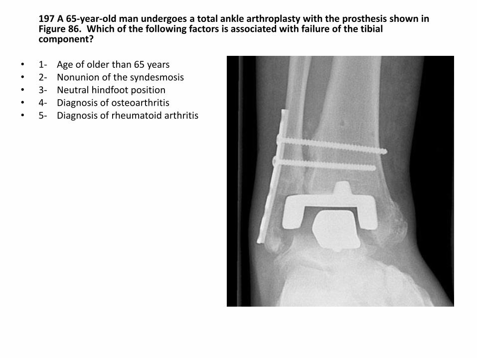
197 A 65-year-old man undergoes a total ankle arthroplasty with the prosthesis shown in Figure 86. Which of the following factors is associated with failure of the tibial component?
• 1- Age of older than 65 years • 2- Nonunion of the syndesmosis • 3- Neutral hindfoot position • 4- Diagnosis of osteoarthritis • 5- Diagnosis of rheumatoid arthritis
PREFERRED RESPONSE: 2 Nonunion of the syndesmosis - Ankle arthroplasty, have to love it. The ideal patient is someone who is over the age of 50, is not too heavy, and is not extremely
active. OA is a good indication, RA not so good but not the reason for this guy’s failure. Despite the fact that syndesmotic screws are intact without radiolucent “halos” the syndesmosis is not radiographically reduced/healed. Part of fixation of the tibial implant involves the distal fibula and you see a radiolucent zone in this area.
RECOMMENDED READINGS: Pyevich MT, Saltzman CL, Callaghan JJ, et al: Total ankle arthroplasty: A unique design. Two to twelve
year followup. J Bone Joint Surg Am 1998;80:1410-1420. Alvine FG: The agility ankle replacement: The good and the bad. Foot Ankle Clin 2002;7:737-753.
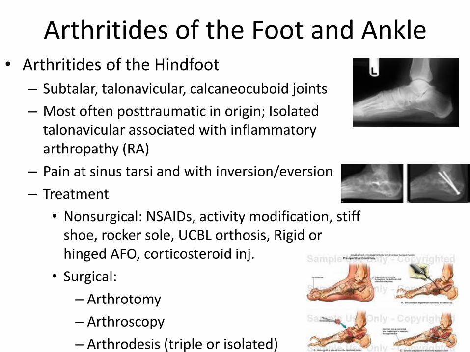
Arthritides of the Foot and Ankle • Arthritides of the Hindfoot
– Subtalar, talonavicular, calcaneocuboid joints
– Most often posttraumatic in origin; Isolated talonavicular associated with inflammatory arthropathy (RA)
– Pain at sinus tarsi and with inversion/eversion
– Treatment
• Nonsurgical: NSAIDs, activity modification, stiff shoe, rocker sole, UCBL orthosis, Rigid or hinged AFO, corticosteroid inj.
• Surgical:
– Arthrotomy
– Arthroscopy
– Arthrodesis (triple or isolated)
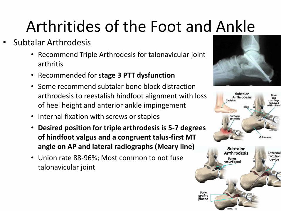
Arthritides of the Foot and Ankle • Subtalar Arthrodesis
• Recommend Triple Arthrodesis for talonavicular joint arthritis
• Recommended for stage 3 PTT dysfunction
• Some recommend subtalar bone block distraction arthrodesis to reestalish hindfoot alignment with loss of heel height and anterior ankle impingement
• Internal fixation with screws or staples
• Desired position for triple arthrodesis is 5-7 degrees of hindfoot valgus and a congruent talus-first MT angle on AP and lateral radiographs (Meary line)
• Union rate 88-96%; Most common to not fuse talonavicular joint
257) Which of the following is a risk factor for delayed union and non-union following a subtalar arthrodesis?
1 Body mass index of greater than 40
2 Use of one versus two screws for fixation
3 pervious ankle arthrodesis adjacent to the subtalar site
4 Presence of a preoperative flatfoot deformity
5 Use of supplemental allograft.
The answer to this question can be found in the article Easley ME, Trnka HJ, Schon LC, et al; Isolated Subtalar Arthrodesis, JBJS Am 2000 (82) 613-624.
This was a retrospective study that looked at 184 cases of isolated subtalar arthritis between 1988 and 1995. The average age of the patient was 43 years old. 46% of patients were smokers. Indications for the procedure included post traumatic arthritis after calcaneus fracture, talus fracture, subtalar dislocation, primary subtalar arthritis, failure of previous subtalar arthrodesis, and residual congenital deformity. Rigid internal fixation with one or two screws was used in all feet. Bone graft, of various forms, was used in 145 feet. 86% union rate was seen after primary arthrodesis, 71% after revision arthrodesis, 92% for non-smokers, 73% for smokers. The rate of union was found to be decreased by smoking, presence of more than 2mm of avascular bone at the arthrodesis site, and failure of previous subtalar arthrodesis. Hence, #3 is the anwer.
A 72 yo woman has medial ankle pain and diffuse hindfoot swelling. Radiographs are shown in figure 11a and
11b. What is the most appropriate surgical treatment for this patient?
1- medial displacement calcaneal osteotomy
2- lateral displacement calcaneal osteotomy
3- lateral column lengthening through the anterior calcaneus
4- lateral column lengthening through the calcaneocuboid joint
5- triple arthrodesis

Answer:
5- triple arthrodesis
- A triple arthrodesis is a fusion of subtalar, talonavicular and calcaneocuboid joints. The vignette really only
tells you that this is an old lady with foot pain and seeing arthritis on the XR that’s really all you need to
know. The XR is crappy but looking carefully at the XR one notes significant talonavicular DJD and
moderate subtalar arthritis. Some ankle arthritis is seen on this foot series XR. Answers 1,2,3 and 4 don’t
make much sense. This lady does not have medial nor lateral column collapse. The lateral does not show
significant flat foot deformity and you don’t have enough data to tell if she needs a calcaneal slide. The
calcaneal cuboid joint doesn’t look bad but indication for triple fusion is subtalar DJD + both or either TN
or CC DJD. Thus the answer is 5.
Arthritides of the Foot and Ankle • Midfoot Arthritides
– Naviculocuneiform and Metatarsocuneiform/cuboid joints
– Etiology: primary, inflammatory, post traumatic (Lisfranc Fx/disloc) or neuropathic (Charcot)
– Untreated 3rd tarsometatarsal (TMT) joint (Lisfranc) fracture-dislocation typically leads to a loss of longitudinal arch and forefoot abduction
– Treatment:
• Nonsurgical: NSAIDs, activity modification, longitudinal arch support, rocker soles, AFO, corticosteroid injection
• Surgical: Successful realignment and arthrodesis of 1st through TMT joint and/or naviculocuneiform joints.
• Note: 4th and 5th TMT joints not fused!
• Select cases of symptomatic 4th and 5th TMT treated with interpositional arthroplasty which maintains lateral column and accomodates gait
• Often in conjunction with achilles tendon lengthening
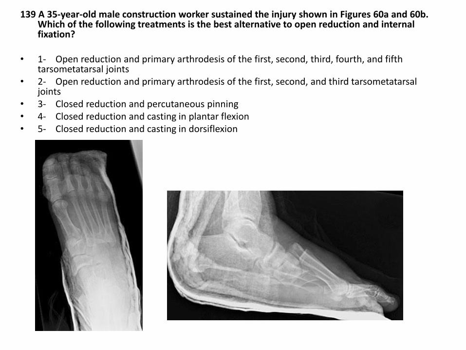
139 A 35-year-old male construction worker sustained the injury shown in Figures 60a and 60b. Which of the following treatments is the best alternative to open reduction and internal fixation?
• 1- Open reduction and primary arthrodesis of the first, second, third, fourth, and fifth
tarsometatarsal joints • 2- Open reduction and primary arthrodesis of the first, second, and third tarsometatarsal
joints • 3- Closed reduction and percutaneous pinning • 4- Closed reduction and casting in plantar flexion • 5- Closed reduction and casting in dorsiflexion
PREFERRED RESPONSE: 2 Open reduction and primary arthrodesis of the first, second, and third tarsometatarsal joints
- Radiographs reveal fx/dislocations of the tarsal/metatarsal joints consistent with a Lisfranc injury in a young laborer.
Badness. Closed reduction and treament is only indicated for displacement less than 2 mm in any plane with the absence of joint line instability on WB views. Closed reduction and non-op rx is not an option unless you want to give him a crappy foot. You need anatomic reduction so this dude needs some type of open reduction. Any instability required anatomic reduction. A little difficult to tell what’s going on but it appears that at least rays 1-4 are dislocated. When thinking about forefoot dynamics you only want to consider fusing the medial rays b/c of the increased normal motion at the 4th/5th metatarsal cuboid joints. Thus answer 2 is the answer.
RECOMMENDED READINGS: Ly TV, Coetzee JC: Treatment of primarily ligamentous Lisfranc joint injuries: Primary arthrodesis
compared with open reduction and internal fixation: A prospective, randomized study. J Bone Joint Surg Am 2006;88:514-520.
Mulier, T, Reynders P, Dereymaeker G, et al: Severe Lisfranc injuries: Primary arthrodesis or ORIF? Foot Ankle Int 2002;23:902-905.
Arthritides of the Foot and Ankle • Forefoot Arthritis
– Most commonly affects 1st MTP joint (Hallux Rigidus) due to repetitive trauma (also, gout,RA)
– Lesser MTP arthritis usually secondary to inflammatory conditions
– Dorsal prominence over MTP joint, swelling of great toe, pain during pushoff, pain with forced dorsiflexion (dorsal impingement), limited hallux ROM
• Classification of Forefoot Arthritis
– I: Mild: MTP joint spaces maintained; dorsal osteophyte
– II: Moderate: MTP joint space narrowing; large dorsal, medial, lateral osteophytes
– III:Severe: Comlete loss of MTP joint space
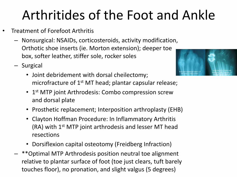
Arthritides of the Foot and Ankle • Treatment of Forefoot Arthritis
– Nonsurgical: NSAIDs, corticosteroids, activity modification, Orthotic shoe inserts (ie. Morton extension); deeper toe box, softer leather, stiffer sole, rocker soles
– Surgical
• Joint debridement with dorsal cheilectomy; microfracture of 1st MT head; plantar capsular release;
• 1st MTP joint Arthrodesis: Combo compression screw and dorsal plate
• Prosthetic replacement; Interposition arthroplasty (EHB)
• Clayton Hoffman Procedure: In Inflammatory Arthritis (RA) with 1st MTP joint arthrodesis and lesser MT head resections
• Dorsiflexion capital osteotomy (Freidberg Infraction)
– **Optimal MTP Arthrodesis position neutral toe alignment relative to plantar surface of foot (toe just clears, tuft barely touches floor), no pronation, and slight valgus (5 degrees)
**Acute/Chronic Injuries of the Ankle
• Acute Lateral Ankle Instability
– Grade I: No ligamentous disruption; Minimal swelling/echymosis/tenderness; No pain with weight bearing
– Grade II: ligamentous stretch w/o rupture; Moderate swelling/echymosis/tenderness; Mild pain with weight bearing
– Grade III: Complete ligamentous rupture; Severe swelling/echymosis/tenderness; Severe pain with weight bearing
Acute/Chronic Injuries of the Ankle
• Acute Lateral Ankle Instability
– Localized tenderness/swelling/echymosis over anterior Talofibular ligament (ATFL) -Plantarflexed and/or calcaneofibular ligament (CFL) - dorsiflexed
– Possible positive tests:
• Anterior Drawer: Anterior talar subluxation in 30 degrees of plantar flexion (10mm or 3mm greater than contralateral side)
• Talar tilt test: >3deg of tilt compared to other side or 10deg of tilt absolutely
• Sulcus sign
Acute/Chronic Injuries of the Ankle
• Acute Lateral Ankle Instability
– Treatment:
• Nonsurgical: rest,ice,compression,elevation (RICE); early weight bearing with protective brace; PT emphasizing isometrics, resistive training, peroneal strengthening, ROM, and proprioceptive training
• Surgical
• **Consider MRI if pain persists 8 weeks after an acute ankle sprain
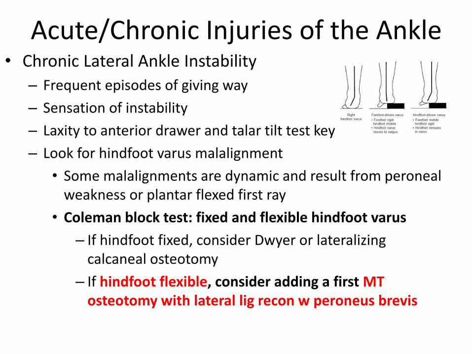
Acute/Chronic Injuries of the Ankle • Chronic Lateral Ankle Instability
– Frequent episodes of giving way
– Sensation of instability
– Laxity to anterior drawer and talar tilt test key
– Look for hindfoot varus malalignment
• Some malalignments are dynamic and result from peroneal weakness or plantar flexed first ray
• Coleman block test: fixed and flexible hindfoot varus
– If hindfoot fixed, consider Dwyer or lateralizing calcaneal osteotomy
– If hindfoot flexible, consider adding a first MT osteotomy with lateral lig recon w peroneus brevis
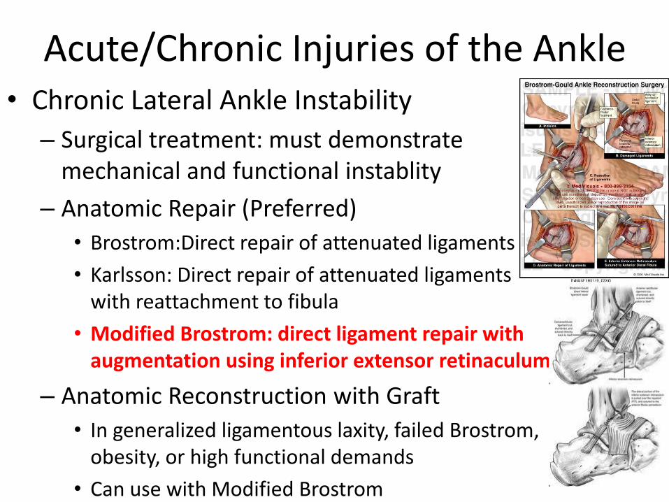
Acute/Chronic Injuries of the Ankle • Chronic Lateral Ankle Instability
– Surgical treatment: must demonstrate mechanical and functional instablity
– Anatomic Repair (Preferred)
• Brostrom:Direct repair of attenuated ligaments
• Karlsson: Direct repair of attenuated ligaments with reattachment to fibula
• Modified Brostrom: direct ligament repair with augmentation using inferior extensor retinaculum
– Anatomic Reconstruction with Graft
• In generalized ligamentous laxity, failed Brostrom, obesity, or high functional demands
• Can use with Modified Brostrom
Acute/Chronic Injuries of the Ankle
• Chronic Lateral Ankle Instability
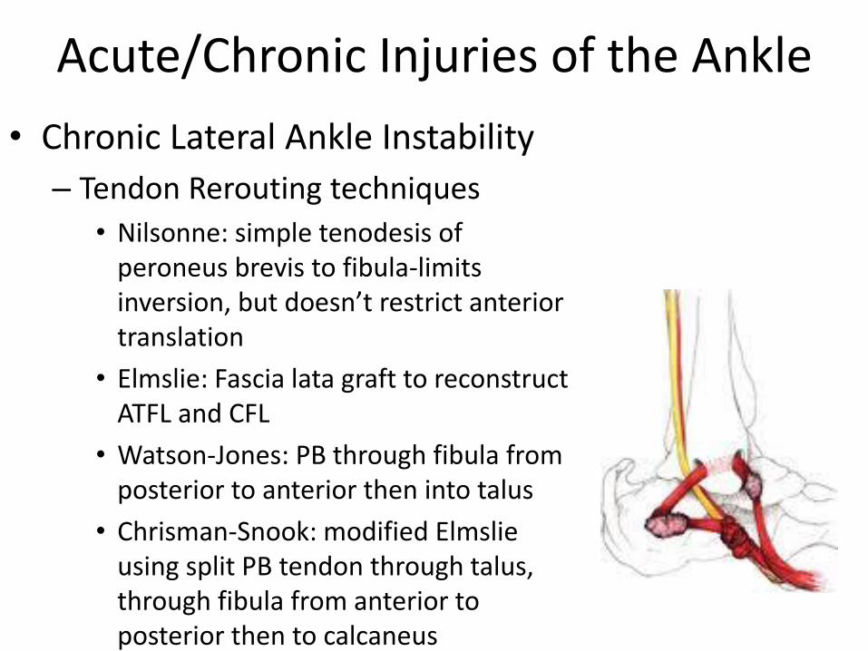
– Tendon Rerouting techniques
• Nilsonne: simple tenodesis of peroneus brevis to fibula-limits inversion, but doesn’t restrict anterior translation
• Elmslie: Fascia lata graft to reconstruct ATFL and CFL
• Watson-Jones: PB through fibula from posterior to anterior then into talus
• Chrisman-Snook: modified Elmslie using split PB tendon through talus, through fibula from anterior to posterior then to calcaneus
Acute/Chronic Injuries of the Ankle • Syndesmotic Injuries
– Combination of dorsiflexion and External Rotation
– Associated deltoid ligament disruption and fibula fracture
– Unable to bear weight, tenderness at syndesmosis and deltoid ligament, (+) Squeeze test
– Decreased tib-fib overlap on AP; Increased tib-fib clear space on mortise;
– Must rule out Maissoneuve fracture;
– Confirm diagnosis with Stress views in ER
– Treatment:
• Stable: RICE, brief immobilization followed by functional brace; delayed WB until pain free
• Unstable: ORIF with syndesmotic screw; controversial: number of cortices, need for future screw removal
Acute/Chronic Injuries of the Ankle • Deltoid Ligament Instability
– Injury occurs with pronation mechanism
– Rupture of deep deltoid ligament renders medial ankle unstable
– Radiographs reveal valgus ankle deformity
– With proper restoration of alignment deltoid ligament often will heal
– Direct ligament repair, ligament augmentation are treatment options with ankle fusion as a possible salvage procedure
Acute/Chronic Injuries of the Ankle
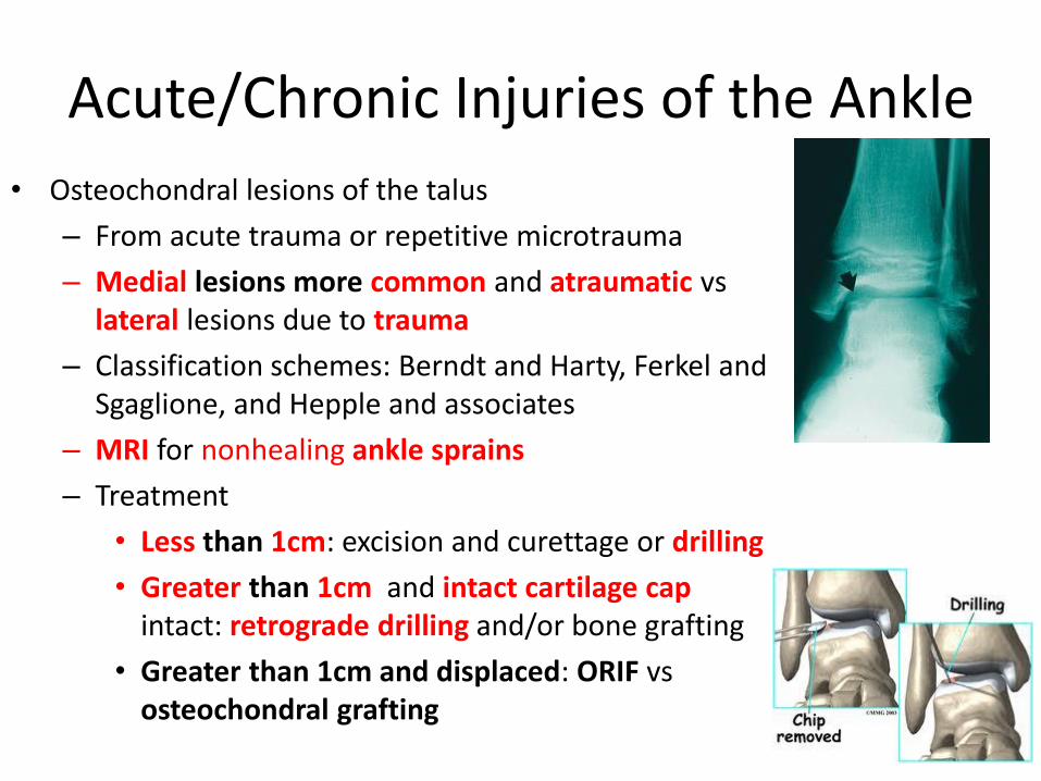
• Osteochondral lesions of the talus
– From acute trauma or repetitive microtrauma
– Medial lesions more common and atraumatic vs lateral lesions due to trauma
– Classification schemes: Berndt and Harty, Ferkel and Sgaglione, and Hepple and associates
– MRI for nonhealing ankle sprains
– Treatment
• Less than 1cm: excision and curettage or drilling
• Greater than 1cm and intact cartilage cap intact: retrograde drilling and/or bone grafting
• Greater than 1cm and displaced: ORIF vs osteochondral grafting
OITE 2006—Foot and Cankle
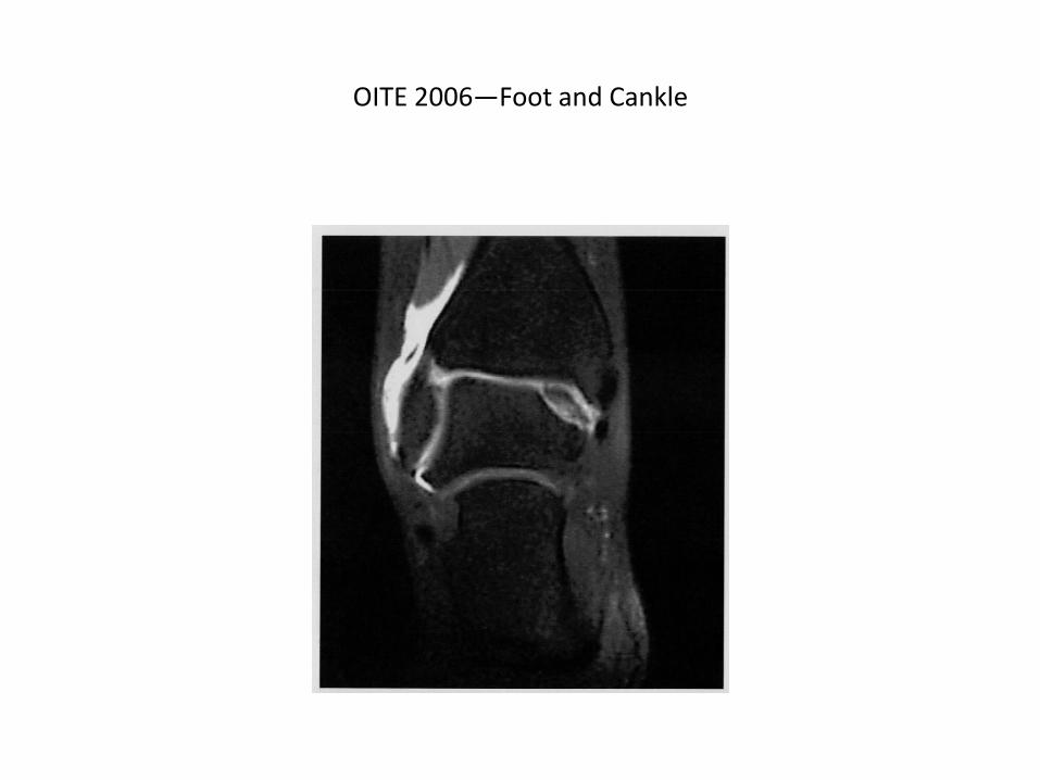
213. A 22-year-old man injured his ankle 12 months ago and has continued, significant posteromedial ankle pain that is relieved with injection. A MRI scan is shown in Figure 84. Surgical management should consist of
1- autologous chondrocyte transplantation. 2- retrograde drilling and bone grafting.
3- excision of the loose fragment. 4- osteoarticular transfer from the knee 5- fragment excision and drilling/micropicking
OITE 2006—Foot and Cankle
OITE 2006—Foot and Cankle
213. A 22-year-old man injured his ankle 12 months ago and has continued, significant posteromedial ankle pain that is relieved with injection. A MRI scan is shown in Figure 84. Surgical management should consist of
1- autologous chondrocyte transplantation. 2- retrograde drilling and bone grafting.
3- excision of the loose fragment. 4- osteoarticular transfer from the knee 5- fragment excision and drilling/micropicking
OITE 2006—Foot and Cankle
• This topic is a bit confusing. Given that the injury is 12 months old, non-operative treatment is not an option (or a choice for that matter). The most data exists regarding microfracture (more data than OATS or autologous chondrocyte transfer). Excision and microfracture has been shown to be superior to excision alone. The reference article by Giannini lists a quick algorithm for treatment in the conclusion. For lesions less than 1.5 cm in pts younger than 50 and less than 3cm in pts older than 50, arthroscopic debridement and drilling/microfracture is the treatment of choice. Lesions 1.5-3 cm in pts younger than 50 and failed arthroscopic treatment should be treated with OATs or ACT. In patients younger than 50 with lesions greater than 3 cm, ACT or allograft is the treatment of choice. Patients older than 50 with lesions larger than 3 cm can be considered for arthrodesis. I find this algorithm a bit confusing because I think this lesion looks larger than 1.5 cm. Perhaps the morbity of and OATs and the cost of ACT detract from these choices.
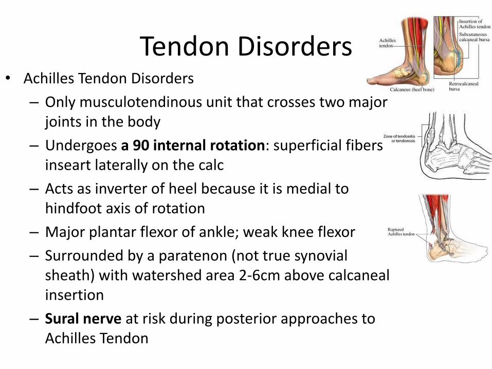
Tendon Disorders • Achilles Tendon Disorders
– Only musculotendinous unit that crosses two major joints in the body
– Undergoes a 90 internal rotation: superficial fibers inseart laterally on the calc
– Acts as inverter of heel because it is medial to hindfoot axis of rotation
– Major plantar flexor of ankle; weak knee flexor
– Surrounded by a paratenon (not true synovial sheath) with watershed area 2-6cm above calcaneal insertion
– Sural nerve at risk during posterior approaches to Achilles Tendon
Tendon Disorders • Achilles Tendon Disorders
– Acute paratenonitis/tendinitis with no nodularity; pain in entire tendon, no effect with ROM; (+) erythema/warmth
• Overuse, inflammatory arthropathy, AS, fluoroquinolones
• Recent change in activity, intensity, or shoewear
– Paratenonitis/tendinitis with tendinosis with (+) nodularity/warmth/erythema and pain in entire tendon; no effect with ROM
– Tendinosis: (+) nodularity; no warmth/erythema and pain moves with ROM
– Chronic tendinitis/tendinosis with chronic degenerative changes within tendon after prolonged acute tendonitis (older patients, htn, obesity, steroid use, estrogen use)
Tendon Disorders • Achilles Tendon Disorders
– Nonsurgical treatment for acute and chronic (65-90% successful):
• decreased activity intensity, physical therapy with eccentric strengthening and modalities (iontophoresis, phonophoresis, and ultrasound), NSAIDs, ice, heel lift, night splint, and cast/removable boot in severe cases
– Surgical treatment
• Acute: surgical debridement of scarred or inflamed paratenon (70-100% successful) in failed nonsurgical treatment
• Chronic: excision of diseased portion of tendon and retrocalcaneal bursa;
– tendon transfer when >50% of tendon involved and in older patients (>55y/o)—most often FHL, but also FDL and PB
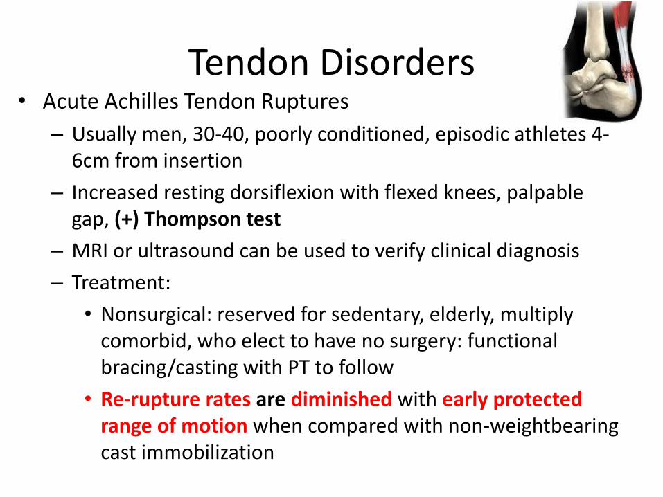
Tendon Disorders • Acute Achilles Tendon Ruptures
– Usually men, 30-40, poorly conditioned, episodic athletes 4-6cm from insertion
– Increased resting dorsiflexion with flexed knees, palpable gap, (+) Thompson test
– MRI or ultrasound can be used to verify clinical diagnosis
– Treatment:
• Nonsurgical: reserved for sedentary, elderly, multiply comorbid, who elect to have no surgery: functional bracing/casting with PT to follow
• Re-rupture rates are diminished with early protected range of motion when compared with non-weightbearing cast immobilization
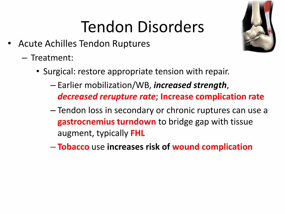
Tendon Disorders • Acute Achilles Tendon Ruptures
– Treatment:
• Surgical: restore appropriate tension with repair.
– Earlier mobilization/WB, increased strength, decreased rerupture rate; Increase complication rate
– Tendon loss in secondary or chronic ruptures can use a gastrocnemius turndown to bridge gap with tissue augment, typically FHL
– Tobacco use increases risk of wound complication
269) Which of the following treatment options for an acute rupture of the Achilles tendon has the lowest risk of re-rupture?
1 Surgical repair followed by immobilization in a long leg cast for three months
2 Surgical repair followed by immobilization in a short leg cast for three months
3 Surgical repair with early range of motion in a functional brace
4 Nonsurgical treatment in a short leg plantar flexed cast for three months
5 Nonsurgical treatment allowing immediate full range of motion in a functional brace
269) Which of the following treatment options for an acute rupture of the Achilles tendon has the lowest risk of re-rupture?
1 Surgical repair followed by immobilization in a long leg cast for three months
2 Surgical repair followed by immobilization in a short leg cast for three months 3 Surgical repair with early range of motion in a functional brace 4 Nonsurgical treatment in a short leg plantar flexed cast for three months 5 Nonsurgical treatment allowing immediate full range of motion in a functional brace
The benefit of treating achilles tendon ruptures surgically is that the chance of re-rupture is lower, however the incidence of wound breakdown is higher. This is the opposite for non-operative treatment. Since the question is asking about options for a lower chance of re-rupture, you can eliminate any non-surgical option. This gets rid of choices 4 and 5.
The correct answer, #3, can be found in the article by Khan RJ, Fick D, Keogh A, et al. Treatment of acute achilles tendon ruptures: A meta-analysis of randomized controlled trials. JBJS Am 2005; (87) 2202-2210.
In their review of the literature they found that:
- Open repair led to a decreased rate of re-rupture compared to non-operative repair.
- Operative repair had increased rates of infection, adhesion, and disturbed skin sensibility
- Percutaneous repair was associated with a lower complication rate compared to open repair
- Patients managed in a functional brace post-op had a lower rate of complications compared to those managed in a cast.
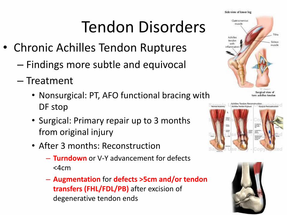
Tendon Disorders • Chronic Achilles Tendon Ruptures
– Findings more subtle and equivocal
– Treatment
• Nonsurgical: PT, AFO functional bracing with DF stop
• Surgical: Primary repair up to 3 months from original injury
• After 3 months: Reconstruction – Turndown or V-Y advancement for defects
<4cm
– Augmentation for defects >5cm and/or tendon transfers (FHL/FDL/PB) after excision of degenerative tendon ends
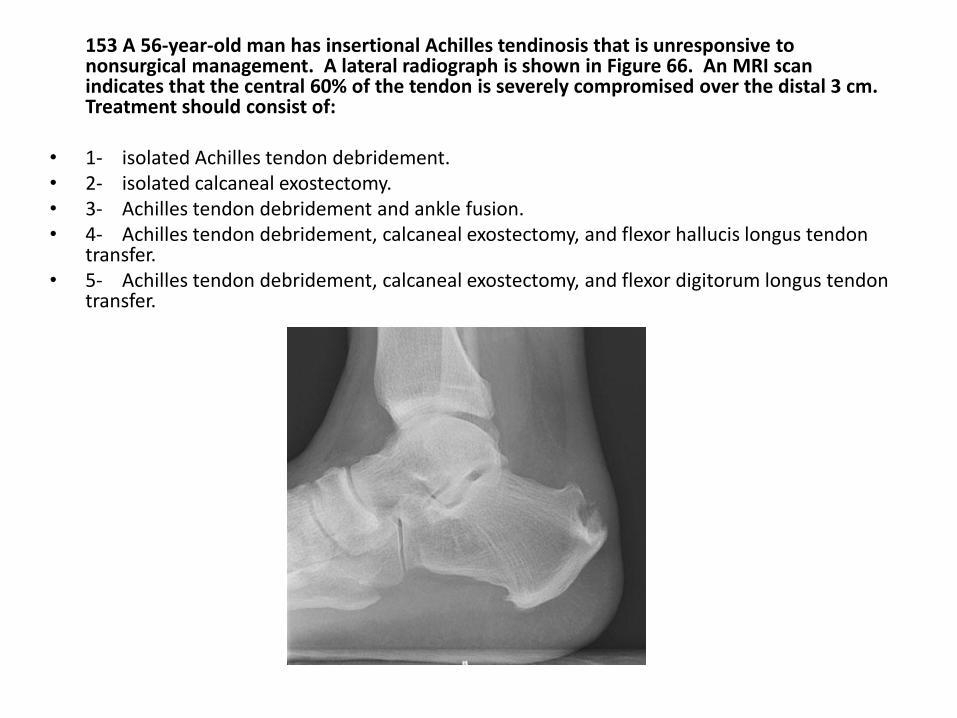
153 A 56-year-old man has insertional Achilles tendinosis that is unresponsive to nonsurgical management. A lateral radiograph is shown in Figure 66. An MRI scan indicates that the central 60% of the tendon is severely compromised over the distal 3 cm. Treatment should consist of:
• 1- isolated Achilles tendon debridement. • 2- isolated calcaneal exostectomy. • 3- Achilles tendon debridement and ankle fusion. • 4- Achilles tendon debridement, calcaneal exostectomy, and flexor hallucis longus tendon
transfer. • 5- Achilles tendon debridement, calcaneal exostectomy, and flexor digitorum longus tendon
transfer.
PREFERRED RESPONSE: 4 Achilles tendon debridement, calcaneal exostectomy, and flexor hallucis longus tendon transfer.
- They tell you the achilles looks like crap on the MRI so you know you need to do some kind of achilles debridement with another
procedure given the amount of tendon involvement. The question is what more you need to do. Look at the Xrays: don’t need to fuse the ankle and if you do don’t really need an achilles debridement do you. So do you choose the FDL or FHL? The FHL is typically used: in-phase transfer, minimal effect on hallux function, more closely reproduces the axial contractile forces of the achilles and provides a local contiguous muscle for revascularization of the scarred tendon bed.
RECOMMENDED READINGS: Den Hartog BD: Flexor hallucis longus transfer for chronic achilles tendonosis. Foot Ankle Int
2003;24:233-237. Kolodziej P, Glisson RR, Nunley JA: Risk of avulsion of the achilles tendon after partial excision for treatment of insertional
tendonitis and Haglund's deformity: A biomechanical study. Foot Ankle Int 1999;20:433-437. McGarvey WC, Palumbo RC, Baxter DE, et al: Insertional achilles tendinosis: Surgical treatment through a central tendon splitting
approach. Foot Ankle Int 2002;23:19-25.
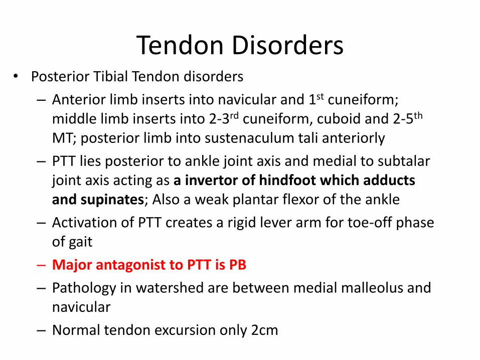
Tendon Disorders • Posterior Tibial Tendon disorders
– Anterior limb inserts into navicular and 1st cuneiform; middle limb inserts into 2-3rd cuneiform, cuboid and 2-5th MT; posterior limb into sustenaculum tali anteriorly
– PTT lies posterior to ankle joint axis and medial to subtalar joint axis acting as a invertor of hindfoot which adducts and supinates; Also a weak plantar flexor of the ankle
– Activation of PTT creates a rigid lever arm for toe-off phase of gait
– Major antagonist to PTT is PB
– Pathology in watershed are between medial malleolus and navicular
– Normal tendon excursion only 2cm
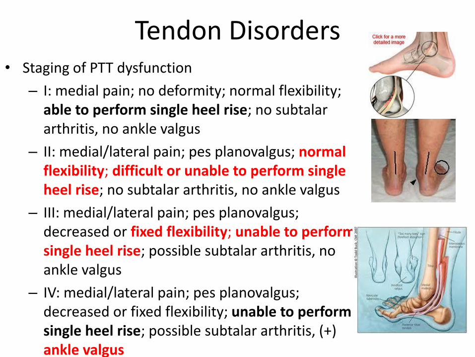
Tendon Disorders • Staging of PTT dysfunction
– I: medial pain; no deformity; normal flexibility; able to perform single heel rise; no subtalar arthritis, no ankle valgus
– II: medial/lateral pain; pes planovalgus; normal flexibility; difficult or unable to perform single heel rise; no subtalar arthritis, no ankle valgus
– III: medial/lateral pain; pes planovalgus; decreased or fixed flexibility; unable to perform single heel rise; possible subtalar arthritis, no ankle valgus
– IV: medial/lateral pain; pes planovalgus; decreased or fixed flexibility; unable to perform single heel rise; possible subtalar arthritis, (+) ankle valgus
Tendon Disorders • Posterior Tibial Tendon Dysfunction
– Most common cause of acquired flatfoot
– Collapse of medial longitudinal arch, hindfoot valgus, forefoot abduction (“too many toes” sign), achilles contracture
– Loss of ability to single heel rise due to
• Inability to lock transverse tarsal joints
• Valgus displacement of calcaneus with weakened achilles tendon moment arm
– Loss of longitudinal arch resulting in fixed equinus deformity
• Achilles tendon contracture
Tendon Disorders • PTT Dysfunction
• Nonsurgical Treatment: – After initial immobilization, custom molded in shoe orthosis with
medial posting for type I & II disease
– Stage III and IV requires bracing that crosses ankle joint (Ankle-foot orthosis- AFO or Arizona)—for sedentary, low demand patients, who cannot tolerate surgery
Tendon Disorders • PTT Dysfunction
• Surgical Treatment: – Stage I: tenosynovectomy
– Stage II: Combo tendon transfer and bony realignment
» FDL tendon transfer and medial calcaneal displacement
» 1st TMT joint arthrodesis, lateral column lengthening, and spring ligament repair
» Cotton osteotomy – plantarflexion opening wedge medial cuneiform osteotomy
– Stage III: Hindfoot Arthrodesis, usually triple arthrodesis
– -Stage IV: With tibiotalar arthritis, pantalar fusion; without tibiotalar arthritis, hindfoot arthrodesis with deltoid reconstruction
OITE 2006—Foot and Cankle
234. Medial displacement calcaneal osteotomy and flexor digitorum longus transfer to the navicular is considered the treatment of choice for which of the following patients?
1- A 24-year-old male runner with posterior tibial tenosynovitis and no hindfoot deformity. 2- A 27-year-old man with cerebral palsy and a spastic cavovarus foot
3- A 35-year-old man with a painful cavovarus foot secondary to Charcot-Marie-Tooth disease 4- A 60-year-old woman with a painful flexible adult-acquired flatfoot deformity secondary to posterior tibial tendon dysfunction 5- A 75-year-old woman with a rigid adult-acquired flatfoot deformity secondary to posterior tibial tendon dysfunction
OITE 2006—Foot and Cankle
• Medializing calcaneal osteotomy and FDL transfer can be used to treat stage II acquired flatfoot deformity or posterior tibial tendon insufficiency. The patient selection is an issue in this question. Patients with a flexible flatfoot deformity with moderate but not severe disease are candidates for this procedure. The patient in choice 1 has tendinitis, not posterior tib insufficiency. A cavovarus foot would be made worse by this procedure. The patient in choice 5 would not be a candidate for the procedure due to the fixed deformity.
OITE 2006—Foot and Ankle
48. An active 46-year-old woman has had an 18 month history of progressive hindfoot pain that has failed to respond to non surgical therapy. She has an asymmetrical planovalgus foot, an equinas contracture, and a flexible deformity. In addition to lengthening of the gastrocsoleus complex, recommended surgery should include:
1. In situ triple arthrodesis
2. Isolated FDL transfer to medial cuneiform
3. Lateral column lengthening, medializing calcaneal osteotomy, FDL transfer to the navicular
4. Lateralizing calcaneal osteotomy, FDL transfer to the navicular, and peroneus longus to peroneus brevis tendon transfer 5. Plantar flexion first tarsometatarsal fusion (Lapidus) and lateralizing calcaneal osteotomy
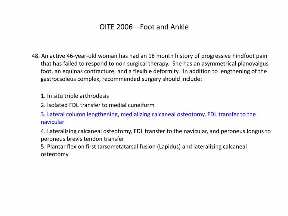
Posterior Tibial Tendon Insufficiency
– Most common cause of Adult Aquired Flatfoot
– First line Treatment is ALWAYS Non operative
• NSAIDs
• ORTHOTIC – accomadative, support medial longitudinal arch
• Ankle brace/AFO – esp with fixed deformities and those involving the ankle
• Corticosteroid injection is NOT recommended as it has been associated with tendon rupture
– Operative treatment only when nonoperative modalities have failed
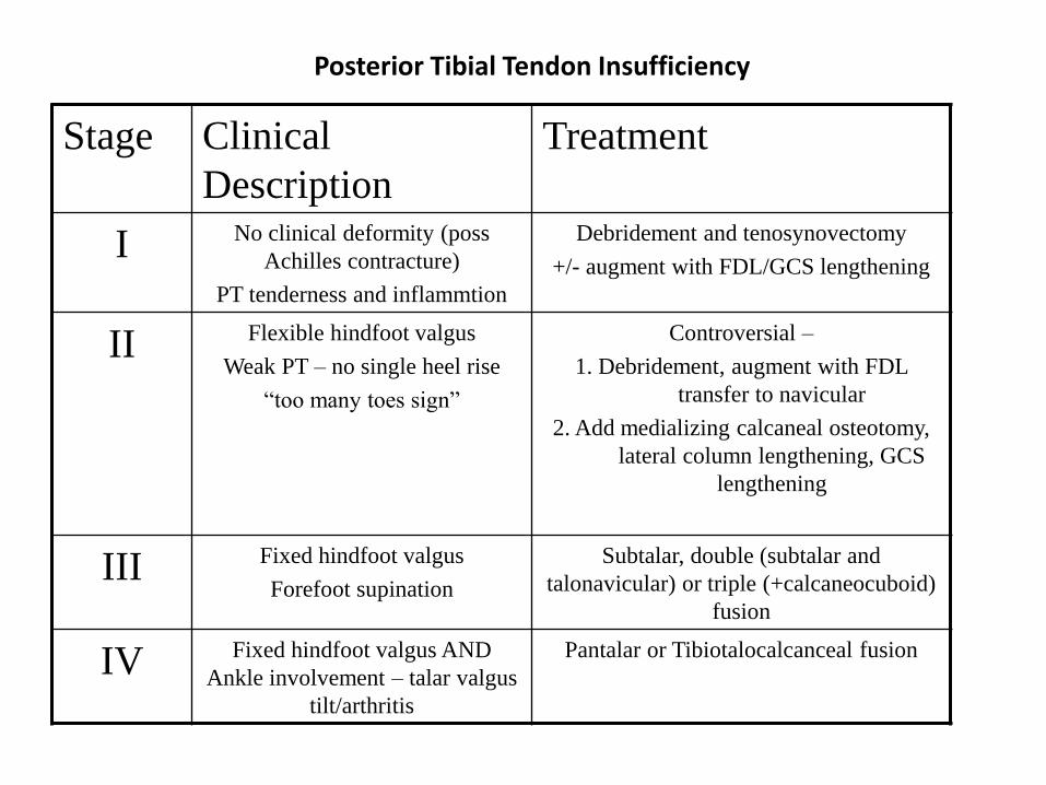
Posterior Tibial Tendon Insufficiency
Stage Clinical
Description
Treatment
I No clinical deformity (poss
Achilles contracture)
PT tenderness and inflammtion
Debridement and tenosynovectomy
+/- augment with FDL/GCS lengthening
II Flexible hindfoot valgus
Weak PT – no single heel rise
“too many toes sign”
Controversial –
1. Debridement, augment with FDL
transfer to navicular
2. Add medializing calcaneal osteotomy,
lateral column lengthening, GCS
lengthening
III Fixed hindfoot valgus
Forefoot supination
Subtalar, double (subtalar and
talonavicular) or triple (+calcaneocuboid)
fusion
IV Fixed hindfoot valgus AND
Ankle involvement – talar valgus
tilt/arthritis
Pantalar or Tibiotalocalcanceal fusion
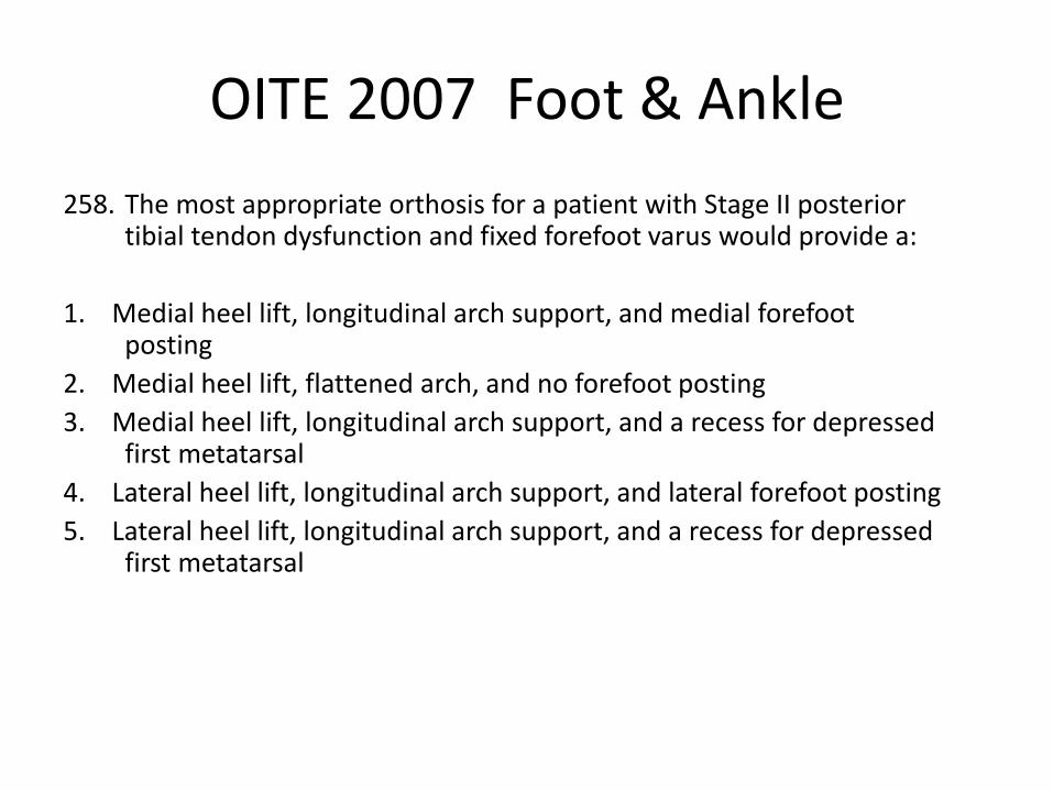
OITE 2007 Foot & Ankle
258. The most appropriate orthosis for a patient with Stage II posterior tibial tendon dysfunction and fixed forefoot varus would provide a:
1. Medial heel lift, longitudinal arch support, and medial forefoot posting
2. Medial heel lift, flattened arch, and no forefoot posting
3. Medial heel lift, longitudinal arch support, and a recess for depressed first metatarsal
4. Lateral heel lift, longitudinal arch support, and lateral forefoot posting
5. Lateral heel lift, longitudinal arch support, and a recess for depressed first metatarsal
OITE 2007 Foot & Ankle
Some helpful info on a very testable Posterior Tibial tendon Insufficiency topic….
40-60 yo; 15% bilateral, 75% female
Pain typically medial around ankle into arch; late may have lateral foot/ankle pain if calcaneus abuts fibula with valgus deformity of foot
“too many toes” sign—abduction of midfoot relative to hindfoot
Single limb heel rise test—unable to do so without vaulting forward and initiating with Achilles
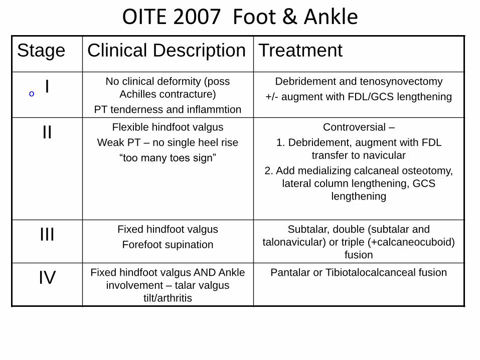
OITE 2007 Foot & Ankle
o
Stage Clinical Description Treatment
I No clinical deformity (poss
Achilles contracture)
PT tenderness and inflammtion
Debridement and tenosynovectomy
+/- augment with FDL/GCS lengthening
II Flexible hindfoot valgus
Weak PT – no single heel rise
“too many toes sign”
Controversial –
1. Debridement, augment with FDL
transfer to navicular
2. Add medializing calcaneal osteotomy,
lateral column lengthening, GCS
lengthening
III Fixed hindfoot valgus
Forefoot supination
Subtalar, double (subtalar and
talonavicular) or triple (+calcaneocuboid)
fusion
IV Fixed hindfoot valgus AND Ankle
involvement – talar valgus
tilt/arthritis
Pantalar or Tibiotalocalcanceal fusion
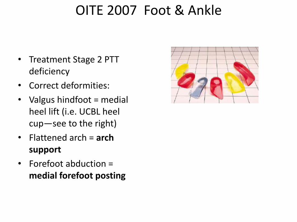
OITE 2007 Foot & Ankle
• Treatment Stage 2 PTT deficiency
• Correct deformities:
• Valgus hindfoot = medial heel lift (i.e. UCBL heel cup—see to the right)
• Flattened arch = arch support
• Forefoot abduction = medial forefoot posting
Foot and Cankle
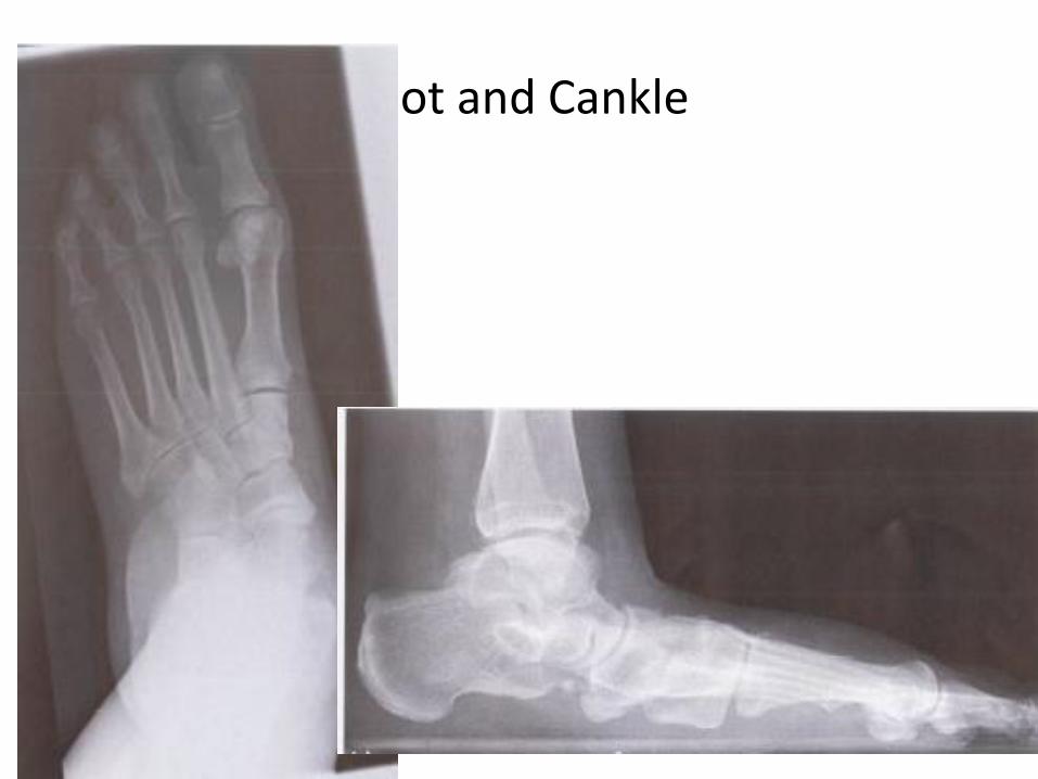
Foot and Cankle
Foot and ankle
Answer 2
Foot and ankle
• Based on the radiographic findings of an increase in Meary’s angle and decreased talonavicular coverage, the condition shown can be defined as adult acquired flatfoot deformity which is a consequence of posterior tibial tendon dysfunction. This disorder can be diagnosed by clinical exam and weight-bearing AP and lateral films of the foot which demonstrate a planovalgus deformity. On the lateral view, Meary’s angle, or the lateral tarsometatarsal angle, is formed by a line drawn through the axis of the talus and down the first metatarsal and should be 0. When these lines diverge, it indicates loss of the medial longitudinal arch. On the AP view, talonavicular coverage is evaluated by drawing a line form the edge of the articular surface of the talar head and from the edge of the articular surface of the navicular. An angle of greater than 10 degrees indicates excessive forefoot abduction.
• AAFD is characterized by a flattening of the medial longitudinal arch and dysfunction of the posteromedial soft tissues. These soft tissues consist of the posterior tibial tendon, the spring ligament and portions of the deltoid ligament. The PTT has a relatively hypovascular zone as it passes behind the medial malleolus. Changes occur in the tendon in this area and are thought to be caused by repetitive microtrauma in midstance. Gastroc-soleus contracture can contribute to the exacerbation of the condition as it causes hindfoot valgus (rule out choice 4). CMT patients have a cavo-varus foot. Not quite sure where the other 2 choices come in.
Question 33
Which of the following ligaments is most likely to be attenuated in a patient with a type II flatfoot deformity secondary to posterior tibial tendon dysfunction?
1. Calcaneonavicular
2. Anterior tibiofibular
3. Anterior talofibular
4. Posterior talofibular
5. Deltoid
Question 33
Which of the following ligaments is most likely to be attenuated in a patient with a type II flatfoot deformity secondary to posterior tibial tendon dysfunction?
1. Calcaneonavicular
2. Anterior tibiofibular
3. Anterior talofibular
4. Posterior talofibular
5. Deltoid
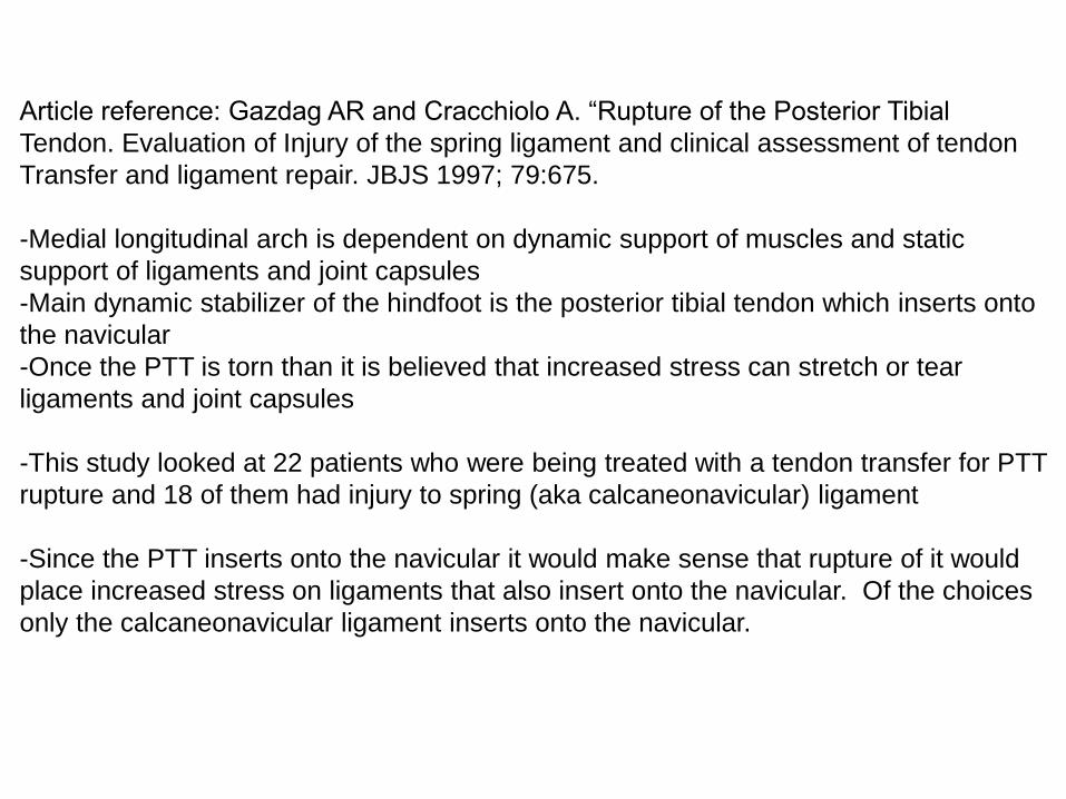
Article reference: Gazdag AR and Cracchiolo A. “Rupture of the Posterior Tibial
Tendon. Evaluation of Injury of the spring ligament and clinical assessment of tendon
Transfer and ligament repair. JBJS 1997; 79:675.
-Medial longitudinal arch is dependent on dynamic support of muscles and static
support of ligaments and joint capsules
-Main dynamic stabilizer of the hindfoot is the posterior tibial tendon which inserts onto
the navicular
-Once the PTT is torn than it is believed that increased stress can stretch or tear
ligaments and joint capsules
-This study looked at 22 patients who were being treated with a tendon transfer for PTT
rupture and 18 of them had injury to spring (aka calcaneonavicular) ligament
-Since the PTT inserts onto the navicular it would make sense that rupture of it would
place increased stress on ligaments that also insert onto the navicular. Of the choices
only the calcaneonavicular ligament inserts onto the navicular.
72. A 55 you woman has a flexible adult-acquired flatfoot deformity characterized by a positive too-many-toes
sign and medial ankle swelling. She is unable to perform a single leg heel raise. These findings are
classified as what stage posterior tibial tendon dysfunction?
1- I
2- II
3- III
4- IV
5- V
Answer:
2- II
- Just have to know the classification. Interestingly the classification for posterior tibialis tendon dysfunction
is one that very commonly pops up each year.
I: degeneration of tendon but no deformity, swelling, tender, able to single leg raise
II: Tendon degeneration with a flexible deformity, too-many-toes, unable/difficulty with single leg raise
III: Fixed deformity, not passively correctible, degenerative changes in subtalar and midfoot
IV: Above with bad DJD and valgus tilt of the talus within the ankle mortise
V: ?
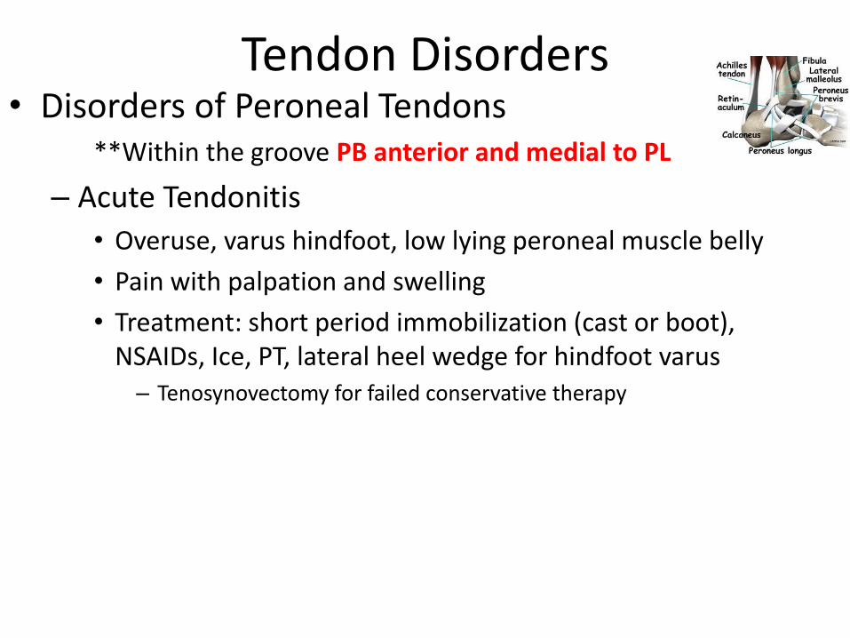
Tendon Disorders • Disorders of Peroneal Tendons
**Within the groove PB anterior and medial to PL
– Acute Tendonitis
• Overuse, varus hindfoot, low lying peroneal muscle belly
• Pain with palpation and swelling
• Treatment: short period immobilization (cast or boot), NSAIDs, Ice, PT, lateral heel wedge for hindfoot varus
– Tenosynovectomy for failed conservative therapy
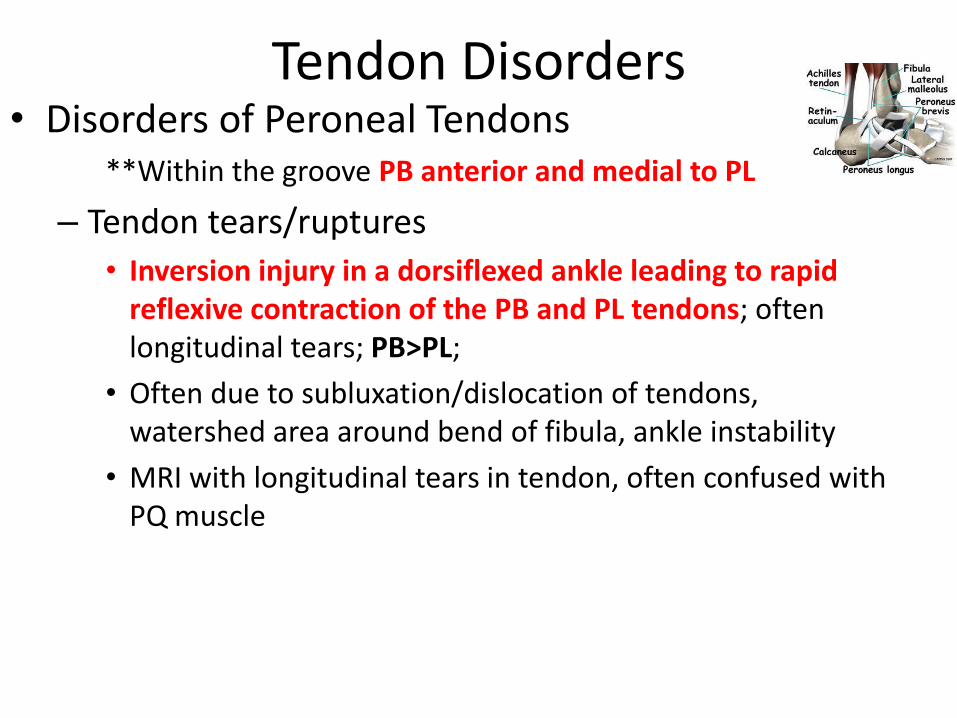
Tendon Disorders • Disorders of Peroneal Tendons
**Within the groove PB anterior and medial to PL
– Tendon tears/ruptures
• Inversion injury in a dorsiflexed ankle leading to rapid reflexive contraction of the PB and PL tendons; often longitudinal tears; PB>PL;
• Often due to subluxation/dislocation of tendons, watershed area around bend of fibula, ankle instability
• MRI with longitudinal tears in tendon, often confused with PQ muscle
Tendon Disorders • Treatment of Peroneal tendon tears/ruptures
• After failed conservative treatment (as above), Surgical Treatment:
– Acute partial rupture:
• Debridement and repair of tendon
• Tenodesis to healthy tendon if ruptured or if more than 50% of the one tendon is abnormal
• If heel varus: Calcaneal slide osteotomy added
– Complete Rupture
• Treatment in acute setting is end to end repair; Rare
– Chronic disease
• Tenodesis to healthy tendon or transfer of the FDL when both tendons are involved
Tendon Disorders
• Peroneal Dislocation/Subluxation
– Due to shallow peroneal groove and “overcrowding”
– Palpable snap over the fibula during ankle dorsiflexion
– Disruption of the superior peroneal retinaculum (SPR) or fibrocartilage ridge
– Variable pain and swelling
– Pain reproduced with resisted eversion
Tendon Disorders
• Peroneal Dislocation/Subluxation – Treatment:
• Acute with cast immobilization to allow SPR to heal; In high level athletes, acute SPR and groove deepening procedures
• Chronic injuries with tendon debridement and repair with a groove deepening procedure
Question
At the level of the ankle joint, what is the position of the peroneus longus tendon relative to the peroneus brevis tendon?
1.Posterior
2.Posterior and medial
3.Anterior and medial
4.Anterior and lateral
5.Directly anterior
Question
At the level of the ankle joint, what is the position of the peroneus longus tendon relative to the peroneus brevis tendon?
1.Posterior
2.Posterior and medial
3.Anterior and medial
4.Anterior and lateral
5.Directly anterior
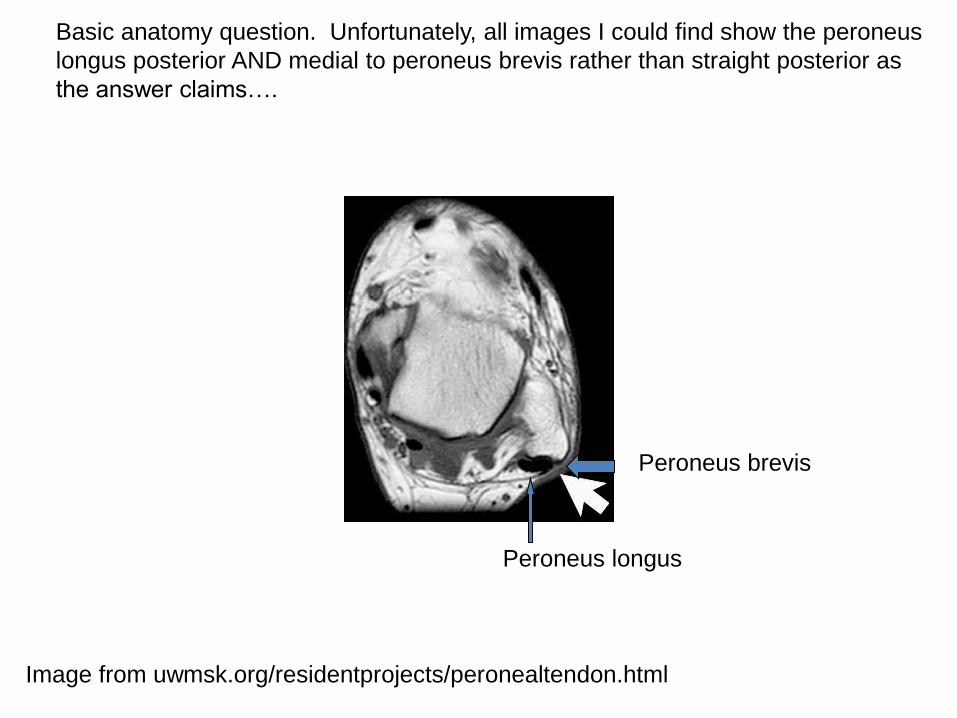
Image from uwmsk.org/residentprojects/peronealtendon.html
Peroneus longus
Peroneus brevis
Basic anatomy question. Unfortunately, all images I could find show the peroneus
longus posterior AND medial to peroneus brevis rather than straight posterior as
the answer claims….
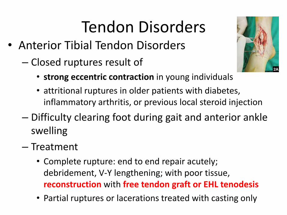
Tendon Disorders • Anterior Tibial Tendon Disorders
– Closed ruptures result of
• strong eccentric contraction in young individuals
• attritional ruptures in older patients with diabetes, inflammatory arthritis, or previous local steroid injection
– Difficulty clearing foot during gait and anterior ankle swelling
– Treatment
• Complete rupture: end to end repair acutely; debridement, V-Y lengthening; with poor tissue, reconstruction with free tendon graft or EHL tenodesis
• Partial ruptures or lacerations treated with casting only
Tendon Disorders • FHL Tendon Disorders
– FHL and FDL cross and have an interconnection at the knot of Henry
– Acute Laceration: most common form of injury
• Loss of active IP joint flexion
• Repair indicated in combined FHL and FHB lacs
Tendon Disorders • FHL Tendon Disorders
– Tensosynovitis:
• May coexist with os trigonum (posterior ankle impingement); common in dancers/gymnasts
• Posteromedial ankle pain
• Pain with resisted greater toe flexion – Great toe triggering
• Treatment – Initial nonsurgical: Rest,ice,NSAIDs,PT
– If symptoms persist, surgery: FHL release from fibro-osseous tunnel, tenosynovectomy, tendinous repair
OITE 2006—Foot and Cankle
187. A 17-year-old female gymnast has persistent pain when her ankle is in maximal plantar flexion. The MRI scan shown in figure 71 shows tenosynovitis of what tendon?
1- tibialis posterior 2- flexor hallucis longus
3- flexor digitorum communis 4- peroneus brevis 5- peroneus longus
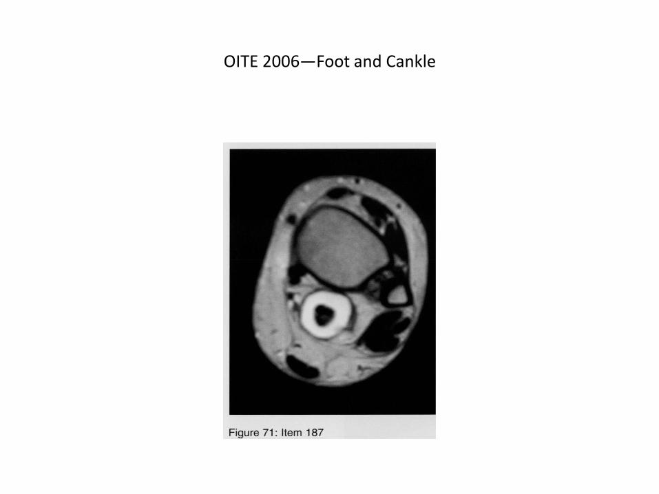
OITE 2006—Foot and Cankle
OITE 2006—Foot and Cankle
187. A 17-year-old female gymnast has persistent pain when her ankle is in maximal plantar flexion. The MRI scan shown in figure 71 shows tenosynovitis of what tendon?
1- tibialis posterior 2- flexor hallucis longus
3- flexor digitorum communis 4- peroneus brevis 5- peroneus longus
OITE 2006—Foot and Cankle
• Knowing the anatomy definitely helps answer this one. On the medial side of the ankle, the order of structures from anterior to posterior can be remembered by the neumonic Tom, Dick, And Now Harry for Tibialis ant., fDl, artery, nerve, fHl. Also, FHL tendinitis is a common cause of posteromedial ankle pain in dancers and gymnasts. The exacerbation of symptoms with plantar flexion suggests some kind of posterior structure being irritated.
Tendon Disorders • EDL and EHL Tendon disorders
– Ruptures (ie attritional) vs lacerations
– Acute EHL lacerations proximal to extensor hood repaired end to end;
– Partial lacerations or those at or distal to extendor hood treated closed with imobilization of hallux in extension
– Chronic EHL injury or attritional ruptures treated with debridement and repair, free tendon grafting, or tenodesis to a healthy extensor tendon
– EDL repair in young active individuals only; untreated will lead to a future claw toe deformity
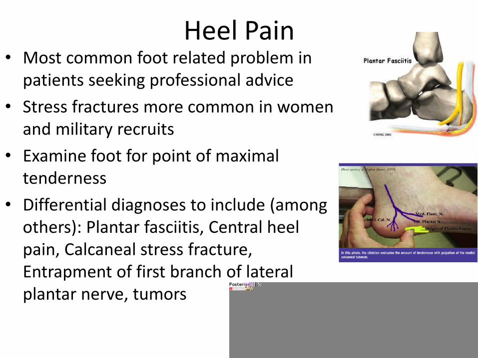
Heel Pain • Most common foot related problem in
patients seeking professional advice
• Stress fractures more common in women and military recruits
• Examine foot for point of maximal tenderness
• Differential diagnoses to include (among others): Plantar fasciitis, Central heel pain, Calcaneal stress fracture, Entrapment of first branch of lateral plantar nerve, tumors
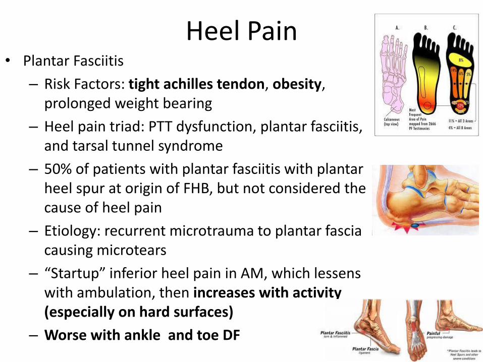
Heel Pain • Plantar Fasciitis
– Risk Factors: tight achilles tendon, obesity, prolonged weight bearing
– Heel pain triad: PTT dysfunction, plantar fasciitis, and tarsal tunnel syndrome
– 50% of patients with plantar fasciitis with plantar heel spur at origin of FHB, but not considered the cause of heel pain
– Etiology: recurrent microtrauma to plantar fascia causing microtears
– “Startup” inferior heel pain in AM, which lessens with ambulation, then increases with activity (especially on hard surfaces)
– Worse with ankle and toe DF
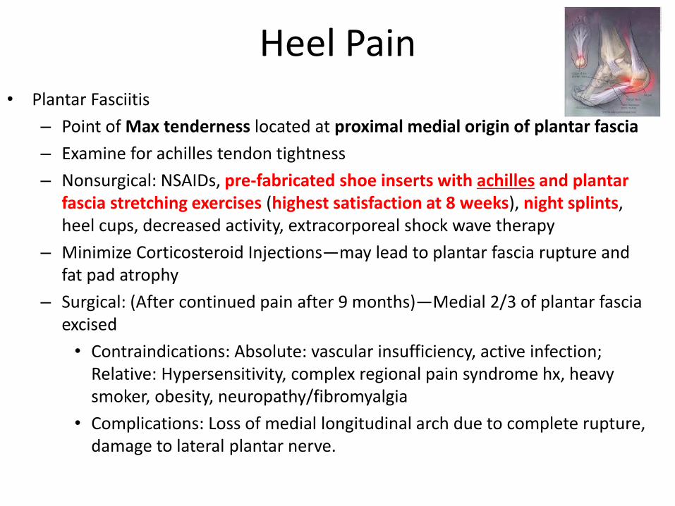
Heel Pain • Plantar Fasciitis
– Point of Max tenderness located at proximal medial origin of plantar fascia
– Examine for achilles tendon tightness
– Nonsurgical: NSAIDs, pre-fabricated shoe inserts with achilles and plantar fascia stretching exercises (highest satisfaction at 8 weeks), night splints, heel cups, decreased activity, extracorporeal shock wave therapy
– Minimize Corticosteroid Injections—may lead to plantar fascia rupture and fat pad atrophy
– Surgical: (After continued pain after 9 months)—Medial 2/3 of plantar fascia excised
• Contraindications: Absolute: vascular insufficiency, active infection; Relative: Hypersensitivity, complex regional pain syndrome hx, heavy smoker, obesity, neuropathy/fibromyalgia
• Complications: Loss of medial longitudinal arch due to complete rupture, damage to lateral plantar nerve.
Heel Pain • Central Heel Pain
– Fat pad atrophy due to corticosteroid injection, advanced age, compression from calcaneal osteophyte, inflammatory disease-with resultant decreased shock reduction
– Pain is localized and does not improve after initial ambulation; Not worse with dorsiflexion of toes
– Point of maximal tenderness in center of weight bearing heel
– Treatment: NS: Heel cups or orthosis; Surgical: None; Possible removal of calcaneal osteophyte
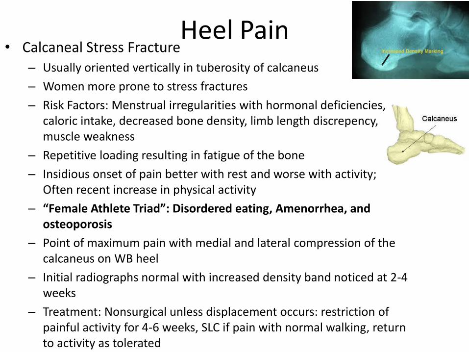
Heel Pain • Calcaneal Stress Fracture
– Usually oriented vertically in tuberosity of calcaneus
– Women more prone to stress fractures
– Risk Factors: Menstrual irregularities with hormonal deficiencies, caloric intake, decreased bone density, limb length discrepency, muscle weakness
– Repetitive loading resulting in fatigue of the bone
– Insidious onset of pain better with rest and worse with activity; Often recent increase in physical activity
– “Female Athlete Triad”: Disordered eating, Amenorrhea, and osteoporosis
– Point of maximum pain with medial and lateral compression of the calcaneus on WB heel
– Initial radiographs normal with increased density band noticed at 2-4 weeks
– Treatment: Nonsurgical unless displacement occurs: restriction of painful activity for 4-6 weeks, SLC if pain with normal walking, return to activity as tolerated
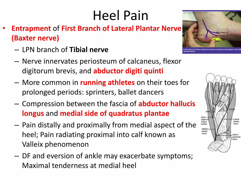
Heel Pain • Entrapment of First Branch of Lateral Plantar Nerve
(Baxter nerve)
– LPN branch of Tibial nerve
– Nerve innervates periosteum of calcaneus, flexor digitorum brevis, and abductor digiti quinti
– More common in running athletes on their toes for prolonged periods: sprinters, ballet dancers
– Compression between the fascia of abductor hallucis longus and medial side of quadratus plantae
– Pain distally and proximally from medial aspect of the heel; Pain radiating proximal into calf known as Valleix phenomenon
– DF and eversion of ankle may exacerbate symptoms; Maximal tenderness at medial heel
Heel Pain
• Entrapment of First Branch of Lateral Plantar Nerve
– Treatment:
• Nonsurgical (6-9months): Rest,Ice, NSAIDs, stretching, shock absorbing inserts with medial arch support
• Surgical (after 9 months): open decompression; medial 1/3 of plantar fascia if concomminant plantar fasciitis
– Deep fascia of abductor hallicus muscle released
OITE 2006- Foot & Ankle
37. A 46 yo woman has had plantar heel pain for the past 5 months. The pain is most severe when she arises out of bed in the morning and when she stands after being seated for a period of time. Initial treatment should consist of
1. Surgical lengthening of the Achilles tendon
2. Surgical release of the plantar fascia
3. A custom orthosis
4. A stretching program and a cushioned insert
5. A corticosteroid injection
Discussion
• Proximal plantar fasciitis or heel pain syndrome is most found in males between 40-70, active, and have unilateral symptoms in a normallly arched foot. Obesity is a predisposing factor. Major complaint is pain beneath the heel that is worse on rising in the morning or after sitting for a while. The pain diminishes after a few steps and the patient is reasonably comfortable the rest of the day.
• Most common physical finding is localized tenderness at the inferomedial aspect of the calcaneal tuberosity. Symptoms may last weeks to months.
• Films may reveal a calcaneal spur in 50% of pts.
• Rarely do pts need sx. Shoe inserts (cups and pads), oral nsaids and local steroid injxns relieve pain in most pts.
• Pfeffer et al. report that in a prospective randomized trial of 236pts, a stretching program in conjunction with a prefabricated shoe insert is likely to improve symptoms as part of initial treatment of proximal plantar fasciitis than a custom orthotic device.
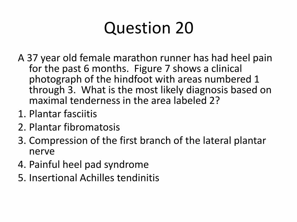
Question 20
A 37 year old female marathon runner has had heel pain for the past 6 months. Figure 7 shows a clinical photograph of the hindfoot with areas numbered 1 through 3. What is the most likely diagnosis based on maximal tenderness in the area labeled 2?
1. Plantar fasciitis 2. Plantar fibromatosis 3. Compression of the first branch of the lateral plantar
nerve 4. Painful heel pad syndrome 5. Insertional Achilles tendinitis
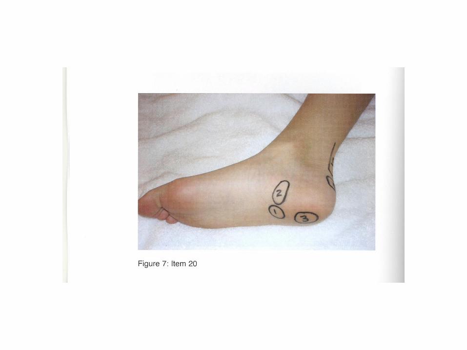
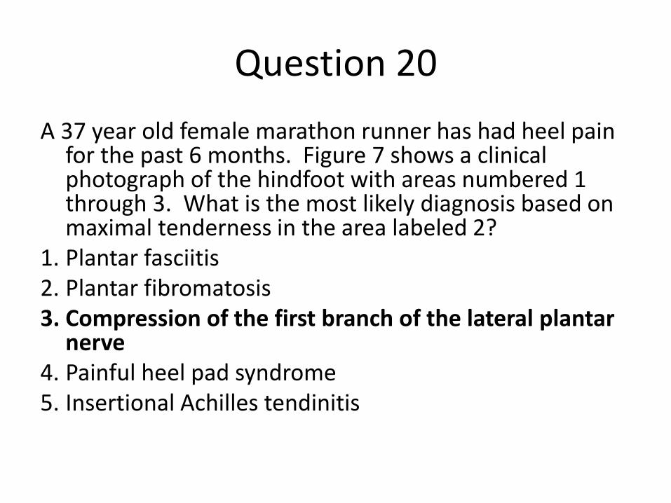
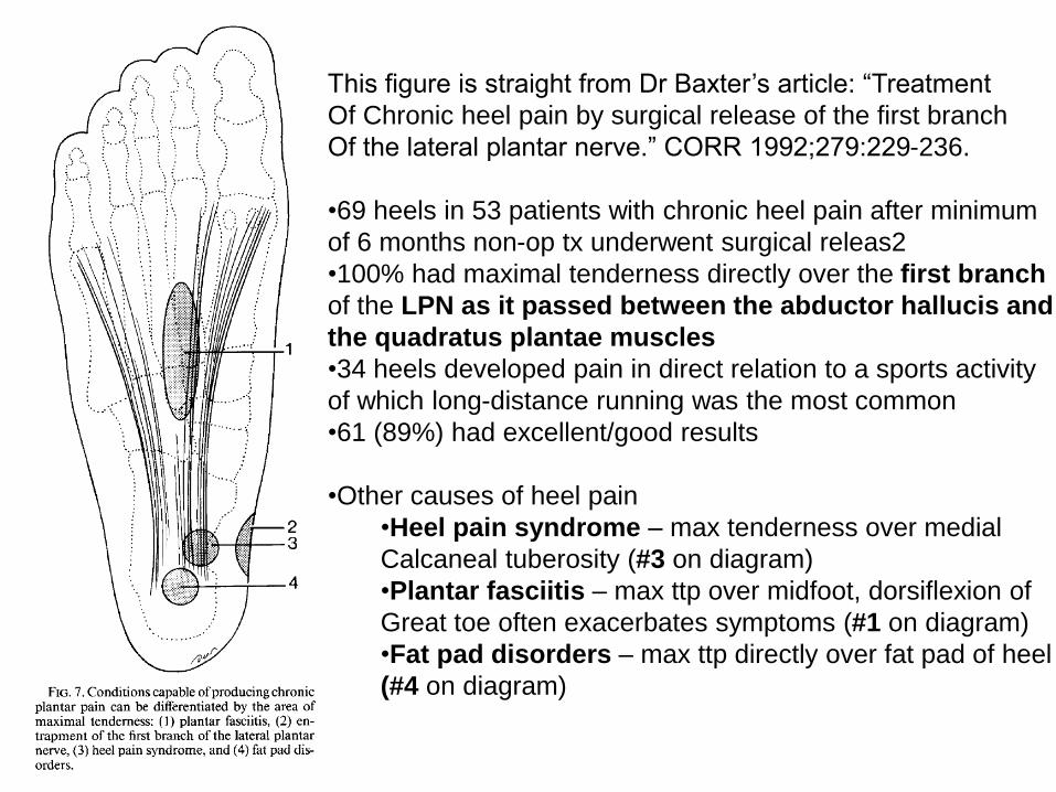
This figure is straight from Dr Baxter’s article: “Treatment
Of Chronic heel pain by surgical release of the first branch
Of the lateral plantar nerve.” CORR 1992;279:229-236.
•69 heels in 53 patients with chronic heel pain after minimum
of 6 months non-op tx underwent surgical releas2
•100% had maximal tenderness directly over the first branch
of the LPN as it passed between the abductor hallucis and
the quadratus plantae muscles
•34 heels developed pain in direct relation to a sports activity
of which long-distance running was the most common
•61 (89%) had excellent/good results
•Other causes of heel pain
•Heel pain syndrome – max tenderness over medial
Calcaneal tuberosity (#3 on diagram)
•Plantar fasciitis – max ttp over midfoot, dorsiflexion of
Great toe often exacerbates symptoms (#1 on diagram)
•Fat pad disorders – max ttp directly over fat pad of heel
(#4 on diagram)
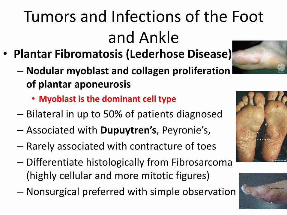
Tumors and Infections of the Foot and Ankle
• Plantar Fibromatosis (Lederhose Disease)
– Nodular myoblast and collagen proliferation of plantar aponeurosis
• Myoblast is the dominant cell type
– Bilateral in up to 50% of patients diagnosed
– Associated with Dupuytren’s, Peyronie’s,
– Rarely associated with contracture of toes
– Differentiate histologically from Fibrosarcoma (highly cellular and more mitotic figures)
– Nonsurgical preferred with simple observation
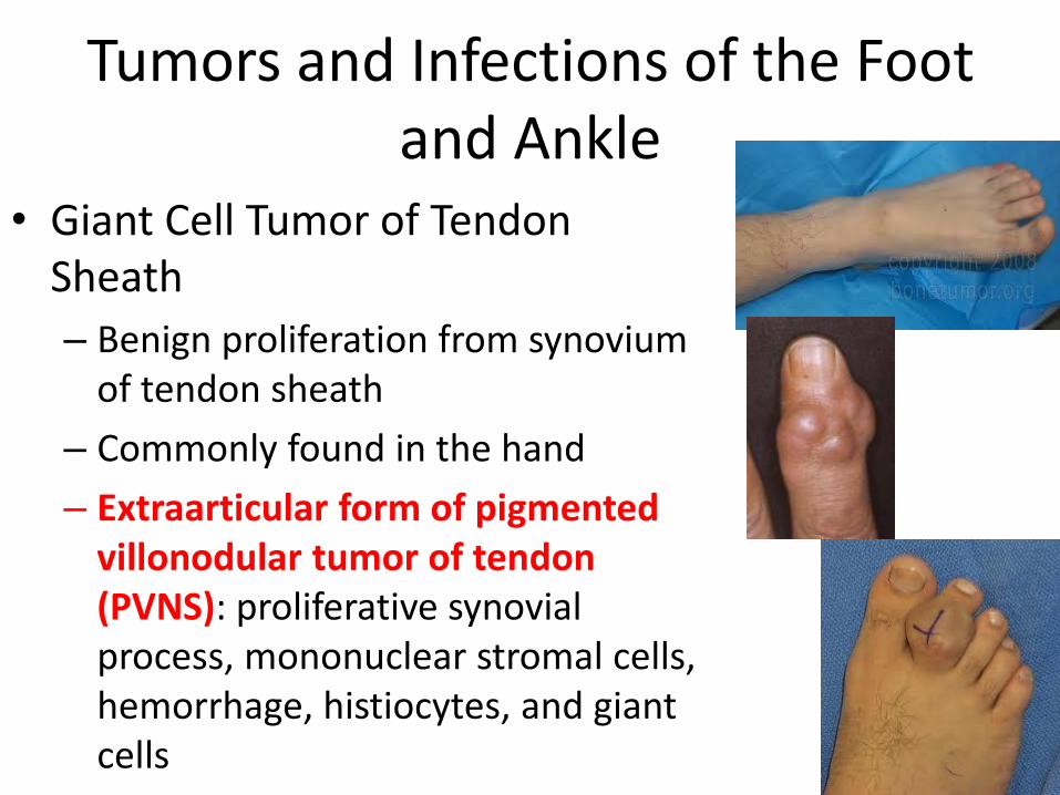
Tumors and Infections of the Foot and Ankle
• Giant Cell Tumor of Tendon Sheath
– Benign proliferation from synovium of tendon sheath
– Commonly found in the hand
– Extraarticular form of pigmented villonodular tumor of tendon (PVNS): proliferative synovial process, mononuclear stromal cells, hemorrhage, histiocytes, and giant cells
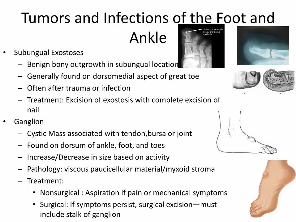
Tumors and Infections of the Foot and Ankle
• Subungual Exostoses
– Benign bony outgrowth in subungual location
– Generally found on dorsomedial aspect of great toe
– Often after trauma or infection
– Treatment: Excision of exostosis with complete excision of nail
• Ganglion
– Cystic Mass associated with tendon,bursa or joint
– Found on dorsum of ankle, foot, and toes
– Increase/Decrease in size based on activity
– Pathology: viscous paucicellular material/myxoid stroma
– Treatment:
• Nonsurgical : Aspiration if pain or mechanical symptoms
• Surgical: If symptoms persist, surgical excision—must include stalk of ganglion
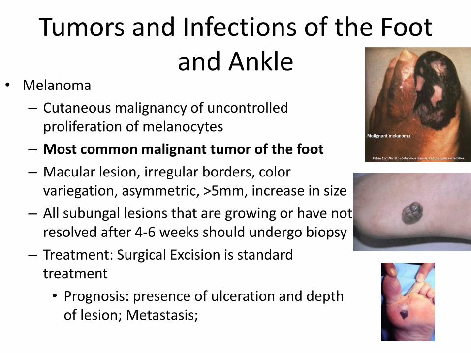
Tumors and Infections of the Foot and Ankle
• Melanoma
– Cutaneous malignancy of uncontrolled proliferation of melanocytes
– Most common malignant tumor of the foot
– Macular lesion, irregular borders, color variegation, asymmetric, >5mm, increase in size
– All subungal lesions that are growing or have not resolved after 4-6 weeks should undergo biopsy
– Treatment: Surgical Excision is standard treatment
• Prognosis: presence of ulceration and depth of lesion; Metastasis;
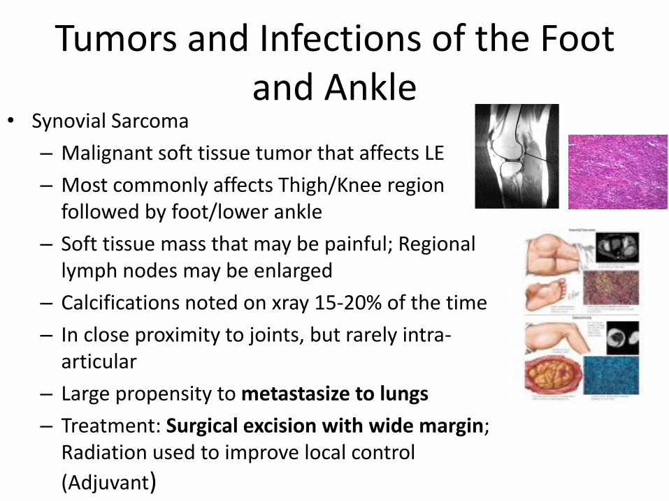
Tumors and Infections of the Foot and Ankle
• Synovial Sarcoma
– Malignant soft tissue tumor that affects LE
– Most commonly affects Thigh/Knee region followed by foot/lower ankle
– Soft tissue mass that may be painful; Regional lymph nodes may be enlarged
– Calcifications noted on xray 15-20% of the time
– In close proximity to joints, but rarely intra-articular
– Large propensity to metastasize to lungs
– Treatment: Surgical excision with wide margin; Radiation used to improve local control
(Adjuvant)
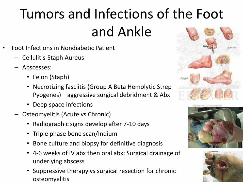
Tumors and Infections of the Foot and Ankle
• Foot Infections in Nondiabetic Patient
– Cellulitis-Staph Aureus
– Abscesses:
• Felon (Staph)
• Necrotizing fasciitis (Group A Beta Hemolytic Strep Pyogenes)—aggressive surgical debridment & Abx
• Deep space infections
– Osteomyelitis (Acute vs Chronic)
• Radiographic signs develop after 7-10 days
• Triple phase bone scan/Indium
• Bone culture and biopsy for definitive diagnosis
• 4-6 weeks of IV abx then oral abx; Surgical drainage of underlying abscess
• Suppressive therapy vs surgical resection for chronic osteomyelitis
Diabetic Foot and Ankle
• Diabetic Neuropathy in somatic/autonomic nervous system
– Sensory neuropathy in almost 70% of patients
• Semmes-Weinstein monofilament test (5.07 gm); ability loss of protective sensation; one of the last to go ;
– Motor neuropathy can lead to claw toes (intrinsics) and achilles contractures
– Autonomic neuropathy-unable to control blood vessel tone and sweat glands in the foot
• Increase blood flow
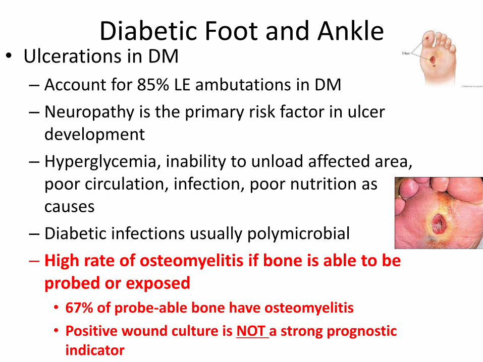
Diabetic Foot and Ankle • Ulcerations in DM
– Account for 85% LE ambutations in DM
– Neuropathy is the primary risk factor in ulcer development
– Hyperglycemia, inability to unload affected area, poor circulation, infection, poor nutrition as causes
– Diabetic infections usually polymicrobial
– High rate of osteomyelitis if bone is able to be probed or exposed
• 67% of probe-able bone have osteomyelitis
• Positive wound culture is NOT a strong prognostic indicator
Diabetic Foot and Ankle • Ulcerations in DM
– Silverskiold test
• Improved ankle dorsiflexion with knee flexed – Gastrocnemius tightness
• Equivalent ankle dorsiflexion with knee flexion and extension
– Achilles tightness
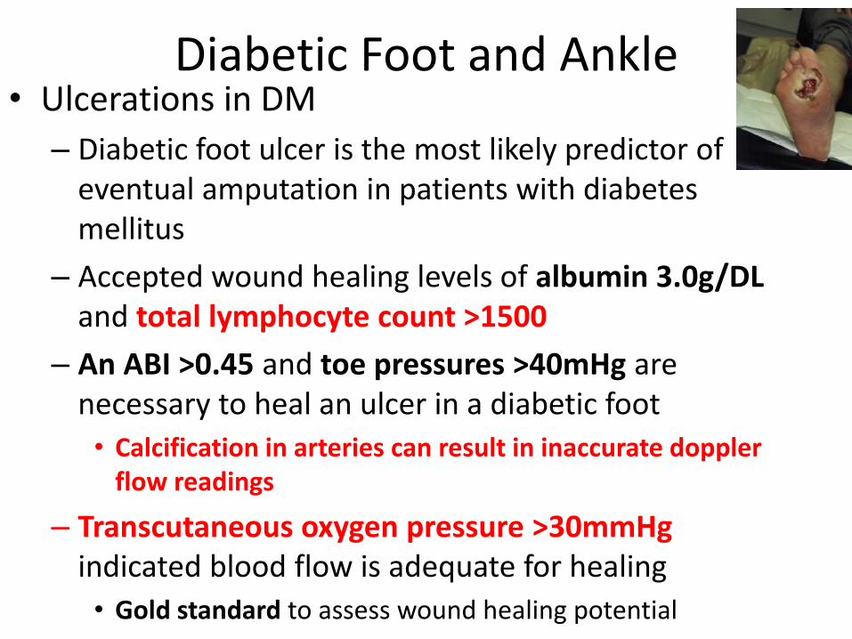
Diabetic Foot and Ankle • Ulcerations in DM
– Diabetic foot ulcer is the most likely predictor of eventual amputation in patients with diabetes mellitus
– Accepted wound healing levels of albumin 3.0g/DL and total lymphocyte count >1500
– An ABI >0.45 and toe pressures >40mHg are necessary to heal an ulcer in a diabetic foot
• Calcification in arteries can result in inaccurate doppler flow readings
– Transcutaneous oxygen pressure >30mmHg indicated blood flow is adequate for healing
• Gold standard to assess wound healing potential
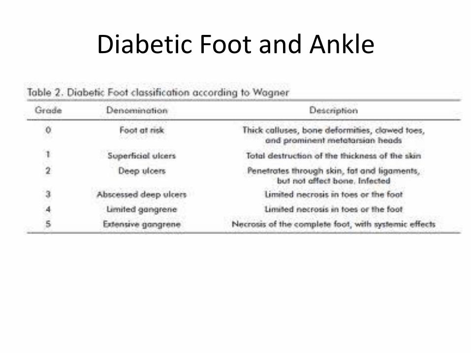
Diabetic Foot and Ankle
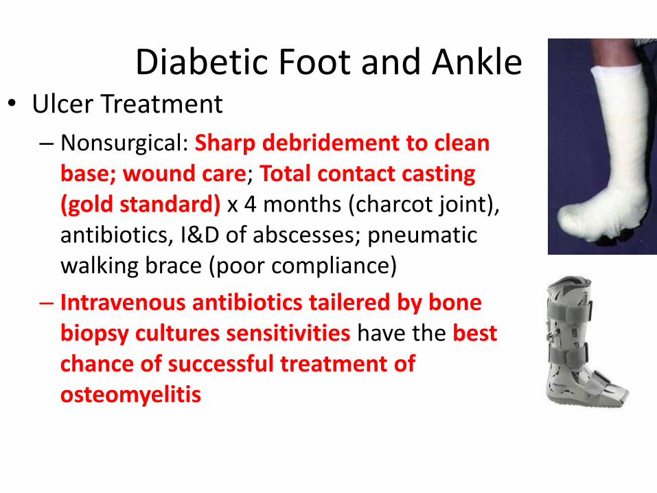
Diabetic Foot and Ankle • Ulcer Treatment
– Nonsurgical: Sharp debridement to clean base; wound care; Total contact casting (gold standard) x 4 months (charcot joint), antibiotics, I&D of abscesses; pneumatic walking brace (poor compliance)
– Intravenous antibiotics tailered by bone biopsy cultures sensitivities have the best chance of successful treatment of osteomyelitis
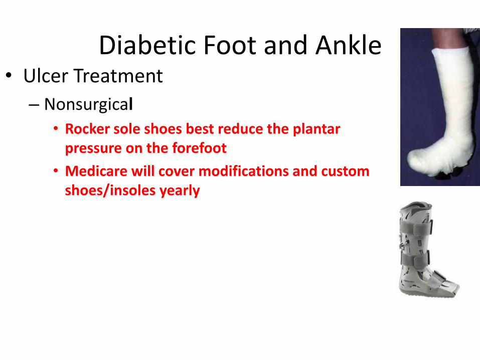
Diabetic Foot and Ankle • Ulcer Treatment
– Nonsurgical
• Rocker sole shoes best reduce the plantar pressure on the forefoot
• Medicare will cover modifications and custom shoes/insoles yearly

Diabetic Foot and Ankle • Ulcer Treatment
– Surgical: Surgical debridement, dressing changes, IV antibiotics; abscess drainage; Ostectomy or realignment arthrodesis (to remove bony prominences), achilles tendon lengthening (reduced forefoot pressures)
• Osteomyelitis: culture by ulcer curettage/bone biopsy/aspiration before antibiotics
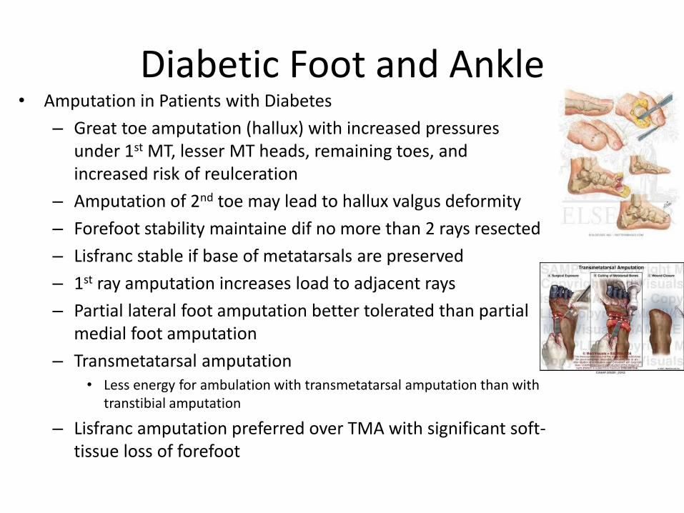
Diabetic Foot and Ankle • Amputation in Patients with Diabetes
– Great toe amputation (hallux) with increased pressures under 1st MT, lesser MT heads, remaining toes, and increased risk of reulceration
– Amputation of 2nd toe may lead to hallux valgus deformity
– Forefoot stability maintaine dif no more than 2 rays resected
– Lisfranc stable if base of metatarsals are preserved
– 1st ray amputation increases load to adjacent rays
– Partial lateral foot amputation better tolerated than partial medial foot amputation
– Transmetatarsal amputation • Less energy for ambulation with transmetatarsal amputation than with
transtibial amputation
– Lisfranc amputation preferred over TMA with significant soft-tissue loss of forefoot
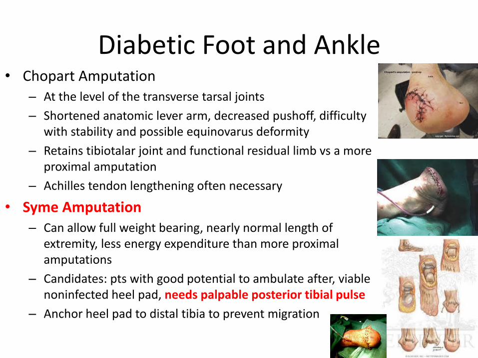
Diabetic Foot and Ankle • Chopart Amputation
– At the level of the transverse tarsal joints
– Shortened anatomic lever arm, decreased pushoff, difficulty with stability and possible equinovarus deformity
– Retains tibiotalar joint and functional residual limb vs a more proximal amputation
– Achilles tendon lengthening often necessary
• Syme Amputation – Can allow full weight bearing, nearly normal length of
extremity, less energy expenditure than more proximal amputations
– Candidates: pts with good potential to ambulate after, viable noninfected heel pad, needs palpable posterior tibial pulse
– Anchor heel pad to distal tibia to prevent migration
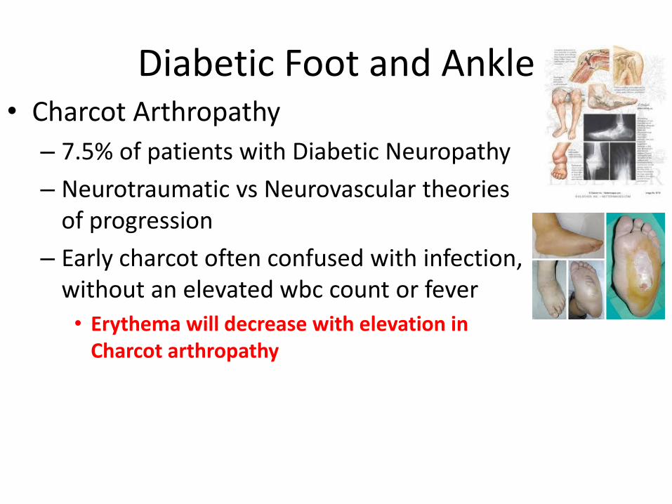
Diabetic Foot and Ankle • Charcot Arthropathy
– 7.5% of patients with Diabetic Neuropathy
– Neurotraumatic vs Neurovascular theories of progression
– Early charcot often confused with infection, without an elevated wbc count or fever
• Erythema will decrease with elevation in Charcot arthropathy
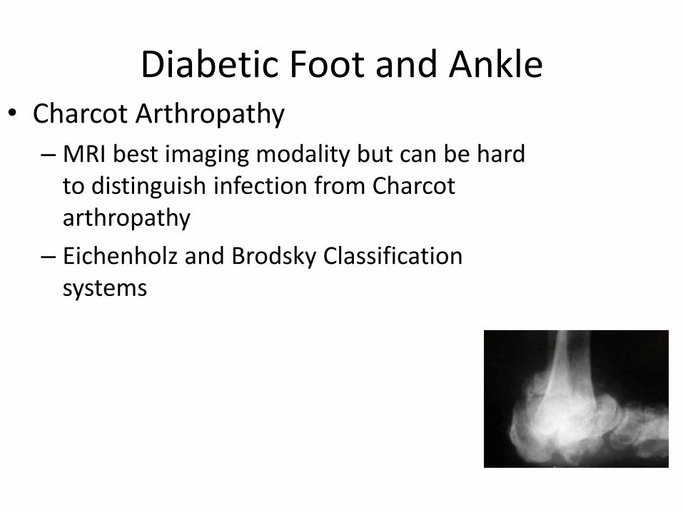
Diabetic Foot and Ankle • Charcot Arthropathy
– MRI best imaging modality but can be hard to distinguish infection from Charcot arthropathy
– Eichenholz and Brodsky Classification systems
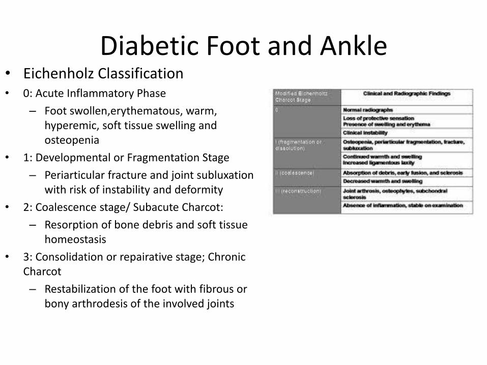
Diabetic Foot and Ankle • Eichenholz Classification • 0: Acute Inflammatory Phase
– Foot swollen,erythematous, warm, hyperemic, soft tissue swelling and osteopenia
• 1: Developmental or Fragmentation Stage
– Periarticular fracture and joint subluxation with risk of instability and deformity
• 2: Coalescence stage/ Subacute Charcot:
– Resorption of bone debris and soft tissue homeostasis
• 3: Consolidation or repairative stage; Chronic Charcot
– Restabilization of the foot with fibrous or bony arthrodesis of the involved joints
Diabetic Foot and Ankle
• Charcot Treatment – Goals: Plantigrade, stable foot able to fit into a shoe; with absence of
recurrent ulceration
– Total Contact Casting (effective 75% with guarded ambulation)—allows even distribution of foot pressure across plantar surface of the foot; Continued for 4 months
– Acute Surgical Correction in the inflammatory phase associated with high rate of nonunion, infection, wound complications, late deformity and eventual amputation
– Late Surgical Correction: Best performed in Eichenholtz stage 3 when inflammatory process has resolved
– Indications for surgery: recurrent ulcers and instability not controlled by a brace: exostectomy or reconstruction with osteotomy and fusion
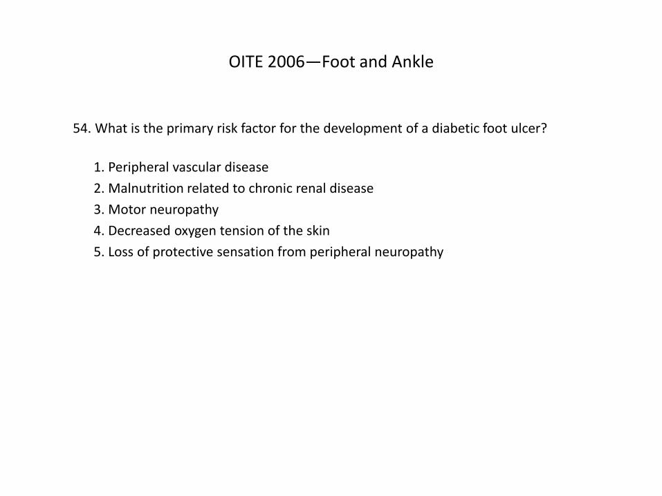
OITE 2006—Foot and Ankle
54. What is the primary risk factor for the development of a diabetic foot ulcer?
1. Peripheral vascular disease
2. Malnutrition related to chronic renal disease
3. Motor neuropathy
4. Decreased oxygen tension of the skin
5. Loss of protective sensation from peripheral neuropathy
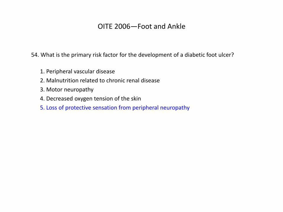
Diabetic Foot Ulcers
– Approximately 20 % of diabetics will have a serious foot infection in their lifetime
– 85 % of amputations are preceeded by a foot ulcer
– Number one risk factor for a diabetic foot ulcer is a history of a previous ulcer
– Inability to respond to mechanical stress because of loss of lg fiber (LT, proprioception) and small fiber (pain, temp) sensory nerves
– High stress – causes immediate puncture or tearing
– Moderate stress – most common cause of injury in insensate foot
• Pressure overcomes plantar skin tensile stress – 20 lbs/sq for >10K reps
• Sensate foot would stop when pain and imflammation are present (day 3)
• Insensate foot keeps walking and ulcerates by day 8
• Moderate stresses can block capillary flow increasing ischemia
Diabetic Foot Ulcers - Evaluation
– Ankle – Brachial index: SBP ankle/SBP brachial artery – want > 0.45 to heal amputation
– Absolute toe pressure of oxygen - > 45 mmHg for amputation healing, > 60 mm Hg for ulcer healing
– Transcutaneous oxygen perfusion TcPO2 is the most accurate measurement of local tissue perfusion and healing potential – (measurements are taken at 45C)
• Healing rate – 50 % if TcO2 1-19, 75% if 20- 29, 92 % if > 32
– Metabolic factors required to heal
• Total protein > 6.2
• Albumin > 3.5
• Lymphocytes > 1500/mm3
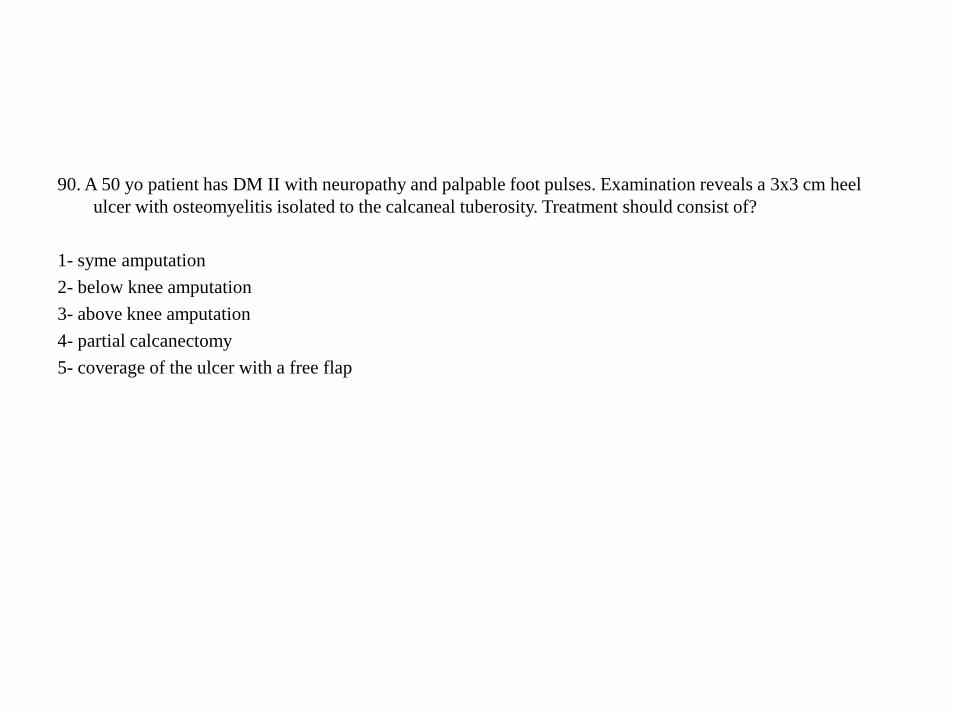
90. A 50 yo patient has DM II with neuropathy and palpable foot pulses. Examination reveals a 3x3 cm heel
ulcer with osteomyelitis isolated to the calcaneal tuberosity. Treatment should consist of?
1- syme amputation
2- below knee amputation
3- above knee amputation
4- partial calcanectomy
5- coverage of the ulcer with a free flap
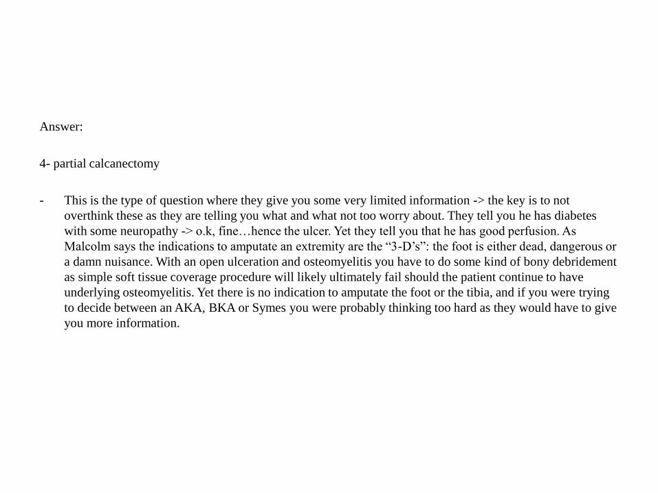
Answer:
4- partial calcanectomy
- This is the type of question where they give you some very limited information -> the key is to not
overthink these as they are telling you what and what not too worry about. They tell you he has diabetes
with some neuropathy -> o.k, fine…hence the ulcer. Yet they tell you that he has good perfusion. As
Malcolm says the indications to amputate an extremity are the “3-D’s”: the foot is either dead, dangerous or
a damn nuisance. With an open ulceration and osteomyelitis you have to do some kind of bony debridement
as simple soft tissue coverage procedure will likely ultimately fail should the patient continue to have
underlying osteomyelitis. Yet there is no indication to amputate the foot or the tibia, and if you were trying
to decide between an AKA, BKA or Symes you were probably thinking too hard as they would have to give
you more information.
Question 72
A 56 year old woman reports a 5 month history of ankle pain and swelling. She denies any significant medical history or history of trauma but admits she has not seen a physician in several years. She remains ambulatory without the use of walking aids. Current radiographs are shown in Figures 27a and 27b. The patient should now undergo testing for
1. Spinal stenosis 2. Compartment syndrome 3. Diabetes mellitus 4. Rheumatoid arthritis 5. Fibromyalgia
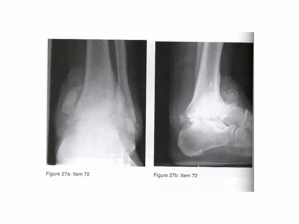
Discussion
• Images show complete destruction of ankle joint with collapse of talus. This picture should immediately suggest Charcot arthropathy
• Then you just need to recognize that Charcot arthropathy is often associated with Diabetes Mellitus
246) When performing a partial foot amputation through the talonavicular and calcaneocuboid joints, which of the following procedures should also be performed?
1 Transfer of the posterior tibial tendon to the calcaneus
2 Transfer of the tibialis anterior tendon to the calcaneal tuberosity
3 Lengthening of the Achilles tendon and transfer of the tibialis anterior to the talar neck
4 Shortening of the Achilles tendon
5 Ankle fusion
A partial foot amputation through the talonavicular and calcaneocuboid joint is known as a Chopart amputation. These are rarely performed today. In this amputation the forefoot and midfoot are removed, and the calcaneus is preserved. This causes loss of the insertion points of the peronei and dorsiflexors. Thus, the plantarflexors have no balancing force, and the residual limb tends to an equinus and varus deformity. To decrease this some soft tissue balancing must be performed. The tibialis anterior is transferred to the talar neck to control the hindfoot deformity. The Achilles tendon can be lengthened to decrease the equinus deformity and aid in casting/splinting that is often necessary after the procedure.
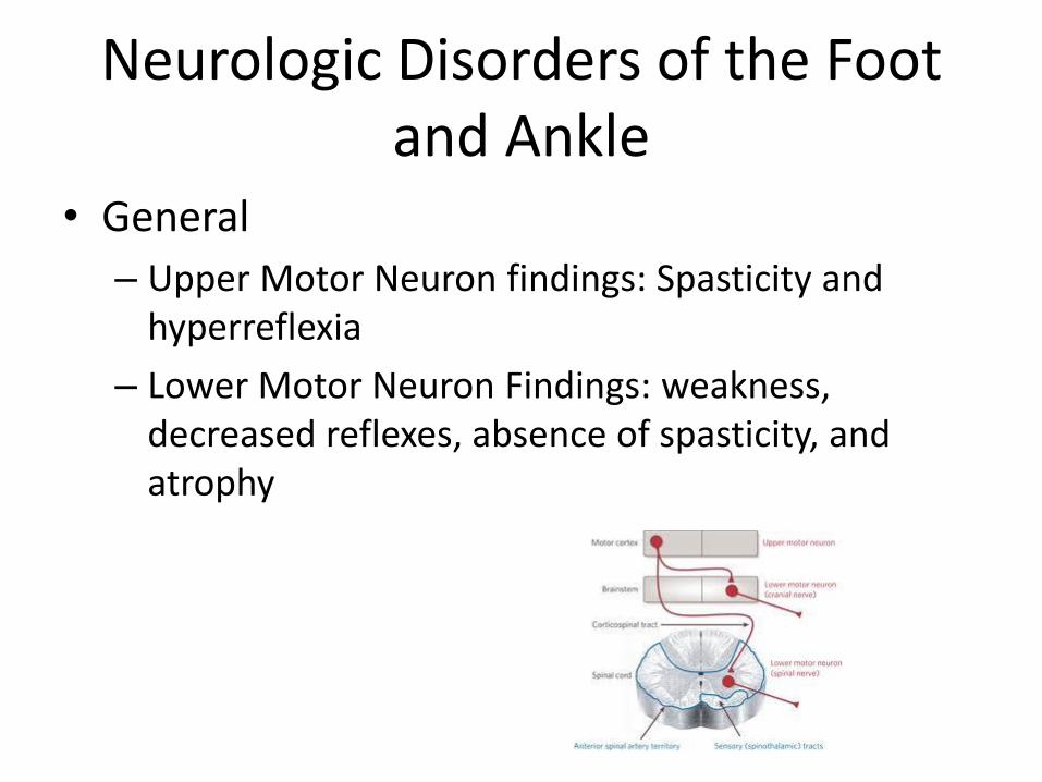
Neurologic Disorders of the Foot and Ankle
• General
– Upper Motor Neuron findings: Spasticity and hyperreflexia
– Lower Motor Neuron Findings: weakness, decreased reflexes, absence of spasticity, and atrophy
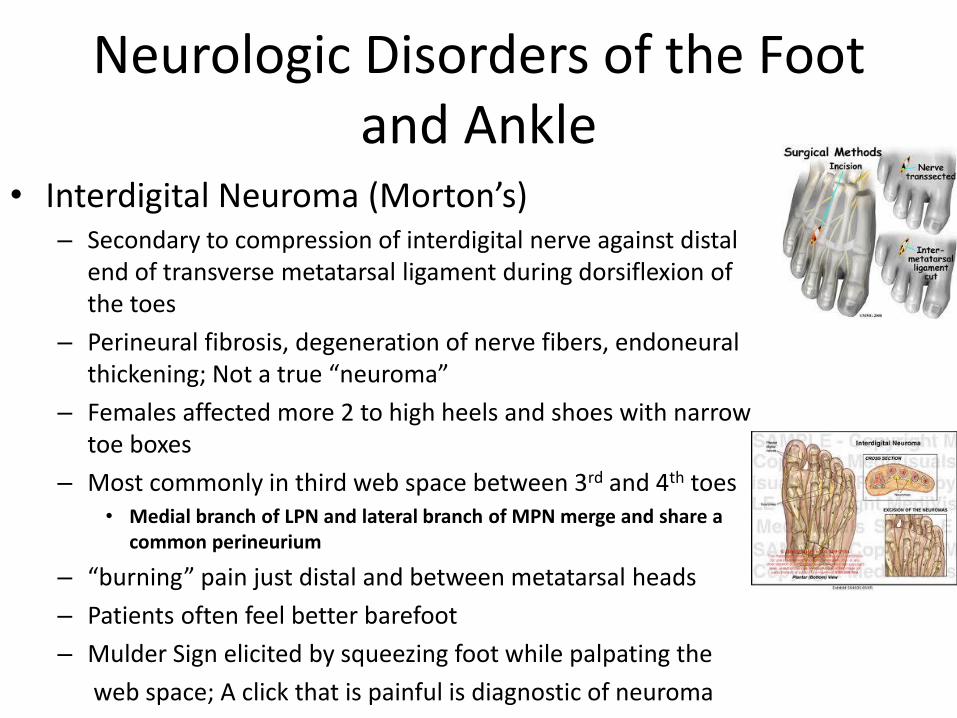
Neurologic Disorders of the Foot and Ankle
• Interdigital Neuroma (Morton’s) – Secondary to compression of interdigital nerve against distal
end of transverse metatarsal ligament during dorsiflexion of the toes
– Perineural fibrosis, degeneration of nerve fibers, endoneural thickening; Not a true “neuroma”
– Females affected more 2 to high heels and shoes with narrow toe boxes
– Most commonly in third web space between 3rd and 4th toes • Medial branch of LPN and lateral branch of MPN merge and share a
common perineurium
– “burning” pain just distal and between metatarsal heads
– Patients often feel better barefoot
– Mulder Sign elicited by squeezing foot while palpating the
web space; A click that is painful is diagnostic of neuroma
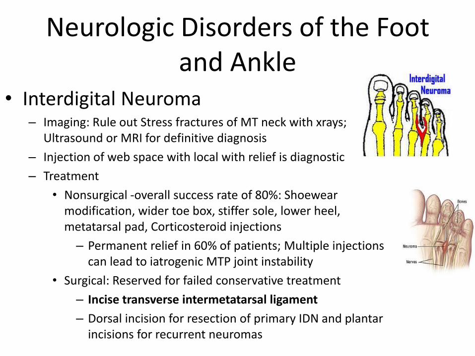
Neurologic Disorders of the Foot and Ankle
• Interdigital Neuroma – Imaging: Rule out Stress fractures of MT neck with xrays;
Ultrasound or MRI for definitive diagnosis
– Injection of web space with local with relief is diagnostic
– Treatment
• Nonsurgical -overall success rate of 80%: Shoewear modification, wider toe box, stiffer sole, lower heel, metatarsal pad, Corticosteroid injections
– Permanent relief in 60% of patients; Multiple injections can lead to iatrogenic MTP joint instability
• Surgical: Reserved for failed conservative treatment
– Incise transverse intermetatarsal ligament
– Dorsal incision for resection of primary IDN and plantar incisions for recurrent neuromas
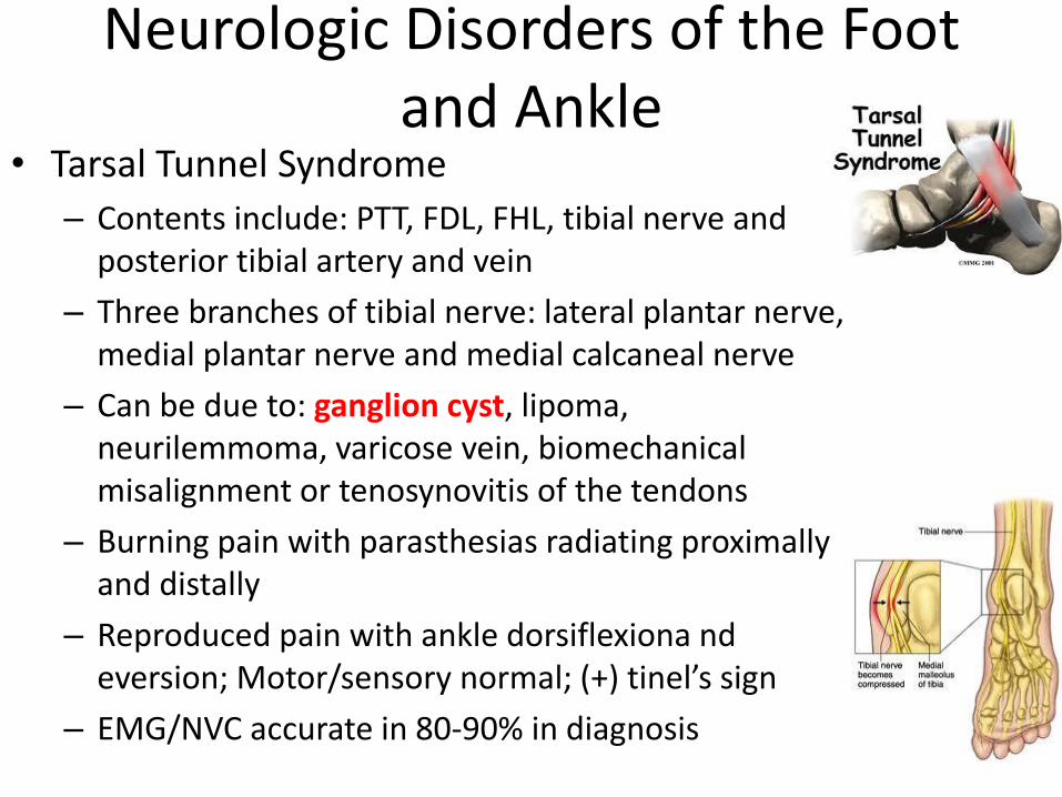
Neurologic Disorders of the Foot and Ankle
• Tarsal Tunnel Syndrome
– Contents include: PTT, FDL, FHL, tibial nerve and posterior tibial artery and vein
– Three branches of tibial nerve: lateral plantar nerve, medial plantar nerve and medial calcaneal nerve
– Can be due to: ganglion cyst, lipoma, neurilemmoma, varicose vein, biomechanical misalignment or tenosynovitis of the tendons
– Burning pain with parasthesias radiating proximally and distally
– Reproduced pain with ankle dorsiflexiona nd eversion; Motor/sensory normal; (+) tinel’s sign
– EMG/NVC accurate in 80-90% in diagnosis
Neurologic Disorders of the Foot and Ankle
• Tarsal Tunnel Syndrome
– Treatment
• Nonsurgical: NSAIDs, cox-2 inhibitors, corticosteroid injections, immobilization in boot or cast after injection to prevent iatrogenic PTT rupture
• Surgical: Decompression of the tibial nerve proximal and distal to tarsal tunnel (medial plantar, lateral plantar, and medial calcaneal nerves); Includes release of the flexor retinaculum;
– Distal tarsal tunnel release for medial plantar heel pain (release of 1st branch of lateral plantar nerve)—nerve to abductor digiti quinti; decompressed by releasing the deep fascia of the abductor hallicus
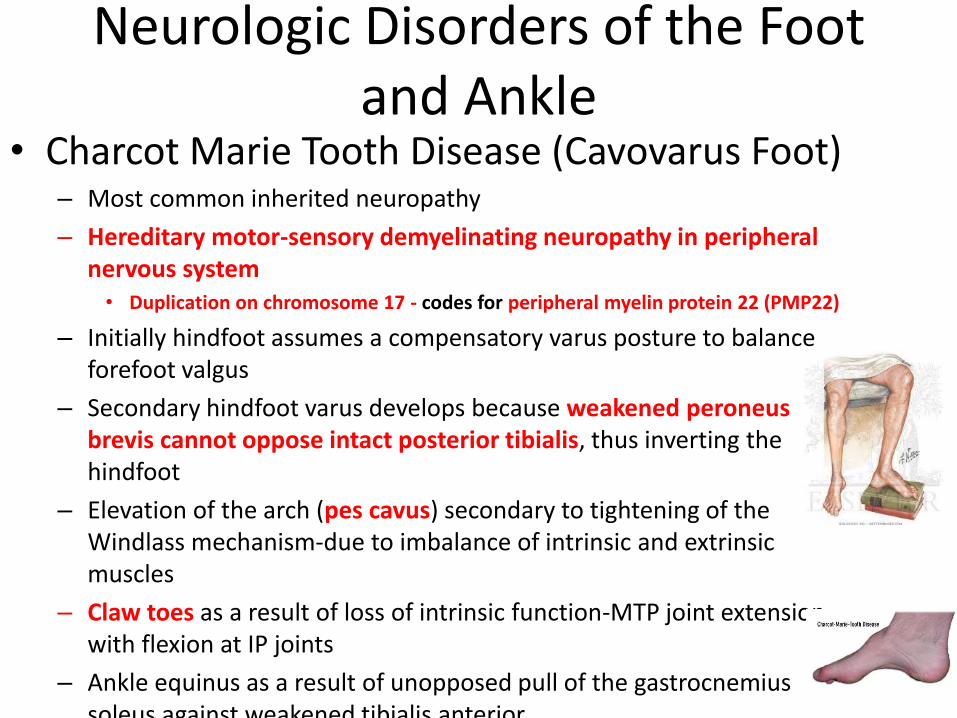
Neurologic Disorders of the Foot and Ankle
• Charcot Marie Tooth Disease (Cavovarus Foot) – Most common inherited neuropathy
– Hereditary motor-sensory demyelinating neuropathy in peripheral nervous system
• Duplication on chromosome 17 - codes for peripheral myelin protein 22 (PMP22)
– Initially hindfoot assumes a compensatory varus posture to balance forefoot valgus
– Secondary hindfoot varus develops because weakened peroneus brevis cannot oppose intact posterior tibialis, thus inverting the hindfoot
– Elevation of the arch (pes cavus) secondary to tightening of the Windlass mechanism-due to imbalance of intrinsic and extrinsic muscles
– Claw toes as a result of loss of intrinsic function-MTP joint extension with flexion at IP joints
– Ankle equinus as a result of unopposed pull of the gastrocnemius soleus against weakened tibialis anterior
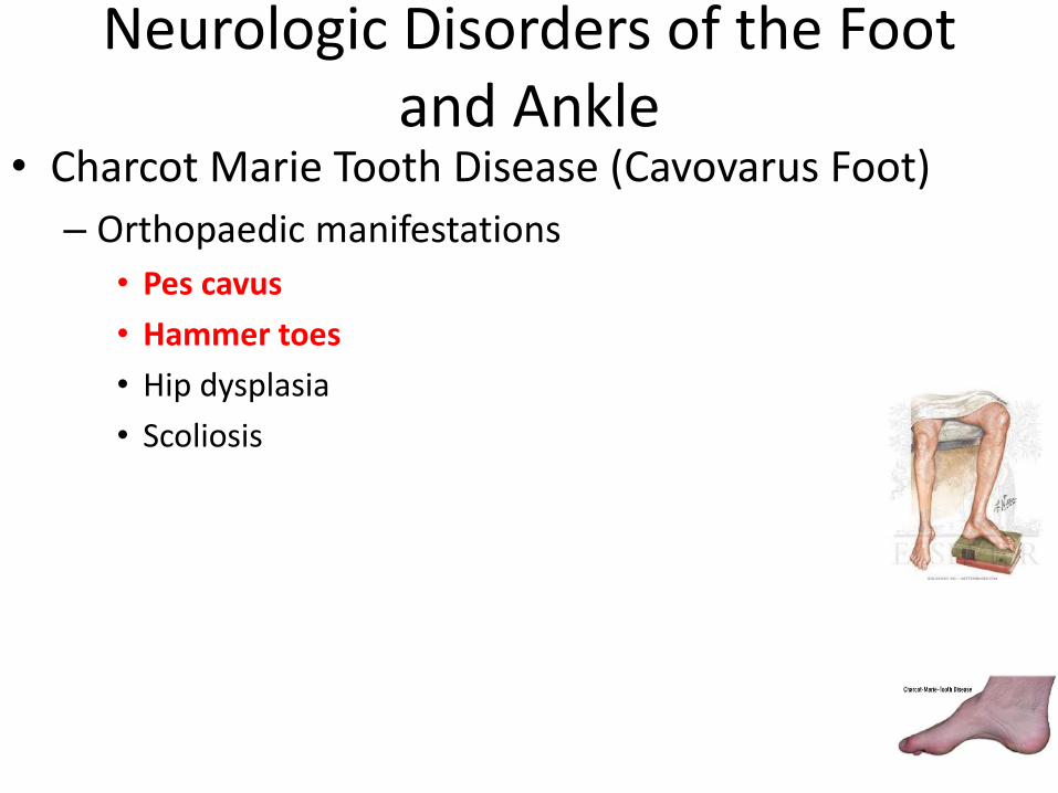
Neurologic Disorders of the Foot and Ankle
• Charcot Marie Tooth Disease (Cavovarus Foot)
– Orthopaedic manifestations
• Pes cavus
• Hammer toes
• Hip dysplasia
• Scoliosis
Neurologic Disorders of the Foot and Ankle
• Charcot Marie Tooth Disease (Cavovarus Foot)
– 94% of patients with claw toes and/or high arch foot
– Intrinsic muscles of foot affected first, followed by involvement of peroneus brevis and tibialis anterior
– Eventually leads to “stork legs” and atrophy of entire leg
– Initial finding is Plantar flexed first ray due to unopposed pull of the peroneus longus (weak TA)
Neurologic Disorders of the Foot and Ankle
• Charcot Marie Tooth Disease (Cavovarus Foot)
– Plantar flexed 1st ray is initial deformity
– Cavus caused by normal peroneus longus overpowering weak tibialis anterior
– Varus caused by normal tibialis posterior overpowering weak peroneus brevis
Neurologic Disorders of the Foot and Ankle
• Charcot Marie Tooth Disease (Cavovarus Foot)
– Hindfoot varus deformity initially is flexible, but with time, it will become fixed
• The Coleman Block test is used to determine whether hindfoot is flexible and whether the deformity is solely result of the plantar flexed first ray
– If hindfoot corrects to neutral or everts, the cavovarus deformity is a result of plantar flexed first ray
» Post lateral forefoot and the lateral heel
– If hindfoot doesn’t correct then both forefoot and hindfoot are involved and need to addressed
Neurologic Disorders of the Foot and Ankle
• Charcot Marie Tooth Disease (Cavovarus Foot)
– Nonsurgical: PT for weakened muscles, accommodative insert, Nonimpact conditioning, stretching, AFO orthoses
– Surgical:
• Delay surgery until progression of deformity
• Soft tissue procedures indicated if deformity is supple:
– Transfer peroneus longus to peroneus brevis
– Transfer PTT to the dorsum of the foot through interosseous membrane
– Transfer of PTT around ankle to cuboid
– Release of plantar fascia to address cavus deformity
– Avoid Achilles tendon lengthening until other procedures done as may lead to hindfoot calcaneus
– Flexor to Extensor transfer (Girdlestone-Taylor) of the lesser toes for flexible claw toes
– The clawed great toe addressed with Jones Procedure with IP joint fusion and transfer of EHL to metatarsal neck
– In skelletally immature patients lengthening of plantar fascia and PTT transfer often sufficient
Neurologic Disorders of the Foot and Ankle
• Surgical (for Charcot Marie Tooth)
– Osseous surgery in addition to soft tissue procedures for rigid deformity
• Patients who overcorrect into slight valgus with Coleman block testing managed with dorsiflexion osteotomy of the 1st MT, plantar fascia release, and peroneus longus to brevis transfer
• Hindfoot varus addressed with Dwyer closing wedge (or lateral displacement) osteotomy of the calcaneus
• For patients with severe rigid deformity, salvage procedure is triple arthrodesis with correction of deformity through bone resections
• After stable plantigrade foot achieved, need for orthotic device (even though PTT transfer restores some degree of DF, there is still a foot drop)
Neurologic Disorders of the Foot and Ankle
• Nerve Entrapment
– Deep Peroneal Nerve (anterior tarsal syndrome)
• Burning pain on dorsum of foot in first dorsal web space
• Worse at night—plantar flexed foot puts strain/stretch on the nerve; Also worse with tight shoes or high heels
• Decreased sensation in 1st dorsal web space, weakness of EDB; (+) Tinel’s
• Treatment: – Nonsurgical: avoid tight shoes/high heels; corticosteroid/local
injections;
– Surgical: Decompression/neurolysis of distal margin of inferior extensor retinaculum to base of 1st and 2nd TMT joints
Neurologic Disorders of the Foot and Ankle
– Superficial Peroneal Nerve Entrapment
• Compression usually causes sensory dysfunction in L5 distribution and pain over anterolateral leg ad ankle pain and dorsal foot pain
• Motor findings usually normal unless compression proximally
• Pain reproduced with plantar flexiona nd inversion; (+) Tinel’s; Delayed Sensory NCV’s
• Treatment – Nonsurgical: corticosteroid/local injections, orthoses, physical
therapy
– Surgical: Decompression (80% with clinical improvement)
– Also, Sural and Saphenous nerve entrapment
Neurologic Disorders of the Foot and Ankle
• CVA and Traumatic Brain Injury – Typical finding in CVA patients is spatic equinovarus deformity of foot and
ankle
– Joint contractures a result of intrinsic joint comonents (capsule), extraarticular soft tissues (shortening of tendons, ligaments, skin), and myogenic shortening
– Treatment
• Nonsurgical: selective nerve/muscle blocks, phenol nerve blocks, botulinim toxin, physiotherapy, bracing (AFO), casting (serial)
• Surgical: tendon lengthening, joint contracture release; Split anterior tibial tendon transfer (continuously active ATT) along with TAL and possible PTT tendon transfer (Muscle strength should be 4 or better to work); osteotomies and fusions for recurrent cases;
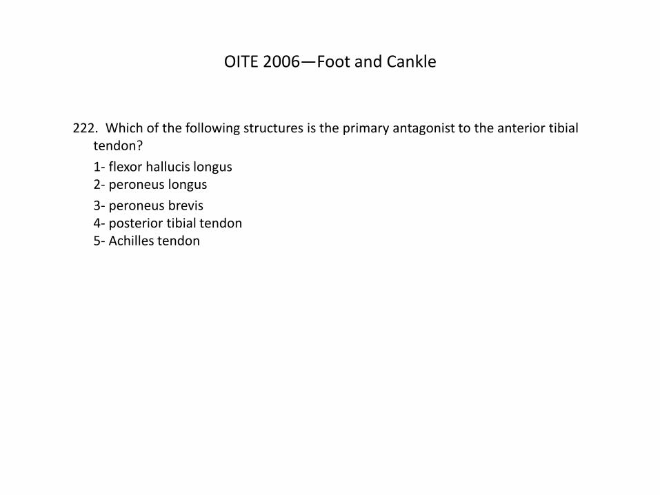
OITE 2006—Foot and Cankle
222. Which of the following structures is the primary antagonist to the anterior tibial tendon?
1- flexor hallucis longus 2- peroneus longus
3- peroneus brevis 4- posterior tibial tendon 5- Achilles tendon
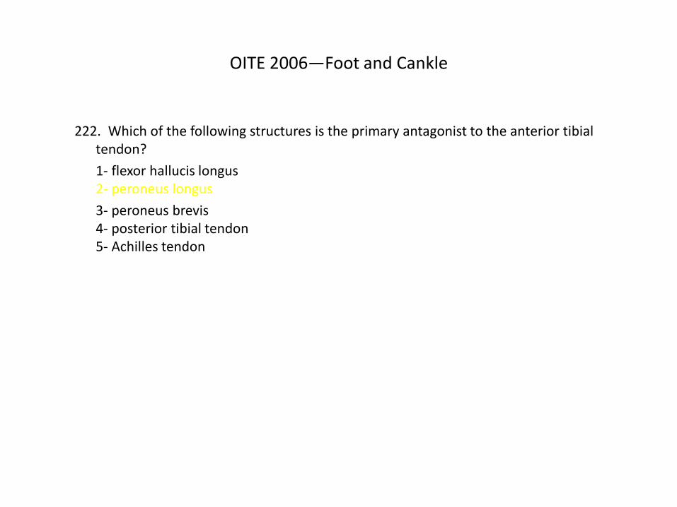
OITE 2006—Foot and Cankle
222. Which of the following structures is the primary antagonist to the anterior tibial tendon?
1- flexor hallucis longus 2- peroneus longus
3- peroneus brevis 4- posterior tibial tendon 5- Achilles tendon
OITE 2006—Foot and Cankle
• Tibialis anterior dorsiflexes and inverts the foot. The opposite action is plantar flexion and eversion which is done by peroneus longus.
OITE 2006—Foot and Ankle
83. Examination of a 24 year old man with hereditary motor sensory neuropathy reveals a cavovarus hindfoot correctable with Coleman block testing. Treatment should consist of
1. Plantar fascial release, dorsal closing wedge osteotomy of the first metatarsal, and peroneus longus to peroneus brevis tendon transfer
2. Split anterior tendon transfer
3. First metatarsophalangeal arthrodesis and Achilles tendon lengthening
4. Dwyer calcaneal osteotomy and posterior tibial tendon transfer
5. Triple arthodesis

Charcot Marie Tooth Disease
– CMT is a hereditary motor sensory neuropathy inherited in an AD (most common) or X linked Dominant (rare) form
– Most common inherited neuropathy in US
– Demyelinating disorder with distal affected first
– Anterior compartment, peroneus brevis and foot intrinsics
Deformity
Weak Agonist
Intact Antagonist
Action
Equinas
Tibialis Anterior
Gastroc-Soleus and
peroneus longus
Pulls foot plantar
Hindfoot varus
Peroneus Brevis
Tibialis posterior
Inverts subtalar joint
Forefoot valgus
Tibialis anterior
Peroneus longus
Pulls first ray plantar
Pes cavus
Foot intrisics
Foot extriniscs
Raises the longitudinal
arch
Toe deformities
Foot intrinsics
Foot extrinsics esp. if pt
uses EHL/EDL for ankle
dorsiflexion
Claw toes, MTP
hyperextension
Charcot Marie Tooth Disease
• Treatment begins with extradepth shoes and MT head support with an
accomodative orthotic
•If the patient has a plantar flexed first ray and a compensitory
flexible hindfoot varus which corrects on lateral block test, the
patient would benefit from an orthotic which posts the lateral
forefoot to correct the inverting effect of the forefoot valgus on the
hindfoot.
•This patient has a correctable hindfoot deformity with Colman block which means hindfoot deformity should correct with forefoot correction
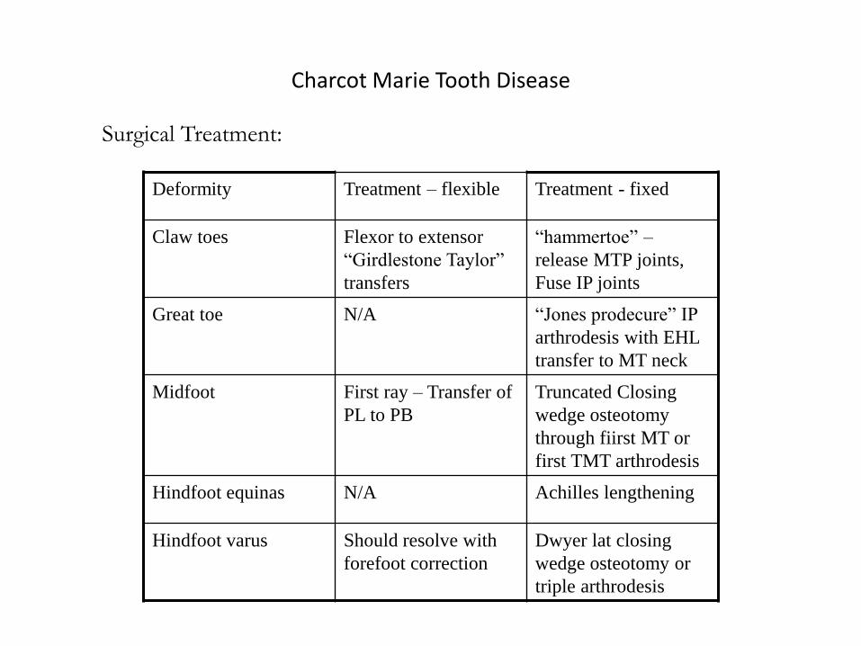
Charcot Marie Tooth Disease
Deformity Treatment – flexible Treatment - fixed
Claw toes Flexor to extensor
“Girdlestone Taylor”
transfers
“hammertoe” –
release MTP joints,
Fuse IP joints
Great toe N/A “Jones prodecure” IP
arthrodesis with EHL
transfer to MT neck
Midfoot First ray – Transfer of
PL to PB
Truncated Closing
wedge osteotomy
through fiirst MT or
first TMT arthrodesis
Hindfoot equinas N/A Achilles lengthening
Hindfoot varus Should resolve with
forefoot correction
Dwyer lat closing
wedge osteotomy or
triple arthrodesis
Surgical Treatment:
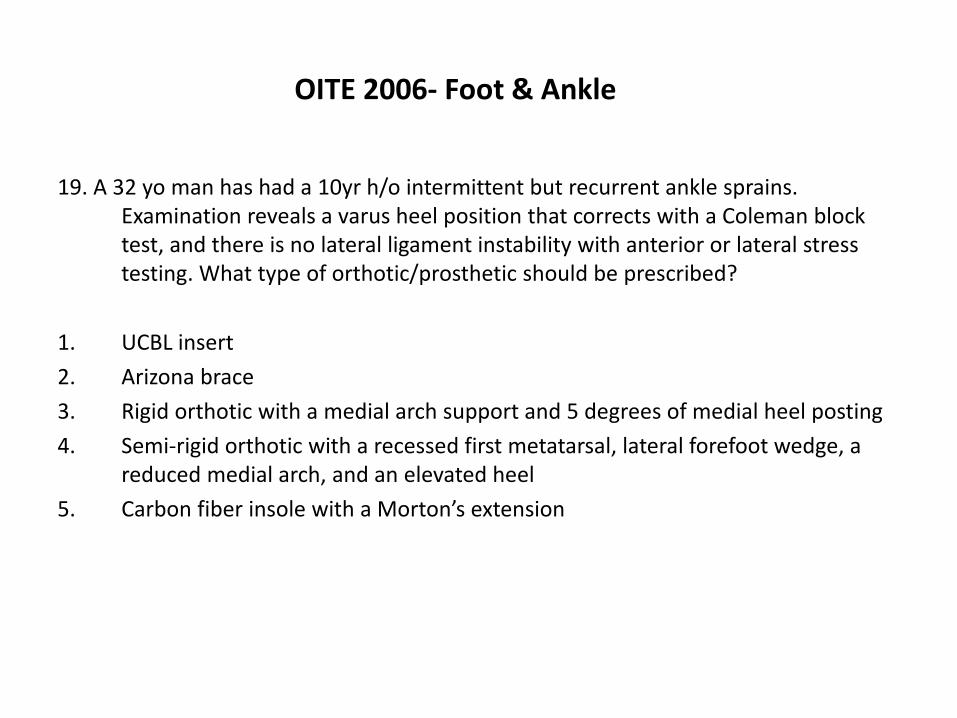
OITE 2006- Foot & Ankle
19. A 32 yo man has had a 10yr h/o intermittent but recurrent ankle sprains. Examination reveals a varus heel position that corrects with a Coleman block test, and there is no lateral ligament instability with anterior or lateral stress testing. What type of orthotic/prosthetic should be prescribed?
1. UCBL insert
2. Arizona brace
3. Rigid orthotic with a medial arch support and 5 degrees of medial heel posting
4. Semi-rigid orthotic with a recessed first metatarsal, lateral forefoot wedge, a reduced medial arch, and an elevated heel
5. Carbon fiber insole with a Morton’s extension
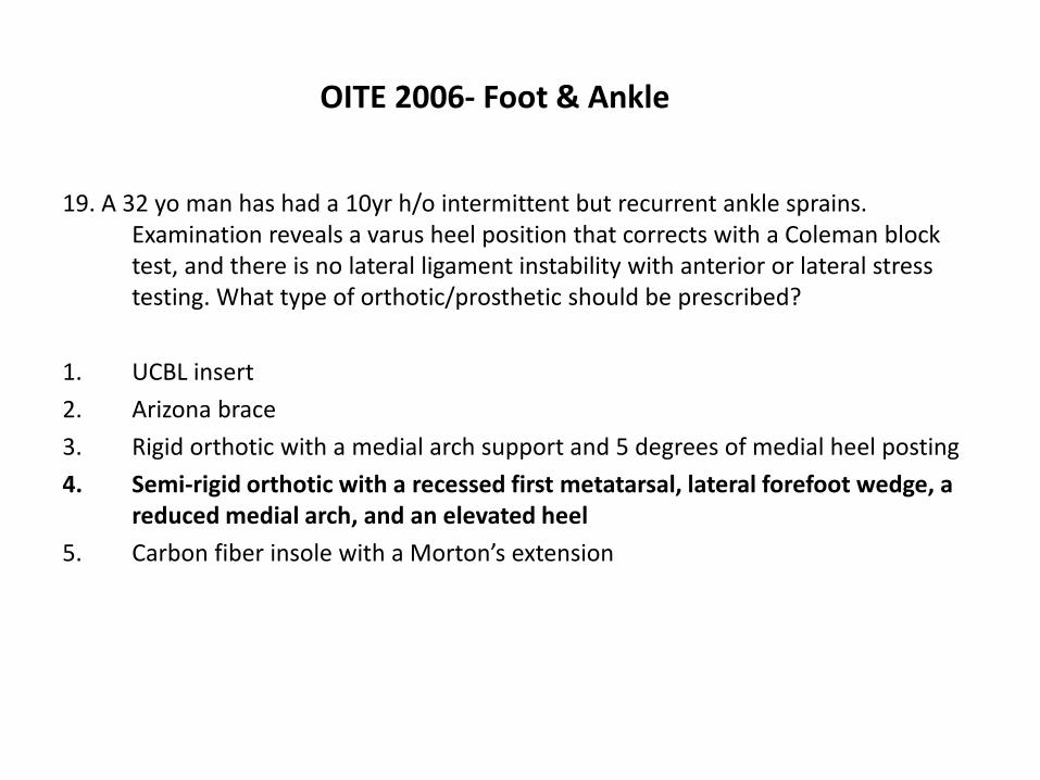
Discussion
• This pt has a cavus foot which is one with an abnormally high arch. With this comes hyperextension of the toes at the MTPJs and hyperflexion at the IPJs; pronation and adduction of the forefoot; a lengthened lateral border of the foot and shortened medial border; calluses beneath the MT heads.
• The cavus deformity has an either fixed or flexible varus deformity of the heel and tightness of the tendo-calcaneus with or without an equinus deformity.
• Most common causes of cavus foot are neuromuscular (CMT and poliomyelitis most common) and trauma (i.e. deep posterior compartment syndrome after fracture of the tibia or by malunion of a midfoot fx. There are those cases in which there is no identifiable cause.
• The Coleman block test determines the hindfoot-forefoot relationship in pes cavus by determining if the hindfoot component is flexible or rigid. This is based on the idea that the hindfoot deformity follows forefoot equinus. The rigid plantar flexed first ray forces the heel into varus and after time, this becomes a fixed deformity. With the Coleman block test, the plantarflexed 1st metatarsal is allowed to hang free off the edge of the block. If the deformity is flexible, the hindfoot will correct and fall out of varus.
Continued Discussion
• In this question, this patient has a flexible cavus deformity and the fact that he has no lateral ligament instability with lateral and anterior stress testing directs us away from ankle instability as the cause of his problems. Therefore, it is the cavus deformity that must be addressed.
• If this required operative tx, which other OITEs will ask, since it is a flexible deformity, he would require a plantar fascia release, elevation of the 1st MT by a basilar closing wedge osteotomy, transfer of the peroneus brevis at the ankle jt and correction of the clawed toes. The goal is to reduce the calcaneal pitch.
• However, the answers offer no surgical choices. The only choice that addresses all the deformities of the felxible cavus foot is choice 4. The reference for this question is pretty weak and unhelpful. Therefore, when answering this question, think of what would be done surgically, then consider how a brace would accomplish those goals.
Foot and Ankle Trauma
• Some Additional Questions…
• Should be covered more extensively in Trauma Review Series
– Ankle and Pilon Fractures
– Talus Fractures
– Calcaneus Fractures
– Lisfranc and Midfoot Injuries
– Fifth Metatarsal Fractures
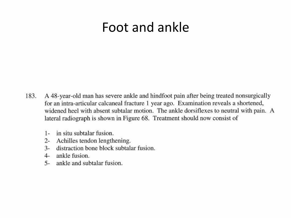
Foot and ankle
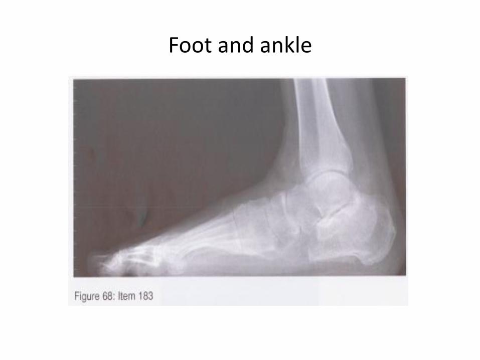
Foot and ankle
Foot and ankle
Answer 3
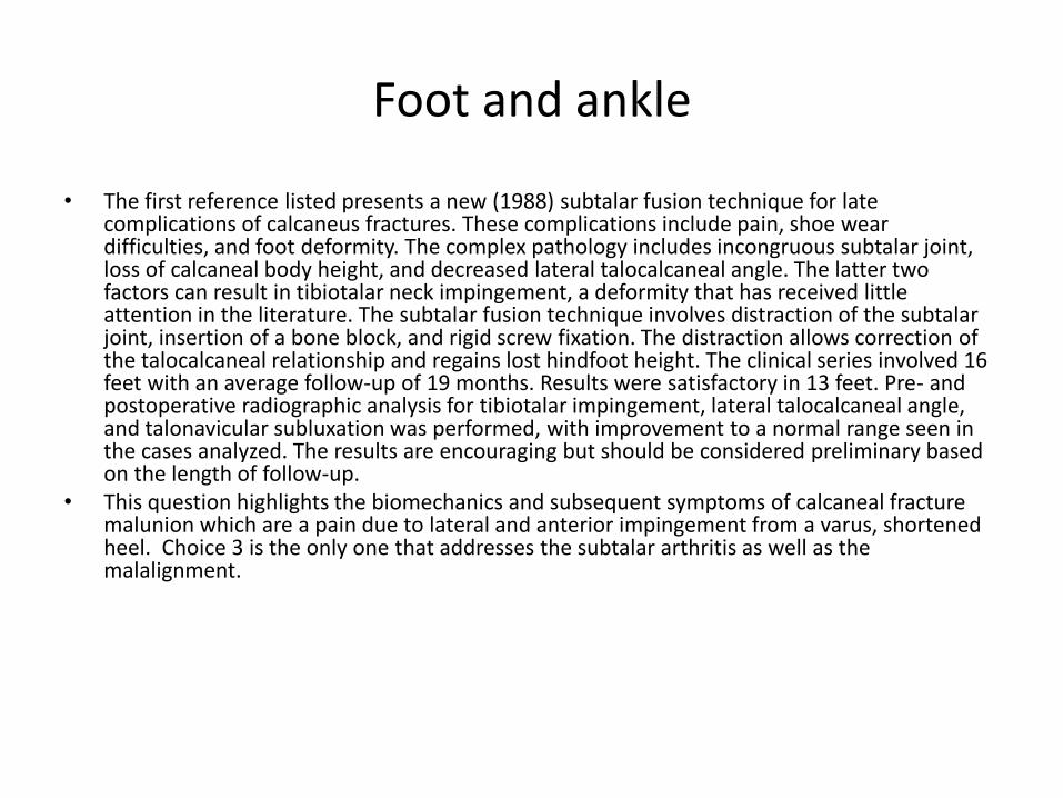
Foot and ankle
• The first reference listed presents a new (1988) subtalar fusion technique for late complications of calcaneus fractures. These complications include pain, shoe wear difficulties, and foot deformity. The complex pathology includes incongruous subtalar joint, loss of calcaneal body height, and decreased lateral talocalcaneal angle. The latter two factors can result in tibiotalar neck impingement, a deformity that has received little attention in the literature. The subtalar fusion technique involves distraction of the subtalar joint, insertion of a bone block, and rigid screw fixation. The distraction allows correction of the talocalcaneal relationship and regains lost hindfoot height. The clinical series involved 16 feet with an average follow-up of 19 months. Results were satisfactory in 13 feet. Pre- and postoperative radiographic analysis for tibiotalar impingement, lateral talocalcaneal angle, and talonavicular subluxation was performed, with improvement to a normal range seen in the cases analyzed. The results are encouraging but should be considered preliminary based on the length of follow-up.
• This question highlights the biomechanics and subsequent symptoms of calcaneal fracture malunion which are a pain due to lateral and anterior impingement from a varus, shortened heel. Choice 3 is the only one that addresses the subtalar arthritis as well as the malalignment.
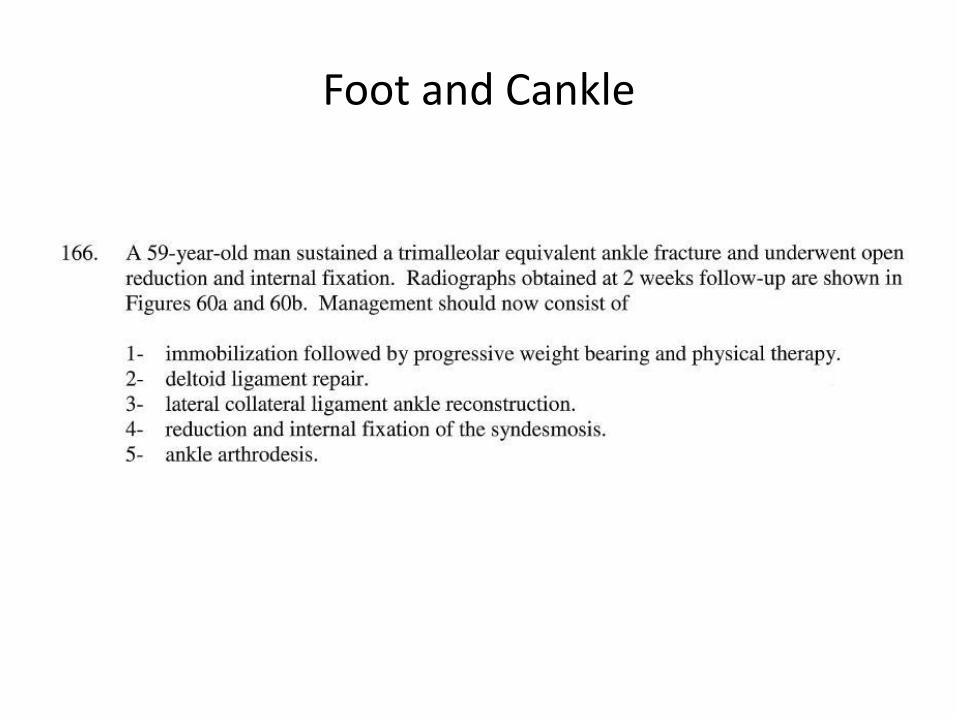
Foot and Cankle
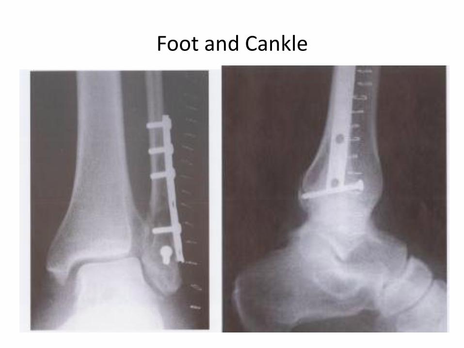
Foot and Cankle
•
Foot and Cankle
Answer 4
Foot and Cankle
• Not really sure what these references add to answering this question. The first concludes that a fibular osteotomy and reconstruction can have good results in patients with ankle pain and fibular malunion. However, this patient’s operative treatment was relatively recent. Perhaps they want you to recognize the radiographic findings of a syndesmotic injury. There is widening of the medial clear space (between talus and tibia) as well as an increase in the tibiofibular clear space (measured 1 cm proximal to the tibiotalar joint line). Some of our attendings will tell us not to get too bogged down in the measurement of these spaces. The best thing to look at is the “Shenton’s line” of the ankle joint, or rather making sure that the joint space on all three sides of the talus is equal on the AP view and that there is a smooth line between the tibial plafond and the medial border of the fibula. Note also the valgus tilt of the talus in the mortise, suggesting that something is not right. Non-operative management is not sufficient for a syndesmotic injury. The deltoid ligament does not require operative repair because it will heal on its own if the ankle is properly reduced and results of operative repair are not good. The lateral collateral ligament is not an issue here. There is no evidence of osteoarthritis suggesting the need for arthrodesis, although if you don’t fix the syndesmosis, there most likely will be down the line.
Foot and Cankle
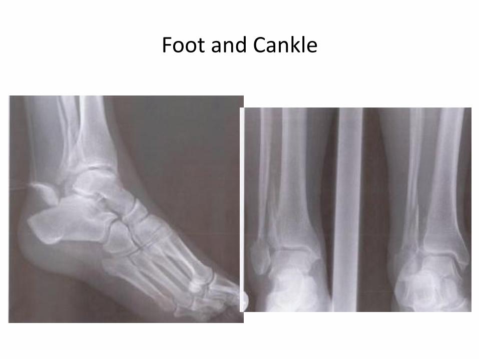
Foot and Cankle
Foot and Cankle
Answer 1
Foot and Cankle
• Not every calcaneal fracture requires a delay in operative fixation. Typically, we use the “wrinkle test” to determine when a calcaneus or pilon fracture has a lesser risk of wound complications after ORIF. However, in this question, the skin on the back of the heel is tented and blanched, suggesting that it will necrose if left alone. One technique to fix this fracture is the Essex-Lopresti technique where a Steinmann or Schanz pin is inserted into the fragment and used as a joysick to aid in reduction. There is an associated pilon fracture in this injury which should not be fixed until the soft tissue swelling has settled down and the skin wrinkles have reappeared. Again, non-operative or delayed management is not an option in this case due to the threatened skin on the back of the heel.
OITE 2006—Foot and Ankle
61. A 17 yo gymnast has worsening midfoot pain and swelling for the past 3 weeks. She denies any trauma but admits to a recent increase in her training regimen. Radiographs are normal. CT and MRI scans are shown in Figures 19a and 19b.
Management should consist of
1. Open reduction and internal fixation with bone grafting
2. A short leg non weightbearing cast
3. Open reduction and internal fixation
4. Percutaneous screw fixation
5. Observation
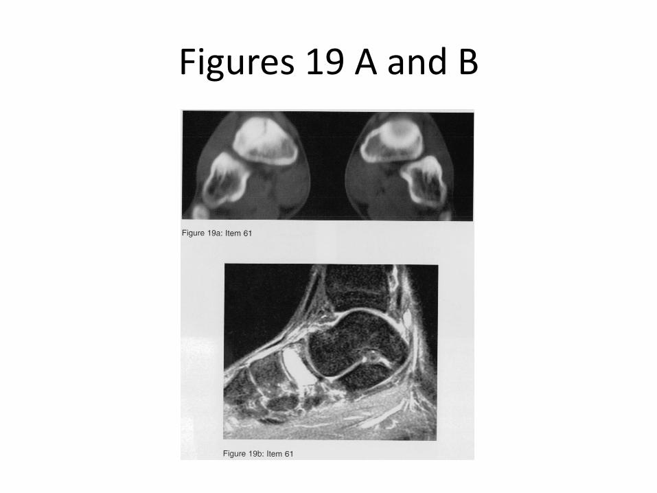
Figures 19 A and B
OITE 2006—Foot and Ankle
61. A 17 yo gymnast has worsening midfoot pain and swelling for the past 3 weeks. She denies any trauma but admits to a recent increase in her training regimen. Radiographs are normal. CT and MRI scans are shown in Figures 19a and 19b.
Management should consist of
1. Open reduction and internal fixation with bone grafting
2. A short leg non weightbearing cast
3. Open reduction and internal fixation
4. Percutaneous screw fixation
5. Observation
Stress fractures of the Navicular
– Stress fractures
• Most commonly in tibia (49.1%), tarsals (25.3%) and metatarsals (8.8%)
• Bilateral in 16 %
• Incidence is 1 % in all athletes but may be up to 20 % in runners
• Occur from excessive, submaximal loads
• Increase in activity may cause an increase in osetoclast activity (osteoblast activity lags)
• Female athlete triad – anorexia, amenorrhea, osteoporosis
• Symptoms – pain with activity, relieved with rest increasing over days to weeks
• Physical exam – Bone point tenderness
• Imaging
– Plain films normal for 2-3 weeks
– Bone scan – hot on all 3 phases
– MRI
Stress fracture of the Navicular
• First step – address predisposing factors – diet, exercise, etc
•Normal XR, CT/MRI – rest for 1-6 weeks
•Normal or abnormal XR, pos CT/MRI (nondisplaced) – non weight
bearing cast for 6 weeks (exception – tension side femoral neck fx req perc
pinning)
•Normal XR, Pos CT/MRI in high level athlete – may require surgery
• Normal or abnormal XR, positive CT, MRI with DISPLACED fracture –
treat as if acute fx - SURGERY
OITE 2006—Foot and Ankle
74. A 20 year old football player reports aching in his leg following vigorous exercise. He denies any resent trauma but does report an untreated severe ankle sprain at 13 years. Radiographs are shown in figures 23a and 23b. What is the most likely diagnosis?
1. Osteochondroma
2. Parosteal osteosarcoma
3. Bizarre parosteal osteochondromatous proliferation
4. Enchondroma
5. Tibiofibular synostosis
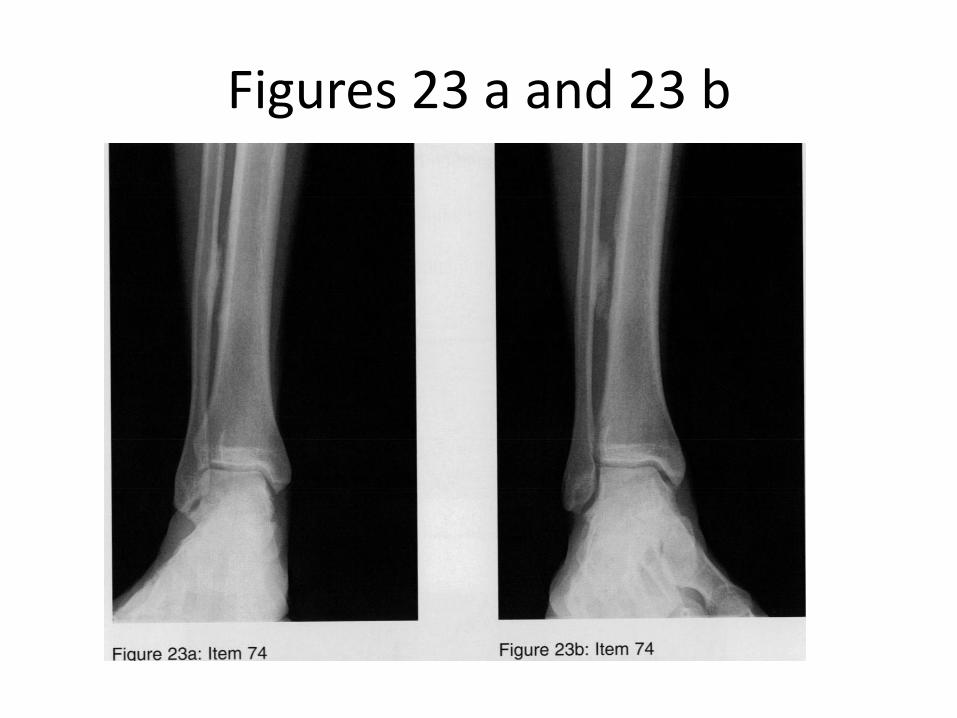
Figures 23 a and 23 b
OITE 2006—Foot and Ankle
74. A 20 year old football player reports aching in his leg following vigorous exercise. He denies any resent trauma but does report an untreated severe ankle sprain at 13 years. Radiographs are shown in figures 23a and 23b. What is the most likely diagnosis?
1. Osteochondroma
2. Parosteal osteosarcoma
3. Bizarre parosteal osteochondromatous proliferation
4. Enchondroma
5. Tibiofibular synostosis
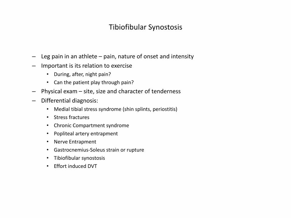
Tibiofibular Synostosis
– Leg pain in an athlete – pain, nature of onset and intensity
– Important is its relation to exercise
• During, after, night pain?
• Can the patient play through pain?
– Physical exam – site, size and character of tenderness
– Differential diagnosis:
• Medial tibial stress syndrome (shin splints, periostitis)
• Stress fractures
• Chronic Compartment syndrome
• Popliteal artery entrapment
• Nerve Entrapment
• Gastrocnemius-Soleus strain or rupture
• Tibiofibular synostosis
• Effort induced DVT
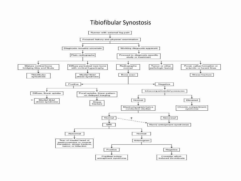
Tibiofibular Synostosis
Tibiofibular Synostosis
• rare cause of leg pain
• occurs after fractures of the tibia or ankle(fibula)
• diagnosis – mature bone bridging from tibia to fibula on XR or CT scans.
• treatment – excision through anterolateral approach
OITE 2006—Foot and Cankle
126. A healthy, active 26-year-old woman sustained a displaced two-piece calcaneal fracture with subluxation of a posterolateral fragment into the subfibular recess. Recommended treatment should consist of 1- open or percutaneous reduction and internal fixation. 2- excision of the posterolateral fragment.
3- non-weight-bearing and early mobilization. 4- casting for 3 months. 5- posterior arthroscopic debridement.
OITE 2006—Foot and Cankle
• The Buckley article listed as a reference is a source of questions each year. It is a prospective, randomized, controlled trial evaluating outcome of operative v. non-operative treatment of displaced intra-articular calcaneal fractures. Their conclusion is that overall, there is no difference in outcome with non-op v. op treatment. The real point of this study lies in the stratification of results. Not much difference is seen in op v non-op treatment until the groups are stratified. Patients who are younger, female, have a light/moderate workload involving the foot, and are not worker’s comp have better results c operative care. This patient fits these criteria. Larger Bohler’s angle and single, simple intra-articular fractures are prognostic of better results. They found it is not possible to salvage a more severely injured foot c a lower Bohler’s angle and ended up doing a subtalar arthrodesis on these regardless of initial treatment. Worker’s comp pts did worse regardless of initial treatment. A comminuted reduction or fracture without a reduction are also predictors of worse outcomes.
OITE 2006—Foot and Cankle
155. Figures 58a and 58b show the radiographs of a 24-year-old professional baseball player who injured his foot sliding into home plate. Which of the following treatments offers the shortest time to union and the lowest risk for the development of a nonunion?
1- open reduction and plate fixation 2- intramedullary screw fixation
3- external fixation 4- short leg weight-bearing cast 5- short leg non-weight-bearing cast
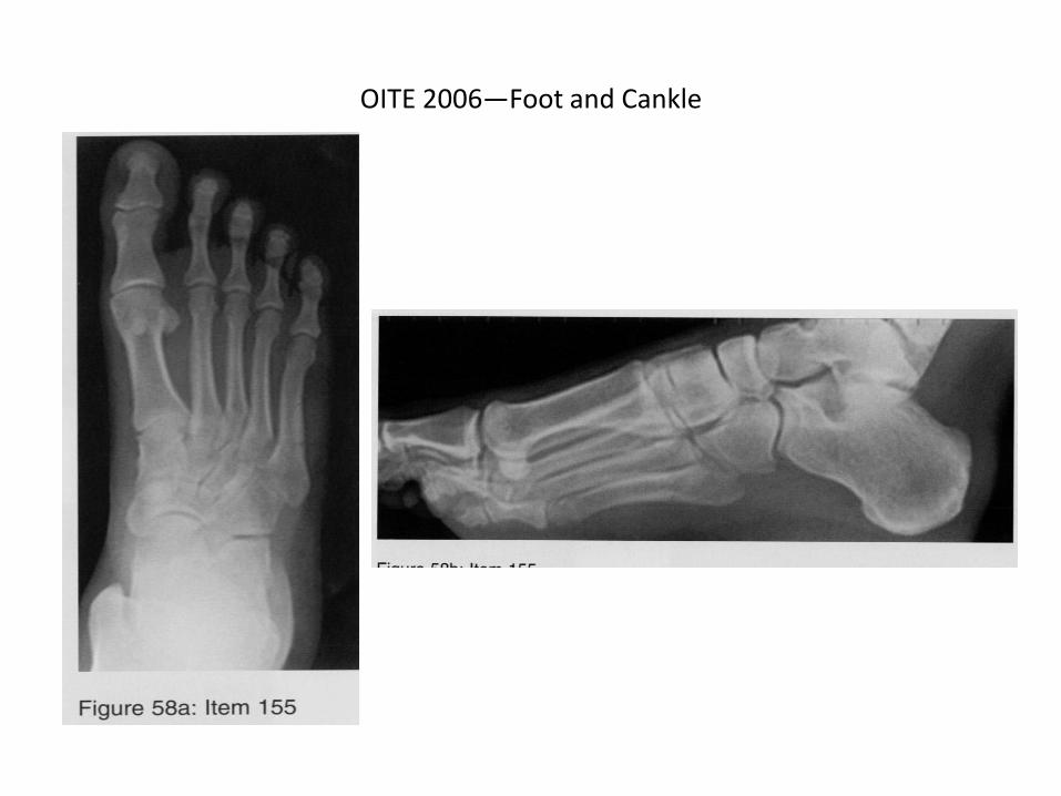
OITE 2006—Foot and Cankle
OITE 2006—Foot and Cankle
155. Figures 58a and 58b show the radiographs of a 24-year-old professional baseball player who injured his foot sliding into home plate. Which of the following treatments offers the shortest time to union and the lowest risk for the development of a nonunion?
1- open reduction and plate fixation 2- intramedullary screw fixation
3- external fixation 4- short leg weight-bearing cast 5- short leg non-weight-bearing cast
OITE 2006—Foot and Cankle
• The guide to choosing an answer to this question lies in the phrases “shortest time to union” and “lowest risk to development of a non-union.” Also, the patient is a professional athlete. There are three patterns of proximal 5th metatarsal fractures-tuberosity avulsion fractures, acute Jones fractures, and proximal diaphyseal stress fractures. Avulsion fractues are thought to be caused by the lateral band of the plantar aponeurosis. They are treated symptomatically in a hard-soled shoe or walking cast. Jones fractures can be confusing as the eponym had been often misused. They are acute transverse fractures of the proximal fifth metatarsal which extend into the articular facet of the 4th and 5th MTs but not distal to this. They are a result of a adduction force with the foot in plantar flexion. Due to confusion of diaphyseal shaft fractures and Jones fractures, the latter have gotten a bad rap for non-union. In actual fact, they heal well treated NWB in a SLC for 6-8 weeks. Time to union and supposedly risk of non-union can be decreased with operative fixation, the means of choice being an IM screw. Because of this, some recommend operative fixation of Jones fractures in athletes, although there have been no prospective studies comparing operative v. non-op treatment.
OITE 2006 - Foot/Ankle
272. A 30-year-old man sustained a calcaneal fracture 4 years ago that was treated nonsurgically. Posttraumatic arthritis has now developed. He elects to be treated with a subtalar bone block distraction arthrodesis. This procedure attempts to correct all of the following abnormalities commonly seen after calcaneal fractures EXCEPT
1. anterior ankle impingement.
2. subfibular peroneal impingement.
3. hindfoot valgus.
4. limb shortening.
5. subtalar arthritis.
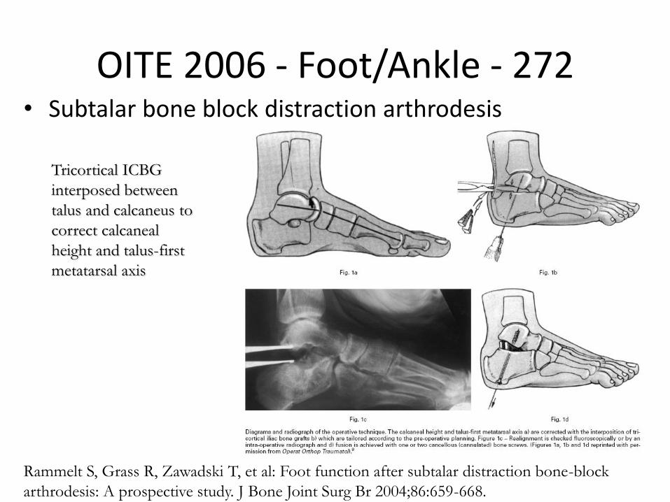
OITE 2006 - Foot/Ankle - 272 • Subtalar bone block distraction arthrodesis
Tricortical ICBG
interposed between
talus and calcaneus to
correct calcaneal
height and talus-first
metatarsal axis
Rammelt S, Grass R, Zawadski T, et al: Foot function after subtalar distraction bone-block
arthrodesis: A prospective study. J Bone Joint Surg Br 2004;86:659-668.
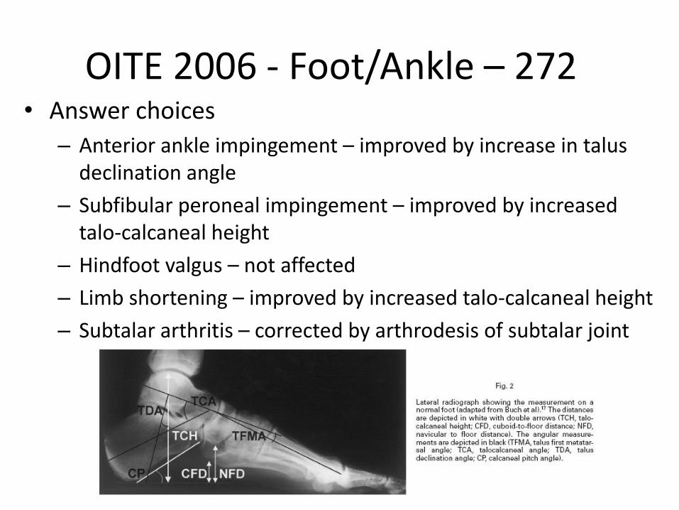
OITE 2006 - Foot/Ankle – 272 • Answer choices
– Anterior ankle impingement – improved by increase in talus declination angle
– Subfibular peroneal impingement – improved by increased talo-calcaneal height
– Hindfoot valgus – not affected
– Limb shortening – improved by increased talo-calcaneal height
– Subtalar arthritis – corrected by arthrodesis of subtalar joint