Embed Size (px)
Citation preview
OESOPHAGEAL CANCER
College voor Oncologie Nationale Richtlijnen
V1.2008 © 2008 College of Oncology
College voor Oncologie Nationale Richtlijnen
College voor Oncologie Nationale Richtlijnen
College voor Oncologie Nationale Richtlijnen
OOe ll
CCOOLLLLEEGGEE OOFF OONNCCOOLLOOGGYY
NNaattiioonnaall CClliinniiccaall PPrraaccttiiccee GGuuiiddeelliinneess
RReeccttuumm CCaanncceerr
VVeerrssiioonn 11..22000044
essoopphhaaggeeaaCCaanncceerr
VVeerrssiioonn 11..22000088
Continue
OESOPHAGEAL CANCER
College voor Oncologie Nationale Richtlijnen
V1.2008 © 2008 College of Oncology
College voor Oncologie Nationale Richtlijnen
College voor Oncologie Nationale Richtlijnen
College voor Oncologie Nationale Richtlijnen College of Oncology National Guidelines
Expert panel
Oesophageal Cancer Guidelines Expert Panel Prof. dr. Marc Peeters Coordinator National Guidelines Oesophageal Cancer University Hospital Ghent
Prof. dr. Tom Boterberg University Hospital Ghent
Prof. Dr. Johan De Mey Universitair Ziekenhuis Brussel
Prof. dr. Pierre Deprez Clinques Universitaires Saint-Luc
Prof. dr. Nadine Ectors University Hospital Leuven
Prof. dr. Patrick Flamen Jules Bordet Institute Brussels
Prof. dr. Antoon Lerut University Hospital Leuven
Prof. dr. B. Neyns Universitair Ziekenhuis Brussel
Prof. dr. Piet Pattyn University Hospital Ghent
Dr. Joan Vlayen Belgian Health Care Knowledge Centre
Dr. Francine Mambourg Belgian Health Care Knowledge Centre
Prof. dr. Jean-Luc Van Laethem ULB Hôpital Erasme Bruxelles
Dr. Margareta Haelterman Federal Public Service Health, Food Chain Safety and Environment
Prof. dr. Jacques De Grève Chairman Working Party Manuals College of Oncology Universitair Ziekenhuis Brussel
Prof. dr. Simon Van Belle Chairman College of Oncology University Hospital Ghent
This report was supported by the Belgian Healthcare Knowledge Centre. The full scientific report can be consulted at the KCE website (www.kce.fgov.be). Reference: Peeters M, Lerut T, Vlayen J, Mambourg F, Ectors N, Deprez P, et al. Wetenschappelijke ondersteuning van het College voor Oncologie: een nationale praktijkrichtlijn voor de aanpak van slokdarm- en maagkanker. Good Clinical Practice (GCP). Brussel: Federaal Kenniscentrum voor de Gezonheidszorg (KCE); 2008. KCE reports 75A (D2008/10.273/16). or Reference: Peeters M, Lerut T, Vlayen J, Mambourg F, Ectors N, Deprez P, et al. Guidelines pour la prise en charge du cancer oesophagien et gastrique: elements scientifiques à destination du Collège d'Oncologie. Bruxelles: Centre fédéral d'expertise des soins de santé (KCE); 2008. KCE reports 75B (D2008/10.273/17).

OESOPHAGEAL CANCER
College voor Oncologie Nationale Richtlijnen
V1.2008 © 2008 College of Oncology
College voor Oncologie Nationale Richtlijnen
College voor Oncologie Nationale Richtlijnen
College voor Oncologie Nationale Richtlijnen
College of Oncology National Guidelines
External reviewers
External reviewers Dr. Didier Verhoeven Dr. Max Mano Belgian Society of Medical Oncology
Dr. Roland Hustinx Belgisch Genootschap voor Nucleaire Geneeskunde / Société belge de Médicine nucléaire
Dr Wim Ceelen Dr Jean-Marie Collard Belgian Society of Surgical Oncology
Dr. Joseph Weerts Dr. Paul Cheyns Koninklijk Belgisch Genootschap Heelkunde / Société Royale belge de Chirurgie
Dr. Jochen Decaestecker Dr. Eric Van Cutsem
Vlaamse Vereniging voor Gastro-enterologie
Dr. Cathy Mahin Association Belge de Radiothérapie-Oncologie / Belgische Vereniging voor Radiotherapie–Oncologie
Dr. Alain Hendlisz Belgian Group of Digestive Oncology
Dr. Hubert Piessevaux Societé Royale Belge de Gastro-enterologie
Dr. Louis Ferrant Dr. Bart Van den Eynden Domus Medica
Dr. Daniel Urbain Dr. Michel Buset The Belgian Society of Gastrointestinal Endoscopy
Dr. Anne Jouret-Mourin Dr. Pieter Demetter Belgian Digestive Pathology Club
External validators
Dr. Harry Bleiberg Jules Bordet Institute Brussels
Dr. Marc De Man Onze Lieve Vrouw Ziekenhuis Aalst
Dr. Hugo W. Tilanus Erasmus MC Rotterdam

OESOPHAGEAL CANCER College voor Oncologie Nationale Richtlijnen
College voor Oncologie Nationale Richtlijnen
College voor Oncologie Nationale Richtlijnen
College voor Oncologie Nationale Richtlijnen
College of Oncology National Guidelines
Table of contents
• Oesophageal cancer guidelines expert panel
• nd external validators External reviewers a
• General algorithm
• National guidelines breast cancer (Full text)
ence gy
• Ddefinitions
lesions
of dysplastic lesions
• Treatment of mucosal cancer
• Table 4: TNM stage grouping
• Introduction • Search for evid• Epidemiolo
efinitions Topographic Early
• Diagnosis • Work-up• Staging
• Treatment of cancer beyond the mucosa Neoadjuvant treatment Surgical treatment Adjuvant treatment Non-surgical treatment with curative intent
• Palliative treatment and metastatic disease • Follow-up • Recurrent disease
• References
• Table 1: Sources
• Table 2: Grade system
• Table 3: TNM classification
V1.2008 © 2008 College of Oncology
OESOPHAGEAL CANCER College voor Oncologie Nationale Richtlijnen
College voor Oncologie Nationale Richtlijnen
College voor Oncologie Nationale Richtlijnen
College voor Oncologie Nationale Richtlijnen
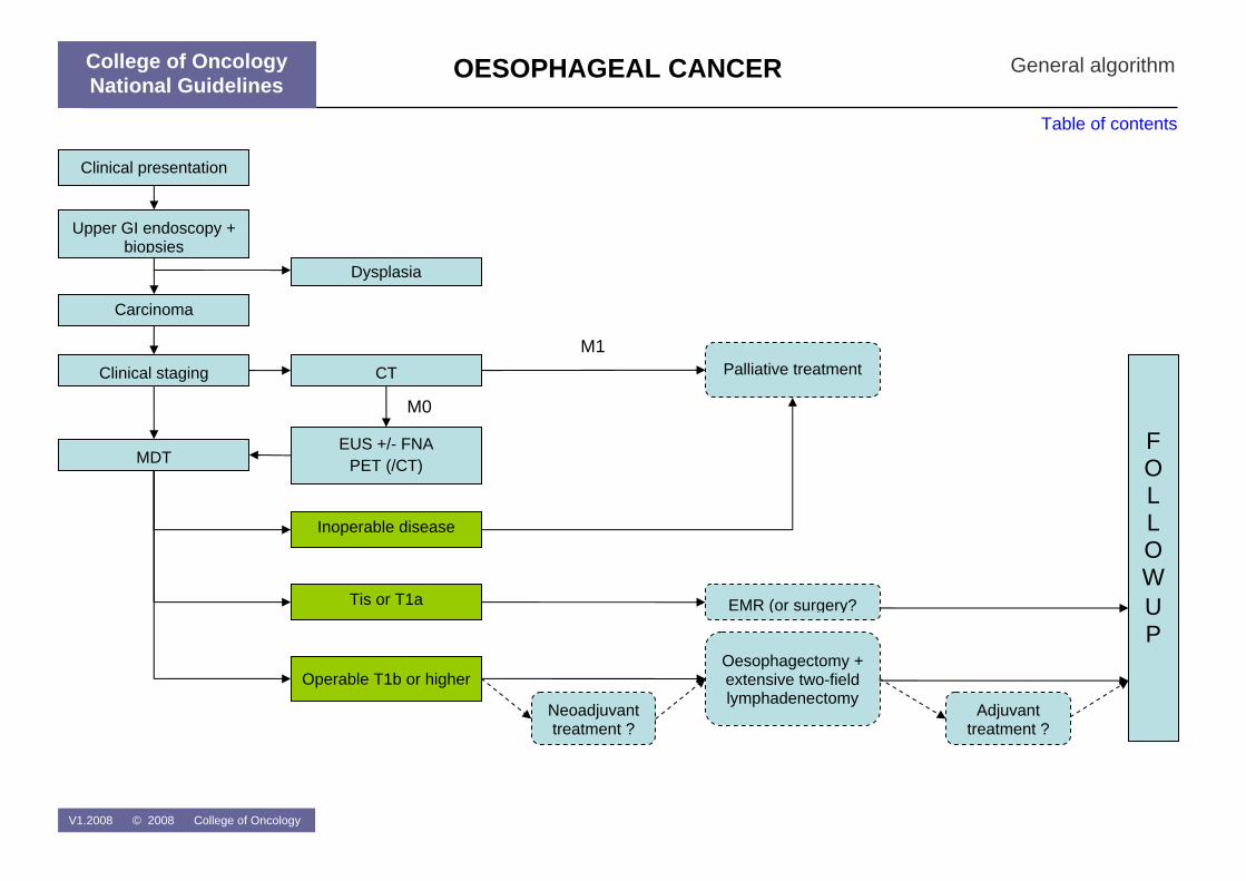
Carcinoma
Tis or T1a
Inoperable disease
Operable T1b or higher
F O L L O W U P
EUS +/- FNA PET (/CT)
Dysplasia
Clinical staging
MDT
Upper GI endoscopy + biopsies
Clinical presentation
EMR (or surgery?
Oesophagectomy + extensive two-field lymphadenectomy
Adjuvant treatment ?
Neoadjuvant treatment ?
Palliative treatment CTM1
M0
General algorithm
Table of contents
College of Oncology National Guidelines
V1.2008 © 2008 College of Oncology
OESOPHAGEAL CANCER College voor Oncologie
Nationale Richtlijnen College voor Oncologie
Nationale Richtlijnen College voor Oncologie
Nationale Richtlijnen College voor Oncologie
Nationale Richtlijnen
National Guidelines Oesophageal Cancer
INTRODUCTION This document provides an overview of the clinical practice guidelines for oesophageal cancer. For more in-depth information and the scientific background, we would like to ask the readers to consult the full scientific report at www.kce.fgov.be. The guidelines are developed by a panel of experts (see 'expert panel') comprising clinicians of different specialties and were reviewed by relevant professional associations (see 'external reviewers') The guidelines are based on the best evidence available at the time they are derived (date restriction 2001-2007). The aim of these guidelines is to assist all care providers involved in the care of patients with oesophageal ancer. c
SEARCH FOR EVIDENCE Clinical practice guidelines
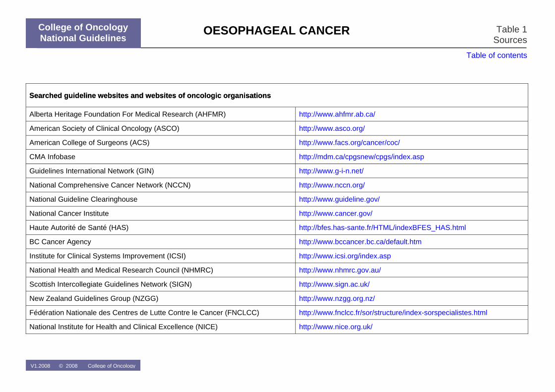
Sources A broad search of electronic databases (Medline, EMBASE), specific guideline websites and websites of oncologic organisations (Table 1) was conducted in July 2007.
In- and exclusion criteria Both national and international clinical practice guidelines (CPGs) on oesophageal cancer were searched. A language (English, Dutch, French)
nd date restriction (2001 – 2007) were used. CPGs without referencea s s without clear recommendations.
Reviews from the search date of the CPG on (search date
tion was assigned to each recommendation using he GRADE system (Table 2).
Table of contents
Full Text
were excluded, as were CPG
Additional evidence For each clinical question, the evidence – identified through the included CPGs – was updated by searching Medline and the Cochrane Database
f SystematicoAugust-September 2007).
Grade of recommendation A grade of recommendat
EPIDEMIOLOGY Oesophageal cancer is the eighth most common cancer in the world and one of the most lethal [1]. Incidence rates of oesophageal cancer show well-known regional disparities. Overall, incidence rates for all types of oesophageal cancer range from four to nine cases per 100.000 males peyear (1975 – 1997) in Western countries [2]. Lower incidence rates are
V1.2008 © 2008 College of Oncology
r
College of Oncology National Guidelines
1
OESOPHAGEAL CANCER
College of Oncology National Guidelines
Full Text
Table of contents
found in Northern Europe (Finland, Norway, and Sweden), whereas the French regions of Burgundy and Calvados have incidence rates of > 14 per 100.000 males per year. In Belgium, the crude incidence rate of oesophageal cancer rose from 8.8 per 100.000 males in 1997 to 10.8 per 100.000 males in 2003, and from 2.2 per 100.000 females in 1997 to 3.5 per 100.000 females in 2003 Belgian Cancer Registry, personal communication). (
DEFINITIONS Topographic definitions [11-16] • If more than 50% of the mass of the tumour is situated in the cardia, the
tumour should be considered to be of cardiac origin and classified as a gastric tumour
• If the mass of the tumour is predominantly found in the oesophagus, it should be classified as an oesophageal tumour.
• Tumours of the gastro-oesophageal junction should be classified and have the same concept of treatment as oesophageal tumours.
Early lesions [17-46] • There is no consensus about the definition of Barrett’s oesophagus. • Several classifications are available for dysplasia. For the physician,
the used classification should be clinically relevant.
DIAGNOSIS [47-54] • Patients presenting with any of the following alarm symptoms within the
clinical context of potential oesophageal pathology should be referred for early endoscopy and biopsies: dysphagia, recurrent vomiting, anorexia, weight loss, gastrointestinal blood loss (1C recommendation).
• Flexible upper gastrointestinal endoscopy with at least biopsies of all suspicious lesions is recommended as the diagnostic procedure of choice in patients with suspected oesophageal cancer (1C recommendation).
• High-resolution endoscopy (HRE) and chromoendoscopy is not routinely recommended, but may be of value in screening and follow-up of high-risk patients (2C recommendation).
WORK-UP DYSPLASTIC LESIONS [47,55-58] • Reduction of risk of progression to adenocarcinoma is not an indication
for anti-reflux surgery in patients with Barrett's oesophagus (2A recommendation).
• In patients with Barrett's oesophagus there should be a structured biopsy protocol with quadrantic biopsies every two centimetres and biopsy of any visible lesion (1C recommendation).
• Pathologists should follow a classification for reporting dysplasia that the multidisciplinary team is familiar with (1C recommendation).
V1.2008 © 2008 College of Oncology 2
OESOPHAGEAL CANCER
College of Oncology National Guidelines
Full Text
Table of contents
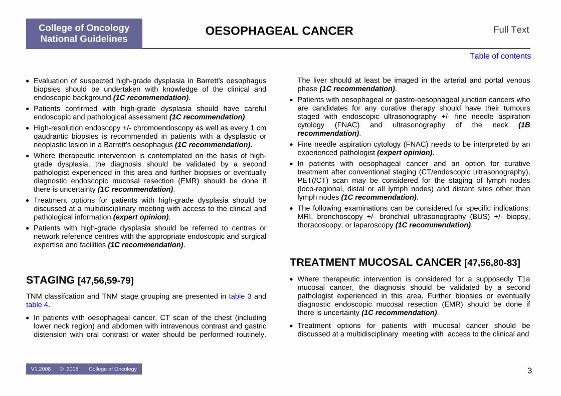
• Evaluation of suspected high-grade dysplasia in Barrett's oesophagus biopsies should be undertaken with knowledge of the clinical and endoscopic background (1C recommendation).
• Patients confirmed with high-grade dysplasia should have careful endoscopic and pathological assessment (1C recommendation).
• High-resolution endoscopy +/- chromoendoscopy as well as every 1 cm qaudrantic biopsies is recommended in patients with a dysplastic or neoplastic lesion in a Barrett's oesophagus (1C recommendation).
• Where therapeutic intervention is contemplated on the basis of high-grade dysplasia, the diagnosis should be validated by a second pathologist experienced in this area and further biopsies or eventually diagnostic endoscopic mucosal resection (EMR) should be done if there is uncertainty (1C recommendation).
• Treatment options for patients with high-grade dysplasia should be discussed at a multidisciplinary meeting with access to the clinical and pathological information (expert opinion).
• Patients with high-grade dysplasia should be referred to centres or network reference centres with the appropriate endoscopic and surgical expertise and facilities (1C recommendation).
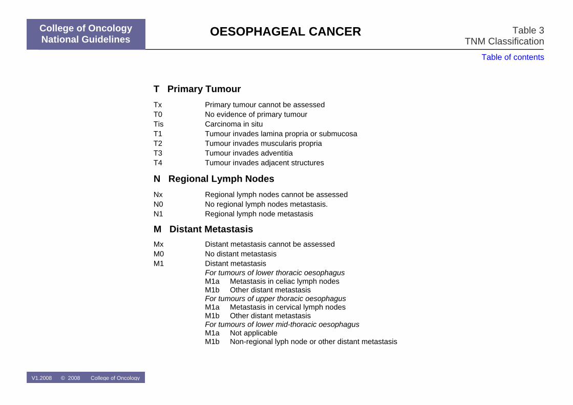
STAGING [47,56,59-79] TNM classifcation and TNM stage grouping are presented in table 3 and table 4.
• In patients with oesophageal cancer, CT scan of the chest (including lower neck region) and abdomen with intravenous contrast and gastric distension with oral contrast or water should be performed routinely.
The liver should at least be imaged in the arterial and portal venous phase (1C recommendation).
• Patients with oesophageal or gastro-oesophageal junction cancers who are candidates for any curative therapy should have their tumours staged with endoscopic ultrasonography +/- fine needle aspiration cytology (FNAC) and ultrasonography of the neck (1B recommendation).
• Fine needle aspiration cytology (FNAC) needs to be interpreted by an experienced pathologist (expert opinion).
• In patients with oesophageal cancer and an option for curative treatment after conventional staging (CT/endoscopic ultrasonography), PET(/CT) scan may be considered for the staging of lymph nodes (loco-regional, distal or all lymph nodes) and distant sites other than lymph nodes (1C recommendation).
• The following examinations can be considered for specific indications: MRI, bronchoscopy +/- bronchial ultrasonography (BUS) +/- biopsy, thoracoscopy, or laparoscopy (1C recommendation).
TREATMENT MUCOSAL CANCER [47,56,80-83] • Where therapeutic intervention is considered for a supposedly T1a
mucosal cancer, the diagnosis should be validated by a second pathologist experienced in this area. Further biopsies or eventually diagnostic endoscopic mucosal resection (EMR) should be done if there is uncertainty (1C recommendation).
• Treatment options for patients with mucosal cancer should be discussed at a multidisciplinary meeting with access to the clinical and
V1.2008 © 2008 College of Oncology 3
OESOPHAGEAL CANCER
College of Oncology National Guidelines
Full Text
Table of contents
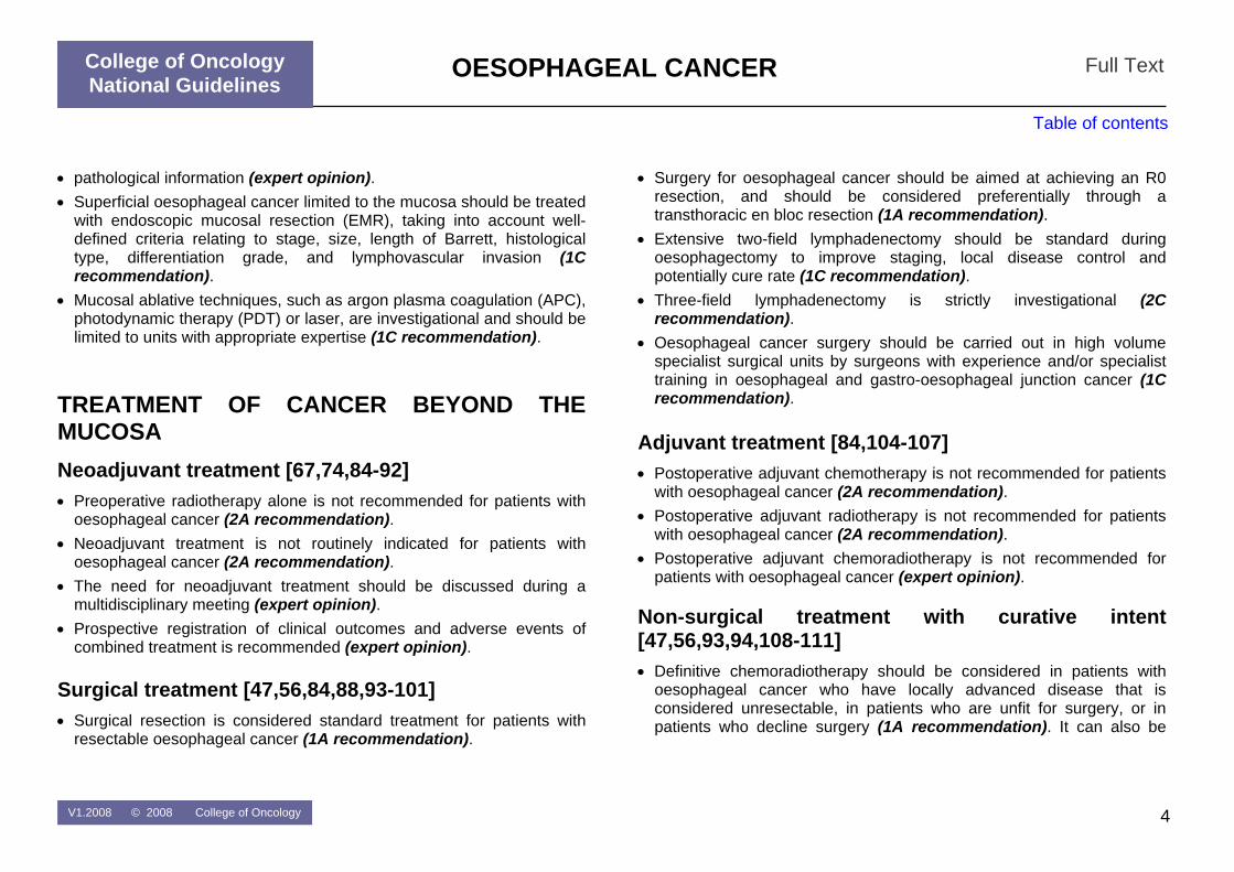
• pathological information (expert opinion). • Superficial oesophageal cancer limited to the mucosa should be treated
with endoscopic mucosal resection (EMR), taking into account well-defined criteria relating to stage, size, length of Barrett, histological type, differentiation grade, and lymphovascular invasion (1C recommendation).
• Mucosal ablative techniques, such as argon plasma coagulation (APC), photodynamic therapy (PDT) or laser, are investigational and should be limited to units with appropriate expertise (1C recommendation).
TREATMENT OF CANCER BEYOND THE MUCOSA Neoadjuvant treatment [67,74,84-92] • Preoperative radiotherapy alone is not recommended for patients with
oesophageal cancer (2A recommendation). • Neoadjuvant treatment is not routinely indicated for patients with
oesophageal cancer (2A recommendation). • The need for neoadjuvant treatment should be discussed during a
multidisciplinary meeting (expert opinion). • Prospective registration of clinical outcomes and adverse events of
combined treatment is recommended (expert opinion).
Surgical treatment [47,56,84,88,93-101] • Surgical resection is considered standard treatment for patients with
resectable oesophageal cancer (1A recommendation).
• Surgery for oesophageal cancer should be aimed at achieving an R0 resection, and should be considered preferentially through a transthoracic en bloc resection (1A recommendation).
• Extensive two-field lymphadenectomy should be standard during oesophagectomy to improve staging, local disease control and potentially cure rate (1C recommendation).
• Three-field lymphadenectomy is strictly investigational (2C recommendation).
• Oesophageal cancer surgery should be carried out in high volume specialist surgical units by surgeons with experience and/or specialist training in oesophageal and gastro-oesophageal junction cancer (1C recommendation).
Adjuvant treatment [84,104-107] • Postoperative adjuvant chemotherapy is not recommended for patients
with oesophageal cancer (2A recommendation). • Postoperative adjuvant radiotherapy is not recommended for patients
with oesophageal cancer (2A recommendation). • Postoperative adjuvant chemoradiotherapy is not recommended for
patients with oesophageal cancer (expert opinion). Non-surgical treatment with curative intent [47,56,93,94,108-111] • Definitive chemoradiotherapy should be considered in patients with
oesophageal cancer who have locally advanced disease that is considered unresectable, in patients who are unfit for surgery, or in patients who decline surgery (1A recommendation). It can also be
V1.2008 © 2008 College of Oncology 4

OESOPHAGEAL CANCER
College of Oncology National Guidelines
Full Text
Table of contents
considered for patients with cervical oesophageal cancer in order to preserve the larynx (1C recommendation).
PALLIATIVE TREATMENT AND METASTATIC DISEASE [47,48,56,112-115] • Control of obstruction caused by oesophageal cancer should be
obtained with stent placement or laser/ argon plasma coagulation (APC) therapy, depending on the local availability and expertise (1A recommendation).
• Partially covered self-expanding metal stents or plastic expandable stents are the best options for palliation of dysphagia caused by oesophageal cancer (1B recommendation).
• Laser therapy, argon plasma coagulation (APC) therapy or re-stenting should be considered for control of tumour ingrowth or overgrowth in stented patients (1C recommendation).
• The use of oesophageal dilatation alone should be avoided (2C recommendation).
• Oesophagectomy (transthoracic or transhiatal) should not be performed with palliative intent in patients with oesophageal cancer (1C recommendation).
• Substernal bypass for oesophageal cancer should not be performed with palliative intent (1C recommendation).
• In patients with locally advanced or metastatic cancer of the oesophagus, chemotherapy or chemoradiotherapy are treatment options that should be discussed in the multidisciplinary team (2A recommendation).
• Palliative external-beam radiotherapy or endoluminal brachytherapy should be considered in patients with dysphagia from oesophageal cancer and with the perspective of a more prolonged survival (2C recommendation).
• Patients with oesophageal cancer should have access to a specialist palliative care team, in particular in relation to comfort and symptom control, nutrition and quality of life (1C recommendation).
FOLLOW-UP [47,56] • It is recommended that the follow-up of patients treated for
oesophageal cancer includes a physical examination every three months, and a CT scan every six months in the first year and afterwards annually until the fifth year (expert opinion).
• Patients treated with endoscopic mucosal resection (EMR) should have a follow-up endoscopy after three months, then every six months in the first two years, and then annually (expert opinion).
RECURRENT DISEASE [116-123] • In patients with recurrent oesophageal cancer, treatment options
should be discussed in the multidisciplinary team (expert opinion). • In patients with a local recurrence or new tumour after endoscopic
mucosal resection (EMR), treatment options, including local treatment, should be discussed in the multidisciplinary team (expert opinion).
V1.2008 © 2008 College of Oncology 5

OESOPHAGEAL CANCER
College of Oncology National Guidelines
References
Table of contents
References
1 Parkin, D.M., et al., Global cancer statistics, 2002. CA Cancer J Clin, 2005.
55(2): p. 74-108. 2 Bollschweiler, E., et al., Demographic variations in the rising incidence of
esophageal adenocarcinoma in white males. Cancer, 2001. 92(3): p. 549-55.
3 Crane, L.M., et al., Oesophageal cancer in The Netherlands: increasing incidence and mortality but improving survival. Eur J Cancer, 2007. 43(9): p. 1445-51.
4 Qiu, D. and S. Kaneko, Comparison of esophageal cancer mortality in five countries: France, Italy, Japan, UK and USA from the WHO mortality database (1960-2000). Jpn J Clin Oncol, 2005. 35(9): p. 564-7.
5 Vizcaino, A.P., et al., Time trends incidence of both major histologic types of esophageal carcinomas in selected countries, 1973-1995. Int J Cancer, 2002. 99(6): p. 860-8.
6 Crew, K.D. and A.I. Neugut, Epidemiology of gastric cancer. World J Gastroenterol, 2006. 12(3): p. 354-62.
7 Wijnhoven, B.P.L., et al., Increased incidence of adenocarcinomas at the gastro-oesophageal junction in Dutch males since the 1990s. Eur J Gastroenterol Hepatol, 2002. 14(2): p. 115-22.
8 Crane, S.J., et al., The changing incidence of oesophageal and gastric adenocarcinoma by anatomic sub-site. Aliment Pharmacol Ther, 2007. 25(4): p. 447-53.
9 Peeters, M., et al., Nationale Richtlijnen van het College voor oncologie: A. Algemeen kader oncologish kwaliteitshandboek B. Wetenschappelijke basis voor klinische paden voor diagnose en behandeling colorectale kanker en testiskanker, in KCE reports. 2006, Federaal Kenniscentrum voor de Gezondheidszorg (KCE): Brussel.
10 Fervers, B., et al., Adaptation of clinical guidelines: literature review and proposition for a framework and procedure. Int J Qual Health Care, 2006. 18(3): p. 167-76.
11 Siewert, J.R. and H.J. Stein, Classification of adenocarcinoma of the oesophagogastric junction. Br J Surg, 1998. 85(11): p. 1457-9.
12 Spechler, S.J., et al., Adenocarcinoma of the esophago-gastric junction., in Pathology and Genetics of Tumours of the Digestive System., S.R. Hamilton and L.A. Aaltonen, Editors. 2000, IARC Press: Lyon, France. p. 31-36.
13 (UICC), I.U.A.C., TNM classification of malignant tumours. 6th ed. ed. 2002, Berlin: Springer-Verlag.
14 Corley, D.A. and A. Kubo, Influence of site classification on cancer incidence rates: an analysis of gastric cardia carcinomas. J Natl Cancer Inst, 2004. 96(18): p. 1383-7.
15 Driessen, A., et al., Identical cytokeratin expression pattern CK7+/CK20- in esophageal and cardiac cancer: etiopathological and clinical implications. Mod Pathol, 2004. 17(1): p. 49-55.
16 Driessen, A., et al., Are carcinomas of the cardia oesophageal or gastric adenocarcinomas? Eur J Cancer, 2003. 39(17): p. 2487-94.
17 Skinner, D.B., et al., Barrett's esophagus. Comparison of benign and malignant cases. Ann Surg, 1983. 198(4): p. 554-65.
18 Paull, A., et al., The histologic spectrum of Barrett's esophagus. N Engl J Med, 1976. 295(9): p. 476-80.
19 Naef, A.P., M. Savary, and L. Ozzello, Columnar-lined lower esophagus: an acquired lesion with malignant predisposition. Report on 140 cases of Barrett's esophagus with 12 adenocarcinomas. J Thorac Cardiovasc Surg, 1975. 70(5): p. 826-35.
20 Haggitt, R.C., et al., Adenocarcinoma complicating columnar epithelium-lined (Barrett's) esophagus. Am J Clin Pathol, 1978. 70(1): p. 1-5.
21 Reid, B.J., et al., Barrett's esophagus. Correlation between flow cytometry and histology in detection of patients at risk for adenocarcinoma. Gastroenterology, 1987. 93(1): p. 1-11.
22 Spechler, S.J. and R.K. Goyal, The columnar-lined esophagus, intestinal metaplasia, and Norman Barrett. Gastroenterology, 1996. 110(2): p. 614-21.
23 Sampliner, R.E., Practice guidelines on the diagnosis, surveillance, and therapy of Barrett's esophagus. The Practice Parameters Committee of the
V1.2008 © 2008 College of Oncology 6
OESOPHAGEAL CANCER
College of Oncology National Guidelines
References
Table of contents
American College of Gastroenterology. Am J Gastroenterol, 1998. 93(7): p. 1028-32.
24 Sharma, P., et al., A critical review of the diagnosis and management of Barrett's esophagus: the AGA Chicago Workshop. Gastroenterology, 2004. 127(1): p. 310-30.
25 British Society of Gastroenterology, Guidelines for the diagnosis and management of Barrett’s columnar-lined oesophagus., B.S.o. Gastroenterology, Editor. 2005, British Society of Gastroenterology: London.
26 Flejou, J.F. and M. Svrcek, Barrett's oesophagus--a pathologist's view. Histopathology, 2007. 50(1): p. 3-14.
27 Takubo, K., et al., Double muscularis mucosae in Barrett's esophagus. Hum Pathol, 1991. 22(11): p. 1158-61.
28 Nigro, J.J., et al., Prevalence and location of nodal metastases in distal esophageal adenocarcinoma confined to the wall: implications for therapy. J Thorac Cardiovasc Surg, 1999. 117(1): p. 16-23; discussion 23-5.
29 Westerterp, M., et al., Outcome of surgical treatment for early adenocarcinoma of the esophagus or gastro-esophageal junction. Virchows Arch, 2005. 446(5): p. 497-504.
30 Watanabe, H., J.R. Jass, and L.H. Sobin, Histological typing of oesophageal and gastric tumours. 2nd ed. ed. 1990, Berlin: Springer-Verlag.
31 Rubio, C.A., F.S. Liu, and H.Z. Zhao, Histological classification of intraepithelial neoplasias and microinvasive squamous carcinoma of the esophagus. Am J Surg Pathol, 1989. 13(8): p. 685-90.
32 Riddell, R.H., et al., Dysplasia in inflammatory bowel disease: standardized classification with provisional clinical applications. Hum Pathol, 1983. 14(11): p. 931-68.
33 Montgomery, E., Is there a way for pathologists to decrease interobserver variability in the diagnosis of dysplasia? Arch Pathol Lab Med, 2005. 129(2): p. 174-6.
34 Montgomery, E., et al., Reproducibility of the diagnosis of dysplasia in Barrett esophagus: a reaffirmation. Hum Pathol, 2001. 32(4): p. 368-78.
35 Reid, B.J., et al., Observer variation in the diagnosis of dysplasia in Barrett's esophagus. Hum Pathol, 1988. 19(2): p. 166-78.
36 Schlemper, R.J., et al., International comparability of the pathological diagnosis for early cancer of the digestive tract: Munich meeting. J Gastroenterol, 2000. 35 Suppl 12: p. 102-10.
37 Schlemper, R.J., et al., Differences in diagnostic criteria for esophageal squamous cell carcinoma between Japanese and Western pathologists. Cancer, 2000. 88(5): p. 996-1006.
38 Schlemper, R.J., et al., Differences in diagnostic criteria for gastric carcinoma between Japanese and western pathologists. Lancet, 1997. 349(9067): p. 1725-9.
39 Antonioli, D.A. and H.H. Wang, Morphology of Barrett's esophagus and Barrett's-associated dysplasia and adenocarcinoma. Gastroenterol Clin North Am, 1997. 26(3): p. 495-506.
40 Haggitt, R.C., Barrett's esophagus, dysplasia, and adenocarcinoma. Hum Pathol, 1994. 25(10): p. 982-93.
41 Rugge, M., et al., Gastric dysplasia: the Padova international classification. Am J Surg Pathol, 2000. 24(2): p. 167-76.
42 Schlemper, R.J., et al., The Vienna classification of gastrointestinal epithelial neoplasia. Gut, 2000. 47(2): p. 251-5.
43 Schlemper, R.J., Y. Kato, and M. Stolte, Diagnostic criteria for gastrointestinal carcinomas in Japan and Western countries: proposal for a new classification system of gastrointestinal epithelial neoplasia. J Gastroenterol Hepatol, 2000. 15 Suppl: p. G49-57.
44 Schlemper, R.J., Y. Kato, and M. Stolte, Review of histological classifications of gastrointestinal epithelial neoplasia: differences in diagnosis of early carcinomas between Japanese and Western pathologists. J Gastroenterol, 2001. 36(7): p. 445-56.
45 Hamilton, S.R. and L.A. Aaltonen, Pathology and genetics of tumours of the digestive system., ed. S.R. Hamilton and L.A. Aaltonen. 2000, Lyon, France: IARC Press.
46 Odze, R.D., Diagnosis and grading of dysplasia in Barrett's oesophagus. J Clin Pathol, 2006. 59(10): p. 1029-38.
47 Scottish Intercollegiate Guidelines Network, Management of oesophageal and gastric cancer. A national clinical guideline., SIGN, Editor. 2006, SIGN: Edinburgh.
V1.2008 © 2008 College of Oncology 7
OESOPHAGEAL CANCER
College of Oncology National Guidelines
References
Table of contents
48 Wang, K.K., M. Wongkeesong, and N.S. Buttar, American Gastroenterological Association technical review on the role of the gastroenterologist in the management of esophageal carcinoma. Gastroenterology, 2005. 128(5): p. 1471-505.
49 Bowrey, D.J., et al., Use of alarm symptoms to select dyspeptics for endoscopy causes patients with curable esophagogastric cancer to be overlooked. Surgical Endoscopy, 2006. 20(11): p. 1725-8.
50 Slee, G.R., S.M. Wagner, and F.S. McCullough, Odynophagia in patients with malignant disorders. Cancer, 1985. 55(12): p. 2877-9.
51 Dubuc, J., et al., Endoscopic screening for esophageal squamous-cell carcinoma in high-risk patients: a prospective study conducted in 62 French endoscopy centers. Endoscopy, 2006. 38(7): p. 690-5.
52 Borovicka, J., et al., Autofluorescence endoscopy in surveillance of Barrett's esophagus: a multicenter randomized trial on diagnostic efficacy. Endoscopy, 2006. 38(9): p. 867-72.
53 Lim, C.H., et al., Randomized crossover study that used methylene blue or random 4-quadrant biopsy for the diagnosis of dysplasia in Barrett's esophagus.[see comment]. Gastrointestinal Endoscopy, 2006. 64(2): p. 195-9.
54 Kara, M.A., et al., High-resolution endoscopy plus chromoendoscopy or narrow-band imaging in Barrett's esophagus: a prospective randomized crossover study. Endoscopy, 2005. 37(10): p. 929-36.
55 Corey, K.E., S.M. Schmitz, and N.J. Shaheen, Does a surgical antireflux procedure decrease the incidence of esophageal adenocarcinoma in Barrett's esophagus? A meta-analysis.[see comment]. American Journal of Gastroenterology, 2003. 98(11): p. 2390-4.
56 Kwaliteitsinstituut voor de Gezondheidszorg (CBO), Diagnostiek en behandeling oesofaguscarcinoom., V.Z.C. B.V., Editor. 2005, CBO: Alphen aan den Rijn.
57 Reid, B.J., et al., Optimizing endoscopic biopsy detection of early cancers in Barrett's high-grade dysplasia. Am J Gastroenterol, 2000. 95(11): p. 3089-96.
58 Reid, B.J., et al., Predictors of progression to cancer in Barrett's esophagus: baseline histology and flow cytometry identify low- and high-risk patient subsets. Am J Gastroenterol, 2000. 95(7): p. 1669-76.
59 Lowe, V.J., et al., Comparison of positron emission tomography, computed tomography, and endoscopic ultrasound in the initial staging of patients with esophageal cancer. Molecular Imaging & Biology, 2005. 7(6): p. 422-30.
60 Lerut, T., et al., Three-field lymphadenectomy for carcinoma of the esophagus and gastroesophageal junction in 174 R0 resections: impact on staging, disease-free survival, and outcome: a plea for adaptation of TNM classification in upper-half esophageal carcinoma. Ann Surg, 2004. 240(6): p. 962-72; discussion 972-4.
61 van Vliet, E.P.M., et al., Publication bias does not play a role in the reporting of the results of endoscopic ultrasound staging of upper gastrointestinal cancers. Endoscopy, 2007. 39(4): p. 325-32.
62 Pfau, P.R., et al., Esophageal dilation for endosonographic evaluation of malignant esophageal strictures is safe and effective. Am J Gastroenterol, 2000. 95(10): p. 2813-5.
63 Shimpi, R.A., et al., Staging of esophageal cancer by EUS: staging accuracy revisited. Gastrointest Endosc, 2007. 66(3): p. 475-82.
64 Egan, J.V., et al., Esophageal dilation. Gastrointest Endosc, 2006. 63(6): p. 755-60.
65 Doldi, S.B., et al., Ultrasonographic evaluation of the cervical lymph nodes in preoperative staging of esophageal neoplasms. Abdom Imaging, 1998. 23(3): p. 275-7.
66 Fédération Nationale des Centres de Lutte Contre le Cancer (FNCLCC), Recommandations pour la pratique clinique: Standards, Options et Recommandations 2003 pour l’utilisation de la tomographie par émission de positons au [18F]-FDG (TEP-FDG) en cancérologie (rapport intégral). in Standards, Options et Recommendations. 2003, FNCLCC: Paris.
67 Cleemput, I., et al., HTA Positron Emission Tomography Imaging in Belgium, in KCE reports. 2005, Belgian Health Care Knowledge Centre (KCE): Brussels.
V1.2008 © 2008 College of Oncology 8

OESOPHAGEAL CANCER
College of Oncology National Guidelines
References
Table of contents
68 Choi, J.Y., et al., 18F-FDG PET in patients with esophageal squamous cell carcinoma undergoing curative surgery: prognostic implications. Journal of Nuclear Medicine, 2004. 45(11): p. 1843-50.
69 Kneist, W., et al., Prospective evaluation of positron emission tomography in the preoperative staging of esophageal carcinoma. Archives of Surgery, 2004. 139(10): p. 1043-9.
70 Leong, T., et al., A prospective study to evaluate the impact of FDG-PET on CT-based radiotherapy treatment planning for oesophageal cancer.[see comment]. Radiotherapy & Oncology, 2006. 78(3): p. 254-61.
71 Moureau-Zabotto, L., et al., Impact of CT and 18F-deoxyglucose positron emission tomography image fusion for conformal radiotherapy in esophageal carcinoma. International Journal of Radiation Oncology, Biology, Physics, 2005. 63(2): p. 340-5.
72 Ott, K., et al., Metabolic imaging predicts response, survival, and recurrence in adenocarcinomas of the esophagogastric junction. Journal of Clinical Oncology, 2006. 24(29): p. 4692-8.
73 Sihvo, E.I.T., et al., Adenocarcinoma of the esophagus and the esophagogastric junction: positron emission tomography improves staging and prediction of survival in distant but not in locoregional disease. Journal of Gastrointestinal Surgery, 2004. 8(8): p. 988-96.
74 Song, S.Y., et al., FDG-PET in the prediction of pathologic response after neoadjuvant chemoradiotherapy in locally advanced, resectable esophageal cancer. International Journal of Radiation Oncology, Biology, Physics, 2005. 63(4): p. 1053-9.
75 Yuan, S., et al., Additional value of PET/CT over PET in assessment of locoregional lymph nodes in thoracic esophageal squamous cell cancer. Journal of Nuclear Medicine, 2006. 47(8): p. 1255-9.
76 Osugi, H., et al., Bronchoscopic ultrasonography for staging supracarinal esophageal squamous cell carcinoma: impact on outcome. World Journal of Surgery, 2003. 27(5): p. 590-4.
77 Wakamatsu, T., et al., Usefulness of preoperative endobronchial ultrasound for airway invasion around the trachea: esophageal cancer and thyroid cancer.[see comment]. Respiration, 2006. 73(5): p. 651-7.
78 Imadahl, A., et al., [Is bronchoscopy a useful additional preoperative examination in esophageal carcinoma?]. Langenbecks Arch Chir, 1990. 375(6): p. 326-9.
79 Mortensen, M.B., et al., Combined preoperative endoscopic and laparoscopic ultrasonography for prediction of R0 resection in upper gastrointestinal tract cancer. British Journal of Surgery, 2006. 93(6): p. 720-5.
80 Oka, S., et al., Advantage of endoscopic submucosal dissection compared with EMR for early gastric cancer. Gastrointest Endosc, 2006. 64(6): p. 877-83.
81 Esaki, M., et al., Risk factors for local recurrence of superficial esophageal cancer after treatment by endoscopic mucosal resection. Endoscopy, 2007. 39(1): p. 41-5.
82 Katada, C., et al., Local recurrence of squamous-cell carcinoma of the esophagus after EMR. Gastrointest Endosc, 2005. 61(2): p. 219-25.
83 Overholt, B.F., et al., Photodynamic therapy with porfimer sodium for ablation of high-grade dysplasia in Barrett's esophagus: international, partially blinded, randomized phase III trial.[see comment][erratum appears in Gastrointest Endosc. 2006 Feb;63(2):359]. Gastrointestinal Endoscopy, 2005. 62(4): p. 488-98.
84 Malthaner, R.A., et al., Neoadjuvant or Adjuvant Therapy for Resectable Esophageal Cancer. Practice Guideline Report #2-11., CCO, Editor. 2005, CCO: Ottawa.
85 Arnott, S.J., et al., Preoperative radiotherapy for esophageal carcinoma.[update of Cochrane Database Syst Rev. 2000;(4):CD001799; PMID: 11034728]. Cochrane Database of Systematic Reviews, 2005(4): p. CD001799.
86 Gebski, V., et al., Survival benefits from neoadjuvant chemoradiotherapy or chemotherapy in oesophageal carcinoma: a meta-analysis.[see comment]. Lancet Oncology, 2007. 8(3): p. 226-34.
87 Malthaner, R.A., S. Collin, and D. Fenlon, Preoperative chemotherapy for resectable thoracic esophageal cancer.[update of Cochrane Database Syst Rev. 2003;(4):CD001556; PMID: 14583936]. Cochrane Database of Systematic Reviews, 2006. 3: p. CD001556.
V1.2008 © 2008 College of Oncology 9
OESOPHAGEAL CANCER
College of Oncology National Guidelines
References
Table of contents
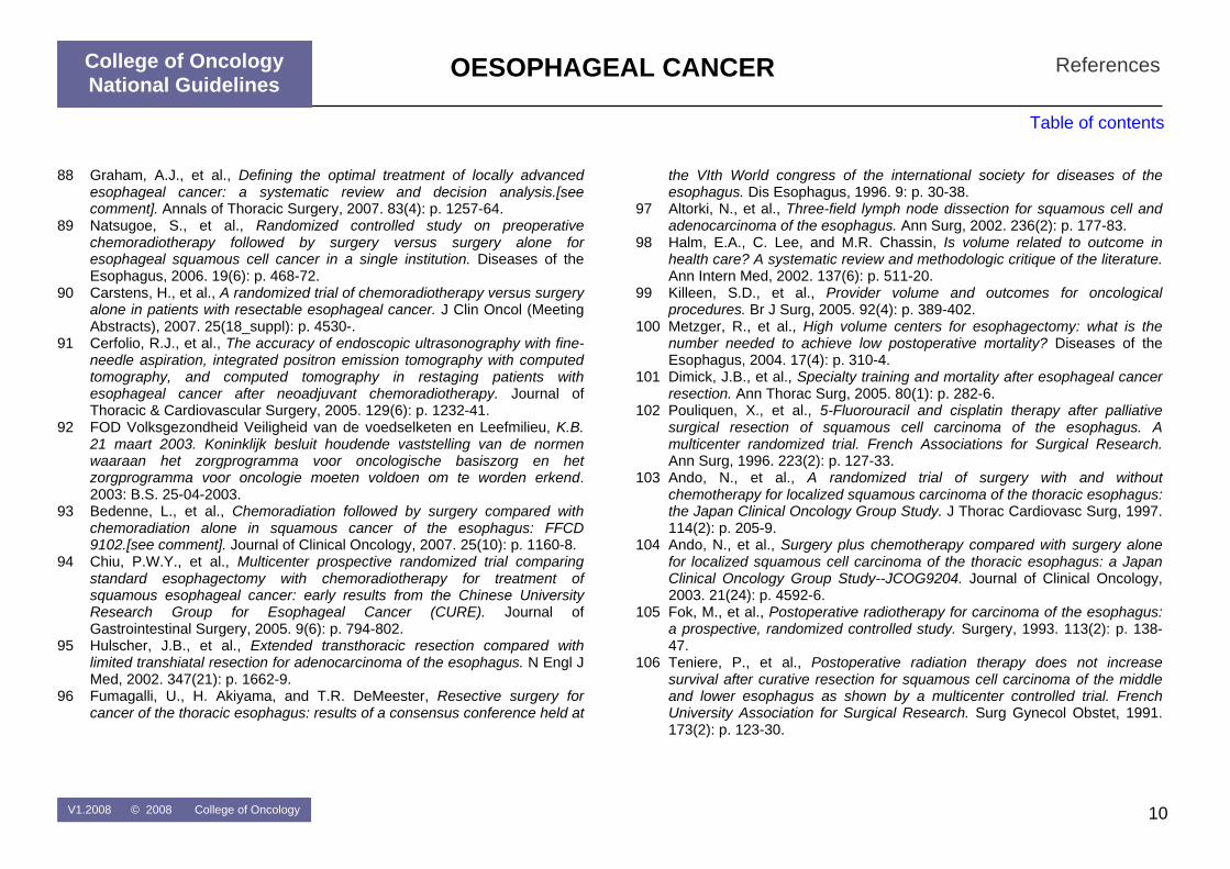
88 Graham, A.J., et al., Defining the optimal treatment of locally advanced esophageal cancer: a systematic review and decision analysis.[see comment]. Annals of Thoracic Surgery, 2007. 83(4): p. 1257-64.
89 Natsugoe, S., et al., Randomized controlled study on preoperative chemoradiotherapy followed by surgery versus surgery alone for esophageal squamous cell cancer in a single institution. Diseases of the Esophagus, 2006. 19(6): p. 468-72.
90 Carstens, H., et al., A randomized trial of chemoradiotherapy versus surgery alone in patients with resectable esophageal cancer. J Clin Oncol (Meeting Abstracts), 2007. 25(18_suppl): p. 4530-.
91 Cerfolio, R.J., et al., The accuracy of endoscopic ultrasonography with fine-needle aspiration, integrated positron emission tomography with computed tomography, and computed tomography in restaging patients with esophageal cancer after neoadjuvant chemoradiotherapy. Journal of Thoracic & Cardiovascular Surgery, 2005. 129(6): p. 1232-41.
92 FOD Volksgezondheid Veiligheid van de voedselketen en Leefmilieu, K.B. 21 maart 2003. Koninklijk besluit houdende vaststelling van de normen waaraan het zorgprogramma voor oncologische basiszorg en het zorgprogramma voor oncologie moeten voldoen om te worden erkend. 2003: B.S. 25-04-2003.
93 Bedenne, L., et al., Chemoradiation followed by surgery compared with chemoradiation alone in squamous cancer of the esophagus: FFCD 9102.[see comment]. Journal of Clinical Oncology, 2007. 25(10): p. 1160-8.
94 Chiu, P.W.Y., et al., Multicenter prospective randomized trial comparing standard esophagectomy with chemoradiotherapy for treatment of squamous esophageal cancer: early results from the Chinese University Research Group for Esophageal Cancer (CURE). Journal of Gastrointestinal Surgery, 2005. 9(6): p. 794-802.
95 Hulscher, J.B., et al., Extended transthoracic resection compared with limited transhiatal resection for adenocarcinoma of the esophagus. N Engl J Med, 2002. 347(21): p. 1662-9.
96 Fumagalli, U., H. Akiyama, and T.R. DeMeester, Resective surgery for cancer of the thoracic esophagus: results of a consensus conference held at
the VIth World congress of the international society for diseases of the esophagus. Dis Esophagus, 1996. 9: p. 30-38.
97 Altorki, N., et al., Three-field lymph node dissection for squamous cell and adenocarcinoma of the esophagus. Ann Surg, 2002. 236(2): p. 177-83.
98 Halm, E.A., C. Lee, and M.R. Chassin, Is volume related to outcome in health care? A systematic review and methodologic critique of the literature. Ann Intern Med, 2002. 137(6): p. 511-20.
99 Killeen, S.D., et al., Provider volume and outcomes for oncological procedures. Br J Surg, 2005. 92(4): p. 389-402.
100 Metzger, R., et al., High volume centers for esophagectomy: what is the number needed to achieve low postoperative mortality? Diseases of the Esophagus, 2004. 17(4): p. 310-4.
101 Dimick, J.B., et al., Specialty training and mortality after esophageal cancer resection. Ann Thorac Surg, 2005. 80(1): p. 282-6.
102 Pouliquen, X., et al., 5-Fluorouracil and cisplatin therapy after palliative surgical resection of squamous cell carcinoma of the esophagus. A multicenter randomized trial. French Associations for Surgical Research. Ann Surg, 1996. 223(2): p. 127-33.
103 Ando, N., et al., A randomized trial of surgery with and without chemotherapy for localized squamous carcinoma of the thoracic esophagus: the Japan Clinical Oncology Group Study. J Thorac Cardiovasc Surg, 1997. 114(2): p. 205-9.
104 Ando, N., et al., Surgery plus chemotherapy compared with surgery alone for localized squamous cell carcinoma of the thoracic esophagus: a Japan Clinical Oncology Group Study--JCOG9204. Journal of Clinical Oncology, 2003. 21(24): p. 4592-6.
105 Fok, M., et al., Postoperative radiotherapy for carcinoma of the esophagus: a prospective, randomized controlled study. Surgery, 1993. 113(2): p. 138-47.
106 Teniere, P., et al., Postoperative radiation therapy does not increase survival after curative resection for squamous cell carcinoma of the middle and lower esophagus as shown by a multicenter controlled trial. French University Association for Surgical Research. Surg Gynecol Obstet, 1991. 173(2): p. 123-30.
V1.2008 © 2008 College of Oncology 10
OESOPHAGEAL CANCER
College of Oncology National Guidelines
References
Table of contents
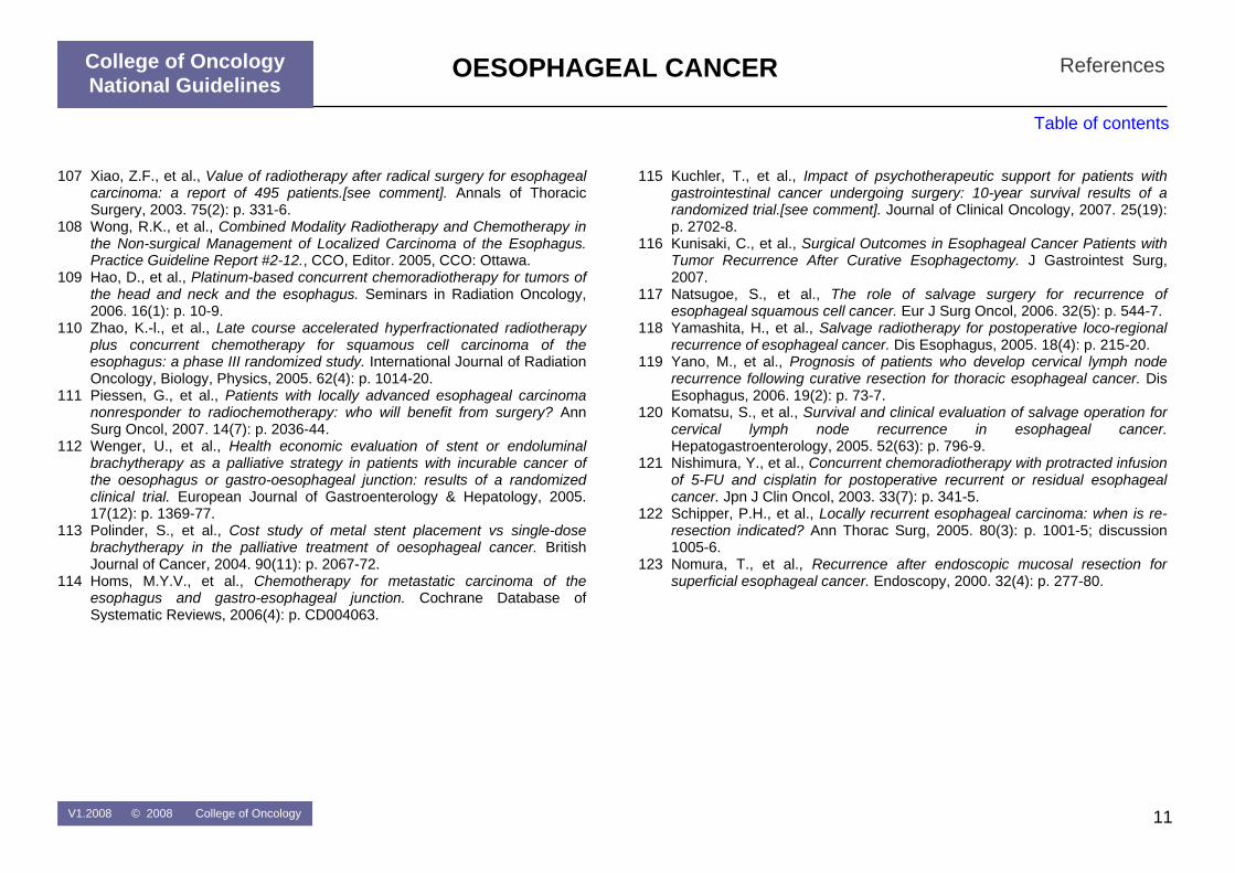
107 Xiao, Z.F., et al., Value of radiotherapy after radical surgery for esophageal carcinoma: a report of 495 patients.[see comment]. Annals of Thoracic Surgery, 2003. 75(2): p. 331-6.
115 Kuchler, T., et al., Impact of psychotherapeutic support for patients with gastrointestinal cancer undergoing surgery: 10-year survival results of a randomized trial.[see comment]. Journal of Clinical Oncology, 2007. 25(19): p. 2702-8. 108 Wong, R.K., et al., Combined Modality Radiotherapy and Chemotherapy in
the Non-surgical Management of Localized Carcinoma of the Esophagus. Practice Guideline Report #2-12., CCO, Editor. 2005, CCO: Ottawa.
116 Kunisaki, C., et al., Surgical Outcomes in Esophageal Cancer Patients with Tumor Recurrence After Curative Esophagectomy. J Gastrointest Surg, 2007. 109 Hao, D., et al., Platinum-based concurrent chemoradiotherapy for tumors of
the head and neck and the esophagus. Seminars in Radiation Oncology, 2006. 16(1): p. 10-9.
117 Natsugoe, S., et al., The role of salvage surgery for recurrence of esophageal squamous cell cancer. Eur J Surg Oncol, 2006. 32(5): p. 544-7.
110 Zhao, K.-l., et al., Late course accelerated hyperfractionated radiotherapy plus concurrent chemotherapy for squamous cell carcinoma of the esophagus: a phase III randomized study. International Journal of Radiation Oncology, Biology, Physics, 2005. 62(4): p. 1014-20.
118 Yamashita, H., et al., Salvage radiotherapy for postoperative loco-regional recurrence of esophageal cancer. Dis Esophagus, 2005. 18(4): p. 215-20.
119 Yano, M., et al., Prognosis of patients who develop cervical lymph node recurrence following curative resection for thoracic esophageal cancer. Dis Esophagus, 2006. 19(2): p. 73-7. 111 Piessen, G., et al., Patients with locally advanced esophageal carcinoma
nonresponder to radiochemotherapy: who will benefit from surgery? Ann Surg Oncol, 2007. 14(7): p. 2036-44.
120 Komatsu, S., et al., Survival and clinical evaluation of salvage operation for cervical lymph node recurrence in esophageal cancer. Hepatogastroenterology, 2005. 52(63): p. 796-9. 112 Wenger, U., et al., Health economic evaluation of stent or endoluminal
brachytherapy as a palliative strategy in patients with incurable cancer of the oesophagus or gastro-oesophageal junction: results of a randomized clinical trial. European Journal of Gastroenterology & Hepatology, 2005. 17(12): p. 1369-77.
121 Nishimura, Y., et al., Concurrent chemoradiotherapy with protracted infusion of 5-FU and cisplatin for postoperative recurrent or residual esophageal cancer. Jpn J Clin Oncol, 2003. 33(7): p. 341-5.
122 Schipper, P.H., et al., Locally recurrent esophageal carcinoma: when is re-resection indicated? Ann Thorac Surg, 2005. 80(3): p. 1001-5; discussion 1005-6.
113 Polinder, S., et al., Cost study of metal stent placement vs single-dose brachytherapy in the palliative treatment of oesophageal cancer. British Journal of Cancer, 2004. 90(11): p. 2067-72. 123 Nomura, T., et al., Recurrence after endoscopic mucosal resection for
superficial esophageal cancer. Endoscopy, 2000. 32(4): p. 277-80. 114 Homs, M.Y.V., et al., Chemotherapy for metastatic carcinoma of the esophagus and gastro-esophageal junction. Cochrane Database of Systematic Reviews, 2006(4): p. CD004063.
V1.2008 © 2008 College of Oncology 11
OESOPHAGEAL CANCER
College of Oncology National Guidelines
Table 1 Sources
Table of contents
Searched guideline websites and websites of oncologic organisations Searched guideline websites and websites of oncologic organisations
Alberta Heritage Foundation For Medical Research (AHFMR) http://www.ahfmr.ab.ca/
American Society of Clinical Oncology (ASCO) http://www.asco.org/
American College of Surgeons (ACS) http://www.facs.org/cancer/coc/
CMA Infobase http://mdm.ca/cpgsnew/cpgs/index.asp
Guidelines International Network (GIN) http://www.g-i-n.net/
National Comprehensive Cancer Network (NCCN) http://www.nccn.org/
National Guideline Clearinghouse http://www.guideline.gov/
National Cancer Institute http://www.cancer.gov/
Haute Autorité de Santé (HAS) http://bfes.has-sante.fr/HTML/indexBFES_HAS.html
BC Cancer Agency http://www.bccancer.bc.ca/default.htm
Institute for Clinical Systems Improvement (ICSI) http://www.icsi.org/index.asp
National Health and Medical Research Council (NHMRC) http://www.nhmrc.gov.au/
Scottish Intercollegiate Guidelines Network (SIGN) http://www.sign.ac.uk/
New Zealand Guidelines Group (NZGG) http://www.nzgg.org.nz/
Fédération Nationale des Centres de Lutte Contre le Cancer (FNCLCC) http://www.fnclcc.fr/sor/structure/index-sorspecialistes.html
National Institute for Health and Clinical Excellence (NICE) http://www.nice.org.uk/
V1.2008 © 2008 College of Oncology

OESOPHAGEAL CANCER
College of Oncology National Guidelines
Table 2 Grade system
Table of contents
Grade of Recommendation/ Description Grade of Recommendation/ Description Benefit vs. Risk and Burdens Benefit vs. Risk and Burdens Methodological Quality of Supporting
Evidence Methodological Quality of Supporting Evidence Implications Implications
1A/ Strong recommendation, high quality evidence
Benefits clearly outweigh risk and burdens, or vice versa
RCTs without important limitations or overwhelming evidence from observational studies
Strong recommendation, can apply to most patients in most circumstances without reservation
1B/ Strong recommendation, moderate quality evidence
Benefits clearly outweigh risk and burdens, or vice versa
RCTs with important limitations (inconsistent results, methodological flaws, indirect, or imprecise) or exceptionally strong evidence from observational studies
Strong recommendation, can apply to most patients in most circumstances without reservation
1C/ Strong recommendation, low quality evidence
Benefits clearly outweigh risk and burdens, or vice versa
Observational studies or case series Strong recommendation, but may change when higher quality evidence becomes available
2A/ Weak recommendation, high quality evidence
Benefits closely balanced with risks and burden
RCTs without important limitations or overwhelming evidence from observational studies
Weak recommendation, best action may differ depending on circumstances or patients’ or societal values
2B/ Weak recommendation, moderate quality evidence
Benefits closely balanced with risks and burden
RCTs with important limitations (inconsistent results, methodological flaws, indirect, or imprecise) or exceptionally strong evidence from observational studies
Weak recommendation, best action may differ depending on circumstances or patients’ or societal values
2C/ Weak recommendation, low quality evidence
Benefits closely balanced with risks and burden
Observational studies or case series Very weak recommendation, other alternatives may be equally reasonable
V1.2008 © 2008 College of Oncology
OESOPHAGEAL CANCER
College of Oncology National Guidelines
Table 3 TNM Classification
Table of contents
T Primary Tumour Tx Primary tumour cannot be assessed T0 No evidence of primary tumour Tis Carcinoma in situ T1 Tumour invades lamina propria or submucosa T2 Tumour invades muscularis propria T3 Tumour invades adventitia T4 Tumour invades adjacent structures
N Regional Lymph Nodes Nx Regional lymph nodes cannot be assessed N0 No regional lymph nodes metastasis. N1 Regional lymph node metastasis
M Distant Metastasis Mx Distant metastasis cannot be assessed
M0
No distant metastasisM1 Distant metastasis
For tumours of lower thoracic oesophagus M1a Metastasis in celiac lymph nodes M1b Other distant metastasis
For tumours of upper thoracic oesophagus M1a Metastasis in cervical lymph nodes M1b Other distant metastasis
For tumours of lower mid-thoracic oesophagus M1a Not applicable M1b Non-regional lyph node or other distant metastasis
V1.2008 © 2008 College of Oncology
OESOPHAGEAL CANCER
College of Oncology National Guidelines
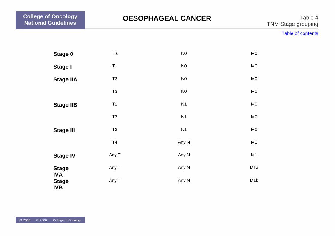
Table 4 TNM Stage grouping
Table of contents
Stage 0 Tis
N0 M0
Stage I T1 N0 M0
Stage IIA T2 N0 M0
T3 N0 M0
Stage IIB T1 N1 M0
T2 N1 M0
Stage III T3 N1 M0
T4 Any N M0
Stage IV Any T Any N M1
Stage IVA
Any T Any N M1a
Stage IVB
Any T Any N M1b
V1.2008 © 2008 College of Oncology