Embed Size (px)
Citation preview
4
Granulomatous Inflammation
INTRODUCTION
Chronic granulomatous inflammation is a proliferative inflam-mation characterized by a cellular infiltrate of epithelioid cells [and sometimes inflammatory giant cells, lymphocytes, plasma cells, polymorphonuclear leukocytes (PMNs), and eosinophils; see Chapter 1].
POST-TRAUMATIC
Sympathetic Uveitis [Sympathetic Ophthalmia (SO), Sympathetic Ophthalmitis]
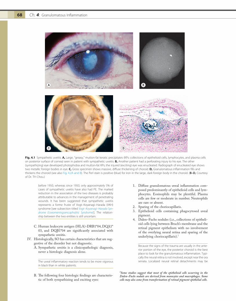
I. SO (Figs 4.1 and 4.2) is a bilateral, diffuse, granuloma-tous, T-cell-mediated uveitis that occurs from two weeks to many years after penetrating or perforating ocular injury and is associated with traumatic uveal incarceration or prolapse.A. Although the uveitis may start as early as five days or
as late as 50 years after injury, well over 90% of cases occur after two weeks but within one year. Most of these (80%) occur within three weeks to three months post-injury.
B. Removal of the injured eye before sympathetic uveitis occurs usually completely protects against inflamma-tion developing in the noninjured eye.1 Once the inflammation starts, however, removal of the injured (“exciting”) eye probably has little effect on the course of the disease, especially after 3–6 months.
SO has been reported in nontraumatized eyes in a few iso-lated cases. However, unless the whole eye is serially sec-tioned and carefully examined for evidence of perforation, the clinician can never be sure that some long-forgotten pen-etrating ocular wound is not present. A diagnosis of sympa-thetic uveitis in the absence of an ocular injury should be viewed with marked skepticism.
C. SO affects approximately 0.03 per 100,000 persons per year and accounts for approximately 1 to 2% of uveitic patients. Greater than 50% of patients maintain a functional visual acuity better than 20/50. The condition seems to be decreasing in occurrences in recent years.
II. Blurred vision and photophobia in the noninjured (sym-pathizing) eye are usually the first symptoms. Vision and photophobia worsen concurrently in the injured (exciting) eye, and a granulomatous uveitis develops (see Fig. 4.1A).
Glaucoma may develop due to blockage of the angle by cellular debris or peripheral anterior synechiae. Hypotony may occur from decreased aqueous output by the inflamed ciliary body.
III. The cause appears to be a delayed-type hypersensitivity reaction of the uvea to antigens localized on the retinal pigment epithelium or on uveal melanocytes.A. The lymphocytic infiltrate consists almost exclusively
of T lymphocytes.B. B cells found in some cases, usually of long duration,
may represent the end stage of the disease.
Phacoanaphylactic endophthalmitis (PE) was found in approximately 25% of patients who had sympathetic uveitis in cases submitted to the Armed Forces Institute of Pathology
1Rarely, sympathetic uveitis has been reported to have developed in the sympathizing eye after the injured eye has been enucleated.
Ch. 4: Granulomatous Inflammation68
1. Diffuse granulomatous uveal inflammation com-posed predominantly of epithelioid cells and lym-phocytes. Eosinophils may be plentiful. Plasma cells are few or moderate in number. Neutrophils are rare or absent.
2. Sparing of the choriocapillaris.3. Epithelioid cells containing phagocytosed uveal
pigment.4. Dalen–Fuchs nodules (i.e., collections of epitheli-
oid cells lying between Bruch’s membrane and the retinal pigment epithelium with no involvement of the overlying neural retina and sparing of the underlying choriocapillaris).2
Because the signs of the trauma are usually in the ante-rior portion of the eye, the posterior choroid is the best place to look for the granulomatous inflammation. Typi-cally the neural retina is not involved, except near the ora serrata. Localized neural retinal detachments may be
before 1950, whereas since 1950, only approximately 5% of cases of sympathetic uveitis have also had PE. The marked reduction in the association of the two diseases is probably attributable to advances in the management of penetrating wounds. It has been suggested that sympathetic uveitis represents a forme fruste of Vogt–Koyanagi–Harada (VKH) syndrome [see subsection titled Vogt–Koyanagi–Harada Syn-drome (Uveomeningoencaphalitic Syndrome)]. The relation-ship between the two entities is still uncertain.
C. Human leukocyte antigen (HLA)-DRB1*04, DQA1* 03, and DQB1*04 are significantly associated with sympathetic uveitis.
IV. Histologically, SO has certain characteristics that are sug-gestive of the disorder but not diagnostic.A. Sympathetic uveitis is a clinicopathologic diagnosis,
never a histologic diagnosis alone.
The uveal inflammatory reaction tends to be more vigorous in black than in white patients.
B. The following four histologic findings are characteris-tic of both sympathizing and exciting eyes:
Fig. 4.1 Sympathetic uveitis. A, Large, “greasy,” mutton-fat keratic precipitates (KPs: collections of epithelioid cells, lymphocytes, and plasma cells on posterior surface of cornea) seen in patient with sympathetic uveitis. B, Another patient had a perforating injury to his eye. The other (sympathizing) eye developed photophobia and mutton-fat KPs; the injured (exciting) eye was enucleated. Radiograph of enucleated eye shows two metallic foreign bodies in eye. C, Gross specimen shows massive, diffuse thickening of choroid. D, Granulomatous inflammation fills and thickens the choroid (see also Fig. 4.2A and B). The Perl stain is positive (blue) for iron in the large, dark foreign body in the choroid. (B–D, Courtesy of Dr. TH Chou.)
A B
C D
2Some studies suggest that most of the epithelioid cells occurring in the Dalen–Fuchs nodule are derived from monocytes and macrophages. Some cells may also come from transformation of retinal pigment epithelial cells.
69Post-Traumatic 69
seen, especially in areas where Dalen–Fuchs nodules coalesce.
C. Other findings:1. Tissue damage caused by the trauma2. Extension of the granulomatous inflammation
into the scleral canals and optic disc
Because uveal tissue is normally found in the scleral canals and in the vicinity of the optic disc, evisceration, which does not reach these areas, does not protect against sympathetic uveitis. If surgery is being done to prevent sympathetic uveitis, the procedure must be an enucleation, not an evisceration.
Phacoanaphylactic (Phacoimmune, Phacoantigenic, or Phacogenic) Endophthalmitis
I. Phacoanaphylactic endophthalmitis (PE) (Fig. 4.3) is a rare, autoimmune, unilateral (sometimes bilateral if the
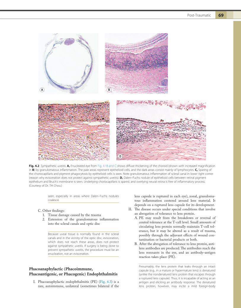
Fig. 4.2 Sympathetic uveitis. A, Enucleated eye from Fig. 4.1B and C shows diffuse thickening of the choroid (shown with increased magnification in B) by granulomatous inflammation. The pale areas represent epithelioid cells, and the dark areas consist mainly of lymphocytes. C, Sparing of the choriocapillaris and pigment phagocytosis by epithelioid cells is seen. Note granulomatous inflammation of scleral canal in lower right corner (reason why evisceration does not protect against sympathetic uveitis). D, Dalen–Fuchs nodule of epithelioid cells between retinal pigment epithelium and Bruch’s membrane is seen. Underlying choriocapillaris is spared, and overlying neural retina is free of inflammatory process. (Courtesy of Dr. TH Chou.)
A B
C D
lens capsule is ruptured in each eye), zonal, granuloma-tous inflammation centered around lens material. It depends on a ruptured lens capsule for its development.
II. The disease occurs under special conditions that involve an abrogation of tolerance to lens protein.A. PE may result from the breakdown or reversal of
central tolerance at the T-cell level. Small amounts of circulating lens protein normally maintain T-cell tol-erance, but it may be altered as a result of trauma, possibly through the adjuvant effects of wound con-tamination or bacterial products or both.
B. After the abrogation of tolerance to lens protein, anti-lens antibodies are produced. The antibodies reach the lens remnants in the eye, and an antibody–antigen reaction takes place (PE).
Presumably, the lens protein that leaks through an intact capsule (e.g., in a mature or hypermature lens) is denatured (unlike the nondenatured lens protein that escapes through a ruptured lens capsule). Thus, it is incapable of acting as an antigen and eliciting an antibody response. The denatured lens protein, however, may incite a mild foreign-body
Ch. 4: Granulomatous Inflammation70
An unusual cause of inflammatory granuloma of the conjunctiva is the synthetic fiber found in teddy bears, called a “teddy-bear” granuloma.
II. Rarely, blood in the vitreous incites a marked foreign-body inflammatory response. When this occurs, the intra-vitreal hemorrhage almost is invariably traumatic in origin rather than spontaneous.
III. Histologically, a zonal type of granulomatous inflamma-tory reaction surrounds the foreign body.
NONTRAUMATIC INFECTIONS
Viral
I. Cytomegalic inclusion disease (salivary gland disease; Fig. 4.4)A. Cytomegalic (CMV) inclusion disease is caused by
systemic infection with the salivary gland virus, cyto-megalovirus, an enveloped herpesvirus formed by an icosahedral capsid and a double-stranded DNA.
CMV is huge, containing more than 200 genes (compared with its modest relative, herpes simplex virus, which contains only 84 genes). It is estimated that CMV infects 80–85% of people by 40 years of age. In otherwise healthy, immunocom-petent people, CMV infection usually runs a benign, asymp-tomatic course (rarely, a heterophile-negative mononucleosis syndrome occurs). After primary exposure, CMV may establish a latent infection and the virus genome may persist in cells undetectable by conventional culture assays.
1. Congenital: Characterized by retinochoroiditis, prematurity, jaundice, thrombocytopenia, anemia,
macrophagic response. The macrophages, swollen with engulfed denatured lens material, may block the anterior chamber drainage angle and cause an acute secondary open-angle glaucoma called phacolytic glaucoma (see Chapter 10).
III. Histologically, in addition to the findings at the site of injury, a zonal granulomatous inflammation is found.A. Activated neutrophils surround and seem to dissolve
or eat away lens material, releasing proteolytic enzymes, arachidonic acid metabolites, and oxygen-derived free radicals.
B. Epithelioid cells and occasional (sometimes in abun-dance) multinucleated inflammatory giant cells are seen beyond the neutrophils.
C. Lymphocytes, plasma cells, fibroblasts, and blood vessels (granulation tissue) surround the epithelioid cells.
D. Usually the iris is encased in, and inseparable from, the inflammatory reaction.
E. The uveal tract usually shows a reactive, chronic nongranulomatous inflammatory reaction. Some-times, however, the same trauma that ruptures the lens and sets off the PE initiates a sympathetic uveitis and results in a diffuse, chronic, granulomatous inflammation.
Foreign-Body Granulomas
I. Foreign-body granulomas may develop around exogenous foreign bodies that are usually introduced into the eye at the time of a penetrating ocular wound, or they may develop around endogenous products such as cholesterol or blood in the vitreous.
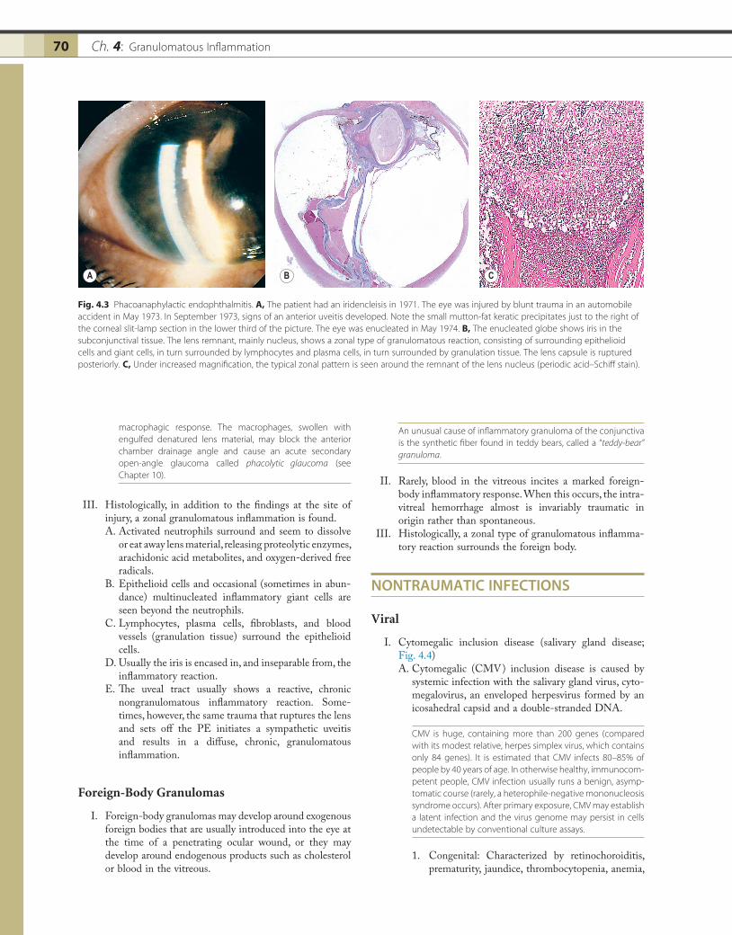
Fig. 4.3 Phacoanaphylactic endophthalmitis. A, The patient had an iridencleisis in 1971. The eye was injured by blunt trauma in an automobile accident in May 1973. In September 1973, signs of an anterior uveitis developed. Note the small mutton-fat keratic precipitates just to the right of the corneal slit-lamp section in the lower third of the picture. The eye was enucleated in May 1974. B, The enucleated globe shows iris in the subconjunctival tissue. The lens remnant, mainly nucleus, shows a zonal type of granulomatous reaction, consisting of surrounding epithelioid cells and giant cells, in turn surrounded by lymphocytes and plasma cells, in turn surrounded by granulation tissue. The lens capsule is ruptured posteriorly. C, Under increased magnification, the typical zonal pattern is seen around the remnant of the lens nucleus (periodic acid–Schiff stain).
A B C
71Nontraumatic Infections 71
iici
A
C
B
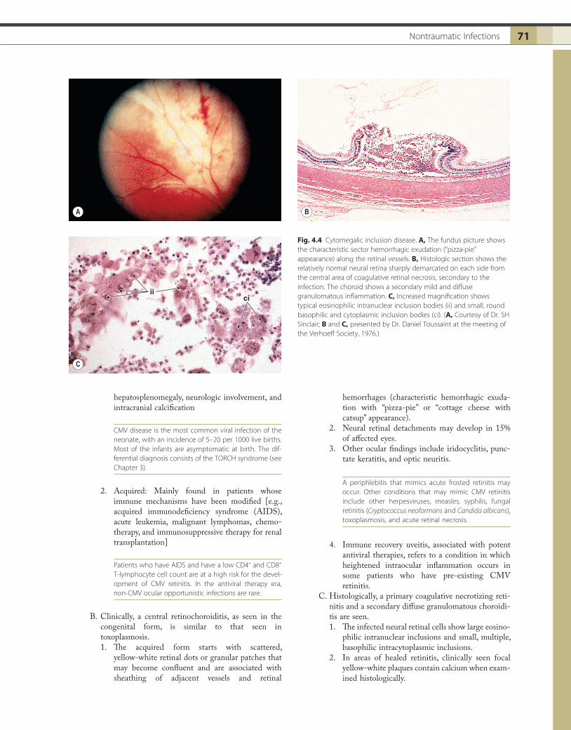
Fig. 4.4 Cytomegalic inclusion disease. A, The fundus picture shows the characteristic sector hemorrhagic exudation (“pizza-pie” appearance) along the retinal vessels. B, Histologic section shows the relatively normal neural retina sharply demarcated on each side from the central area of coagulative retinal necrosis, secondary to the infection. The choroid shows a secondary mild and diffuse granulomatous inflammation. C, Increased magnification shows typical eosinophilic intranuclear inclusion bodies (ii) and small, round basophilic and cytoplasmic inclusion bodies (ci). (A, Courtesy of Dr. SH Sinclair; B and C, presented by Dr. Daniel Toussaint at the meeting of the Verhoeff Society, 1976.)
hepatosplenomegaly, neurologic involvement, and intracranial calcification
CMV disease is the most common viral infection of the neonate, with an incidence of 5–20 per 1000 live births. Most of the infants are asymptomatic at birth. The dif-ferential diagnosis consists of the TORCH syndrome (see Chapter 3).
2. Acquired: Mainly found in patients whose immune mechanisms have been modified [e.g., acquired immunodeficiency syndrome (AIDS), acute leukemia, malignant lymphomas, chemo-therapy, and immunosuppressive therapy for renal transplantation]
Patients who have AIDS and have a low CD4+ and CD8+ T-lymphocyte cell count are at a high risk for the devel-opment of CMV retinitis. In the antiviral therapy era, non-CMV ocular opportunistic infections are rare.
B. Clinically, a central retinochoroiditis, as seen in the congenital form, is similar to that seen in toxoplasmosis.1. The acquired form starts with scattered,
yellow-white retinal dots or granular patches that may become confluent and are associated with sheathing of adjacent vessels and retinal
hemorrhages (characteristic hemorrhagic exuda-tion with “pizza-pie” or “cottage cheese with catsup” appearance).
2. Neural retinal detachments may develop in 15% of affected eyes.
3. Other ocular findings include iridocyclitis, punc-tate keratitis, and optic neuritis.
A periphlebitis that mimics acute frosted retinitis may occur. Other conditions that may mimic CMV retinitis include other herpesviruses, measles, syphilis, fungal retinitis (Cryptococcus neoformans and Candida albicans), toxoplasmosis, and acute retinal necrosis.
4. Immune recovery uveitis, associated with potent antiviral therapies, refers to a condition in which heightened intraocular inflammation occurs in some patients who have pre-existing CMV retinitis.
C. Histologically, a primary coagulative necrotizing reti-nitis and a secondary diffuse granulomatous choroidi-tis are seen.1. The infected neural retinal cells show large eosino-
philic intranuclear inclusions and small, multiple, basophilic intracytoplasmic inclusions.
2. In areas of healed retinitis, clinically seen focal yellow-white plaques contain calcium when exam-ined histologically.
Ch. 4: Granulomatous Inflammation72
B. Ocular complications occur in approximately 50% of cases of herpes zoster ophthalmicus:1. Cornea: dendritic ulcer (rare), ulceration, per-
foration, peripheral erosions, bullous keratopathy, epidermidalization (keratinization), band kera-topathy, pannus formation, stromal vasculariza-tion, hypertrophy of corneal nerves, ring abscess, granulomatous reaction to Descemet’s membrane, and endothelial degeneration
2. Anterior segment: iridocyclitis followed by periph-eral anterior synechiae, exudate, and hyphema
3. Iris: patchy necrosis and postnecrotic atrophy (mimics iris after attack of acute angle-closure glaucoma), chronic nongranulomatous inflamma-tion, anterior-surface fibrovascular membrane, patchy necrosis of anterior portion of ciliary body, especially of circular and radial portions of ciliary muscle, and cataract and posterior synechiae
4. Posterior segment: chronic nongranulomatous choroiditis (commonly, granulomatous inflamma-tion), retinal perivasculitis and vasculitis, mild mononuclear vitreal inflammatory infiltrate, and acute or chronic episcleritis and scleritis; optic nerve: perivasculitis and chronic leptomeningitis; and long posterior ciliary nerves and vessels: strik-ing perineural and, less commonly, intraneural nongranulomatous and occasionally granuloma-tous inflammation and perivasculitis and vasculitis
C. Histologically, the most characteristic findings are lymphocytic (chronic nongranulomatous) infiltrations involving the posterior ciliary nerves and vessels, often
The cytoplasmic inclusions consist of numerous virions closely associated with dense masses of matter (periodic acid–Schiff-positive on light microscopy) that are highly characteristic of CMV. An additional highly characteristic feature is the presence of the virions in a mass of viral subunit material that forms a lacy, centrally located pattern in the nucleus. The nucleolus is marginated and free of virions. Clumping of peripheral chromatin is lacking.
3. The location and character of the retinal vascular changes in AIDS indicate an ischemic pathogen-esis, most profound in CMV retinitis.
II. Varicella/herpes zoster virus (VZV; Figs 4.5 and 4.6)A. VZV causes varicella (chickenpox) and herpes zoster
(shingles).1. The virus, a member of the herpesvirus family,
consists of a lipid envelope surrounding an icosa-hedral nucleocapsid with a central, double-stranded DNA core; only the enveloped virions are infectious.
2. Congenital infection is rare (differential diagnosis consists of the TORCH syndrome; see Chapter 3).
3. In immunocompetent individuals, VZV is a major cause of the acute retinal necrosis syndrome (see Chapter 11).
Following herpes zoster ophthalmicus, patients have a nine times greater risk of developing cancer than patients without herpes zoster ophthalmicus. Also, the risk of anterior uveitis increases the year following VZV.
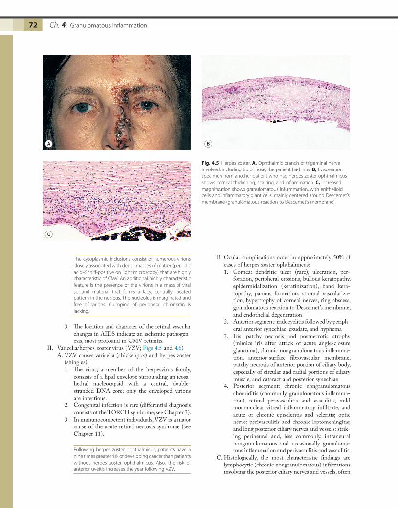
Fig. 4.5 Herpes zoster. A, Ophthalmic branch of trigeminal nerve involved, including tip of nose; the patient had iritis. B, Evisceration specimen from another patient who had herpes zoster ophthalmicus shows corneal thickening, scarring, and inflammation. C, Increased magnification shows granulomatous inflammation, with epithelioid cells and inflammatory giant cells, mainly centered around Descemet’s membrane (granulomatous reaction to Descemet’s membrane).
A
C
B

73Nontraumatic Infections 73
in a segmental distribution, and a diffuse or patchy necrosis involving the iris and pars plicata of the ciliary body. Granulomatous inflammatory lesions also may be seen. Inclusion bodies have not been demonstrated in the chronic inflammatory lesions.
Bacterial
I. Tuberculosis (Mycobacterium tuberculosis; Figs 4.7 and 4.8)A. Tuberculosis has re-emerged as a serious public health
problem, mainly because of the human immunodefi-ciency virus (HIV) epidemic and newly developed resistance to standard antibiotic therapy.
It is estimated that approximately one-third of the world’s population is infected by Mycobacterium tuberculosis. It may present in children initially as a preceptal cellulitis unrespon-sive to systemic antibiotic therapy.
B. Tubercle bacilli reach the eye through the blood-stream, after lung infection.
Tubercle bacilli survive within macrophages because they secrete eukaryocyte-like serine/threonine protein kinase G within macrophage phagosomes, inhibiting phagosome–lysosome fusion and mediating intracellular survival of myco-bacterium. Rarely, intraocular tuberculosis can occur without obvious systemic infection.
Fig. 4.6 Herpes zoster. A, Patient with herpes zoster ophthalmicus developed chronic herpes keratitis and then corneal perforation; the eye was enucleated. B, Nongranulomatous inflammatory infiltrates centered around ciliary nerves in posterior episclera (no granulomatous inflammation present), shown with increased magnification in C.
A
C
B
1. The most common form of ocular involvement is a cyclitis that rapidly becomes an iridocyclitis and may also spread posteriorly to cause a choroiditis.
2. Clinically, mutton-fat keratic precipitates are seen on the posterior surface of the cornea and deep infiltrates in the choroid, often in the posterior pole.
Retinal tuberculosis usually spreads from an underlying choroiditis. The involvement may become massive to form a large tuberculoma involving all the coats of the eye. Tuberculoprotein hypersensitivity may play a role in the pathogenesis of phlyctenules and Eales’ disease. Tuberculous choroiditis may simulate serpigi-nous choroiditis (called tuberculous serpiginous-like choroiditis).
C. Miliary tuberculosis usually causes a multifocal, discrete (sarcoidal, tuberculoidal) granulomatous choroiditis.
D. Histologically, the classic pattern of caseation necrosis consists of a zonal type of granulomatous reaction around the area of coagulative necrosis.1. Smooth, acid-fast bacilli can be demonstrated by
acid-fast (Ziehl–Neelsen) or fluorescent acid-fast stains.
2. The polymerase chain reaction, prepared from formaldehyde-fixed and paraffin-embedded tissue, can be helpful in making the diagnosis.
Ch. 4: Granulomatous Inflammation74
The bacteria may grow better in the cooler, anterior portion of the eye rather than in the warmer, post-erior portion, just as they do in the cooler skin instead of in the warmer, deeper structures of the body.
B. In tuberculoid leprosy, the lepromin test is positive, sug-gesting immunity. The prognosis is good.1. A neural involvement, particularly the ulnar nerve
(leads to claw hand), predominates with hypopig-mented (vitiliginous), hypoesthetic lesions, and thickened nerves.
2. The ocular adnexa and orbital structures are involved, especially the ciliary nerves, but not the eyeballs.
3. Histologically, a discrete (sarcoidal, tuberculoidal) type of granulomatous inflammatory reaction is seen, mainly centered around nerves.a. The individual nodules tend to be much more
variably sized than those in sarcoidosis or miliary tuberculosis.
b. Organisms are extremely difficult to find (good immunity) and are usually located in an area of nerve degeneration.
II. Leprosy (Hansen’s disease; M. leprae; Fig. 4.9)A. In lepromatous leprosy, the lepromin test (analogous to
the tuberculin test) is negative, suggesting little or no immunity. The prognosis is poor.
Genes belonging to the leukocyte immunoglobulin-like receptor (LIR) family are significantly upregulated in lesions of lepromatous patients suffering from the disseminated form of the infection.
1. Lepromas of the skin result in leonine facies and neurologic changes. The eyeballs are involved, usually in their anterior portions.
2. Histologically, a diffuse type of granulomatous inflammatory reaction, known as a leproma, is present.a. Lepromas, which involve mainly cornea, ante-
rior sclera, and iris, show large, pale-staining histiocytes that are called lepra cells when their cytoplasm is amorphous and Virchow’s cells when vacuolated.
b. The lepra cells and Virchow’s cells teem with beaded bacilli (no immunity).
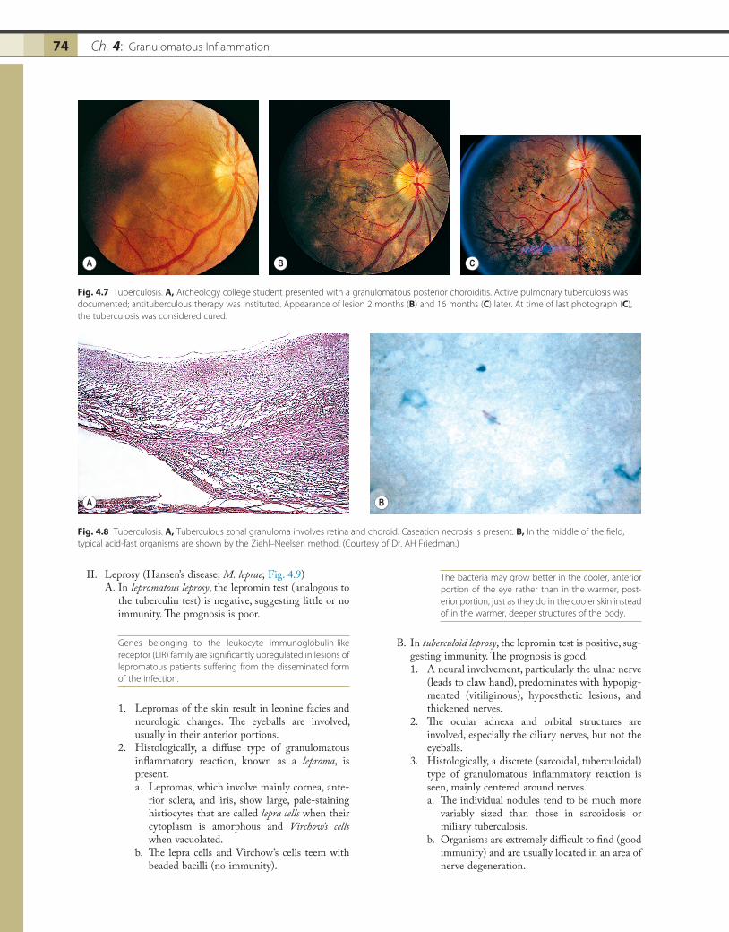
Fig. 4.7 Tuberculosis. A, Archeology college student presented with a granulomatous posterior choroiditis. Active pulmonary tuberculosis was documented; antituberculous therapy was instituted. Appearance of lesion 2 months (B) and 16 months (C) later. At time of last photograph (C), the tuberculosis was considered cured.
A B C
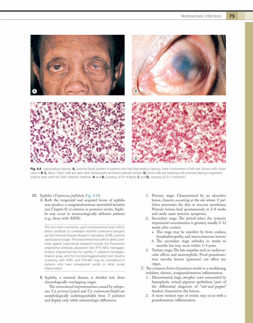
Fig. 4.8 Tuberculosis. A, Tuberculous zonal granuloma involves retina and choroid. Caseation necrosis is present. B, In the middle of the field, typical acid-fast organisms are shown by the Ziehl–Neelsen method. (Courtesy of Dr. AH Friedman.)
A B
75Nontraumatic Infections 75
1. Primary stage: Characterized by an ulcerative lesion, chancre, occurring at the site where T. pal-lidum penetrates the skin or mucous membrane. Primary lesions heal spontaneously in 2–8 weeks and rarely cause systemic symptoms.
2. Secondary stage: The period when the systemic treponemal concentration is greatest, usually 2–12 weeks after contact.a. This stage may be manifest by fever, malaise,
lymphadenopathy, and mucocutaneous lesions.b. The secondary stage subsides in weeks to
months but may recur within 1–4 years.3. Tertiary stage: The late sequelae such as cardiovas-
cular effects and neurosyphilis. Focal granuloma-tous vascular lesions (gummas) can affect any organ.
C. The common form of posterior uveitis is a smoldering, indolent, chronic, nongranulomatous inflammation.1. Disseminated, large, atrophic scars surrounded by
hyperplastic retinal pigment epithelium (part of the differential diagnosis of “salt-and-pepper” fundus) characterize the lesions.
2. A more virulent type of uveitis may occur with a granulomatous inflammation.
III. Syphilis (Treponema pallidum; Fig. 4.10)A. Both the congenital and acquired forms of syphilis
may produce a nongranulomatous interstitial keratitis (see Chapter 8) or anterior or posterior uveitis. Syphi-lis may occur in immunologically deficient patients (e.g., those with AIDS).
The two most commonly used nontreponemal tests (which detect antibody to cardiolipin–lecithin–cholesterol antigen) are the Venereal Disease Research Laboratory (VDRL) and the rapid plasma reagin. The treponemal tests (which detect anti-body against treponemal antigens) include the fluorescent treponemal antibody absorption test (FTA-ABS), hemagglu-tination treponemal test for syphilis, T. pallidum hemagglu-tination assay, and the microhemagglutination test. Routine screening with VDRL and FTA-ABS may be considered in patients who have unexplained uveitis or other ocular inflammation.
B. Syphilis, a venereal disease, is divided into three chronologically overlapping stages.
The nonvenereal treponematoses caused by subspe-cies T. p. pertenue (yaws) and T. p. endemicum (bejel) are morphologically indistinguishable from T. pallidum and display only subtle immunologic differences.
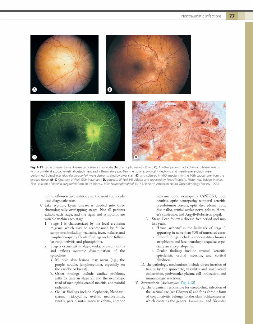
Fig. 4.9 Lepromatous leprosy. A, Leonine facies present in patient who had lepromatous leprosy. Note involvement of left eye, shown with closer view in B. C, Many “clear” cells are seen with hematoxylin and eosin-stained section. D, Same cells are teeming with acid-fast leprous organisms (red) as seen with the Ziehl–Neelsen method. (A and B, Courtesy of Dr. B Blaise; C and D, courtesy of Dr. P Henkind.)
A B
C D

Ch. 4: Granulomatous Inflammation76
a. The inflammatory process usually involves the choroid and the overlying neural retina and is quite vascular. Epithelioid cells, lymphocytes, and plasma cells are seen.
b. Spirochetes can be demonstrated in the inflam-matory tissue.
3. The preceding two types of reactions may also involve the anterior uvea.
Spirochetes may be obtained by aspiration of aqueous from the anterior chamber and identified by dark-field microscopy.
IV. Lyme disease (Borrelia burgdorferi; Fig. 4.11)A. Lyme disease is a worldwide, tickborne, multisystem
disorder, heralded by a red rash and erythema migrans, which forms at the site of the tick bite, usually within 4–20 days. The rash enlarges with central clearing (forming a ring), can last several weeks, and may return and become chronic (erythema chronicum migrans).
B. The tick, an Ixodes species, transmits the infectious agent, B. burgdorferi, through its bite. The enzyme-linked immunosorbent assay (ELISA) and the indirect
D. Histologic findings1. The chronic nongranulomatous disseminated
form of posterior choroiditis:a. In the atrophic scar, the outer neural retinal
layers, the retinal pigment epithelium, and the inner choroidal layers disappear. Dehiscences in Bruch’s membrane may be present through which neural retinal elements may “invade” the choroid.
b. Bruch’s membrane may be folded into the atro-phic, sclerosed choroid. Scattered lymphocytes and plasma cells may be present.
c. The Treponema spirochete is a helical bacterium 5–15 µm in length and less than 0.18 µm in width, and it can be demonstrated in the ocular tissue with special stains, often in areas devoid of inflammatory cells.
Treponema pallidum belongs to the same family (Spi-rochaetaceae) as Borrelia (see later) and Leptospira.
2. The granulomatous form of posterior chorioreti-nitis:
Fig. 4.10 Syphilis. A, Small, round translucent nodules are seen in the conjunctiva of the inferior fornix. B, Biopsy of nodules shows numerous granulomas under the conjunctival epithelium (ce, surface conjunctival epithelium; gr, granulomatous reaction in substantia propia). C, Increased magnification reveals epithelioid cells in the inflammatory nodules. D, A special stain, Dieteria, demonstrates spirochetes (s) in the inflammatory infiltrate. (Case reported in Spektor FE, Eagle RC, Nichols CW: Granulomatous conjunctivitis secondary to Treponema pallidum. Ophthalmology 88:863. © PubMed Central 1981.)
ce
gr
s
A B
C D
77Nontraumatic Infections 77
ischemic optic neuropathy (ANION), optic neuritis, optic neuropathy, temporal arteritis, pseudotumor cerebri, optic disc edema, optic disc pallor, cranial ocular nerve palsies, Horn-er’s syndrome, and Argyll–Robertson pupil.
3. Stage 3 can follow a disease-free period and may last years.a. “Lyme arthritis” is the hallmark of stage 3,
appearing in more than 50% of untreated cases.b. Other findings include acrodermatitis chronica
atrophicans and late neurologic sequelae, espe-cially an encephalopathy.
c. Ocular findings include stromal keratitis, episcleritis, orbital myositis, and cortical blindness.
D. The pathologic mechanisms include direct invasion of tissues by the spirochete, vasculitis and small-vessel obliteration, perivascular plasma cell infiltration, and immunologic reactions.
V. Streptothrix (Actinomyces; Fig. 4.12)A. The organism responsible for streptothrix infection of
the lacrimal sac (see Chapter 6) and for a chronic form of conjunctivitis belongs to the class Schizomycetes, which contains the genera Actinomyces and Nocardia.
immunofluorescence antibody are the most commonly used diagnostic tests.
C. Like syphilis, Lyme disease is divided into three chronologically overlapping stages. Not all patients exhibit each stage, and the signs and symptoms are variable within each stage.1. Stage 1 is characterized by the local erythema
migrans, which may be accompanied by flulike symptoms, including headache, fever, malaise, and lymphadenopathy. Ocular findings include follicu-lar conjunctivitis and photophobia.
2. Stage 2 occurs within days, weeks, or even months and reflects systemic dissemination of the spirochete.a. Multiple skin lesions may occur (e.g., the
purple nodule, lymphocytoma, especially on the earlobe or breast).
b. Other findings include cardiac problems, arthritis (rare in stage 2), and the neurologic triad of meningitis, cranial neuritis, and painful radiculitis.
c. Ocular findings include blepharitis, blepharo-spasm, iridocyclitis, uveitis, neuroretinitis, vitritis, pars planitis, macular edema, anterior
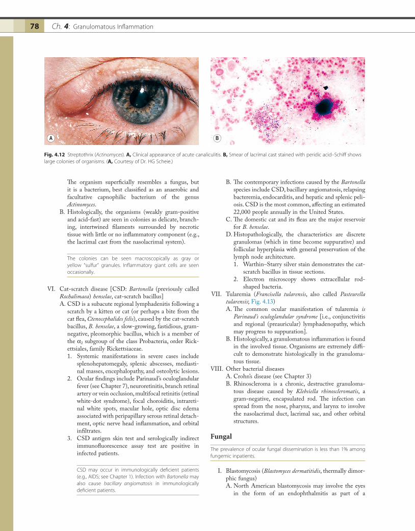
Fig. 4.11 Lyme disease. Lyme disease can cause a choroiditis (A) or an optic neuritis (B and C). Another patient had a chronic bilateral uveitis with a unilateral exudative retinal detachment and inflammatory pupillary membrane. Surgical iridectomy and membrane excision were performed. Spirochetes (Borrelia burgdorferi) were demonstrated by silver stain (D) and cultured in MKP medium (in the 16th subculture) from the excised tissue. (A–C, Courtesy of Prof. GOH Naumann; D, courtesy of Prof. HE Völcker and reported by Preac-Mursic V, Pfister HW, Spiegel H et al.: First isolation of Borrelia burgdorferi from an iris biopsy. J Clin Neuroophthalmol 13:155. © North American Neuro-Ophthalmology Society 1993.)
A B
C D
Ch. 4: Granulomatous Inflammation78
B. The contemporary infections caused by the Bartonella species include CSD, bacillary angiomatosis, relapsing bacteremia, endocarditis, and hepatic and splenic peli-osis. CSD is the most common, affecting an estimated 22,000 people annually in the United States.
C. The domestic cat and its fleas are the major reservoir for B. henselae.
D. Histopathologically, the characteristics are discrete granulomas (which in time become suppurative) and follicular hyperplasia with general preservation of the lymph node architecture.1. Warthin–Starry silver stain demonstrates the cat-
scratch bacillus in tissue sections.2. Electron microscopy shows extracellular rod-
shaped bacteria. VII. Tularemia (Francisella tularensis, also called Pasteurella
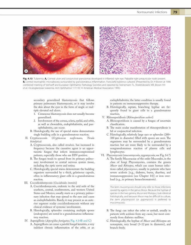
tularensis; Fig. 4.13)A. The common ocular manifestation of tularemia is
Parinaud’s oculoglandular syndrome [i.e., conjunctivitis and regional (preauricular) lymphadenopathy, which may progress to suppuration].
B. Histologically, a granulomatous inflammation is found in the involved tissue. Organisms are extremely diffi-cult to demonstrate histologically in the granuloma-tous tissue.
VIII. Other bacterial diseasesA. Crohn’s disease (see Chapter 3)B. Rhinoscleroma is a chronic, destructive granuloma-
tous disease caused by Klebsiella rhinoscleromatis, a gram-negative, encapsulated rod. The infection can spread from the nose, pharynx, and larynx to involve the nasolacrimal duct, lacrimal sac, and other orbital structures.
Fungal
The prevalence of ocular fungal dissemination is less than 1% among fungemic inpatients.
I. Blastomycosis (Blastomyces dermatitidis, thermally dimor-phic fungus)A. North American blastomycosis may involve the eyes
in the form of an endophthalmitis as part of a
The organism superficially resembles a fungus, but it is a bacterium, best classified as an anaerobic and facultative capnophilic bacterium of the genus Actinomyces.
B. Histologically, the organisms (weakly gram-positive and acid-fast) are seen in colonies as delicate, branch-ing, intertwined filaments surrounded by necrotic tissue with little or no inflammatory component (e.g., the lacrimal cast from the nasolacrimal system).
The colonies can be seen macroscopically as gray or yellow “sulfur” granules. Inflammatory giant cells are seen occasionally.
VI. Cat-scratch disease [CSD: Bartonella (previously called Rochalimaea) henselae, cat-scratch bacillus]A. CSD is a subacute regional lymphadenitis following a
scratch by a kitten or cat (or perhaps a bite from the cat flea, Ctenocephalides felis), caused by the cat-scratch bacillus, B. henselae, a slow-growing, fastidious, gram-negative, pleomorphic bacillus, which is a member of the α2 subgroup of the class Probacteria, order Rick-ettsiales, family Rickettsiaceae.1. Systemic manifestations in severe cases include
splenohepatomegaly, splenic abscesses, mediasti-nal masses, encephalopathy, and osteolytic lesions.
2. Ocular findings include Parinaud’s oculoglandular fever (see Chapter 7), neuroretinitis, branch retinal artery or vein occlusion, multifocal retinitis (retinal white-dot syndrome), focal choroiditis, intrareti-nal white spots, macular hole, optic disc edema associated with peripapillary serous retinal detach-ment, optic nerve head inflammation, and orbital infiltrates.
3. CSD antigen skin test and serologically indirect immunofluorescence assay test are positive in infected patients.
CSD may occur in immunologically deficient patients (e.g., AIDS; see Chapter 1). Infection with Bartonella may also cause bacillary angiomatosis in immunologically deficient patients.
Fig. 4.12 Streptothrix (Actinomyces). A, Clinical appearance of acute canaliculitis. B, Smear of lacrimal cast stained with peridic acid–Schiff shows large colonies of organisms. (A, Courtesy of Dr. HG Scheie.)
A B
79Nontraumatic Infections 79
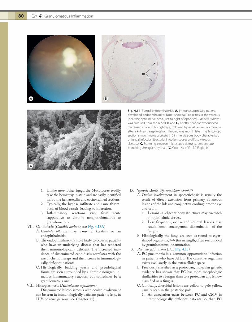
endophthalmitis; the latter condition is usually found in patients on immunosuppressive therapy.
B. Histologically, septate, branching hyphae are fre-quently found in giant cells in a granulomatous reaction.
V. Rhinosporidiosis (Rhinosporidium seeberi)A. Rhinosporidiosis is caused by a fungus of uncertain
classification.B. The main ocular manifestation of rhinosporidiosis is
lid or conjunctival infection.C. Histologically, relatively large sacs or spherules (200–
300 µm in diameter) filled with spores are seen. The organisms may be surrounded by a granulomatous reaction but are more likely to be surrounded by a nongranulomatous reaction of plasma cells and lymphocytes.
VI. Phycomycosis (mucormycosis, zygomycosis; see Fig. 14.7)A. The family Mucoraceae of the order Mucorales, in the
class of fungi Phycomycetes, contains the genera Mucor and Rhizopus, which can cause human infec-tions called phycomycoses, usually in patients who have severe acidosis [e.g., diabetes, burns, diarrhea, and immunosuppression (see Chapter 14)] or iron over-load (e.g., in primary hemochromatosis).
The term mucormycosis should only refer to those infections caused by agents in the genus Mucor. Because the hyphae of species in the two genera, Mucor and Rhizopus, look identical histologically, and because Mucor may be difficult to culture, the term phycomycosis (or zygomycosis) is preferred to mucormycosis.
B. The fungi can infect the orbit or eyeball, usually in patients with acidosis from any cause, but most com-monly from diabetes mellitus.
C. Histologically, the hyphae of Mucor and Rhizopus are nonseptate, very broad (3–12 µm in diameter), and branch freely.
secondary generalized blastomycosis that follows primary pulmonary blastomycosis, or it may involve the skin about the eyes in the form of single or mul-tiple elevated red ulcers.1. Cutaneous blastomycosis does not usually become
generalized.2. Involvement of the cornea, sclera, eyelid, and orbit,
as well as choroiditis, endophthalmitis, and pan-ophthalmitis, can occur.
B. Histologically, the use of special stains demonstrates single budding cells in a granulomatous reaction.
II. Cryptococcosis (Cryptococcus neoformans, Torula histolytica)A. Cryptococcosis, also called torulosis, has increased in
frequency because the causative agent is an oppor-tunistic fungus that infects immunocompromised patients, especially those who are HIV-positive.
B. The fungus tends to spread from its primary pulmo-nary involvement to central nervous system tissue, including the optic nerve and retina.
C. Histologically, special stains demonstrate the budding organism surrounded by a thick, gelatinous capsule, often in inflammatory giant cells in a granulomatous reaction.
III. Coccidioidomycosis (Coccidioides immitis)A. Coccidioidomycosis, endemic to the arid soils of the
southern, central, southwestern, and western United States and Mexico, usually starts as a primary pulmo-nary infection that may spread to the eyes and cause an endophthalmitis. Rarely, it may present as an ante-rior segment ocular coccidioidomycosis without any clinical evidence of systemic involvement.
B. Histologically, spherules containing multiple spores (endospores) are noted in a granulomatous inflamma-tory reaction.
IV. Aspergillosis (Aspergillus fumigatus; Fig. 4.14B and C)A. Aspergillosis can cause a painful fungal keratitis, a very
indolent chronic inflammation of the orbit, or an
Fig. 4.13 Tularemia. A, Corneal ulcer and conjunctival granulomas developed in inflamed right eye. Palpable right preauricular node present. B, Central neutrophilic microabscess surrounded by granulomatous inflammation. Francisella tularensis cultured. (Presented by Dr. H Brown at 1996 combined meeting of Verhoeff and European Ophthalmic Pathology Societies and reported by Steinemann TL, Sheikholeslami MR, Brown HH et al.: Oculoglandular tularemia. Arch Ophthalmol 117:132. © American Medical Association 1999.)
A B
Ch. 4: Granulomatous Inflammation80
IX. Sporotrichosis (Sporotrichum schenkii)A. Ocular involvement in sporotrichosis is usually the
result of direct extension from primary cutaneous lesions of the lids and conjunctiva eroding into the eye and orbit.1. Lesions in adjacent bony structures may encroach
on ophthalmic tissues.2. Less frequently, ocular and adnexal lesions may
result from hematogenous dissemination of the fungus.
B. Histologically, the fungi are seen as round to cigar-shaped organisms, 3–6 µm in length, often surrounded by granulomatous inflammation.
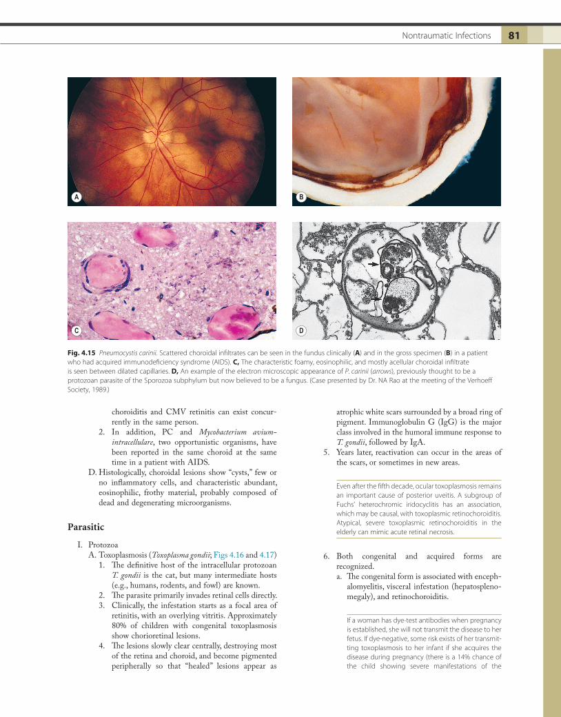
X. Pneumocystis carinii (PC; Fig. 4.15)A. PC pneumonia is a common opportunistic infection
in patients who have AIDS. The causative organism exists exclusively in the extracellular space.
B. Previously classified as a protozoan, molecular genetic evidence has shown that PC has more morphologic similarities to a fungus than to a protozoan and is now classified as a fungus.
C. Clinically, choroidal lesions are yellow to pale yellow, usually seen in the posterior pole.1. An association exists between PC and CMV in
immunologically deficient patients so that PC
1. Unlike most other fungi, the Mucoraceae readily take the hematoxylin stain and are easily identified in routine hematoxylin and eosin-stained sections.
2. Typically, the hyphae infiltrate and cause throm-bosis of blood vessels, leading to infarction.
3. Inflammatory reactions vary from acute suppurative to chronic nongranulomatous to granulomatous.
VII. Candidiasis (Candida albicans; see Fig. 4.13A)A. Candida albicans may cause a keratitis or an
endophthalmitis.B. The endophthalmitis is most likely to occur in patients
who have an underlying disease that has rendered them immunologically deficient. The increased inci-dence of disseminated candidiasis correlates with the use of chemotherapy and the increase in immunologi-cally deficient patients.
C. Histologically, budding yeasts and pseudohyphal forms are seen surrounded by a chronic nongranulo-matous inflammatory reaction, but sometimes by a granulomatous one.
VIII. Histoplasmosis (Histoplasma capsulatum)Disseminated histoplasmosis with ocular involvement
can be seen in immunologically deficient patients (e.g., in HIV-positive persons; see Chapter 11).
Fig. 4.14 Fungal endophthalmitis. A, Immunosuppressed patient developed endophthalmitis. Note “snowball” opacities in the vitreous (near the optic nerve head, just to right of opacities). Candida albicans was cultured from the blood. B and C, Another patient experienced decreased vision in his right eye, followed by renal failure two months after a kidney transplantation. He died one month later. The histologic section shows microabscesses (m) in the vitreous body characteristic of fungal infection (bacterial infection causes a diffuse vitreous abscess). C, Scanning electron microscopy demonstrates septate branching Aspergillus hyphae. (C, Courtesy of Dr. RC Eagle, Jr.)
m
A
C
B
81Nontraumatic Infections 81
atrophic white scars surrounded by a broad ring of pigment. Immunoglobulin G (IgG) is the major class involved in the humoral immune response to T. gondii, followed by IgA.
5. Years later, reactivation can occur in the areas of the scars, or sometimes in new areas.
Even after the fifth decade, ocular toxoplasmosis remains an important cause of posterior uveitis. A subgroup of Fuchs’ heterochromic iridocyclitis has an association, which may be causal, with toxoplasmic retinochoroiditis. Atypical, severe toxoplasmic retinochoroiditis in the elderly can mimic acute retinal necrosis.
6. Both congenital and acquired forms are recognized.a. The congenital form is associated with enceph-
alomyelitis, visceral infestation (hepatospleno-megaly), and retinochoroiditis.
If a woman has dye-test antibodies when pregnancy is established, she will not transmit the disease to her fetus. If dye-negative, some risk exists of her transmit-ting toxoplasmosis to her infant if she acquires the disease during pregnancy (there is a 14% chance of the child showing severe manifestations of the
choroiditis and CMV retinitis can exist concur-rently in the same person.
2. In addition, PC and Mycobacterium avium-intracellulare, two opportunistic organisms, have been reported in the same choroid at the same time in a patient with AIDS.
D. Histologically, choroidal lesions show “cysts,” few or no inflammatory cells, and characteristic abundant, eosinophilic, frothy material, probably composed of dead and degenerating microorganisms.
Parasitic
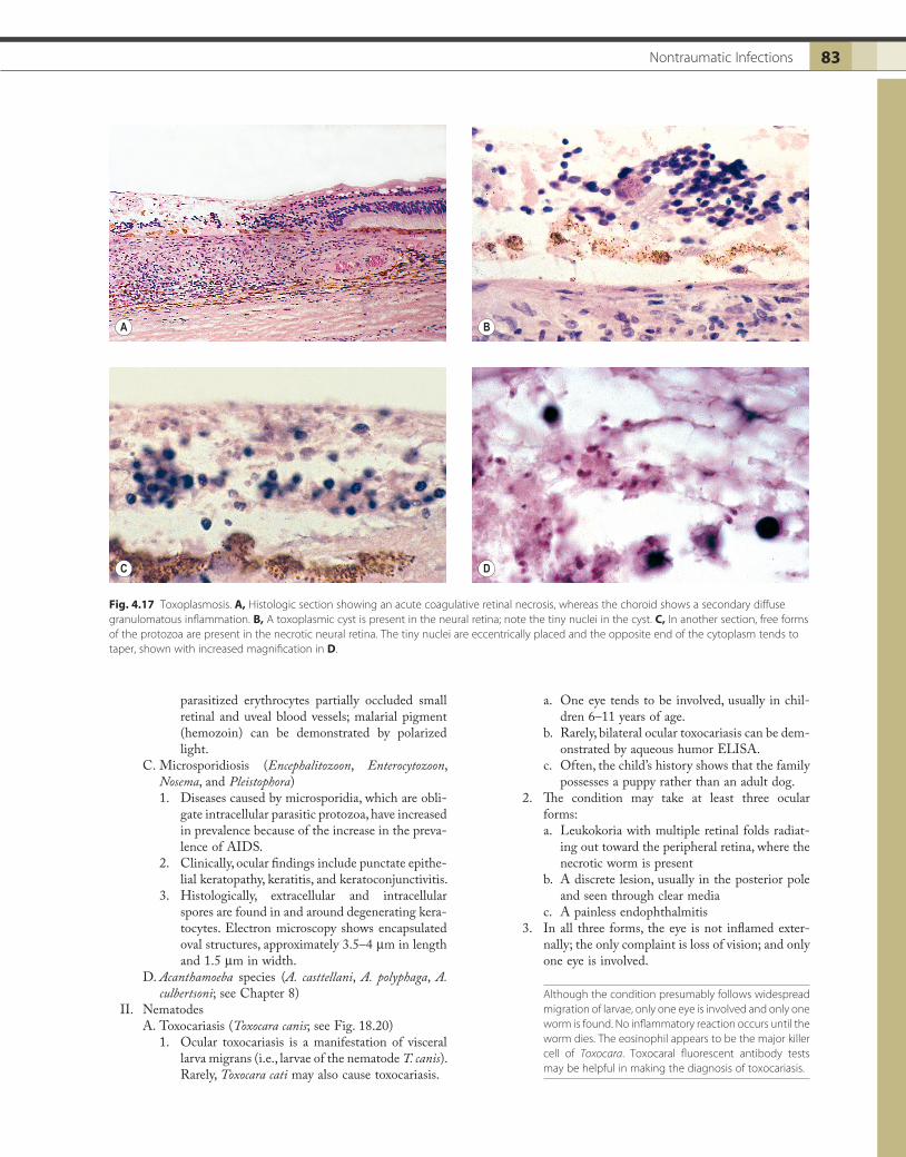
I. ProtozoaA. Toxoplasmosis (Toxoplasma gondii; Figs 4.16 and 4.17)
1. The definitive host of the intracellular protozoan T. gondii is the cat, but many intermediate hosts (e.g., humans, rodents, and fowl) are known.
2. The parasite primarily invades retinal cells directly.3. Clinically, the infestation starts as a focal area of
retinitis, with an overlying vitritis. Approximately 80% of children with congenital toxoplasmosis show chorioretinal lesions.
4. The lesions slowly clear centrally, destroying most of the retina and choroid, and become pigmented peripherally so that “healed” lesions appear as
Fig. 4.15 Pneumocystis carinii. Scattered choroidal infiltrates can be seen in the fundus clinically (A) and in the gross specimen (B) in a patient who had acquired immunodeficiency syndrome (AIDS). C, The characteristic foamy, eosinophilic, and mostly acellular choroidal infiltrate is seen between dilated capillaries. D, An example of the electron microscopic appearance of P. carinii (arrows), previously thought to be a protozoan parasite of the Sporozoa subphylum but now believed to be a fungus. (Case presented by Dr. NA Rao at the meeting of the Verhoeff Society, 1989.)
A B
C D

Ch. 4: Granulomatous Inflammation82
and multiplies in the confines of the cell mem-brane. All that is seen histologically, therefore, is a group of protozoa surrounded by the retinal cell membrane; the whole assemblage is called a pseudocyst.
c. If the environment becomes inhospitable, an intracellular protozoan (trophozoite) may transform itself into a bradyzoite, surround itself with a self-made membrane, multiply, and then form a true cyst that extrudes from the cell and lies free in the tissue.1) It is found in the late stage of the disease,
at the time of remission.2) The true cyst is resistant to the host’s
defenses and can remain in this latent form indefinitely.
d. The underlying choroid, and sometimes sclera, contains a secondary diffuse granulomatous inflammation.
B. Malaria (Plasmodium)1. Ocular complications occur in approximately
10–20% of malarial patients and include conjunc-tival pigmentation; conjunctival, epibulbar, and retinal hemorrhages; keratitis; optic neuritis; peri-papillary edema; and temporary loss of vision.
2. Histologically, in a case of Plasmodium falci-parum malaria, cytoadherence and rosetting of
disease). If the woman acquires toxoplasmosis during the first trimester, pregnancy may cause activation of ocular disease in the mother.
b. The acquired form usually presents as a poste-rior uveitis and sometimes as an optic neuritis.
The acquired form, usually a retinitis, rarely a scleritis, may occur in persons who have immunologic abnor-malities of many types, especially in AIDS.
7. Histologically, the protozoa are found in three forms: free, in pseudocysts, or in true cysts.a. Rarely, the protozoa may be found in a free form
in the neural retina.1) The free parasite, called a trophozoite, resides
in an intracellular vacuole that is completely unable to fuse with other endocytic or bio-synthetic vacuoles.
2) The protozoa are seen in an area of coagula-tive necrosis of the neural retina, sharply demarcated from the contiguous normal-appearing neural retina, or rarely in the optic nerve.
b. Commonly, a protozoan enters a retinal cell (neural retina or retinal pigment epithelium)
Fig. 4.16 Toxoplasmosis. A, Acute attack in right eye in 12-year-old girl (white spots on blood vessels represent granulomatous cellular reaction on surface of retina). B, Early pigmentation present 7 years later. C, Twelve years later, lesion looks like “typical” toxoplasmosis.
A
C
B
83Nontraumatic Infections 83
a. One eye tends to be involved, usually in chil-dren 6–11 years of age.
b. Rarely, bilateral ocular toxocariasis can be dem-onstrated by aqueous humor ELISA.
c. Often, the child’s history shows that the family possesses a puppy rather than an adult dog.
2. The condition may take at least three ocular forms:a. Leukokoria with multiple retinal folds radiat-
ing out toward the peripheral retina, where the necrotic worm is present
b. A discrete lesion, usually in the posterior pole and seen through clear media
c. A painless endophthalmitis3. In all three forms, the eye is not inflamed exter-
nally; the only complaint is loss of vision; and only one eye is involved.
Although the condition presumably follows widespread migration of larvae, only one eye is involved and only one worm is found. No inflammatory reaction occurs until the worm dies. The eosinophil appears to be the major killer cell of Toxocara. Toxocaral fluorescent antibody tests may be helpful in making the diagnosis of toxocariasis.
parasitized erythrocytes partially occluded small retinal and uveal blood vessels; malarial pigment (hemozoin) can be demonstrated by polarized light.
C. Microsporidiosis (Encephalitozoon, Enterocytozoon, Nosema, and Pleistophora)1. Diseases caused by microsporidia, which are obli-
gate intracellular parasitic protozoa, have increased in prevalence because of the increase in the preva-lence of AIDS.
2. Clinically, ocular findings include punctate epithe-lial keratopathy, keratitis, and keratoconjunctivitis.
3. Histologically, extracellular and intracellular spores are found in and around degenerating kera-tocytes. Electron microscopy shows encapsulated oval structures, approximately 3.5–4 µm in length and 1.5 µm in width.
D. Acanthamoeba species (A. casttellani, A. polyphaga, A. culbertsoni; see Chapter 8)
II. NematodesA. Toxocariasis (Toxocara canis; see Fig. 18.20)
1. Ocular toxocariasis is a manifestation of visceral larva migrans (i.e., larvae of the nematode T. canis). Rarely, Toxocara cati may also cause toxocariasis.
Fig. 4.17 Toxoplasmosis. A, Histologic section showing an acute coagulative retinal necrosis, whereas the choroid shows a secondary diffuse granulomatous inflammation. B, A toxoplasmic cyst is present in the neural retina; note the tiny nuclei in the cyst. C, In another section, free forms of the protozoa are present in the necrotic neural retina. The tiny nuclei are eccentrically placed and the opposite end of the cytoplasm tends to taper, shown with increased magnification in D.
A B
C D

Ch. 4: Granulomatous Inflammation84
4. Histologically, a granulomatous inflammatory infiltrate, usually with many eosinophils, sur-rounds the necrotic worm. The infiltrate is zonal, with the necrotic worm surrounded by an abscess containing eosinophils, neutrophils, and necrotic debris; granulomatous inflammation surrounds the abscess.
Splendore–Hoeppli phenomenon is a local eosinophilic, amorphous precipitate consisting of debris (mainly from eosinophils) and granular material (probably an antigen–antibody complex). It is presumed to be caused by a para-site, perhaps a nematode, but the exact cause is unclear.
B. Diffuse unilateral subacute neuroretinitis (DUSN; unilateral wipe-out syndrome)1. DUSN, which typically affects young, healthy
people, is probably caused by more than one type of motile, subneural retinal, nematode round-worm, which if seen clinically can be destroyed by focal photocoagulation.
2. The early stage of the disease is characterized by unilateral vision loss, vitritis, mild optic disc edema, and successive crops of multiple, evanes-cent, gray-white, deep retinal lesions.
3. Over a period of many months, widespread, diffuse, focal depigmentation of the retinal pigment epi-thelium develops, accompanied by retinal arterial narrowing, optic atrophy, severe vision loss, and electroretinographic abnormalities.
4. Worms seen in the fundi of patients from the southern United States seem to be approximately one-half the size of those seen in patients from the northern and western United States, and the exact type of the small variant roundworm is not known. The large nematode variant is probably not caused by Toxocara but by the raccoon roundworm larva, Baylisascaris procyonis.
C. Trichinosis (Trichinella spiralis; Fig. 4.18)1. The nematode T. spiralis is obtained by eating
undercooked meat, classically pork that contains the trichina cysts.
2. Clinically, the lids and extraocular muscles may be involved as the larvae migrate systemically.
3. Histologically, the larvae encapsulate or encyst in striated muscle and cause little or no inflammatory reaction. If the larvae die before they encapsulate, however, a zonal granulomatous inflammatory reaction around the necrotic worm results.
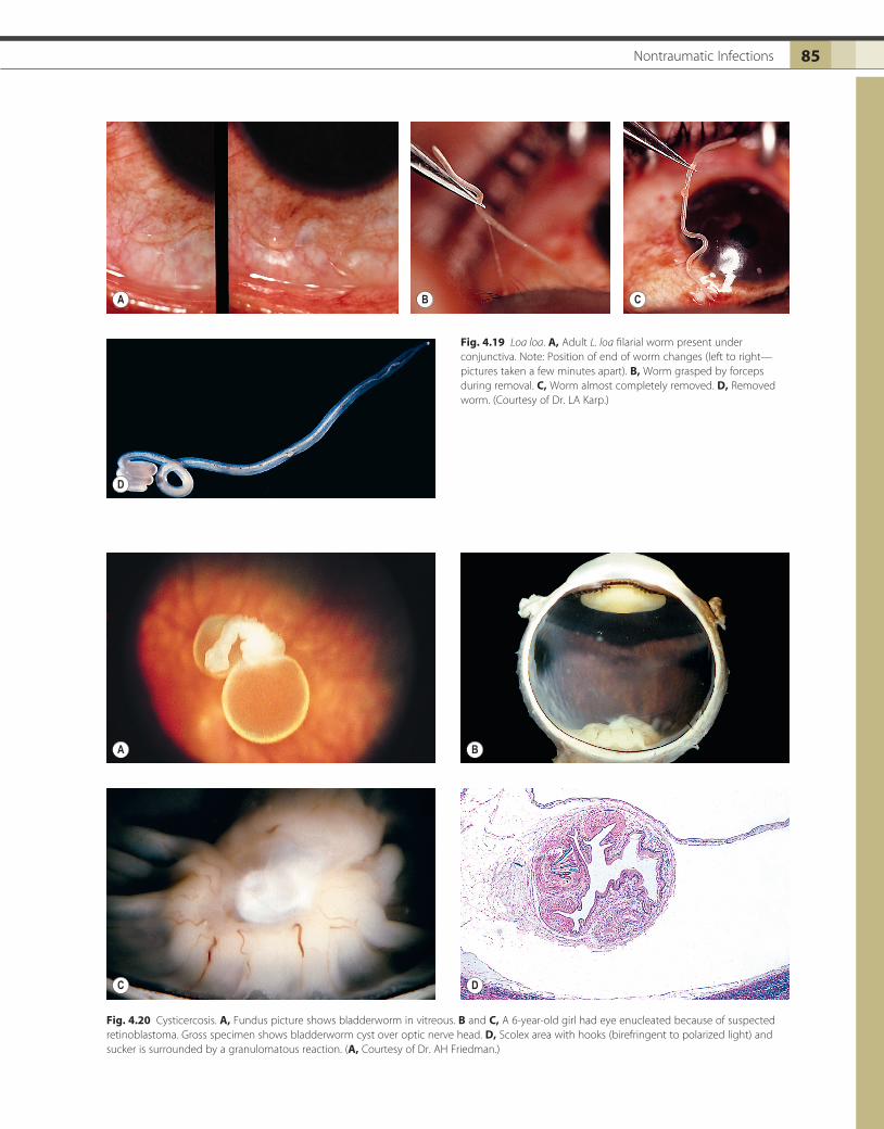
D. Loa loa (Fig. 4.19)1. The adult L. loa filarial worm wanders in the sub-
cutaneous tissues. It may wander into the perior-bital tissues and eyelids and often into the subconjunctival tissues, where its length makes it easily visible.
2. Histologically, little inflammatory reaction occurs while the worm is alive.
E. Dracunculiasis (Dracunculus medinensis; guinea worm; serpent worm)
Fig. 4.18 Trichinosis. A, Acute trichinosis with orbital involvement. Note swelling of lids. B, Top two cysts are empty; bottom cyst shows larva of Trichinella spiralis (pork nematode); seen with increased magnification in C. (A, Courtesy of Dr. ME Smith.)
A
B
C
1. Dracunculiasis, caused by the obligate, nematode parasite, D. medinensis, affects the skin, subcutane-ous tissues, and orbit.
2. Histologically, the worm, when dead, is sur-rounded by an abscess.
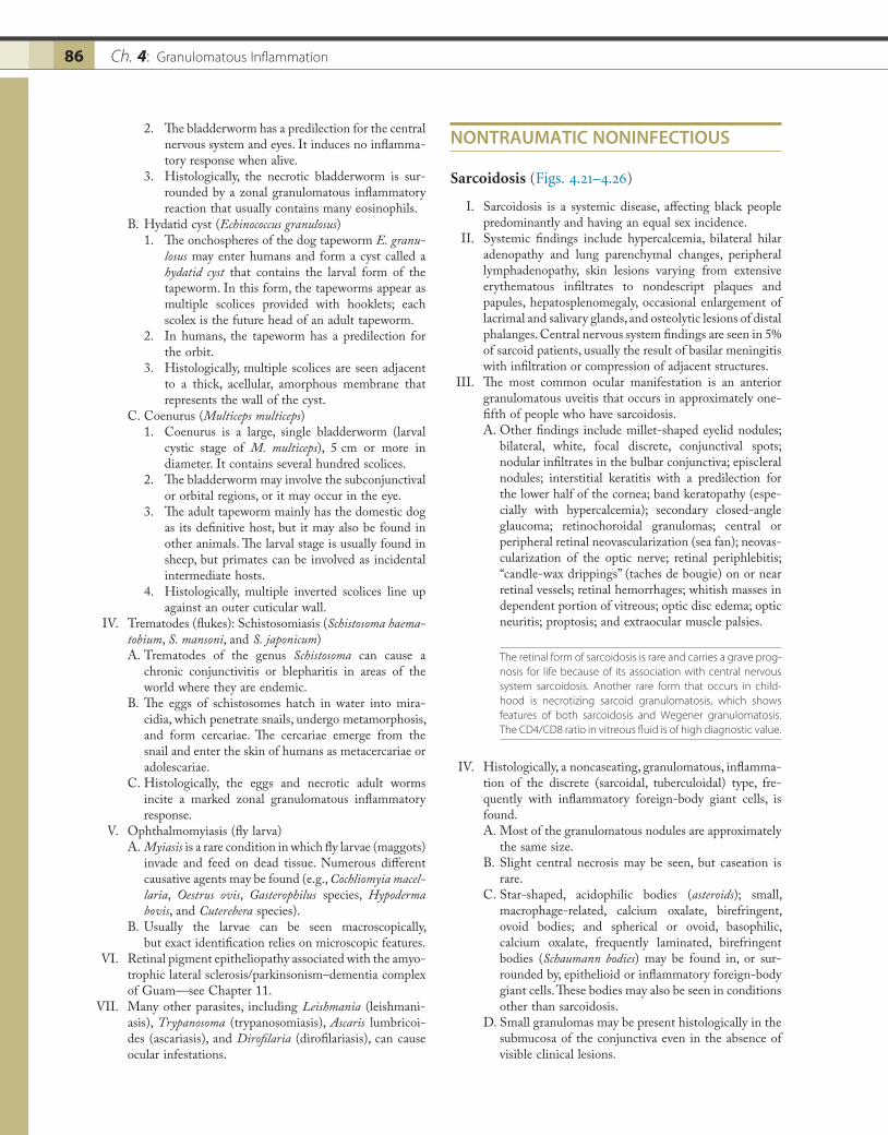
III. Cestoidea (tapeworms)A. Cysticercosis (Cysticercus cellulosae; Fig. 4.20)
1. Cysticercus cellulosae is the larval stage of the pork tapeworm Taenia solium. The larvae, or bladder-worms, hatch in the intestine, and the resultant systemic infestation is called cysticercosis.
Cysticercosis is the most common ocular tapeworm infestation and the most common parasitic infection of the central nervous system. The prognosis in untreated cases is uniformly poor. The best chance for cure is early surgical removal, although destruction of the parasite in situ by diathermy, light coagulation, or cryoapplication may prove successful.
85Nontraumatic Infections 85
Fig. 4.19 Loa loa. A, Adult L. loa filarial worm present under conjunctiva. Note: Position of end of worm changes (left to right—pictures taken a few minutes apart). B, Worm grasped by forceps during removal. C, Worm almost completely removed. D, Removed worm. (Courtesy of Dr. LA Karp.)
A
D
B C
Fig. 4.20 Cysticercosis. A, Fundus picture shows bladderworm in vitreous. B and C, A 6-year-old girl had eye enucleated because of suspected retinoblastoma. Gross specimen shows bladderworm cyst over optic nerve head. D, Scolex area with hooks (birefringent to polarized light) and sucker is surrounded by a granulomatous reaction. (A, Courtesy of Dr. AH Friedman.)
A B
C D
Ch. 4: Granulomatous Inflammation86
2. The bladderworm has a predilection for the central nervous system and eyes. It induces no inflamma-tory response when alive.
3. Histologically, the necrotic bladderworm is sur-rounded by a zonal granulomatous inflammatory reaction that usually contains many eosinophils.
B. Hydatid cyst (Echinococcus granulosus)1. The onchospheres of the dog tapeworm E. granu-
losus may enter humans and form a cyst called a hydatid cyst that contains the larval form of the tapeworm. In this form, the tapeworms appear as multiple scolices provided with hooklets; each scolex is the future head of an adult tapeworm.
2. In humans, the tapeworm has a predilection for the orbit.
3. Histologically, multiple scolices are seen adjacent to a thick, acellular, amorphous membrane that represents the wall of the cyst.
C. Coenurus (Multiceps multiceps)1. Coenurus is a large, single bladderworm (larval
cystic stage of M. multiceps), 5 cm or more in diameter. It contains several hundred scolices.
2. The bladderworm may involve the subconjunctival or orbital regions, or it may occur in the eye.
3. The adult tapeworm mainly has the domestic dog as its definitive host, but it may also be found in other animals. The larval stage is usually found in sheep, but primates can be involved as incidental intermediate hosts.
4. Histologically, multiple inverted scolices line up against an outer cuticular wall.
IV. Trematodes (flukes): Schistosomiasis (Schistosoma haema-tobium, S. mansoni, and S. japonicum)A. Trematodes of the genus Schistosoma can cause a
chronic conjunctivitis or blepharitis in areas of the world where they are endemic.
B. The eggs of schistosomes hatch in water into mira-cidia, which penetrate snails, undergo metamorphosis, and form cercariae. The cercariae emerge from the snail and enter the skin of humans as metacercariae or adolescariae.
C. Histologically, the eggs and necrotic adult worms incite a marked zonal granulomatous inflammatory response.
V. Ophthalmomyiasis (fly larva)A. Myiasis is a rare condition in which fly larvae (maggots)
invade and feed on dead tissue. Numerous different causative agents may be found (e.g., Cochliomyia macel-laria, Oestrus ovis, Gasterophilus species, Hypoderma bovis, and Cuterebera species).
B. Usually the larvae can be seen macroscopically, but exact identification relies on microscopic features.
VI. Retinal pigment epitheliopathy associated with the amyo-trophic lateral sclerosis/parkinsonism–dementia complex of Guam—see Chapter 11.
VII. Many other parasites, including Leishmania (leishmani-asis), Trypanosoma (trypanosomiasis), Ascaris lumbricoi-des (ascariasis), and Dirofilaria (dirofilariasis), can cause ocular infestations.
NONTRAUMATIC NONINFECTIOUS
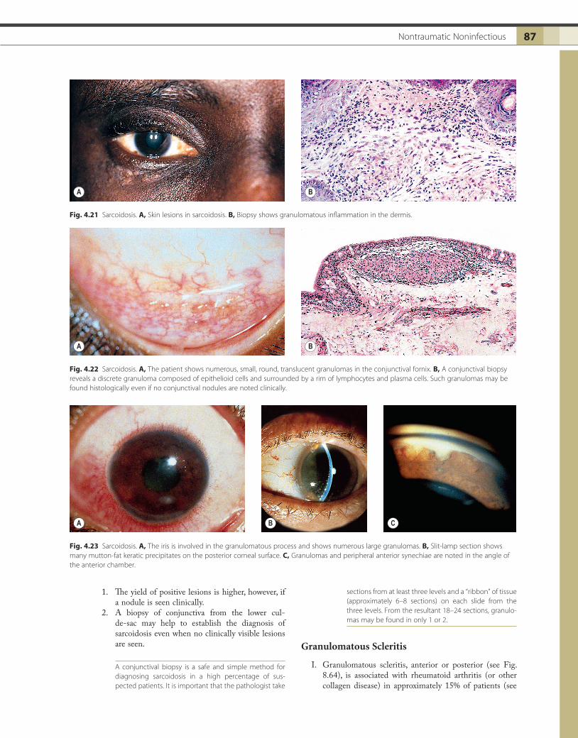
Sarcoidosis (Figs. 4.21–4.26)
I. Sarcoidosis is a systemic disease, affecting black people predominantly and having an equal sex incidence.
II. Systemic findings include hypercalcemia, bilateral hilar adenopathy and lung parenchymal changes, peripheral lymphadenopathy, skin lesions varying from extensive erythematous infiltrates to nondescript plaques and papules, hepatosplenomegaly, occasional enlargement of lacrimal and salivary glands, and osteolytic lesions of distal phalanges. Central nervous system findings are seen in 5% of sarcoid patients, usually the result of basilar meningitis with infiltration or compression of adjacent structures.
III. The most common ocular manifestation is an anterior granulomatous uveitis that occurs in approximately one-fifth of people who have sarcoidosis.A. Other findings include millet-shaped eyelid nodules;
bilateral, white, focal discrete, conjunctival spots; nodular infiltrates in the bulbar conjunctiva; episcleral nodules; interstitial keratitis with a predilection for the lower half of the cornea; band keratopathy (espe-cially with hypercalcemia); secondary closed-angle glaucoma; retinochoroidal granulomas; central or peripheral retinal neovascularization (sea fan); neovas-cularization of the optic nerve; retinal periphlebitis; “candle-wax drippings” (taches de bougie) on or near retinal vessels; retinal hemorrhages; whitish masses in dependent portion of vitreous; optic disc edema; optic neuritis; proptosis; and extraocular muscle palsies.
The retinal form of sarcoidosis is rare and carries a grave prog-nosis for life because of its association with central nervous system sarcoidosis. Another rare form that occurs in child-hood is necrotizing sarcoid granulomatosis, which shows features of both sarcoidosis and Wegener granulomatosis. The CD4/CD8 ratio in vitreous fluid is of high diagnostic value.
IV. Histologically, a noncaseating, granulomatous, inflamma-tion of the discrete (sarcoidal, tuberculoidal) type, fre-quently with inflammatory foreign-body giant cells, is found.A. Most of the granulomatous nodules are approximately
the same size.B. Slight central necrosis may be seen, but caseation is
rare.C. Star-shaped, acidophilic bodies (asteroids); small,
macrophage-related, calcium oxalate, birefringent, ovoid bodies; and spherical or ovoid, basophilic, calcium oxalate, frequently laminated, birefringent bodies (Schaumann bodies) may be found in, or sur-rounded by, epithelioid or inflammatory foreign-body giant cells. These bodies may also be seen in conditions other than sarcoidosis.
D. Small granulomas may be present histologically in the submucosa of the conjunctiva even in the absence of visible clinical lesions.
87Nontraumatic Noninfectious 87
Fig. 4.21 Sarcoidosis. A, Skin lesions in sarcoidosis. B, Biopsy shows granulomatous inflammation in the dermis.
A B
Fig. 4.22 Sarcoidosis. A, The patient shows numerous, small, round, translucent granulomas in the conjunctival fornix. B, A conjunctival biopsy reveals a discrete granuloma composed of epithelioid cells and surrounded by a rim of lymphocytes and plasma cells. Such granulomas may be found histologically even if no conjunctival nodules are noted clinically.
A B
Fig. 4.23 Sarcoidosis. A, The iris is involved in the granulomatous process and shows numerous large granulomas. B, Slit-lamp section shows many mutton-fat keratic precipitates on the posterior corneal surface. C, Granulomas and peripheral anterior synechiae are noted in the angle of the anterior chamber.
A B C
1. The yield of positive lesions is higher, however, if a nodule is seen clinically.
2. A biopsy of conjunctiva from the lower cul- de-sac may help to establish the diagnosis of sarcoidosis even when no clinically visible lesions are seen.
A conjunctival biopsy is a safe and simple method for diagnosing sarcoidosis in a high percentage of sus-pected patients. It is important that the pathologist take
sections from at least three levels and a “ribbon” of tissue (approximately 6–8 sections) on each slide from the three levels. From the resultant 18–24 sections, granulo-mas may be found in only 1 or 2.
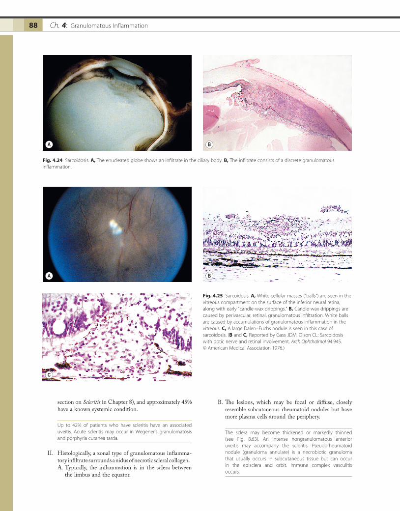
Granulomatous Scleritis
I. Granulomatous scleritis, anterior or posterior (see Fig. 8.64), is associated with rheumatoid arthritis (or other collagen disease) in approximately 15% of patients (see
Ch. 4: Granulomatous Inflammation88
B. The lesions, which may be focal or diffuse, closely resemble subcutaneous rheumatoid nodules but have more plasma cells around the periphery.
The sclera may become thickened or markedly thinned (see Fig. 8.63). An intense nongranulomatous anterior uveitis may accompany the scleritis. Pseudorheumatoid nodule (granuloma annulare) is a necrobiotic granuloma that usually occurs in subcutaneous tissue but can occur in the episclera and orbit. Immune complex vasculitis occurs.
section on Scleritis in Chapter 8), and approximately 45% have a known systemic condition.
Up to 42% of patients who have scleritis have an associated uveitis. Acute scleritis may occur in Wegener’s granulomatosis and porphyria cutanea tarda.
II. Histologically, a zonal type of granulomatous inflamma-tory infiltrate surrounds a nidus of necrotic scleral collagen.A. Typically, the inflammation is in the sclera between
the limbus and the equator.
Fig. 4.25 Sarcoidosis. A, White cellular masses (“balls”) are seen in the vitreous compartment on the surface of the inferior neural retina, along with early “candle-wax drippings.” B, Candle-wax drippings are caused by perivascular, retinal, granulomatous infiltration. White balls are caused by accumulations of granulomatous inflammation in the vitreous. C, A large Dalen–Fuchs nodule is seen in this case of sarcoidosis. (B and C, Reported by Gass JDM, Olson CL: Sarcoidosis with optic nerve and retinal involvement. Arch Ophthalmol 94:945. © American Medical Association 1976.)
A
C
B
Fig. 4.24 Sarcoidosis. A, The enucleated globe shows an infiltrate in the ciliary body. B, The infiltrate consists of a discrete granulomatous inflammation.
A B
89Nontraumatic Noninfectious 89
Chédiak–Higashi Syndrome
See Chapter 11.
Allergic Granulomatosis and Midline Lethal Granuloma Syndrome
See section on Collagen Diseases in Chapter 6.
Weber–Christian Disease (Relapsing Febrile Nodular Nonsuppurative Panniculitis)
See Chapter 6.
Vogt–Koyanagi–Harada Syndrome (Uveomeningoencephalitic Syndrome)
I. VKH syndrome (Fig. 4.27) is a multisystem disorder that reflects the integration of Vogt–Koyanagi syndrome with Harada’s disease.A. Although mainly a syndrome of adults, it rarely occurs
in children, even those as young as four years of age.
Chalazion
See Chapter 6.
Xanthogranulomas (Juvenile Xanthogranuloma and Langerhans’ Granulomatoses; Histiocytosis X)
See Chapter 9 and subsection on Reticuloendothelial System in Chapter 14.
Granulomatous Reaction to Descemet’s Membrane
I. In approximately 10% of eyes with corneal ulcer or kera-titis that are examined histologically, a granulomatous reaction to Descemet’s membrane is found (see Fig. 4.5B and C). Most frequently, the corneas have a disciform keratitis with or without a history of herpes simplex or zoster keratitis.
II. The peculiar reaction to Descemet’s membrane may be the result of altered antigenicity of the membrane and subsequent development of an autosensitivity reaction.
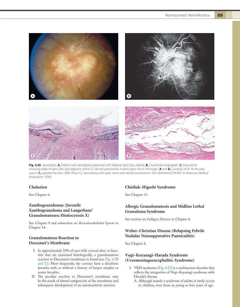
Fig. 4.26 Sarcoidosis. A, Patient with sarcoidosis presented with bilateral optic disc edema. B, Fluorescein angiogram. C, Granuloma involving edge of optic disc and adjacent retina. D. Sarcoid granulomas involve optic nerve meninges. (A and B, Courtesy of Dr. AJ Brucker; case in C, reported by Gass JDM, Olson CL: Sarcoidosis with optic nerve and retinal involvement. Arch Ophthalmol 94:945. © American Medical Association 1976.)
A
C
B
D

Ch. 4: Granulomatous Inflammation90
C. Also, T lymphocytes are decreased in the peripheral blood.
III. Histologically, a chronic, diffuse, granulomatous uveitis, closely resembling sympathetic uveitis, is seen.A. Multiple histologic sections, however, usually show
one or more areas in the posterior segment where the inflammatory reaction does not spare the choriocapil-laris and involves the overlying neural retina.
B. An accompanying disciform degeneration of the macula is common. Immunocytology shows that the uveal infiltrates are composed of T lymphocytes and HLA-DR+ macrophages; nondendritic-appearing CD1 (Leu-6)-positive cells are localized to the choroid in close proximity to melanocytes. Scattered plasma cells and T lymphocytes occur in the retina.
Familial Chronic Granulomatous Disease of Childhood
I. Familial chronic granulomatous disease (FCGD) is char-acterized by chronic suppurative lymphadenitis, eczema-toid dermatitis, osteomyelitis, hepatosplenomegaly, pulmonary infiltrates, abscesses of soft tissues caused by saprophytic organisms, pigmented lipid histiocytosis, and hypergammaglobulinemia.
Approximately 60% have an X-linked, 40% an autosomal reces-sive, and less than 1% an autosomal dominant inheritance pattern.
II. FCGD is a heterogeneous group of disorders of phago-cytic, oxidative metabolism.A. A lesion anywhere in the biochemical pathway that
leads to hydrogen peroxide production has the poten-tial to cause the disease.
B. The patients have a common phenotype of recurrent bacterial infections with catalase-positive microbes
B. VKH syndrome consists of a severe, acute, often bilateral, anterior uveitis associated with vitiligo (leukodermia), poliosis (whitened hair or canities), alopecia, and dysacusia. Perilimbal vitiligo in VKH syndrome is called Sugiura sign.1. Harada’s disease consists primarily of a posterior
granulomatous uveitis, usually bilateral and associ-ated with bilateral serous retinal detachments, accompanied by fluctuating meningeal symptoms, both central and peripheral.
2. Glaucoma, cataract, subretinal neovascularization, late subretinal fibrosis, and Sugiura’s sign (perilim-bal vitiligo) may occur.
C. The cerebrospinal fluid shows increased protein levels and pleocytosis.
Melanin-laden macrophages may be found in the cerebrospi-nal fluid in the early stages (within 25 days) of the onset of VKH syndrome.
II. Autoaggressive cell-bound responses to uveal pigment may play a role in the histogenesis of VKH syndrome.A. VKH syndrome is associated with HLA-DR53,
HLA-DR4, and HLA-DQ4 antigens (and HLA-DR1 in Hispanic patients).
B. VKH may be a syndrome of combined allelic predis-position in which DQA1*0301 acts as the primary and HLA-DR4 acts as an additive factor, whereas DQB1*0604 may be protective, in the development of the prolonged form of the syndrome.
It has been suggested that sympathetic uveitis represents a forme fruste of VKH syndrome. Rarely, VKH syndrome can occur after cutaneous injury such as laceration, burn, and contact dermatitis. Thus, both the clinical manifestations and the immunogenetic background of sympathetic uveitis and VKH syndrome are quite similar.
Fig. 4.27 Vogt–Koyanagi–Harada syndrome. A, Patient shows vitiligo, poliosis, and alopecia. B, Diffuse thickening of choroid by granulomatous inflammation resembles that seen in sympathetic uveitis. However, unlike in sympathetic uveitis, inflammation does not spare choriocapillaris and has broken through the retinal pigment epithelium into the subneural retinal area. (A, Case reported in Fine BS, Gilligan JH: The Vogt–Koyanagi syndrome: A variant of sympathetic ophthalmia. Report of two cases. Am J Ophthalmol 43:433. © Elsevier 1957; B, case presented at the 1980 Verhoeff Society meeting by Dr. H Inomata and reported in Fine BS, Gilligan JH: The Vogt-Koyanagi syndrome: report of two cases. Am J Ophthalmol 43:433, 1957.)
A B
91Nontraumatic Noninfectious 91
III. Ocular findings include lid dermatitis, keratoconjunctivi-tis, and chorioretinitis.
IV. Histologically, suppurative and granulomatous inflamma-tory lesions characteristically coexist.A. The suppurative component may be secondary to
infection, whereas the granulomatous component is likely caused by inadequate breakdown of antigenic debris or inadequate feedback inhibition of inflamma-tion by toxic oxygen products.
B. The choroid and sclera show multiple foci of granulo-matous inflammation.
(e.g., Staphylococcus aureus and Serratia, Pseudomonas, Klebsiella, Chromobacterium, Escherichia, Nocardia, and Aspergillus species).
PMNs in patients with FCGD ingest bacteria but do not kill them because of a deficiency in leukocyte hydrogen peroxide metabolism. Furthermore, lyso-somal hydrolytic enzymes (acid phosphatase and β-glucuronidase) are released in decreased amounts by PMNs during phagocytosis, resulting in abnormal (lessened) degranulation of the PMNs.
C. Humoral immunity, cell-mediated immunity, and inflammatory responses are normal.
Access the complete reference list online at

91.e1Bibliography 91.e1
BIBLIOGRAPHY
Sympathetic Uveitis
Boyd SR, Young S, Lightman S: Immunopathology of the noninfec-tious posterior and intermediate uveitides. Surv Ophthalmol 46:209, 2001
Chan C-C, Roberge FG, Whitcup SM et al.: 32 cases of sympathetic ophthalmia. Arch Ophthalmol 113:597, 1995
Davis JL, Mittal KK, Freidlin V et al.: HLA associations and ancestry in Vogt–Koyanagi–Harada disease and sympathetic ophthalmia. Ophthalmology 97:1137, 1990
Easom HA, Zimmerman LE: Sympathetic ophthalmia and bilateral phacoanaphylaxis: A clinicopathologic correlation of the sympatho-genic and sympathizing eyes. Arch Ophthalmol 72:9, 1964
Fine BS, Gilligan JH: The Vogt–Koyanagi syndrome: A variant of sympathetic ophthalmia. Report of two cases. Am J Ophthalmol 43:433, 1957
Font RL, Fine BS, Messmer E et al.: Light and electron microscopic study of Dálen–Fuchs nodules in sympathetic ophthalmia. Ophthal-mology 89:66, 1982
Galor A, Davis JL, Flynn WW Jr et al.: Sympathetic ophthalmia: incidence of ocular complications and vision loss in the sympathizing eye. Am J Ophthalmol 148:704, 2009
Gass JDM: Sympathetic ophthalmia following vitrectomy. Am J Oph-thalmol 93:552, 1982
Green WR, Maumenee AE, Saunders TE et al.: Sympathetic uveitis following evisceration. Trans Am Acad Ophthalmol Otolaryngol 76:625, 1972
Kay ML, Yanoff M, Katowitz JA: Sympathetic uveitis: Development in spite of corticosteroid therapy. Am J Ophthalmol 78:90, 1974
Kinyoun JL, Bensinger RE, Chuang EL: Thirty-year history of sym-pathetic ophthalmia. Ophthalmology 90:59, 1983
Lubin JR, Albert DM: Early enucleation in sympathetic ophthalmia. Ocul Inflamm Ther 1:47, 1983
Marak GE Jr, Font RL, Zimmerman LE: Histologic variations related to race in sympathetic ophthalmia. Am J Ophthalmol 78:935, 1974
Sen HN, Nussenblatt RB: Sympathetic ophthalmia: What have we learned? Am J Ophthalmol 148:632, 2009
Sisk RA, Davis JL, Dubovy Sr et al.: Sympathetic ophthalmia follow-ing vitrectomy for endophthalmitis after intravitreal bevacizumab. Ocul Immunol Inflamm 16:236, 2008
Stafford WR: Sympathetic ophthalmia: Report of a case occurring ten and one half days after injury. Arch Ophthalmol 74:521, 1965
Yanoff M: Pseudo-sympathetic uveitis. Trans Pa Acad Ophthalmol Oto-laryngol 30:118, 1977
Phacoanaphylactic Endophthalmitis
Apple DJ, Mamalis N, Steinmetz RL et al.: Phacoanaphylactic endo-phthalmitis associated with extracapsular cataract extraction and posterior chamber intraocular lens. Arch Ophthalmol 102:1528, 1984
Caudill JW, Streeten BW, Tso MOM: Phacoanaphylactoid reaction in persistent hyperplastic primary vitreous. Ophthalmology 92:1153, 1985
Chan C-C: Relationship between sympathetic ophthalmia, phacoana-phylactic endophthalmitis, and Vogt–Koyanagi–Harada disease. Oph-thalmology 95:619, 1988
Chishti M, Henkind P: Spontaneous rupture of anterior lens capsule (phacoanaphylactic endophthalmitis). Am J Ophthalmol 69:264, 1970
Easom HA, Zimmerman LE: Sympathetic ophthalmia and bilateral phacoanaphylaxis: A clinicopathologic correlation of the sympatho-genic and sympathizing eyes. Arch Ophthalmol 72:9, 1964
Habil I, Cohen E, Karshai I et al.: Spontaneous involution of autologous lenses and phacoanaphylaxis reaction in Stickler syn-drome. Br J Ophthalmol 89:1532, 2005
Inomota H, Yoshikawa H, Rao NA: Phacoanaphylaxis in Behçet’s disease: A clinicopathologic and immunohistochemical study. Oph-thalmology 110:1942, 2003
Marak GE: Phacoanaphylactic endophthalmitis. Surv Ophthalmol 36:325, 1992
Yanoff M, Scheie HG: Cytology of human lens aspirate: Its relation-ship to phacolytic glaucoma and phacoanaphylactic endophthalmitis. Arch Ophthalmol 80:166, 1968
Foreign-Body Granulomas
Ferry AP: Synthetic fiber granuloma: “Teddy bear” granuloma of the conjunctiva. Arch Ophthalmol 112:1339, 1994
Naumann GOH, Völcker HE: Endophthalmitis haemogranulomatosa (eine spezielle Reaktionsform auf intraokulare Blutungen). Klin Monatsbl Augenheilkd 171:352, 1977
Riddle PJ, Font RL, Johnson FB et al.: Silica granuloma of eyelid and ocular adnexa. Arch Ophthalmol 99:683, 1981
Wilson MW, Grossniklaus HE, Heathcote JG: Focal choroidal gran-ulomatous inflammation. Am J Ophthalmol 121:397, 1996
Viral
Ausayakhun, S, Keenan JD, Ausayakhun S et al.: Clinical features of newly diagnosed cytomegalovirus retinitis in northern Thailand. Am J Ophthalmol 153:923, 2012
Bresnahan WA, Shenk T: A subset of viral transcripts packaged within human cytomegalovirus particles. Science 288:2373, 2000
Chen J, Ausayaklun S, Tangmonkongvoragul CT et al.: Incidence of cytomegalovirus in patients with human immunodeficiency virus fol-lowing negative initial screening examination results. Arch Ophthalmol 130:527, 2012
Friedman AH: The retinal lesions of the acquired immune deficiency syndrome. Trans Am Ophthalmol Soc 82:447, 1984
Ghaznawi N, Virdi A, Dayan A et al.: Herpes zoster ophthalmicus: Comparison of disease in patients 60 years and older versus younger than 60 years. Ophthalmology 118:2242, 2011
Ho J-D, Xirasagar S, Lin H-C: Increased risk of a cancer diagnosis after herpes zoster ophthalmicus: A nationwide population-based study. Ophthalmology 118:1076, 2011
Jabs DA: Cytomegalovirus retinitis and the acquired immunodeficiency syndrome—Bench to bedside.: LXVII Edward Jackson Memorial Lecture. Am J Ophthalmol 151:198, 2011
Jabs DA, Martin BK, Forman MS et al.: Cytomegalovirus resistance to ganciclovir and clinical outcomes of patients with cytomegalovirus retinitis. Am J Ophthalmol 135:26, 2003
Karbassi M, Raizman MB, Schuman JS: Herpes zoster ophthalmicus. Surv Ophthalmol 36:395, 1992
Kempen JH, Jabs DA, Wilson LA et al.: Risk of vision loss in patients with cytomegalovirus and the acquired immunodeficiency syndrome. Arch Ophthalmol 121:466, 2003
Kunavisarut P, Sirirungsi W, Pathanapitoon K et al.: Clinical mani-festations of human immunodeficiency virus-induced uveitis. Oph-thalmology 119:1455, 2012
Kuo IC, Kempen JH, Dunn JP et al.: Clinical characteristics and out-comes of cytomegalovirus retinitis in persons without human immu-nodeficiency virus infection. Am J Ophthalmol 138:338, 2004
Nguyen QD, Kempen JH, Bolton SG et al.: Immune recovery uveitis in patients with AIDS and cytomegalovirus retinitis following highly active retroviral therapy. Am J Ophthalmol 129:634, 2000
Ch. 4: Granulomatous Inflammation91.e2
Rummelt V, Rummelt C, Jahn G et al.: Triple retinal infection with human immunodeficiency virus type 1, cytomegalovirus, and herpes simplex virus type 1: Light and electron microscopy, immuno-histochemistry, and in situ hybridization. Ophthalmology 101:270, 1994
Sorenson RL, Jeng BH: Ophthalmic manifestations of human immu-nodeficiency virus infection in the era of highly active antiretroviral therapy. Arch Ophthalmol 130:1621, 2012
Sugar EA, Jabs DA, Ahuja A et al.: Incidence of cytomegalovirus reti-nitis in the era of highly active antiretroviral therapy. Am J Ophthalmol 153:1016, 2012
Walter KA, Coulter VL, Palay DA et al.: Corneal endothelial deposits in patients with cytomegalovirus retinitis. Am J Ophthalmol 121:391, 1996
Wang T-J, Hu C-C, Lin H-C: Increased risk of anterior uveitis fol-lowing herpes zoster: A nationwide population-based study. Arch Ophthalmol 130:451, 2012
Bacterial
Ang M, Jap A, Chee S-P: Prognostic factors and outcomes in endog-enous Klebsiella pneumonia endophthalmitis. Am J Ophthalmol 151:338, 2011
Babu RB, Sudharshan S, Kumaraasamy N et al.: Ocular tuberculosis in acquired immunodeficiency syndrome. Am J Ophthalmol 142:413, 2006
Barile GR, Llynn TE: Syphilis exposure in patients with uveitis. Oph-thalmology 104:1605, 1997
Bleharski JR, Li H, Meinken C et al.: Use of genetic profiling in leprosy to discriminate clinical forms of the disease. Science 301:1527, 2003
Bullington RH Jr, Lanier JD, Font RL: Nontuberculous mycobacterial keratitis: Report of two cases and review of the literature. Arch Oph-thalmol 110:519, 1992
Chao JR, Khurana RN, Fawzi AA et al.: Syphilis: Reemergence of an old adversary. Ophthalmology 113:2074, 2006
Chi SL, Stinnett S, Eggenberger E et al.: Clinical characteristics in 53 patients with cat scratch optic neuropathy. Ophthalmology 119:183, 2012
Cunningham ET Jr, Koehler JE: Ocular bartonellosis. Am J Ophthalmol 130:340, 2000
Dana M-R, Hochman MA, Viana MAG et al.: Ocular manifestations of leprosy in a noninstitutionalized community in the United States. Arch Ophthalmol 112:626, 1994
Flach AJ, Lavoie PE: Episcleritis, conjunctivitis, and keratitis as ocular manifestations of Lyme disease. Ophthalmology 97:973, 1990
Fraser CM, Norris SJ, Weinstock GM et al.: Complete genome sequence of Treponema pallidum, the syphilis spirochete. Science 281:375, 1998
Gopal L, Rao SK, Biswas J et al.: Tuberculous granuloma managed by full thickness eye wall resection. Am J Ophthalmol 135:93, 2003
Gupta V, Gupta A, Arora S et al.: Presumed tubercular serpiginous choroiditis: Clinical presentations and management. Ophthalmology 110:1744, 2003
Haas CJ, Zink A, Pàlfi G et al.: Detection of leprosy in ancient human skeletal remains by molecular identification of Mycobacterium leprae. Am J Clin Pathol 114:427, 2000
Isogai E, Isogai H, Kotake S et al.: Detection of antibodies against Borrelia burgdorferi in patients with uveitis. Am J Ophthalmol 112:23, 1991
Kestelyn P: Rhinoscleroma with bilateral orbital involvement. Am J Ophthalmol 101:381, 1986
Lee WR, Chawla JC, Reid R: Bacillary angiomatosis of the conjunc-tiva. Am J Ophthalmol 118:152, 1994
Mikkilä HO, Seppälä IJT, Viljanen MK et al.: The expanding clinical spectrum of ocular Lyme borreliosis. Ophthalmology 107:581, 2000
Parikh R, Thomas S, Mutiyil J et al.: Ocular manifestations in treated Multibacillary Hansen’s disease. Ophthalmology 117:2051, 2009
Patel SS, Saraiya NV, Tessler HH et al.: Mycobacterial ocular inflam-mation: Delay in diagnosis and other factors impacting morbidity. JAMA Ophthalmol 131:752, 2013
Preac-Mursic V, Pfister HW, Spiegel H et al.: First isolation of Bor-relia burgdorferi from an iris biopsy. J Clin Neuroophthalmol 13:155, 1993
Raina UK, Jain S, Monga S et al.: Tubercular preseptal cellulites in children: A presenting sign of underlying systemic tuberculosis. Oph-thalmology 111:291, 2004
Rao NA, Hidayat AA: Endogenous mycotic endophthalmitis: Varia-tions in clinical and histologic changes in candidiasis compared with aspergillosis. Am J Ophthalmol 132:244, 2001
Solley WA, Martin DF, Newman NJ et al.: Cat scratch disease: Pos-terior segment manifestations. Ophthalmology 106:1546, 1999
Spektor FE, Eagle RC, Nichols CW: Granulomatous conjunctivitis secondary to Treponema pallidum. Ophthalmology 88:863, 1981
Steinemann TL, Sheikholeslami MR, Brown HH et al.: Oculoglan-dular tularemia. Arch Ophthalmol 117:132, 1999
Vasconcelos-Santos DV, Rao PQ, Davies JB et al.: Clinical features of tuberculous serpiginous choroiditis in contrast to classic serpiginous choroiditis. Arch Ophthalmol 128:853, 2010
Wade NK, Levi L, Jones MR et al.: Optic edema associated with peripapillary retinal detachment: an early sign of systemic Bartonella henselae infection. Am J Ophthalmol 130:327, 2000
Walburger A, Koul A, Ferrari G et al.: Protein kinase G from patho-genic mycobacteria promotes survival within macrophages. Science 304:1800, 2004
Whitcup SM, Fenton RM, Pluda JM et al.: Pneumocystis carinii and Mycobacterium infection of the choroid. Retina 12:331, 1992
Wickremasinghe S, Ling C, Stawell R et al.: Syphylitic punctuate inner retinitis in immunocompetent gay men. Ophthalmology 116:1196, 2009
Wroblewski KJ, Hidayat AA, Neafie RC et al.: Ocular tuberculosis: A clinicopathologic and molecular study. Ophthalmology 118:772, 2011
Fungal
Bartley GB: Blastomycosis of the eyelid. Ophthalmology 102:2020, 1995Charles NC, Boxrud CA, Small EA: Cryptococcosis of the anterior
chamber in acquired immune deficiency syndrome. Ophthalmology 99:813, 1992
Cohen J, Carvalho RC, Guimarães R et al.: Oculosporidiosis. Arch Ophthalmol 115:1340, 1997
Cohen DB, Glascow BJ: Bilateral optic nerve cryptococcosis in sudden blindness in patients with acquired immune deficiency syndrome. Ophthalmology 100:1689, 1993
Cunningham ET, Seiff SR, Berger TG et al.: Intraocular coccidioido-mycosis diagnosed by skin biopsy. Arch Ophthalmol 116:674, 1998
Custis PH, Haller JA, de Juan E Jr: An unusual case of cryptococcal endophthalmitis. Retina 15:300, 1995
Donahue SP, Greven CM, Zuravleff JJ et al.: Intraocular candidiasis in patients with candidemia: Clinical implications derived from a prospective multicenter study. Ophthalmology 101:1302, 1994
Dozier CC, Tarantola RM, Jiramongkolchai K et al.: Fungal eye disease at a tertiary care center: The utility of routine inpatient con-sultation. Ophthalmology 118:1671, 2011
Fairley C, Sullivan TJ, Bartley P et al.: Survival after rhino-orbital-cerebral mucormycosis in an immunocompetent. Ophthalmology 107:555, 2000
91.e3Bibliography 91.e3
Font RL, Jakobiec FA: Granulomatous necrotizing retinochoroiditis caused by Sporotrichum schenkii: Report of a case including immuno-fluorescence and electron microscopical studies. Arch Ophthalmol 94:1513, 1976
Glasgow BJ, Brown HH, Foos RY: Miliary retinitis in coccidioidomy-cosis. Am J Ophthalmol 104:24, 1987
Gottlieb JL, McAllister IL, Guttman FA et al.: Choroidal blastomy-cosis: A report of two cases. Retina 15:248, 1995
Levy MG, Meuten DJ, Breitschwerdt EB: Cultivation of Rhinospo-ridium seeberi in vitro: Interaction with epithelial cells. Science 234:474, 1986
Li S, Perlman JI, Edward DP et al.: Unilateral Blastomyces dermatitidis endophthalmitis and orbital cellulitis: A case report and literature review. Ophthalmology 105:1467, 1998
Lin JC, Ward TP, Belyea DA et al.: Treatment of Nocardia asteroides keratitis with polyhexamethylene biguanide. Ophthalmology 104:1306, 1997
McNab AA, McKelvie P: Iron overload is a risk factor for zygomycosis. Arch Ophthalmol 115:919, 1997
Moorthy RS, Rao NA, Sidikaro Y et al.: Coccidioidomycosis iridocy-clitis. Ophthalmology 101:1923, 1994
Pushker N, Meel R, Kashyap S et al.: Invasive aspergillosis of orbit in immunocompetent patients: Treatment and outcome. Ophthalmology 118:1886, 2011
Safneck JR, Hogg GA, Napier LB: Endophthalmitis due to Blastomy-ces dermatitidis: Case report and review of the literature. Ophthalmol-ogy 97:212, 1990
Sheu S-J, Chen Y-C, Kuo N-W et al.: Endogenous cryptococcal endo-phthalmitis. Ophthalmology 105:377, 1998
Slade MP, McNab AA: Fatal mucormycosis therapy associated with deferoxamine (letter). Am J Ophthalmol 112:594, 1991
Specht CS, Mitchell KT, Bauman AE et al.: Ocular histoplasmosis with retinitis in a patient with acquired immune deficiency syndrome. Ophthalmology 98:1356, 1991
Sponsler TA, Sassani JW, Johnson LN et al.: Ocular invasion in mucormycosis. Surv Ophthalmol 36:345, 1992
Stone JL, Kalina RE: Ocular coccidioidomycosis. Am J Ophthalmol 116:249, 1993
Parasitic
Agrawal S, Agrawal J, Agrawal TP: Orbital cysticercosis-associated scleral indentation presenting with pseudo-retinal detachment. Am J Ophthalmol 137:1153, 2004
Bernard EM, Sepkowitz KA, Telzak EE et al.: Pneumocystinosis. Med Clin North Am 76:107, 1992
Biswas J, Fogla R, Srinivasan P et al.: Ocular malaria: A clinical and histopathologic study. Ophthalmology 103:1471, 1996
Buettner H: Ophthalmomyiasis interna. Arch Ophthalmol 120:1582, 2002
Burnier M, Hidayat AA, Neafdiue R: Dracunculiasis of the orbit and eyelid: Light and electron microscopic observations of two cases. Ophthalmology 98:919, 1991
Campbell RJ, Steele JC, Cox TA et al.: Pathologic findings in the retinal pigment epitheliopathy associated with the amyotrophic lateral sclerosis/Parkinsonism–dementia complex (ALS/PDC) of Guam. Ophthalmology 100:37, 1993
Chan CML, Theng JTS, Li L et al.: Microsporidial keratoconjunctivi-tis in healthy individuals: A case series. Ophthalmology 110:1420, 2003
Chang GY, Keane JR: Visual loss in cysticercosis. Neurology 57:545, 2001Chuck RS, Olk RJ, Weil GJ et al.: Surgical removal of a subretinal
proliferating cysticercus of Taeniaeformis crassiceps. Arch Ophthalmol 115:562, 1997
Fahnehjelm KT, Malm G, Ygge J et al.: Ophthalmological findings in children with congenital toxoplasmosis: Report from a Swedish pro-spective screening study of congenital toxoplasmosis with two years of follow-up. Acta Ophthalmol Scand 78:569, 2000
Filho CADAG, Gomes AHB, Soares ACMDAGet al.: Clinical fea-tures of 121 patients with diffuse unilateral subacute neuroretinitis. Am J Ophthalmol 153:743, 2012
Font RL, Samaha AN, Keener MJ et al.: Corneal microsporidiosis: Report of a case, including electron microscopic observations. Oph-thalmology 107:1769, 2000
Gendelman D, Blumberg R, Sadun A: Ocular Loa loa with cryoprobe extraction of subconjunctival worm. Ophthalmology 91:300, 1984
Glasgow BJ, Maggiano JM: Cuterebra ophthalmomyiasis. Am J Oph-thalmol 119:512, 1995
Gupta NK, Sachdev MS, Bhardwaj Y et al.: Intraocular Ascaris larva. Arch Ophthalmol 106:880, 1988
Hidayat AA, Nalbandian RM, Sammons DW et al.: The diagnostic histopathologic features of ocular malaria. Ophthalmology 100:1183, 1993
Holland GN: Ocular toxoplasmosis: A global reassessment. Part I: Epidemiology and course of the disease (LX Edward Jackson memo-rial lecture). Am J Ophthalmol 136:973, 2003
Holland GN: Ocular toxoplasmosis: A global reassessment. Part II: Disease manifestations and management (LX Edward Jackson memorial lecture). Am J Ophthalmol 137:1, 2004
Jakobiec FA, Gess L, Zimmerman LE: Granulomatous dacryoadenitis caused by Schistosoma haematobium. Arch Ophthalmol 95:278, 1977
Joshi G, Parchand S, Dogra MR et al.: Live juvenile strobilate tape-worm in the anterior chamber of the human eye. Arch Ophthalmol 130:1464, 2012
Kırath H, Biliç S, Öztürkmen C et al.: Intramuscular hydatid cyst of the medial rectus. Am J Ophthalmol 135:98, 2003
Labalette P, Delhaes L, Margaron F et al.: Ocular toxoplasmosis after the fifth decade. Am J Ophthalmol 133:506, 2002
Lawson JMM, Dart JKG, McCartney ACE: Conjunctival nodules associated with the Splendore–Hoeppli phenomenon. Arch Ophthal-mol 109:426, 1991
Lima LH, Laud K, Rosenberg P et al.: Ophthalmomyasis with a sin-gular subretinal track. Am J Ophthalmol 150:731, 2010
Mohammad AE-NA, Ray CJ, Karcioglu ZA: Echinococcus cysts of the orbit and substernum. Am J Ophthalmol 118:676, 1994
Morales LR, Cialdini AP, Avila MP et al.: Identifying live nematodes in diffuse unilateral subacute neuroretinitis by using the scanning laser ophthalmoscope. Arch Ophthalmol 120:135, 2002
Perry HD, Donnenfeld ED, Font RL: Intracorneal ophthalmomyasis. Am J Ophthalmol 109:741, 1990
Raithinam S, Fritsche TR, Srinivascan M et al.: An outbreak of trematode-induced granulomas of the conjunctiva. Ophthalmology 108:1223, 2001
Rath S, Honavar SG, Naik M et al.: Orbital cysticercosis: Clinical manifestations, diagnosis, management, and outcome. Ophthalmology 117:600, 2010
Risco JM, Al-Dosari F, Millar L: Sheep nasal botfly (Oestrus ovis) larvae infestation of the conjunctiva. Arch Ophthalmol 113:529, 1995
Roberts F, Mets MB, Ferguson DJP et al.: Histopathological features of ocular toxoplasmosis in the fetus and infant. Arch Ophthalmol 119:51, 2001
Sakai R, Kawashima H, Shibui H et al.: Toxocara cati-induced ocular toxocariasis. Arch Ophthalmol 116:1686, 1998
Schwab IR: The epidemiologic association of Fuchs’ heterochromic iridocyclitis and toxoplasmosis. Am J Ophthalmol 111:356, 1991
Schwartz DA, Visvesvara GS, Diesenhouse MC et al.: Pathologic fea-tures and immunofluorescent antibody demonstration of ocular micro-sporidiosis (Encephalitozoon hellem) in seven patients with acquired immunodeficiency syndrome. Am J Ophthalmol 115:285, 1993

Ch. 4: Granulomatous Inflammation91.e4
Sekhar GC, Hanavar SG: Mycocysticercosis: Experience with imaging and therapy. Ophthalmology 106:2336, 1999
Sekhar GC, Lemke BN: Orbital cysticercosis. Ophthalmology 104:1599, 1997
Shami MJ, Freeman W, Friedberg D et al.: A multicenter study of Pneumocystis choroidopathy. Am J Ophthalmol 112:15, 1991
Sharma T, Sinha S, Shah N et al.: Intraocular cysticercosis: Clinical characteristics and visual outcome after vitrectomy surgery. Ophthal-mology 110:1005, 2003
Vasconcelos-Santos DV, Azevedo DOM, Campos WR et al.: Con-genital toxoplasmosis in southeastern Brazil: Results of early oph-thalmologic examination of as large cohort of neonates. Ophthalmology 116:2199, 2009
Whitcup SM, Fenton RM, Pluda JM et al.: Pneumocystis carinii and Mycobacterium infection of the choroid. Retina 12:331, 1992
Wilder HC: Nematode endophthalmitis. Trans Am Acad Ophthalmol Otolaryngol 54:99, 1950
Wilder HC: Toxoplasma chorioretinitis in adults. Arch Ophthalmol 48:127, 1952
Woodhall D, Starr MC, Montgomery SP et al.: Ocular toxoplas-mosis: Epidemiologic, anatomic, and therapeutic variations based on a survey of ophthalmic subspecialists. Ophthalmology 119:1211, 2012
Sarcoidosis
Boyd SR, Young S, Lightman S: Immunopathology of the noninfec-tious posterior and intermediate uveitides. Surv Ophthalmol 46:209, 2001
Demirci H, Christianson MD: Orbital and adnexal involvement in sarcoidosis: Analysis of clinical features and systemic disease in 30 cases. Am J Ophthalmol 151:1074, 2011
Farr AK, Jabs DA, Green WR: Optic disc sarcoid granuloma. Arch Ophthalmol 118:728, 2000
Frohman L, Grigorian R, Slamovits T: Evolution of sarcoid granulo-mas of the retina. Am J Ophthalmol 131:661, 2001
Garcia GH, Harris GJ: Sarcoid inflammation and obstruction of the nasolacrimal system. Arch Ophthalmol 118:719, 2000
Gass JDM, Olson CL: Sarcoidosis with optic nerve and retinal involve-ment. Arch Ophthalmol 94:945, 1976
Hall JG, Cohen KL: Sarcoidosis of the eyelid and skin. Am J Ophthal-mol 119:100, 1995
Khalatbari D, Stinnett S, McCallum RM et al.: Demographic-related variations in posterior segment ocular sarcoidosis. Ophthalmology 111:357, 2004
Kojima K, Manuyama K, Inaba T et al.: The CD4/CD8 ratio in vitre-ous fluid is of high diagnostic value in sarcoidosis. Ophthalmology 119:2386, 2012
Nichols CW, Eagle RC Jr, Yanoff M et al.: Conjunctival biopsy as an aid in the evaluation of the patient with suspected sarcoidosis. Oph-thalmology 87:287, 1980
Nichols CW, Mishkin M, Yanoff M: Presumed orbital sarcoidosis: Report of a case followed by computerized axial tomography and conjunctival biopsy. Trans Am Ophthalmol Soc 76:67, 1978
Pelton RW, Lee AG, Orengo-Nania SD et al.: Bilateral optic disk edema caused by sarcoidosis mimicking pseudotumor cerebri. Am J Ophthalmol 127:229, 1999
Qazi A, Thorne JE, Jabs DA: Scleral nodule associated with sarcoidosis. Am J Ophthalmol 136:752, 2003
Schaumann J: On nature of certain peculiar corpuscles present in tissue of lymphogranulomatosis benigna. Acta Med Scand 106:239, 1941
Schumacher C, Lohr A, Kaup M et al.: Necrotizing sarcoid granulo-matosis: Ocular manifestations in 3 children of African origin. Arch Ophthalmol 129:1370, 2011
Granulomatous Scleritis
Brod RD, Saul RF: Nodular scleritis. Arch Ophthalmol 108:1170, 1990Fong LP, de la Maza MS, Rice BA et al.: Immunopathology of scleritis.
Ophthalmology 98:472, 1991Hinzpeter EN, Naumann G, Gartelheimer HK: Ocular histopathol-
ogy in Still’s disease. Ophthalmic Res 2:16, 1971Lebowitz MA, Jakobiec FA, Donnenfeld ED et al.: Bilateral epibulbar
rheumatoid nodulosis: A new ocular entity. Ophthalmology 95:1256, 1988
de la Maza MS, Foster CS, Jabbur NS: Scleritis associated with rheu-matoid arthritis and with other systemic immune-mediated diseases. Ophthalmology 101:1281, 1994
de la Maza MS, Foster CS, Jabbur NS: Scleritis associated with sys-temic vasculitic diseases. Ophthalmology 102:687, 1995
Sacks RD, Stock EL, Crawford SE et al.: Scleritis and Wegener’s granulomatosis in children. Am J Ophthalmol 111:430, 1991
Salmon JF, Strauss PC, Todd G et al.: Acute scleritis in porphyria cutanea tarda. Am J Ophthalmol 109:400, 1990
Tuft SJ, Watson PG: Progression of scleral disease. Ophthalmology 98:467, 1991
Watson PG, Hayreh SS: Scleritis and episcleritis. Br J Ophthalmol 60:163, 1976
Granulomatous Reaction to Descemet’s Membrane
Green WR, Zimmerman LE: Granulomatous reaction to Descemet’s membrane. Am J Ophthalmol 64:555, 1967
Holbach LM, Font RL, Naumann GOH: Herpes simplex stromal and endothelial keratitis: Granulomatous cell reactions at the level of Descemet’s membrane, the stroma, and Bowman’s layer. Ophthalmol-ogy 97:722, 1990
Vogt–Koyanagi–Harada Syndrome
Boyd SR, Young S, Lightman S: Immunopathology of the noninfec-tious posterior and intermediate uveitides. Surv Ophthalmol 46:209, 2001
Fine BS, Gilligan JH: The Vogt–Koyanagi syndrome: A variant of sympathetic ophthalmia: Report of two cases. Am J Ophthalmol 43:433, 1957
Gocho K, Kondo I, Yamaki K: Identification of autoreactive T cells in Vogt–Koyanagi–Harada disease. Invest Ophthalmol Vis Sci 42:2037, 2001
Goldberg AC, Yamamota JH, Chiarella JM et al.: DRB1*0405 is the predominant allele in Brazilian patients with Vogt–Koyanagi–Harada disease. Hum Immunol 59:183, 1998
Inomata H, Rao NA: Depigmented atrophic lesions in sunset glow fundi of Vogt–Koyanagi–Harada disease. Am J Ophthalmol 131:607, 2001
Keino H, Goto H, Mori H et al.: Association between severity of inflammation in CNS and development of sunset glow fundus in Vogh–Koyanagi–Herada disease. Am J Ophthalmol 141:1140, 2006
Kim M-H, Seong M-C, Kwak N-H et al.: Association of HLA with Vogt–Koyanagi–Harada syndrome in Koreans. Am J Ophthalmol 129:173, 2000
Moorthy RS, Inomata H, Rao NA: Vogt–Koyanagi–Harada syn-drome. Surv Ophthalmol 39:265, 1995
Najman-Vainer J, Levinson RD, Graves MC et al.: An association between Vogt–Koyanagi–Harada disease and Guillain Barré syn-drome. Am J Ophthalmol 131:615, 2001
91.e5Bibliography 91.e5
Nakamura S, Nakazawa M, Yoshioka M et al.: Melanin-laden macro-phages in Vogt–Koyanagi–Harada syndrome. Arch Ophthalmol 114:1184, 1996
Rao NA, Gupta A, Dustin L et al.: Frequency of distinguishing clinical features in Vogt–Koyanagi–Harada disease. Ophthalmology 117:591, 2010
Rathinam SR, Namperumalsamy P, Nozik RA et al.: Vogt–Koyanagi–Harada syndrome after cutaneous injury. Ophthalmology 106:635, 1999
Read RW, Holland GN, Rao NA et al.: Revised diagnostic criteria for Vogt–Koyanagi–Harada disease: Report of an international commit-tee on nomenclature. Am J Ophthalmol 131:647, 2001
Read RW, Rechodouni A, Butani N et al.: Complications and prog-nostic factors in Vogt–Koyanagi–Harada disease. Am J Ophthalmol 131:599, 2001
Shindo Y, Ohno S, Yamamoto T et al.: Complete association of the HLA-DRB*1 and -DQB1*04 alleles with Vogt–Koyanagi–Harada’s disease. Hum Immunol 39:169, 1994
Tabbara KF: Reversal of poliosis and vitilligo following Vogt–Koyanagi–Harada disease. Arch Ophthalmol 130:394, 2012
Taylor S, Lightman S: Recurrent anterior uveitis in patients with Vogt–Koyanagi–Harada syndrome. Arch Ophthalmol 122:922, 2004
Weisz JM, Holland GN, Roer LN et al.: Association between Vogt–Koyanagi–Harada syndrome and HLA-DR1 HLA-DR4 in His-panic patients living in Southern California. Ophthalmology 102:1012, 1995
Familial Chronic Granulomatous Disease of Childhood
Gallin JI, Malech HL: Update on chronic granulomatous diseases of childhood: Immunotherapy and potential for gene therapy. JAMA 263:1533, 1990
Grossniklaus HE, Frank KE, Jacobs G: Chorioretinal lesions in chronic granulomatous disease of childhood: Clinicopathologic cor-relations. Retina 8:270, 1988
White CJ, Kwon-Chung KJ, Gallin JI: Chronic granulomatous disease of childhood: An unusual case of infection with Aspergillus nidulans var. echinulatus. Am J Clin Pathol 90:312, 1988