Embed Size (px)
Citation preview
Occipital Condyle Fractures: Epidemiology, Classification, and
Treatment
Sabih T Effendi, Kevin C Morrill, Howard Morgan, David P Chason, Richard A Suss, Christopher J Madden
Department of Neurosurgery University of Texas Southwestern Medical Center
Dallas, TX
Disclosure Statement
• Nothing to disclose
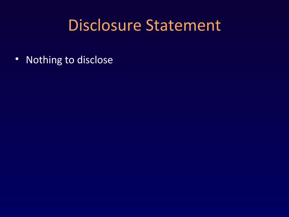
History
• Sir Charles Bell (1817)• Rare entity• Increasingly diagnosed
– Imaging enhancements– Routine imaging
Middlesex Hospital Journal 4:469-470, 1817

REVIEW OF LITERATURE
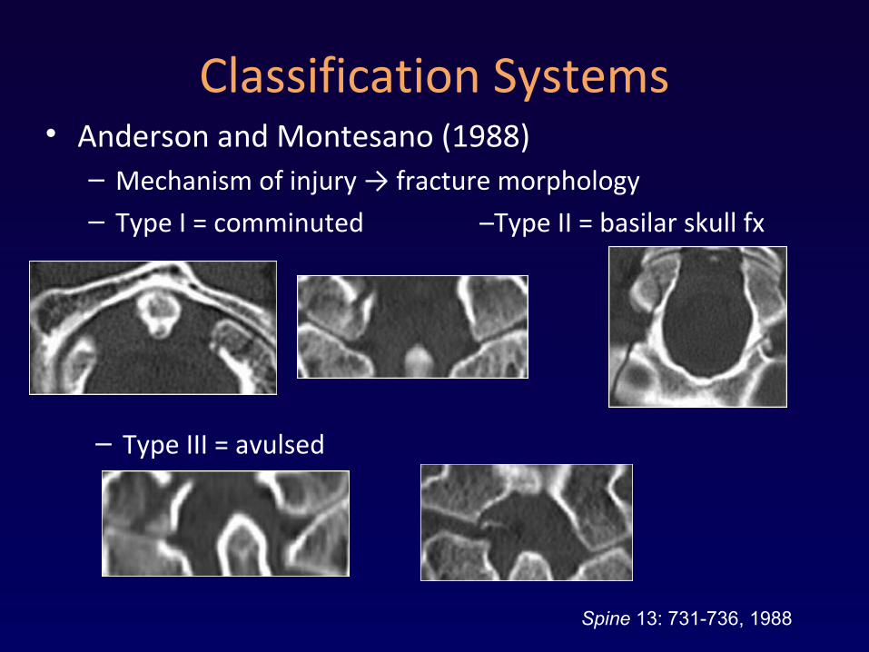
Classification Systems• Anderson and Montesano (1988)
– Mechanism of injury → fracture morphology – Type I = comminuted –Type II = basilar skull fx
– Type III = avulsed
Spine 13: 731-736, 1988
Classification Systems• Tuli et al (1997)
– Type 1 = non-displaced– Type 2 = displaced (2A – stable, 2B – unstable)– Instability
• CT/Xray – subluxation OR MRI – avulsed transverse ligament
• Newer systems– A-M system– Stability assessment
• Hanson et al (2001) – bilateral O-C1-C2 joint complex injury• Malham et al (2009) – displaced fracture or malalignment of joint
Neurosurgery 41:368-377, 1997
American Roent Ray Soc 178: 1261-68, 2002Emergency Radiology Online, 2009
Treatment• Experience or non-radiographic outcome:
– wide range of treatments suggested
• Radiographic outcome data:– Capuano et al (2004)
• 10 pts, CT for fusion• All isolated OCF healed well with cervical collar
– Malham et al (2009)• 24 pts, CT for fusion and alignment & pain and disability scales• Isolated type I and II heal well with C collar• Isolated type III may benefit from halo vs collar
Acta Neurochirurgica 146: 779-784, 2004Emergency Radiology Online, 2009
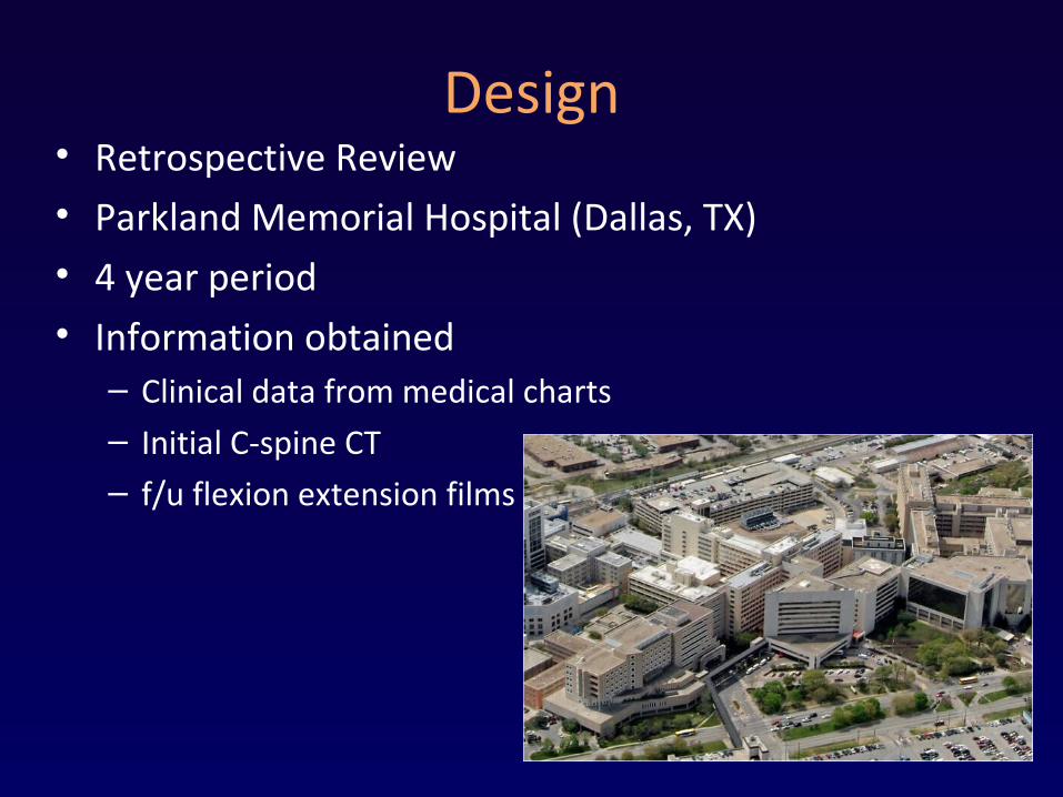
Design• Retrospective Review• Parkland Memorial Hospital (Dallas, TX)• 4 year period• Information obtained
– Clinical data from medical charts– Initial C-spine CT – f/u flexion extension films
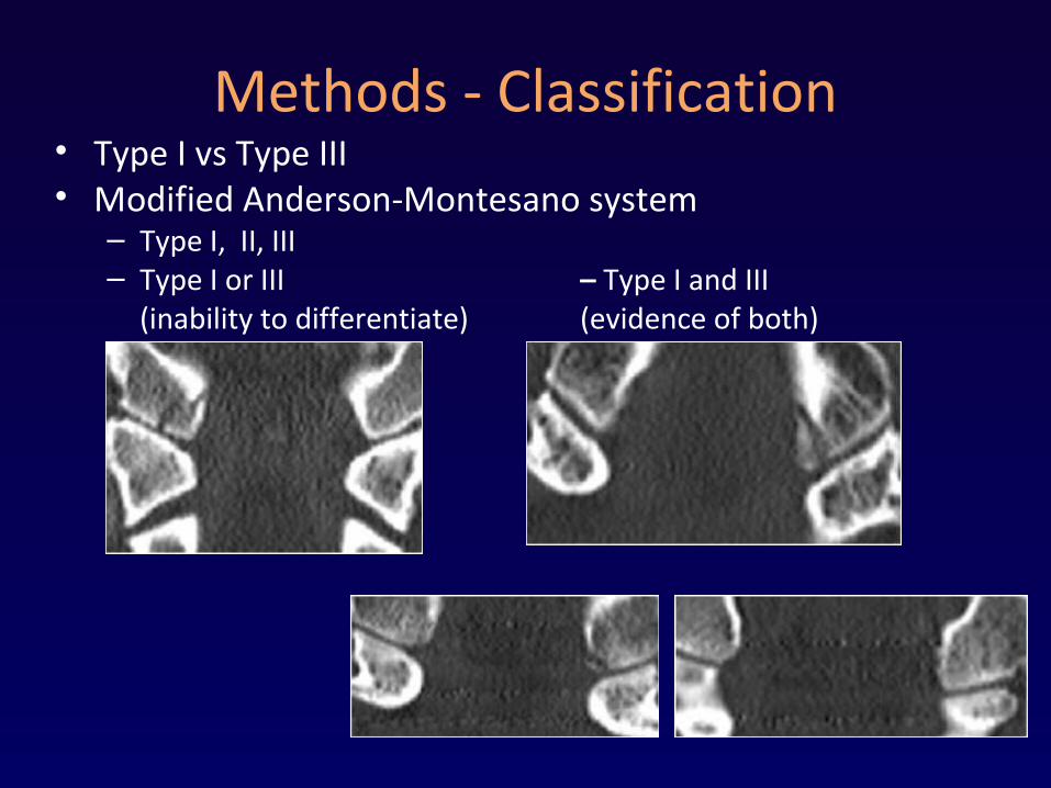
Methods - Classification• Type I vs Type III• Modified Anderson-Montesano system
– Type I, II, III– Type I or III – Type I and III
(inability to differentiate) (evidence of both)
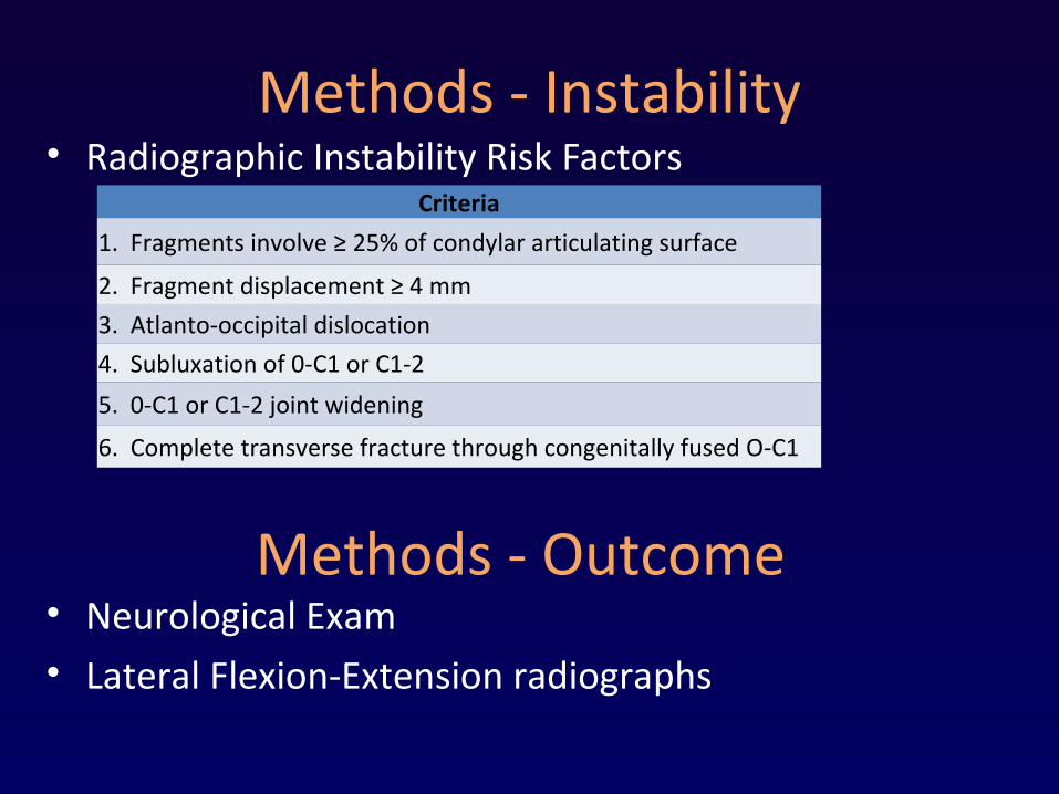
Methods - Instability• Radiographic Instability Risk Factors
Criteria
1. Fragments involve ≥ 25% of condylar articulating surface
2. Fragment displacement ≥ 4 mm
3. Atlanto-occipital dislocation
4. Subluxation of 0-C1 or C1-2
5. 0-C1 or C1-2 joint widening
6. Complete transverse fracture through congenitally fused O-C1
Methods - Outcome• Neurological Exam• Lateral Flexion-Extension radiographs
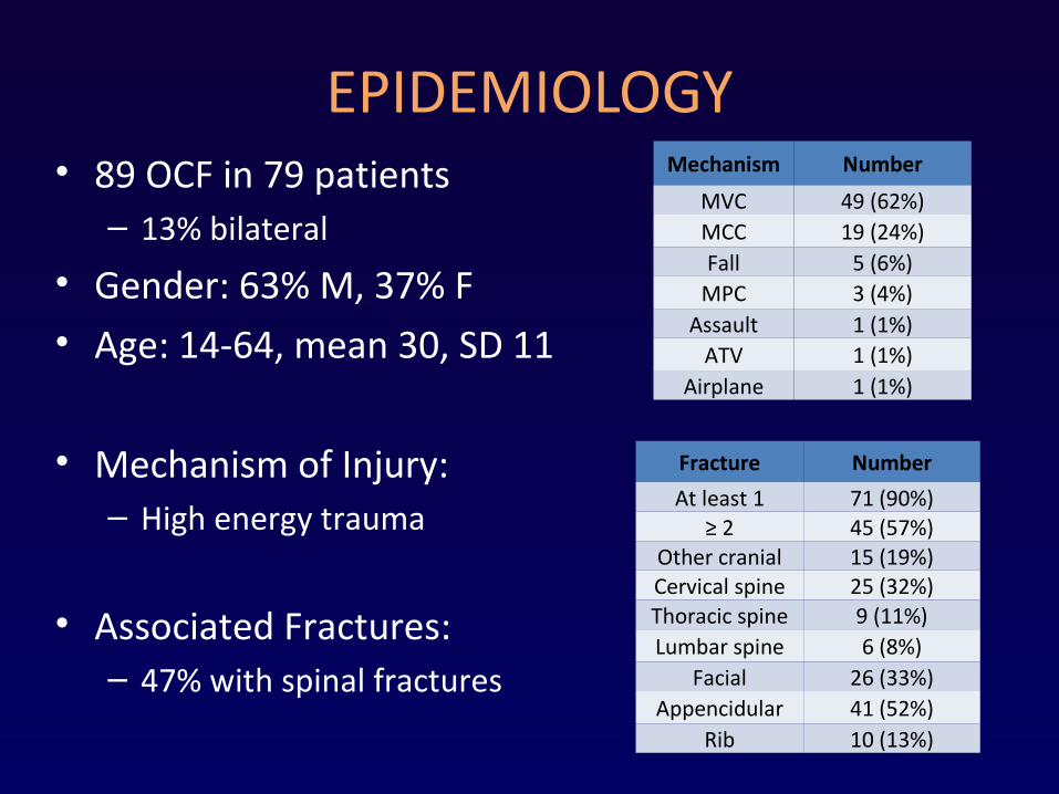
EPIDEMIOLOGY• 89 OCF in 79 patients– 13% bilateral
• Gender: 63% M, 37% F• Age: 14-64, mean 30, SD 11
• Mechanism of Injury:– High energy trauma
• Associated Fractures:– 47% with spinal fractures
Mechanism Number
MVC 49 (62%)MCC 19 (24%)Fall 5 (6%)
MPC 3 (4%)Assault 1 (1%)
ATV 1 (1%)Airplane 1 (1%)
Fracture Number
At least 1 71 (90%)≥ 2 45 (57%)
Other cranial 15 (19%)Cervical spine 25 (32%)Thoracic spine 9 (11%)Lumbar spine 6 (8%)
Facial 26 (33%)Appencidular 41 (52%)
Rib 10 (13%)
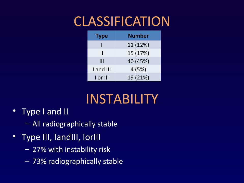
CLASSIFICATIONType Number
I 11 (12%)II 15 (17%)III 40 (45%)
I and III 4 (5%)I or III 19 (21%)
INSTABILITY• Type I and II– All radiographically stable
• Type III, IandIII, IorIII– 27% with instability risk– 73% radiographically stable
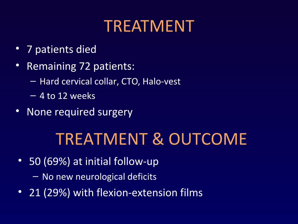
TREATMENT• 7 patients died• Remaining 72 patients:– Hard cervical collar, CTO, Halo-vest– 4 to 12 weeks
• None required surgery
TREATMENT & OUTCOME• 50 (69%) at initial follow-up– No new neurological deficits
• 21 (29%) with flexion-extension films
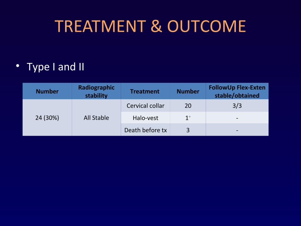
TREATMENT & OUTCOME
• Type I and II
NumberRadiographic
stabilityTreatment Number
FollowUp Flex-Exten stable/obtained
24 (30%) All Stable
Cervical collar 20 3/3
Halo-vest 1+ -
Death before tx 3 -
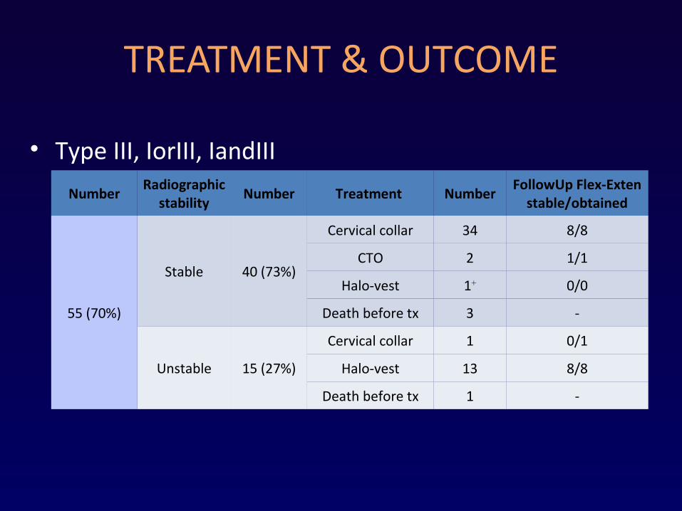
TREATMENT & OUTCOME
• Type III, IorIII, IandIII
NumberRadiographic
stabilityNumber Treatment Number
FollowUp Flex-Exten stable/obtained
55 (70%)
Stable 40 (73%)
Cervical collar 34 8/8
CTO 2 1/1
Halo-vest 1+ 0/0
Death before tx 3 -
Unstable 15 (27%)
Cervical collar 1 0/1
Halo-vest 13 8/8
Death before tx 1 -
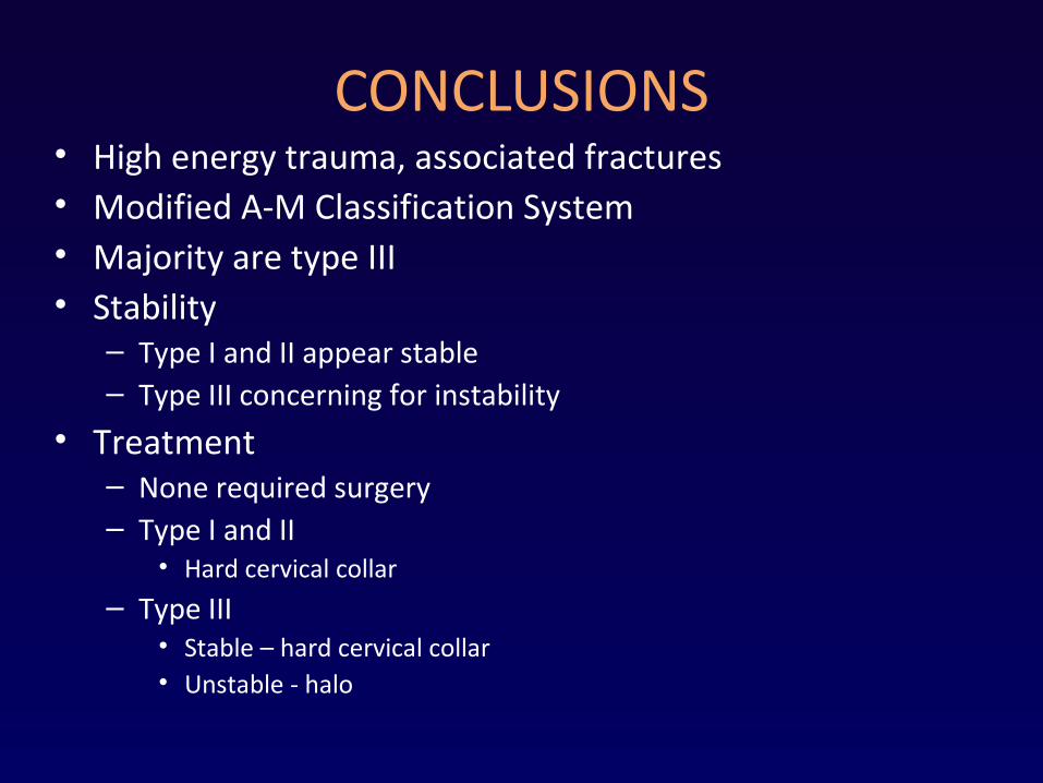
CONCLUSIONS• High energy trauma, associated fractures• Modified A-M Classification System• Majority are type III• Stability
– Type I and II appear stable– Type III concerning for instability
• Treatment– None required surgery– Type I and II
• Hard cervical collar
– Type III• Stable – hard cervical collar• Unstable - halo
LIMITATIONS• Limited number with complete outcome data• Others
FUTURE INVESTIGATION• Assessing stability in type III fracture• Do all type I and II need collar immobilization?• Can some “unstable” type III be treated with collars?
Thank You• Dr. Christopher Madden• Dept of Neurosurgery at UT Southwestern