Embed Size (px)
Citation preview
Observations on the Stomach and Digestive Diverticula ofthe Lamellibranchia
I. The Anisomyaria and Eulamellibranchia
By G. OWEN
(From the Department of Zoology, University of Glasgow)
With 3 plates (figs, i, 2, and 6)
SUMMARY
A study of the digestive diverticula of the Anisomyaria and Eulamellibranchiarevealed certain features hitherto undescribed. The diverticula consist of blind-endingtubules which open into ciliated main ducts by way of short, non-ciliated secondaryducts. The main ducts open into the intestinal groove. In all the species examined theciliated epithelium of the main ducts was restricted to a well-defined groove, the re-mainder of the lumen being surrounded by a non-ciliated, brush-border epithelium.
Each tubule is surrounded by a system of smooth muscle fibres. Cilia associated withthe darkly staining cells of the crypts were demonstrated in sections of the tubules.After feeding with titanium dioxide in suspension, this substance was later found inthe large vacuolated cells of the tubules. The particles of titanium dioxide were largerthan o-i^t.
A continuous circulation is maintained within the main ducts solely as a result ofciliary activity. The exhalant current in the ciliated portion of the main ducts producesan inhalant counterpart current in the non-ciliated portion. It is suggested that freshfluid is drawn into the tubules as a consequence of the absorptive functions of the largevacuolated cells. Indigestible material accumulates in the large vacuolated cells and isextruded into the main ducts where it is conveyed out of the diverticula by the exhalantciliary current.
In both the Anisomyaria and Eulamellibranchia the flap-like major typhlosoleprevents material entering the mid-gut except by the intestinal groove, and isolatesthe rejectory currents of the intestinal groove from the general circulation of particlesin the stomach. In the Eulamellibranchia the major typhlosole also acts as a valvewhich controls the entry and exit of material into and out of the inhalant and exhalantportions of the main ducts.
INTRODUCTION
MANY conflicting views have been expressed regarding the functions ofthe digestive diverticula of the Lamellibranchia. An absorptive func-
tion has been attributed to them by many authors, while others have regardedthem as secretory organs (for details and bibliography see Yonge, 1926a).From a comparative study of the structure and functions of the diverticulaof numerous lamellibranchs Yonge concluded that • they were organs ofabsorption and intracellular digestion and this view has received generalacceptance by the majority of workers. Recently, Mansour (1946 a and b,1949) and Mansour-Bek (1946) have questioned the occurrence of intracellulardigestion in the Lamellibranchia. They claim that extracellular proteases andlipases are secreted into the lumen of the stomach by the digestive diverticula.[Quarterly Journal of Microscopical Science, Vol. 96, part 4, pp. 517-537, December 1955.J
518 Owen—Observations on the Lamellibranchia
Despite this recent interest in the functions of the diverticula little has beenadded to our knowledge of the structure and mode of functioning of theseorgans.
Observations on the digestive diverticula of Cardium edule carried outduring the course of feeding experiments revealed structural features hithertoundescribed, and these were subsequently investigated in other species oflamellibranchs. In this paper the results obtained for the Anisomyaria andEulamellibranchia are described and their possible significance discussed.
The work was carried out on a variety of lamellibranchs with both fresh andfixed material. Fixed material was embedded in ester-wax (Steedman, 1947)and sections cut at 2-6,11 were in most cases stained in Heidenhain's ironhaematoxylin, alcian blue 8GS (Steedman, 1950) and orange G in clove oil.Before embedding, the material was cleared in monochlorisothymol (Steed-man, 1955).
THE STRUCTURE OF THE DIGESTIVE DIVERTICULA
The digestive diverticula surround the stomach and consist of numerousblind-ending tubules which have the form of globular (e.g. Cardium) orelongate (e.g. Anodonta) sacs or, as in the Mytilidae (List, 1902), of irregularlybranched tubes with numerous saccular outgrowths. They communicate withthe stomach by a system of ducts whose structure is distinct from that of thetubules.
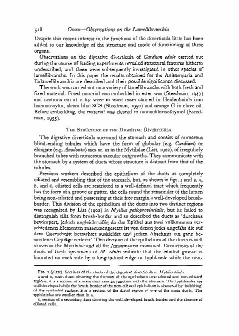
Previous workers described the epithelium of the ducts as completelyciliated and resembling that of the stomach, but, as shown in figs. 1 and 2, A,B, and c, ciliated cells are restricted to a well-defined tract which frequentlyhas the form of a groove or gutter, the cells round the remainder of the lumenbeing non-ciliated and possessing at their free margin a well-developed brush-border. This division of the epithelium of the ducts into two distinct regionswas recognized by List (1902) in Mytilus galloprovincialis, but he failed todistinguish cilia from brush-border and so described the ducts as 'durchausbewimpert, jedoch ungleichmafiig da das Epithel aus zwei vollkommen ver-schiedenen Elementen zusammengesetzt ist von denen jedes ungefahr die aufdem Querschnitt betrachtet auskleidet und jedem Abschnitt ein ganz be-sonderes Geprage verleiht'. This division of the epithelium of the ducts is wellshown in the Mytilidae and all the Anisomyaria examined. Dissections of theducts of fresh specimens of M. edulis indicate that the ciliated groove isbounded on each side by a longitudinal ridge or typhlosole while the non-
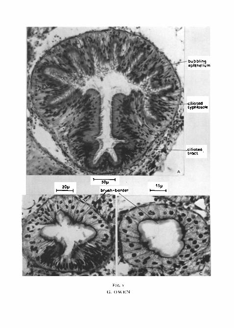
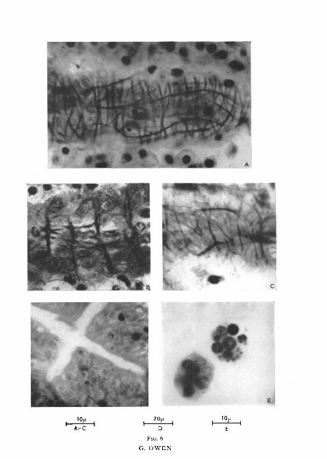
FIG. 1 (plate). Sections of the ducts of the digestive diverticula of Mytilus edulis.A and B, main ducts showing the division of the epithelium into ciliated and non-ciliated
regions. A is a section of a main duct near its junction with the stomach. The typhlosoles arewell developed while the brush-border of the non-ciliated epithelium is obscured by 'bubbling'of the epithelial surface. B is a section of the distal region of one of the main ducts. Thetyphlosoles are smaller than in A.
c, section of a secondary duct showing the well-developed brush-border and the absence ofciliated cells.
bubblingepithelium
FIG. I
G. OWEN
I. The Anisomyaria and Eulamellibranchia 519
ciliated portion of the duct has a metallic sheen, possibly due to refractionfrom the surface of the brush-border epithelium. The typhlosoles partiallydivide the ducts into ciliated and non-ciliated regions, the division being mostmarked in the larger ducts near their junction with the stomach (comparefig. i, A and B).
In M. edulis, the brush-border epithelium consists of cells of varying heightcontaining spherical nuclei and possessing at the free margin a well-defineddarkly staining region (fig. 1, B). In sections of fixed material the epitheliumfrequently includes numerous, apparently empty, vacuoles and the brush-border is often obscured by a characteristic 'bubbling' of the epithelial surface(fig. 1, A). The outline of the lumen of the non-ciliated portion is undulatingowing to variation in height of the epithelium, and the bubbling cells arearranged in longitudinal bands coinciding with the taller epithelial cells.Knight-Jones (1953) described similar bubbling cells in the gut wall ofSaccoglossus but he concluded that they were probably artifacts due to violentcontraction on fixation. In contrast to the brush-border cells, the nuclei ofthe ciliated cells are elongate and only occasional vesicular outpushings arepresent at the epithelial surface (fig. 1, B). The typhlosoles are formed of tallciliated cells, there being a gradual decrease in height of the epithelium towardthe depth of the groove.
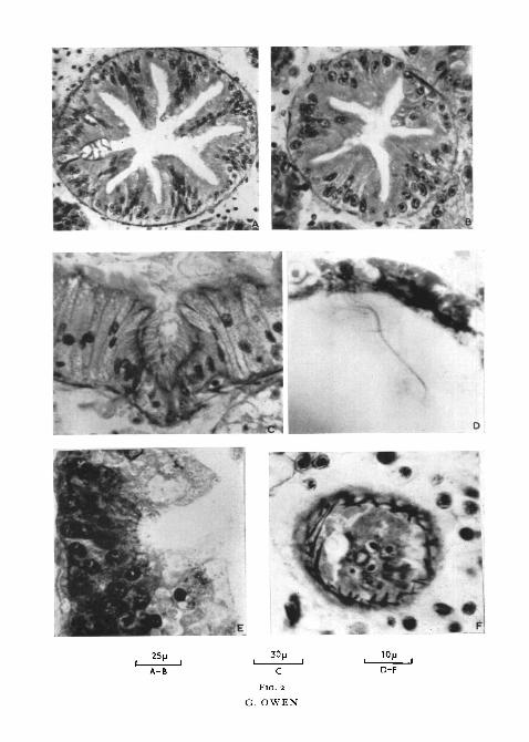
The ducts of the digestive diverticula of all the Anisomyaria examined aresimilar in cross-section to those of Mytilus edulis but members of the Eulamelli-branchia exhibit variation in form both of the ciliated groove and of the brush-border epithelium. In Spisula subtruncata (fig. 2, c) the ciliated groove is welldefined but small relative to the size of the ducts. Numerous mucous glandsstaining with alcian blue 8GS (Steedman, 1950) are present in the brush-border epithelium; they are not found in the majority of eulamellibranchs. InZirphaea crispata (fig. 2, A) numerous villi project into the lumen of the ducts,the ciliated tract being situated between two villi. As in Mytilus edulis, how-ever, dissections of these ducts show only two longitudinal typhlosoles, oneon each side of the ciliated tract. In many Eulamellibranchia the ciliatedgroove is not as well defined as in the above species and the brush-borderof the non-ciliated cells is frequently obscured by the bubbling appearanceof the epithelial surface. Nevertheless, in all species of Eulamellibranchiaexamined ciliated cells were restricted to a well-defined tract.
FIG. Z (plate), A, section of a main duct of the digestive diverticula of Zirphaea crispatashowing the well-developed ciliated tract between two villi.
B, section of a secondary duct of Zirphaea crispata. Cilia are absent. Compare with A.c, portion of main duct of the digestive diverticula of Spisula subtruncata showing the
ciliated tract.D, portion of a tubule of Aloidis gibba showing the cilia of the darkly staining cells.E, portion of a tubule of Ostrea edulis showing the basal granules associated with the darkly
staining cells. Staining of the cilia has been lost in order to differentiate the basal granulesfrom the cytoplasm of the darkly staining cells.
F, section through the tip of a tubule of Cardium edule showing the network of smoothmuscle fibres. See fig. 6.
520 Owen—Observations on the Latnellibranchia
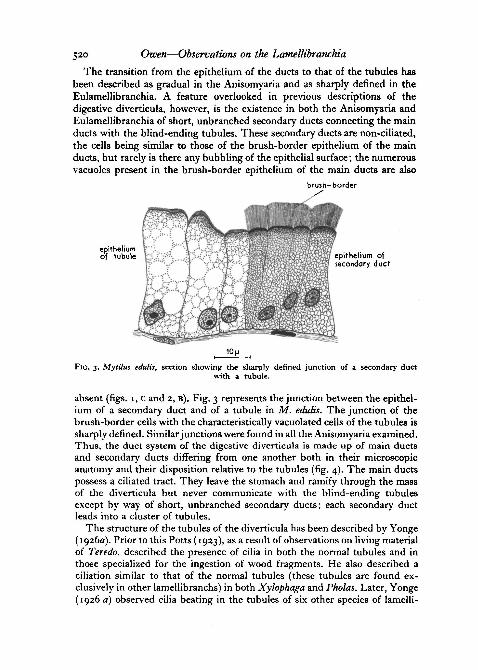
The transition from the epithelium of the ducts to that of the tubules hasbeen described as gradual in the Anisomyaria and as sharply defined in theEulamellibranchia. A feature overlooked in previous descriptions of thedigestive diverticula, however, is the existence in both the Anisomyaria andEulamellibranchia of short, unbranched secondary ducts connecting the mainducts with the blind-ending tubules. These secondary ducts are non-ciliated,the cells being similar to those of the brush-border epithelium of the mainducts, but rarely is there any bubbling of the epithelial surface; the numerousvacuoles present in the brush-border epithelium of the main ducts are also
brush-border
epitheliumof tubule epithelium of
secondary duct
FIG. 3. Mytilus edulis, section showing the sharply defined junction of a secondary ductwith a tubule.
absent (figs. 1, c and 2, B). Fig. 3 represents the junction between the epithel-ium of a secondary duct and of a tubule in M. edulis. The junction of thebrush-border cells with the characteristically vacuolated cells of the tubules issharply defined. Similar junctions were found in all the Anisomyaria examined.Thus, the duct system of the digestive diverticula is made up of main ductsand secondary ducts differing from one another both in their microscopicanatomy and their disposition relative to the tubules (fig. 4). The main ductspossess a ciliated tract. They leave the stomach and ramify through the massof the diverticula but never communicate with the blind-ending tubulesexcept by way of short, unbranched secondary ducts; each secondary ductleads into a cluster of tubules.
The structure of the tubules of the diverticula has been described by Yonge(1926a). Prior to this Potts (1923), as a result of observations on living materialof Teredo, described the presence of cilia in both the normal tubules and inthose specialized for the ingestion of wood fragments. He also described aciliation similar to that of the normal tubules (these tubules are found ex-clusively in other lamellibranchs) in both Xylophaga and Pholas. Later, Yonge(1926 a) observed cilia beating in the tubules of six other species of lamelli-
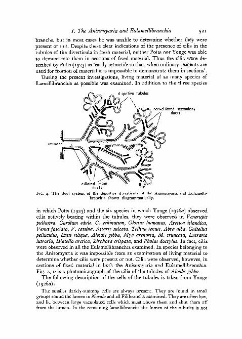
/ . The Anisomyaria and Eulamellibranchia 521
branchs, but in most cases he was unable to determine whether they werepresent or not. Despite these clear indications of the presence of cilia in thetubules of the diverticula in fresh material, neither Potts nor Yonge was ableto demonstrate them in sections of fixed material. Thus the cilia were de-scribed by Potts (1923) as 'easily retractile so that, when ordinary reagents areused for fixation of material it is impossible to demonstrate them in sections'.
During the present investigations, living material of as many species ofLamellibranchia as possible was examined. In addition to the three species
diqestive tubules
stomach
non-ciliated secondaryducts
ciliated mainducts
FIG. 4. The duct system of the digestive diverticula of the Anisomyaria and Eulamelli-branchia shown diagrammatically.
in which Potts (1923) and the six species in which Yonge (1926a) observedcilia actively beating within the tubules, they were observed in Venerupispullastra, Cardiutn edule, C. echinatum, Glossus humanus, Arctica tslandica,Venus fasciata, V. cassina, Astarte sulcata, Tellina tenuis, Abra alba, Cultelluspellucidus, Ensis siliqua, Aloidis gibba, Mya arenaria, M. truncata, Lutrarialutraria, Hiatella arctica, Zirphaea crispata, and Pholas dactylus. In fact, ciliawere observed in all the Eulamellibranchia examined. In species belonging tothe Anisomyaria it was impossible from an examination of living material todetermine whether cilia were present or not. Cilia were observed, however, insections of fixed material in both the Anisomyaria and Eulamellibranchia.Fig. 2, D is a photomicrograph of the cilia of the tubules of Aloidis gibba.
The following description of the cells of the tubules is taken from Yonge(1926a):
The smaller darkly-staining cells are always present. They are found in smallgroups round the lumen in Nucula and all Filibranchs examined. They are often low,and lie between large vacuolated cells which meet above them and shut them offfrom the lumen. In the remaining lamellibranchs the lumen of the tubules is not
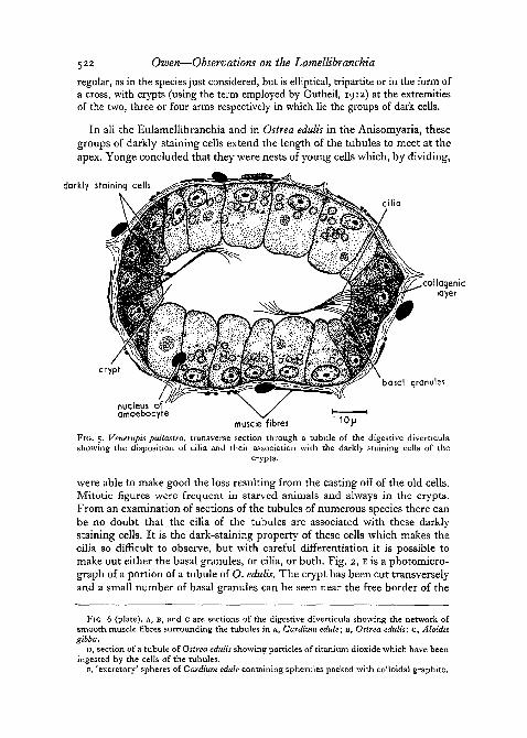
5 2 2 Owen—Observations on the Lamellibranchia
regular, as in the species just considered, but is elliptical, tripartite or in the form ofa cross, with crypts (using the term employed by Gutheil, 1912) at the extremitiesof the two, three or four arms respectively in which lie the groups of dark cells.
In all the Eulamellibranchia and in Ostrea edulis in the Anisomyaria, thesegroups of darkly staining cells extend the length of the tubules to meet at theapex. Yonge concluded that they were nests of young cells which, by dividing,
darkly staining cells
cilia
collogeniclayer
cryptbasal granules
nucleus ofamoebocyre
muscle fibresFIG. 5. Venerupis pullastra, transverse section through a tubule of the digestive diverticulashowing the disposition of cilia and their association with the darkly staining cells of the
crypts.
were able to make good the loss resulting from the casting off of the old cells.Mitotic figures were frequent in starved animals and always in the crypts.From an examination of sections of the tubules of numerous species there canbe no doubt that the cilia of the tubules are associated with these darklystaining cells. It is the dark-staining property of these cells which makes thecilia so difficult to observe, but with careful differentiation it is possible tomake out either the basal granules, or cilia, or both. Fig. 2, E is a photomicro-graph of a portion of a tubule of O. edulis. The crypt has been cut transverselyand a small number of basal granules can be seen near the free border of the
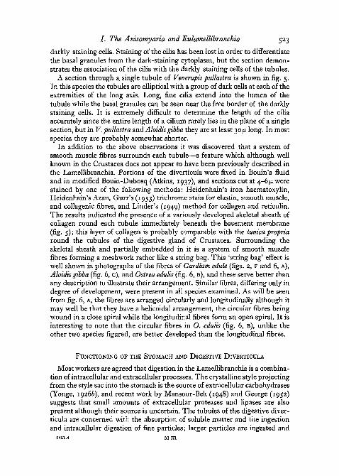
FIG. 6 (plate), A, B, and c are sections of the digestive diverticula showing the network ofsmooth muscle fibres surrounding the tubules in A, Cardium edule; B, Ostrea edulis; c, Aloidisgibba.
D, section of a tubule of Ostrea edulis showing particles of titanium dioxide which have beeningested by the cells of the tubules.
B, 'excretory' spheres of Cardium edule containing spherules packed with colloidal graphite.
A-C
20)j
D
FIG. 6
G. OWEN
IOJJ
E
/ . The Anisomyaria and Eulamellibranchia 523
darkly staining cells. Staining of the cilia has been lost in order to differentiatethe basal granules from the dark-staining cytoplasm, but the section demon-strates the association of the cilia with the darkly staining cells of the tubules.
A section through a. single tubule of Venerupis pullastra is shown in fig. 5.In this species the tubules are elliptical with a group of dark cells at each of theextremities of the long axis. Long, fine cilia extend into the lumen of thetubule while the basal granules can be seen near the free border of the darklystaining cells. It is extremely difficult to determine the length of the ciliaaccurately since the entire length of a cilium rarely lies in the plane of a singlesection, but in V. pullastra and Aloidisgibba they are at least 30fi long. In mostspecies they are probably somewhat shorter.
In addition to the above observations it was discovered that a system ofsmooth muscle fibres surrounds each tubule—a feature which although wellknown in the Crustacea does not appear to have been previously described inthe Lamellibranchia. Portions of the diverticula were fixed in Bouin's fluidand in modified Bouin-Dubosq (Atkins, 1937), and sections cut at 4-6/x werestained by one of the following methods: Heidenhain's iron haematoxylin,Heidenhain's Azan, Gurr's (1953) trichrome stain for elastin, smooth muscle,and collagenic fibres, and Linder's (1949) method for collagen and reticulin.The results indicated the presence of a variously developed skeletal sheath ofcollagen round each tubule immediately beneath the basement membrane(fig. 5); this layer of collagen is probably comparable with the tunica propriaround the tubules of the digestive gland of Crustacea. Surrounding theskeletal sheath and partially embedded in it is a system of smooth musclefibres forming a meshwork rather like a string bag. This 'string bag' effect iswell shown in photographs of the fibres of Cardium edule (figs. 2, F and 6, A),Aloidis gibba (fig. 6, c), and Ostrea edulis (fig. 6, B), and these serve better thanany description to illustrate their arrangement. Similar fibres, differing only indegree of development, were present in all species examined. As will be seenfrom fig. 6, A, the fibres are arranged circularly and longitudinally although itmay well be that they have a helicoidal arrangement, the circular fibres beingwound in a close spiral while the longitudinal fibres form an open spiral. It isinteresting to note that the circular fibres in O. edulis (fig. 6, B), unlike theother two species figured, are better developed than the longitudinal fibres.
FUNCTIONING OF THE STOMACH AND DIGESTIVE DIVERTICULA
Most workers are agreed that digestion in the Lamellibranchia is a combina-tion of intracellular and extracellular processes. The crystalline style projectingfrom the style sac into the stomach is the source of extracellular carbohydrases(Yonge, 19266), and recent work by Mansour-Bek (1948) and George (1952)suggests that small amounts of extracellular proteases and Upases are alsopresent although their source is uncertain. The tubules of the digestive diver-ticula are concerned with the absorption of soluble matter and the ingestionand intracellular digestion of fine particles; larger particles are ingested and

524 Owen—Observations on the Lamellibranchia
digested by amoebocytes (Yonge, 1926 a and b). Thus, fluid together with fineparticles must be conveyed from the stomach to the digestive diverticula whilewaste material, consisting of rejected particles and excretory products, must beconveyed in the opposite direction, i.e. from the diverticula to the stomach.The role of the digestive diverticula as organs of intracellular digestion hasrecently been questioned (Mansour, 1949). Support for the view of intracellulardigestion is based largely on the results of feeding experiments with Indianink, carmine (List, 1902; Vonk, 1924), and iron saccharate (Yonge, 1926a).Krijgsman (1928) and G. and S. Horstadius (1940) claim that these substancescontain particles small enough to pass through the cells by some physicalmeans and that phagocytosis, as suggested by Hirsch (1925), should be dennedas a process of ingesting particles larger than o-i/n. To test the phagocyticproperties of the digestive diverticula Zaki (1951) fed animals with powderedgold-fibrin. No free gold particles were found in the cells of the diverticulaand this was taken to indicate a lack of phagocytic properties on the part ofthese cells. Both Mansour and Zaki (1946) claim that the digestive diverticulaare organs of secretion. There can be no doubt, however, from feeding experi-ments with iron saccharate, that the digestive diverticula are certainly organsof absorption and that fluid at least must be conveyed from the stomach to thediverticula.
To determine whether fine particles are also conveyed from the stomach tothe diverticula, there to undergo phagocytosis, animals were fed with a suspen-sion of titanium dioxide which was specially prepared by Acheson ColloidsLtd. It contained no particles below, 0-5^, the majority being about i-o/x withoccasional particles up to 2-o/x. Animals were placed in suspensions of titan-ium dioxide in filtered sea-water and fixed after definite periods. Beforefixation, fresh preparations of the diverticula were examined under themicroscope. Four species were used—Ostrea edulis, Venerupis pullastra, Car-diutn edule, and Tellina tenuis—and in each case free particles of titaniumdioxide were present in the lumen of the tubules. From an examination offresh material it was impossible to tell whether any particles were present withinthe cells or not. In sections of fixed material, however, particles of titaniumdioxide, showing black by transmitted light and a characteristic white byreflected light, were easily identified within the large vacuolated cells of thetubules. Fig. 6, D shows a section of the diverticula of Ostrea edulis fixed 3hours after feeding with titanium dioxide. It is hoped to discuss further detailsof this experiment elsewhere, but in so far as it concerns the problem of thefunctioning of the diverticula it demonstrates convincingly the passage ofmaterial from the stomach into the diverticula and the ability of the cells of thetubules to ingest particles larger than o-1 fj, in size. Thus a two-way circulationmust be maintained within the diverticula, fluid and solid particles beingconveyed from the stomach to the tubules while waste materials, with orwithout secretions, are conveyed in the opposite direction. Previous workers(Yonge, 1937) have suggested that cilia maintain a continuous circulationwithin the diverticula, while recently Graham (1949), Owen (1953), and
7. The Anisomyaria and Eulamellibranchia 525
Purchon (1955) have all suggested that the part played by muscular activityin the functioning of the gut of lamellibranchs, and in particular of thestomach and digestive diverticula, has not been fully appreciated.
Muscular activity
In the Crustacea the tubules of the digestive gland are surrounded by anetwork of striated muscles, the passage of fluid from and into the diverticulabeing controlled by the pumping action of the walls of the tubules (Pump,1914). Morse (1902) described similar pumping movements of the 'stomachalglands' of brachiopods, and sections of Terebratulina retusa show a system ofmuscle fibres round the tubules similar to that found in the Lamellibranchia(personal observation). Thus changes in the contents of the diverticula oflamellibranchs could be brought about by the contraction and relaxation ofthe muscle fibres surrounding the tubules. But pulsations of the tubules werenever observed in living specimens and there is no direct evidence, therefore,that in the Lamellibranchia these muscle fibres effect changes in volume ofthe tubules.
Patterson (1933), working on the comparative physiology of the gastrichunger mechanism, introduced a rubber balloon into the stomach of the largegaper clam, Schizothoerus nuttallii, and so recorded pressure changes there.Slight stomach contractions occurred at the rate of one per minute regardlessof the presence or absence of the crystalline style; the contractions alsocontinued in the absence of foot movements. Changes in pressure broughtabout by contractions of the stomach would aid the entry of material into thedigestive diverticula and an attempt was made to repeat Patterson's experi-ment with Cardium edule and a different recording apparatus. Slight gastriccontractions were recorded but it was impossible to determine whether theseresulted solely from the irritation of the stomach epithelium or not. Directobservations of the stomachs of dissected animals did not reveal any rhythmiccontractions and thus there is little to suggest that muscular contractions playan important part in the circulation of material within the diverticula. In thelarval oyster, particles are drawn into the diverticula and returned to thestomach by the rhythmic expansion and contraction of the diverticula. Butthese are simple and sac-like in the oyster larva and unlike the much dividedstructure of the adult (Millar, 1955). Complicated and much-branched diverti-cula are present in the majority of adult lamellibranchs, and it is unlikely thatrhythmic contractions similar to those which occur in the larva would resultin an efficient circulation of material. Also, it is difficult to visualize suchcontractions taking place in species in which the digestive diverticula areclosely invested by the surrounding connective tissue, e.g. Mytilus edulis andOstrea edulis.
Ciliary activity
Three species, Mytilus edulis (figs. 1, A and B), Ostrea edulis, and Zirphaeacrispata (fig. 2, A), provided suitable material for determining the pattern of
526 Owen—Observations on the Lamellibranchia
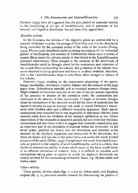
ciliary activity within the main ducts of the digestive diverticula. In all threespecies the cilia in the depth of the groove beat along the length of the ductsand away from the tubules. The cilia on the lower sides of the typhlosolesbeat into the groove while those on the upper sides of the typhlosoles beatobliquely out of the groove (fig. 7). There are no cilia beating from the stomachtowards the tubules. Although this pattern of ciliary activity was confirmedin only three species, it is probable that similar currents are present in allAnisomyaria and Eulamellibranchia where the cilia of the main ducts arerestricted to a well-defined tract.
The cilia of the tubules were observed in fresh preparations of the diverti-
non-ciliated inhalantportion of duct
ciliated exhalanrportion of duct
FIG. 7. A diagrammatic representation of a main duct of the digestive diverticula. Unbrokenarrows indicate the direction of ciliary currents in the exhalant portion of the duct and on thetyphlosoles, while the broken arrow represents the non-ciliary, inhalant, counterpart current.
cula of numerous species of Eulamellibranchia and are interesting in that theybeat with an undulating or sinusoidal movement rather than a simple flexingor bending motion; they do not exhibit metachronal rhythm nor do theyappear to produce any directive currents. Particles present in the lumen of thetubules appear to move solely as a result of direct contact with these cilia.
The cilia of the main ducts and of the tubules represent the entire ciliationof the digestive diverticula, and although there are no cilia in the main ductsbeating towards the blind-ending tubules it is possible that a two-way circula-tion is maintained within these ducts solely as a result of ciliary activity. In anyclosed fluid system, as represented by the digestive diverticula, a ciliary currentin the one direction inevitably carries a counterpart current in the oppositedirection elsewhere. As a consequence of this, material is conveyed out of thediverticula in the ciliated portion of the main ducts while an inhalant counter-part current carries material in the opposite direction in the non-ciliated por-tion. The cilia of the typhlosoles, beating into and out of the ciliated groove,keep the two currents distinct. Fig. 7 is a diagrammatic representation of thistwo-way flow within the main ducts. Although there is morphologically only asingle duct it serves functionally as two ducts, the ciliated portion carrying anexhalant current out of the diverticula and the non-ciliated portion an inhalant
/ . The Anisomyaria and Eulamellibranchia 527
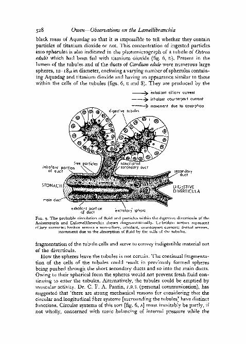
current in the opposite direction. In this way a continuous circulation could bemaintained within the main ducts solely as a result of ciliary activity. Thiscirculation does not, however, include the lumen of the blind-ending tubulessince between them and the main ducts are the short, non-ciliated secondaryducts (fig. 4). Interchange of material between the tubules and the main ductsmust result from some means other than ciliary activity.
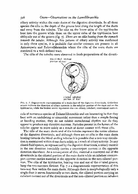
A feature which has been overlooked in considering the entry of fluid andparticles into the lumen of the tubules is the absorptive function of the largevacuolated cells. That these cells possess an absorptive function has been
'excretory" spheres
muscle fibrescollaqenic layer
FIG. 8. Cells of a tubule of the digestive diverticula of Cardium edule 6 hours after feeding witha mixture of titanium dioxide and colloidal graphite.
demonstrated by various workers using carmine, Indian ink (List, 1902; Vonk,1924), and iron saccharate (Yonge, 1926a). The short, unbranched secondaryducts open into the non-ciliated portion of the main ducts, and thus absorptionby the cells of the tubules must inevitably result in fresh fluid being drawn intothe lumen of the tubules from the inhalant current of the main ducts. Such amechanism explains why only very fine particles are found in the tubules,since only those remaining in suspension in the fluid medium can enter inthis way; the rate of absorption controls the size of particle available.
A few cells of a tubule of Cardium edule 6 hours after feeding with a mixtureof titanium dioxide and Aquadag (colloidal graphite) are represented in fig. 8.Similar experiments producing identical results have been described by List(1902), Vonk (1924), and Yonge (1926a). Only an abbreviated account is there-fore presented here. Both Aquadag and titanium dioxide have been ingestedby the cells and are concentrated in spherules 3-5/x in diameter. Some of thespherules contain titanium dioxide particles together with relatively smallamounts of Aquadag scattered round the periphery while others are a solid
528 Owen—Observations on the Lamellibranchia
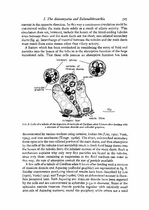
black mass of Aquadag so that it is impossible to tell whether they containparticles of titanium dioxide or not. This concentration of ingested particlesinto spherules is also indicated in the photomicrograph of a tubule of Ostreaedulis which had been fed with titanium dioxide (fig. 6, D). Present in thelumen of the tubules and of the ducts of Cardium edule were numerous largespheres, 12-18/x in diameter, enclosing a varying number of spherules contain-ing Aquadag and titanium dioxide and having an appearance similar to thosewithin the cells of the tubules (figs. 6, E and 8). They are produced by the
^ exhalant ciliary current
^ inhalant counterpart current
movement due to absorption
inhalant portionof duct -
STOMACH
main duct
exhalant portionof duct excretory' sphere
FIG. 9. The probable circulation of fluid and particles within the digestive diverticula of theAnisomyaria and Eulamellibranchia shown diagrammatically. Unbroken arrows representciliary currents; broken arrows a non-ciliary, inhalant, counterpart current; dotted arrows,
movement due to the absorption of fluid by the cells of the tubules.
fragmentation of the tubule cells and serve to convey indigestible material outof the diverticula.
How the spheres leave the tubules is not certain. The continual fragmenta-tion of the cells of the tubules could result in previously formed spheresbeing pushed through the short secondary ducts and so into the main ducts.Owing to their spherical form the spheres would not prevent fresh fluid con-tinuing to enter the tubules. Alternatively, the tubules could be emptied bymuscular activity. Dr. C. F. A. Pantin, F.R.S. (personal communication), hassuggested that 'there are strong mechanical reasons for considering that thecircular and longitudinal fibre systems [surrounding the tubules] have distinctfunctions. Circular systems of this sort [fig. 6, A] must inevitably be partly, ifnot wholly, concerned with tonic balancing of internal pressure while the
I. The Anisomyaria and Eulamellibranchia 529
longitudinal fibres have necessarily much less to do with this and, in speciesin which they are well developed [e.g. Cardium edule], their prime functionmay be the emptying of the tubules.' Thus, it could be that the longitudinalfibres, from time to time, contract slowly to empty the contents of the tubulesinto the main ducts; the circular fibres, on the other hand, merely balance theweak pressure resulting from the absorptive functions of the cells.
In fig. 9 an attempt has been made to represent diagrammatically thesuggested circulation of fluid and particles within the digestive diverticula. Acontinuous circulation is maintained within the main ducts solely as a resultof ciliary activity, the inhalant counterpart current together with suspendedparticles being carried in the non-ciliated portion of the duct. As a consequenceof the absorptive activities of the large vacuolated cells, fluid enters the lumenof the tubules by way of the short, non-ciliated secondary ducts. Solublematter is absorbed and fine particles ingested. Indigestible material and wasteproducts are concentrated in spherules and returned to the main ducts withinlarge 'excretory' spheres. These excretory spheres are conveyed out of thedigestive diverticula by the exhalant ciliary currents. The transfer of materialfrom the inhalant to the exhalant portion probably occurs in the distal regionsof the main.ducts since here the ciliated typhlosoles which separate the twoportions are not as well developed as in the proximal regions (compare fig.1, A and B). They finally terminate near the distal ends of the main ducts(fig. 9). Thus the main ducts can be regarded as tubes bent on themselves toform U-shaped structures within which a continuous circulation is maintained.The spheres are unable to re-enter the tubules from the main ducts since theyare almost certainly too large to be affected by the relatively weak absorptiveflow. In this way, waste material is separated from the inhalant 'food' particlesand soluble matter.
The stomach
It is now possible to discuss the function of some of the more obviousmorphological features of the stomach. One of the most constant features ofthe stomach of the Anisomyaria and Eulamellibranchia is the prominent flap-like major typhlosole which extends from the combined style sac and mid-gutor, if they are separate, from the mid-gut anteriorly across the floor of thestomach to enter the caecum or its representative (Graham, 1949). In discus-sing the lamellibranch stomach Graham points out that 'elaborate mechanismsare necessary to ensure the movement of food and waste into and out of theducts of the diverticula, where absorption and digestion of food particlesoccur. This is provided by the complex course and arrangement of the majortyphlosole.' The importance of the major typhlosole in isolating the rejectorycurrents of the intestinal groove from the main stomach cavity has alreadybeen emphasized (Owen, 1953) but how the major typhlosole ensures theentry of material into the diverticula has not been described.
A detailed account of the ciliary currents of the stomach of Zirphaea crispatahas been given by Purchon (1955) and a similar account for the stomach of
530 Owen—Observations on the Lamellibranchia
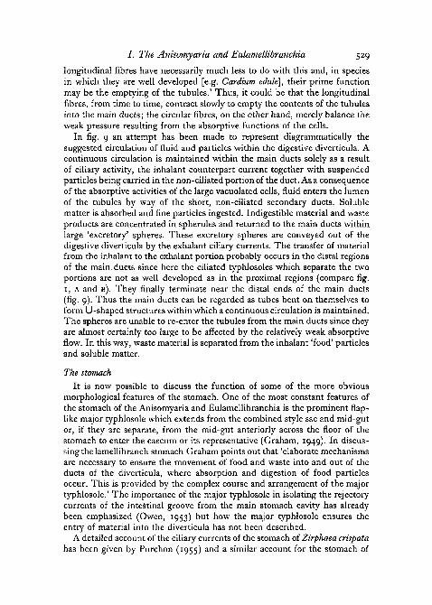
Glossus humanus by Owen (1953). The ciliary currents of the stomach ofCardium edule, apart from minor differences, are similar to those of Glossushumanus. Particles entering the stomach from the oesophagus are caught upby the revolving crystalline style and brushed on to the ridged surface of theposterior sorting area. Coarse particles are directed to the mid-gut by way ofthe rejection groove and intestinal groove while fine particles are directed firstdorsally into the dorsal hood and then ventrally over the posterior wall of thestomach. Finally, they are caught up by the strong anteriorly directed currentsof the major typhlosole and conveyed towards the openings of the right and
A B C
aperture leodinqto stpmach
.». aoerture of duct (c^??^ J K ^ "/MANTERIOR
POSTERIOR
intestinal groove5*m m
major typhlosoleVENTRAL
FIG. IO. The right caeca of A, Glossus humanus; B, Cardium edule; c, Zirphaea crispata. Un-broken arrows indicate the course of ciliary currents over the major typhlosole and broken
arrows the rejectory currents of the intestinal groove.
left caeca. In all three species, particles failing to enter the caeca continueanteriorly to rejoin material entering the stomach from the oesophagus and socirculate again (fig. 13).
In the caeca of G. humanus, the ciliary currents over the extension of themajor typhlosole are directed from the stomach to the apertures of the ducts ofthe diverticula (fig. 10, A). At the entrance to each duct, however, the ciliarycurrents are directed at right angles to the long axis of the duct. They do notconvey particles directly into the ducts. In Cardium edule the pattern of ciliary-activity in this region is more confusing (fig. 10, B). While the general trendof the currents is towards the apertures of the ducts there are a number ofcurrents directed away from the ducts. This results in the formation of vorticeswhere, at least in dissected specimens, particles entering the caecum from thestomach tend to collect; they do not enter the ducts. In Zirphaea crispata thepattern of ciliary activity is again a simple one, but, unlike Glossus humanus,the currents are all directed away from the openings of the ducts and towardsthe stomach (fig. 10, c). Thus in all three species ciliary currents do not conveyparticles directly into the ducts of the diverticula.
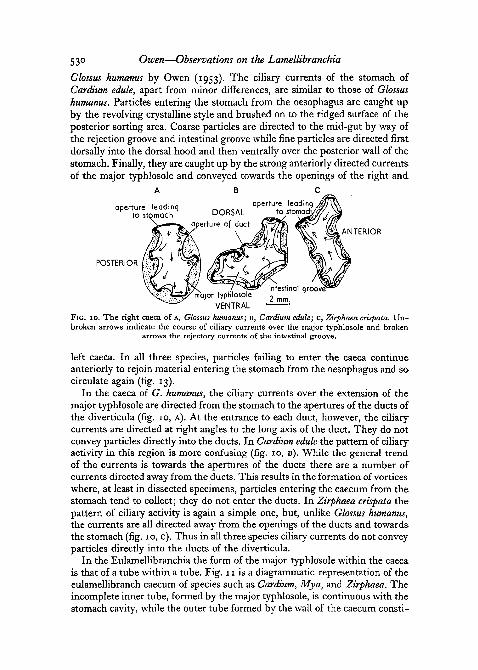
In the Eulamellibranchia the form of the major typhlosole within the caecais that of a tube within a tube. Fig. 11 is a diagrammatic representation of theeulamellibranch caecum of species such as Cardium, Mya, and Zirphaea. Theincomplete inner tube, formed by the major typhlosole, is continuous with thestomach cavity, while the outer tube formed by the wall of the caecum consti-
/ . The Anisomyaria and Eulamellibranchia 531
tutes the intestinal groove and is therefore continuous with the mid-gut.Extensions of the typhlosole project a short distance into the non-ciliatedportions of each of the main ducts, while the ciliated portion opens on to theintestinal groove. As a consequence of this disposition of the major typhlosolewithin the caecum, waste material passing out of the diverticula in the ciliatedportions of the main ducts is caught up by the rejectory currents of the in-testinal groove, so preventing its return to the circulation within the stomach:fluid and fine particles conveyed from the stomach to the proximity of theducts by the ciliary currents of the major typhlosole are drawn into the non-
main du
sion of typhlosoleinto opening of duct
FIG. I I . A diagrammatic representation of the form of the major typhlosole within the caecumof most eulamellibranchs. Ciliary currents are indicated by arrows. The direction of those over
the major typhlosole will vary in the different species (see fig. 10).
ciliated portions of the main ducts by the inhalant counterpart current. Thusthe extension of the typhlosole into the caecum acts as a valve, allowing fluidand fine particles to pass from the stomach into the ducts but preventing thepassage of material in the opposite direction. The muscular properties of theshort extensions of the typhlosole into the openings of the ducts were notedby Graham (1949) who described them as performing 'slight twistings, ex-pansions and contractions so that the whole structure has a gently lobed edge,the size and disposition of the lobes continuously undergoing slight changes'.Zirphaea crispata differs from Cardium edule and Glossus humanus in that theciliary currents over the extension of the major typhlosole are directed awayfrom the apertures of the ducts, but, as shown in fig. 10, c, the typhlosole islong and strap-shaped and an inhalant counterpart current almost certainlyoperates within the caecum as well as the non-ciliated portions of the mainducts. As suggested by Purchon (1955), the ciliary currents of Zirphaeacrispata probably prevent blocking of the ducts and caeca, particles failing toremain in suspension in the fluid medium being returned to the stomach.Blocking is unlikely to occur in Glossus humanus where the stomach
532 Owen—Observations on the Lamellibranchia
undoubtedly possesses a more efficient sorting mechanism and only relativelyfine particles are ingested (Owen, 1953).
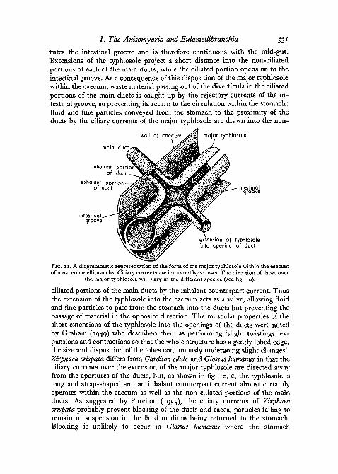
Thus the pattern of ciliary activity within the eulamellibranch stomach issuch that fluid and fine particles are conveyed to the proximity of the aper-tures of the caeca and ducts rather than directly into the ducts. Material isdrawn into the non-ciliated portion of the main ducts by the inhalant counter-part current. Although the details are different, this is also the case in theAnisomyaria. The ciliary currents at the junction of a main duct with thestomach in Ostrea edulis and Mytilus edulis are represented diagrammaticallyin fig. 12 (in these species extensions of the major typhlosole do not projectinto the apertures of the ducts). The ciliated epithelium of the stomach
/DIGESTIVE DIVERTICULAN
non-ciliated inhalant ciliated exhalantportion of duct portion of duct
FIG. 12. Anisomyaria, junction of a main duct of the digestive diverticula with the stomach,shown diagrammatically. The unbroken arrows represent ciliary currents, the broken arrows
an inhalant, counterpart current.
produces a current which sweeps across the mouth of the duct from the non-ciliated, inhalant side to the ciliated, exhalant side. Thus fresh fluid andparticles are continuously drawn into the non-ciliated portion of the ductswhile waste material is carried away from the ciliated portion.
In both the Anisomyaria and Eulamellibranchia yet another function maybe attributed to the major typhlosole. At the junction of the mid-gut with thepostero-ventral region of the stomach, the flap-like major typhlosole archesover the opening to the mid-gut, so preventing material from entering thelatter except by the intestinal groove. It is interesting to note that in manyspecies the opening of the oesophagus into the stomach is also protected, beingslit-like and situated between fleshy, protuberant lips; a feature which reducesthe possibility of regurgitation of the stomach contents. Thus, of the threepossible exits from the stomach (the oesophagus, the mid-gut, and the ductsof the diverticula), two (the oesophagus and mid-gut) are 'guarded'.
A diagrammatic representation of the suggested functioning of the stomachand digestive diverticula of the Eulamellibranchia is shown in fig. 13. Particlesentering the stomach from the oesophagus are subjected to a sorting mechan-ism and to the action of extracellular enzymes present in the stomach. Coarseparticles are directed to the mid-gut by way of the intestinal groove and fine
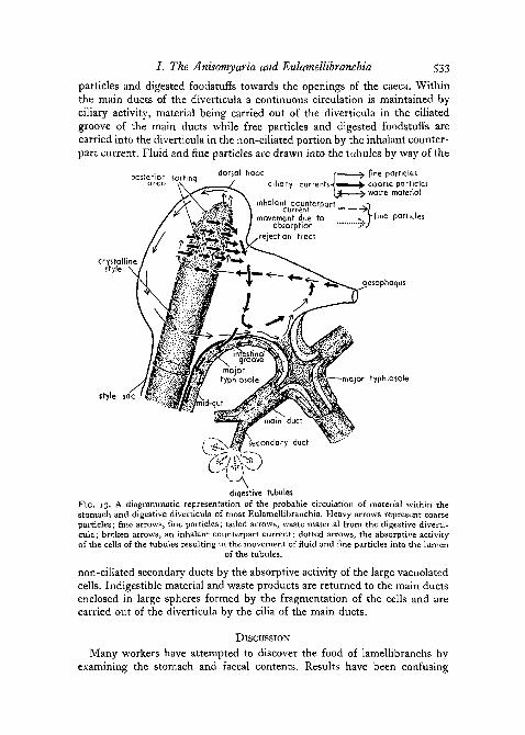
7. The Anisomyaria and Eulamellibranchia 533
particles and digested foodstuffs towards the openings of the caeca. Withinthe main ducts of the diverticula a continuous circulation is maintained byciliary activity, material being carried out of the diverticula in the ciliatedgroove of the main ducts while free particles and digested foodstuffs arecarried into the diverticula in the non-ciliated portion by the inhalant counter-part current. Fluid and fine particles are drawn into the tubules by way of the
posterior sortinqarea vv ^
crystallinestyle •
dorsal hood r ^ fine particlesciliary currenh-|^»^^^ coarse particles
{£ ^ waste materialinhalant counterpart o
current >[movement due to ... f flne particles
absorption "rJrejection tract
oesophagus
style sac
major typhlosole
diqestive tubulesFIG. 13. A diagrammatic representation of the probable circulation of material within thestomach and digestive diverticula of most Eulamellibranchia. Heavy arrows represent coarseparticles; fine arrows, fine particles; tailed arrows, waste material from the digestive diverti-cula; broken arrows, an inhalant counterpart current; dotted arrows, the absorptive activityof the cells of the tubules resulting in the movement of fluid and fine particles into the lumen
of the tubules.
non-ciliated secondary ducts by the absorptive activity of the large vacuolatedcells. Indigestible material and waste products are returned to the main ductsenclosed in large spheres formed by the fragmentation of the cells and arecarried out of the diverticula by the cilia of the main ducts.
DISCUSSION
Many workers have attempted to discover the food of lamellibranchs byexamining the stomach and faecal contents. Results have been confusing
534 Owen—Observations on the Lamellibranchia
owing largely to a failure to understand the functional morphology of thestomach of bivalves. Recently Mansour (1946a), after examining the stomachcontents of a number of bivalves, claimed that animal plankton constitutes amuch greater part of the food than is generally accepted and that this fact hasbeen overlooked owing to the very rapid digestion of the animal food. Assupport for rapid digestion and in the belief that, as in many animals, food ispropelled along the digestive tract of lamellibranchs at a rate which permitseffective digestion, Mansour (1949) adds that the time taken for material topass through the whole of the gut of Unto prasidens may be as short as 2 hours.Mansour-Bek (1948) tested filtered stomach juice for the presence of extra-cellular lipases and proteases. She obtained positive results but found that thestrength of the enzymes as indicated by experiments did not correspond tothe quick disintegration recorded by Mansour. Rosen (1949) also obtainedonly a weak protease reaction with filtered stomach juice. He suggests thatthis extracellular protease is much too weak to account for all the splitting ofproteins and that the abundant occurrence of cathepsin bound intracellularlyin the digestive diverticula indicates a considerable intracellular digestion ofproteins.
The very rapid rate of digestion recorded by Mansour and the weak pro-teolytic and lipolytic enzymes present in the stomach would appear to becontradictory. The rate of passage of material through the gut of lamelli-branchs, however, bears little relation to the effective duration of enzymeaction (Yonge, 1937). The flap-like major typhlosole extending over theopening of the mid-gut prevents material entering the latter except by theintestinal groove, and this material is derived from the sorting areas ofthe stomach and the exhalant currents of the digestive diverticula (fig. 13).Material rejected by the sorting areas passes rapidly into the mid-gut and soto the anus and as a consequence of this may pass through the entire alimen-tary canal in a very short time (from personal observation material may passthrough the entire gut of Cardium edule within 45 minutes). This feature of thelamellibranch stomach explains why several workers have observed that atleast part of the 'food' of lamellibranchs passes through the alimentary canalunchanged. In addition to material which passes rapidly out of the stomach,particles too large to enter either the digestive diverticula or the intestinalgroove may also be swallowed, since, under certain conditions, the gills andpalps may pass into the mouth relatively large particles (Nelson, 1933)-Yonge (1949) measured the maximum size of sand grains found in the pseudo-faeces, the stomach, and faecal pellets of Tellina tenuis. The measurements were400n, 320JU., and 80fj, respectively, but particles of 80/u. were rare in the faecalpellets, the majority not exceeding 40/x. Thus only particles a quarter of thediameter of the largest particles found in the stomach appear in the faeces.This retention of large particles in the stomach may explain the disintegratingzooplankton observed by Nelson (1933) and Mansour (1946a). The zoo-plankton organisms recorded by them were of relatively large size, e.g.nematodes, copepods, larvae of Crustacea, &c, and the flap-like major
/ . The Anisomyaria and Eulamellibranchia 535
typhlosole 'guarding' the entrance to the intestine would prevent such largeobjects entering the mid-gut (fig. 13). They must, therefore, remain in thegastric cavity until disintegrated. This could result from the triturating effectof the crystalline style (Yonge, 1949) aided possibly by any weak proteases andUpases present in the stomach cavity.
Mansour and Zaki (1946) injected into the blood of starved specimens ofUnio prasidens a solution of chlorophyll, and subsequent examination revealedthe presence of coloured globules in the large vacuolated cells of the tubules;similar globules were later found in the faeces. This was taken as evidence ofthe secretory function of the diverticula, the cells fragmenting and passing,together with their contained enzymes, into the stomach. Yonge (1946) re-mains convinced that the digestive diverticula are organs of intracellulardigestion. He suggests that, while true extracellular lipases and proteases areabsent from the gut of lamellibranchs, small amounts of these enzymes mayoccur owing to the presence in the lumen of numerous amoebocytes.
To test the phagocytic properties of the cells of the diverticula, Zaki (1951)fed U. prasidens with powdered gold-fibrin. No free gold particles were foundin the cells of the diverticula and he assumed that these cells did not possessphagocytic properties. Similar techniques have been used by G. and S.Horstadius (1940) using gold-gelatine and gold-fibrin, and Rosen (1941) usingcarbon-casein, the use of carmine and Indian ink having been criticized byHirsch (1925) and Krijgsman (1928) on the grounds that they contain diffusibleparticles. Horstadius's experiments with Helix pomatia were negative, freegold particles being absent from the cells of the digestive gland. This experi-ment was repeated by Rosen (1951) with similar results, but he found that'the gold-fibrin had not passed up into the mid-gut gland', probably becausethe gold-fibrin contained particles larger than the cells themselves. Feedingexperiments with carbon-casein, whipped pigeon's blood, and edestin demon-strated that phagocytosis does take place in the cells of the digestive gland ofH. pomatia. Rosen states that there is thus 'not only a lower critical limit butalso an upper critical limit' for the size of particle ingested by phagocytosis.Certainly this is true of the Anisomyaria and Eulamellibranchia, where onlyvery fine particles enter the tubules of the diverticula. Zaki (1951) in hisaccount of feeding Unio prasidens with gold-fibrin does not give measurementsof particle size. Furthermore, the animals were fed by mouth with a finepipette, so increasing the possibility of using particles larger than thosenormally ingested. The positive results obtained with titanium dioxide pro-vide clear evidence that the cells of the tubules ingest particles larger thanO-I/JL.
Irrespective of whether the digestive diverticula are organs of secretion,feeding experiments with iron saccharate and other substances provide ampleevidence that they are organs of absorption. Thus a two-way circulation isnecessary to ensure the movement of the products of extracellular digestioninto, and waste (with or without secretions) out of, the ducts of the diverticula.A continuous circulation is maintained within the main ducts solely as a result
536 Owen—Observations on the Lamellibranchia
of ciliary activity, but the entry of fluid into the blind-ending tubules ispossibly a consequence of the absorptive functions of the large vacuolatedcells. The entry of fresh fluid into the tubules will in this case be dependenton the rate of absorption, and though the resultant movement of fluid will beslow, solid particles able to remain in suspension in the fluid medium will alsoenter the lumen of the tubules. To prevent blocking of the tubules indigestibleparticles must be returned to the stomach on the way to the mid-gut andprevented from re-entering the tubules. As demonstrated by feeding experi-ments with titanium dioxide, the large vacuolated cells phagocytose solidparticles present in the lumen of the tubules, the ingested particles being thenconcentrated in spherules and returned to the main ducts enclosed in largeexcretory spheres formed by the fragmentation of the cells. These spheres areconveyed out of the diverticula by the cilia of the main ducts. Thus in additionto an absorptive function, the large vacuolated cells of the tubules also servean excretory function, which is an essential feature of their phagocyticproperties.
The results obtained by Mansour-Bek (1948), Rosen (1949), and George(1952) indicate that weak extracellular proteases and lipases are present in thestomach although their source is uncertain. Preliminary extracellular digestionwould certainly increase the amount of finely divided material received intothe digestive diverticula for absorption and the completion of digestion intra-cellularly. The excretory spheres produced by the fragmentation of the tubulecells may contain proteases and lipases, and traces of these enzymes could beliberated into the stomach in this way, as suggested by Morton (1951) for thegastropod Struthiolaria. In both the Anisomyaria and Eulamellibranchia,however, such an explanation is improbable since the ciliary mechanisms ofthe stomach would appear to prevent material carried by the intestinal groovefrom returning to the general circulation within the stomach (Owen, 1953).Yonge's (1946) suggestion that traces of proteolytic and lipolytic enzymes areliberated into the lumen of the gut as a result of cytolysis of phagocytes wouldappear more likely.
This account of the functioning of the stomach and digestive diverticula ofthe Anisomyaria and Eulamellibranchia is intended to be of general interest,there being little doubt that individual species vary, particularly in the relativedevelopment and importance of muscular activity: e.g. the Anomalodesmata,in which the diverticula are relatively simple and sac-like. There would alsoappear to be some variation in the form and disposition of the major typhlo-sole, e.g. Pandora (Allen, 1954). There can be little doubt, however, that theorganization of the stomach and diverticula in the majority of Anisomyariaand Eulamellibranchia is directed towards efficient intracellular digestion andthat 'secretions' by the cells of the diverticula can play little part in theprocesses of digestion in these animals.
I should like to express my gratitude to Professor C. M. Yonge, F.R.S., forhis continued encouragement and guidance, to Dr. H. F. Steedman for
/ . The Anisomyaria and Eulamellibranchia 537
technical assistance, and to Dr. C. F. A. Pantin, F.R.S., who provided ideasand criticisms of great value. Acknowledgement is also due to the CarnegieTrust for financial assistance.
REFERENCES
ALLEN, J. A., 1954. Quart. J. micr. Sci., 95, 473.ATKINS, D., 1937. Ibid., 79, 423.GEORGE, W. C, 1952. Biol. Bull., 102, 118.GRAHAM, A., 1949. Trans. Roy. Soc. Edinb., 61, 737.GURR, E., 1953. A practical manual of medical and biological staining techniques. London
(Hill).GUTHEIL, F., 1912. Z. wiss. Zool., 99, 444.HIRSCH, G. C, 1925. Z. vergl. Physiol., 2, 183.HORSTADIUS, G. and S., 1940. Pubbl. Staz. zool. Napoli, 18, 151.KNIGHT-JONES, E. W., 1953. Proc. Zool. Soc. Lond., 123, 637.KRIJGSMAN, B. J., 1928. Z. vergl. Physiol., 8, 187.LINDER, J. E., 1949. Quart. J. micr. Sci., 90, 427.LIST, T., 1902. 'Die Mytiliden', Fauna und Flora des Golfes von Neapel, 27.MANSOUR, K., 1946a. Nature, 157, 482.
19466. Ibid., 158, 378.1949. C.R. 13c Congr. int. Zool. Paris, 444.
MANSOUR, K., and ZAKI, F. G., 1946. Proc. Egypt. Acad. Sci., 2, 38.MANSOUR-BEK, J. J., 1946. Nature, 158, 378.
1948. Enzymologia, 12, 221.MILLAR, R. H., 1955. Quart. J. micr. Sci., 96, 539.MORSE, E. S., 1902. Mem. Boston Soc. nat. Hist., 5, 313.MORTON, J. E., 1951. Quart. J. micr. Sci., 92, 1.NELSON, T. C, 1933. Proc. Soc. exp. Biol. N.Y., 30, 1287.OWEN, G-, 1953. J. mar. biol. Ass. U.K., 32, 85.PATTERSON, T. L., 1933. Ann. N.Y. Acad. Sci., 34, 59.POTTS, F. A., 1923. Proc. Camb. phil. Soc. biol. Sci., 1, 1.PUMP, W., 1914. Arch. mikr. Anat., 85, 167.PURCHON, R. D., 1955. Proc. Zool. Soc. Lond., 124, 859.ROSEN, B., 1941. Zool. Jb., 60, 241.
1949- Ark. Kemi, 1, 205.1951- Ark. Zool., Serie 2, 3, 33.
STEEDMAN, H. F., 1947. Quart. J. micr. Sci., 88, 123.1950. Ibid., 91, 477.!9SS' (In preparation.)
VONK, D. J., 1924. Z. vergl. Physiol., i , 607.YONGE, C. M., 1926a. Trans. Roy. Soc. Edinb., 54, 703.
19266. J. mar. biol. Ass. U.K., 14, 295.1937- Biol. Rev., 12, 87.1946. Nature, 157, 729.1949- Phil. Trans. B, 234, 29.
ZAKI, F. G., 1951. Proc. Egypt. Acad. Sci., 7, 18.